Embed Size (px)
Citation preview
From M113en 2014OCT10 Page 1 of 21
This Procedural Bulletin is intended to provide a ready outline reference for performance of the assay. These abbreviated directions for use are not intended to replace the complete Package Insert. It is the obligation of every manufacturer of medical devices labeled FOR IN VITRO DIAGNOSTIC USE to provide a complete Package Insert in accordance with FDA labeling regulation (21 CFR 809.10).
Quidel Corporation provides CLSI procedures for your use. The procedures are required to include the same information as listed in the Package Insert. Any modifications to this document are the sole responsibility of the Laboratory.
_____________________________________________________________________________________________
Lyra Adenovirus Assay
CLIA Complexity: High
INTENDED USE The Lyra Adenovirus Assay is a real‐time polymerase chain reaction (PCR) in vitro diagnostic test for the qualitative detection of human adenovirus (HAdV) viral DNA isolated from nasal and nasopharyngeal swab specimens obtained from individuals exhibiting signs and symptoms of acute respiratory infections. The intended use of this test is to aid in the diagnosis of HAdV in conjunction with other clinical and laboratory findings.
The test detects, but does not differentiate, HAdV species (A, B, C, D, E, and F) or serotypes (HAdV 1-52). Negative results do not preclude HAdV infection and should not be used as the sole basis for diagnosis, treatment or other patient management decisions. SUMMARY AND EXPLANATION Adenoviruses (HAdV; family Adenoviridae) are non-enveloped, double stranded DNA viruses. At the present time there are 52 serotypes, further divided into 6 groups, A to F. Most HAdVs are associated with respiratory and ocular infections. Generally, HAdV infections in adults have a low morbidity with the exceptions of immunocompromised individuals and those living in overcrowded conditions, in which infections can cause atypical pneumonia. Virus spread is commonly through aerosolized droplets and fomites with infection of mucous membranes of the eye, respiratory tract and gut. PRINCIPLE OF THE PROCEDURE The assay detects viral nucleic acids that have been extracted from a patient sample. A real-time polymerase chain reaction (PCR) is carried out under optimized conditions in a single tube generating amplicons for HAdV and the Process Control (PRC). Identification of HAdV and the PRC occurs by the use of target-specific primers and fluorescent-labeled probes that hybridize to conserved regions in the genomes of HAdV and the PRC.
Lyra Probe Labels
Target Dye
HAdV FAM
PRC Cy5
The following is a summary of the procedure:
From M113en 2014OCT10 Page 2 of 21
1. Sample Collection: Obtain nasal or nasopharyngeal swabs specimens using standard techniques from symptomatic patients. Transport, store, and process these specimens according to established laboratory procedures.1
2. Nucleic Acid Extraction: Extract nucleic acids from the specimens with the NucliSENS® easyMAG® System following the manufacturer’s instructions and using the appropriate reagents (See Materials Required but Not Provided). Use of other extraction systems with the Lyra Adenovirus Assay has not been validated. Validation of these other systems is the responsibility of the end user. Prior to the extraction procedure, add 20 µL of the PRC to each 180 µL aliquot of specimen. The PRC serves to monitor inhibitors in the extracted specimen, assures that adequate amplification has taken place, and confirms that the nucleic acid extraction was sufficient.
3. Rehydration of Master Mix: Rehydrate the lyophilized Master Mix using the Rehydration Solution. The Master Mix contains oligonucleotide primers, fluorophore and quencher-labeled probes targeting conserved regions of HAdV, as well as the PRC sequence.
4. Nucleic Acid Amplification and Detection: Add 15 µL of the rehydrated Master Mix to each plate well. Then add 5 µL of extracted nucleic acids (specimen with PRC) to the plate well. Place the plate into the Applied Biosystems® (ABI) 7500 Fast Dx Real-Time PCR Instrument (“ABI 7500 Fast Dx Instrument”). Once the plate is added to the instrument, initiate the assay protocol. This protocol initiates amplification of the DNA target amplicons. The Lyra Adenovirus Assay is based on TaqMan® chemistry and uses an enzyme with reverse transcriptase, DNA polymerase, and 5’-3’ exonuclease activities. During DNA amplification, this enzyme cleaves the probe bound to the complementary DNA sequence, separating the quencher dye from the reporter dye. This step generates an increase in fluorescent signal upon excitation by a light source of the appropriate wavelength. With each cycle, additional dye molecules are separated from their quenchers resulting in additional signal. If sufficient fluorescence is achieved by 45 cycles on the ABI 7500 Fast Dx Instrument, the sample is reported as positive for the detected nucleic acid.
MATERIALS PROVIDED Cat. #M113 Detection Kit (96 Reactions) – Store at 2°C to 8°C
Component Quantity
Rehydration Solution Part M5003 1 vial/kit 1.9 mL
Lyra Adenovirus Master Mix Part M5072 Lyophilized Contents: DNA polymerase enzyme Primers and probes dNTPs Stabilizers
12 vials/kit, 8 reactions/vial
Process Control (PRC) Part M5005 Identity: MS2 Bacteriophage
1 vial/kit 2.0 mL
OPTIONAL MATERIALS External controls for HAdV (i.e. Lyra Adenovirus Control Set, #M110 which serves as an external
processing and extraction control).
MATERIALS REQUIRED BUT NOT PROVIDED Micropipettors (range between 1 to 10 μL and 100 to 1000 μL) Non-aerosol pipette tips
From M113en 2014OCT10 Page 3 of 21
ABI 7500 Fast Dx instrument (software version 1.4 or higher)
96 well PCR plate Optical plate films
Plate centrifuge for ABI 96 well plate bioMérieux NucliSENS easyMAG software (version 2.0) bioMérieux NucliSENS easyMAG Buffers 1, 2, 3 bioMérieux NucliSENS easyMAG Lysis Buffer bioMérieux NucliSENS easyMAG Silica Magnetic Beads bioMérieux NucliSENS easyMAG disposables Biohit pipettor
WARNINGS AND PRECAUTIONS For in vitro diagnostic use The assay has been validated using bioMérieux NucliSENS easyMAG software version 2.0 and ABI
7500 Fast Dx Software version 1.4 or higher. Please contact Quidel Technical Support prior to modifying or upgrading beyond these versions of software.
Performance characteristics of this test have been established with the specimen types listed in the Intended Use Section only. The performance of this assay with other specimen types or samples has not been evaluated.
Use of nucleic acid extraction systems other than the bioMérieux NucliSENS easyMAG System has not been validated. Validation of these systems is the responsibility of the end-user.
Using cycling conditions other than those indicated in the Set Up of Thermocycler Program (ABI 7500 Fast Dx Instrument) section may give erroneous results.
Use of this product should be limited to personnel with sufficient training in PCR and RT-PCR techniques.
Treat all specimen/samples as potentially infectious. Follow universal precautions when handling samples, this kit, and its contents.
Proper sampl e collection, storage, and transport are essential for correct results. Store assay reagents as indicated on their individual labels. Wear suitable protective clothing, gloves, eye, and face protection when using this kit. For accurate results, pipette carefully using only calibrated equipment.
Thoroughly clean and disinfect all surfaces with a 10% bleach solution followed by molecular grade water.
Use micropipettes with an aerosol barrier or positive displacement tips for all procedures. Avoid microbial and cross contamination of the kit reagents. Follow Good Laboratory Procedures. Do not mix reagents from kits with different lot numbers. Do not use reagents from other manufacturers with this kit. Do not use product after its expiration date. Proper workflow planning is essential to minimize contamination risk. Always plan laboratory
workflow in a uni-directional manner, beginning with pre-amplification and moving through amplification and detection.
Use dedicated supplies and equipment in pre-amplification and amplification areas. Do not allow cross movement of personnel or equipment between areas. Keep amplification supplies separate from pre-amplification supplies at all times. Do not open sample tubes or unseal plates post amplification.
Dispose of amplified material carefully and in accordance with local laws and regulations in order to minimize the risk of amplicon contamination.
Do not use supplies dedicated for reagent or sample preparation for processing target nucleic acid. MSDS is available upon request or can be accessed on the product website.
From M113en 2014OCT10 Page 4 of 21
STORAGE AND HANDLING OF KIT REAGENTS Store the unopened kit at 2°C to 8°C until the expiration date listed on the outer kit box. The rehydrated Master Mix may be kept at room temperature or at 2°C to 8°C for up to 4 hours or at
–20°C for up to 3 days. The rehydrated Master Mix should be recapped, sealed with parafilm, and stored in an upright
position. Protect the Master Mix from light during storage.
INDICATIONS OF INSTABILITY OR DETERIORATION OF REAGENTS Cloudiness of the Rehydration Solution may indicate deterioration of this reagent. Contact Quidel Technical Support for a replacement.
SPECIMEN COLLECTION, STORAGE, AND HANDLING Specimens used for the validation of the Lyra Adenovirus Assay were obtained using standard techniques from patients with upper respiratory tract infection symptoms. These specimens were collected, transported, stored, and processed according to CLSI M41-A. Briefly, samples should be
transported refrigerated at 2C to 8C. Unextracted samples can be stored refrigerated (2C to 8C) or frozen at –20°C for up to 7 days before processing. A series of studies were preformed evaluating a number of routinely used viral transport medium at a volume of 2mL: M4, M4-RT, M5, M6, and UTM. No significant difference in assay performance was seen between the five different types of viral transport media.
NUCLEIC ACID EXTRACTS STORAGE
Nucleic acid extracts can be stored at room temperature (20C to 25C), 2C to 8C, or at ≤–20°C for up to 30 days. The extracted DNA is stable for up to three freeze/thaw cycles when stored at ≤–20°C.
SET UP OF NUCLEIC ACID EXTRACTION PROGRAM (BIOMÉRIEUX NUCLISENS EASYMAG SYSTEM) Note: A HAdV positive processing/extraction control (i.e. Lyra Adenovirus Control Set, Cat. #M110 or previously characterized positive HAdV specimen) and a negative external control (i.e. viral transport media or previously characterized HAdV negative specimen) should be included in each extraction run.
1. Turn on the instrument and wait for instrument light to appear orange. Then switch on the computer/launch the bioMérieux NucliSENS easyMAG System software.
2. Barcode reagents after pressing the ‘Instrument’ and ‘Reagent Inventory’ buttons.
3. To enter samples, press the ‘Daily Use’ button, which will default to the ‘Define Request’
screen. Select the following settings: a. Sample ID: Enter the sample name using the keyboard. b. Matrix: Select Other from the drop-down menu. c. Request: Select Generic from the drop-down menu. d. Volume (mL): Select 0.200 from the drop-down menu. e. Eluate (µL): Select 50 from the drop-down menu. f. Type: Primary g. Priority: Normal

From M113en 2014OCT10 Page 5 of 21
4. Upon pressing the ‘Save’ button, the sample will appear in the ‘Unassigned Sample’ window
on the left side of the screen. Press the ‘Enter New Extraction Request’ button, and repeat the process for additional samples. Alternatively, multiple samples can be entered by pressing the
‘Auto Create New Extraction Requests’ button.
5. Once all samples are created, go to ‘Organize Runs’ by clicking on the icon near the top of
the page. Create a run by pressing the ‘Create Run’ button. Enter a run name, or use the default.
6. Add samples to the run by using the ‘Auto Fill Run’ button (auto fills up to 24 samples from the ‘Unassigned Sample list’ on the left hand side of the screen). Alternatively, individual samples
may be moved into and out of the run by using the left and right ‘Positioning icons’ after selecting the appropriate sample. The sample order within the run may be changed using the
‘Move Extraction Request Up/Down’ buttons . 7. Obtain 1 to 3 (for 8 to 24 samples, respectively) sample vessel(s), and add 20 µL of PRC to each
sample well used. 8. Add 180 µL of each sample to the appropriate well as designated.
9. Go to ‘Load Run’ by pressing the button near the top of the screen. Insert tips and sample vessel(s) into the instrument.
10. Enter the barcode(s) of the sample vessel(s). 11. Enter the barcode(s) of silica beads to be used. 12. Assign silica beads to samples as follows:
a. Click the reagents symbol (number 1 in the picture below). The lot number of the silica beads should appear below the Silica tab (number 2 in the picture below).
b. Highlight and select the samples for which beads need to be assigned (number 3 in the picture below).
c. Click the positioning icon (number 4 in the picture below) to assign the silica lot number to the selected samples.
d. If the bead symbol (to the right of number 5 in the picture below) is selected, the silica bead lot number should be displayed for each sample.

From M113en 2014OCT10 Page 6 of 21
13. Print work list by touching ‘Load Run’ icon followed by pressing the ‘Print Work List’ icon .
14. Press the ‘Dispense Lysis’ button. The on-board lysis will take approximately 12 minutes to complete.
15. For each sample vessel, prepare magnetic particles using the Biohit pipettor and tips for up to eight reactions as follows:
a. Using 1 tip and Program 1, aspirate 550 µL nuclease-free water and dispense into a 1.5 mL DNAse/RNAse free microfuge tube.
b. Vortex the magnetic silica. Using 1 tip and Program 1, aspirate 550 µL of magnetic silica, dispense into the water and mix by vortexing.
c. Using 1 tip and Program 2, aspirate 1050 µL of the magnetic silica mixture and dispense 25 µL back into the same tube.
d. Dispense 125 µL magnetic silica mixture 8 times into 8 wells of an ELISA strip plate. Discard tip.
e. After Lysis is complete (Note: the ‘Instrument Status’ at the bottom of the screen must be ‘IDLE’!), using 8 tips and Program 3, aspirate 100 µL of magnetic silica mixture from the strip wells, then dispense 100 µL of magnetic silica mixture into the strip wells, and aspirate 100 µL of magnetic silica mixture into the strip wells again to mix the solution.
f. Insert tips into liquid within the sample vessels. Aspirate 800 µL, then dispense 900 µL of magnetic silica mixture back into the vessel. Aspirate 1000 µL of magnetic silica mixture from the vessel, and dispense 1000 µL of magnetic silica back into the vessel. Repeat aspiration / dispensing of 1000 µL two more times.
16. Close the instrument, and press the ‘Start’ button to begin the run. 17. Upon completion of the run, transfer purified nucleic acid to nuclease-free tubes. Eluates can be
stored at room temperature (20C to 25C), 2C to 8C, or at ≤–20°C for 1 month. The extracted DNA is stable for up to three freeze/thaws cycles when stored at –20°C.
1
4 2
3
5
From M113en 2014OCT10 Page 7 of 21
SET UP OF THERMOCYCLER PROGRAM (ABI 7500 FAST DX INSTRUMENT) 1. Launch the ABI 7500 Fast Dx Software package (version 1.4 or higher). 2. The Quick Startup document dialog window will open. Select the Create New Document button to
start the New Document Wizard. Follow the steps below to initiate the Lyra Adenovirus Assay protocol.
a. Define Document: Most of the following should be the default setting. If not, change accordingly.
i. Confirm or enter the following information:
Assay: Standard Curve (Absolute Quantitation)
Container: 96-Well Clear
Template: Blank Document
Run Mode: Fast 7500
Operator: your operator name
Comments: SDS v1.4
Plate Name: ’Lyra HAdV’
ii. Select the Next button.
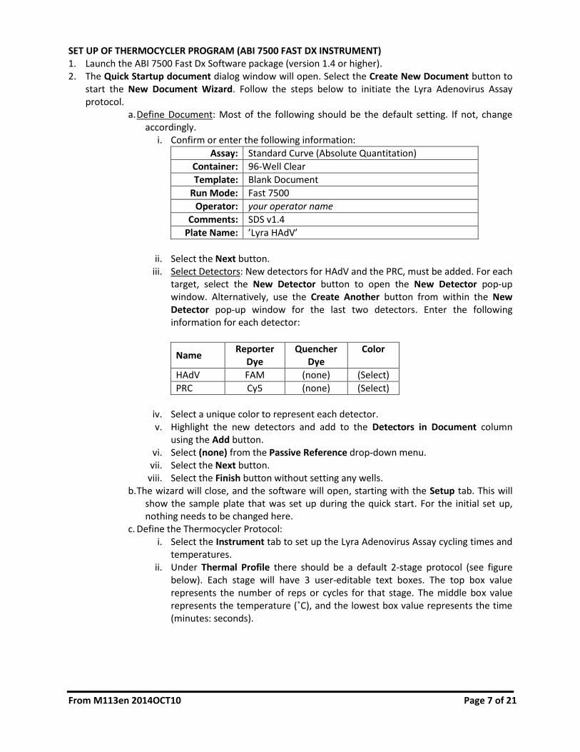
iii. Select Detectors: New detectors for HAdV and the PRC, must be added. For each target, select the New Detector button to open the New Detector pop-up window. Alternatively, use the Create Another button from within the New Detector pop-up window for the last two detectors. Enter the following information for each detector:
Name Reporter
Dye Quencher
Dye Color
HAdV FAM (none) (Select)
PRC Cy5 (none) (Select)
iv. Select a unique color to represent each detector. v. Highlight the new detectors and add to the Detectors in Document column
using the Add button. vi. Select (none) from the Passive Reference drop-down menu.
vii. Select the Next button. viii. Select the Finish button without setting any wells.
b. The wizard will close, and the software will open, starting with the Setup tab. This will show the sample plate that was set up during the quick start. For the initial set up, nothing needs to be changed here.
c. Define the Thermocycler Protocol: i. Select the Instrument tab to set up the Lyra Adenovirus Assay cycling times and
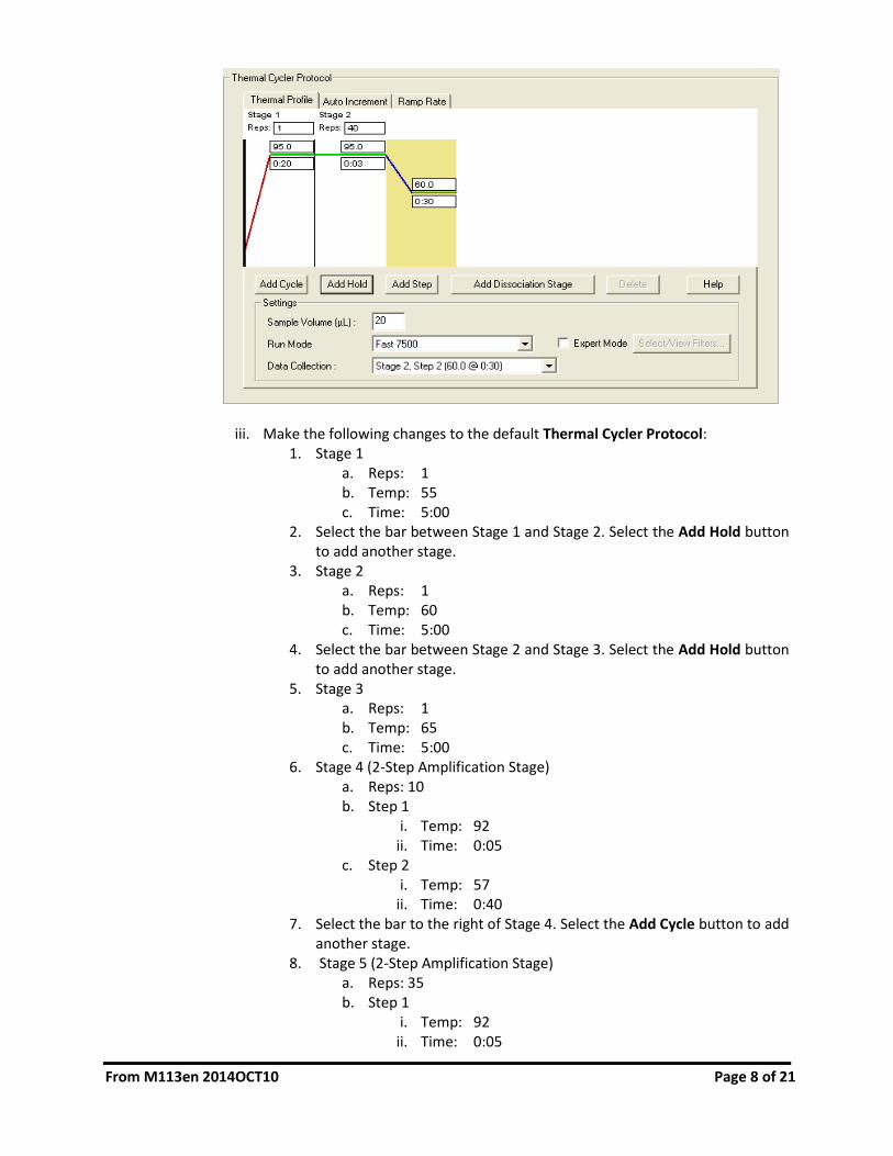
temperatures. ii. Under Thermal Profile there should be a default 2-stage protocol (see figure
below). Each stage will have 3 user-editable text boxes. The top box value represents the number of reps or cycles for that stage. The middle box value represents the temperature (˚C), and the lowest box value represents the time (minutes: seconds).
From M113en 2014OCT10 Page 8 of 21
iii. Make the following changes to the default Thermal Cycler Protocol: 1. Stage 1
a. Reps: 1 b. Temp: 55 c. Time: 5:00
2. Select the bar between Stage 1 and Stage 2. Select the Add Hold button to add another stage.
3. Stage 2 a. Reps: 1 b. Temp: 60 c. Time: 5:00
4. Select the bar between Stage 2 and Stage 3. Select the Add Hold button to add another stage.
5. Stage 3 a. Reps: 1 b. Temp: 65 c. Time: 5:00
6. Stage 4 (2-Step Amplification Stage) a. Reps: 10 b. Step 1
i. Temp: 92 ii. Time: 0:05
c. Step 2 i. Temp: 57
ii. Time: 0:40 7. Select the bar to the right of Stage 4. Select the Add Cycle button to add
another stage. 8. Stage 5 (2-Step Amplification Stage)
a. Reps: 35 b. Step 1
i. Temp: 92 ii. Time: 0:05
From M113en 2014OCT10 Page 9 of 21
c. Step 2 i. Temp: 57
ii. Time: 0:40 9. If a wrong stage is added, the stage may be removed by pressing the
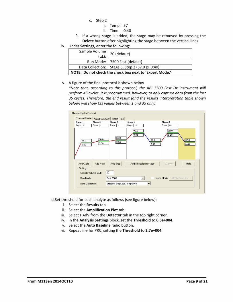
Delete button after highlighting the stage between the vertical lines. iv. Under Settings, enter the following:
Sample Volume (μL):
20 (default)
Run Mode: 7500 Fast (default)
Data Collection: Stage 5, Step 2 (57.0 @ 0:40)
NOTE: Do not check the check box next to ‘Expert Mode.’
v. A figure of the final protocol is shown below
*Note that, according to this protocol, the ABI 7500 Fast Dx Instrument will perform 45 cycles. It is programmed, however, to only capture data from the last 35 cycles. Therefore, the end result (and the results interpretation table shown below) will show Cts values between 1 and 35 only.
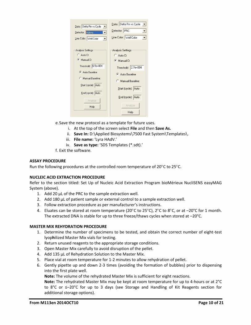
d. Set threshold for each analyte as follows (see figure below): i. Select the Results tab.
ii. Select the Amplification Plot tab. iii. Select HAdV from the Detector tab in the top right corner. iv. In the Analysis Settings block, set the Threshold to 6.5e+004. v. Select the Auto Baseline radio button.
vi. Repeat iii-v for PRC, setting the Threshold to 2.7e+004.
From M113en 2014OCT10 Page 10 of 21
e. Save the new protocol as a template for future uses. i. At the top of the screen select File and then Save As.
ii. Save In: D:\Applied Biosystems\7500 Fast System\Templates\. iii. File name: ‘Lyra HAdV.’ iv. Save as type: ‘SDS Templates (*.sdt).’
f. Exit the software.
ASSAY PROCEDURE
Run the following procedures at the controlled room temperature of 20C to 25C.
NUCLEIC ACID EXTRACTION PROCEDURE Refer to the section titled: Set Up of Nucleic Acid Extraction Program bioMérieux NucliSENS easyMAG System (above).
1. Add 20 µL of the PRC to the sample extraction well. 2. Add 180 µL of patient sample or external control to a sample extraction well. 3. Follow extraction procedure as per manufacturer’s instructions.
4. Eluates can be stored at room temperature (20C to 25C), 2C to 8C, or at –20°C for 1 month. The extracted DNA is stable for up to three freeze/thaws cycles when stored at –20°C.
MASTER MIX REHYDRATION PROCEDURE 1. Determine the number of specimens to be tested, and obtain the correct number of eight-test
lyophilized Master Mix vials for testing. 2. Return unused reagents to the appropriate storage conditions. 3. Open Master Mix carefully to avoid disruption of the pellet. 4. Add 135 µL of Rehydration Solution to the Master Mix. 5. Place vial at room temperature for 1-2 minutes to allow rehydration of pellet. 6. Gently pipette up and down 2-3 times (avoiding the formation of bubbles) prior to dispensing
into the first plate well. Note: The volume of the rehydrated Master Mix is sufficient for eight reactions. Note: The rehydrated Master Mix may be kept at room temperature for up to 4-hours or at 2°C to 8°C or ≤–20°C for up to 3 days (see Storage and Handling of Kit Reagents section for additional storage options).

From M113en 2014OCT10 Page 11 of 21
PCR SET-UP PROCEDURE: 1. Add 15 µL of the rehydrated Master Mix to each plate well. 2. Add 5 µL of extracted nucleic acid (specimen with the PRC) into the plate wells. Mixing of
reagents is not required. Note: Use a micropipettor with a new non-aerosol tip with each extracted specimen.
3. Seal the plate. Note: Quidel suggests each thermocycler run should include a well with a HAdV external positive control and a negative control (containing a PRC). Run controls in keeping with Local, State, and/or Federal regulations or accreditation requirements and your laboratory's standard quality control procedures.
4. Centrifuge the plate for a minimum of 15 seconds. Ensure that all liquid is at the bottom of the tube.
5. Insert plate into the thermocycler.
AMPLIFICATION PROTOCOL – ABI 7500 FAST DX INSTRUMENT 1. Switch on ABI 7500 Fast Dx Instrument. 2. Launch the ABI 7500 Fast Dx Software package (version 1.4 or higher). 3. The Quick Startup document dialog window will open. 4. Click on Create a new document. 5. Most of the following should be the default setting. If not, change accordingly.
Assay: Standard Curve (Absolute Quantitation)
Container: 96-Well Clear
Template: Lyra HAdV
Run Mode: Fast 7500
Operator: your operator name
Comments: SDS v1.4 (add more if needed)
Plate Name: YYMMDD-Lyra HAdV
6. Set Up Sample Plate:
a. Under the Setup and Plate tabs the plate setup will appear. b. Select all wells that will contain sample, right-click and select the Well Inspector from the
drop-down menu. When the Well Inspector pop-up window opens, select the detectors for HAdV and PRC.
c. Use the Well Inspector to enter the sample names. Patient IDs may be entered in the Well Inspector window; however, it is recommended that this is done prior to resuspending the lyophilized master mix, post run, or using the import function to minimize the time the PCR reactions will sit at room temperature prior to starting the run.
d. Save the run as YYMMDD-Lyra HAdV.sds. e. A window will open asking for the “Reason for change of entry”. Enter “Setup” and any
other comments relevant to the run. 7. Starting the PCR:
a. Select the Instrument tab. b. Insert the 96 well PCR plate into the machine. c. Under Instrument Control, select the Start button to initiate the run.

From M113en 2014OCT10 Page 12 of 21
8. Post PCR: a. IMPORTANT: When the run is finished, press OK. Analyze the data by pressing the
“Analyze” button in the top menu, and save the file. b. Save the file by pressing Save Document in the task bar. A window will open asking for the
“Reason for change of entry.” Enter “Data analysis post run” and any other comments relevant to the run.
INTERPRETATION OF RESULTS
Interpretation of the Lyra Adenovirus Assay Results
Assay Result Detector: HAdV
Detector: PRC
Interpretation of Results
HAdV Negative Ct <5.0 or Ct >35.0 5.0 ≤ Ct ≤ 35.0 No HAdV viral DNA detected; PRC Detected
HAdV Positive
5.0 ≤ Ct ≤ 35.0 N/A* HAdV viral DNA detected
Invalid Ct <1.0 or Ct >35.0 Ct <1.0 or Ct >35.0
No HAdV viral DNA and no PRC detected; invalid test. Retest the same purified sample. If this test is also invalid, re‐extract and retest another aliquot of the same sample or obtain a new sample and retest.
Invalid Undetermined Undetermined
Not determined. Retest the same purified sample. If the test is also invalid, re-extract and retest another aliquot of the same sample or obtain a new sample and retest.
*No Ct value is required for the PRC to make a positive call.
QUALITY CONTROL The Lyra Adenovirus Assay incorporates several controls to monitor assay performance. 1. The PRC should be used during extraction and amplification in the assay. This control should be
added to each sample aliquot prior to extraction (please refer to the section titled Principle of the Procedure for further information).
2. Commercially available external positive HAdV controls may be treated as a patient specimen and should be used in accordance with local, state, and/or federal regulations or accreditation requirements and your laboratory's standard quality control procedures.. Previously characterized positive HAdV specimens may be used in lieu of a commercial HAdV control.
3. Viral transport media (UTM, M4, M4-RT, M5, or M6) or previously characterized negative specimen (containing a PRC) may be used as an external negative control. This must be treated as a patient specimen and should be performed in accordance with Local, State, and/or Federal regulations or accreditation requirements and your laboratory's standard quality control procedures.
LIMITATIONS
This test does not differentiate HAdV serotypes or species. Additional testing is required if serotype or species differentiation is required.
Negative results do not preclude infection with HAdV and should not be the sole basis of a treatment decision.
Always include one negative control (containing a PRC) and at least one positive control in each amplification/detection run performed.

From M113en 2014OCT10 Page 13 of 21
Failure of controls (positive, negative, and/or PRC) invalidates the run and results should not be reported.
If the positive control is not positive within the specified Ct range, but the negative control is valid, repeat testing should be done starting from the purified nucleic acid and using a new aliquot of the positive control. If repeat results are still invalid, results should not be reported and testing should be repeated from the original sample or a new sample should be collected and tested.
If the PRC is not positive or the negative control is invalid, repeat testing should be done starting from the original sample using new aliquots of the PRC and negative control. If repeat results are still invalid, results should not be reported and a new sample should be collected and tested.
Inhibitors present in the sample and/or errors in following the assay procedure may lead to false negative results.
A trained health care professional should interpret assay results in conjunction with the patient’s medical history, clinical signs and symptoms, and the results of other diagnostic tests.
The performance of the assay has not been established in individuals who received nasally administered corticosteroids.
The performance of the assay has not been established in individuals who received nasally administered Influenza vaccine.
The effect of interfering substances has only been evaluated for the substances listed in the labeling. Interference by substances other than those described in the “Analytical Specificity – Interfering Substances” section below can lead to erroneous results.
The effect of cross-reactivity and interference has only been evaluated with the microorganisms listed in the labeling. Cross-reactivity with respiratory tract organisms other than those listed in the “Microbial Cross-Reactivity and Interference” section below can lead to erroneous results.
The analyte target of the assay (HAdV nucleic acid sequence) may persist in vivo, independent of virus viability. Detection of the analyte target does not imply that the corresponding virus is infectious, nor does it imply that the virus is the causative agent of clinical symptoms.
The detection of a viral sequence is dependent upon proper specimen collection, handling, transportation, storage, and preparation (including extraction). Failure to observe proper procedures in any one of these steps can lead to incorrect results. There is a risk of false negative values resulting from improperly collected, transported, or handled specimens.
There is a risk of false positive values resulting from cross-contamination by the target organism, its nucleic acids or amplified product, or from non-specific signals in the assay.
There is a risk of false negative values due to the presence of sequence variants in the viral target of the assay.
Performance of the Lyra Adenovirus Assay was not established in immunocompromised patients.
In the analytical reactivity (inclusivity) study, three HAdV types tested required repeat analysis and were ultimately detected at 250x LOD (ADV-21/Serotype B) and 6x LOD (ADV-15 and ADV-17/Serotype D) for their respective serotypes.
Positive and negative predictive values are highly dependent on prevalence and may vary depending on the population tested. Performance of the Lyra Adenovirus Assay using the ABI 7500 Fast Dx Instrument was established during the 2013 and 2014 winter seasons.
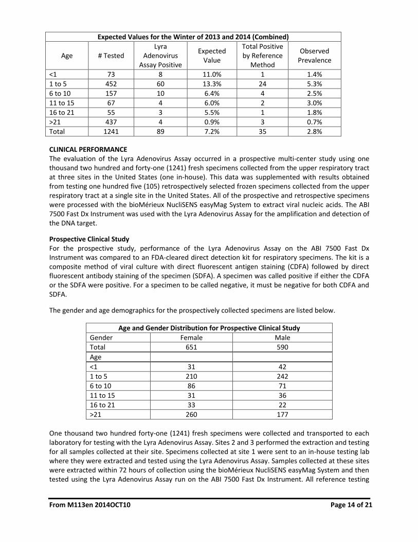
EXPECTED VALUES A prospective clinical study was performed with the Lyra Adenovirus Assay using the ABI 7500 Fast Dx Instrument. Testing was performed with one thousand two hundred and forty-one (1241) nasal and nasopharyngeal swab specimens collected from three sites throughout the United States during the 2013 and 2014 winter seasons (January-March 2013 and December-February 2014). The overall prevalence (all sites combined) for HAdV averaged 2.8%. The table below provides the expected values for HAdV.
From M113en 2014OCT10 Page 14 of 21
Expected Values for the Winter of 2013 and 2014 (Combined)
Age # Tested Lyra
Adenovirus Assay Positive
Expected Value
Total Positive by Reference
Method
Observed Prevalence
˂1 73 8 11.0% 1 1.4%
1 to 5 452 60 13.3% 24 5.3%
6 to 10 157 10 6.4% 4 2.5%
11 to 15 67 4 6.0% 2 3.0%
16 to 21 55 3 5.5% 1 1.8%
>21 437 4 0.9% 3 0.7%
Total 1241 89 7.2% 35 2.8%
CLINICAL PERFORMANCE The evaluation of the Lyra Adenovirus Assay occurred in a prospective multi-center study using one thousand two hundred and forty-one (1241) fresh specimens collected from the upper respiratory tract at three sites in the United States (one in-house). This data was supplemented with results obtained from testing one hundred five (105) retrospectively selected frozen specimens collected from the upper respiratory tract at a single site in the United States. All of the prospective and retrospective specimens were processed with the bioMérieux NucliSENS easyMag System to extract viral nucleic acids. The ABI 7500 Fast Dx Instrument was used with the Lyra Adenovirus Assay for the amplification and detection of the DNA target.
Prospective Clinical Study For the prospective study, performance of the Lyra Adenovirus Assay on the ABI 7500 Fast Dx Instrument was compared to an FDA-cleared direct detection kit for respiratory specimens. The kit is a composite method of viral culture with direct fluorescent antigen staining (CDFA) followed by direct fluorescent antibody staining of the specimen (SDFA). A specimen was called positive if either the CDFA or the SDFA were positive. For a specimen to be called negative, it must be negative for both CDFA and SDFA.
The gender and age demographics for the prospectively collected specimens are listed below.
Age and Gender Distribution for Prospective Clinical Study
Gender Female Male
Total 651 590
Age
˂1 31 42
1 to 5 210 242
6 to 10 86 71
11 to 15 31 36
16 to 21 33 22
>21 260 177
One thousand two hundred forty-one (1241) fresh specimens were collected and transported to each laboratory for testing with the Lyra Adenovirus Assay. Sites 2 and 3 performed the extraction and testing for all samples collected at their site. Specimens collected at site 1 were sent to an in-house testing lab where they were extracted and tested using the Lyra Adenovirus Assay. Samples collected at these sites were extracted within 72 hours of collection using the bioMérieux NucliSENS easyMag System and then tested using the Lyra Adenovirus Assay run on the ABI 7500 Fast Dx Instrument. All reference testing

From M113en 2014OCT10 Page 15 of 21
with the comparator method was conducted at an in-house testing lab. Specimens were shipped to this location daily with cold packs and were cultured within 72 hours of collection. Two (2) specimens were invalid when initially tested with the Lyra Adenovirus Assay. The specimens were re-tested according to the instructions for use and were invalid upon repeat testing. The specimens have been removed from the data presented below. The table below details the HAdV results for the remaining one thousand two hundred thirty-nine (1239) specimens.
Combined Site Data – Prospective Samples
Comparator: CDFA with SDFA
Lyra Adenovirus Assay Positive Negative Total
Positive 35 54* 89
Negative 0 1150 1150
Total 35 1204 1239
95% CI
Sensitivity 35/35 100% 90.1% to 100%
Specificity 1150/1204 95.5% 94.2 % to 96.5%
*Forty-five (45) of the fifty-four (54) positives were positive by an additional FDA cleared PCR assay. Four (4) of the fifty-four (54) positives were negative by an additional FDA cleared PCR assay. Two (2) of the fifty-four (54) positives were invalid by an additional FDA cleared PCR assay. Three (3) of the fifty-four (54) positives had insufficient volume for additional testing.
Retrospectively Selected Specimens Due to the low prevalence of adenovirus at the clinical sites during the study period, a set of retrospectively selected samples were also tested. One hundred five (105) frozen nasopharyngeal swab specimens were obtained from a pediatric hospital in the Southern United States. These specimens were selected based on a qualitative result previously generated by an FDA-cleared respiratory panel. All specimens were stored frozen at –70°C until they were shipped to an in-house testing lab where they were extracted and tested with the Lyra Adenovirus Assay and the comparator, an FDA-cleared PCR assay. The operator performing the testing was blinded to the identity of these specimens. The table below shows the agreement of the Lyra Adenovirus Assay with the comparator (an FDA-cleared PCR assay).
Data from a Single Site – Retrospectively Selected Samples
Comparator: FDA-Cleared PCR Assay
Lyra Adenovirus Assay Positive Negative Total
Positive 27 1* 28
Negative 0 77 77
Total 27 78 105
95% CI
Positive Percent Agreement 27/27 100% 87.5% to 100%
Negative Percent Agreement 77/78 98.7% 93.1% to 99.8%
*One (1) of one (1) positive was positive by a third FDA-cleared PCR assay.

From M113en 2014OCT10 Page 16 of 21
ANALYTICAL PERFORMANCE Limit of Detection The analytical sensitivity (limit of detection or LOD) of the Lyra Adenovirus Assay was determined using quantified (TCID50/mL) stocks of representative serotypes from the six (6) species of HAdV serially diluted in negative nasal matrix. Each dilution tested was extracted in replicates of 20 per concentration using the bioMérieux NucliSENS easyMag System and tested on the ABI 7500 Fast Dx Instrument. Testing was performed with three manufactured device lots. LOD is defined as the lowest concentration at which 95% of all replicates tested positive.
Summary of LOD Study
Species/ Serotype LOD (TCID50/mL) Avg. Ct Value
A/31 8.00 x10⁻² 28.5
B/3 8.00 x10⁻² 28.3
C/1 8.00 x10⁻² 28.1
D/19 1.61 x101 28.6
E/4 1.00 x100 27.7
F/41 3.20 x10⁻² 28.9
Analytical Reactivity (Inclusivity) To verify the Lyra Adenovirus Assay detects the fifty-two (52) known serotypes of HAdV, 49 serotypes were initially tested at or near the species LOD (see above). Higher concentrations were tested if the organism was not detected at the LOD. Each of the 49 serotypes tested was detected by the Lyra Adenovirus Assay. Three types were not detected at 2x LOD. These types were subjected to repeat analysis and were ultimately detected at 250x LOD (ADV-21/Serotype B) and 6x LOD (ADV-ADV-15 and ADV-17/Serotype D) for their respective serotypes.
HAdV Serotypes Reactivity Summary
Species Serotype Concentration Tested
(TCID50/mL) Multiple of LOD
Detected
A HAdV-12 1.60 x10
-1 2x LOD
HAdV-18 1.60 x10⁻¹
2x LOD
B
HAdV-7 1.60 x10⁻¹
2x LOD
HAdV-11 1.60 x10⁻¹
2x LOD
HAdV-14 1.60 x10⁻¹
2x LOD
HAdV-16 1.60 x10⁻¹
2x LOD
HAdV-21 2.00 x101 250x LOD*
HAdV-34 1.60 x10⁻¹
2x LOD
HAdV-35 1.60 x10⁻¹
2x LOD
HAdV-50 1.60 x10⁻¹
2x LOD
C
HAdV-2 1.60 x10⁻¹
2x LOD
HAdV-5 1.60 x10⁻¹
2x LOD
HAdV-6 1.60 x10⁻¹
2x LOD
F HAdV-40 6.40 x10⁻²
2x LOD

From M113en 2014OCT10 Page 17 of 21
HAdV Serotypes Reactivity Summary
Species Serotype Concentration Tested
(TCID50/mL) Multiple of LOD
Detected
D
HAdV-8 3.22 x10⁻¹
2x LOD
HAdV-9 3.22 x10⁻¹
2x LOD
HAdV-10 3.22 x10⁻¹
2x LOD
HAdV-13 3.22 x10⁻¹
2x LOD
HAdV-15 9.66 x101 6x LOD*
HAdV-17 9.66 x101 6x LOD*
HAdV-20 3.22 x10⁻¹
2x LOD
HAdV-22 3.22 x10⁻¹
2x LOD
HAdV-23 3.22 x10⁻¹
2x LOD
HAdV-24 3.22 x10⁻¹
2x LOD
HAdV-25 3.22 x10⁻¹
2x LOD
HAdV-26 3.22 x10⁻¹
2x LOD
HAdV-27 3.22 x10⁻¹
2x LOD
HAdV-28 3.22 x10⁻¹
2x LOD
HAdV-29 3.22 x10⁻¹
2x LOD
HAdV-30 3.22 x10⁻¹
2x LOD
HAdV-32 3.22 x10⁻¹
2x LOD
HAdV-33 3.22 x10⁻¹
2x LOD
HAdV-36 3.22 x10⁻¹
2x LOD
HAdV-37 3.22 x10⁻¹
2x LOD
HAdV-39 3.22 x10⁻¹
2x LOD
HAdV-43 3.22 x10⁻¹
2x LOD
HAdV-44 3.22 x10⁻¹
2x LOD
HAdV-45 3.22 x10⁻¹
2x LOD
HAdV-46 3.22 x10⁻¹
2x LOD
HAdV-47 3.22 x10⁻¹
2x LOD
HAdV-48 3.22 x10⁻¹
2x LOD
HAdV-49 3.22 x10⁻¹
2x LOD
HAdV-51 3.22 x10⁻¹
2x LOD
*HAdV-15, HAdV-17, and HAdV-21 required repeat testing and were ultimately found to be detected at 6x LOD (HAdV-15 and HAdV-17) and 250x LOD (HAdV-21). NOTE: Most viruses were re-grown and quantified in TCID50 (50% Tissue Culture Infectious Dose) for LOD determination. The unit TCID50 is a measure of infectivity or cytotoxicity rather than number of organisms or copies of nucleic acid. Variability in TCID50/mL may not accurately reflect differences in the relative sensitivity of detection between different strains of the same organism.
HAdV Species D serotypes 38 and 42 and Species G serotype 52 (often associated with conjunctivitis and gastroenteritis) were not evaluated for inclusivity as the sponsor could not obtain these strains. Instead, the manufacturer conducted an in silico analysis where they aligned their probe and primers with the
From M113en 2014OCT10 Page 18 of 21
target regions from a single sequence (obtained from GenBank) for each of these adenovirus types. The alignments showed that 100% agreement of the forward primer with types 38, 42, and 52. For the reverse primer, there were 3 mismatches detected on the sequences for types 38 and 42 (84% homology) and 2 mismatches with the sequence for type 52 (89.5% homology). For the probe, there was 100% agreement with types 38 and 42 and 3 mismatches with the sequence for type 52 (85% homology).
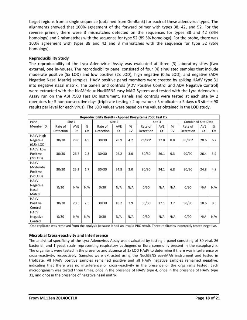
Reproducibility Study The reproducibility of the Lyra Adenovirus Assay was evaluated at three (3) laboratory sites (two external, one in-house). The reproducibility panel consisted of four (4) simulated samples that include moderate positive (5x LOD) and low positive (2x LOD), high negative (0.5x LOD), and negative (ADV Negative Nasal Matrix) samples. HAdV positive panel members were created by spiking HAdV type 31 into negative nasal matrix. The panels and controls (ADV Positive Control and ADV Negative Control) were extracted with the bioMérieux NucliSENS easy MAG System and tested with the Lyra Adenovirus Assay run on the ABI 7500 Fast Dx Instrument. Panels and controls were tested at each site by 2 operators for 5 non-consecutive days (triplicate testing x 2 operators x 3 replicates x 5 days x 3 sites = 90 results per level for each virus). The LOD values were based on the values obtained in the LOD study.
Reproducibility Results - Applied Biosystems 7500 Fast Dx
Panel Member ID
Site 1 Site 2 Site 3 Combined Site Data
Rate of Detection
AVE Ct
% CV
Rate of Detection
AVE Ct
% CV
Rate of Detection
AVE Ct
% CV
Rate of Detection
AVE Ct
% CV
HAdV High Negative (0.5x LOD)
30/30 29.0 4.9 30/30 28.9 4.2 26/30* 27.8 8.8 86/90* 28.6 6.2
HAdV Low Positive (2x LOD)
30/30 26.7 2.3 30/30 26.2 3.0 30/30 26.1 9.3 90/90 26.4 5.9
HAdV Moderate Positive (5x LOD)
30/30 25.2 1.7 30/30 24.8 3.0 30/30 24.1 6.8 90/90 24.8 4.8
HAdV Negative Nasal Matrix
0/30 N/A N/A 0/30 N/A N/A 0/30 N/A N/A 0/90 N/A N/A
HAdV Positive Control
30/30 20.5 2.5 30/30 18.2 3.9 30/30 17.1 3.7 90/90 18.6 8.5
HAdV Negative Control
0/30 N/A N/A 0/30 N/A N/A 0/30 N/A N/A 0/90 N/A N/A
*One replicate was removed from the analysis because it had an invalid PRC result. Three replicates incorrectly tested negative.
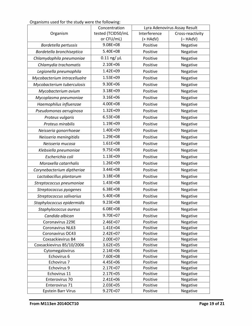
Microbial Cross-reactivity and Interference The analytical specificity of the Lyra Adenovirus Assay was evaluated by testing a panel consisting of 30 viral, 26 bacterial, and 1 yeast strain representing respiratory pathogens or flora commonly present in the nasopharynx. The organisms were tested in the presence and absence of 2x LOD HAdV to determine if there was interference or cross-reactivity, respectively. Samples were extracted using the NucliSENS easyMAG instrument and tested in triplicate. All HAdV positive samples remained positive and all HAdV negative samples remained negative, indicating that there was no interference or cross-reactivity in the presence of the organisms tested. Each microorganism was tested three times, once in the presence of HAdV type 4, once in the presence of HAdV type 31, and once in the presence of negative nasal matrix.
From M113en 2014OCT10 Page 19 of 21
Organisms used for the study were the following:
Organism Concentration
tested (TCID50/mL or CFU/mL)
Lyra Adenovirus Assay Result
Interference (+ HAdV)
Cross-reactivity (– HAdV)
Bordetella pertussis 9.08E+08 Positive Negative
Bordetella bronchiseptica 5.40E+08 Positive Negative
Chlamydophila pneumoniae 0.11 ng/ μL Positive Negative
Chlamydia trachomatis 2.10E+06 Positive Negative
Legionella pneumophila 1.42E+09 Positive Negative
Mycobacterium intracellualre 1.53E+09 Positive Negative
Mycobacterium tuberculosis 9.30E+06 Positive Negative
Mycobacterium avium 3.18E+09 Positive Negative
Mycoplasma pneumoniae 3.16E+06 Positive Negative
Haemophilus influenzae 4.00E+08 Positive Negative
Pseudomonas aeruginosa 1.32E+09 Positive Negative
Proteus vulgaris 6.53E+08 Positive Negative
Proteus mirabilis 1.19E+09 Positive Negative
Neisseria gonorrhoeae 1.40E+09 Positive Negative
Neisseria meningitidis 1.29E+08 Positive Negative
Neisseria mucosa 1.61E+08 Positive Negative
Klebsiella pneumoniae 9.75E+08 Positive Negative
Escherichia coli 1.13E+09 Positive Negative
Moraxella catarrhalis 1.26E+09 Positive Negative
Corynebacterium diptheriae 3.44E+08 Positive Negative
Lactobacillus plantarum 3.18E+08 Positive Negative
Streptococcus pneumoniae 1.43E+08 Positive Negative
Streptococcus pyogenes 6.38E+08 Positive Negative
Streptococcus salivarius 5.40E+08 Positive Negative
Staphylococcus epidermidis 9.23E+08 Positive Negative
Staphylococcus aureus 6.08E+08 Positive Negative
Candida albican 9.70E+07 Positive Negative
Coronavirus 229E 2.46E+07 Positive Negative
Coronavirus NL63 1.41E+04 Positive Negative
Coronavirus OC43 2.42E+07 Positive Negative
Coxsackievirus B4 2.00E+07 Positive Negative
Coxsackievirus B5/10/2006 3.62E+05 Positive Negative
Cytomegalovirus 2.14E+06 Positive Negative
Echovirus 6 7.60E+08 Positive Negative
Echovirus 7 4.45E+06 Positive Negative
Echovirus 9 2.17E+07 Positive Negative
Echovirus 11 2.17E+05 Positive Negative
Enterovirus 70 2.41E+06 Positive Negative
Enterovirus 71 2.03E+05 Positive Negative
Epstein Barr Virus 9.27E+07 Positive Negative

From M113en 2014OCT10 Page 20 of 21
Organism Concentration
tested (TCID50/mL or CFU/mL)
Lyra Adenovirus Assay Result
Interference (+ HAdV)
Cross-reactivity (– HAdV)
HSV Type 1 MacIntyre Strain 5.89E+06 Positive Negative
HSV Type 2 Strain G 1.96E+06 Positive Negative
Human Metapneumovirus (A1) 3.66E+05 Positive Negative
Human Rhinovirus 45 2.94E+04 Positive Negative
Human Rhinovirus 52 2.63E+04 Positive Negative
Influenza A/Mexico/4108/2009 4.08E+05 Positive Negative
Influenza A/Port Chalmers 3.55E+08 Positive Negative
Influenza B/Florida/04/2006 1.54E+06 Positive Negative
Measles 1.95E+06 Positive Negative
Mumps Virus 2.75E+08 Positive Negative
Parainfluenza Type 1 3.97E+06 Positive Negative
Parainfluenza Type 2 3.15E+08 Positive Negative
Parainfluenza Type 3 2.36E+07 Positive Negative
Parainfluenza Type 4A 1.04E+05 Positive Negative
RSV A (Long) 4.36E+04 Positive Negative
RSV B (Wash/18537/62) 3.43E+05 Positive Negative
Varicella Zoster Virus 1.11E+04 Positive Negative
Analytical Specificity – Interfering Substances The performance of Lyra Adenovirus Assay was evaluated with potentially interfering substances that may be present in nasopharyngeal specimens. The 11 potentially interfering substances were evaluated using HAdV at a concentration of 2x LOD. Each interfering substance was tested three times, once in the presence of HAdV type 4, once in the presence of HAdV type 31, and once in the presence of negative nasal matrix. All HAdV positive samples remained positive and all HAdV negative samples remained negative, indicating that there was no interference in the presence of the substances tested.
Substance Name Concentration Tested
Mucin (Bovine Submaxillary Gland, type I-S) 60 µg/mL
Blood (human), EDTA anticoagulated 2% (vol/vol)
Neo-Synephrine 15% (vol/vol)
Afrin Nasal Spray 15% (vol/vol)
Zicam Homeopathic Non-Drowsy Allergy Relief No Drip Liquid Nasal Gel
5% (vol/vol)
Saline Nasal Spray 15% (vol/vol) of dose
Throat Lozenges 0.68 g/mL; 1/18 drop, crushed; active ingredients: 1.7 mg/mL
menthol
Zanamivir 3.3-5 mg/mL
Tobramycin 4.0 µg/mL
Mupirocin 6.6-10 mg/mL
Oseltamivir phosphate 7.5-25 mg/mL

From M113en 2014OCT10 Page 21 of 21
Carry-over and Cross-contamination Study A carry-over study was conducted using a 96 sample panel consisting of 48 high positive (1.0 x 10⁵ TCID50/mL HAdV type 4) and 48 negative specimens (negative nasal matrix). The high positive samples were extracted and added to the plate in a checkerboard pattern that alternated with the negative samples. This testing was repeated over a 5 day period. There was no evidence of carry-over/cross contamination with the Lyra Adenovirus Assay using the NucliSens easyMAG automated nucleic acid extraction instrument and the ABI 7500 Fast Dx instrument.
Additional Source Material 1. Guidance on Informed Consent for In Vitro Diagnostic Device Studies Leftover Human Specimens
that are Not Individually Identifiable (April 2006) – http://www.fda.gov/cdrh/oivd/guidance/ 1588.pdf.
2. Draft Guidance on Nucleic Acid Based In Vitro Diagnostic Devices for Detection of Microbial Pathogens (Dec 2005) – http://www.fda.gov/cdrh/oivd/guidance/1560.html.
3. CLSI EP17-A: Guidance for Protocols for Determination of Limits of Detection and Limits of Quantitations (Vol. 2, No. 34) (Oct 2004).
4. CLSI MM13-A: Guidance for the Collection, Transport, Preparation and Storage of Specimens for Molecular Methods (Vol. 25, No. 31) (Dec 2005).
5. CLSI EP7-A2: Guidance for Interference Testing in Clinical Chemistry (Vol. 25, No.27 Second Ed) (Nov 2005).
6. CLSI EP12-A: Guidance for User Protocol for Evaluation of Qualitative Test Performance (Vol. 22, No. 14) (Sept 2002).
7. CLSI MM6-A: Guidance for the Quantitative Molecular Methods for Infectious Diseases (Vol. 23, No.28) (Oct 2003).
8. CLSI EP5-A2: Guidance for Evaluation of Precision Performance of Quantitative Measurement Methods (Vol. 24, No. 25 Second Ed.) (Aug 2004).
CUSTOMER AND TECHNICAL SUPPORT To place an order or for technical support, please contact a Quidel Representative at 800.874.1517 (in the U.S.) or 858.552.1100 (outside the U.S.A), Monday through Friday, from 8:00 a.m. to 5:00 p.m., Eastern Time. Orders may also be placed by fax at 740.592.9820. For e-mail support contact [email protected] or [email protected]. For services outside the U.S., please contact your local distributor. Additional information about Quidel, our products, and our distributors can be found on our website quidel.com.
INTELLECTUAL PROPERTY The purchase of this product grants the purchaser rights under certain Roche patents to use it solely for providing human in vitro diagnostic services. No general patent or other license of any kind other than this specific right of use from purchase is granted hereby. Dye compounds in this product are sold under license from Biosearch Technologies, Inc. and protected by U.S. and world-wide patents either issued or in application.
REFERENCES 1. Clinical and Laboratory Standards Institute. Viral Culture; Approved Guidelines. CLSI document
M41-A [ISBN 1562386239] Clinical and Laboratory Standards Institute, 940 West Valley Road,
Suite 1400, Wayne, Pennsylvania 19087-1898, USA 2006.