Embed Size (px)
Citation preview

Physica D 43 (1990) 1-16 North-Holland
THERE IS A THEORY OF HEART
Leon GLASS’ and P. HUNTERb ‘Department of Physiology, McGill University, 3655 Drummond Street, Montreal, Quebec, Canada H3G 1 Y6 “Department of Engineering Science, Universiry of Auckland Auckland New Zealand
Received 29 November 1989 Accepted 18 December 1989 Communicated by A.T. Winfree
We review a recent workshop entitled “Theory of Heart,” held July lo-18,1989 at the Institute for Nonlinear Sciences in the University of California at San Diego. Recent advances have been made in the application of mathematical techniques used in the physical sciences to the analysis of mechanics and dynamics in the heart. The theoretical models are in many cases sufficiently well developed that theoretical predictions can be made that can be confronted with experimental and clinical data. Topics covered include mechanics of the passive and active heart, ionic mechanisms of cardiac arrhythmias, reentrant reexcitation and its connection with cardiac tachycardia and fibrillation, and clinical implications of the basic research.
1. Introhction
The physical sciences employ theoretical models, usually posed in the form of mathematical equations, to predict the outcome of experiments. Experiments test the theories, or better yet, demonstrate unex- pected new effects demanding novel theoretical insights. The biological sciences seem to be different. Few biologists have received technical mathematical training and the research style in which there is a continual intercourse between mathematical theory and experiment is not common. But there are exceptions. One area in which mathematical theory is helping to shape and define experiments is in cardiac physiology. A recent workshop held at the University of California at San Diego on July 10-18, 1989 dealt with the Theory of Heart. The workshop brought together about 80 mathematicians, physicists, engineers, physiolo- gists and cardiologists, all sharing a common interest in applying theory to study the heart. The first three days were devoted to didactic talks, and the remaining six days were more oriented towards talks describing original research. There were about twenty invited speakers, but many of the participants gave brief presentations of their research in afternoon workshop sessions. This is a brief summary of the conference and a review of the use of mathematical theories in cardiac physiology. We concentrate on summarizing the invited talks, but in some instances will also mention results presented in the afternoon discussions. A multi-authored volume containing contributions from the workshop participants will also be published [ 11.
2. A primer of cardiac physiology and pathophysiology: structural complexity of the heart [2]
All theoretical analyses of the heart make simplifying assumptions. Before examining some of these simplifications, let us examine some of the complexities of the intact heart.
0167-2789/90/$03.50 Q Elsevier Science Publishers B.V. (North-Holland)
L Glass and P. Hunter/ There is a theory of heart
Superior vena cava
Sinoatriol node
Atrioventricular node
Bundle branches
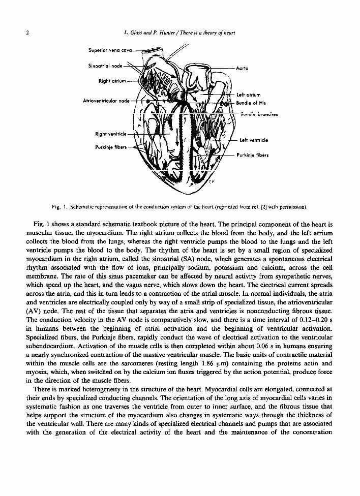
Fig. 1. Schematic representation of the conduction system of the heart (reprinted from ref. [2] with permission).
Fig. 1 shows a standard schematic textbook picture of the heart. The principal component of the heart is muscular tissue, the myocardium. The right atrium collects the blood from the body, and the left atrium collects the blood from the lungs, whereas the right ventricle pumps the blood to the lungs and the left ventricle pumps the blood to the body. The rhythm of the heart is set by a smaIl region of specialized myocardium in the right atrium, called the sinoatrial (SA) node, which generates a spontaneous electrical rhythm associated with the flow of ions, principally sodium, potassium and calcium, across the cell membrane. The rate of this sinus pacemaker can be affected by neural activity from sympathetic nerves, which speed up the heart, and the vagus nerve, which slows down the heart. The electrical current spreads across the atria, and this in turn leads to a contraction of the atrial muscle. In normal individuals, the atria and ventricles are electrically coupled only by way of a small strip of specialized tissue, the atrioventricular (AV) node. The rest of the tissue that separates the atria and ventricles is nonconducting fibrous tissue. The conduction velocity in the AV node is comparatively slow, and there is a time interval of 0.12-0.20 s in humans between the beginning of atrial activation and the beginning of ventricular activation. Specialized fibers, the Purkinje fibers, rapidly conduct the wave of electrical activation to the ventricular subendocardium. Activation of the muscle cells is then completed within about 0.06 s in humans ensuring a nearly synchronized contraction of the massive ventricular muscle. The basic units of contractile material within the muscle cells are the sarcomeres (resting length 1.86 urn) containing the proteins actin and myosin, which, when switched on by the calcium ion fluxes triggered by the action potential, produce force in the direction of the muscle fibers.
There is marked heterogeneity in the structure of the heart. Myocardial cells are elongated, connected at their ends by specialized conducting channels. The orientation of the long axis of myocardial cells varies in systematic fashion as one traverses the ventricle from outer to inner surface, and the fibrous tissue that helps support the structure of the myocardium also changes in systematic ways through the thickness of the ventricular wall. There are many kinds of specialized electrical channels and pumps that are associated with the generation of the electrical activity of the heart and the maintenance of the concentration

L. Glass and P. Hunter/ There is a theory of heart 3
gradients of various ionic species across myocardial cells. Different parts of the heart have different electrical properties, and this is believed to be due to differences in the types and relative numbers of channels in the various parts of the heart.
The electrical properties of the heart can be monitored noninvasively by recording the electrocardiogram (ECG), which measures potential differences on the surface of the body. The spread of the electrical activity in the heart leads to characteristic features on the ECG. Cardiologists are trained to recognize and interpret variations from normality. Abnormal cardiac rhythms (arrhythmias) can usually be readily identified based on the ECG. Common abnormalities include situations in which the heart rate is too slow (bradycardia) or too fast (tachycardia). Electrical conduction can be wholly or partially blocked in such structures as the AV node or the branches of the Purkinje network (fig. 1). Excitation can arise from abnormal (ectopic) foci in the heart. Uncoordinated spread of excitation can take place in the atria (atria1 fibrillation), or the ventricles (ventricular fibrillation). Although atrial fibrillation is usually not fatal, ventricular fibrillation is always fatal within a few minutes of initiation due to inadequate blood flow to the brain.
There are many different cardiac abnormalities and diseases, which can have varying effects, ranging from benign conditions to death. There are a large number of different congenital structural abnormalities such as holes between the two ventricles, and accessory conducting pathways between the atria and ventricles. Diseases can lead to impairment of the heart valves or impaired myocardial function (cardiomyopathy). One of the most common cardiac diseases in developed countries is coronary artery disease in which coronary arteries become partially blocked. A complete sudden blockage of coronary arteries, a myocardial infarction (a “heart attack”), has varying severity depending on the extent of tissue damaged due to lack of adequate oxygenation. The damage can lead to impaired pumping action of the heart, or sometimes make the heart susceptible to arrhythmias. Changes in the ionic composition of the blood, such as occurs in some diseases, can also lead to arrhythmias.
3. Mechanics
3.1. The continuum mechanics framework
A review of the continuum mechanics approach to modeling the mechanics of the heart was given by Peter Hunter and Andrew McCulloch. The aim of the modeling is to provide a link between the mechanical behavior of the sarcomeres and the mechanical function of the intact heart [3]. A model can be used in the “forward” sense of predicting intact heart behavior from a knowledge of sarcomere dynamics, or in the “inverse” sense of revealing muscle properties from observations of the motion of the intact heart. The key elements in a mechanics model are: (1) the equations of motion (essentially Newton’s laws applied to a continuously deformable material), which hold for all materials; (2) the stress-strain or “constitutive” law for the particular material under study, in this case cardiac muscle; (3) the kinematic relations which ensure that adjacent particles move in a compatible fashion; and (4) the boundary conditions which specify the externally applied displacements or forces, such as those generated by ventricular blood pressure.
The deformations occurring in the cardiac cycle are sufhciently large that both the kinematic relations, coupling strain and displacement, and the constitutive law, coupling stress and strain, are nonlinear. This, together with the three-dimensional complexity of the cardiac geometry and muscle fiber orientations,
4 L.. Glass and P. Hunter/ There is a theory of heart
means that the models must employ numerical analysis. Little insight is gained from oversimplified analytic models.
The most promising numerical analysis technique for this type of problem is the finite element method, which is widely used in engineering analysis. A common feature of the finite element method is that the dependent variable field (e.g. displacement field) is described by piecewise parametric functions (usually low-order polynomials) which employ point (“nodal”) values of the field variable as coefficients in a way which implicitly guarantees continuity of the field variable across the boundaries of the piecewise regions (“elements”) [4]. Hunter and McCulloch showed how the complex three-dimensional geometry and fibrous structure of the heart can be modelled efficiently with a prolate spheroidal coordinate system and a combination of linear and cubic Hermite finite element basis functions. This description of heart anatomy provided the reference configuration for the mechanical analysis of heart motion described later in the workshop. The kinematics of large deformation elasticity and the various types of stress tensor used in mechanics modeling were described in detail as a background to the later talks [5].
3.2. Material properties of myocardium
A challenging task in cardiac mechanics research is the formulation of a constitutive law that accurately describes the mechanical properties of passive cardiac muscle that has been arrested with a high concentration potassium medium. Several contributors addressed the problem of formulating a three- dimensional stress-strain law, both as a completely “black box” empirical description and by using available knowledge of tissue microstructure to guide the formulation.
Bruce Smaill described the microstructure of cardiac muscle. Recent studies have revealed an extensive network of connective tissue in the myocardium with the extracellular collagen organized into a hierarchy of different structures: endomysium is the connective tissue network surrounding individual myocytes or coupling adjacent myocytes; perimysium is the collagen network surrounding groups or bundles of fibers and linking contiguous bundles (fairly loosely); and epimysium is the connective tissue coat surrounding the entire muscle (in papillary muscles or trabeculae) [6]. Smaill also emphasized the importance of biaxial testing procedures for measuring the stress-strain laws of incompressible soft tissue. Loads are applied to the cut edges of a thin tissue specimen in such a way as to produce a uniform stress field in the central region of the sample, where strain is measured by videometric techniques.
Several complementary approaches addressed the formulation of constitutive laws. Arie Horowitz showed how a constitutive law can be formulated using- knowledge of tissue microstructure [7]. He proposed that the stress tensor be derived from the gradient of a strain energy function by summing up the individual strain energies of its constituents including the muscle fibers, the collagen fibers and the fluid matrix which embeds them. The orientation and “waviness” of the two types of fiber are described with distribution functions. The resulting stress-strain law has eight parameters associated with the muscle fibers and five with the collagen -but only the collagen was found to contribute significantly to the stresses measured in biaxial tests.
Jay Humphrey presented an alternative approach to formulating a constitutive law for passive my- ocardium [8]. He proposed that the strain energy function, expressed in terms of strain invariants, be written as a power series. A program of biaxial tests was then used to identify both the functional form of the strain energy and the values of the material parameters.
Myocardial tissue includes both a solid and a fluid phase. Dan Bogen addressed this aspect of myocardial mechanics by considering the role of fluid compartments in the formulation of the strain energy function [9, lo]. He showed that the distribution of fluid among compartments is a significant

L. Glass and P. Hunter/ There is a theory of heart 5
determinant of tissue elasticity. Extracellular fluid movement can be substantial and rapid. Using an axisymmetric model he showed that this may be an important mechanism in stress equilibration of the left ventricle.
The research on the stress-strain relationships in passive potassium arrested heart in vitro is an important step in understanding the mechanical properties of the intact heart since it enables one to carry out precise mechanical studies on cardiac tissue. Yet, the experiments here are difficult since measurements must be carried out rapidly following potassium arrest to prevent deterioration of the tissue. It is also not straightforward to extrapolate from these studies to the properties of the intact, beating heart.
3.3. Mechanics of the intact heart
With a knowledge of the geometry and fibrous architecture of the heart wall, the three-dimensional mechanical properties of the resting and contracting cardiac muscle, and the pressure loads acting on the boundaries of the heart wall, it should be possible to formulate and solve a model of the mechanics of the intact myocardium using the laws of continuum mechanics. Such a model could predict the distributions of internal force (stress) and local shape-change (strain) throughout the heart wall. Since stress and strain are local measures of mechanics rather than global measures such as ventricular pressures and ejection fractions, they tell us about the regional function of the heart. This is particularly important since the properties of the myocardium are highly nonhomogeneous and cardiac pathologies are commonly characterized by regional abnormalities, such as an ischemic zone or infarction resulting from a coronary artery occlusion. Knowing the stresses and strains in and around the infarction would allow the cardiologist to identify the region of altered properties, the extent of injury, the prospects for recovery and the vulnerability of the region to a failure like an aneurysm. The stresses and strains predicted by a model are also directly related to important variables such as the regional myocardial blood flow and oxygen demand, and the signals for growth and remodelling of the wall [ll]. To test models of cardiac mechanics, experimental measurements of myocardial stresses and strains are required. However, the experimental measurement of force in the intact ventricular wall is fraught with difficulty, and the few published findings have been controversial and inconsistent. On the other hand, the measurement of regional wall strains using length transducers (such as pairs of ultrasonic crystals implanted close together in the myocardium) or by imaging of discrete markers placed in or on the wall has been much more successful.
The experimental measurement of cardiac wall strains in various experimental preparations was described by Andrew McCulloch, Jeffrey Omens and Lewis Waldman, and the implications of their findings for models of ventricular mechanics were discussed. It is well known that with increased diastolic filling, the output and work done by the beating heart also increases. This relationship, known as the Frank-Starling law of the heart, is an intrinsic property of cardiac muscle contraction, the force of which is directly related to the length of the muscle sarcomeres. Therefore, any model of the mechanics of the beating heart must first be able to predict the distribution of sarcomere length, and hence the distribution of strain, in the myocardium at end-diastole. Andrew McCulloch described the measurement of left ventricular wall strains on the epicardium of the isolated potassium-arrested canine heart. A biplane video recording technique was used to measure the displacements of triplets of markers attached lo-20 mm apart on the left ventricular free wall as the ventricle was loaded to various diastolic pressures [12]. Under physiological loading the relationship between ventricular pressure and volume is nonlinear, characteristic of the nonlinear stress-strain relationship of the resting myocardium and most other soft tissues [13]. The epicardial extensions exceeded 20% at moderate pressures-too high to be treated by the classical linear strain theory of engineering mechanics. The magnitudes of epicardial strains varied significantly with

6 L. Glass and P. Hunter/ There is a theory of heart
location on the free wall being up to twice as great near the apex as near the base. But the pattern of surface deformation had some features common to all regions. In particular, lengthening of the epicardium during filling was nonuniform. It was consistently greatest along a preferred axis that was rotated clockwise from the circumference by about 30-50”, which coincides with the local epicardial fiber direction [14]. Consequently, the ventricle exhibited twisting about its long axis (torsion). The observation of ventricular torsion in the passive heart, just as is seen in the actively contracting heart but opposite in direction, suggests that the resting myocardium is anisotropic with respect to its local fiber axis. McCulloch showed how experimental measurements of wall strain could be used not only to validate a model of ventricular mechanics but to predict unknown material parameters. Using a simplified cylindrical model of the equatorial region of the left ventricular wall and a nonlinear material model that included anisotropy with respect to a fiber axis that changed continuously across the wall thickness, he showed the nonlinear governing equations were only satisfied subject to the known boundary conditions and epicardial deformations for certain values of the material parameters. Optimizing these, it was predicted that the intact myocardium is signiGcantly stiffer along the fiber axis than in the transverse plane. The prediction was in agreement with mechanical tests performed on isolated tissue specimens by other workers. The model also served to demonstrate the beneficial effect of the ventricular torsion on reducing the peaks in stress and strain on the endocardium that isotropic models have predicted [ll].
In the experiments described by McCulloch, the reference state for measuring strain was chosen to be the state at which the pressure inside and outside the ventricular wall was zero. However, the unloaded state of a body is not necessarily the “stress-free” state, since “residual” or initial stresses can exist. In engineering, residual stresses can be created during fabrication or manufacture. Prsstressing of concrete with steel bars in tension is an example. In tissues, residual stresses arise as cells grow and interact with each other. Jeffrey Omens described new experimental work which revealed the presence of residual stresses in the wall of the unloaded potassium-arrested rat heart. All contractile activity of the muscle was blocked. Residual stresses are revealed by cutting a body. If enough cuts are made, the body will tend to become stress-free and the shape-changes that accompany the cuts can be measured. Omens called these shape-changes residual strains. To identify residual strains, he cut a cross-sectional ring from the arrested rat heart. The multiply connected ring topology will admit various residual stress fields. Making a single radial cut in the ring was found to relive all or most of the residual stress. The ring instantaneously springs open to form an arc. The average angle of opening in 11 hearts was 45’. Direct measurements of the residual strains showed that in the intact and unloaded state the left ventricular wall is compressed on the endocardium and in tension on the epicardium. Models show that the corresponding gradient of residual stress serves to counteract the opposing gradient of stress that occurs during diastolic filling. Hence, Omens postulated that residual stress in the heart wall is a direct reflection of inhomogeneous growth of the myocardium to achieve optimal function by minimizing stress concentrations in the wall [15].
Lewis Waldman described the experimental measurement of strain in the beating heart during systolic contraction. His laboratory has developed a technique using biplane radiography of columns of small metal beads implanted across the thickness of the ventricular wall [16]. From the three-dimensional coordinates of the metal markers reconstructed from the X-ray, the fully three-dimensional deformation of the heart wall could be determined at different transmural locations. The large amount of information produced by this method included some unexpected results. In particular, although systolic shortening tended to be greatest in the direction of the muscle fibers on the epicardium of the left ventricle, the direction of greatest shortening only changed by 20-30” moving through the wall, whereas the orientation of the muscle fibers changes by 120” or more. Therefore, on the endocardium, the greatest shortening actually occurs in a direction perpendicular to the muscle fibers. These results have important implications

L Glass and P. Hunter/ There is a theory of heart I
for models of ventricular wall stress. The active shortening of the wall is aliected by the extracellular collagen connections between the cells as well as the uniaxial contractile properties of the sarcomeres. Therefore, models that treat the ventricular walls as contracting fibers embedded in a nonviscous fluid, or as uncoupled concentric shells, are inappropriate.
Cardiac disease is commonly characterized by an increase in the heterogeneity of the mechanical function of the myocardium. A good example is acute myocardial ischemia following the interruption of blood supply to a region of the ventricular wall due to an occlusion of a major coronary artery. Normal systolic shortening gives way to lengthening in the ischemic region as the ischemic myocardium loses its ability to develop active tension and shortening (171. A complex pattern of deformation can occur in the border zone between normal and damaged tissue. Dyskinesis can also be seen in some arrhythmias and can be produced in an experimental preparation by pacing the heart from a ventricular site. Wilbur Lew described the clinical consequences for ventricular function of regional heterogeneity. In one set of studies, heterogeneous mechanical function was produced by a local infusion of isoproterenol. Although, this agent increases cardiac contractility, the performance of the heart was impaired by the resulting inhomogeneity, which limited the ability of the heart to fill. Lew concluded that cardiac efficiency may be directly related to the homogeneity of regional function.
The development of a model of the actively contracting heart within the continuum mechanics framework requires further research in a number of areas. First, models of sarcomere dynamics are needed. These must include a description of calcium ion fluxes in order to couple the electrical activation of the heart muscle to the mechanics. Second, since the activation of the ventricular muscle is not synchronous but occurs over 60 ms, a second goal is to model the activation sequence in parallel with the mechanics. Third, the progress of the activation wave itself depends on the mechanics, both through path length changes during contraction and through the effect of mechanical strain on the permeability of cell membranes to the ions underlying the action potential. As an initial step towards a fully active model, Peter Hunter described a constant wavespeed activation model based on the finite element model developed for the mechanics. The three-dimensional geometry was represented by 24 finite elements each with 53 computational points. Each point, once active, gives rise to an ellipsoidal wavefront emanating from that point, whose major axis lies in the local muscle fiber direction. The model was used to generate isochronal maps showing the contours of simultaneous activation on the epicardium, resulting from various initial stimulus points, and these were compared with experimental measurements. The general features of the predicted maps (contour spacing and orientation) agreed quite well with the experimental observations provided the wavespeeds in the fiber and transverse directions were taken to be 1.5 and 0.5 m/s, respectively, everywhere except in the subendocardial region where faster conducting fibers required a speed of 5 m/s.
4. Ionics and arrhythmias
4.1. Ionic models
The ionic basis for the electrical activity in nerve cells was elucidated in the early 1950s by Hodgkin and Huxley, who carried out voltage-clamp studies in squid giant axon [18]. They were able to isolate two main currents, carried by sodium and potassium ions, by manipulating the transmembrane voltage and the composition of the bathing medium. A system of coupled nonlinear differential equations based on the

8 L Glaw and P. Hunter/ There is a theory of heart
experimental data was formulated. This work has formed the cornerstone for subsequent electrophysiolog cal studies in nerve and other excitable tissues.
Michael Guevara reviewed ionic modeling of cardiac tissue. Current evidence indicates that a number of different types of ionic channels and currents play a role in cardiac excitation and pacemaker activity. For example, the recent ionic model of excitation in Purkinje fiber by DiFrancesco and Noble includes more than ten different types of channels activated at various stages in the cardiac cycle [19]. Many of these channels are described by nonlinear equations in which a current becomes activated and in some cases inactivated with different kinetic features that depend on the transmembrane potential. Since ionic species cross the membrane during the action potential, it is necessary to have pump mechanisms that restore and maintain the transmembrane potential gradients. The resulting equations are extraordinarily complex involving more than 10 coupled nonlinear equations with more than 50 parameters.
The DiFrancesco-Noble equations deal with one tissue in one species. Different tissues and different species are known to have different ionic mechanisms. John Clark discussed the ionic modeling of the sinus venosus pacemaker in the bullfrog [20] and the SA node of rabbits [21]. The model of the bullfrog sinus venosus cell was based on single-cell voltage-clamp data which permitted quantitative description for the principal membrane components including the potassium outward delayed rectifier current and the electrogenic calcium extrusion pump. Other important components, such as the inward calcium current and the sodium-potassium pump, were added from models available in the literature. Material balance equations were included for sodium, potassium and calcium ions, and calcium buffering in the intracellular medium was also included. The model for the rabbit SA node was a modification of a model by Noble and Noble [22], but many of the parameters were modified to obtain better agreement with the data. One feature of the models was to propose a formulation for an acetylcholine (ACh)-sensitive potassium channel. In the normal heart, ACh is released by the vagus nerve, leading to a slowing of the heart. Clark described the effects of pulsatile delivery of ACh at various phases of the pacemaker cycle, as well as the effects of periodic application of ACh frequencies [23]. There was good agreement between the models and experimental data.
Wayne Giles summarized recent advances in the development of mathematical models of the calcium current(s) and the sodium-calcium exchange process in cardiac muscle. Experimental measurements and computer simulations have been used in attempts to identify an exchanger current generated by an electrogenic sodium-calcium exchanger in single cells from bullfrog atrium [24]. At potentials near the resting potential of the atrial cell, the net inward current due to sodium-calcium exchange may be significant. Theoretical results suggest that in the intact single atria1 cell, calcium-binding proteins such as calmodulin and troponin could be physiologically important modulators of the amplitude, polarity and kinetics of the exchange current.
Using examples of his work with Dr. Clark’s group at Rice University, Giles stressed the need to base mathematical formulations and assumptions on quantitative experimental data and thus to involve electrophysiologists, bioengineers, biophysicists, and applied mathematicians. The development of tech- niques for the recording and modeling of ionic currents from single isolated myocytes has been very important in this regard. Nonetheless, many recording conditions which yield reliable quantitative data characterizing the biophysical variables are unphysiological. For example, external sodium may be removed, or the contents of recording electrodes can enter into the intracellular milieu by dialysis. Hence, development of mathematical models must take into account the experimental limitations of data collection. Researchers should strive to carry out model development in an environment which allows a continuous dialogue between those carrying out electrophysiological studies and those developing com- puter models.
L. Glass and P. Hunter/ There is a theory of heart 9
The ionic models are complex. One might hope that by approaching the problem in some different way (i.e. not using the Hodgkin-Huxley paradigm), it will be possible to reformulate the whole ionic story in some conceptually simpler fashion. Yet the current evidence is that this cannot be done. People are now able to measure ionic currents passing through individual channels confirming the presence of many different types of channels whose existence had been inferred using less direct approaches. Most workers in the field believe that the complexity in the current models is a reflection of the physiological reality.
4.2. Low-dimensional dynamics in the heart
It is sometimes possible to reduce the complex dynamics of the heart to problems involving low-dimen- sional dynamical models. Several different ways in which this may come about were described by Leon Glass, and this theme was dominant in many of the presentations.
Even though the ionic models generating periodic cardiac activity are formulated as multi-dimensional nonlinear differential equations, the attractor in these equations is a limit cycle oscillation. A perturbation delivered to such equations usually leads to a rapid reestablishment of the oscillation, but with a phase shift relative to the unperturbed cycle. Consequently, once the phase shift induced by a single stimulus at different phases is known, it is possible to predict the effects of periodic stimuli by iteration of a one-dimensional finite difference equation (a circle map). Theoretical predictions and experimental observations in in vitro systems show many of the familiar features found in circle maps including complex bifurcations, Farey sequences of phase-locking ratios, quasiperiodicity, period-doubling bifurcations and
chaos [25, 261. Interaction of a periodic stimulation and a spontaneous cardiac oscillation also occurs in clinical
circumstances in which the normal sinus rhythm interacts with an ectopic pacemaker. This leads to an arrhythmia called parasystole, and theoretical and experimental work in this area was discussed by Glass. Experimental studies and mathematical modeling of parasystole was carried out first by Jalife, Moe and collaborators [27]. They were able to show the presence of complex bifurcations and phase locking between the sinus and ectopic pacemakers in various ratios. For weak coupling between the sinus and ectopic pacemakers, the parasystole problem is equivalent to a problem in number theory, the “gaps and steps” problem. In this case, novel numerological rules have been derived concerning the sequences of normal and ectopic beats in subjects with parasystolic rhythms [28]. The rules have been confirmed in clinical subjects [29].
In the theory described above, the effects of a stimulus depend only on its phase in a cycle. Thus if one repeatedly stimulates a cardiac oscillator with a fixed delay from the action potential to the stimulus, each stimulus would have an identical effect. This is not necessarily the case [30]. However, by taking account of finite relaxation times to the limit cycle following a stimulus in simple models, the dynamics resulting from repeated stimulation at a fixed delay can be reduced to a one-dimensional map [31]. Iteration of the map once again leads to complex bifurcations and chaotic dynamics, but there are not yet good experimental studies using this different stimulation protocol.
Since most of the myocardium is not normally spontaneously oscillating, it is important to consider the effects of stimulation of excitable, but not spontaneously oscillating cardiac tissue. Under rapid stimula- tion, excitable tissue does not always conduct excitation in a 1: 1 fashion. An important example discussed by Guevara is AV heart block in which some atrial stimuli are blocked in the passage through the AV node. The earliest theoretical model of AV block was presented by Mobitz [32], one of the pioneers of cardiac electrophysiology. The basic idea is that the time that it takes for the passage of an impulse
10 L Glass and P. Hunter/ There is a theory of heart
through the AV node depends on the recovery time since the passage of the last impulse. Once the recovery curve describing this recovery process is measured the effects of periodic stimulation at various frequencies can be predicted. In fact, the bifurcations in the class of maps that arise is well understood [33], and generates a “devil’s staircase” in which all rational ratios N : M, N > 44, between atria1 : ventricu- lar activations are expected. Clinical studies were carried out in which recovery curves are measured and used to predict AV conduction at various stimulation frequencies [34]. Finally, numerical integration of nonlinear partial differential equations for cardiac propagation, in which there is a region of depressed conduction stimulating the AV node, also are well described by one-dimensional maps [35]. An interesting sidelight noted by Guevara is that the basic idea originally found by Mobitz has been rediscovered in each generation since it is not intelligibly or accurately described in any text in cardiac electrophysiology (one-dimensional maps are not yet too popular with cardiologists!).
Although AV heart block in people involves the propagation of excitation through heterogeneous tissue, Jose (Pepe) Jalife described experiments in which analogous phenomena have been observed from periodic stimulation of single ventricular cells from guinea pigs [36]. The advantage here is that one can have a reasonable idea of the ionic mechanisms that underlie the phenomena. A simplified ionic model showed that recovery processes involving potassium currents are responsible for the complex rhythms found in this case. A one-dimensional map was derived based on the dynamics of the delayed rectifier potassium current [37]. Theoretical computations of the effects of periodic stimulation obtained by iteration of this map showed excellent agreement with the experimental results.
4.3. Reentrant arrhythmias and Jibrillation
In the normal heart, the excitation for each cardiac cycle originates in the SA node, and spreads through the heart. In some arrhythmias, the excitation reenters cardiac tissue previously excited, forming a circus or reentrant rhythm. In these rhythms, the cycle time is set by the conduction properties of the cardiac tissue, rather than by the SA node. This usually leads to a tachycardia. Ventricular tachycardia is dangerous because it may not permit the heart to pump enough blood, and it often degenerates into ventricular fibrillation.
In order to carry out experiments on the spatial spread of excitation in two and three dimensions, it is necessary to devise instrumentation measuring activity at many different spatial locations simultaneously. The traditional way to do this, as described by Ray Ideker, involves recording using an array of extracellular microelectrodes. Such procedures have been used to map reentrant excitation in a large number of different preparations including rabbit atrial tissue in vitro [38] and dog ventricular tissue in vivo [39]. These mapping techniques have even been employed successfully in humans to guide surgery to alleviate potentially fatal arrhythmias [40].
Bruce Hill described an alternative technique for mapping the spread of excitation using voltage-sensi- tive dyes that change optical properties concomitantly with changes in membrane potential. Tissues stained with these dyes do not appear to have altered electrophysiological properties, and appear to produce a true membrane potential signal that is insensitive to electrical shocks delivered to the tissue. Such artifacts are found with microelectrodes and interfere with electrical recording of the activity immediately following electrical shocks. There are three principal optical recording techniques including laser scanning [41, 421, simultaneous recording from a photodiode array [43], and use of a fiber-optic system [44]. Optical techniques are limited to two dimensions and at present to study the epicardium. The technology involved in these systems is complex enough to warrant their use only in situations where a

L. Glass and P. Hunter/ There is a theory of heart 11
clear advantage is gained over more traditional extracellular arrays of wire electrodes. In particular, optical techniques may be favored where it is desirable to have: (i) measurements without physical contact; (ii) flexibility in array size and configuration; or (iii) multi-point action potential duration measurements.
A mathematician’s approach to the study of cardiac action potential propagation was described by James Keener. In one dimension, it is possible to prove existence, uniqueness and stability properties of wave propagation in a class of parabolic nonlinear partial differential equations that model excitable systems [45]. Continuous cable theory has been used for many years to describe plane wave propagation in myocardium. In normal, well-recovered myocardium, the predictions of this simplified theory agree quite well with observed phenomena. In the study of two-dimensional systems, Keener focused on issues related to the formulation of mathematical models of the anisotropic myocardium, particularly in conditions of reduced excitability. The myocardium consists of cells, connected by gap junctions. In the numerical integration of such systems, the grid size is often taken to reflect the cell size, with the resistance between cells being a measure of the coupling. Keener argued that it was not adequate to account for gap junctions solely by adjusting the effective resistance between cells as in the standard cable theory. It is also necessary to incorporate modifications to formulae for space constants, speed of propagation, and stimulus strength-duration threshold. With these changes the modified theory could account for the following experimental observations that are not accounted for using continuous cable theory: (i) Early experiments on anisotropic two-dimensional tissue showed longitudinal propagation failure and transverse propagation success following premature stimulation [46]. (ii) More recently, it was found that the ratio of transverse to longitudinal propagation velocities in sheep epicardium was a decreasing function of increasing heptanol concentration, eventually leading to transverse propagation failure and longitudinal propagation success [47]. (iii) Finally, in normal tissue, the stimulus strength-duration threshold for transverse propagation is smaller that for longitudinal propagation at long stimulus intervals, and at shorter stimulus intervals this order is reversed [46].
J. Mailen Kootsey also discussed the modeling of the spread of excitation in two-dimensional anisotropic tissue. He focused on experimental findings that contradict the predictions of classical theory for uniform, isotropic media [48], including detailed observations relating to the morphology of the action potential during transverse and longitudinal propagation. Kootsey tried to incorporate the anatomy of the cardiac tissue in a precise way in the theoretical modeling. A “ brick wall” model was proposed in which there were multiple one-dimensional cables with staggered transverse couplings linking them into a two-dimensional sheet. Although original simulations seemed to give good agreement with experimental data [49], a programming error of a factor of 2 was later found; when corrected, there was no longer good agreement between the “brick wall” model and the experiments.
Some features of electrical activity in ventricular myocardium are analogous to propagation in idealized continuous, excitable media. Arthur Winfree discussed some of the properties of wave excitation in such media as well as the implications of these findings for the establishment of tachycardia and fibrillation in the heart. One mechanism for the establishment of reentrant waves in excitable media is the delivery of a geographically graded shock in the wake of a propagating wave of excitation [50, 511. This might be analogous to a premature stimulus delivered to the normal myocardium during the “vulnerable” phase, when some of the cardiac tissue is refractory. If this were the case, then ventricular reentry should appear to surface electrodes as a rotating vortex, possibly distorted by anisotropy. In two dimensions, the contours that mark the points of simultaneous activation of the ventricle, called isochrons, converge to a singular pivot point. In three dimensions, the isochrons are two-dimensional surfaces, converging on a one-dimensional vortex filament. Some years ago, Winfree and Strogatz showed several possible morpholo- gies for reentrant waves in three dimensions [52]. At the workshop, Keener discussed his analytical

12 L Glass and P. Hunier/ There is a theory of heart
derivation of the equation of motion of the vortex filament in various morphologies [53], and showed computer animation of the solutions of related vortex equations (unpublished).
One of the extraordinary features of this work is that Winfree has been able to make a number of theoretical predictions concerning the establishment of vortex-like reentrant waves by an electrical stimulus delivered during the vulnerable phase of the heart [50, 511. These vortices should arise in mirror-image pairs rotating so as to send waves retrograde to the prior activation front through the stimulus site. They should be located by the intersection of two “critical contours” on the myocardium, one identifying a locus of critical timing and one identifying a locus of critical stimulus size. In situations admitting no intersections there should be no reentry. Consequently, there is only a limited range of stimulus amplitudes and phases that lead to reentry. Thus, the vulnerable “period” forms a compact “bull’s eye” in the two-dimensional amplitude and phase parameter space. Outside the bull’s eye there is either no reentry or a short self-terminating burst of reentry waves. These predictions have been experimentally confirmed in a series of papers by Ideker and his colleagues [39, 54, 551. Predictions of the theory for three-dimensional vortices remain to be tested.
5. Fractals and deterministic chaos
Probably the most contentious issue at the workshop revolved around the discussion of the implications of chaos and fractals to the study of the heart. Although it is clear that these are important mathematical concepts that will likely be important in the study of the heart, the lack of good operational definitions for identification of fractals and chaos leads sensible people to reach differing conclusions about the same data.
Bruce West discussed some of the basic concepts concerning fractals. Fractal is a term invented by Mandelbrot to describe geometrical structures that are self-similar across multiple size scales [56]. Deterministic chaos is aperiodic dynamics in deterministic equations with sensitive dependence of the dynamics to the initial starting conditions. The geometrical concept of fractals has been extended to dynamics by considering trajectories that have fluctuations at all temporal scales. For example, temporal fluctuations with a broad l/f (inverse power law) frequency spectrum at low frequencies are sometimes considered as examples of “fractal time”.
Two possible applications of fractals to cardiac physiology were discussed by West, one related to geometry and the other to dynamics. The Purkinje fiber network is a highly ramified structure. It was proposed that it is a fractal, and that one consequence of this should be an inverse power law spectrum at high frequencies of the ECG associated with a single ventricular depolarization complex. This prediction is supported by spectral analysis of ECG waveforms [57] and also by recent computer simulations of the spread of excitation from a fractal network by Abhoud and collaborators [58]. The possibility that various anatomical structures of the body are fractals has deep implications for development and normal physiology that still need to be probed.
West also cited a study by Babloyantz and Destexhe [59] that used the Grassberger and Procaccia dimension algorithm to show that the ECG associated with the normal heart-beat has a dimension of about 3-6 depending on the particular individual and the particular episode tested. This was cited as evidence that the normal heart may be chaotic. Other studies have observed l/f fluctuations in the normal heart-beat variability [60, 611. Although complex temporal fluctuations in the normal heartbeat and other physiological functions such as respiration has been recognized for many years [62], the possibility that these fluctuations represent deterministic chaos is novel [63]. However, the evidence in favor of this is still

L Glass and P. Hunter/ There is a theory of heart 13
scanty. There are stochastic [64] as well as deterministic [65] theoretical models for l/f noise, so no conclusion about deterministic chaos in the heartbeat is possible based solely on the observation of l/f noise. Fluctuations in the environment will lead to variability of the heart rate, and these fluctuations are difficult to control and assess when considering normal heart rate variability.
There is intense interest in analysis of the dimension in cardiac data. In the workshop sessions, Zbilut reported on the computation of dimension in heart transplant patients and perfused rat hearts [66], and Skinner discussed the computation of dimension associated with sinus rate variability in pigs under conditions of stress [67]. In other very recent work, not reported in the workshop Kaplan has found that there was not a low dimensional attractor in surface ECG data from induced ventricular fibrillation in dog hearts [68]. On the other hand, a group at Cedars-Sinai Hospital in Los Angeles found a low-dimensional attractor D = 2-5 in epicardial electrograms in perfused human hearts during cardiac by-pass surgery [69].
There are many difficulties associated with the application of the algorithms for the determination of the dimension in experimental data. The data requirements in terms of the number of points needed, the choice of embedding dimensions and delays, and the effects of geometry of the attractor are all subject to question. Grebogi et al. have noted that not all “strange attractors” are chaotic, and not all chaotic systems have strange attractors [70], so that even if one could measure the dimension with certainty, a firm conclusion about presence or absence of chaos is not possible. Consequently, it would seem that all conclusions based on the analysis of dimension algorithms should be tentative at the present.
Of course, computation of the dimension is not necessarily the only method for identification of chaos in experimental data. In many circumstances, it has been possible to manipulate control parameters in experimental systems and observe bifurcations in the dynamics corresponding to theoretically studied paths to chaos. Such a method was used several years ago by Guevara and colleagues to identify chaos in periodically stimulated spontaneously oscillating cardiac tissue [25]. More recently, several groups have been examining the effects of periodic stimulation of excitable, but not periodically oscillating tissue. In the workshop sessions, Chialvo and Jalife discussed the presence of chaos in experimental and theoretical studies of periodically stimulated Purkinje fiber [71], and Lewis and Guevara reported on a novel route to chaos observed in theoretical studies of models of periodically stimulated excitable ventricular tissue during one-dimensional propagation [72].
There is a need in this area for critical examination of the techniques and for the different groups to examine the same data. At present, it is difficult and perhaps impossible to reach definite conclusions concerning the existence or nonexistence of deterministic chaos based on dimension calculations of experimental data. Besides, what many people consider most interesting is the underlying mechanisms of the dynamics, and this is not transparent from a determination of the dimension.
6. Clinical implications
Studies of nonlinear dynamics in physical systems often do not have an immediate impact on practical questions. The same is true of studies of nonlinear dynamics of the heart. Many of the motivations for basic research on the heart to date have come about as a consequence of efforts to find interesting phenomena in the heart that can be treated using the same theoretical tools that are being used in physical systems. Yet, since cardiac disease is the primary cause of death in developed countries, the possibility exists that basic research will eventually yield novel insights that have a direct impact on human health.
One area that seems closest to the realization of such a goal is the analysis of defibrillation. Defibrillation refers to the delivery of a strong electric shock to a fibrillating heart. If successful, this shock

14 L. Ghs and P. Hunter/ There is a theory of heart
will cause the fibrillation to cease, the normal sinus rhythm will take over, and no long term damage is done. If the fibrillation lasts for too long (a few minutes) brain damage occurs. There are now implantable defibrillators that will deliver defibrihating shocks automatically when fibrillation is sensed. The lifetime of such devices is maximized by delivering the smallest shock possible. Thus understanding the design of the defibrillating electrodes, and determining optimal strategies for delivering defibrillating shocks, is of direct clinical significance. Ray Ideker described work from this group in which mechanisms of defibrillation were examined by recording simultaneously from 128 electrodes placed on and in the hearts of animals [73]. Cardiac activation was recorded during ventricular fibrillation just before the shock; potentials generated throughout the heart were recorded during the defibrillation shock; and cardiac activation was again recorded quickly after the shock. The potential gradient distribution created by the shock was calculated from the electrode potentials and locations.
Earliest activations following a subthreshold defibrillation shock occur in regions of low potential gradient generated by the shock. Activation fronts after subthreshold shocks are not continuations of fronts present just before the shock. An upper limit exists to the strengths of shocks that induce fibrillation during the vulnerable period during normal sinus rhythm, and this correlates with the defibrillation threshold. To defibrillate, a shock must halt the activation fronts of fibrillation without giving rise to new activation fronts that reinduce fibrillation. The response to shocks during regular rhythm just below the upper limit of vulnerability is similar to the response to subthreshold defibrillation shocks. Shocks during regular rhythm initiate reentry when a critical point is formed at which a certain critical value of shock field strength intersects with a certain critical degree of myocardial refractoriness. This critical point may explain the existence of the upper limit of vulnerability. This critical point may also partially explain the probability function of defibrillation as stimulus strength varies.
Ary Goldberger gave a number of examples to illustrate clinical phenomena in the heart that seem analogous with phenomena in nonlinear dynamics. For example, there can be abrupt changes in dynamics, sustained and complex oscillations, chaotic-appearing behavior, and hysteresis. Altemant rhythms, in which electrocardiographic complexes show different morphologies on alternate beats, are well known, and may in fact be associated with period-doubling bifurcations. However, there are not yet reports of cascades of period-doubling bifurcations leading to chaos in clinical data. Modification of ongoing rhythms is often the desired end result of clinical therapy, and a battery of drugs and electrical devices are available to help. One problem is that it is often a difficult task to decide when to initiate therapy, since all interventions themselves also are associated with risk. Goldberger pointed out that one potential clinical tool may be power spectral analysis of heart rate variability or perhaps other measures such as dimension calculations. Goldberger emphasized that the normal sinus rhythm does fluctuate in a seemingly erratic fashion and absence of such fluctuation or the appearance of relatively low-frequency (e.g. 0.1-0.6 Hz) oscillation may be associated with pathology, and even sometimes with a high risk for sudden cardiac death [74].
7. Conclusions
There are three major conclusions from the workshop. (i) A large number of individuals are studying the theory of heart from diverse perspectives. (ii) This work is interdisciplinary- experimental and clinical groups are seeking the assistance of theoreticians and theoreticians are collaborating with experimentalists in the design of experiments and testing of hypotheses. (iii) There is a sign&ant body of excellent work in which theory is being used to help understand complex phenomena in the heart. Yes, there is a theory of heart.

L. Glass and P. Hunter/ There b a theory of heart 15
Acknowledgements
Financial support for this workshop was provided by the US Department of Energy and by the Medtronic Corporation. We thank the Institute for Nonlinear Sciences, University of California at San Diego for acting as the host institution. Particular thanks to Henry Abarbanel for suggesting holding the workshop and to Peggy Orr and Robert Siverson for their excellent handling of the administrative details.
References
[l] L. Glass and P. Hunter, eds., Theory of Heart (Springer, New York, 1990). [2] R.M. Beme and M.N. Levy, Cardiovascular Physiology, 4th Ed. (C.V. Mosby, St. Louis, 1981). [3] P.J. Hunter and B.H. Smaill, Prog. Biophys. Mol. Biol. 52 (1988) 101. [4] J.T. Oden, Finite Elements of Nonlinear Continua (McGraw-Hill, New York, 1972). [5] A.J.M. Spencer, Continuum Mechanics (Longman, London, 1980). [6] T.F. Robinson, L. Cohen-Gould and S.M. Factor, Lab. Invest. 49 (1983) 482. [7] A. Horowitz, Y. Lanir, F.C.P. Yin, M. Perl, I. Sheinman and R.K. Strumpf, ASME J. Biomech. Eng. 110 (1988) 200. [8] J.D. Humphrey and F.C.P. Yin, ASME J. Biomech. Eng. 109 (1987) 298. [9] D.K. Bogen, ASME J. Biomech. Eng. 109 (1987) 252.
[lo] D.K. Bogen, ASME J. Biomech. Eng. 109 (1987) 257. [ll] F.C.P. Yin, Circ. Res. 49 (1981) 829. [12] A.D. McCulloch, B.H. Smaill and P.J. Hunter, Circ. Res. 64 (1989) 721. [13] Y.C. Fung, Biomechanics: Mechanical Properties of Living Tissues (Springer, New York, 1984). [14] D.D. Streeter Jr., H.M. Spot&z, D.D. Patel, J. Ross Jr. and E.H. Sonnenbhck, Circ. Res. 24 (1969) 339. [15] Y.C. Fung, Biodynamics: Circulation (Springer, New York, 1984) p. 54. [16] L.K. WaIdman, D. Nossan, F.J. Villareal and J.W. Covell, Circ. Res. 63 (1988) 550. [17] B.D. Hoit and W.Y.W. Lew, Am. J. Physiol. 254 (1988) H1065. [18] A.L. Hodgkin and A.F. Huxley, J. Physiol. (London) 117 (1952) 500. [19] D. DiFrancesco and D. Noble, Phil, Trans. R. Sot. London Ser. B 307 (1985) 353. 1201 W.R. Giles and E.F. Shibata, J. Physiol. (London) 368 (1985) 265. [21] R.D. Nathan, Am. J. Physiol. 250 (1986) H325. [22] D. Noble and S. Noble, Proc. Roy. Sot. B 222 (2984) 295. [23] J.M. Shumaker, J.W. Clark and W.R. Giles, J. Theor. Biol., submitted for publication. [24] D.L. Campbell, W.R. Giles, K. Robinson and E.F. Shibata, J. Physiol. (London) 403 (1988) 317. [25] M.R. Guevara, L. Glass and A. Shrier, Science 214 (1981) 1350. [26] M.R. Guevara, A. Shrier and L. Glass, Am. J. Physiol. 254 (1988) Hl. [27] J. Jalife and G.K. Moe, Circ. Res. 39 (1976) 801. [28] M. Courtemanche, L. Glass, J. Belair, D. Scaghotti and D. Gordon, Physica D 40 (1989) 299. [29] M. Courtemanche, L. Glass, M.D. Rosengarten and A.L. Goldberger, Am. J. Physiol. 257 (1989) H693. [30] M.N. Levy, T. Iano and H. Zieske, Circ. Res. 30 (1972) 186. [31] J. Lewis, M. Bachoo, L. Glass and C. Polosa, Phys. Lett. A 125 (1987) 119. [32] W. Mob&, Z. Gesamte Exp. Med. 41 (1924) 180. [33] J.P. Keener, J. Math. Biol. 12 (1981) 215. [34] A. Shrier, H. Dubarsky, M. Rosengarten, M.R. Guevara, S. Nattel and L. Glass, Circulation 76 (1987) 1196. [35] M.R. Guevara, in: From Chemical to Biological Organization, eds. M.R. Markus, S. MuIler and G. Nicolis (Springer, Berlin,
1988) p. 273. [36] M. Delmar, D.C. Michaels and J. Jalife, Circ. Res. 65 (1989) 761. [37] M. Delmar, L. Glass, D.C. Michaels and J. Jahfe, Circ. Res. 65 (1989) 775. [38] M.A. AIlessie, F.I.M. Bonke and F.J.G. Schopman, Circ. Res. 41 (1977) 9. [39] P.S. Chen, P.D. Wolf, E.G. Dixon, N.D. Daniely, D.W. Frazier, W.M. Smith and R.E. Ideker, Circ. Res. 62 (1988) 1191. [40] E. Downar, I.D. Parson, L.L. Mickelborough, D.A. Cameron, L.C. Yao and M.B. Waxman, Am. Coll. Cardiol. 4 (1984) 703. [41] S.M. Dillon and M. Morad, Science 214 (1981) 453. [42] B.C. Hill and K.R. Courtney, Ann. Biomed. Eng. 15 (1987) 567. [43] G. SaIama, R. Lombardi and J. Elson, Am. J. Physiol. 252 (1987) H384. [44] SM. Dillon and A.L. Wit, Biophys. J. 53 (1988) 641a

16 L Glass and P. Hunter/ There is a theory of heart
[45] P.C. Fife, Mathematical Aspects of Reacting and Diffusing Systems (Springer, Berlin, 1979). [46] M.S. Spach, W.T. Miller, D.B. Geselowitz, R.C. Barr, J.M. Kootsey and E.A. Johnson, Circ. Res. 48 (1981) 39. [47] M. Delmar, D.C. MichaeIs, T. Johnson and J. Jalife, Circ. Res. 60 (1987) 780. [48] M.S. Spach and J.M. Kootsey, Am. J. Physiol. 244 (1983) H3. [49] J.M. Kootsey and J. Wu, IEEE Eng. Med. Biol. Sot., New Orleans (1988). [50] A.T. Winfree, When Time Breaks Down (Princeton Univ. Press, Princeton, 1987). [51] A.T. Winfree, J. Theor. Biol. 183 (1989) 353. [52] A.T. Winfree and S.H. Strogatz, Physica D 13 (1984) 221. [53] J.P. Keener and J.J. Tyson, Science 239 (1988) 1284. [54] D.W. Frazier, P.D. Wolf, J.M. Wharton, A.S.L. Tang, W.M. Smith and R.E. Ideker, J. Clin. Invest. 83 (1989) 1039. (551 N. Shibata, P.S. Chen, E.G. Dixon, P.D. Wolf, Daniely, W.M. Smith and R.E. Ideker, Am. J. Physiol. 255 (1988) H891. [56] B.B. Mandelbrot, Fractals: Form, Chance and Dimension (Freeman, San Francisco, 1977). [57] A.L. Goldberger, B.J. Bhargava, B.J. West and A.J. Mandell, Biophys. J. 48 (1985) 525. [58] D. Eylon, D. Sadeh, Y. Kantor and S. Abhoud, IEEE Comput. Sot. (1989), in press. [59] A. Babloyantx and A. Destexhe, Biol. Cybem. 58 (1988) 203. [60] M. Kobayashi and T. Musha, IEEE Trans. BME Eng. 29 (1982) 456. [61] M.L. Appel, R.D. Berger, J.P. Saul, J.M. Smith and R.J. Cohen, preprint (1989). [62] R.I. Kitney and 0. Rompelman, eds., The Study of Heart Rate Variability (Clarendon Press, Oxford, 1980). [63] A.L. Goldberger and B.J. West, Ann. NY Acad. Sci. 504 (1987) 195. [64] B.J. West and M.F. Shlesinger, Int. J. Mod. Phys. (1989), in press. [65] I. Procaccia and H. Schuster, Phys. Rev. A 28 (1983) 1210. [66] J.P. ZbiIut, G. Mayer-Kress, P.A. Sobotka, M. O’Toole and J.X. Thomas Jr., Biol. Cybem. 61 (1989) 371. [67] J.E. Skinner, C. Carpeggiani, C.E. Landisman and K.W. F&on, preprint (1989). [68] D.T. Kaplan, The Dynamics of Cardiac Electrical Instability, Ph.D. Thesis, MIT (1989). [69] S.J. Evans, S.S. Khan, A. Garfinkel, R.M. Kass, A. AIbano and G.A. Diamond, Abstract, American Heart Association, New
Orleans, 1989. [70] C. Grebogi, E. Ott, S. Pelikan and J.A. Yorke, Physica D 13 (1984) 261. [71] D.R. Chialvo and J. Jalife, Nature 330 (1987) 749. [72] T. Lewis and M.R. Guevara, unpublished (1989). [73] R.E. Ideker, P.-S. Chen, N. Shibata, P.G. Colavita and J.M. Wharton, in: Non-Pharmacological Therapy of Tacharrhythmias,
eds. G. Breithardt, M. Borggrefe and D. Zipes (Futura, Mount Kisco, 1987) p. 449. [74] A.L. Goldberger, D.R. Rigney, J. Mietus, E.M. Antman and S. Greenwald, Experientia 44 (1988) 983.