Embed Size (px)
Citation preview

Therapeutic Remodeling of Postnatal Cerebrovasculature
Arinzechukwu Nkemdirim Okere
Mentor: Dr. Grant Anderson, PhD
University of MinnesotaCollege of Pharmacy, Duluth


Mom and Daughter Doing Great
Older Sister,not too sure
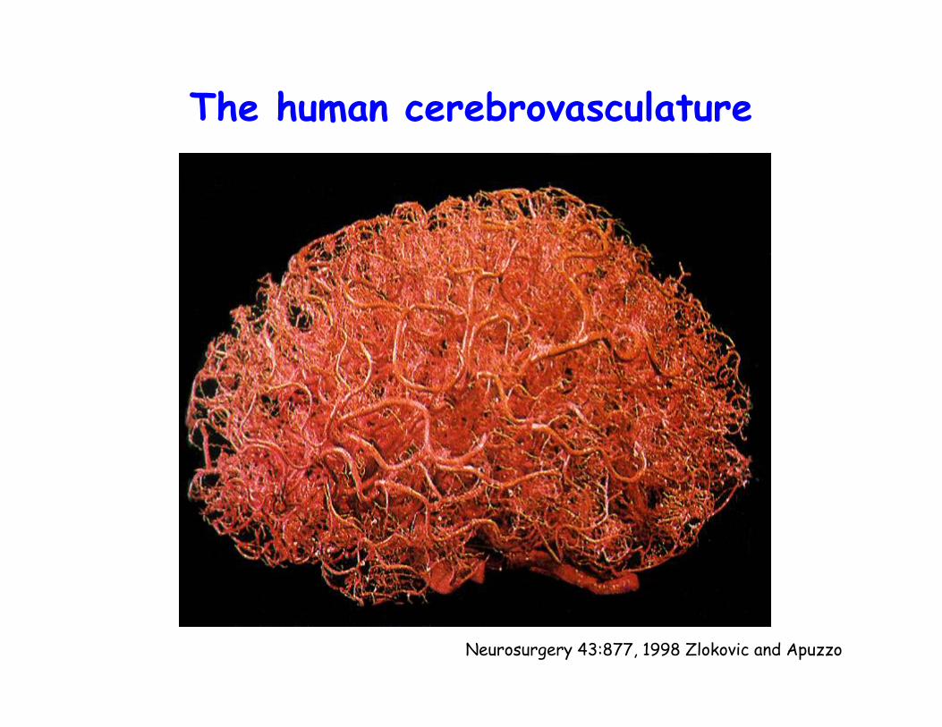
The human cerebrovasculature
Neurosurgery 43:877, 1998 Zlokovic and Apuzzo
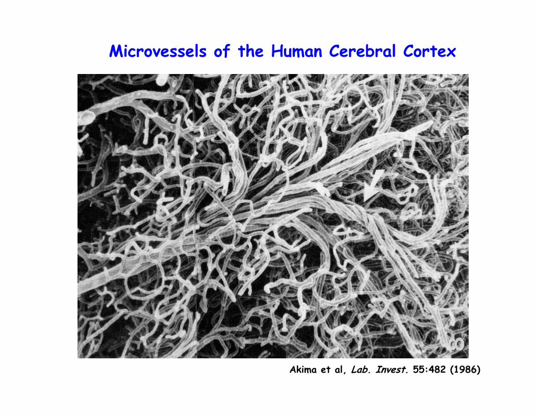
Microvessels of the Human Cerebral Cortex
Akima et al, Lab. Invest. 55:482 (1986)
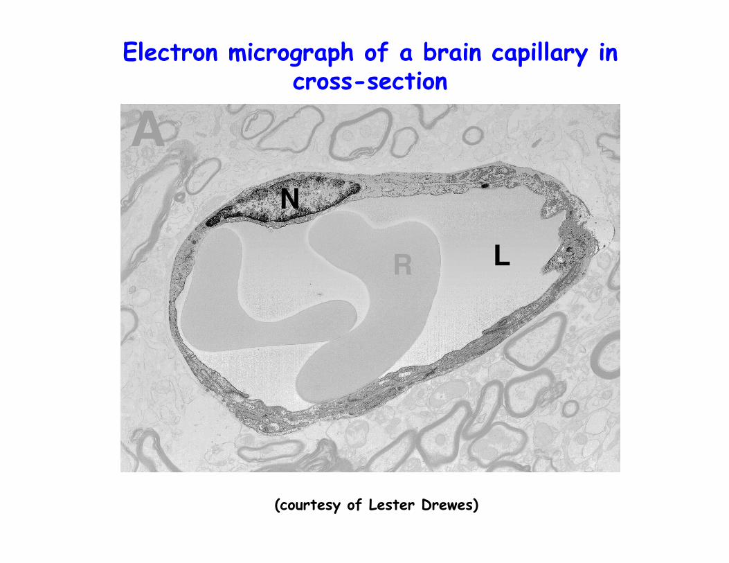
(courtesy of Lester Drewes)
Electron micrograph of a brain capillary in cross-section
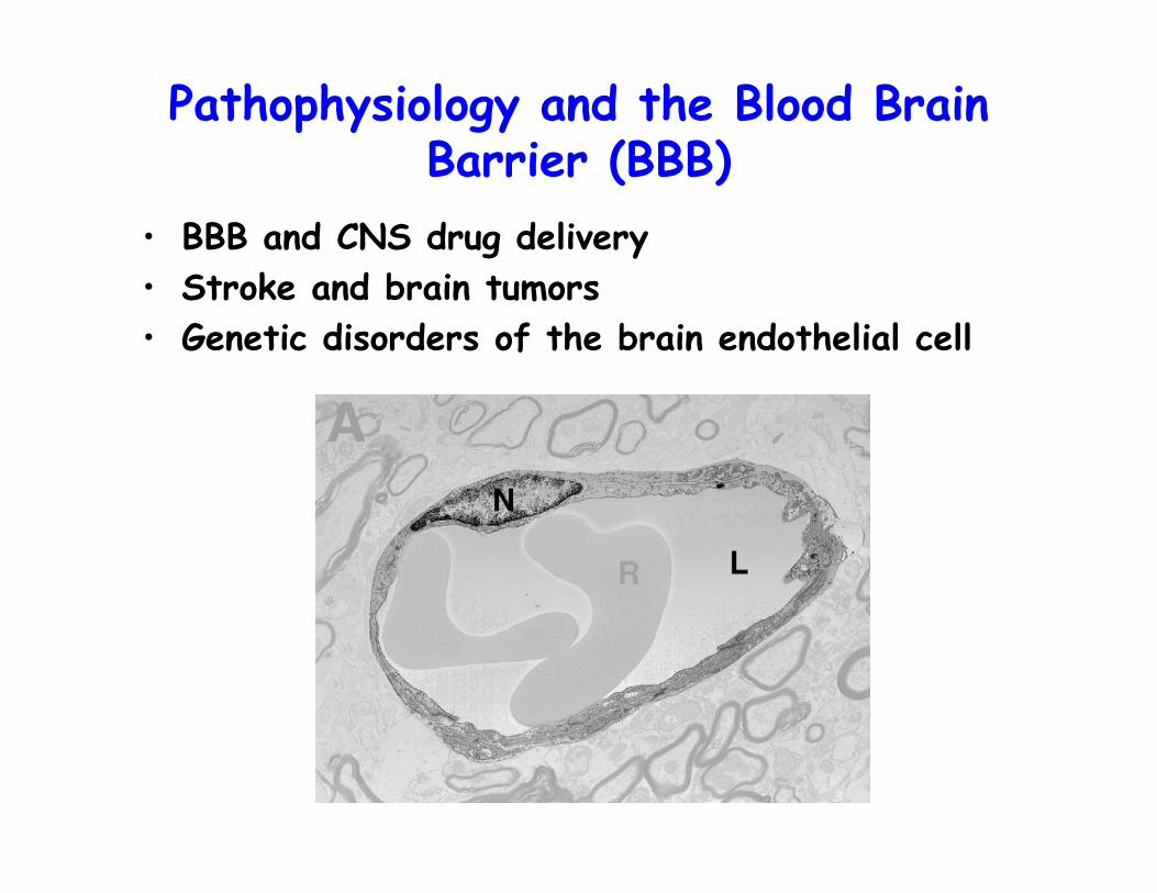
Pathophysiology and the Blood Brain Barrier (BBB)
• BBB and CNS drug delivery• Stroke and brain tumors • Genetic disorders of the brain endothelial cell
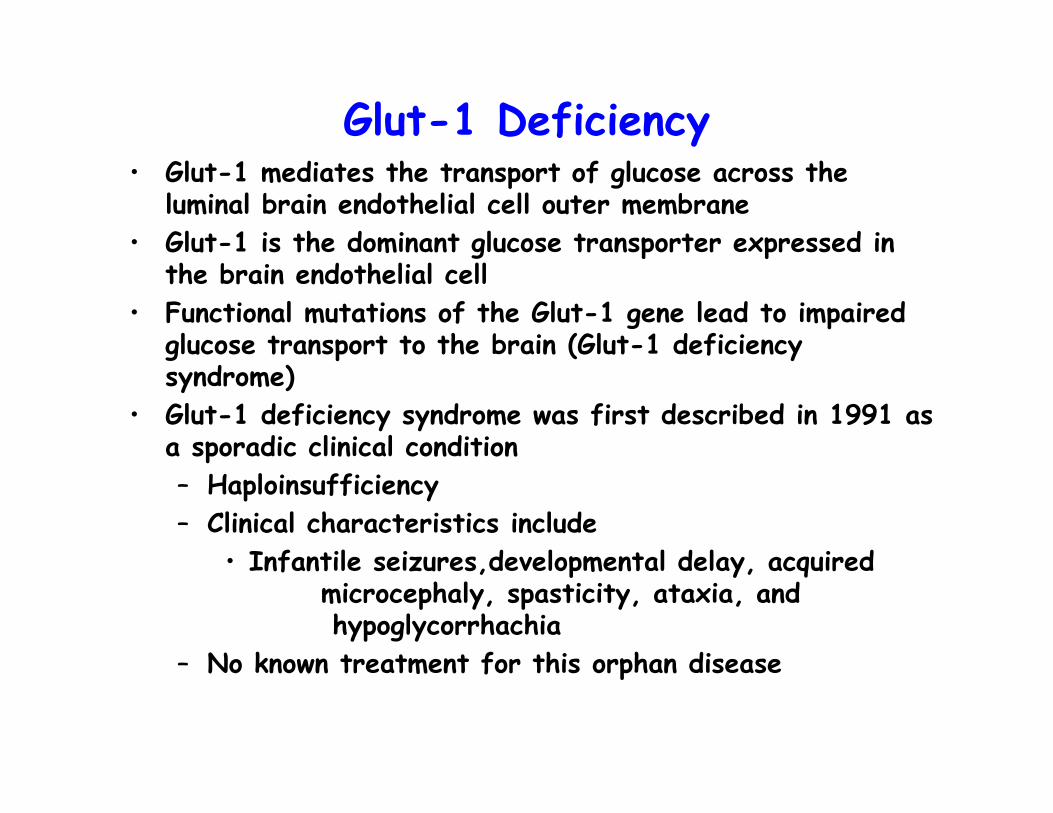
Glut-1 Deficiency• Glut-1 mediates the transport of glucose across the
luminal brain endothelial cell outer membrane• Glut-1 is the dominant glucose transporter expressed in
the brain endothelial cell• Functional mutations of the Glut-1 gene lead to impaired
glucose transport to the brain (Glut-1 deficiency syndrome)
• Glut-1 deficiency syndrome was first described in 1991 as a sporadic clinical condition– Haploinsufficiency– Clinical characteristics include
• Infantile seizures,developmental delay, acquired microcephaly, spasticity, ataxia, and hypoglycorrhachia
– No known treatment for this orphan disease

Clinical Goals
• To correct Glut 1 deficiency by populating the brain cerebrovasculature with wild type Glut-1 expressing endothelial cells
• Use the developed methodology to provide a therapeutic approach for treating other brain disorders
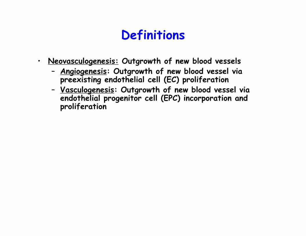
Definitions
• Neovasculogenesis: Outgrowth of new blood vessels– Angiogenesis: Outgrowth of new blood vessel via
preexisting endothelial cell (EC) proliferation– Vasculogenesis: Outgrowth of new blood vessel via
endothelial progenitor cell (EPC) incorporation and proliferation

Hypotheses
• Physiologic response of the brain cerebrovasculature to chronic mild hypoxia– Neovasculogenesis
• Hypotheses– Hypoxia induced brain neovasculogenesis is resultant
from the process of vasculogenesis– Isolated EPCs i.v. infused during chronic mild hypoxia
treatment will home to the brain and form nascent blood vessels
– Genetically engineered EPCs infused during chronic hypoxia will allow therapeutic engineering of the BBB

Overview of Research Project
Incorporate Glut-1 ACE enginneered ex vivo cultured EPCs into brain neovasculature
Incorporate ACE engineered ex vivo cultured EPCs into brain neovasclature
Assess contribution of vasculogenesis to hypoxia induced brain neovasculature
Establish hypoxia induced brain neovasculogenesismethod
Incorporate ex vivo cultured EPCs into brain neovasculature
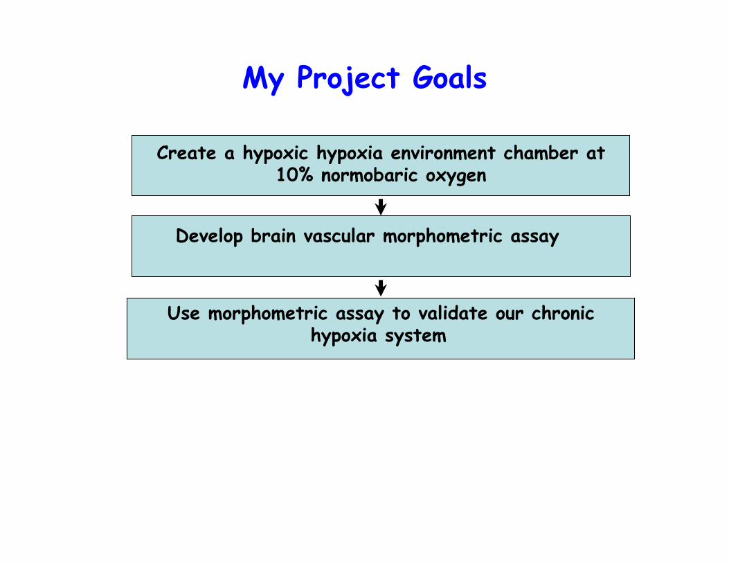
My Project Goals
Develop brain vascular morphometric assay
Create a hypoxic hypoxia environment chamber at 10% normobaric oxygen
Use morphometric assay to validate our chronic hypoxia system

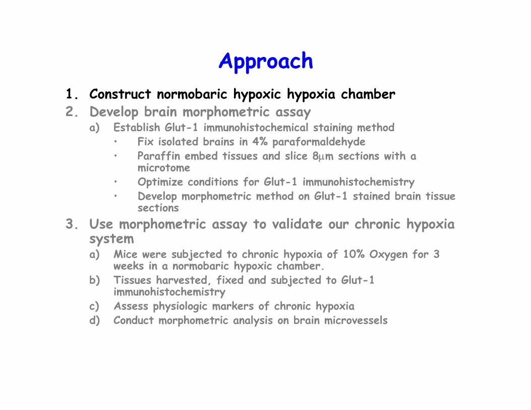
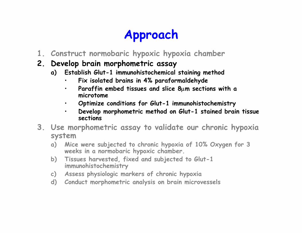
Approach1. Construct normobaric hypoxic hypoxia chamber 2. Develop brain morphometric assay
a) Establish Glut-1 immunohistochemical staining method• Fix isolated brains in 4% paraformaldehyde• Paraffin embed tissues and slice 8μm sections with a
microtome• Optimize conditions for Glut-1 immunohistochemistry• Develop morphometric method on Glut-1 stained brain tissue
sections3. Use morphometric assay to validate our chronic hypoxia
systema) Mice were subjected to chronic hypoxia of 10% Oxygen for 3
weeks in a normobaric hypoxic chamber.b) Tissues harvested, fixed and subjected to Glut-1
immunohistochemistryc) Assess physiologic markers of chronic hypoxiad) Conduct morphometric analysis on brain microvessels
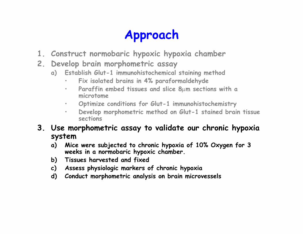
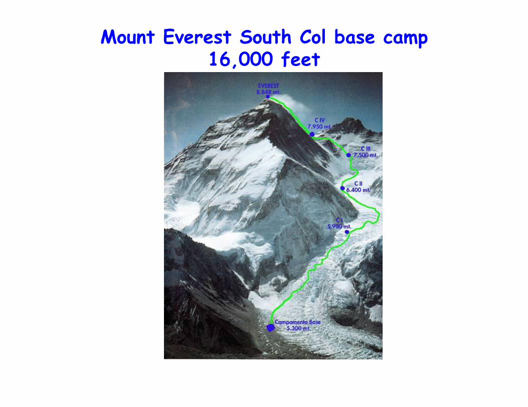
Mount Everest South Col base camp 16,000 feet

Mount Everest Base Camp

The city of Puno on the Peruvian Altiplano plain (12,420 feet)

Potala Palace, Lhasa Tibet (12,100 feet)

Horsemen on the Tibetan plains
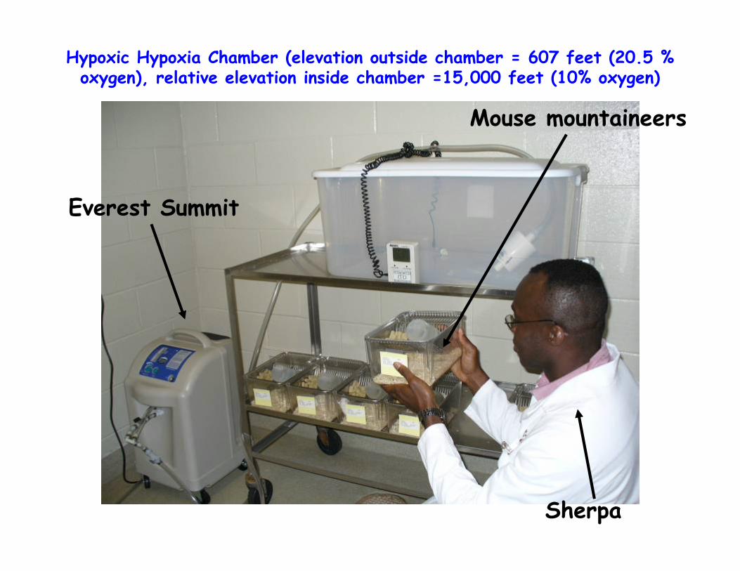
Hypoxic Hypoxia Chamber (elevation outside chamber = 607 feet (20.5 % oxygen), relative elevation inside chamber =15,000 feet (10% oxygen)
Everest Summit
Mouse mountaineers
Sherpa
Approach1. Construct normobaric hypoxic hypoxia chamber2. Develop brain morphometric assay
a) Establish Glut-1 immunohistochemical staining method• Fix isolated brains in 4% paraformaldehyde• Paraffin embed tissues and slice 8μm sections with a
microtome• Optimize conditions for Glut-1 immunohistochemistry• Develop morphometric method on Glut-1 stained brain tissue
sections3. Use morphometric assay to validate our chronic hypoxia
systema) Mice were subjected to chronic hypoxia of 10% Oxygen for 3
weeks in a normobaric hypoxic chamber.b) Tissues harvested, fixed and subjected to Glut-1
immunohistochemistryc) Assess physiologic markers of chronic hypoxiad) Conduct morphometric analysis on brain microvessels

Glut-1 immunofluorescence identifies brain microvessels
Glut-1 positive microvessels
Nuclei

Morphometric method
Approach1. Construct normobaric hypoxic hypoxia chamber 2. Develop brain morphometric assay
a) Establish Glut-1 immunohistochemical staining method• Fix isolated brains in 4% paraformaldehyde• Paraffin embed tissues and slice 8μm sections with a
microtome• Optimize conditions for Glut-1 immunohistochemistry• Develop morphometric method on Glut-1 stained brain tissue
sections3. Use morphometric assay to validate our chronic hypoxia
systema) Mice were subjected to chronic hypoxia of 10% Oxygen for 3
weeks in a normobaric hypoxic chamber.b) Tissues harvested and fixed c) Assess physiologic markers of chronic hypoxiad) Conduct morphometric analysis on brain microvessels

Hematocrit, hemoglobin and ceruloplasmin levels are all elevated after 3 weeks of mild hypoxia
***
p<0.0002
*
p<0.027
p<0.0074
**

HypoxicNormoxic
**
p<0.0019
Morphometric analysis reveals cortical neovasculogenesis
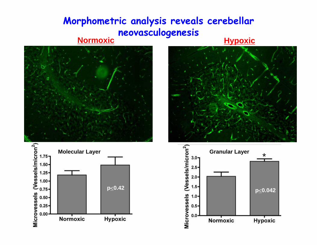
p<0.042
Granular Layer
HypoxicNormoxic
Morphometric analysis reveals cerebellarneovasculogenesis
*
p<0.42
Molecular Layer

Overview of Research Project
Incorporate Glut-1 ACE enginneered ex vivo cultured EPCs into brain neovasculature
Incorporate ACE engineered ex vivo cultured EPCs into brain neovasclature
Assess contribution of vasculogenesis to hypoxia induced brain neovasculature
Establish hypoxia induced brain neovasculogenesismethod
Incorporate ex vivo cultured EPCs into brain neovasculature
Acknowledgements
• Dr.Grant Anderson– Dan Westholm– Kevin Viken
• Members of Hypoxia Group– Dr. Les Drewes– Dr. Ed Perkins
• Funding support for this project– Arinze Okere
• Office of Clinical Research AHC-University of Minnesota
– Grant Anderson• 2007 AHC Faculty Seed Grant, NIH R21