Embed Size (px)
Citation preview
Structural analysis and therapeutic modulation of axonal remodeling following spinal cord
injury
Dissertation
der Fakultät für Biologie der Ludwig-Maximilians-Universität München
prepared at the Institute of Clinical Neuroimmunology, LMU Munich
submitted by
Claudia Nicole Lang
Munich, 2012

.
Erstgutachter: Prof. Dr. Hans Straka Zweitgutachter: PD. Dr. Mario Wullimann Externer Gutachter: Prof. Dr. Martin Kerschensteiner Tag der Abgabe: 26th April 2012 Tag der mündlichen Prüfung: 31st July 2012

.
''Research should not be reckless, but it does need to be fearless'' Christopher Reeve (2003)

Table of Contents
i
List of Abbreviations ...........................................................................................................................iii
Abstract ................................................................................................................................................1
Zusammenfassung..............................................................................................................................3
I. Introduction...................................................................................................................................5
1.1. The pathophysiological response of the cord to injury .......................................................7 1.1.1. The acute phase: 3-6hrs post injury ......................................................................7 1.1.2. The sub-acute phase: 6-72 hrs post injury.............................................................7 1.1.3. The late phase: weeks to months post injury.........................................................8
1.2. Clinical care and perspectives ............................................................................................10
1.3. Experimental models for SCI research .............................................................................11
1.4. Spontaneous corticospinal outgrowth and remodeling after SCI.......................................13 1.5. Therapy-induced modulation of corticospinal remodeling ................................................16
1.6. The Jak/STAT3 Pathway: A pathway part of the intrinsic growth program......................18
II. Aims of the Thesis .................................................................................................................. 22
III. Results- Original Publications ..........................................................................................24
3.1. Single collateral reconstructions reveal distinct phases of corticospinal remodeling after spinal cord injury ....................................................................................25
3.2. In vivo imaging reveals a phase-specific role of STAT3 during central and peripheral nervous system axon regeneration ....................................................................37
3.3. STAT3 promotes axonal reorganization and regeneration following spinal cord injury ...............................................................................................................49
IV. Discussion..................................................................................................................................77
4.1. Structural remodeling of corticospinal tract collaterals .............................................78 4.1.1. Summary of key findings .....................................................................................78 4.1.2. Stages of collateral remodeling ...........................................................................80 4.1.3. Contribution of main and minor CST components ..............................................82 4.1.4. Reorganization following SCI and the implications for functional recovery .....83

Table of Contents
ii
4.2. Enhancing and modulating axonal outgrowth and remodeling..........................................86 4.2.1. Summary of key findings ..................................................................................86 4.2.2. STAT3 as an initiator of the intrinsic neuronal growth program ......................88 4.2.3. Integration of STAT3 into the signaling pathways that regulate neuronal growth ...............................................................................................................90 4.2.4. STAT3 as a therapeutic target...........................................................................92 4.3. Outlook and future directions.........................................................................................96
4.4. Conclusions.......................................................................................................................99
V. Bibliography ............................................................................................................................101
VI. Acknowledegments..............................................................................................................121
VII. CV.............................................................................................................................................123
VII. Eidesstattliche Versicherung............................................................................................125

List of Abbreviations
iii
List of Abbreviations
5-HT – Serotonin Akt – Protein Kinase B (PKB), serine-threonine proteine kinase AAV – Adeno-associated virus Actb – Actin beta Adcyap1 – Adenylate cyclase activating polypeptide 1 AF – Alexa fluor ASIAIS – American Spinal Injury Association Impairment Scale Atf-3 – Cyclic AMP-dependent transcription factor BDNF – Brain-derived neurotrophic factor BOLD MRI – Blood-Blood-Oxygen-Level Dependence Magnetic Resonance Imaging C# – Cervical cord level# cAMP – Cyclic adenosine monophosphate CFP – Cyan fluorescent protein CNS – Central nervous system CPG – Ccentral pattern generator CREB – cAMP response element binding ChABC – Chodroitinase ABC CSPGs – Chondroitin sulfate proteoglycans CST – Corticospinal tract hCST – Hindlimb corticospinal tract fCST – Forelimb corticospinal tract dlCST – Dorsolateral corticospinal tract vCST – Ventral corticospinal tract mCST – Main corticospinal tract DNA – Deoxyribonucleic acid DRG – Dorsal root ganglia E – Embryonic Day eGFP – Enhanced green fluorescent protein EMGs – Electromyograms FES – Functional Electrical Stimulation fMRI – Functional magnetic resonance imaging Gadd45a – Growth arrest and DNA-damage-inducible protein GAP43 – Growth-associated protein 43 kDa GFP – Green Fluorescent protein GlyT2 – Glycine Transporter 2 gp130 – Glycoprotein 130 GSK-3 – Glycogen synthase kinase 3 Hspb1 – Heat shock 27kDa protein 1 HSP-27 – Heat shock protein 27 ICCP – International Campaign for Cures of Spinal Cord Injury Paralysis IGF-1 – Insulin-like growth factor-1 IRF1 – Regulatory factor 1 Jak – Janus kinase (JAK, or "Just another kinase") LTD – long-term depression MAGs – Myelin associated glycoproteins mTOR – Mammalian target of rapamycin n – Number of samples
List of Abbreviations
iv
NCS1 – Neuronal calcium sensor1 NgR – Nogo receptor NMDAR – N-methyl-D-aspartate receptor NR2d – Expressing NMDA-receptor2d NT3 – Neurotrophin 3 OECs – Olfactory ensheathing cells OMgp – Olidgodendrocyte myeline glycoprotein P – Post natal day PB – Phosphate buffer PBS – Phosphate buffer saline PCR – Polymerase chain reaction PFA – Paraformaldehyde PI3K – Phosphatidylinositol 3-kinases PIP3 – Phosphatidylinositol (3,4,5)-triphosphate PSNs – Propriospinal neurons LPSNS – Long propriospinal neurons SPSNS – Short propriospinal neurons PTEN – Phosphatase and tensin homolog Ptx – Pyramidotomy RNA – Ribonucleic acid RNAi – RNA interference ROS – Reactive oxygen species RAGs – Regenerative associated genes RST – Reticulospinal tract SEM – Standard error of the mean Ser – Serine amino acid SCI – Spinal cord injury SOCS3 – Suppressor of cytokine signaling 3 SPRR1A – Small proline rich protein 1a STAT3 – Signal transducer and activator of transcription 3 STPYFP – Stop Yellow fluoroscent protein (mouse line) T# – Thoracic cord level# (eg T8= thoracic cord level 8) TF – Transcription factor Trk – Tyrosine kinase TSA – Tyramide Signal Amplification Tubb3 – Tubulin beta-3 chain TYC9 – Brainbow mouse line Tyr – Tyrosine VSD – Voltage-sensitive dye imaging

Abstract
1
Abstract
Functional recovery following spinal cord injury (SCI) depends on the remodeling of
the preserved neuronal circuits. Injury-induced remodeling can be studied using the
corticospinal tract (CST), an important descending motor tract that is involved in fine-skilled
limb movements, as a model system. Previously it has been shown that the CST responds
to a thoracic lesion by the formation of an intraspinal detour circuit that contributes to
functional recovery. However the underlying principles that govern this CST remodeling are
not fully understood. By reconstructing single CST collaterals after lesion, we reveal that
CST remodeling occurs in three distinct phases. Initially following lesion, newly formed
collaterals undergo a growth phase that is then followed by a collateral formation phase
where newly formed collaterals develop a more mature and complex structure. Finally there
is a maturation phase during which there are small scale refinements of the contact pattern.
While such endogenous remodeling processes can lead to some degree of functional
recovery, in many cases severe deficits persist. To this date there is no effective therapeutic
treatment that restores sensorimotor function following SCI. In the injured peripheral nervous
system (PNS), the activation of the intrinsic growth response can support axonal
regeneration and functional recovery. In this thesis we investigated whether and how the
initiation of the intrinsic neuronal growth program can improve axonal remodeling and
functional recovery after injury. To initiate the intrinsic neuronal growth response we targeted
the transcription factor STAT3, the expression of which had been shown to be associated
with axonal regeneration. In a collaborative study, we used conditional genetics, viral gene
transfer and in vivo timelapse imaging to show that sustained STAT3 expression is essential
for the timely initiation of axonal regeneration in the PNS. In contrast to the PNS, STAT3
expression is only transiently induced following a central nervous system (CNS) lesion.
Therefore we next investigated whether and how intrinsic growth initiation by STAT3
expression can be used to support the regeneration and remodeling of corticospinal fibers
after spinal cord injury. Sustained expression of STAT3 induced by viral gene transfer was
found to cause an increase in CST axonal sprouting and regeneration following a thoracic
lesion. Interestingly, STAT overexpression could also stimulate axonal growth in the
absence of any lesion. This led us to utilize a unilateral lesion pyramidotomy model to
investigate whether sustained STAT3 expression can recruit the unlesioned tract to
compensate for the loss of innervation in the lesioned side. Indeed STAT3 overexpression

Abstract
2
was found to induce compensatory sprouting and remodeling of the unlesioned tract. Fibers
from the unlesioned tract exited the unlesioned CST in the cervical spinal cord and grew
across the midline into the denervated side of the cord. In addition these crossing collaterals
were found to form contacts onto the interneurons and motorneurons responsible for
forelimb movement. Behavioral and electrophysiological assessment validated that a new
intraspinal circuit that was formed enabled functional recovery.
Taken together, our results show that axonal remodeling occurs in defined stages.
Targeting the initial growth phase by viral gene transfer of STAT3, a transcription factor that
can initiate the intrinsic neuronal growth program, is an effective strategy to enhance axonal
remodeling and thereby promote functional recovery following injury. In this thesis, we were
able to contribute to the further understanding of the mechanisms that underlie axonal
remodeling. In addition we have identified a promising strategy to improve axonal
remodeling following injury.

Zusammenfassung
3
Zusammenfassung
Die Wiederherstellung motorischer und sensibler Funktionen (oder die funktionelle
Erholung) nach Rückenmarksverletzung (SCI) ist abhängig von der Reorganisationsfähigkeit
der noch unversehrten neuronalen Schaltkreise. Die auf eine Verletzung folgende neuronale
Umstrukturierung kann modellhaft am kortikospinalen Trakt (CST) untersucht werden. Es
handelt sich hierbei um eine wichtige absteigende motorische Bahn, die vor allem an der
Ausführung feinmotorischer Bewegungen beteiligt ist. Unsere Arbeitsgruppe konnte in
vorangegangenen Studien zeigen, dass der CST nach einer thorakalen Rückenmarksläsion
einen intraspinalen Umgehungsschaltkreis ausbildet, der maßgeblich zur funktionellen
Erholung beiträgt. Jedoch hat man bislang die Prinzipien, auf denen diese Fähigkeit zur
Reorganisation beruht, noch nicht vollständig verstanden.
Durch Rekonstruktion einzelner axonaler Kollateralen des CST nach Läsion konnte
ich nun zeigen, dass der Umbau des CST in drei Phasen erfolgt. Initial durchlaufen
neugebildete Kollateralen eine Wachstumsphase. Diese wird dann von einer Phase gefolgt,
in der Kollateralen ausgebildet werden, welche zunehmend eine reife komplexe Struktur
einnehmen. Die letzte Phase ist charakterisiert durch Verfeinerungen auf Ebene der
interneuronalen Kontaktmuster.
Während diese endogenen Reorganisationsprozesse zu einem gewissen Grad
funktionelle Erholung herbeiführen, bleiben in vielen Fällen schwerwiegende Defizite zurück.
Bislang gibt es keine effizienten Therapien, die nach einer Querschnittslähmung sensible
und motorische Funktionen wiederherstellen. Im peripheren Nervensystems (PNS)
hingegen kann durch die Aktivierung des intrinsischen neuronalen Wachstumgsprogrammes
axonale Regeneration und funktionelle Erholung erfolgreich induziert werden. In einem
weiteren Abschnitt meiner Arbeit untersuchte ich, ob und in wie weit die Induktion des
neuronalen Wachstumsprogramms auch im zentralen Nervensystem (ZNS) axonales
Remodeling und die Wiederherstellung von Funktionen verbessern würde. Um das
intrinische neuronale Wachstumsprogramm zu initiieren, konzentrierte ich mich auf den
Transkriptionsfaktor STAT3, dessen Expression während der axonaler Regeneration im
peripheren Nervensystem induziert wird. Mithilfe konditioneller knock-out Mäuse, viralen
Gentransfers und in vivo timelapse Mikroskopie konnten wir zeigen, dass die anhaltende
Expression von STAT3 essentiell für den zeitgemässen Beginn axonaler Regeneration im
PNS ist. Im Gegensatz zum PNS wird die STAT3 Expression nach einer Läsion im ZNS nur

Zusammenfassung
4
transient gesteigert. Daher untersuchte ich im letzten Abschnitt meiner Arbeit, ob und wie
die Initiierung des intrinischen neuronalen Wachstumsprograms durch STAT3 genützt
werden könnte, um die Regeneration und Reorganisation kortikospinaler Fasern nach
Rückenmarksläsion anzukurbeln. Ich konnte zeigen, dass die Überexpression von STAT3
mittels viralen Gentransfers eine Zunahme des axonalen Aussprossens und der
Regeneration des CSTs nach einer thorakalen Läsionen bewirkte. Interessanterweise
konnte die Überexpression von STAT 3 dabei auch das axonale Aussprossen in
Abwesenheit einer Läsion fördern. Infolge führte ich dann eine einseitige Verletzung des
CST auf der Höhe der Medulla oblongata (Pyramidotomie) durch und untersuchte, ob die
Überexpression von STAT3 in kortikalen Neuronen in der Lage ist, die unverletzte Seite des
CST dazu zu stimulieren, den Innervationsverlust der lädierten Seite auszugleichen.
Tatsächlich konnte ich zeigen, dass STAT3-Überexpression eine kompensatorische
Reorganisation des unverletzten CST bewirkte. Der unverletzten CST bildete zunächst neue
Kollateralen im zervikalen Rückenmark aus. Diese wuchsen dann über die Mittellinie auf die
denervierte Seite des Rückenmarks, wo sie Kontakte mit kurzen propriospinalen Neuronen
und ventralen Motoneuronen – welche die Bewegung der oberen Extremitäten kontrollieren
– ausbildeten. Verhaltensexperimente und elektrophysiologische Messungen konnten
bestätigen, dass diese neuen intraspinale Verschaltungen die funktionelle Erholung
verbesserten.
In der Gesamtschau meiner Ergebnisse kann man schlussfolgern, dass axonales
Remodeling nach einer Rückenmarksverletzung in distinkten sukzessiven Phasen abläuft.
Ich konnte weiterhin zeigen, dass der virale Gentransfer von STAT3 – einem
Transkriptionsfaktor, der das intrinsiche neuronale Wachstumsprogramm aktiviert – eine
effektive Strategie ist, diesen axonalen Umbau weiter zu verbessern und auf diese Weise
die funktionelle Erholung nach Verletzung zu fördern. Ich hoffe, dass diese Arbeit dazu
beitragen konnte, das Verständnis der Mechanismen, die axonale Umbauprozesse
regulieren, zu erweitern. Aufbauend auf diesen Befunden konnten wir weiterhin eine
vielversprechende Strategie identifizieren, welche axonalen Umbau und funktionelle
Erholung nach ZNS-Verletzungen weiter verbessern kann.

Chapter One
5
General Introduction
The worldwide incidence of spinal cord injury (SCI) is estimated to be 22
people/million population in the western and developing world (source: International
Campaign for Cures of Spinal Cord Injury Paralysis [ICCP]). In Germany, an estimated 18.5
cases/million of the population suffer from SCI, with 1500 new cases reported per year
(ICCP). SCI results from either direct or indirect trauma to the cord. Spinal cord lesions are
commonly due to acute contusion caused by the displacement of bone fragments into the
spinal cord (Schwab and Bartholdi 1996; Kraus 1996; Schwartz and Flanders 2006). A
majority of injuries are caused by motor vehicle and sport accidents, while other causes
include falls, acts of violence from stabbings or gunshot wounds, and sport-related injuries
(Figure 1). SCI patients are commonly young males, therefore SCI presents an economical
burden for society. Due to the organization of the spinal cord and the poor capacity of the
central nervous system (CNS) to repair following injury, SCI disrupts descending and
ascending motor pathways, causing a transient or permanent loss of sensorimotor and/or
autonomic function below the level of the injury.
Figure 1. Spinal Cord Injury facts and figures for the North American and European populations. (A) The economical impact of traumatic spinal cord injury. Data compiled from the National Spinal Cord Injury Statistical Center 2005 and 2011, Birmingham, Alabama, USA. (B) Etiology of traumatic spinal cord injuries for the European population (including Germany) by WHO 1959–2008 (Cripps et al., 2011)

Introduction
6
Over 130,000 people worldwide are affected by a traumatic spinal cord injury
resulting in paralysis and loss of sensory-motor function below the level of injury (source:
ICCP). Depending on the severity of the injury, the ability to control a majority of
autonomous bodily functions that includes bowel, bladder and sexual function can be lost to
various degrees (Figure 2; Ditunno et al., 1994; American Spinal Injury Association 2000;
Rhee et al., 2006). Human spinal cord injury is classified clinically by the segmental level of
injury, the completeness of the injury, and the mechanism of injury (Figure 2). An injury
above the C4 cervical level leads to tetraplegia where there is paralysis in both the arms and
legs. Conversely, patients with an injury at the lower thoracic to lumbar level can experience
paralysis or reduced movement of their legs. In such cases of severe traumatic injury, the
SCI patient will require long term care with elevated lifetime cost (Figure 1; The University of
Alabama National Spinal Cord Injury Statistical Center 2002). In addition, the quality of life of
many SCI patients is severely affected as they are both paralyzed and bound for life to a
wheelchair (Westgren and Levi 1998; Krause 2003; Budh and Osteråker 2007).
Figure 2. Spinal cord injury severity classification using the American Spinal Injury Association (ASIA) Impairment Scale (Modified from Thuret et al., 2006, Copyright © Nature Publishing Group 2006).

Introduction
7
1.1. The pathophysiological response of the cord to injury
Information regarding the pathology of human SCI is limited, though the available
data indicates there are strong similarities between humans and rodent spinal contusion
injuries (Kakulas 1984; Bunge et al., 1993; Kalb and Strittmatter 2010). In response to injury,
the spinal cord undergoes three time-dependent phases (Tator 1995; Schwab and Bartholdi
1996; Tartor 1998; Bareyre and Schwab 2003; Schwartz and Flanders 2006): The acute,
sub-acute and chronic phase.
1.1.1. The acute phase: 3-6hrs post injury
The acute phase happens immediately to a few hours after injury. Injury to the cord
can be caused by the displacement of bone fragments or direct compression of the cord.
During this phase the cord undergoes biochemical and structural changes. Interneuronal
tracts are damaged, blood flow is reduced, intracellular calcium levels rise, and there is
cellular death and degeneration of axons (Tartor 1995; Martirosyan et al., 2011). The edema
develops and there is a change in electrolyte levels, with an increase of extracellular
potassium (Schwab and Bartholdi 1996). The biochemical changes lead to a state of spinal
shock, where there is temporary flaccid paralysis and loss of tendon reflexes below the level
of the lesion (Hiersemenzel et al., 2000; Ditunno et al., 2004).
1.1.2. The sub-acute phase 6-72 hrs post injury
During this phase there is an increase in free radical production and the release of
excitatory neurotransmitters, such as glutamate and aspartate, up to cytotoxic levels (Park et
al., 2004). Within hours and lasting for several days following injury, an inflammatory
response develops (Balentine 1978; Dusart and Schwab 1994), with endothelial damage,
release of inflammatory mediators, invasion of peripheral inflammatory cells and activation of
microglia. Secondary damage is caused by immune cells such as neutrophils (appearing 6-
24h post-injury) macrophages (appearing 24h-2wks post-injury), and T-cells (Blight 1992;
Schnell et al., 1999; Bethea and Dietrich, 2002; David and Kroner 2011).

Introduction
8
At the lesion site a cavity and scar tissue are formed, consisting of extracellular
matrix (ECM) proteins, mainly collagen and chondroitin sulphate proteoglycans (CSPGs),
which secrete inhibitors hindering axonal regrowth (Fitch and Silver 2008; Fehlings and
Hawryluk 2010). Fibroblast, Schwann cells and macrophages form the scar tissue by the
deposit of ECM laminin, fibronectin and collagen. The sealing of the scar is dependent on
pericytes, perivascular cells that associate with the endothelial cells of capillaries and are the
source of scar-forming cells (Göritz et al., 2011). The glial/fibrotic scar is a hindrance to
regeneration as it is an inhibitory environment that is made up of fibroblast-like cells,
collagen surrounded by reactive astrocytes and microglial cells (Silver and Miller 2004; Xu et
al., 2011; Leal-Filho 2011). The reactive astrocytes are responsible for the upregulation, at
the site of injury, of molecules such as semaphorin 3 (Pasterkamp et al., 2001), ephrin-B2
(Bundesen et al., 2003), slit proteins (Hagino et al., 2003), and chondroitin sulfate
proteoglycans (McKeon et al., 1995; Jones et al., 2003; Rhodes and Fawcett 2004). The
scar however does serve a function as it separates the damaged tissue from healthy, hence
protecting viable tissue from necrosis (Lindsay 1986; Reier and Houlse 1988; Schwab and
Bartholdi 1996; Fitch and Silver 2001; Leal-Filho 2011).
1.1.3. The late phase: weeks to months post injury
In the late phase, there is the disappearance of phagocytic macrophages from the
lesion area and what remains is a fluid-filled cyst (Figure 3). There is also the formation of
cavities, which are filled with cerebrospinal fluid (Balentine 1978; Zhang et al., 1997).
Examination of damaged human tissue found that necrosis is similar to animal models,
including the cavities formed (Hughes 1974; Bunge et al., 1993). The dense network of
reactive astrocytes makes up the major component of the scar, another albeit minor
component is the reactive microglial cell and macrophages. At 3 weeks there is Wallerian
degeneration (Blight and Descrecito 1986; Zhang et al., 1997), and to some extent,
particularly in smaller lesions, remyelination by oligodendrocytes (Gledhill and McDonald
1977; Harrison and McDonald 1977; Schwartz and Flanders 2006). There is some attempt of
CNS axons to sprout after injury but the newly formed growth become dystrophic (Li and
Raisman 1995; Kerschensteiner et al., 2005; Misgeld et al., 2007) after exposure to a
gradient of inhibitory extracellular matrix molecules (Fitch and Silver 2008). Growth-
associated inhibitors such as myelin associated glycoproteins (MAGs), Nogo and
oligodendrocyte myelin glycoprotein (OMgp) are expressed in the vicinity of the lesion area
hindering any attempt of growth (Schwab and Bartholdi 1996; Sekhon and Fehlings 2001;

Introduction
9
Filbin 2003). Near the vicinity of the lesion site, chronic demyelination occurs in both humans
(Bunge et al., 1993; Guest et al., 2005) and experimental animals (Blight, 1983; Blight 1993;
Cao et al., 2005; Totoiu and Keirstead 2005). Overall there is loss of myelin in the white
matter and conduction deficits due to the biochemical and molecular changes that occur,
interruption of tracts and demyelination (Waxman 1989; Waxman 1992; Taoka and Okajima
1998; Sekhon and Fehlings 2001).
Figure 3. Response of the spinal cord following injury. (A) Dissected mouse spinal cord highlighting the thoracic spinal cord (red box). The events mentioned occur in the acute and subacute phase following a lesion. Scale bar represents 1mm (B) Sagittal section of a thoracic spinal cord 3 weeks following a dorsal T8 hemisection (neurons in blue and the corticospinal tract (CST) in red). Scale bar represents 100µm. (C) After spinal cord injury there is damage of ascending and descending tracts, upregulation of growth and inhibitory factors, accumulation of immune cells in the lesion site and the formation of the glial scar. The damaged axonal tracts can respond by sprouting new collaterals and by the formation of new circuits (Image C is modified from Thuret et al., 2006, Copyright © Nature Publishing Group 2006).
Introduction
10
1.2. Clinical care and perspectives
At present, in humans there is no treatment that is able to fully restore sensorimotor
function following injury. Patients admitted with injury undergo surgery to remove bone
fragments and have their spine stabilized. After surgery care involves the prevention of
secondary damages and rehabilitation in the form of physiotherapy (Dietz 2002; Edgerton et
al., 2006; Mehrholz et al., 2008; Markandaya et al., 2012). The acute standard of care
involves the administration of corticosteroids such as methylprednisolone, which works by
decreasing inflammation thereby limiting secondary damage (Bracken et al., 1985; Bracken
et al., 1997; Bracken 2002; Hurlbert and Hamilton 2008). The use of this drug is somewhat
controversial with some studies highlighting the benefits (Bracken et al., 1985; Bracken et
al., 1997; Bracken and Holford 2002) and others finding the effects modest in light of
associated side effects that can include glaucoma, hyperglycemia, depression, psychosis
and the cessation of the natural production of cortisol (Nesathurai 1998; Sayer et al., 2006).
Strategies for future treatment and management of spinal cord injury in humans are
based on the pathophysiological changes that occur in the acute, sub-acute and late phases
following injury. Research aims at understanding the different pathophysiological
mechanisms that arise following injury in experimental models and that could be potentially
targeted for therapeutic treatments. For example neuroprotective strategies have been
developed by preventing excitotoxicity (Feldblum et al., 2000; Abdelkarim et al., 2001;
Mazzone and Nistri 2011), by controlling the inflammatory response (Popovich et al., 1999;
Fitch et al., 1999; Alexander and Popovich 2009; Pajoohesh-Ganji and Byrnes 2011), or by
preventing apoptosis (Nicholson 2000; Nesic et al., 2001; Demjen et al, 2004). Other lines of
work aim at promoting axonal growth or regeneration through the use of cell grafts and
scaffolds that can act as bridges, through the application of growth promoting factors (Xu et
al., 1995; Li et al., 1997; Li et al., 1998; Menei et al., 1998; Ramon-Cueto et al., 2000;
Bamber et al., 2001;, Takami et al., 2002; Li et al., 2003; Bradbury and McMahon 2006) or
through the manipulation of the intrinsic growth program of neurons (Qiu et al., 2002; Yip et
al., 2010; Liu et al., 2010, Bareyre et al., 2011; Sun et al., 2011). Laboratory research has
provided useful information in expanding the knowledge and contributing to the treatment of
spinal cord injury.

Introduction
11
1.3. Experimental models for SCI research
In SCI research the use of appropriate animal models is important. In humans, spinal
cord injuries can be diverse and it can be difficult to reproduce the same sort of injury in an
experimental setting. The two main commonly used models of spinal cord injury employed
by most laboratories are (Figure 4A):
(1) The contusion injury performed using a NYU-MASCIS (New York University -
Multicenter Animal Spinal Cord Injury Study) impactor device, which drops a weight from
specific heights, can perform standardized grades of spinal cord injuries (Gruner 1992;
Agrawal et al., 2010). This injury model is based on the Allen technique, developed in 1911,
whereby a weight dropped through a tube onto the exposed thoracic cord of a dog resulting
in a reproducible model. The contusion model is the most similar to the histological
mechanics of human spinal cord injury (Schwab and Bartholdi 1996), however in this type of
injury there is haemorrhage and variable tissue damage making the comparison of neuronal
damage between different animals more challenging (David and Kroner 2011).
(2) The dorsal hemisection injury involves transection of the dorsal half of the spinal
cord at the thoracic level with fine iridectomy scissors. This model produces a smaller
inflammatory response, is highly reproducible and allows for defined parts of the cord to be
spared. This model is thus best suited for the precise investigation of axonal remodeling and
was used in our studies on CST regeneration and remodeling.
In addition to spinal lesion models that focus on the analysis of the corticospinal tract,
we also used the dorsal root ganglia (DRG) model system to study the growth initiation of
axons (Figure 4B). The DRG model system is often employed in outgrowth studies as it
provides access to both the PNS, (known for its regenerative capabilities), and to the CNS,
(where there is no successful regeneration) (Richardson and Issa 1984; Richardson and
Verge 1987; Sjoberg and Kanje 1990; Chong et al., 1994; Neumann and Woolf 1999).

Introduction
12
Figure 4. Localization of the Corticospinal tract (CST) and Dorsal root ganglia (DRG) neuron with its peripheral and central branch. (A) The corticospinal tract (red) can be labeled or manipulated via stereotaxic injection of a compound (either a tracer or virus) of interest into defined coordinates in the cortex. In a dorsal T8 hemisection, only half of the cord is transected (dashed line) sparing fiber tracts and interneuronal pools located in the ventral grey and white matter. For a contusion injury, an impactor drops a weight from a fixed distance onto the cord enabling standardized grades of injuries. (B) The DRG can be exposed through surgery and also injected with a finely pulled micropipette. The neuronal cell body of the DRG, located in the ganglia, sends one branch into the periphery (peripheral branch) and another branch into the spinal cord (central branch). The peripheral branch is known for its regenerative capabilities. Regeneration of the central branch can occur only if the peripheral branch is first injured, an effect known as a conditioning lesion. The DRG injection image is courtesy of Fabian Laage-Gaupp. Scale bar equals 1mm in A and B.

Introduction
13
1.4. Spontaneous corticospinal outgrowth and remodeling after SCI
The corticospinal tract (CST), as one of the most important descending motor tract
for skilled movements in all mammalian species (Nudo and Masterton 1988, 1990; Maier et
al., 2008), has been a frequent model used to investigate axonal growth, regeneration and
remodeling within the adult CNS (Schnell et al., 1994; Weidner et al., 2001; Zhou and Shine
2003; Bareyre et al., 2004; Liu et al., 2010; Rosenzweig et al., 2010). In the mammalian
system the CST is responsible for fine skilled movements, for example grasping and
handling (Whishaw et al., 1998) and locomotor functions such as stride length (Bregman et
al., 1995). In humans the CST has an even more important role and controls locomotion,
posture as well as voluntary skilled movements (Ferguson et al., 2001; Hutson et al., 2011).
The pyramidal neurons of the corticospinal tract originate in lamina V of the cortex. The
fibers of this tract descend from the cerebral cortex sending axons via the internal capsule to
the spinal cord (Dottori et al., 1998). The CST is composed of two components: a main
component also known as the main CST which comprises of 95% of all CST axons; and a
minor component constituted of the ventral and dorsolateral CST (Vahlsing and Feringa
1980; Joosten et al., 1992; Brösamle and Schwab 1997; Weidner et al., 2001; Steward et al.,
2004). The tract in rodents decussates at the spinomedullary junction with the main
component crossing over in the dorsal funiculus. In humans the CST fibers descend
contralaterally in the lateral funiculus (Harel and Strittmatter 2006; Hutson et al., 2011). The
remaining minor components of the CST do not decussate and the fibers run in the white
matter of the ipsilateral dorsolateral side (Hutson et al., 2011). At 4 weeks of age the rodent
CST is matured and establishes contacts with the appropriate interneurons and with a
subset of motorneurons (Ghosh et al., 2009). The topological organization of the CST is
such that the forelimb CST (fCST) projects its collaterals in the cervical cord, while hindlimb
CST (hCST) axons innervate the appropriate target in the lumbar cord (Akintunde and
Buxton 1992; Ghosh et al., 2009).
The CNS for a long time has had the reputation for being a “static” system in
adulthood, where successful axonal growth, sprouting and regeneration does not occur. As
research progresses, we are beginning to now realize that in the adult mammal the CNS has
the capability for some spontaneous recovery of function in rodents and to some extent in
humans (Schwab and Bartholdi 1996; Burns et al., 1997; Dietz et al., 1998; Rossignol et al.,
1999) Many examples of injury-induced plasticity stem from the study of lesioned CST

Introduction
14
connections (Bernstein and Stelzner 1983; Kuang and Kalil 1990; Schnell et al., 1994;
Terashima 1995; Bregman et al., 1995; Li et al., 1997; Weidner et al., 2001; Bareyre et al.,
2002; Bareyre et al., 2005; Demjen et al., 2004). Prominent findings have found that the
reorganization of neural circuits plays a key role in spontaneous recovery of function
(Bareyre et al., 2004; Courtine et al., 2008; Ghosh et al., 2009; Alilain et al., 2011). In 2001,
Fouad et al., (2001) observed that following an incomplete thoracic lesion in rats there were
rearrangements in the cortical motor map. Stimulation of what was previously an area
responsible for hindlimb muscle response instead invoked forelimb responses. The shift in
the cortical map was accompanied by spontaneous growth of hindlimb corticospinal axons in
the cervical cord. However it was unknown what the neuronal targets of these newly formed
collaterals were.
Bareyre et al., (2004), then made a seminal discovery whereby functional recovery
after SCI was attained via the formation of an intraspinal detour circuit (Figure 4). This study
revealed that neural circuits are able to undergo spontaneous functional reorganization that
lead to the reconnection of lesioned CST fibers with their original targets. Following a partial
lesion of the CST, lesioned hindlimb CST axons sprouted newly formed collaterals in the
cervical cord rostral to the lesion site. These collaterals were found to form contacts with a
pool of excitatory interneurons known as propriospinal neurons. Propriospinal neurons were
first described in 1902, as a network of axons extending from the proximal to distal spinal
cord (Sherrington and Laslett 1902, 1903). These neurons are an important part of an
intraspinal network of interneurons involved in motor reflexes, voluntary movement and
sensory processing (Kostyuk and Vasilenko 1979; Jankowska 1992; Foreman 2000; Pierrot-
Deseilligny and Burke 2005; Alstermark et al., 2007; Conta and Stelzner, 2009; Cowley et
al., 2010; Flynn et al., 2011). There are two main populations of propriospinal neurons. Short
prospriospinal neurons (SPSNs), involved in the fine tuning of forelimb movement, have their
cell bodies in C4 and their axons terminate at T2. Long propriospinal neurons (LPSNS) are
known to coordinate forelimb and hindlimb movement. This population of PSNs has their cell
bodies in C5/6 and their axons extend till T12/T13 (Nicolas et al., 2001; Dietz 2002). In this
study, it was found that initially, hCST collaterals contacted both long and short PSNs
equally (Bareyre et al., 2004). Overtime, contacts onto SPSNs were removed while contacts
onto LPSNs were maintained (Figure 5). Long PSNs are an ideal target as they are able to
bypass the lesion site and contact motorneurons in the lumbar cord. Further examination
revealed that the LPSNs had increased their contacts onto lumbar motorneurons, hence
forming a new intraspinal circuit (Bareyre et al., 2004). Behavioral and electrophysiological

Introduction
15
experiments further proved that this circuit was functional and responsible for the
spontaneous functional recovery seen following SCI.
Figure 5. Reorganization of hindlimb CST collaterals following injury. Timeline of the formation of a new intraspinal detour circuit following a dorsal T8 hemisection. (A) Normal connectivity, in absence of a lesion, in which the hindlimb CST (hCST) sprouts its collaterals into the lumbar cord to contact interneurons or motorneurons. (B) Three weeks post lesion, hCST collaterals are induced to sprout in the cervical cord where they were seen to form equal contacts onto both short and long propriospinal spinal relay neurons. (C) By 12 weeks post lesion contacts onto long propriospinal neurons (LPSNs) that bypass the lesion site and are involved in coordinating hindlimb and forelimb movement are strengthened and maintained. While contacts onto short propriospinal neurons that do not directly aid in functional recovery are pruned. LPSNs were seen to form contacts with lumbar motorneurons, thereby creating a new intraspinal circuit that can enable functional recovery. (D-F) Confocal photomicrographs showing contacts of hCST collaterals to (D) interneurons at 3 weeks following injury, (E) to short propriospinal neurons 3 weeks following injury, and to (F) LPSNs 3 weeks following injury. (D) is taken from Lang et al., 2012.
Further examining the importance of this newly formed intraspinal circuit for
functional recovery after incomplete injuries, Courtine et al., (2008) performed a series of
elegant experiments to examine the effects of eliminating the propriospinal relay connection.
They demonstrated that severing the newly formed propriospinal relay connections whether
Introduction
16
by a staggered hemisection or a high dose of N-methyl-D-asparate (NMDA), which in high
doses can act as an excitotoxin, abolishes recovery. By severing the supraspinal axon
connection, kinematic and physiological analyses showed that this results in permanent
paralysis of the hindlimbs. The various lesion paradigms performed in this study, where
lesions were separated both temporally and spatially, in combination with refined kinematic
analysis, illustrated that the propriospinal relay circuit is responsible for the restoration of full,
weight-bearing hindlimb locomotor function seen following even a severe injury. In summary,
these studies revealed upon electrophysiological, behavioral and anatomical examination
that a new detour circuit involving LPSNs can be formed and that this axonal remodeling
contributed to functional recovery (Fouad et al., 2001; Bareyre et al., 2004; Courtine et al.,
2008).
After injury, intraspinal circuits are spontaneously created and able to transmit
descending supraspinal input to the lumbar motor circuits (Bareyre et al., 2004;
Kerschensteiner et al., 2004; Ballermann and Fouad 2006; Courtine et al., 2008). These
studies not only highlight the positive impacts that injury can induce in the form of
remodeling, they can also help to identify the mechanisms that govern spontaneous
remodeling and functional recovery. Exploiting this innate phenomenon by reinforcing
beneficial axonal remodeling would be an advantageous strategy in facilitating functional
recovery.
1.5. Therapy-induced modulation of corticospinal remodeling
About half of SCI patients, approximately 54%, suffer from an incomplete injury
where at an anatomical level there is some sparing of tissue and descending fibers
(Raineteau and Schwab 2001; Spinal injury network 2011). In such cases, under the right
conditions extensive remodeling can occur due to the preservation of the cortical, subcortical
and remaining intact fibers and spinal circuitry. Molecular interventions to take advantage of
spared fibers and circuits are one of the main types of therapies that are researched in the
field of SCI (Thuret et al., 2006). In the search for successful therapeutic strategies one
needs to consider the extrinsic and the intrinsic barriers that limit this process in the CNS.

Introduction
17
Extrinsic cues that limit axonal remodeling appear to accumulate over time in the
CNS. For example, in young animals the CST displays great plasticity and if injured the CST
axons are able to pass the lesion site to innervate the appropriate area (Bregman et al.,
1989; Liu et al., 2011). As the animal matures the regenerative and plastic capability of the
CNS is reduced, coinciding with the production of myelin and of inhibitory proteins such as
associated neurite growth inhibitory proteins (Kapfhammer and Schwab 1994a,b; Steeves et
al., 1994; Harel and Strittmatter 2006). The glial scar forms an inhibitory environment and
acts as a barrier to axonal regeneration. Injured axons when faced with such a barrier are
unable to grow further and instead form retraction bulbs (Fitch and Silver 1997; Silver and
Miller 2004; Leal-Filho 2011). Neutralization of the inhibitory environment that is formed
following injury is one strategy that has been used to enhance regeneration of lesioned
axons. For example, the removal or neutralization of oligodendrocytes or myelin can improve
regeneration (Bregman et al., 1995). When an antibody aimed against neutralizing inhibitory
factors like Nogo-A (IN-1 antigen) is used, regenerative sprouts and long distance elongation
is seen (Schnell and Schwab, 1990; Brosamle et al., 2000; Chen et al., 2000). Likewise
treatment with the bacterial enzyme chondroitinase ABC (chABC), which has the ability to
digest CSPG in the scar, also has beneficial effects in spinal cord injury models from
contusion (Caggiano et al., 2005) hemisections (Bradbury et al., 2002; Yick et al., 2003;
Barritt et al., 2006; Houle et al., 2006) and transections (Fouad et al., 2005). While some
beneficial effects on axonal remodeling have been reported as well (Thallmair et al., 1998;
Z’Graggen et al., 1998; Z'Graggen et al., 2000; Bradbury et al., 2002; Bareyre et al., 2002),
the removal of extracellular inhibitory molecules has so far proven to be insufficient for
extensive axonal remodeling and complete functional recovery. This might at least partially
be due to the fact that this approach targets growth inhibitors at the lesion site, whereas
axonal remodeling commonly occurs remote from this area.
The intrinsic barrier, attributed to the decline in the intrinsic growth competence of a
CNS neuron as it matures, affects the capabilities of injured CNS neurons to both regenerate
and remodel. To promote axonal growth and remodeling, one commonly used strategy is the
application of factors such as neurotrophins. When a unilateral lesion of the CST is
performed at the pyramidal decussation (pyramidotomy lesion) in the medulla oblongata, the
unlesioned CST tract can be recruited to compensate for the loss of the original input and
enable functional recovery. This model system can be used to evaluate the effectiveness of
potential molecules to promote remodeling. Following a pyramidotomy lesion, when
motorneurons were transduced to express NT3, unlesioned CST axons were found to sprout

Introduction
18
across the midline towards the transduced motorneurons (Zhou et al., 2003). Sustained
concomitant expression of neurotrophic factors BDNF and NT3 in the cortex and spinal cord,
respectively, further enhanced the axonal sprouting effect from the intact tract (Zhou and
Shine 2003). An alternative and more direct approach is the manipulation of key regulatory
genes involved in the intrinsic growth program and known to be activated in regenerating
PNS neurons. The forced upregulation of molecules that contribute to axonal growth and
that are normally downregulated in the CNS after injury can induce compensatory sprouting
from the unlesioned tract. Genetically overexpressing factors such as neuronal calcium
sensor-1 (NCS1) (Yip et al., 2010) and mTOR (Liu et al., 2010) has been reported to
stimulate spared fibers to send their axons into to the denervated side leading to functional
recovery of the injured limb. Remodeling requires axonal growth, as it has been shown that
manipulation of the PI3K-Akt (of which NCS1 is a member) and PTEN/mTOR pathways can
induce growth and lead to recovery following CNS injuries (Park et al., 2010; Yip et al., 2010;
Liu et al., 2010). Hence the approach to induce growth through exploiting the intrinsic growth
potential of a neuron would be an effective strategy to promote axonal remodeling.
1.6. The Jak/STAT Pathway: A component of the intrinsic growth program
In the adult mammalian system, it is well known that only in the peripheral nervous
system (PNS) there is successful regeneration of lesioned axons. Following injury, even
though successful regeneration does not occur in the CNS, CNS neurons are capable of
mounting a transient regenerative response as evidenced by the expression of regeneration
associated proteins and genes (RAGs) (Mikucki and Oblinger 1991; Tetzlaff et al., 1994;
Fournier and McKerracher 1997; Neumann and Woolf 1999; Bulsara et al., 2002; Storer et
al., 2003; Kruse et al., 2011). This indicates that the intrinsic growth program inherent to
neurons is activated in response to injury, however it is not sustained. This is in contrast to
what has been observed in the PNS, where following injury there is regeneration and a high
expression of transcription factors and proteins associated with regeneration and growth. In
the classical ‘conditioning lesion’ paradigm, regeneration of both the peripheral and central
branch of the DRG neuron can occur, although regeneration of the central branch is
dependent on prior injury of the peripheral branch (Richardson and Issa 1984; Richardson
and Verge 1987; Sjoberg and Kanje 1990; Chong et al., 1994; Neumann and Woolf 1999;

Introduction
19
Cai et al., 2002). Injury of the peripheral branch activates the intrinsic growth program and is
thus the key for regeneration in both branches (Cai et al., 2002; Yang and Yang 2011) as
this program is not activated in the mature CNS following an injury (Stam et al., 2007).
Targeting factors involved in the intrinsic growth program would therefore be an effective
strategy to promote axonal growth and regeneration following CNS injuries. Most commonly
upregulated genes and transcription factors after injury that are found to be associated with
regeneration are: c-Jun, Atf-3, Hspb1, HSP-27, Adcyap1, Gadd45a, Gap43, Actb, Tubb3
and in particular the Signal transducer and activator of transcription 3 (STAT3) (Broude et
al., 1997; Qiu et al., 2005; MacGillavry et al., 2009; Sun and He 2010; Smith et al., 2011).
STAT3 has dual roles as a signal transducer and a transcription factor. The STAT3
protein is essential during development as a complete knockout results in embryonic lethality
(Takeda et al., 1997; Aaronson and Horvath 2002). In adulthood it is known to participate in
various functions including neuronal cell survival, axonal growth, protection and remodeling
(Levy and Darnell 2002). In the immune system, STAT3 plays a key role in the signal
transduction of anti-inflammatory responses mediated via macrophages and neutrophils, in
turn regulating the inflammation process (Kühn et al., 1993; Akira 2000; Shuai and Liu
2003). The Janus kinase (Jak/STAT) pathway has also been implicated to be involved in
synaptic plasticity, with pharmacological inhibition or knockdown of STAT3 blocking the
induction of NMDAR (N-methyl-D-aspartate receptor)-LTD (long-term depression) (Nicolas
et al., 2012). In normal conditions, STAT3 exists in the cytoplasm of a cell in an inactive form
where it is associated with the glycoprotein 130 (gp130). The Jak/STAT3 signaling pathway
is activated by cytokines interleukin 6 (Il6) (Zhong et al., 1994), ciliary neurotrophic factor
(CNTF) (Rajan et al., 1996) and the leukemia inhibitory factor (LIF) (Kunisada et al., 1996).
While the intensity and duration that the Jak/STAT pathway is activated for is tightly
regulated and controlled by members of the suppressor of cytokine signaling (SOCS) family
(Croker et al., 2008). Once activated by cytokines, JAK kinases phosphorylates the tyrosine
residue (Tyr-705) of STAT3, leading to homodimerization or STAT1/3 heterodimerization
(Figure 5). In its activated phosphorylated form, the dimerized complex is translocated to the
nucleus where it binds to specific DNA-response elements activating the transcription of
specific genes (Zhong et al., 1994; Akira 2000; Ng et al., 2006). STAT3 can also be
phosphorylated at serine 727 (Ser-727), and it has been suggested that Ser-727
phosphorylation enables STAT3 to achieve its maximal transcriptional activity (Ceresa and
Pessin 1996; Lim and Cao 1999; Ng et al., 2006).

Introduction
20
Figure 6. Signal transducer and activator of transcription 3 (STAT3) is part of the Janus-family kinases (JAKs)/STAT3 pathway. This pathway can be activated in response to autocrine or paracrine signals, including cytokines. Upon activation, JAKs and SRC tyrosine kinases are able to phosphorylate STAT3 at its tyrosine residue. Following phosphorylation, STAT3 is able to dimerize and translocates to the nucleus where it will transcribe target genes. STAT3 signaling is tightly regulated by inhibitory molecules that include suppressor of cytokine signaling (SOCS) proteins, protein inhibitor of activated STAT (PIAS) proteins and protein tyrosine phosphatases (PTPases).

Introduction
21
The following findings suggest that STAT3 signaling could be a key event in the
regulation of axonal outgrowth:
(i) The expression levels of phosphorylated STAT3 is increased in regenerating
axons (Schwaiger et al., 2000; Sheu et al., 2000; Xia et al., 2002) and is associated with
axonal remodeling (Bareyre et al., 2002).
(ii) Molecules that have an influence on axonal regeneration and are expressed
following a peripheral nerve injury, for example cytokines IL-6, ciliary neurotrophic factor,
and leukemia inhibitory factor have an effect on STAT3 signaling (Curtis et al., 1994; Zhong
et al., 1999; Cafferty et al., 2001; Cafferty et al., 2004).
(iii) In cultured CNS neurons, STAT3 has been found to promote neuronal outgrowth
(Smith et al., 2011) and in the DRG ‘conditioning lesion’ paradigm STAT3 is activated in the
cell bodies of the DRG neurons only when the peripheral branch is injured (Schwaiger et al.,
2000; Qiu et al., 2005). Taken together these findings suggest that STAT3 is an interesting
candidate regulator of the intrinsic neuronal growth program.

Chapter Two
22
Aims of the Thesis
The overall goals of this thesis were to contribute to a better understanding of the
structural principles underlying axonal remodeling following spinal cord injury and to
determine whether we could enhance axonal growth, regeneration and remodeling of the
corticospinal tract through the genetic manipulation of intrinsic neuronal growth pathways.
Therefore the following questions were investigated within this thesis:
1. What are the processes that underlie the maturation and remodeling of newly
formed collaterals following injury?
In aim I of this thesis we examined the manner in which the corticospinal tract
remodels its axons following spinal cord injury. Our laboratory has previously shown that in
response to injury, the CST can spontaneously sprout collaterals rostrally to the lesion site.
In turn these collaterals contact interneuronal populations in the cervical cord creating a
“detour circuit” to mediate functional recovery. Through the use of transgenic mice, genetic
tracing methods and dye tracers, we selectively labeled the CST as well as different
interneuronal populations in the spinal cord. Using bulk analysis and reconstructions of
single CST collaterals we were able to follow the formation, maturation and refinement of
CST collaterals over several months following lesion.
2. How does the regenerative-associated transcription factor STAT3 regulate
axonal regeneration in the PNS and CNS?
In aim II of this thesis, we wanted to investigate whether the transcription factor
STAT3 is a suitable tool to manipulate the intrinsic neuronal growth response. To reveal the
role of STAT3 during PNS and CNS regeneration we focused on the DRG system which
provides access to both the CNS and the PNS branches of the same neuron. The
expression levels of STAT3 were manipulated in both the PNS (where STAT3 was ablated)
and CNS (where STAT3 was overexpressed) through the combined use of transgenic mice
and adeno-associated viral (AAV) viruses. With confocal and repetitive in vivo timelapse

Aims of the Thesis
23
microscopy, we were able to identify sustained STAT3 expression is a key requirement for
the timely initiation of PNS axon regeneration.
3. Can the sustained expression of STAT3 enhance axonal regeneration,
remodeling and functional recovery following a CNS injury?
In aim III of the thesis, we expanded on our previous studies in order to elucidate
whether the initiation of an intrinsic neuronal growth response by STAT3 expression could
also promote outgrowth of CNS axons following spinal cord injury. To address this question
we performed two sets of experiments. First we deleted endogenous STAT3 expression in
cortical projection neurons and analyzed the effects on endogenous CST axonal outgrowth.
Second, through gene therapy we overexpressed STAT3 in upper corticospinal
motorneurons and used several CST-lesion paradigms, along with behavioral and
electrophysiological assessments to reveal the role of STAT3 in axonal remodeling,
regeneration and functional recovery after spinal cord injury.

Chapter Three
24
Results
The work during this doctoral thesis has resulted in two peer-reviewed publications and one submitted manuscript. They are included in the thesis and constitute Chapter 3.
• Lang C*, Guo, X*, Kerschensteiner, M., and Bareyre, F.M. (2012). Single Collateral Reconstructions Reveal Distinct Phases of Corticospinal Remodeling after Spinal Cord Injury. PLoS ONE. 7:e30461.
• Bareyre FM, Garzorz N, Lang C, Misgeld T, Büning H, Kerschensteiner M. (2011). In vivo imaging reveals a phase-specific role of STAT3 during central and peripheral nervous system axon regeneration. Proc Natl Acad Sci U S A.108(15):6282-7.
• Lang C, Bradley P, Kerschensteiner M, Bareyre FM. (2012). STAT3 promotes corticospinal remodeling, regeneration and functional recovery after spinal cord injury. An article submitted to the Journal of Neuroscience

Chapter Three
25
Single collateral reconstructions reveal distinct phases of corticospinal remodeling after spinal cord injury
Lang C, Guo X, Kerschensteiner M, Bareyre FM
An article published in PLOS One 2012; 7(1) e30461

Single Collateral Reconstructions Reveal Distinct Phasesof Corticospinal Remodeling after Spinal Cord InjuryClaudia Lang., Xiaoli Guo., Martin Kerschensteiner*", Florence M. Bareyre*"
Research Unit Therapy Development, Institute of Clinical Neuroimmunology, Ludwig-Maximilians-Universitat Munchen, Munich, Germany
Abstract
Background: Injuries to the spinal cord often result in severe functional deficits that, in case of incomplete injuries, can bepartially compensated by axonal remodeling. The corticospinal tract (CST), for example, responds to a thoracic transectionwith the formation of an intraspinal detour circuit. The key step for the formation of the detour circuit is the sprouting ofnew CST collaterals in the cervical spinal cord that contact local interneurons. How individual collaterals are formed andrefined over time is incompletely understood.
Methodology/Principal Findings: We traced the hindlimb corticospinal tract at different timepoints after lesion to showthat cervical collateral formation is initiated in the first 10 days. These collaterals can then persist for at least 24 weeks.Interestingly, both major and minor CST components contribute to the formation of persistent CST collaterals. We thendeveloped an approach to label single CST collaterals based on viral gene transfer of the Cre recombinase to a small numberof cortical projection neurons in Thy1-STP-YFP or Thy1-Brainbow mice. Reconstruction and analysis of single collaterals for upto 12 weeks after lesion revealed that CST remodeling evolves in 3 phases. Collateral growth is initiated in the first 10 daysafter lesion. Between 10 days and 3–4 weeks after lesion elongated and highly branched collaterals form in the gray matter,the complexity of which depends on the CST component they originate from. Finally, between 3–4 weeks and 12 weeksafter lesion the size of CST collaterals remains largely unchanged, while the pattern of their contacts onto interneuronsmatures.
Conclusions/Significance: This study provides a comprehensive anatomical analysis of CST reorganization after injury andreveals that CST remodeling occurs in distinct phases. Our results and techniques should facilitate future efforts to unravelthe mechanisms that govern CST remodeling and to promote functional recovery after spinal cord injury.
Citation: Lang C, Guo X, Kerschensteiner M, Bareyre FM (2012) Single Collateral Reconstructions Reveal Distinct Phases of Corticospinal Remodeling after SpinalCord Injury. PLoS ONE 7(1): e30461. doi:10.1371/journal.pone.0030461
Editor: Mark L. Baccei, University of Cincinnatti, United States of America
Received August 5, 2011; Accepted December 21, 2011; Published January 24, 2012
Copyright: � 2012 Lang et al. This is an open-access article distributed under the terms of the Creative Commons Attribution License, which permitsunrestricted use, distribution, and reproduction in any medium, provided the original author and source are credited.
Funding: Work in F.M.B.’s lab is supported by grants from the Deutsche Forschungsgemeinschaft (DFG, SFB 870) and the German Federal Ministry of Educationand Research (BMBF). Work in M.K.’s lab is financed through grants from the Deutsche Forschungsgemeinschaft (DFG; Emmy-Noether Program, SFB 571 and SFB870), the BMBF Competence Network Multiple Sclerosis and the ‘‘Verein Therapieforschung fur MS-Kranke e.V.’’. The funders had no role in study design, datacollection and analysis, decision to publish, or preparation of the manuscript.
Competing Interests: The authors have declared that no competing interests exist.
* E-mail: [email protected] (FMB); [email protected] (MK)
. These authors contributed equally to this work.
" These authors also contributed equally to this work.
Introduction
Injury to the spinal cord leads to a disruption of ascending and
descending fiber tracts followed by loss of sensation and voluntary
movements below the level of the lesion [1]. Whereas a complete
transection of the spinal cord often leads to permanent disabilities,
incomplete injuries can be followed by spontaneous functional
recovery [2–4]. An important anatomical feature underlying this
functional recovery is the remodeling of damaged axonal
connections [5–8]. Many insights into how axons remodel after
lesion stems from the study of the corticospinal tract (CST). The
CST is a major descending motor pathway that mediates skilled
movements in all mammalian species [9], [10]. The CST in
rodents consists of a main component that runs at the base of the
dorsal funiculus and minor components in the dorso-lateral and
ventral funiculus [11–13]. In recent years we and others have
studied how the hindlimb portion of the CST responds to a
thoracic dorsal hemisection. Using a combination of anterograde,
retrograde and trans-synaptic tracing techniques we have
previously shown that the formation of intraspinal detour circuits
are a key component of CST remodeling after injury [6], [14].
Detour circuits are formed in the following steps: First, the
lesioned CST fibers sprout new collaterals in the cervical spinal
cord above the level of lesion. These collaterals then extend to the
intermediate layers of the cervical gray matter. There they form
contacts with different populations of spinal interneurons,
including long propriospinal neurons, a population of interneurons
that are involved in coupling of forelimb and hindlimb movement
[15–18]. These long propriospinal neurons, the axons of which
bypass the lesion in the ventral funiculus, in return increase their
projections to hindlimb motoneurons in the lumbar spinal cord.
Electrophysiological and detailed behavioral and kinematic
analysis show that this and similar detour circuits play a key role
for the recovery of CST function [6], [7].
PLoS ONE | www.plosone.org 1 January 2012 | Volume 7 | Issue 1 | e30461

While it is thus established that the formation of CST collaterals
is a key step of axonal remodeling after injury, we still know very
little about how long these collaterals persist, from which CST
components they originate and how their complexity and
projection pattern evolves over time. Analysis of mice traced by
injection with the anterograde tracer BDA (Biotin Dextran Amine)
in the hindlimb motor cortex and perfused at 10 days to 24 weeks
after a dorsal hemisection of the mid-thoracic spinal cord now
revealed the following findings: CST collaterals primarily started
to grow in the first 10 days after injury. Both major and minor
CST components contributed to this emergence of collaterals.
Once emerged, the majority of CST collaterals persisted at least
for up to 24 weeks after lesion. To study how these collaterals
evolve over a long period of time (for up to 12 weeks after lesion),
we labeled single CST collaterals by viral gene transfer of Cre
recombinase to a small number of cortical projection neurons in
Thy1-Stp-YFP [13] and Thy1-Brainbow mice [19]. The reconstruc-
tion of single collaterals emerging from main and minor CST
components showed that dramatic changes in collateral length and
complexity occur between 10 days and 4 weeks after injury. These
parameters remain largely stable between 4 weeks and 12 weeks
after lesion. Analysis of the CST contacts onto interneurons
however indicated that while the morphology of the collaterals
remained largely unchanged during the late stage of the
remodeling process, their synaptic projections were still refined.
We can further show that while the overall timing of the
remodeling is similar in main and minor CST collaterals their
individual complexity differed depending on their origin. Taken
together our result suggest that CST remodeling after SCI occurs
in 3 subsequent phases: a growth initiation phase (within the first
10 days after injury), which is followed by a collateral formation
phase (between 10 days and 3–4 weeks after injury) and a later
maturation phase (between 3–4 weeks and 12 weeks after injury).
Results
Cervical CST collaterals primarily emerge in the first 10days after lesion and persist over time
Injection of BDA 10,000 into the hindlimb motor cortex
revealed three components of the hindlimb CST in the spinal cord
(Fig. 1A). The main CST component runs at the base of the
dorsal funiculus and contains 97.660.27% (n = 8 mice) of labeled
CST fibers. The minor CST components run in the dorso-lateral
and ventral funiculus and contain 2.160.23% (n = 8 mice) and
0.360.04% (n = 8 mice) of labeled CST fibers, respectively.
In unlesioned adult mice, axons arising from all hindlimb CST
components sent only very few collaterals into the gray matter of
the cervical spinal cord (level C3–C5, Fig. 1 A, D). However, as
early as 10 days following a mid-thoracic lesion, the number of
CST collaterals in the cervical cord gray matter increased more
than 4-fold (Fig. 1 B, D). Over the following weeks the number of
cervical collaterals slowly decreased. Still the majority of the
collaterals persisted long-term and was still detected as late as 24
weeks after injury (Fig. 1 C, D). Over this timeframe the
collaterals, which in most cases have just started to enter the spinal
gray matter at 10 days after lesion (Fig. 1 B), extended further and
mainly projected to the intermediate layers of the spinal cord
(Fig. 1 C). When we analyzed the contribution of different CST
components to the formation of cervical collaterals, we found that,
while in absolute number most of the collaterals arose from the
main CST, the relative number of new collaterals that emerge per
labeled fiber was several-fold higher for the minor dorso-lateral
and ventral CST components (Fig. 1 E–P). Notably, while the
number of newly formed CST collaterals emerging from the main
CST significantly declined over time (Fig. 1 H), the number of
collaterals derived from the minor CST components remained
stable for the entire observation period (Fig. 1 L, P).
Complex CST collaterals form between 10 days and 4weeks after lesion
To label single CST collaterals, we took advantage of Thy1-Stp-
YFP [13] and Thy1-Brainbow [19] mice. In these mouse lines the
presence of Cre recombinase either starts (in the case of Thy1-Stp-YFP)
or changes (in the case of Thy1-Brainbow mice) the expression of
fluorescent proteins in the affected neurons. Expression of Cre
recombinase was restricted to a small number of cortical projection
neurons by stereotactically injecting small amounts of a recombi-
nant Adeno-Associated Virus expressing Cre recombinase (rAAV-
Cre) into the hindlimb motor cortex (Fig. 2 A, B). Single
collaterals emerging from the axons of transduced cortical
projection neurons could than be identified based on their unique
labeling in the cervical spinal cord and reconstructed from serial
cross-sections (Fig. 2 C–G).
We used this approach to analyze the structure of cervical
collaterals emerging from main and minor CST components at 10
days, 4 weeks and 12 weeks after a mid-thoracic hemisection of the
spinal cord (Fig. 3). At 10 days following the injury, CST
collaterals emerging from all CST components were fairly short
(Fig. 3 A–C, J), had a simple, mostly unbranched structure (Fig. 3K) and showed very few, if any, boutons (Fig. 3 L). At 4 weeks
after lesion, the collaterals were substantially longer (Fig. 3 D–F,J), had a complex often highly branched structure (Fig. 3 K) and a
higher number of boutons (Fig. 3 L). At this time, the anatomical
structure of a collateral depended on its white matter origin.
Compared to main CST collaterals, collaterals emerging from the
ventral CST were long but showed a relatively simple structure
with few branch points and boutons (Fig. 3 E, J–L). In contrast,
collaterals emerging from the dorso-lateral CST component had a
highly complex structure and significantly more branchpoints and
boutons compared to both ventral and main CST collaterals
(Fig. 3 J–L). While the structure of CST collaterals thus evolved
substantially between 10 days and 4 weeks after lesion, collaterals
emerging from all CST components remain largely unchanged
between 4 weeks and 12 weeks after injury (Fig. 3 G–L).
Consequently, at 12 weeks after lesion dorso-lateral CST
collaterals still had significantly more branchpoints than main
CST collaterals and more branchpoints and boutons than ventral
CST collaterals (Fig. 3 J–L).
Synaptic differentiation of newly formed CST boutonsTo determine the synaptic differentiation of the newly formed
CST boutons we traced the hindlimb CST and then stained
cervical and lumbar spinal cord sections with antibodies against
two synaptic markers: bassoon, a marker of the presynaptic active
zone and synapsin I, a protein that regulates neurotransmitter
release at the synapse (Fig. 4). We first determined the percentage
of boutons that are immunoreactive for the synaptic markers in the
lumbar spinal cord of unlesioned mice (n = 2 mice). Of these
‘‘control’’ boutons 51% were immunoreactive for synapsin I and
52% were immunoreactive for bassoon. As these values likely
represent the mature expression pattern, this value was set as
100% and the immunoreactivity in newly formed boutons was
expressed as a percentage of the mature expression pattern. The
analysis of CST boutons in the cervical spinal cord at 10 days and
3 weeks after lesion then showed that the expression of both
bassoon (Fig 4 A–C) and synapsin I (Fig. 4 D–F) is low at 10 days
after lesion but is comparable to the expression pattern observed in
the lumbar spinal cord of unlesioned mice by 3 weeks. Double-
Axonal Remodeling after Spinal Cord Injury
PLoS ONE | www.plosone.org 2 January 2012 | Volume 7 | Issue 1 | e30461

immunostaining experiments further showed that 3 weeks after
lesion 80.564.5% of the immunoreactive CST boutons are double
positive for synapsin I and bassoon while comparably few of them
showed the expression of only one marker (862% are only
immunoreactive for synapsin I and 11.566.5% are only
immunoreactive for bassoon, Fig. 4 G).
Figure 1. Population analysis of hindlimb CST collateral formation at different timepoints after SCI. (A–C) Reconstruction of hindlimbCST collaterals (black) from 5 consecutive sections in the cervical spinal cord of control mice (A) and of mice perfused 10 days (B) and 24 weeks (C)following SCI. (D) Quantification of the total numbers of collaterals emerging from all CST components in the cervical gray matter of control mice andof mice at different timepoints following SCI. (E–G) Confocal images of main CST (BDA, yellow) and the adjacent gray matter (Neurotrace, blue; bordershown by dashed white line) in control mice (E) and in mice perfused 10 days (F, arrow indicates CST collateral emerging from main CST) and 24weeks (G, arrow indicates CST collateral emerging from main CST) following SCI. (H) Quantification of the number of collaterals emerging from themain CST component at different timepoints following SCI. (I–K) Confocal images of the minor dorso-lateral CST (BDA, yellow) and the adjacent graymatter (Neurotrace, blue; border shown by dashed white line) in control mice (I) and in mice perfused 10 days (J) and 24 weeks (K, arrow indicatesCST collateral emerging from dorso-lateral CST) following SCI. (L) Quantification of the number of collaterals emerging from the minor dorso-lateralCST component at different timepoints following SCI. (M–O) Confocal images of the minor ventral CST (BDA, yellow) and the adjacent gray matter(Neurotrace, blue; border shown by dashed white line) in control mice (M, arrow indicates ventral CST fiber) and in mice perfused 10 days (N) and 24weeks (O, arrow indicates collateral emerging from ventral CST) following SCI. (P) Quantification of the number of collaterals emerging from theminor ventral CST component at different timepoints following SCI. Asterisks indicate significance compared to the unlesioned controls. Pound signsindicate significance compared to the 10-day timepoint. Scale bar in A (also for B,C), 500 mm; in M (also for E–O), 100 mm.doi:10.1371/journal.pone.0030461.g001
Axonal Remodeling after Spinal Cord Injury
PLoS ONE | www.plosone.org 3 January 2012 | Volume 7 | Issue 1 | e30461

CST collaterals refine their contacts on interneuronsbetween 3 and 12 weeks after lesion
To investigate how the projection pattern of CST collaterals
evolves over time, we analyzed the number of contacts that an
individual collateral formed with the cell bodies of spinal
interneurons (Fig. 5). We first determined the mature projection
pattern by evaluating contacts of hindlimb CST collaterals onto
interneurons in the lumbar spinal cord. Here, we found that in
most cases (85.564.7%, n = 2 animals and 59 collaterals) a CST
collateral forms 1 and in some cases (14.564.7%) 2 contacts on
spinal interneurons (Fig. 5 C). In contrast, the majority of newly
emerging CST collaterals in the cervical spinal cord displayed
multiple (up to 4) contacts on spinal interneurons at 10 days after
lesion (Fig. 5 C). This ‘‘multiple contact’’ pattern still persisted at
3 weeks after lesion (Fig. 5 A, C). The mature contact pattern was
only present at 12 weeks after lesion and at this time more than
80% (81.163.1%, n = 3 animals and 168 collaterals) of collaterals
only showed 1 contact per interneuron (Fig. 5 B, C). The mature
pattern then persisted over time and was still present at 24 weeks
after injury (Fig. 5C). The analysis of the individual CST
components showed that at 3, 8 and 12 weeks after lesion most of
the contacts on interneurons were formed by collaterals emerging
from the main CST tract (Fig. 5D).
Discussion
The plastic reorganization of axonal connections is an
important element of the recovery process after CNS damage.
This is exemplified by the remodeling of lesioned CST fibers after
spinal cord injury. Previous work has shown that the sprouting of
new CST collaterals above the level of the lesion is a key step in
the formation of intraspinal detour circuits that contribute to
functional recovery after traumatic and inflammatory lesions of
the spinal cord [6], [7], [14], [20]. Here, we can show that these
collaterals form and mature in distinct phases (Fig. 6). In the
growth initiation phase that encompasses the first 10 days after
lesion, CST collaterals emerge and, at least in the case of the main
and dorso-lateral CST, start to enter the cervical gray matter. In
the collateral formation phase that covers the ensuing weeks, these
collaterals elongate, branch and form synaptic contacts in the
cervical gray matter. The final maturation phase, 12 weeks after
injury, is then characterized by the small-scale refinements of the
projection pattern that includes the removal of excessive inputs
Figure 2. Strategies for labeling individual CST collaterals. (A, B) Confocal images of the cortex of a Thy1-Brainbow (A; YFP, yellow; CFP, blue)and Thy1-Stp-YFP mouse (B; YFP, green; Neurotrace, red) after local injection of rAAV-Cre. Boxed areas are magnified 2 times in insets. (C,D) Confocalimages of CST collaterals in the cervical spinal cord of a Thy1-Brainbow (C; YFP, yellow; CFP, blue) and Thy1-Stp-YFP (D; YFP, green; Neurotrace, red)mouse after injection of rAAV-Cre in the cortex. Boxed areas are magnified 2 times in insets. Arrows in inset in C indicate different collateralsexpressing either CFP (blue), YFP (yellow) or a combination of both (white). Dashed white line indicates the outline of the spinal gray matter. (E–G)Confocal images of individual collaterals (white) emerging from the main CST (F), the dorso-lateral CST (E) and the ventral CST (G) following SCI.Arrows indicate individual collaterals. Dashed white lines indicate the outline of the spinal gray matter. Scale bar in B (also for A),100 mm; Scale bar inD (also for C),100 mm; Scale bar in G (also for E,F), 50 mm.doi:10.1371/journal.pone.0030461.g002
Axonal Remodeling after Spinal Cord Injury
PLoS ONE | www.plosone.org 4 January 2012 | Volume 7 | Issue 1 | e30461

from interneurons. Another example of this refinement process is
provided by our previous analysis [6] of CST contacts onto two
distinct interneuronal populations, the long propriospinal neurons
which connect the cervical spinal cord to the lumbar spinal cord
[15–18] and the short propriospinal neurons that form intracervi-
cal connections [21], [22]. The cell bodies of these interneurons
are located side by side in the cervical gray matter. Indeed, at 3
weeks after lesion - at the end of the formation phase - similar
fractions of long and short propriospinal neurons are contacted by
CST collaterals. However at 12 weeks - at the end of the
maturation phase - many of the contacts on short propriospinal
neurons have been removed while contacts on long propriospinal
neurons persisted [6]. Taken together with the results of this study,
it seems that the main aim of the maturation phase is the removal
of excessive connections and the strengthening of pertinent
connections. The emergence and selection of CST collaterals
thus shows interesting parallels to the initial formation and
refinement of neuronal connections in development. In the
neuromuscular system it has been shown that initially exuberant
connections between motor neurons and muscle fibers are formed,
leading to the innervation of single neuromuscular junctions by
multiple axons [23]. Over time, most of these inputs are then
removed and only a single axon remains to innervate the junction
[24], [25].
Similarly, during the development of the CNS initially excessive
connections are formed that are later pruned. A classical example
for the removal of excessive connections is the pruning of early
corticospinal projections that originate from the occipital cortex
[26], [27]. The refinement of CST connections during develop-
ment however extends beyond this large scale pruning. For
example, it is known that in different species the initial termination
pattern of the CST encompasses the entire gray matter from
dorsal to ventral horn and becomes more restricted later on [28–
31]. In addition, while most corticospinal fibers in adults terminate
contralateral to their origin in the cortex, during development
CST axons also show extensive projections to the ipsilateral spinal
gray matter. This can be at least partially explained by the findings
that during development a larger proportion of axons appear to
descend ipsilaterally without decussating in the pyramid while
other projections cross not only in the decussation but again in the
Figure 3. Reconstruction of individual hindlimb CST collaterals at different timepoints after spinal cord injury. (A–C) Reconstruction ofindividual collaterals (blue asterisks indicate the entry point of the collateral in the gray matter) emerging from the main dorsal (A) and the minorventral (B) and dorso-lateral (C) CST components at 10 days following SCI. (D–F) Reconstruction of individual collaterals (green) emerging from themain dorsal (D) and the minor ventral (E) and dorso-lateral (F) CST components at 4 weeks following SCI. (G–I) Reconstruction of individual collaterals(red) emerging from the main dorsal (G) and the minor ventral (H) and dorso-lateral (I) CST components at 12 weeks following SCI. (J–L)Quantification of the total collateral length (J), the number of branchpoints/collateral (K) and the number of boutons/collateral (L) measured inindividually reconstructed collaterals at different timepoints after SCI. Blue bars, 10-day timepoint; green bars, 3-week timepoint; red bars, 12-weektimepoint. Asterisks indicate significant differences compared to the 10-day timepoint. Pound signs indicate significant differences betweencollaterals emerging from different CST components at 3 weeks (green) and 12 weeks (red) after injury. Scale bar in A (also for B–I), 50 mm.doi:10.1371/journal.pone.0030461.g003
Axonal Remodeling after Spinal Cord Injury
PLoS ONE | www.plosone.org 5 January 2012 | Volume 7 | Issue 1 | e30461

spinal cord [32]. At least some of these initial CST connections,
including many of the double-crossed or uncrossed collaterals
appear to be transient and are removed during the maturation of
the CST [32], [33]. Further work will be necessary to determine to
what extend these structural commonalities between developmen-
tal and post-injury remodeling also imply common regulatory
mechanisms. For example, it will be interesting to see if neuronal
activity patterns, that are important determinants of competition
at the neuromuscular junction [34–36] or during CST develop-
ment [37] also regulate the fate of newly formed CST collaterals.
Likewise it will be important to explore to what extend molecules
that affect pruning such as the semaphorins/plexins [38], [39],
ephrins [40] or components of the wlds pathway [41] also
influence the removal of CST collaterals.
The excessive formation and subsequent sorting of connec-
tions is one way how the specificity of new CST connections
Figure 4. Synaptic differentiation of newly formed CST boutons. (A–B) Confocal images of bassoon immmunostaining (green) in the cervicalspinal cord of mice with a traced hindlimb CST (BDA, red) perfused 10 days (A) and 3 weeks following SCI (B). Yellow arrows indicate boutons thatwere immunoreactive for bassoon, white arrows indicate those that were not. (A9–A90) Single plane confocal image of the boutons boxed in Ashowing the collateral (A9; BDA, white), bassoon immunostaining (A0; white) and the overlay (A90; BDA, red; bassoon, green) at 10 days after SCI. (B9–B90) Single plane confocal image of the bouton boxed in B showing the collateral (B9; BDA, white), bassoon immunostaining (B0, white) and theoverlay (B90; BDA, red; bassoon, green) at 3 weeks after SCI. (C) Quantification of the number of boutons on hindlimb CST collaterals that wereimmunopositive for bassoon at 10 days and 3 weeks following SCI in the cervical cord. The percentages were normalized to the expression pattern inthe lumbar cord (L) of control animals (which was set to 100%). (D–E) Confocal images of synapsin I immunostaining in the cervical spinal cord ofmice with a traced hindlimb CST (BDA, red) perfused 10 days following SCI (D) and at 3 weeks post-injury (E). Yellow arrows indicate boutons thatwere immunoreactive for synapsin I, white arrows indicate those that were not. (D9–D90) Single plane confocal image of the bouton boxed in Dshowing the collateral (D9, BDA, white), the synapsin I staining (D0; white) and the overlay (D90; BDA, red; synapsin I, green) at 10 days after SCI. (E9–E90) Single plane confocal image of the bouton boxed in E showing the collateral (E9; BDA, white), the bassoon staining (E0; white) and the overlay(D90; BDA, red; synapsin I, green) at 3 weeks after SCI. (F) Quantification of the number of boutons on hindlimb CST collaterals that wereimmunnopositive for synapsin I at 10 days and 3 weeks following SCI in the cervical cord. The percentages were normalized to the expression patternin the lumbar cord (L) of control animals (which was set to 100%). (G) Quantification of the co-expression of bassoon and synapsin I in boutons of CSTcollaterals of animals perfused at 3 weeks after injury (expressed as percentages of all immunoreactive boutons). Scale bar in A (also for B, D, E),10 mm and in A9 (also for A0–E90), 3 mm.doi:10.1371/journal.pone.0030461.g004
Figure 5. Analysis of CST contacts onto cervical interneurons after SCI. (A,B) Confocal images of contacts (arrows, defined as boutons inapposition to neuronal cell bodies) between hindlimb CST collaterals (YFP, green) and the cell bodies of cervical interneurons (Neurotrace, red) at 4weeks (A) and 12 weeks (B) following SCI. (C) Quantification of the number of contacts a given hindlimb CST collateral makes with the cell body of asingle interneuron at different timepoints after SCI as well as in the lumbar spinal cord of unlesioned animals. (D) Quantification of the percentage ofNeurotrace (NT)-stained interneurons contacted by collaterals emerging from the different CST components at multiple timepoints following thelesion. Asterisks indicate significant difference compared to main CST collaterals. Scale bar in A (also for B), 15 mm.doi:10.1371/journal.pone.0030461.g005
Axonal Remodeling after Spinal Cord Injury
PLoS ONE | www.plosone.org 6 January 2012 | Volume 7 | Issue 1 | e30461

can be established. Another measure to ensure specificity is the
targeting of axons to specific neurons or regions of the spinal
cord. In the case of the newly formed CST connections, the
refinement of initial connections during the maturation phase
suggests that initial targeting is not established by specific
recognition of single neurons. On the other hand, the
distribution of newly formed CST collaterals in the spinal
cord gray matter indicates that specific regions of the spinal
cord, in particular the intermediate layers V–VII are
preferentially targeted by CST collaterals (see Fig. 1C). Taken
together our findings thus suggest that a combination of
‘‘region-specific’’ targeting that guides collaterals to the
intermediate layers of the spinal cord and a subsequent
refinement process that removes excessive connections collab-
orate to ensure specific targeting of newly formed CST
connections to intraspinal relay neurons.
Figure 6. Schematic representation of hindlimb CST remodeling following SCI. Scheme illustrating the formation of cervical collateralsderived from the main CST (upper row) and the minor dorsolateral (2nd row) and ventral (3rd row) CST components at 10 days (blue), 3–4 weeks(green) and 12 weeks (red) after SCI. Bottom row illustrates the refinement over time of the contacts between CST collaterals and cervicalinterneurons.doi:10.1371/journal.pone.0030461.g006
Axonal Remodeling after Spinal Cord Injury
PLoS ONE | www.plosone.org 7 January 2012 | Volume 7 | Issue 1 | e30461

It should be noted however, that while most newly formed CST
collaterals end in the intermediate layers of the spinal cord, some
fibers reach the ventral horn and might form contacts with ventral
motoneurons [42]. These direct connections of hindlimb CST
axons to forelimb motoneurons could be one anatomical substrate
that underlies the shift of motor maps that occurs both in animal
and humans in response to spinal cord injury [43–45].
A second important finding of our study is that different CST
components contribute to the remodeling of the CST. Antero-
grade tracing revealed three distinct localizations of CST fibers in
the spinal cord, with the majority of fibers located in the main
CST component at the bottom of the dorsal funiculus and a
smaller proportion of fibers located in the dorso-lateral funiculus.
Only very few fibers were observed in the ventral funiculus,
contralateral to the main and dorso-lateral CST. These ventral
fibers together with the fibers in the dorso-lateral funiculus form
the minor CST components. This structure of the CST is in
accordance with previous reports in mice, where the ventral CST
component is relatively small [13], [46], [47], as well as in other
rodents [11], [12], [48–50]. We further observed that following
injury the number of fibers in the ventral component that is spared
by the lesion is increased. This is probably due to sprouting of
additional CST collaterals that can enter the ventral white matter
tract as previously described [51]. Our analysis of collateral
formation reveals both commonalities and differences between the
distinct CST components. For example, the overall timecourse of
collateral initiation, formation and maturation appears mostly
similar in major and minor CST components. The comparably
lower number of CST collaterals derived from the minor CST
tracts detected in the gray matter at 10 days after injury likely does
not reflect a different growth initiation but rather the longer
distance between the parental axons and the gray matter border. It
is interesting to note that while the overall timecourse of collateral
formation is similar, the number of collaterals an individual CST
axon sends to the gray matter differs substantially between the
CST components. A CST axon running in the ventral funiculus,
for example, extends more than 10-fold more collaterals into the
gray matter at 3 weeks after lesion than a main CST axon (Fig. 1H, P). These findings suggest an important role for ventral CST
fibers in the CST remodeling process. As after a midthoracic
lesion, the ventral funiculus consists of both pre-existing ventral
fibers as well as newly sprouted collaterals likely derived from
other CST components, it is possible that both unlesioned fibers
and new collaterals emerging from lesioned CST fibers contribute
to this response. An important role of ventral fibers is in line with
previous experiments in rats that have demonstrated that ventral
CST fibers can play an important role for the recovery of CST
function [52]. However, it appears that not only the relative
number of collaterals emerging from a given CST component but
also their complexity differs between CST components. For
example, collaterals emerging from the dorso-lateral CST contain
several-fold more branch points and boutons at 4 weeks after
lesion than collaterals emerging from main or ventral CST. This
might suggest that dorso-laterals CST contact different target cell
populations. The idea that distinct CST components target distinct
cell populations is consistent with our previous observation that
dorso-lateral CST collaterals appear to be primarily responsible
for direct contacts on motoneurons in the lumbar spinal cord [13].
Taken together the characteristic differences between main and
minor CST components strongly suggest that individual compo-
nents might play distinct roles in the recovery process. The single
collateral tracing techniques established in this study can in the
future help to further define these distinct roles in different lesion
paradigms. This is of interest as the remodeling process after a
spinal lesion likely extends beyond the corticospinal tract to other
supra- and intraspinal tract systems. For example, the reticulosp-
inal tract has been shown to spontaneously sprout after SCI [53],
[54] and the spontaneous restoration of serotonergic activity, likely
mediated by the remodeling of serotonergic circuits, was found to
contribute to functional recovery [55], [56].
Finally our study demonstrates that once collaterals from all
CST components are formed they by and large persist long-term -
in our experiments at least up to the end of the observation period
(24 weeks after lesion for the population analysis and 12 weeks
after lesion for the analysis of individual collaterals). Our analysis
further shows that after early formation and refinement of the
collaterals, very little changes to the collateral number, structure
and contact pattern are observed beyond 12 weeks after lesion.
This suggests that an early critical period for CST remodeling
exists during which the formation or refinement of collaterals can
be influenced. However after this period, newly formed connec-
tions appear to remain stable. This defines a time-window for
therapeutic interventions that are likely most effective in the first
10 days after lesion if the aim is to improve collateral initiation,
between 10 days and 3–4 weeks if they aim to support collateral
formation and between 3–4 and 12 weeks if the aim to modulate
target connections. This is of interest as despite the spontaneous
remodeling of axonal connections substantial functional impair-
ments often remain following experimental and clinical spinal cord
injuries. It will therefore be important to develop therapeutic
strategies that can enhance the remodeling process. One
promising approach could be to foster the intrinsic neuronal
growth response of cortical projection neurons targeting, for
example, c-AMP and its downstream mediators [57], the growth
cone-associated proteins GAP43 and CAP23 [58], components of
the PTEN/mTOR pathway [59], [60] or the JAK-STAT pathway
[60], [61]. Another possible way to enhance axonal remodeling is
through rehabilitation. Several studies [43], [62–64] have already
demonstrated the positive effect of rehabilitation on axonal
sprouting following spinal cord injury. Care needs to be taken
however not to favor task-specific rewiring at the cost of other
tasks. Several studies have indeed shown that experimental
rehabilitation schemes in which one task is trained repetitively
will lead to improvement in this task to the detriment of other tasks
[43], [65–67]. To our mind, the analysis techniques introduced in
this study can in the future help to evaluate whether and how these
therapeutic approaches can improve axonal remodeling after
injury.
Materials and Methods
Ethics StatementAll animal experiments conformed to the institutional guidelines
and were approved by the Animal Study Committee of the
Regierung von Oberbayern. Approval ID: 55.2-1-54-2531-127-
05.
AnimalsAdult C57BL/6 female mice 6–8 weeks old, Thy1-Stp-YFP and
Thy1-Brainbow mice (line TYC9, kindly provided by J. Livet,
INSERM) were used in this study. C57BL/6 mice were used for
all conventional CST tracing experiments. Thy1-Stp-YFP mice
express yellow fluorescent protein (YFP) in neurons after Cre-
mediated excision of a floxed STOP-sequence [13], [68]. Thy1-
Brainbow mice show a combinatorial expression of different
fluorescent proteins after Cre-mediated excision of Lox sites [19].
Briefly, in the brainbow-1.0 mice used in this study, lox P sites
alternate with incompatible lox variant (lox2272) sites. The Cre
Axonal Remodeling after Spinal Cord Injury
PLoS ONE | www.plosone.org 8 January 2012 | Volume 7 | Issue 1 | e30461

recombinase thus randomly chooses between different initial excision
events. As the initial excision between a pair of identical lox sites
removes one of the other pair, it prevents multiple recombination.
Before Cre action, only the gene following the promoter is
expressed (RFP). The recombination then switches expression to
either YFP or M-CFP depending on the site of the initial
recombination event. Further, the presence of multiple copies of
the brainbow construct that recombine independently can lead to
the co-expression of different fluorescent proteins [19]. Thy1-Stp-
YFP and Thy1-Brainbow mice were used for single collateral
analysis.
Spinal Cord InjuryMice were anesthetized with a subcutaneous injection of
Ketamin/Xylazin (Ketamine 150 mg/kg, Xylazine 10 mg/kg).
A laminectomy was performed at thoracic level 8 (T8) and a dorsal
hemisection of the spinal cord was performed with fine iridectomy
scissors. This lesion interrupts the main dorsal and the minor
dorso-lateral CST components but not the minor ventral CST
component (Fig. 7). After surgery animals were heated,
rehydrated and treated with analgesics (which were also
administrated immediately before surgery).
rAAV-CreAAV1/2-CAG-HA-NLS-Cre-WPRE-BGH-polyA expression
vectors were used to generate viral particles in which the CAG
promoter consists of the chicken b-actin promoter hybridized with
the CMV immediate early enhancer sequence. The CAG
promoter drives the expression of the P1 Cre recombinase, the N-
terminus of which is fused to an HA-tag followed by a nuclear
localization signal (NLS). The woodchuck post-transcriptional
regulatory element (WPRE) and the presence of the bovine growth
hormone (BGH) polyadenylation sequence ensure high transcrip-
tion following transduction. AAV1/2 particles were generated by
GeneDetect.com Ltd.
Anatomical tracing of hindlimb corticospinal tract (CST)Population analysis of CST collaterals. To study
reorganization of both the major and minor CST components
we traced the hindlimb CST in adult C57BL/6 mice by bilateral
pressure injections as previously described [14]. For this purpose,
1 ml of a 10% solution of biotinylated dextran amine (BDA
10 000, Molecular Probes) was slowly injected with a glass
capillary (tip diameter of about 20 mm) into lamina V of the
hindlimb motor cortex (coordinates: 21.3 mm posterior to
bregma, 1 mm lateral to bregma, 0.6 mm depth). The
micropipette remained in place for 2 minutes after completion
of the injection to minimize backflow and diffusion of the tracer.
Single CST collateral analysis. To study the projection
pattern of individual collaterals, we first determined the amount of
rAAV necessary to label single hindlimb CST collaterals by
varying the injected volume. We then performed bilateral pressure
injections of 0.3 ml of a rAAV-Cre (titer: 161012 genomic
particles/ml) into lamina V of the hindlimb motor cortices of
Thy1-Stp-YFP and Thy1-Brainbow mice. The micropipette remained
in place for 2 minutes after completion of the injection to
minimize backflow and diffusion of the virus.
Tissue preparation and immunohistochemistryAnimals were perfused transcardially with 4% paraformalde-
hyde (PFA). Brains and spinal cords were dissected, postfixed
overnight and cryoprotected in 30% sucrose for 3 days. For the
population analysis of CST collaterals we analyzed the cervical
spinal cord of C57BL/6 mice between the spinal level C3 and C5,
where the cell bodies of long propriospinal neurons are located.
For this purpose coronal sections (50 mm thickness) were cut on a
vibratome and processed as described previously [69]. The
hindlimb CST was revealed after BDA tracing using 0.4%
ammonium nickel sulfate (Sigma), 0.015% DAB (Sigma), 0.004%
H2O2 in 50 mM Tris buffer (pH 8) resulting in a black reaction
product. For the analysis of individual CST collaterals consecutive
coronal sections (100 mm thickness) of the cervical spinal cord of
Thy1-Stp-YFP and Thy1-Brainbow mice were cut on a vibratome
and mounted on gelatinized glass slides. Sections were then
incubated with a rabbit anti-GFP antibody (diluted 1:500,
Invitrogen) overnight at 4uC and on the next day with a goat-
anti-rabbit secondary antibody conjugated to Alexa Fluor 488
(Invitrogen). Finally, sections were counterstained with Neurotrace
435 (diluted 1:500, Invitrogen) to identify the cell bodies of spinal
interneurons.
For analysis of synaptic maturation 20 mm thick cryostat
sections derived from animals, in which the hindlimb CST was
Figure 7. Illustration of a dorsal hemisection of the thoracic spinal cord. (A) Confocal image of a cross-section of the thoracic (T8) spinalcord of a mouse perfused 12 weeks after dorsal hemisection (counterstained with Neurotrace). Dashed line indicates lesion border. (B) Schematicrepresentation of the location of the different CST components (highlighted in different shades of green) in relation to this lesion (outlined by dashedline from A). Scale bar in A, 200 mm.doi:10.1371/journal.pone.0030461.g007
Axonal Remodeling after Spinal Cord Injury
PLoS ONE | www.plosone.org 9 January 2012 | Volume 7 | Issue 1 | e30461

labeled with BDA, were immunostained for synapsin I or bassoon
as follows. Sections were incubated with ABC (Vector Laborato-
ries) and primary polyclonal antibodies reactive against either
synapsin I (Millipore, 1:500) or bassoon (Synaptic System, 1:500)
in Tris buffer containing 0.3% Triton X-100 (Sigma) and 2.5%
goat serum serum (Invitrogen) overnight at 4uC. For double
immunostaining the polyclonal anti-synapsin I antibody (dilution
same as above) was combined with a mouse monoclonal anti-
bassoon antibody (dilution 1:100, Enzo Life Sciences). After a
20 min tyramide amplification (Biotin-XX, TSA Kit #21,
Invitrogen) to detect BDA, the sections were incubated overnight
with Streptavidin conjugated to Alexa Fluor 594 (1:500, Invitro-
gen) and a goat-anti-rabbit antibody conjugated to Alexa Fluor
488 (1:500, Invitrogen). Counterstaining was performed using
NeuroTrace 435 (1:500, Invitrogen) and sections were mounted in
Vectashield (Vector Laboratories).
Quantification of anatomical reorganizationPopulation analysis of CST collaterals. Fibers exiting
from main and minor CST components and entering the grey
matter were counted in 30 consecutive coronal sections of the
cervical spinal cord using a IX71 microscope (Olympus) with a
640 (NA 0.65) objective. To correct for inter-animal differences in
tracing efficiency, the number of CST collaterals was divided by
the number of traced fibers in the respective CST component and
expressed as a ratio of collaterals per CST fiber.Single collateral analysis. Consecutive coronal sections of
the cervical spinal cord of Thy1-Stp-YFP and Thy1-Brainbow mice
were imaged on an Olympus FV1000 confocal microscope. Image
stacks were acquired with a 620 oil objective and processed using
ImageJ (http://rsbweb.nih.gov/ij/) and Adobe Photoshop
software. Alignment and tracing of collaterals in consecutive
sections was performed in Adobe Photoshop. Collateral properties
(collateral length, number of branch points) were measured using
the NeuronJ plugin in ImageJ.Contacts on interneuronal cell bodies. To quantify the
contacts onto interneuronal cell bodies 20 mm sections of the
cervical spinal were scanned with a 620 (NA 0.85) oil immersion
objective. Single hindlimb CST collaterals (labelled with BDA)
were followed and the number of boutons in contact with the cell
body of an interneuron (labelled with Neurotrace) was counted.Expression of synaptic markers. To determine the
percentage of boutons that express the synaptic markers
synapsin I and bassoon image stacks were acquired with an
Olympus FV1000 confocal microscope equipped with standard
filter sets and a 660 (NA 1.45) oil immersion objective. Tissue
from the population analysis was used for this analysis as the high
number of BDA-labelled collaterals in this tissue allowed us to
analyse sufficiently large numbers of CST boutons. The total
number of boutons as well as the number of these boutons that
expressed synapsin I or bassoon were counted. To analyze the co-
expression of synapsin I and bassoon, sections from the cervical
spinal cord of animals perfused 3 weeks after lesion (n = 3 mice)
were used. The number of CST boutons immunoreactive for
either bassoon, synapsin I or both was determined and expressed
as percentage of all immunoreactive boutons. All quantifications
were performed by a blinded observer.
Image processingImage stacks obtained with confocal microscopy were processed
using ImageJ software to generate maximum intensity projections.
To obtain final images, these maximum intensity projections were
processed in Adobe Photoshop using gamma adjustments to
enhance visibility of intermediate gray values and median filtering
to suppress noise when necessary. For the representation of CST
collaterals (Fig. 1 A–C) 5 consecutive sections were reconstructed
and overlaid.
Statistical evaluationResults are given as mean 6 SEM unless indicated otherwise.
For paired comparison data were analyzed by the Student’s t test.
For multiple comparisons a two-way ANOVA followed by a
Tukey’s or a Bonferroni post hoc was performed using Graphpad
Prism 5.01 for Windows (GraphPad Software). Significance levels
are indicated as follows: *p,0.05; **p,0.01; ***p,0.001.
Acknowledgments
We would like to thank Geraldine Heitmann and Anja Schmalz for
excellent technical assistance and Edgar Meinl and Markus Krumbholz for
critical reading of the manuscript.
Author Contributions
Conceived and designed the experiments: FMB MK. Performed the
experiments: CL XG. Analyzed the data: FMB CL XG MK. Wrote the
paper: FMB MK.
References
1. Schwab ME (2002) Repairing the injured spinal cord. Science 295(5557):
1029–31.
2. Blight AR (1993) Remyelination, revascularization, and recovery of function in
experimental spinal cord injury. Adv Neurol 59: 91–104.
3. Burns SP, Golding DG, Rolle WA, Jr., Graziani V, Ditunno JF, Jr. (1997)
Recovery of ambulation in motor-incomplete tetraplegia. Arch Phys Med
Rehabil 78(11): 1169–72.
4. Little JW, Ditunno JF, Jr., Stiens SA, Harris RM (1999) Incomplete spinal cord
injury: neuronal mechanisms of motor recovery and hyperreflexia. Arch Phys
Med Rehabil 80(5): 587–99.
5. Raineteau O, Schwab ME (2001) Plasticity of motor systems after incomplete
spinal cord injury. Nat Rev Neurosci 2(4): 263–73.
6. Bareyre FM, Kerschensteiner M, Raineteau O, Mettenleiter TC, Weinmann O,
et al. (2004) The injured spinal cord spontaneously forms a new intraspinal
circuit in adult rats. Nat Neurosci 7(3): 269–77.
7. Courtine G, Gerasimenko Y, van den Brand R, Yew A, Musienko P, et al. (2009)
Transformation of nonfunctional spinal circuits into functional states after the
loss of brain input. Nat Neurosci 12(10): 1333–42.
8. Dobkin BH (2000) Spinal and supraspinal plasticity after incomplete spinal cord
injury: correlations between functional magnetic resonance imaging and
engaged locomotor networks. Prog Brain Res 128: 99–111.
9. Nudo RJ, Masterton RB (1988) Descending pathways to the spinal cord: a
comparative study of 22 mammals. J Comp Neurol 277(1): 53–79.
10. Nudo RJ, Masterton RB (1990) Descending pathways to the spinal cord, III:
Sites of origin of the corticospinal tract. J Comp Neurol 296(4): 559–83.
11. Brosamle C, Schwab ME (1997) Cells of origin, course, and termination patterns
of the ventral, uncrossed component of the mature rat corticospinal tract.
J Comp Neurol 386(2): 293–303.
12. Brosamle C, Schwab ME (2000) Ipsilateral, ventral corticospinal tract of the
adult rat: ultrastructure, myelination and synaptic connections. J Neurocytol
29(7): 499–507.
13. Bareyre FM, Kerschensteiner M, Misgeld T, Sanes JR (2005) Transgenic
labeling of the corticospinal tract for monitoring axonal responses to spinal cord
injury. Nat Med 11(12): 1355–60.
14. Kerschensteiner M, Bareyre FM, Buddeberg BS, Merkler D, Stadelmann C,
et al. (2004) Remodeling of axonal connections contributes to recovery in an
animal model of multiple sclerosis. J Exp Med 200(8): 1027–38.
15. Jankowska E, Lundberg A, Roberts WJ, Stuart D (1974) A long propriospinal
system with direct effect on motoneurones and on interneurones in the cat
lumbosacral cord. Exp Brain Res 21(2): 169–94.
16. Miller S, van Berkum R, van der Burg J, van der Meche FG (1997) Interlimb co-
ordination in stepping in the cat. J Physiol 230(1): 30P–31P.
17. Alstermark B, Lundberg A, Pinter M, Sasaki S (1987) Vestibular effects in long
C3–C5 propriospinal neurones. Brain Res 404(1–2): 389–94.
18. Alstermark B, Lundberg A, Pinter M, Sasaki S (1987) Subpopulations and
functions of long C3–C5 propriospinal neurones. Brain Res 404(1–2): 395–400.
Axonal Remodeling after Spinal Cord Injury
PLoS ONE | www.plosone.org 10 January 2012 | Volume 7 | Issue 1 | e30461

19. Livet J, Weissman TA, Kang H, Draft RW, Lu J, et al. (2007) Transgenic
strategies for combinatorial expression of fluorescent proteins in the nervoussystem. Nature 450(7166): 56–62.
20. Rosenzweig ES, Courtine G, Jindrich DL, Brock JH, Ferguson AR, et al. (2010)Extensive spontaneous plasticity of corticospinal projections after primate spinal
cord injury. Nat Neurosci 13(12): 1505–10.
21. Alstermark B, Kummel H, Pinter MJ, Tantisira B (1987) Branching and
termination of C3–C4 propriospinal neurones in the cervical spinal cord of thecat. Neurosci Lett 74(3): 291–6.
22. Alstermark B, Isa T, Kummel H, Tantisira B (1990) Projection from excitatory
C3–C4 propriospinal neurones to lamina VII and VIII neurones in the C6-Th1
segments of the cat. Neurosci Res 8(2): 131–7.
23. Colman H, Nabekura J, Lichtman JW (1997) Alterations in synaptic strengthpreceding axon withdrawal. Science 275(5298): 356–61.
24. Balice-Gordon RJ, Lichtman JW (1993) In vivo observations of pre- andpostsynaptic changes during the transition from multiple to single innervation at
developing neuromuscular junctions. J Neurosci 13(2): 834–55.
25. Balice-Gordon RJ, Chua CK, Nelson CC, Lichtman JW (1993) Gradual loss ofsynaptic cartels precedes axon withdrawal at developing neuromuscular
junctions. Neuron 11(5): 801–15.
26. Stanfield BB, O’Leary DD, Fricks C (1982) Selective collateral elimination in
early postnatal development restricts cortical distribution of rat pyramidal tractneurones. Nature 298(5872): 371–3.
27. Stanfield BB, O’Leary DD (1985) Fetal occipital cortical neurones transplantedto the rostral cortex can extend and maintain a pyramidal tract axon. Nature
313(5998): 135–7.
28. Cabana T, Martin GF (1985) Corticospinal development in the North-American
opossum: evidence for a sequence in the growth of cortical axons in the spinalcord and for transient projections. Brain Res 355(1): 69–80.
29. Curfs MH, Gribnau AA, Dederen PJ (1994) Selective elimination of transient
corticospinal projections in the rat cervical spinal cord gray matter. Brain Res
Dev Brain Res 78(2): 182–90.
30. Li Q, Martin JH (2002) Postnatal development of connectional specificity ofcorticospinal terminals in the cat. J Comp Neurol 447(1): 57–71.
31. Li Q, Martin JH (2000) Postnatal development of differential projections fromthe caudal and rostral motor cortex subregions. Exp Brain Res 134(2): 187–98.
32. Joosten EA, Schuitman RL, Vermelis ME, Dederen PJ (1992) Postnatal
development of the ipsilateral corticospinal component in rat spinal cord: a light
and electron microscopic anterograde HRP study. J Comp Neurol 326: 133–46.
33. Martin JH (2005) The corticospinal system: from development to motor control.Neuroscientist 11(2): 161–73.
34. Buffelli M, Burgess RW, Feng G, Lobe CG, Lichtman JW, et al. (2003) Geneticevidence that relative synaptic efficacy biases the outcome of synaptic
competition. Nature 424: 430–4.
35. Ridge RM, Betz WJ (1984) The effect of selective, chronic stimulation on motorunit size in developing rat muscle. J Neurosci 4: 2614–20.
36. Callaway EM, Soha JM, Van Essen DC (1987) Competition favouring inactiveover active motor neurons during synapse elimination. Nature 328: 422–6.
37. Martin JH, Lee SJ (1999) Activity-dependent competition between developing
corticospinal terminations. Neuroreport 10(11): 2277–82.
38. Bagri A, Cheng HJ, Yaron A, Pleasure SJ, Tessier-Lavigne M (2003)
Stereotyped Pruning of Long Hippocampal Axon Branches Triggered byRetraction Inducers of the Semaphorin Family. Cell 113(3): 285–299.
39. Low LK, Liu XB, Faulkner RL, Coble J, Cheng HJ (2008) Plexin signalingselectively regulates the stereotyped pruning of corticospinal axons from visual
cortex. Proc Natl Acad Sci U S A 105(23): 8136–41.
40. O’Leary DD, Wilkinson DG (1999) Eph receptors and ephrins in neural
development. Curr Opin Neurobiol 9(1): 65–73.
41. Hoopfer ED, McLaughlin T, Watts RJ, Schuldiner O, O’Leary DD, et al. (2006)Wlds Protection Distinguishes Axon Degeneration following Injury from
Naturally Occurring Developmental Pruning. Neuron 50(6): 883–95.
42. Fouad K, Pedersen V, Schwab ME, Brosamle C (2001) Cervical sprouting of
corticospinal fibers after thoracic spinal cord injury accompanies shifts in evokedmotor responses. Curr Biol 11(22): 1766–70.
43. Girgis J, Merrett D, Kirkland S, Metz GA, Verge V, et al. (2007) Reachingtraining in rats with spinal cord injury promotes plasticity and task specific
recovery. Brain 130(Pt 11): 2993–3003.
44. Kaas JH, Florence SL, Jain N (1999) Subcortical contributions to massive
cortical reorganizations. Neuron 22: 657–660.
45. Donoghue JP (1995) Plasticity of adult sensorimotor representations. Curr Opin
Neurobiol 5: 749–754.46. Gianino S, Stein SA, Li H, Lu X, Biesiada E, et al. (1999) Postnatal growth of
corticospinal axons in the spinal cord of developing mice. Brain Res Dev Brain
Res 112(2): 189–204.47. Uematsu J, Ono K, Yamano T, Shimada M (1996) Development of
corticospinal tract fibers and their plasticity I: quantitative analysis of thedeveloping corticospinal tract in mice. Brain Dev 18(1): 29–34.
48. Terashima T (1995) Anatomy, development and lesion-induced plasticity of
rodent corticospinal tract. Neurosci Res 22(2): 139–61.49. Brown LT, Jr. (1971) Projections and termination of the corticospinal tract in
rodents. Exp Brain Res 13(4): 432–50.50. Oudega M, Varon S, Hagg T (1994) Distribution of corticospinal motor neurons
in the postnatal rat: quantitative evidence for massive collateral elimination andmodest cell death. J Comp Neurol 347(1): 115–26.
51. Steward O, Zheng B, Tessier-Lavigne M, Hofstadter M, Sharp K, et al. (2008)
Regenerative growth of corticospinal tract axons via the ventral column afterspinal cord injury in mice. J Neurosci 28(27): 6836–47.
52. Weidner N, Ner A, Salimi N, Tuszynski MH (2001) Spontaneous corticospinalaxonal plasticity and functional recovery after adult central nervous system
injury. Proc Natl Acad Sci U S A 98(6): 3513–8.
53. Hill CE, Beattie MS, Bresnahan JC (2002) Degeneration and Sprouting ofIdentified Descending Supraspinal Axons after Contusive Spinal Cord Injury in
the Rat. Exp Neurol 171(1): 153–169.54. Ballermann M, Fouad K (2006) Spontaneous locomotor recovery in spinal cord
injured rats is accompanied by anatomical plasticity of reticulospinal fibers.Eur J Neurosci 23: 1988–1996.
55. Fouad K, Rank MN, Vavrek R, Murray KC, Sanelli L, et al. (2010) Locomotion
after spinal cord injury depends on constitutive activity in serotonin receptors.J Neurophysiol 104: 2975–2984.
56. Murray KC, Nakae A, Stephens MJ, et al. (2010) Recevery of motoneuron andlocomotor function after spinal cord injury depends on constitutive activity in 5-
HT2C receptors. Nat Med 16: 694–700.
57. Qiu J, Cai D, Dai H, McAtee M, H man PN, et al. (2002) Spinal axonregeneration induced by elevation of cyclic AMP. offNeuron 34(6): 895–903.
58. Bomze HM, Bulsara KR, Iskandar BJ, Caroni P, Skene JH (2001) Spinal axonregeneration evoked by replacing two growth cone proteins in adult neurons.
Nat Neurosci 4(1): 38–43.59. Liu K, Lu Y, Lee JK, Samara R, Willenberg R, et al. (2010) PTEN deletion
enhances the regenerative ability of adult corticospinal neurons. Nat Neurosci
13(9): 1075–81.60. Sun F, Park KK, Belin S, Wang D, Lu T, et al. (2011) Sustained axon
regeneration induced by co-deletion of PTEN and SOCS3. Nature 480(7377):372–5.
61. Bareyre FM, Garzorz N, Lang C, Misgeld T, Buning H, et al. (2011) In vivo
imaging reveals a phase-specific role of STAT3 during central and peripheralnervous system axon regeneration. Proc Natl Acad Sci U S A 108(15): 6282–7.
62. van Hedel HJ, Dietz V (2010) Rehabilitation of locomotion after spinal cordinjury. Restor Neurol Neurosci 28(1): 123–34.
63. de Leon RD, Roy RR, Edgerton VR (2001) Is the recovery of stepping followingspinal cord injury mediated by modifying existing neural pathways or by
generating new pathways? A perspective. Phys Ther 81: 1904–1911.
64. Ichiyama RM, Courtine G, Gerasimenko YP, Yang G, Van den Brand R, et al.(2008) Step training reinforces specific spinal locomotor circuitry in adult spinal
rats. J Neurosci 28: 7370–5.65. Garcıa-Alıas G, Barkhuysen S, Buckle M, Fawcett JW (2009) Chondroitinase
ABC treatment opens a window of opportunity for task-specific rehabilitation.
Nat Neurosci 12(9): 1145–51.66. de Leon RD, Hodgson JA, Roy RR, Edgerton VR (1998) Locomotor capacity
attributable to step training versus spontaneous recovery after spinalization inadult cats. J Neurophysiol 79(3): 1329–40.
67. Bigbee AJ, Crown ED, Ferguson AR, Roy RR, Tillakaratne NJ, et al. (2007)
Two chronic motor training paradigms differentially influence acute instrumen-tal learning in spinally transected rats. Behav Brain Res 180(1): 95–101.
68. Feng G, Mellor RH, Bernstein M, Keller-Peck C, Nguyen QT, et al. (2000)Imaging neuronal subsets in transgenic mice expressing multiple spectral
variants of GFP. Neuron 28(1): 41–51.69. Herzog A, Brosamle C (1997) ‘Semifree-floating’ treatment: a simple and fast
method to process consecutive sections for immunohistochemistry and neuronal
tracing. J Neurosci Methods 72(1): 57–63.
Axonal Remodeling after Spinal Cord Injury
PLoS ONE | www.plosone.org 11 January 2012 | Volume 7 | Issue 1 | e30461

Chapter Three
37
In vivo imaging reveals a phase-specific role of STAT3 during central and peripheral nervous system axon regeneration
Bareyre FM, Garzorz N, Lang C, Misgeld T, Büning H, Kerschensteiner M
An article published in PNAS 2011, 108(15), 6282-7

In vivo imaging reveals a phase-specific role ofSTAT3 during central and peripheral nervoussystem axon regenerationFlorence M. Bareyrea,1,2, Natalie Garzorza,1, Claudia Langa, Thomas Misgeldb,c, Hildegard Büningd,e,and Martin Kerschensteinera,2
aResearch Unit Therapy Development, Institute of Clinical Neuroimmunology, Ludwig-Maximilians Universität München, 81377 Munich, Germany; bChair forBiomolecular Sensors, Center for Integrated Protein Sciences (Munich) at the Institute of Neuroscience and cInstitute for Advanced Study, TechnischeUniversität München, 80802 Munich, Germany; and dDepartment I of Internal Medicine and eCenter for Molecular Medicine Cologne, University of Cologne,50931 Cologne, Germany
Edited* by Joshua R. Sanes, Harvard University, Cambridge, MA, and approved March 8, 2011 (received for review October 11, 2010)
In the peripheral nervous system (PNS), damaged axons regeneratesuccessfully, whereas axons in the CNS fail to regrow. In neurons ofthe dorsal root ganglia (DRG), which extend branches to both thePNS and CNS, only a PNS lesion but not a CNS lesion induces axonalgrowth. How this differential growth response is regulated in vivois only incompletely understood. Here, we combine in vivo time-lapse fluorescence microscopy with genetic manipulations in miceto reveal how the transcription factor STAT3 regulates axonalregeneration. We show that selective deletion of STAT3 in DRGneurons of STAT3-floxed mice impairs regeneration of peripheralDRG branches after a nerve cut. Further, overexpression of STAT3induced by viral gene transfer increases outgrowth and collateralsprouting of central DRG branches after a dorsal column lesion bymore than 400%. Notably, repetitive in vivo imaging of individualfluorescently labeled PNS and CNS axons reveals that STAT3selectively regulates initiation but not later perpetuation of axonalgrowth. With STAT3, we thus identify a phase-specific regulator ofaxonal outgrowth. Activating STAT3might provide an opportunityto “jumpstart” regeneration, and thus prime axons in the injuredspinal cord for application of complementary therapies that im-prove axonal elongation.
in vivo microscopy | spinal cord injury | peripheral nerve lesion | intrinsicgrowth program
Lesioned peripheral nervous system (PNS) axons regeneratesuccessfully, whereas lesioned CNS axons fail to regrow. This
differential behavior is exemplified by neurons located in thedorsal root ganglia (DRG), which extend one branch into the PNSand another into the CNS. In these neurons, a cut in the PNS butnot in the CNS is followed by neuronal outgrowth (1). If, however,the DRG neuron is “conditioned” by a transection of its periph-eral branch, a subsequent central lesion can be followed by ex-tensive outgrowth (2, 3). This suggests the existence of a commonintrinsic neuronal growth program that can, in principle, supportboth PNS and CNS growth but is normally initiated only aftera PNS lesion. In recent years, several intracellular componentsthat might regulate this intrinsic growth program have beenidentified (4, 5). They include a number of transcriptional regu-lators such as the transcription factors cJun (6), SMAD1 (7),ATF3 (8), AKRD1 (9), NFIL3 (10), and several KLF familymembers (11). One particularly interesting transcriptional regu-lator is STAT3, which is activated as part of the JAK–STAT sig-naling pathway (12). The following findings make STAT3 a goodcandidate for regulating axon growth: first, increased levels ofSTAT3 expression and phosphorylation are associated with axo-nal regeneration (13–15) and axonal remodelling (16). Second,molecules that can affect STAT3 signaling such as the neuro-poietic cytokines IL-6, ciliary neurotrophic factor, and leukemiainhibitory factor, as well as the intracellular regulator SOCS3,have been shown to influence axonal regeneration (17–20). Third,
STAT3 expression promotes neuronal outgrowth in cultured CNSneurons (21) and increased STAT3 expression is directly involvedin the conditioning response of DRG neurons (22).The identification of STAT3 and other transcription factors
indicates that multiple transcriptional programs exist that can, inprinciple, influence the neuronal growth response to injury.Whether they operate in concert or in succession, e.g., by affectingspecific phases of the growth response, such as growth initiation orelongation, is not known. A direct way to elucidate how a givenfactor affects different phases of axonal growth is to visualizeprogress of regenerating axons in vivo (3, 23–25) in the presenceor absence of such a factor.Here we use in vivo imaging in combination with selective ge-
netic manipulations to address whether and when the transcrip-tion factor STAT3 influences the divergent growth pattern oflesioned PNS and CNS axons. We show that deletion of STAT3 issufficient to impair PNS axon regeneration. By comparing the invivo growth pattern of regrowing STAT3-competent and STAT3-deficient axons, we discovered that STAT3 regulates the timing ofgrowth induction but not subsequent axon elongation. In line withthis finding, viral gene delivery of either STAT3 or its constitu-tively active version, STAT3c, to DRG neurons significantlyimproves terminal and collateral sprouting after a CNS lesion bypromoting growth induction but not elongation. Thus, STAT3acts as a phase-specific regulator of axonal regeneration that se-lectively controls the timing of growth induction after CNS andPNS lesions.
ResultsSTAT3 Deletion Impairs the Regeneration of PNS Axons. To confirmthat STAT3 is activated after a PNS lesion, we studied the ex-pression of STAT3 and its active, phosphorylated form P-STAT3in DRG neurons by immunohistochemistry at different timepoints after creation of bilateral lesions of the saphenous nerves,which contain the axons of the third lumbar (L3) DRGs (26).Starting within a few hours and lasting for weeks after transection,we observed a significant increase in the number of P-STAT3–positive nuclei in L3 DRG neurons (Fig. 1 A, B, and E). STAT3expression in DRG neurons overall followed a similar time course(Fig. S1). However, at early time points, hours after the lesion,
Author contributions: F.M.B., N.G., and M.K. designed research; F.M.B., N.G., C.L., T.M.,and M.K. performed research; T.M. and H.B. contributed new reagents/analytic tools;F.M.B., N.G., and M.K. analyzed data; and F.M.B. and M.K. wrote the paper.
The authors declare no conflict of interest.
*This Direct Submission article had a prearranged editor.1F.M.B. and N.G. contributed equally to this work.2Towhomcorrespondencemaybeaddressed. E-mail:[email protected] or [email protected].
This article contains supporting information online at www.pnas.org/lookup/suppl/doi:10.1073/pnas.1015239108/-/DCSupplemental.
6282–6287 | PNAS | April 12, 2011 | vol. 108 | no. 15 www.pnas.org/cgi/doi/10.1073/pnas.1015239108
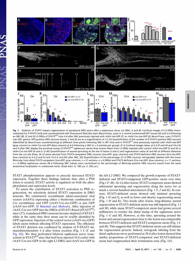
STAT3 phosphorylation appears to precede increased STAT3expression. Together these findings indicate that, after a PNSlesion is created, STAT3 activity is regulated on both the phos-phorylation and expression levels.To assess the contribution of STAT3 activation to PNS re-
generation, we selectively deleted STAT3 expression in DRGneurons. We constructed recombinant adeno-associated viralvectors (rAAVs) expressing either a bicistronic combination ofCre recombinase and GFP (rAAV-Cre-ires-GFP) or just GFP(rAAV-ires-GFP; SI Materials and Methods). After injection ofrAAV-Cre-ires-GFP into the DRGs of STAT3-floxed (STAT3fl/fl)mice (27), transduced DRG neurons become depleted of STAT3,while at the same time their axons can be readily identified byGFP expression. Injection of the control vector (rAAV-ires-GFP)labels axons without affecting STAT3 expression. The efficiencyof STAT3 deletion was confirmed by analysis of P-STAT3 im-munohistochemistry 4 d after lesion creation (Fig. 1 C–E andFig. S2). We then performed bilateral saphenous nerve trans-ections in STAT3fl/fl mice that had been injected 10 d earlier withrAAV-Cre-ires-GFP in the right L3 DRG and rAAV-ires-GFP in
the left L3 DRG. We compared the growth response of STAT3-deficient and STAT3-competent GFP-positive axons over time(Fig.1 F–M). At 4 d after lesion, STAT3-competent axons showedsubstantial sprouting and regeneration along the nerve for asmuch a several hundred micrometers (Fig. 1 F, J and K). In con-trast, STAT3-deficient axons showed only minimal sprouting(Fig. 1 H and J), as well as fewer and shorter regenerating axons(Fig. 1 H and K). Two weeks after lesion, long-distance axonalregeneration in STAT3-deficient axons was still impaired (Fig. 1 Iand M), while many STAT3-competent axons had grown severalmillimeters to reenter the distal stump of the saphenous nerve(Fig. 1 G and M). However, at this time, sprouting around thelesion and axonal regeneration close to the lesion was comparablebetween STAT3-deficient and STAT3-competent axons (Fig. 1 LandM). This suggests that STAT3-deficient axons can still initiatethe regeneration process. Indeed, retrograde labeling from thedistal saphenous nerve performed at 28 d after lesion showed thatsimilar proportions of STAT3-competent and STAT3-deficientaxons had reapproached their termination zone (Fig. 1N).
Fig. 1. Deletion of STAT3 impairs regeneration of peripheral DRG axons after a saphenous nerve cut (SNC; A and B). Confocal images of L3 DRGs immu-nostained for P-STAT3 (red) and counterstained with fluorescent Nissl-like stain (NeuroTrace, cyan) in a control (unlesioned) WT mouse (A) and 2 d followingan SNC (B). (C and D) L3 DRGs of STAT3fl/fl mice 4 d after SNC previously injected with rAAV-ires-GFP (C) or rAAV-Cre-ires-GFP (D; NeuroTrace, cyan; P-STAT3,red; GFP, green). GFP-positive DRG neurons (Insets, C and D) are at a magnification of ×3. (E) Quantification of the number of P-STAT3–positive DRG neurons(identified by NeuroTrace counterstaining) at different time points following SNC in WT mice and in STAT3fl/fl mice previously injected with rAAV-ires-GFP(gray column) or rAAV-Cre-ires-GFP (blue column) at 4 d following a SNC (n = 6 animals per group). (F–I) Confocal images taken at 4 d (F and H) and 14 d (Gand I) after SNC display the proximal stump of STAT3fl/fl saphenous nerves that receive fibers from L3 DRGs injected with control rAAV-ires-GFP (F and G) orrAAV-Cre-ires-GFP (H and I). (J–M) Quantification of axonal sprouting at the site of lesion (J and L) and regeneration ratios (K and M) at different distancesfrom the cut site (lines, H) of axons derived from STAT3-competent DRG neurons (ires-GFP, gray columns) and STAT3-deficient DRG neurons (Cre-ires-GFP,blue columns) at 4 d (J and K) and 14 d (L and M) after SNC. (N) Quantification of the percentage of L3 DRG neurons retrogradely labeled with the tracerMiniruby from distal STAT3-competent (ires-GFP, gray columns; n = 21 sections, n = 6 DRGs) and STAT3-deficient (Cre-ires-GFP, blue columns; n = 11 sections,n = 4 DRGs) saphenous nerves 28 d following SNC (values were normalized to the percentage of Miniruby-positive DRG neurons traced from the sameanatomical localization in unlesioned mice). (Scale bars: A, 100 μm; F, 250 μm.)
Bareyre et al. PNAS | April 12, 2011 | vol. 108 | no. 15 | 6283
NEU
ROSC
IENCE

STAT3 Deletion Affects Initiation but Not Perpetuation of PNS AxonRegeneration in Vivo. Impaired regeneration of STAT3-deficientPNS axons could be caused by delayed growth induction or re-duced elongation of regenerating axons. To differentiate thesepossibilities, we followed the outgrowth of fluorescently labeledSTAT3-competent and STAT3-deficient axons by repetitive invivo imaging. We first imaged regrowing axons on consecutivedays when axonal regeneration is induced (day 2–4 after lesion).Many STAT3-competent DRG axons initiate growth within 2 dafter lesion and progress with an average speed of 132 ± 23 μm/dfrom day 2 to day 3 (Fig. 2 A and C). In contrast, the vast majorityof STAT3-deficient axons fail to initiate growth during the first 2 dafter lesion. Accordingly, the speed of regeneration is reducedmore than threefold in STAT3-deficient axons in this initiationphase (33 ± 6 μm/d; Fig. 2 B and C). Notably during the axonelongation phase 7 to 8 d after lesion, when regeneration speedhas increased to approximately 400 μm/d, there is no differencebetween axons derived from STAT3-competent and STAT3-deficient DRG neurons (411 ± 48 μm/d for axons from rAAV-ires-GFP-injected DRGs vs. 341 ± 53 μm/d for axons from rAAV-Cre-ires-GFP-injected DRGs; Fig. 2D). Taken together, theseresults indicate that STAT3 is crucial for the timing of growthinitiation but not for subsequent elongation of PNS axons.
STAT3 and STAT3c Gene Therapy Can Initiate but Not Perpetuate CNSAxon Outgrowth. The failure to initiate growth is a key impedimentto successful regeneration in the CNS. Therefore, we examinedwhether STAT3 overexpression is sufficient to induce outgrowthof CNS axons in vivo. We first evaluated the activation of en-dogenous STAT3 after a CNS lesion by P-STAT3 and STAT3immunostaining of cervical DRGs after a bilateral dorsal columntransection, which interrupts central projections from cervicalDRG neurons. In contrast to the sustained activation of STAT3after a PNS lesion (cf. Fig. 1), a dorsal column lesion induces nosignificant changes in the P-STAT3 and STAT3 immunoreactivityin DRG neurons (Fig. 3 A and D and Fig. S1). To exogenouslyincrease STAT3 expression, we produced rAAVs expressing ei-ther a control protein (enhanced CFP or Cre recombinase; con-trol rAAV), STAT3 (rAAV-STAT3), or a constitutively activevariant of STAT3 (rAAV-STAT3c) and confirmed the efficiencyof viral gene transfer by immunofluorescence (Fig. 3 B–D). Wethen injected the respective rAAVs into the DRGs of Thy1-GFPs
mice, which express GFP in a subset of DRG neurons (28). Ten to12 d later we surgically reexposed the spinal cord, lesioned in-dividual GFP-positive axons emerging from the injected DRGs inthe dorsal funiculus using a hand-held small-diameter needle, andimaged their growth response over time. As expected, 2 d afterlesion, only 9% of axons emerging from DRGs injected withcontrol rAAV had formed sprouts (Fig. 3 E, F, andK and Fig. S3).In contrast, 53% of r-AAV-STAT3–transduced axons (Fig. 3G,Hand K and Fig. S3) and 46% of the rAAV-STAT3c–transducedaxons (Fig. 3 I, J, and K and Fig. S3) showed an early growth re-sponse. Interestingly, STAT3 expression not only increased ter-minal sprouting, but also collateral sprouting along the axon (Fig.3 G, I, and L). The finding that a similar growth induction wasobserved after injection of rAAV-STAT3 and rAAV-STAT3cindicates that overexpression of STAT3 alone is sufficient to in-duce downstream effects on regeneration.To determine how STAT3 overexpression affects different
phases of axonal outgrowth, we used repetitive multiphoton im-aging to follow the growth pattern of individual GFP-labeledDRG axons emerging from DRGs injected with control-rAAV,rAAV-STAT3, or rAAV-STAT3c at 2, 4, and 10 d after lesion.STAT3 and STAT3c overexpression increased the speed of axo-nal growth in the early phase (2–4 d) of regeneration (Fig. 4 A-C).However, this early growth cannot be sustained, and only verylimited axonal extension can be observed in all groups between 4and 10 d after lesion (Fig. 4 A, B, and D). Thus, in the CNS, as inthe PNS, STAT3 regulates the initiation of axonal growth but notthe elongation of regenerating axons.
DiscussionThe present study identifies the transcription factor STAT3 asa phase-specific regulator of neuronal outgrowth in both the PNSand CNS. InDRGneurons, the endogenous expression of STAT3parallels the regenerative response. By using conditional deletionof STAT3 in combination with in vivo imaging, we now also showthat STAT3 expression is not only associated with axonal re-generation, but is in fact crucial for the timing of axonal growthinitiation after a PNS lesion. It is interesting to note, however, thataxons from STAT3-deficient neurons can still mount a growthresponse, albeit with a prolonged “lag” phase compared with theirSTAT3-competent counterparts. This suggests that in the PNScompensatory mechanisms are in place that can induce neuronal
0
400
800
D7-D8
Spe
ed o
f gro
wth
(µm
/day
)
C
D
Spe
ed o
f gro
wth
(µm
/day
)
D2-D3
***
ires-GFP Cre-ires-GFP
ires-GFP Cre-ires-GFP
0
200
400*
*
*
*
*
*
A B
D0
D0 D2 D3
D2
D3
rAAV-ires-GFP
D0 D2 D3
D0
D2
D3
rAAV-Cre-ires-GFP
Fig. 2. In vivo imaging reveals that growth of STAT3-deficient axons is reduced in the initial but not the later stages of regeneration. (A and B) In vivo time-lapse images of saphenous nerve axons (GFP, green) in sparsely labeled animals emerging from DRGs that were injected with control rAAV-ires-GFP (A) orrAAV-Cre-ires-GFP (B). Axons were imaged immediately after transection (D0) as well as 2 and 3 d after transection (asterisk indicates lesion site, orange latexbeads visible on the left were used as fiduciary markers). Lower row of boxes show the boxed axon tips at magnifications of ×3 over time. (C and D)Quantification of the growth speed of axons from STAT3-competent DRG neurons (ires-GFP, gray columns) and STAT3-deficient DRG neurons (Cre-ires-GFP,blue columns) during the early (C, 2–3 d after transection) and the late phase (D, 7–8 d after transection) of the regeneration process. (Scale bar: A, 100 μm.)
6284 | www.pnas.org/cgi/doi/10.1073/pnas.1015239108 Bareyre et al.

growth in the absence of STAT3. Notably, additional over-expression of STAT3 or STAT3c did not further improve PNSregeneration (Fig. S4), indicating that a PNS lesion alone is suf-ficient to induce optimal levels of STAT3 activation for re-generation. When regeneration has been initiated, STAT3-deficient axons grow with the same speed as STAT3-competentaxons. Thus, STAT3 is primarily needed to induce a neuronalgrowth program. When the growth program has been initiated,STAT3 is no longer required for perpetuation of axonal out-growth. Given this role of STAT3 in PNS regeneration, it istempting to speculate that the failure of CNS lesions to up-regulateSTAT3 expression is directly linked to the failure of CNS axons toinitiate outgrowth. Indeed, we can show that overexpression ofSTAT3 by viral vector gene transfer alone is sufficient to initiateaxonal growth initiation in more than half the lesioned CNS axons.However, as the baseline sprouting response of CNS neurons is notaffected by STAT3 deletion (12% of axons with sprouts areemerging from STAT3-competent DRGs, compared with 15.4%emerging from STAT3-depleted DRG axons; n= 28–30 axons pergroup), it is likely that, as in the PNS, additional regulators caninduce CNS outgrowth independent of STAT3.
The observation that early stages of axon growth can be initiatedin many transected axons, even in the hostile CNS environment, byexpression of a single intracellular molecule highlights the impor-tance of intrinsic mediators of axonal growth. During recent years,a number of molecules that can influence the intrinsic neuronalgrowth response have been identified. These include c-AMP and itsdownstream mediators (29), the growth cone-associated proteinsGAP43 and CAP23 (30), components of the PTEN/mTOR path-way (31), as well as a number of transcription factors (6–11). Fur-ther, a number of recent studies in Caenorhabditis elegans havedemonstrated an essential role of the DLK-1MAP kinase pathwayfor axon regeneration, and in particular growth cone formationand migration (32–34). As more and more components of the in-trinsic growth response are emerging, it becomes increasingly im-portant to understand how they act in concert to regulate thecomplex process of axonal outgrowth.The present study provides evidence that, in vivo, this intrinsic
growth response can be divided into at least two distinct phases:initiation and elongation. The concept of a multiphasic growthresponse suggests a number of conclusions. One is that the distinctphases of axonal growth are likely regulated by distinct molecularmechanisms. STAT3, for example, controls the timely initiation of
Fig. 3. Viral vector gene transfer of STAT3 and STAT3c induces terminal and collateral sprouting of DRG branches after a central lesion. (A–C) Confocalimages of cervical DRGs immunostained for P-STAT3 (red) and counterstained with fluorescent Nissl-like stain (NeuroTrace, cyan) in a WT mouse 2 d followinga dorsal column lesion (DCL, A) and in lesioned Thy1-GFPs mice (GFP, green) injected with control rAAV (B) or rAAV-STAT3 (C). Insets: Higher-magnification(×3) of the GFP-positive neurons boxed in the images. (D) Quantification of the number of P-STAT3–positive DRG neurons (identified by NeuroTracecounterstaining) at different time points following DCL in WT mice and in mice previously injected with control rAAV (gray column), rAAV-STAT3 (redcolumn), or rAAV-STAT3c (orange column) at 2 d after a central lesion (n = 6 animals per group). (E–J) Confocal images of lesioned spinal axon endings derivedfrom DRGs injected with control rAAV (bulbs, E and F), rAAV-STAT3 (terminal sprout, G and H), or rAAV-STAT3c (terminal sprout, I and J). (F, H, and J) Higher-magnification views of details boxed in E, G, and I. Additional insets (G and I) show a magnification ×2 of the boxed collateral sprouts. (K and L) Quantificationof terminal (K) and collateral (L) sprouting of axons derived from DRGs injected with control rAAV, rAAV-STAT3, or rAAV-STAT3c and analyzed 2 d aftertransection. (Scale bars: A and I, 100 μm; J, 25 μm.)
Bareyre et al. PNAS | April 12, 2011 | vol. 108 | no. 15 | 6285
NEU
ROSC
IENCE

axonal growth but does not affect axonal elongation. The molec-ular mechanisms by which STAT3 initiates this axonal growthprogram are currently not known. However, as STAT3 is a tran-scription factor, it is likely that the downstream effects are medi-ated by induction of gene expression. A large number of genes thatare affected by STAT3 have already been identified. Some of thesedownstream targets like the cell cycle inhibitor P21/Cip1/Waf1 (35)or the small proline rich protein 1a (SPRR1A) (36) can directlyaffect neuronal outgrowth (37, 38). Notably, a recent transcrip-tional profiling study has identified several additional genes thatare specifically regulated by STAT3 in DRG neurons. At least oneof these genes, the IFN regulatory factor 1 (IRF1), is sufficient toincrease neuronal outgrowth in cultured cerebellar neurons (21).Our results further suggest that it is likely that environmental
cues play key roles in shaping the distinct stages of axonal regrowth.For example, in the PNS, it is conceivable, that the transition fromgrowth initiation to elongation is induced by the interaction of axonswith Schwann cells. Schwann cells align after injury to form tubes,also called bands of Büngner, that guide the axons to their targetcells (39). Axons could require STAT3 to initiate growth in-dependently of Schwann cell guidance; however, when a regrowingaxon has contacted the Schwann cell tube, it shifts to the elongationmode and no longer requires STAT3. In line with this scenario, wecan show that PNS crush lesions that do not interrupt Schwann cellguidance do not induce STAT3 expression in the correspondingDRG neurons (Fig. S5). Although the reasons behind this lack ofSTAT induction are not yet understood and might include the in-duction of differential injury signals by crush and cut lesions (40),the rapid outgrowth of crushed axons (41) indicates that axonalgrowth along glial support structures does not require STAT3. Likein the PNS, changes in the environmentmay also help to explain thetransition (or lack thereof) between different growth phases aftera CNS lesion. Although, in the PNS, the regrowing axon can reachSchwann cell support at some point, CNS axons that initiate growthin response to STAT3 continue to encounter a growth-inhibitoryCNS environment that becomes even more hostile with the de-velopment of a glial scar. These changes in the lesion environmentmight help to explain why STAT3-transduced axons can initiategrowth early but fail to support it later. In line with this assumption,when we combined the induction of STAT3 (by viral vector gene
transfer of rAAV-STAT3) with the neutralization of inhibitory scarcomponents (by application of chondroitinase ABC) (42), the av-erage axonal outgrowth over a period of 10 d after a CNS lesion wasincreased more than twofold (141 ± 53 μm for axons treated withrAAV-STAT3 and chondroitinase ABC vs. 42 ± 26 μm for axonstreated with rAAV-STAT3 alone and 64 ± 28 μm for axons treatedwith chondroitinase alone; n = 14–20 axons per group). Theseresults underline the importance of developing combined thera-peutic strategies that target the molecularly distinct phases of theaxonal growth response. In this concept, phase-specific regulators ofaxonal growth initiation such as STAT3 would be used to “jump-start” the regenerationprocess andprime axons in the injured spinalcord for application of complementary therapies that can sustainaxonal elongation in the growth-inhibitory CNS environment (43).
Materials and MethodsMice. Animals used in this study were adult female WT mice on a C57BL/6background (weight 20–30 g, 6–12 wk of age) with the following exceptions:STAT3 was deleted by using STAT3fl/fl mice, which are maintained on a BL6background (27). Further, central axon regeneration was investigated inThy1-GFPs mice (28), which express GFP in a subset of neurons and aremaintained on a mixed background. All animal experiments were per-formed in accordance with regulations of the animal welfare act and pro-tocols approved by the Regierung von Oberbayern.
AAV Vector Construction, Production, and Purification. The adeno-associatedviral vectors used in this study have been cloned into the pAAV-MCS vectorfrom Stratagene and were produced by the adenovirus-free AAV productionmethod as detailed in SI Materials and Methods.
Tissue Processing, Immunohistochemistry, and STAT Expression Analysis. Ani-mals were deeply anesthetized with isoflurane and perfused transcardiallywith saline followed by 4% paraformaldehyde in 0.01 M phosphate buffer.DRGs were dissected out, immunostained for P-STAT3 and STAT3 and ana-lyzed as detailed in SI Materials and Methods.
Gene Therapy with Recombinant Adeno-associated Viral Vectors. For analysisof PNS regeneration, the left and right L3 DRGs of anesthetized STAT3fl/fl
mice were surgically exposed after a dorsal laminectomy. Then, 1 μL ofrAAV-ires-GFP was slowly injected into the left L3 DRG with a thinly drawnglass capillary and the same amount of rAAV-Cre-ires-GFP was injected intothe right L3 DRG of the same animal. Ten days after the injection, the sa-
D4
D10
A *
D2
*
*
*
D4
*
D2
*
D10
B
**
STAT3
Control
0
20
40
60
80
100
Control rAAV rAAV-STAT3
D2-D4C
Spee
d of
gro
wth
(µ
m/d
ay)
D
Spee
d of
gro
wth
(µ
m/d
ay)
200 **
STAT3c
D4-D10
020
40
60
80
100
STAT3
Control
STAT3c
n=12 n=14 n=11
n=10 n=9 n=9
Fig. 4. In vivo imaging reveals successful initiation but not elongation of CNS axons after STAT3 and STAT3c gene therapy. (A and B) Multiphoton images ofthe growth pattern of spinal DRG axons emerging from DRGs injected with control rAAV (A) or rAAV-STAT3 (B) imaged 2, 4, and 10 d following a centrallesion. Insets: Magnifications (×2) of boxed axon ends. (C and D) Quantification of axonal growth speed in vivo after injection of control rAAV (gray bars),rAAV-STAT3 (red bars), or rAAV-STAT3c (orange bars) analyzed early (C, 2–4 d) and late (D, 4–10 d) after transection. (Scale bar: B, 200 μm.)
6286 | www.pnas.org/cgi/doi/10.1073/pnas.1015239108 Bareyre et al.

phenous nerve was bilaterally transected at the midthigh level using fineiridectomy scissors as previously described (23).
For analysis of CNS regeneration, a cervical dorsal laminectomy wasperformed in Thy1-GFPs mice anesthetized by an i.p. injection of ketamine/xylazine (ketamine 87 mg/kg, xylazine 13 mg/kg) as previously described(24). DRGs, from which suitably labeled axons emerged, were identified byin vivo imaging (SI Materials and Methods) and surgically exposed. Then,1 μL of rAAV-STAT3, rAAV-STAT3c, rAAV-eCFP, or rAAV-Cre (control rAAV)was slowly injected into the DRG with a thinly drawn glass capillary. Ten to12 d later, Thy1-GFPs mice were reanesthetized, the spinal cord laminectomysite was reaccessed, and selected fluorescently labeled axons were trans-ected with a hand-held 32-gauge hypodermic needle.
Confocal Microscopy.Weobtained confocal images offixed tissue on a FV1000confocal system mounted on an upright BX61 microscope (Olympus) andequipped with 20×/0.85 and 60×/1.42 oil immersion objectives. We recordedstacks of 12-bit images that were processed using MetaMorph software(Universal Imaging) or the freeware ImageJ/Fiji (http://rsbweb.nih.gov/ij).
In Situ and in Vivo Analysis of Axon Regeneration. The regeneration oftransected peripheral and central DRG axons was evaluated as detailed in SIMaterials and Methods.
Statistical Analysis. Results are given as mean ± SEM unless indicated other-wise. Statistical significance was determined using GraphPad Prism software(GraphPad). All data were analyzed by using a one-way ANOVA followedby a Tukey post-hoc test for multiple comparisons or a t test for single com-parisons. For the statistical evaluation of the proportion of terminal sproutsfollowing rAAV treatment after CNS lesion, a frequency analysis was madeusing a χ2 test.
ACKNOWLEDGMENTS. We thank A. Schmalz and H. Janicki for excellenttechnical assistance and R. Hohlfeld, K. Dornmair, E. Meinl, and P. Williamsfor critical reading of the manuscript. We would also like to thank S. Akirafor providing STAT3fl/fl mice and J. Sanes and I. J. Kim for help with viralproduction. This work was supported by a grant from the International In-stitute for Research in Paraplegia (to F.M.B. and M.K.). M.K. was supportedby the Deutsche Forschungsgemeinschaft (DFG; Emmy-Noether Program,SFB 571 and SFB 870) and by Verein “Therapieforschung für MS-Kranke e.V.”F.M.B. was supported by DFG (SFB 870) and is the recipient of an Indepen-dent Group Leader Award from the Federal Ministry of Education andResearch of Germany (BMBF). T.M. was supported by the Technische Univer-sität München Institute for Advanced Study, Alexander von Humboldt Foun-dation, Center for Integrated Protein Science (Munich), and a Christopher andDana Reeve Foundation grant. H.B. was supported by the Center for MolecularMedicine Cologne (ZMMK).
1. Schwab ME, Bartholdi D (1996) Degeneration and regeneration of axons in thelesioned spinal cord. Physiol Rev 76:319–370.
2. Neumann S, Woolf CJ (1999) Regeneration of dorsal column fibers into and beyondthe lesion site following adult spinal cord injury. Neuron 23:83–91.
3. Ylera B, et al. (2009) Chronically CNS-injured adult sensory neurons gain regenerativecompetence upon a lesion of their peripheral axon. Curr Biol 19:930–936.
4. Raivich G, Makwana M (2007) The making of successful axonal regeneration: Genes,molecules and signal transduction pathways. Brain Res Brain Res Rev 53:287–311.
5. Rossi F, Gianola S, Corvetti L (2007) Regulation of intrinsic neuronal properties foraxon growth and regeneration. Prog Neurobiol 81:1–28.
6. Raivich G, et al. (2004) The AP-1 transcription factor c-Jun is required for efficientaxonal regeneration. Neuron 43:57–67.
7. Zou H, Ho C, Wong K, Tessier-Lavigne M (2009) Axotomy-induced Smad1 activationpromotes axonal growth in adult sensory neurons. J Neurosci 29:7116–7123.
8. Seijffers R, Mills CD, Woolf CJ (2007) ATF3 increases the intrinsic growth stateof DRG neurons to enhance peripheral nerve regeneration. J Neurosci 27:7911–7920.
9. Stam FJ, et al. (2007) Identification of candidate transcriptional modulators involvedin successful regeneration after nerve injury. Eur J Neurosci 25:3629–3637.
10. MacGillavry HD, et al. (2009) NFIL3 and cAMP response element-binding protein forma transcriptional feedforward loop that controls neuronal regeneration-associatedgene expression. J Neurosci 29:15542–15550.
11. Moore DL, et al. (2009) KLF family members regulate intrinsic axon regenerationability. Science 326:298–301.
12. Aaronson DS, Horvath CM (2002) A road map for those who don’t know JAK-STAT.Science 296:1653–1655.
13. Schwaiger FW, et al. (2000) Peripheral but not central axotomy induces changes inJanus kinases (JAK) and signal transducers and activators of transcription (STAT). Eur JNeurosci 12:1165–1176.
14. Xia XG, Hofmann HD, Deller T, Kirsch M (2002) Induction of STAT3 signaling inactivated astrocytes and sprouting septal neurons following entorhinal cortex lesionin adult rats. Mol Cell Neurosci 21:379–392.
15. Sheu JY, Kulhanek DJ, Eckenstein FP (2000) Differential patterns of ERK and STAT3phosphorylation after sciatic nerve transection in the rat. Exp Neurol 166:392–402.
16. Bareyre FM, Haudenschild B, Schwab ME (2002) Long-lasting sprouting and geneexpression changes induced by the monoclonal antibody IN-1 in the adult spinal cord.J Neurosci 22:7097–7110.
17. Cafferty WB, et al. (2001) Leukemia inhibitory factor determines the growth status ofinjured adult sensory neurons. J Neurosci 21:7161–7170.
18. Cafferty WB, et al. (2004) Conditioning injury-induced spinal axon regeneration failsin interleukin-6 knock-out mice. J Neurosci 24:4432–4443.
19. Zhong J, Dietzel ID, Wahle P, Kopf M, Heumann R (1999) Sensory impairments anddelayed regeneration of sensory axons in interleukin-6-deficient mice. J Neurosci 19:4305–4313.
20. Smith PD, et al. (2009) SOCS3 deletion promotes optic nerve regeneration in vivo.Neuron 64:617–623.
21. Smith RP, et al. (2011) Transcriptional profiling of intrinsic PNS factors in the postnatalmouse. Mol Cell Neurosci 46:32–44.
22. Qiu J, Cafferty WB, McMahon SB, Thompson SW (2005) Conditioning injury-inducedspinal axon regeneration requires signal transducer and activator of transcription 3activation. J Neurosci 25:1645–1653.
23. Pan YA, Misgeld T, Lichtman JW, Sanes JR (2003) Effects of neurotoxic andneuroprotective agents on peripheral nerve regeneration assayed by time-lapseimaging in vivo. J Neurosci 23:11479–11488.
24. Kerschensteiner M, Schwab ME, Lichtman JW, Misgeld T (2005) In vivo imaging ofaxonal degeneration and regeneration in the injured spinal cord. Nat Med 11:572–577.
25. Dray C, Rougon G, Debarbieux F (2009) Quantitative analysis by in vivo imaging of thedynamics of vascular and axonal networks in injured mouse spinal cord. Proc NatlAcad Sci USA 106:9459–9464.
26. Jankowski MP, et al. (2009) Sensitization of cutaneous nociceptors after nervetransection and regeneration: possible role of target-derived neurotrophic factorsignaling. J Neurosci 29:1636–1647.
27. Matsukawa A, et al. (2003) Aberrant inflammation and lethality to septic peritonitisin mice lacking STAT3 in macrophages and neutrophils. J Immunol 171:6198–6205.
28. Feng G, et al. (2000) Imaging neuronal subsets in transgenic mice expressing multiplespectral variants of GFP. Neuron 28:41–51.
29. Qiu J, et al. (2002) Spinal axon regeneration induced by elevation of cyclic AMP.Neuron 34:895–903.
30. Bomze HM, Bulsara KR, Iskandar BJ, Caroni P, Skene JHP (2001) Spinal axonregeneration evoked by replacing two growth cone proteins in adult neurons. NatNeurosci 4:38–43.
31. Park KK, et al. (2008) Promoting axon regeneration in the adult CNS by modulation ofthe PTEN/mTOR pathway. Science 322:963–966.
32. Hammarlund M, Nix P, Hauth L, Jorgensen EM, Bastiani M (2009) Axon regenerationrequires a conserved MAP kinase pathway. Science 323:802–806.
33. Yan D, Wu Z, Chisholm AD, Jin Y (2009) The DLK-1 kinase promotes mRNA stability andlocal translation in C. elegans synapses and axon regeneration. Cell 138:1005–1018.
34. Ghosh-Roy A, Wu Z, Goncharov A, Jin Y, Chisholm AD (2010) Calcium and cyclic AMPpromote axonal regeneration in Caenorhabditis elegans and require DLK-1 kinase.J Neurosci 30:3175–3183.
35. Coqueret O, Gascan H (2000) Functional interaction of STAT3 transcription factor withthe cell cycle inhibitor p21WAF1/CIP1/SDI1. J Biol Chem 275:18794–18800.
36. Pradervand S, et al. (2004) Small proline-rich protein 1A is a gp130 pathway- andstress-inducible cardioprotective protein. EMBO J 23:4517–4525.
37. Tanaka H, et al. (2002) Cytoplasmic p21(Cip1/WAF1) regulates neurite remodeling byinhibiting Rho-kinase activity. J Cell Biol 158:321–329.
38. Bonilla IE, Tanabe K, Strittmatter SM (2002) Small proline-rich repeat protein 1A isexpressed by axotomized neurons and promotes axonal outgrowth. J Neurosci 22:1303–1315.
39. Höke A (2006) Mechanisms of disease: What factors limit the success of peripheralnerve regeneration in humans? Nat Clin Pract Neurol 2:448–454.
40. Bussmann KA, Sofroniew MV (1999) Re-expression of p75NTR by adult motor neuronsafter axotomy is triggered by retrograde transport of a positive signal from axonsregrowing through damaged or denervated peripheral nerve tissue. Neuroscience 91:273–281.
41. Nguyen QT, Sanes JR, Lichtman JW (2002) Pre-existing pathways promote preciseprojection patterns. Nat Neurosci 5:861–867.
42. Bradbury EJ, et al. (2002) Chondroitinase ABC promotes functional recovery afterspinal cord injury. Nature 416:636–640.
43. Kadoya K, et al. (2009) Combined intrinsic and extrinsic neuronal mechanismsfacilitate bridging axonal regeneration one year after spinal cord injury. Neuron 64:165–172.
Bareyre et al. PNAS | April 12, 2011 | vol. 108 | no. 15 | 6287
NEU
ROSC
IENCE
Supporting InformationBareyre et al. 10.1073/pnas.1015239108SI Materials and MethodsAAV Vector Construction, Production, and Purification. The adeno-associated viral vectors used in this study have been cloned into thepAAV-MCS vector from Stratagene as follows. Briefly, for thecontrol pAAV-ires-GFP vector, a strong modified internal ribo-some entry site (ires) of the encephalomyocarditis virus, whichpermits the translation of two genes of interest from a single bi-cistronic mRNA, was cut with BamHI and BstXI from the pires2-DsRed2 plasmid (Clontech) and inserted at the HincII site ofpAAV-MCS. Then the DNA coding for GFP was inserted at theXhoI site of pAAV-MCS to create the pAAV-ires-GFP construct.The pAAV-Cre-ires-GFP construct was cloned by excising thecoding sequence for Cre recombinase from pBS185 (gift ofThomas Hughes, Montana State University, MT) with XhoI andMluI and inserting it into the EcoRI site of pAAV-ires-GFP. Forthe pAAV-STAT3, the STAT3 gene was excised from pcDNA3STAT3 (Addgene plasmid 8706) with BamHI and XhoI andcloned in the pAAV-MCS at the HincII site. For cloning thepAAV-STAT3c, the STAT3c gene was excised from pRc/CMVSTAT3c Flag (Addgene plasmid 8722) with NotI and SwaI andinserted in the HincII site of pAAV-MCS. Control pAAV-eCFPwas engineered by excising the eCFP gene from the peCFP N1plasmid at BamHI and NotI and cloning it in the pAAV-MCS atthe HincII site.AAV serotype 2 particles were then produced in HEK293 cells
by the adenovirus-free AAV production method (1, 2). Briefly,HEK293 cells were seeded at 80% confluence and cotransfectedusing the calcium phosphate method with 7.5 μg of pRC (3), 7.5 μgof transgene plasmid, and 22.5 μg of pXX6 (1, 2). After 48 h, cellswere harvested and pelleted by low-speed centrifugation. Cellswere resuspended in 150 mM NaCl/50 mM Tris-HCl (pH 8.5),freeze/thawed three times, and treated with Benzonase (50 U/mLlysate) for 30 min at 37 °C. To purify viral vector preparations, celldebris was removed by centrifugation at 3,700 × g for 20 min at4 °C, and the supernatant was loaded onto a discontinuous io-dixanol gradient (3). Following harvesting of the 40% phase, aheparin affinity chromatography was conducted. Briefly, a hepa-rin affinity column (HiTrap heparin HP; GE Healthcare) wasequilibrated with 1× PBS solution/MgCl2 (1 mM)/KCl (2.5 mM).The vector solution was diluted 1:10 in the same buffer and ap-plied to the column. After a washing step with 1× PBS solution/MgCl2 (1 mM)/KCl (2.5 mM), the vector was eluted with 1× PBS/MgCl2 (1 mM)/KCl (2.5 mM) plus 1 M NaCl in 500-μL steps.To determine the genomic particle titer, vector genomes were
isolated by DNeasy Tissue Kit (Qiagen) and subjected to quan-titative PCR analysis by using the LightCycler rapid thermal cyclersystem (Roche Diagnostics) and the SYBR Green kit (RocheDiagnostics). Genomic titers were as follows: rAAV-STAT3, 9 ×10¹² genome copies/mL; rAAV-STAT3c, 5.5 × 10¹² genomecopies/mL; rAAV-eCFP, 9.2 × 10¹² genome copies/mL; rAAV-Cre, 9 × 10¹² genome copies/mL; rAAV-Cre-ires-GFP, 5.7–7.1 ×10¹¹ genome copies/mL; and rAAV-ires-GFP, 1.4–3.8 × 10¹²
genome copies/mL.
Tissue Processing, Immunohistochemistry, and STAT Expression Analysis.Animals were deeply anesthetized with isoflurane and perfusedtranscardially with saline solution followed by 4% paraformal-dehyde in 0.01 M phosphate buffer. Tissues were postfixed in 4%paraformaldehyde at 4 °C for 24 h. For immunofluorescenceanalysis, DRGs were dissected out, transferred to 30% sucrose forat least 24 h, and embedded in Tissue-Tek optimal cutting tem-perature compound (Sakura Finetek). Then, 20-μm-thick coronal
sections were cut on a cryostat. Immunofluorescence staining ofDRG sections was performed as described previously (4). Beforeimmunostaining, sections were heated in a microwave to improveantigen retrieval. Antibodies were diluted in a working solution ofPBS containing 0.2% Triton X-100 (Sigma) and 10% normal horseserum (Jackson Laboratory), and incubation was performed atroom temperature or at 4 °C overnight. Details of the primaryantibodies are as follows: STAT3 (dilution 1:500; Cell Signaling),P-STAT3 (dilution 1:50; Cell Signaling), and cJun (dilution 1:500;Santa Cruz Biotechnology). Secondary antibodies (goat anti-rabbit594, goat anti-rabbit 488) were obtained from Jackson Laboratoryand used at a dilution of 1:500. Nuclei were counterstained usingNeuroTrace 435 (Invitrogen) and mounted in Vectashield (VectorLaboratories).P-STAT3 and STAT3 immunoreactivity was analyzed in L3
DRGs of animals perfused at 1 h, 6 h, 2 d, 1 wk, and 2 wk aftera bilateral saphenous nerve transection. P-STAT3 immunoreac-tivity was also analyzed in L3 DRGs of mice perfused 1 h, 6 h, 2 d,1 wk, and 2 wk after the saphenous nerve was double-crushedwith hand-held forceps. Immunofluorescence analysis of cJuninduction in DRG neurons and confocal microscopy analysis ofWallerian degeneration in distal nerve segments of Thy1-YFP16
mice were used to confirm the success of the crush lesion (Fig. S5).P-STAT3 and STAT3 immunoreactivity was further analyzedin C6 DRGs of animals perfused at 1 h, 6 h, 2 d, 1 wk, and 2 wkafter a bilateral dorsal column lesion. For the dorsal column le-sion, animals were anesthetized with isoflurane, and a dorsallaminectomy was performed at the C4 to C6 spinal cord level aspreviously described (4). A small incision was made in the dura,and the dorsal column was then cut with fine iridectomy scissors.Unlesioned animals were used as controls. All DRG sections werecounterstained with NeuroTrace 435 (as described earlier) toreveal the total number of neuronal nuclei in the DRG, and theproportion of DRG neurons showing STAT3 or P-STAT3 im-munoreactivity was determined.
In Situ Analysis of Peripheral Axon Regeneration. To evaluate theregeneration of peripheral DRG branches, STAT3fl/fl mice (5)injected with rAAV-Cre-ires-GFP or rAAV-ires-GFP weretranscardially perfused with 4% paraformaldehyde at 4 or 14 dafter saphenous nerve transection. Cross-sections of the saphe-nous nerve were cut rostrally to the injury site to quantify thenumber of GFP-labeled axons above the lesion. The distal part ofthe nerve containing the lesion site was dissected with the sur-rounding muscular tissue and flat-mounted in Vectashield (Vec-tor Laboratories). High-resolution image stacks of the axons weretaken on a FV 1000 confocal microscope system (Olympus). Thenlines were positioned on the confocal maximum intensity pro-jections at 0.1 mm, 0.2 mm, 0.4 mm, 0.8 mm, 1.4 mm, 2 mm, and 5mm distal from the lesion site (Fig. 1H). The number of axonscrossing these lines was counted. A ratio of regenerating axonswas calculated by dividing the number of regenerating axons ata given distances from the lesion by the number of labeled axonsrostral to the lesion.To determine the percentage of DRG neurons that reach the
proximity of their original target area, retrograde tracingexperiments were performed as follows: At 28 d following theSNC, a gel foam impregnated with the tracer Miniruby (Invi-trogen) was applied to the distal-most part of the saphenous nerve(located approximately 10 mm distal to the cut nerve). Three dayslater, mice were deeply anesthetized and transcardially perfusedwith PFA. Then, L3 DRGs were dissected and 20-μm sections
Bareyre et al. www.pnas.org/cgi/content/short/1015239108 1 of 5

were cut on the cryostat as described earlier. Confocal images ofthe sections were acquired on an Olympus FV 1000 confocalmicroscope and the number of Miniruby-positive DRG neuronsand total DRGneurons (identified by costaining with NeuroTrace435) were counted. The percentage of Miniruby-positive DRGneurons was calculated and normalized to the percentage ofMiniruby-positive neurons that were retrogradely traced from thesame anatomical localization in unlesioned mice (45 ± 6% ofMiniruby-positive L3 DRG neurons; n= 14 sections, n= 2mice).Mice traced from the same anatomical localization immediatelyafter the saphenous nerve was cut showed no labeling, indicatingthat the labeling at 28 d is caused by long-distance regeneration.To determinewhether the overexpression of STAT3or STAT3c
could increase peripheral axon regeneration, we injected 1 μL ofrAAV-STAT3, rAAV-STAT3c, or rAAV-Cre (control rAAV)into the L3 DRG of Thy1-YFP16 mice (6), in which a large pro-portion of DRG neurons are fluorescently labeled with the YFP.We then performed an SNC and evaluated axonal sprouting andregeneration at 4 d after the lesion as described earlier.
In Vivo Analysis of Peripheral Axon Regeneration. For in vivo imagingexperiments, we used sparsely labeled animals, lesioned the sa-phenous nerve as described earlier, and marked the position of theaxons proximally and distally from the lesion site with a suspensionof 1-μm-diameter orange FluoSpheres (Invitrogen). Before eachimaging session, animals were anesthetized and placed directlyunder a fluorescence dissection microscope (Olympus). An in-cision was made on top of the lesion site and the axons in the sa-phenous nerve were imaged with 20×magnification at day 2 and 3or at day 7 and 8 after lesion. Images were captured with a cooledSensicam QE CCD camera (pco.imaging). To determine thegrowth of the transected axon ends, we measured the distancefrom the axon end to the FluoSpheres proximal to the lesion site at2 and 3 d or 7 and 8 d after lesion and calculated the speed ofgrowth per day.
In Vivo and in Situ Analysis of Central Axon Outgrowth. To imagecentral branches of fluorescently labeled DRG neurons, weadapted our previously established spinal in vivo imaging ap-proach (4). Briefly, Thy1-GFPs mice were anesthetized by an i.p.injection of ketamine/xylazine. To access the cervical spinal cord,a laminectomy was performed and the dorsal surface of thespinal cord was exposed. During the imaging session, the spinalcord was superfused with mouse artificial cerebrospinal fluid. Tofollow the outgrowth of individual axons in vivo, we first iden-tified single fluorescently labeled axons emerging from a DRGusing a widefield set-up based on an Olympus BX51 microscopeequipped with ×4/0.13 dry, ×10/0.3 dry, and ×20/0.5 dipping conewater-immersion objectives. To document these axons in vivo,image streams of 25 to 50 images were acquired with a cooledSensicam QE CCD camera controlled by MetaMorph softwareas previously described (4). The DRG from which the selectedaxon was emerging was then surgically prepared and 1 μL of oneof the rAAVs was injected by using a glass capillary as describedearlier. Then, 10 to 12 d after the DRG injection, the dorsalsurface of the spinal cord was surgically reexposed. The pre-viously identified axon was transected by using a hand-heldsmall-diameter needle and documented in vivo. Two differentimaging protocols were used as follows.To determine the frequency of axonal sprouting, the animal was
perfused transcardially with 4% PFA 2 d after the lesion and thepreviously imaged spinal cord segment was dissected. Thetransected axon was then documented by using high-resolutionconfocal microscopy as described earlier, first in the intact spinalcord and then in 100- to 250-μm-thick vibratome sections.Confocal stacks were processed as described earlier and the
transected axons were reconstructed from the DRG root to thelesion site. Axons were then evaluated by two blinded observersand terminal axon ends were classified as “sprouts” or “bulbs”based on their characteristic morphological appearances. Fur-ther, the number of collateral sprouts emerging from the trans-ected axons was counted.To determine the speed of axonal outgrowth we reimaged the
transected axon ends at 2 and 4 d after lesion in vivo using acustom-built multiphoton imaging setup based on an OlympusFV 300 scanner equipped with a femtosecond pulsed Ti:Sapphirelaser (Mai Tai HP; Newport/Spectra-Physics). We acquired imagestacks of 50 to 200 images per stack (spaced at 1–2 μm in z di-mension) for each frame with a ×20/0.5 dipping cone water-immersion objective. Animals were then perfused transcardiallywith 4% PFA 10 d after the lesion. The imaged part of the spinalcord was dissected out. The transected axons were reidentified inthe fixed tissue and imaged in the intact spinal cord by using anOlympus FV1000 MPE multiphoton microscope or an OlympusFV1000 confocal microscope. Image stacks of 20 to 100 frameswere acquired with a ×25/1.05 water-immersion or a ×20/0.85oil-immersion objective. Imaging stacks were processed by usingMetaMorph or ImageJ/Fiji software. Frames containing the axonwere selected, and the transected axons were reconstructed usingPhotoshop. To determine the growth of the transected axonends, we measured the distance from the axon end to a charac-teristic proximal structure (in most cases the Y-branch point inthe dorsal root; in some cases a crossing point with anotheraxon) at 2, 4, and 10 d after lesion. To compensate for tissuechanges caused by fixation in the perfused samples, we measureda “constant” distance e.g., between two branch points in thesame unlesioned axon both in vivo and in the correspondingfixed tissue to determine a sample-specific “correction factor.”The length of the transected axon end measured in fixed tissuewas then multiplied by this correction factor. Axons that showedsubstantial die-back were excluded from the analysis.To determine the influence of endogenous STAT3 expression
on axonal sprouting after a CNS lesion, we injected 1 μL of rAAV-eCFP or a combination of rAAV-eCFP and rAAV-Cre into thecervical DRGs of STAT3fl/fl mice. Ten days after the injection,fluorescently labeled spinal DRG axons were lesioned by a pin asdescribed earlier. Then, 2 d following the lesion, animals wereperfused and axonal sprouting was evaluated based on confocalstacks of the fixed intact spinal cord as described earlier.To determine the effects of a combination therapy with STAT3
and chondroitinase ABC, we performed the following experi-ment. We first injected 1 μL of rAAV-STAT3 or rAAV-Cre intothe cervical DRGs of Thy1-GFPs mice as described earlier. Tendays after this injection, we then lesioned individual GFP-posi-tive spinal axons and administered a first bolus of 6 μL ofchondroitinase ABC (10 U/mL in saline solution with 0,01%BSA; Sigma-Aldrich) or vehicle only (saline solution with 0.01%BSA) immediately following lesion creation. At the same time,an osmotic minipump (1007B; Alzet), which was connected toa brain infusion kit (Alzet) inserted into the lateral ventricle, wasinstalled and started to deliver 0.5 μL/h of chondroitinase ABC(10 U/mL in saline solution with 0.01% BSA) or vehicle only for7 d. At 10 d following the lesion, the animals were perfused withPFA, the spinal cords were dissected, and the transected axonswere imaged in the intact fixed spinal cord by confocal micros-copy as described earlier. On confocal image stacks, we thenmeasured the length of axonal sprouts to determine the axonaloutgrowth (axonal terminals that ended in tips or bulbs wereconsidered to be nongrowing axons and their outgrowth was setto 0 μm).
Bareyre et al. www.pnas.org/cgi/content/short/1015239108 2 of 5
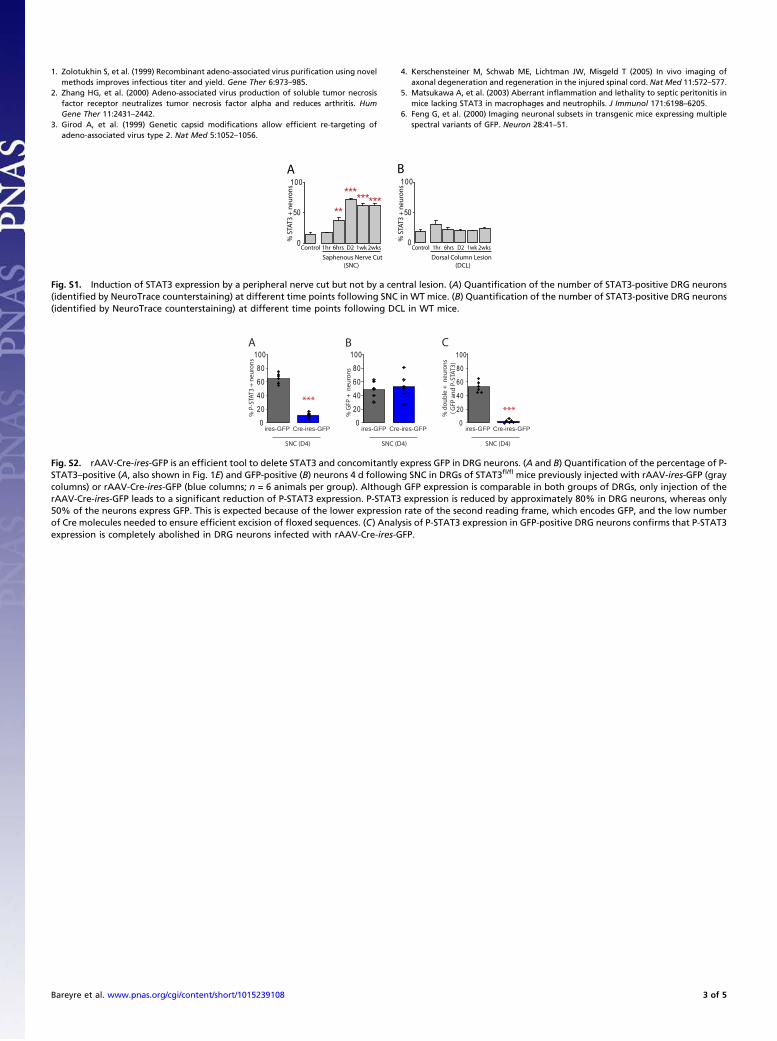
1. Zolotukhin S, et al. (1999) Recombinant adeno-associated virus purification using novelmethods improves infectious titer and yield. Gene Ther 6:973–985.
2. Zhang HG, et al. (2000) Adeno-associated virus production of soluble tumor necrosisfactor receptor neutralizes tumor necrosis factor alpha and reduces arthritis. HumGene Ther 11:2431–2442.
3. Girod A, et al. (1999) Genetic capsid modifications allow efficient re-targeting ofadeno-associated virus type 2. Nat Med 5:1052–1056.
4. Kerschensteiner M, Schwab ME, Lichtman JW, Misgeld T (2005) In vivo imaging ofaxonal degeneration and regeneration in the injured spinal cord. Nat Med 11:572–577.
5. Matsukawa A, et al. (2003) Aberrant inflammation and lethality to septic peritonitis inmice lacking STAT3 in macrophages and neutrophils. J Immunol 171:6198–6205.
6. Feng G, et al. (2000) Imaging neuronal subsets in transgenic mice expressing multiplespectral variants of GFP. Neuron 28:41–51.
Control
Saphenous Nerve Cut (SNC)
A
0
50
100
1hr 6hrs D2 1wk 2wks
% S
TAT3
+ n
euro
ns
0
50
100
Control
Dorsal Column Lesion (DCL)
1hr 6hrs D2 1wk 2wks
% S
TAT3
+ n
euro
ns
B
**
*********
Fig. S1. Induction of STAT3 expression by a peripheral nerve cut but not by a central lesion. (A) Quantification of the number of STAT3-positive DRG neurons(identified by NeuroTrace counterstaining) at different time points following SNC in WT mice. (B) Quantification of the number of STAT3-positive DRG neurons(identified by NeuroTrace counterstaining) at different time points following DCL in WT mice.
0
20
40
60
80
100
***
ires-GFP Cre-ires-GFP
SNC (D4)
% P
-STA
T3 +
neu
rons
0
20
40
60
80
100
ires-GFP Cre-ires-GFP
SNC (D4)
% G
FP +
neu
rons
A B
0
20
40
60
80
100
ires-GFP Cre-ires-GFP
SNC (D4)%
dou
ble
+ n
euro
ns (
GFP
and
P-S
TAT3
)
C
***
Fig. S2. rAAV-Cre-ires-GFP is an efficient tool to delete STAT3 and concomitantly express GFP in DRG neurons. (A and B) Quantification of the percentage of P-STAT3–positive (A, also shown in Fig. 1E) and GFP-positive (B) neurons 4 d following SNC in DRGs of STAT3fl/fl mice previously injected with rAAV-ires-GFP (graycolumns) or rAAV-Cre-ires-GFP (blue columns; n = 6 animals per group). Although GFP expression is comparable in both groups of DRGs, only injection of therAAV-Cre-ires-GFP leads to a significant reduction of P-STAT3 expression. P-STAT3 expression is reduced by approximately 80% in DRG neurons, whereas only50% of the neurons express GFP. This is expected because of the lower expression rate of the second reading frame, which encodes GFP, and the low numberof Cre molecules needed to ensure efficient excision of floxed sequences. (C) Analysis of P-STAT3 expression in GFP-positive DRG neurons confirms that P-STAT3expression is completely abolished in DRG neurons infected with rAAV-Cre-ires-GFP.
Bareyre et al. www.pnas.org/cgi/content/short/1015239108 3 of 5

Fig. S3. Gallery of central DRG axon endings after treatment with control rAAV, rAAV-STAT3, and r-AAV-STAT3c. (A–C) Representative images of axonendings 2 d after a central lesion derived from DRG neurons treated with control rAAV (A), rAAV-STAT3 (B), or rAAV-STAT3c (C). Axonal sprouts (asterisks) arerarely found in control axons (A) but are frequent in axons emerging from DRG neurons expressing STAT3 (B) or STAT3c (C). (Scale bar: A, 25 μm.)
D
0
20
40
60
% o
f axo
nal s
prou
ts
D4 E
% re
gene
rate
d ax
ons Control n=7
STAT3 n=7
D4
Control STAT3 STAT3c0
10
20
30
0,1 0,2 0,4 0,8 1,4 2 5 [mm]
STAT3c n=7
A BSTAT3 STAT3cD4 D4
C ControlD4
Fig. S4. Overexpression of STAT3 or STAT3c does not increase peripheral axon regeneration following a SNC. (A–C). Confocal images taken at 4 d after SNCdisplaying the proximal stump of saphenous nerve that receives fibers from L3 DRGs injected with rAAV-STAT3 (A), rAAV-STAT3c (B), or control rAAV (C).Axons are fluorescently labeled with YFP (white) in Thy1-YFP16 mice. (D and E) Quantification of axonal sprouting at the site of the lesion (D) and regenerationratios (E) at different distances from the cut site (Fig. 1H) of axons derived from DRG neurons that express normal levels of STAT3 (gray columns) or overexpressSTAT3 (red columns) or STAT3c (orange columns) at 4 d after SNC. (Scale bar: A, 250 μm.)
Bareyre et al. www.pnas.org/cgi/content/short/1015239108 4 of 5
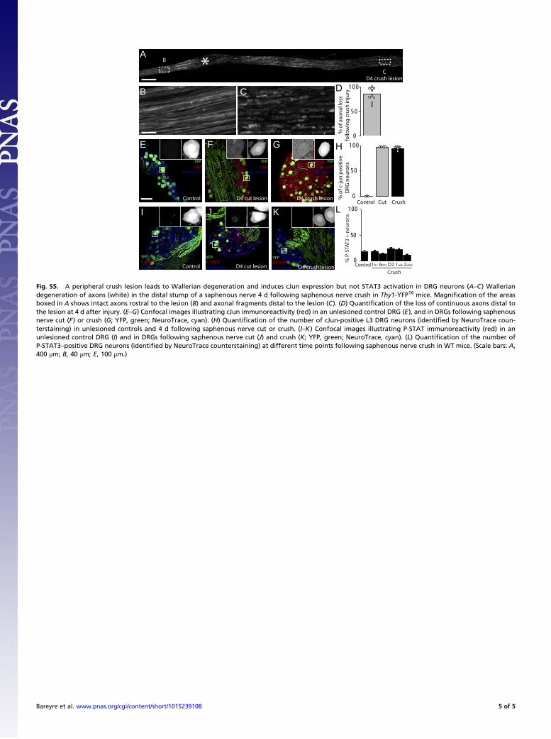
0
50
100
% o
f axo
nal
loss
fo
llow
ing
cru
sh in
juryB C
*D
0
50
100
% o
f c-j
un
po
siti
ve
DRG
neu
ron
s
H
Control Cut Crush
E G
Control D4 crush lesion
B
CD4 crush lesion
Control
Crush
0
50
100
1hr 6hrs D2 1wk 2wks
% P
-STA
T3 +
neu
rons
L
A
F
D4 cut lesion
YFP cJunNeurotrace
YFP cJunNeurotrace
YFP cJunNeurotrace
D4 cut lesion
KJ
D4 crush lesion
YFPP-STAT3Neurotrace
YFPP-STAT3Neurotrace
YFPP-STAT3Neurotrace Control
I
*
Fig. S5. A peripheral crush lesion leads to Wallerian degeneration and induces cJun expression but not STAT3 activation in DRG neurons (A–C) Walleriandegeneration of axons (white) in the distal stump of a saphenous nerve 4 d following saphenous nerve crush in Thy1-YFP16 mice. Magnification of the areasboxed in A shows intact axons rostral to the lesion (B) and axonal fragments distal to the lesion (C). (D) Quantification of the loss of continuous axons distal tothe lesion at 4 d after injury. (E–G) Confocal images illustrating cJun immunoreactivity (red) in an unlesioned control DRG (E), and in DRGs following saphenousnerve cut (F) or crush (G; YFP, green; NeuroTrace, cyan). (H) Quantification of the number of cJun-positive L3 DRG neurons (identified by NeuroTrace coun-terstaining) in unlesioned controls and 4 d following saphenous nerve cut or crush. (I–K) Confocal images illustrating P-STAT immunoreactivity (red) in anunlesioned control DRG (I) and in DRGs following saphenous nerve cut (J) and crush (K; YFP, green; NeuroTrace, cyan). (L) Quantification of the number ofP-STAT3–positive DRG neurons (identified by NeuroTrace counterstaining) at different time points following saphenous nerve crush in WT mice. (Scale bars: A,400 μm; B, 40 μm; E, 100 μm.)
Bareyre et al. www.pnas.org/cgi/content/short/1015239108 5 of 5

Chapter Three
49
STAT3 promotes corticospinal remodeling, regeneration and functional recovery after spinal cord injury
Lang C, Bradley P, Kerschensteiner M, Bareyre FM
An article submitted to Journal of Neuroscience

1
STAT3 promotes corticospinal remodeling, regeneration and
functional recovery after spinal cord injury
Claudia Lang, Peter M. Bradley, Martin Kerschensteiner* and Florence M. Bareyre*
Research Unit Therapy Development, Institute of Clinical Neuroimmunology, Ludwig-
Maximilians Universität, Munich, Germany
Correspondance should be addressed to FMB ([email protected])
or MK ([email protected])
* Authors contributed equally to this work
Running Title: STAT3 promotes corticospinal remodeling
Key words: spinal cord injury, axonal remodeling, corticospinal tract, axonal regeneration,
STAT3, pyramidotomy, viral gene transfer
Words: 3581 Figures: 5 + 2 Supplementary Figures References: 34

2
Abstract
In contrast to peripheral neurons, neurons of the central nervous system (CNS) fail
to grow after injury. As a result axonal regeneration and remodeling after CNS lesions
remain limited and functional deficits persist. The transcription factor Signal Transducer
and Activator of Transcription 3 (STAT3) has recently been identified as a key element of
axonal growth induction and its sustained expression is required for timely growth
initiation in the peripheral nervous system.
Here we use the corticospinal tract (CST) to investigate the role of STAT3 during
axon regeneration and remodeling in the CNS. We show that cortical projection neurons
only transiently overexpress STAT3 after spinal cord injury. This transient STAT3
expression is insufficient to initiate axonal outgrowth and its conditional deletion in CST
projection neurons affects neither axon regeneration nor sprouting at or remote from the
lesion. If however sustained expression and activation of STAT3 is induced in these
neurons by viral gene transfer, their axons show increased terminal and collateral sprouting
as well as regeneration after spinal cord injury. Furthermore, after a unilateral
pyramidotomy, sustained STAT3 expression and activation initiates the de novo formation
of collaterals from unlesioned CST fibers that cross the spinal midline and contact
interneurons and motoneurons that control forelimb function. Behavioral and
electrophysiological recordings indicate that these newly formed ‘midline crossing circuits’
establish ipsilateral forelimb activation and contribute to forelimb function recovery. These
findings identify intrinsic growth induction by STAT3 as a promising approach to promote
regeneration and remodeling and improve functional recovery after CNS injury.

3
Introduction
Traumatic, inflammatory or ischemic lesions to the spinal cord lead to the
transection of axonal tract systems and as a result are often followed by devastating motor
and sensory deficits (1). If the lesion of the spinal cord is complete, i.e. if the entire spinal
cord is transected, severe deficits persist. If the lesion is however incomplete, some
functional recovery can occur in rodents (2-5) as well as in humans (6,7). Over recent years
a number of studies have investigated the anatomical basis of this recovery process often
using the corticospinal tract (CST), a major descending motor tract, as a model system (2,3,
8-10). The results show that – while long-distance regeneration of transected CST fibers
fails – lesioned CST connections spontaneously attempt to remodel after injury (2,3,11).
We have previously identified sprouting of CST collaterals into the cervical cord and their
contacts to long propriospinal neurons as key components of endogenous CST remodeling
(2,12). Despite the formation of this detour circuit, however, spontaneous functional
recovery in most cases remains incomplete. To further improve functional recovery we thus
need to devise strategies that can not only enhance axonal regeneration but also improve
endogenous remodeling.
How the induction of axonal remodeling is regulated is however so far only
incompletely understood. One of the reasons is that so far many studies have focused on
the induction of axonal regeneration based on the neutralization of extrinsic growth-
inhibitory signals at the lesion site (13-16). These strategies are however less likely to
affect axonal remodeling, which often requires the formation of new collaterals remote
from the lesion site. Here, strategies that affect the intrinsic growth capability of the entire
neuron are conceptually more suited (17). We have previously identified the sustained
expression of the transcription factor STAT3 (Signal Transducer and Activator of
Transcription 3) is a key requirement for the timely induction of the intrinsic growth
4
program in DRG neurons (18). A key role for STAT3 in the induction of the intrinsic
growth program is supported by the following findings: (i) The expression and
phosphorylation of STAT3 correlates with the regenerative response of the neuron after
injury; neurons that express STAT3 start to regenerate while those that don´t generally fail
to grow (18-21). (ii) Deletion of STAT3 impairs the timely initiation of PNS regeneration
(18) and STAT3 inhibition blocks the growth-promoting effect of a conditioning lesion in
the CNS (22). (iii) STAT3 over-expression or the deletion of its inhibitor, SOCS3, can
improve sprouting of central DRG projections (18) and promote optic nerve regeneration in
vivo (23,24). Whether and how STAT 3 can initiate the remodeling of central neurons and
their projections is so far not known.
Here, we investigate how growth initiation by STAT3 affects CST regeneration and
remodeling as well as functional recovery in different spinal cord injury paradigms. We use
conditional genetics to delete endogenous STAT3 expression and viral gene transfer to
induce sustained STAT3 expression. Stereotactic tracing of hindlimb CST fibers then
allowed us to reveal the effects of STAT3 deletion or over-expression on CST regeneration
and remodeling, while behavioural testing and electrophysiological recordings were used to
assess the resulting functional recovery.
Results
Cortical projection neurons only transiently express and activate STAT3 after spinal
cord injury
To examine whether STAT is expressed and activated in lesioned cortical projection
neurons, we investigated the expression of STAT3 and its activated, phosphorylated form

5
(p-STAT3) immunohistochemically in the hindlimb motor cortices of mice perfused at
different timepoints from 6 hrs to 3 weeks following a dorsal midthoracic hemisection. In
unlesioned animals, only very few layer V pyramidal neurons in the motor cortex (that
were identified based on their typical morphology after neurotrace labeling) expressed
either p-STAT3 (Fig. 1 A, C) or STAT3 (Supplementary Fig. 1A, C). The number of
STAT3 and p-STAT3-positive cortical projection neurons was then significantly increased
at 24 hrs after lesion (Fig. 1 B, C and Supplementary Fig. 1 B, C). However, even at this
time only a subset of cortical projection neurons expressed STAT3 (Supplementary Fig. 1
C) or p-STAT (Fig. 1 C). Moreover, in these neurons STAT3 expression and
phosphorylation is only transiently induced and has returned to baseline levels at 1 week
after injury (Fig. 1 C and Supplementary Fig. 1 C).
Transient expression of STAT3 does not contribute to endogenous axonal
regeneration and remodeling after spinal cord injury
To assess to what extend the transient STAT expression supports endogenous
attempts of axonal regeneration and remodeling, we selectively deleted STAT3 expression
in cortical projection neurons. For this purpose we crossed Emx-Cre mice, which express
Cre recombinase in the forebrain (25) with STAT3fl/fl mice. As expected no STAT3 or p-
STAT3 expression is detected in cortical projection neurons of Cre-positive STAT3fl/fl mice
(Fig. 1 D-F and Supplementary Fig. 1 D-F). We then performed midthoracic dorsal
hemisections in STAT3 competent (Cre–) and conditional STAT3 deficient (Cre+) mice
and examined the effects of STAT3 deletion on CST sprouting, regeneration, remodeling
and functional recovery. Our analysis revealed no differences between STAT3 competent
and conditional STAT3 deficient mice in all parameters analyzed: Axonal sprouting and
regeneration at the lesion site was comparable between both groups both at 1 week
6
(Supplementary Fig. 1 G-K) as well as at 3 weeks after lesion (Fig. 1 G, H). Likewise, no
differences in the formation of intraspinal detour circuits were detected as similar numbers
of cervical collaterals form, which do not differ in length, complexity and number of
boutons and contact similar proportions of long propriospinal neurons (Fig. 1 I, J
Supplementary Fig. 1 L-O and data not shown). Consistent with these observations no
differences in the recovery of hindlimb locomotion assessed by BMS and rotarod tests were
detected between STAT3 competent and conditional STAT3 deficient mice
(Supplementary Fig. 2 A-C).
Sustained STAT3 expression induces corticospinal sprouting and regeneration
As the transient induction of STAT3 expression after central lesions is thus
insufficient to contribute to axonal regeneration and remodeling, we next investigated
whether the exogenous induction of sustained STAT3 expression would be sufficient to
promote axonal outgrowth. To induce sustained STAT3 expression in cortical neurons, we
injected a recombinant adeno-associated viruses (rAAV) expressing STAT3 into the
cortical hindlimb motor area (Fig. 2 A-C). Immunohistochemical analysis of STAT3
expression and phosphorylation demonstrated that transfer of rAAV-STAT3 is sufficient to
induce sustained expression and phosphorylation of STAT3 for at least 3 weeks after the
injection of the virus.
To assess the effect of sustained STAT3 expression on regeneration and remodeling
of lesioned CST fibers, we injected rAAV-STAT3 or Control rAAV into the hindlimb
motor cortex, lesioned the main dorsal and the minor dorso-lateral component of the CST
by a midthoracic hemisection and performed the following analyses: First, to assess effects
on CST regeneration we counted the number of sprouts at the lesion site as well as the
number of regenerating CST fibers that extend for up to 500 µm beyond the lesion site

7
(Fig. 2 A, E). Our analysis showed that sustained STAT3 expression is sufficient to induce
a significant increase in CST sprouting at the lesion site (Fig. 2 E-H, J). Moreover
significantly more regenerated CST fibers were detected in mice injected with rAAV
STAT3 at 200 – 400 µm from the lesion site (Fig. 2 E-I). Sustained expression of STAT3 is
thus sufficient to induce CST sprouting at and regeneration beyond the lesion site.
To assess to what extent sustained STAT3 expression can also support the
remodeling of CST fibers distant from the lesion site, we quantified the formation of
intraspinal detour circuits after midthoracic dorsal hemisections in mice injected with
rAAV-STAT3 and Control rAAV (Fig. 3 A). Indeed mice injected with rAAV-STAT3
showed an increased formation of cervical CST collaterals at 3 weeks after injury (Fig. 3
B-D). However the intraspinal targeting of these collaterals is not affected and thus a
similar proportion of long propriospinal neurons are contacted in mice injected with rAAV-
STAT3 and Control rAAV (Fig. 3 E, F). Notably, mice injected with rAAV-STAT3
displayed the formation of cervical collaterals (Fig. 3 G-J) and contacts onto long
propriospinal neurons (Fig. 3 K, L) even in the absence of a spinal lesion. This indicates
that sustained STAT3 expression can also induce the remodeling of fibers that have not
been primed to grow by their previous transection.
Sustained expression of STAT3 induces the de novo formation of midline-crossing
CST circuits after pyramidotomy
To further investigate the capability of STAT3 to recruit unlesioned fibers to the
remodeling process, we induced a unilateral lesion of the left CST at the level of the
medulla oblongata (‘unilateral pyramidotomy’, Fig. 4 A). We then assessed whether and
how unlesioned fibers from the contralateral, right forelimb portion of the CST remodel in
response to the unilateral denervation. In animals injected with Control rAAV no

8
significant increase in the number of CST fibers that exit the right CST is detected at 1, 3 or
12 weeks after pyramidotomy (Fig. 4 B, E). Further CST fibers that exit the CST rarely
crossed the spinal midline (Fig. 4 B, F). In contrast in animals injected with rAAV-STAT3
additional CST collaterals exit the CST at 3 weeks after injury (Fig. 4 C, E). These newly
formed collaterals extend towards the denervated side of the spinal cord resulting in a
significant increase in the number of midline-crossing fibers that is first detected at 3 weeks
(Fig. 4 C, F) and persist for at least 12 weeks after lesion (Fig. 4 D, F). We next examined
the projection pattern of these newly formed midline crossing CST collaterals and found
that in animals injected with rAAV-STAT3, CST collaterals extended significantly further
into the denervated spinal cord and often projected to the intermediate and ventral laminae
VI-IX of the spinal cord (Fig. 4 G, H). As the cell bodies of short propriospinal neurons
and spinal motoneurons that control forelimb movement are located in these laminae we
next assessed to what extend these neurons are targeted by midline crossing CST collaterals
at 12 weeks after pyramidotomy. Our results show that the proportions of short
propriospinal neurons and spinal motoneurons that are contacted by CST collaterals are
increased more than 4- and 20-fold, respectively, in animals injected with rAAV-STAT3
(Fig. 4 I-L).
De novo formation of midline crossing circuits improves behavioral and
electrophysiological recovery after injury
To investigate whether the newly formed midline crossing CST circuits induced by
sustained STAT3 expression can foster functional recovery we performed the following
analyses after unilateral pyramidotomy. First, we used the staircase test which measures the
capability of mice to remove sugar pellets placed on different stairs of a staircase to
evaluate skilled forelimb grasping as described in SI Materials and Methods (Fig. 5 A,).
9
While mice from both experimental groups showed similarly impaired forelimb grasping
immediately after injury, mice injected with rAAV-STAT3 recovered quicker and
performed significantly better in the staircase reaching task compared to mice injected with
Control rAAV from 5 weeks after pyramidotomy onwards (Fig. 5 B). To study the
contribution of midline-crossing circuits to this functional recovery, we recorded forelimb
flexor electromyographs (EMGs) after intracortical stimulations (Fig. 5 C, D). Unlesioned
animals elicited EMG responses in 100% of the cases (n=24 stimulations, Fig. 5 E). In the
days following pyramidotomy, this response was basically abolished (Fig. 5 E). When we
investigated the response to stimulation twelve weeks following pyramidotomy, we
observed that animals treated with the Control rAAV exhibited 53±9% while animals
treated with rAAV-STAT3 exhibited 94±4% of responsive sites in the ipsilateral cortex
(Fig. 5 E). The finding that cortical stimulation in animals injected with rAAV-STAT3
evoked an increased ipsilateral EMG response is consistent with the idea that newly formed
midline crossing CST fibers mediate this recovery. To confirm the contribution of new
CST connections below the level of the pyramids to the recovery we performed an
additional pyramidotomy of the intact side in mice that had previously recovered
responsiveness to stimulation at 12 weeks after lesion and injection with rAAV-STAT3. In
these mice the second pyramidotomy completely abolishes the response to cortical
stimulation (Fig. 5 E). Further analysis of the cortical stimulation parameters revealed
lower stimulation thresholds and shorter latencies to an EMG response in mice 12 weeks
after injection with rAAV-STAT3 compared with mice injected with Control rAAV (Fig. 5
F, G). Taken together our electrophysiological and behavioral analysis strongly suggests
that the midline crossing CST circuits induced by sustained expression of STAT3 are
functional and contribute to improved recovery of forelimb function after injury.
10
Discussion
The sustained expression of p-STAT3 is required for the timely initiation of
neuronal growth in the lesioned peripheral nervous system (18). Here, we show that in
response to a central lesion STAT3 and p-STAT3 expression are only transiently
upregulated in a subset of cortical projection neurons. This transient upregulation is
consistent with previous reports that investigated STAT3 expression in layer V cortical
projection neurons following transient cerebral ischemia in rats (26) and the expression of
JAK-STAT family members (JAK1 and STAT3 in particular) in the injured spinal cord
(19). The conditional deletion of STAT3 expression now shows that this transient
expression of STAT3 does not contribute to the induction of endogenous attempts of
axonal regeneration and remodeling. This suggests that at least in cortical projection
neurons other pathways might be responsible for endogenous induction of axonal
sprouting.
While the transient STAT3 expression induced by central lesions can thus not
influence axonal growth, we can now show that sustained expression of STAT3 induced by
viral gene transfer is sufficient to substantially improve the neuronal growth induction of
transduced cortical projection neuron and affect multiple aspects of the CST response to
injury: First, sustained STAT3 expression improves axonal sprouting and regeneration at
the lesion site. This pro-regenerative effect of STAT3 expression is consistent with
previous reports that have shown that STAT3 over-expression can induce sprouting of
central DRG projections (18) and that deletion of its negative regulator SOCS3 can
improve optic nerve regeneration (23,24). The molecular mechanisms by which STAT3
initiates this axonal growth are currently not fully understood. However, several genes that
are induced by the transcription factor STAT3 have already been identified and include the
cell cycle inhibitor P21/Cip1/Waf1 (27), the small proline rich protein 1a (SPRR1A, 28)
11
and the interferon regulatory factor 1 (IRF1; 21), all of which can directly affect neuronal
outgrowth (21,29,30). When evaluating the effects of STAT3 in regeneration, it should be
noted that axonal growth initiation by STAT3 appears to primarily increase the number of
CST axons that attempt to regenerate after a lesion. As these axons still have to grow
through the growth-inhibitory environment of the lesioned CNS (13-16,31) it is probably
not surprising that even after STAT3 induction most regenerating fibers terminate within
the first 500 µm from the lesion. By itself this pro-regenerative effect of STAT3 is thus
unlikely to contribute to functional recovery, however improved growth induction by
STAT3 can be an ideal therapeutic complement to strategies that counteract growth-
inhibitory signals in the glial scar and central myelin (13-16,31,32). In line with this
concept we could previously show synergistic effects on axonal outgrowth of central
sensory connections when we combined STAT3 induction with the neutralization of
growth-inhibitory chondroitin sulfate proteoglycans (18).
While the contribution of axonal regeneration to functional recovery is thus limited, at least
in the untreated spinal cord, the remodeling of axonal connections can promote some
recovery of function even in the absence of therapeutic support. We and others have
previously shown that the formation of intraspinal detour circuits is a key component of the
endogenous recovery process following spinal cord injury (2,3,12,33). For detour circuits
to be formed lesioned CST fibers first extend new collaterals distant from the lesion site.
These collaterals enter the intermediate layers of the cervical spinal cord and there contact
different interneurons including long propriospinal neurons. Long propriospinal neurons
act as relay neurons and increase their projections to the denervated target area of the
transected hindlimb CST thereby completing the detour circuit. We can now show that
sustained STAT3 expression can further increase the number of cervical CST collaterals
that form after lesion. However increased collateral formation does not result in significant
12
changes to the CST connections to the long propriospinal relay neurons. This might
indicate that the endogenous growth response of lesioned CST projection neurons is
sufficient to promote the formation of detour circuits at an “optimal” rate that is not
improved further by the presence of additional CST collaterals.
While lesioned CST fibers thus spontaneously reorganize after injury, unlesioned fibers
generally do not adapt in response to injury. Our results now indicate that the sustained
expression of STAT3 is sufficient to recruit unlesioned fibers to the remodeling process.
After a unilateral lesion of the CST at the level of the pyramids, sustained expression of
STAT3 induces the formation of collaterals from the unlesioned CST. These collaterals
enter the cervical gray matter, cross the spinal midline and form contacts to the previously
denervated spinal interneurons and motoneurons that control forelimb function. The
findings that neuronal growth initiation by STAT3 is sufficient to induce the de novo
formation of these midline crossing circuits indicate that the guidance signals that attract
newly formed collaterals are endogenously present in the denervated spinal cord. It is
interesting to note in this context that the remodeling of unlesioned CST fibers can not only
be induced by sustained STAT3 expression. Indeed previous studies have, for example,
shown that activation of the mTOR pathway through deletion of PTEN leads to sprouting
of uninjured CST axons and regeneration of injured CST axons past the lesion site (10) and
that the over-expression of the neuronal calcium sensor1 (NCS1), which acts via the
PI3K/Akt pathway, induces sprouting and midline crossing of unlesioned CST fibers (9).
The therapeutic potential of interventions that promote the remodeling of unlesioned
connections is illustrated by our behavioral and electrophysiological analyses that
demonstrate meaningful recovery of forelimb function in animals treated with rAAV-
STAT3 following pyramidotomy. One caveat of strategies that promote axonal remodeling
is the induction of “unwanted” new connections that could in principle augment pain or
13
spasticity after spinal cord injury. Our results from this and previous studies however
suggest that endogenous regulatory mechanisms might be in place that prevents the
formation of functionally “unwanted” connections. For example, we could previously show
that the formation of collaterals is followed by a phase of sprout sorting and refinement
during which excessive connections e.g. to short propriospinal neurons that do offer a
detour around the lesion are at least partially removed (2,33). Further, as discussed above
our current experiments provide evidence for the existence of endogenous guidance cues
that target sprouting collaterals to denervated areas of the spinal cord. Finally, the
combination of strategies that promote axonal reorganization with neurorehabilitation
approaches might further help to enhance desired and limit unwanted consequences of
nervous system remodeling (34). Thus we believe that the support of endogenous axonal
remodeling, for example by the induction of sustained expression of STAT3 or selective
manipulation of its downstream targets, is a promising therapeutic avenue that can help to
improve functional recovery in many neurological conditions in which trauma,
inflammation or ischemia cause permanent axon damage.
Materials and Methods
Animals:
To delete STAT3 expression in cortical projection neurons, we crossed STAT3fl/fl
mice, in which deletion of the STAT3 gene depends on Cre-mediated excision of loxP
sites, and EMX-Cre mice (25) in which regulatory elements of the Emx1 gene drive Cre
expression in the forebrain. Adult female mice homozygous for the floxed STAT3 allele
and either expressing Cre (STAT3-deficient group) or Cre-negative (control group) were
used for experiments. For all other experiments we used adult female C57/Bl6 mice (6-8

14
weeks old). All animal experiments were performed in accordance with regulations of the
animal welfare act and protocols approved by the Regierung von Oberbayern.
Generation and production of AAV vectors
We cloned and produced rAAV-STAT3 and Control rAAV as previously described
(25). Additional details can be found in SI Materials and Methods. Genomic titers were as
follows: rAAV-STAT3, 9 ×10¹² genome copies/ml; Control rAAV-eCFP, 9.2 × 10¹²
genome copies/ml; Control rAAV-mbYFP, 8.6 x 10¹² genome copies/ml.
Surgical procedures
We performed midthoracic hemisections and pyramidotomies and labelled CST
fibers and propriospinal neurons as described in detail in SI Materials and Methods.
Tissue processing and histological analysis
Animals were perfused transcardially with 4% paraformaldehyde (PFA). Brains and
spinal cords were dissected and postfixed overnight in PFA. The tissue was then
cryoprotected in 30% sucrose for 3 days. Coronal sections (50µm thick) were cut on a
cryostat. To visualize CST collaterals, BDA detection was performed as described in SI
Materials and Methods.
Confocal microscopy and image processing
Stained sections were scanned using an Olympus FV1000 confocal microscope
equipped with x20/0.85 and x60/1.42 oil immersion objectives. Image stacks were then
later processed using the freeware ImageJ/Fiji (http://rsbweb.nih.gov/ij) to generate
maximum intensity projections. To obtain final representations, these maximum intensity
projections were further processed in Photoshop (Adobe) using gamma adjustments to
enhance visibility of intermediate gray values.

15
Quantification of CST regeneration and remodelling
The extent of CST regeneration and remodelling was quantified as detailed in SI
Materials and Methods.
Behavioral analysis and Electrophysiology
To assess behavioural recovery we monitored the BMS score and performed Food
pellet grasping and the Rotarod tests as described in SI Materials and Methods. We
stimulated the forelimb motor cortex and performed electrophysiological recordings as
described in SI Materials and Methods.
Statistical evaluation
Data were analyzed by the Student's t test in case of comparisons of two groups or
two-way ANOVA with Tukey's post hoc test in case of multiple comparisons using
Graphpad Prism 5.01 for Windows (GraphPad Software).
Acknowledgments
We thank A. Schmalz and G. Heitmann for excellent technical assistance, D. Matzek for
help with animal husbandry and S. McMullan, Macquarie University for helpful advice on
bipolar EMG recordings. We would also like to thank S. Akira for providing the STAT3
floxed mice. Work in F.M.B’s lab is supported by the Deutsche Forschungsgemeinschaft
(SFB 870) and by the Federal Ministry of Education and Research (BMBF, Independent
Groups in the Neurosciences). Work in M.K.`s lab is supported by the DFG (Emmy-
Noether Program, SFB 571 and SFB 870), the BMBF (Competence Network Multiple
Sclerosis) and by the Verein “Therapieforschung für MS-Kranke e.V.”
16
References
1. Schwab ME, Bartholdi D. (1996) Degeneration and regeneration of axons in the lesioned spinal cord. Physiol Rev. 76: 319-70. 2. Bareyre FM et al. (2004) The injured spinal cord spontaneously forms a new intraspinal circuit in adult rats. Nat Neurosci. 7: 269-77. 3. Courtine G et al. (2008) Recovery of supraspinal control of stepping via indirect propriospinal relay connections after spinal cord injury. Nat Med. 14: 69-74. 4. Ghosh A et al. (2009) Functional and Anatomical Reorganization of the Sensory-Motor Cortex after Incomplete Spinal Cord Injury in Adult Rats. J Neurosci. 29: 12210-9. 5. Alilain WJ, Horn KP, Hu H, Dick TE, Silver J. (2011) Functional regeneration of respiratory pathways after spinal cord injury. Nature 475: 196-200. 6. Burns SP, Golding DG, Rolle Jr WA, Graziani V, Ditunno Jr JF. (1997) Recovery of ambulation in motor-incomplete tetraplegia. Arch Phys Med Rehabil. 78: 1169-72. 7. Dietz V, Wirz M, Colombo G, Curt A. (1998) Locomotor capacity and recovery of spinal cord function in paraplegic patients: a clinical and electrophysiological evaluation. Electroencephalogr Clin Neurophysiol. 109: 140-53. 8. Weidner N, Ner A, Salimi N, Tuszynski MH. (2001) Spontaneous corticospinal axonal plasticity and functional recovery after adult central nervous system injury. Proc Natl Acad Sci USA 98: 3513-3518. 9. Yip PK, Wong L.-F, Sears TA, Yáñez-Muñoz RJ, McMahon SB. (2010) Cortical Overexpression of Neuronal Calcium Sensor-1 Induces Functional Plasticity in Spinal Cord Following Unilateral Pyramidal Tract Injury in Rat. PLoS Biol. 8: e1000399 10. Liu K et al. (2010) PTEN deletion enhances the regenerative ability of adult corticospinal neurons. Nat Neurosci. 13: 1075-81.
11. Fouad K, Pedersen V, Schwab ME, Brösamle C. (2001) Cervical sprouting of corticospinal fibers after thoracic spinal cord injury accompanies shifts in evoked motor responses. Curr Biol. 11(22):1766-70.
12. Kerschensteiner M et al. (2004) Remodeling of Axonal Connections Contributes to Recovery in an Animal Model of Multiple Sclerosis. J Exp Med. 200: 1027-38. 13. Schnell L, Schwab ME. (1990) Axonal regeneration in the rat spinal cord produced by an antibody against myelin-associated neurite growth inhibitors. Nature. 343: 269-72. 14. Bradbury EJ et al. (2002) Chondroitinase ABC promotes functional recovery after spinal cord injury. Nature. 416: 636-40. 15. Cafferty WBJ, Duffy P, Huebner E, Strittmatter SM. (2010) MAG and OMgp Synergize with Nogo-A to Restrict Axonal Growth and Neurological Recovery after Spinal Cord Trauma. J. Neurosci. 30: 6825-37.
17
16. Blesch A and Tuszynski MH. (2009) Spinal cord injury: plasticity, regeneration and the challenge of translational drug development. Trans Neuosci. 32: 41-7. 17. Liu K, Tedeschi A, Park KK, He Z. (2011) Neuronal Intrinsic Mechanisms of Axon Regeneration. Annu Rev Neurosci. 34:131-152. 18. Bareyre FM et al. (2011) In vivo imaging reveals a phase-specific role of STAT3 during central and peripheral nervous system axon regeneration. Proc Natl Acad Sci USA 108: 6282-7. 19. Schwaiger FW. et al. (2000) Peripheral but not central axotomy induces changes in Janus kinases (JAK) and signal transducers and activators of transcription (STAT). Eur J Neurosci. 12: 1165-1176. 20. Xia XG, Hofmann HD, Deller T, Kirsch M. (2002) Induction of STAT3 Signaling in Activated Astrocytes and Sprouting Septal Neurons Following Entorhinal Cortex Lesion in Adult Rats. Mol Cell Neurosci. 21: 379-392. 21. Smith RP. et al. (2011) Transcriptional profiling of intrinsic PNS factors in the postnatal mouse. Mol Cell Neurosci. 46: 32-44. 22. Qiu J, Cafferty WBJ, McMahon SB, Thompson SWN. (2005) Conditioning Injury-Induced Spinal Axon Regeneration Requires Signal Transducer and Activator of Transcription 3 Activation. J Neurosci. 25: 1645-1653. 23. Smith PD. et al. (2009) SOCS3 Deletion Promotes Optic Nerve Regeneration In Vivo. Neuron. 64: 617-623. 24. Sun F. et al. (2011) Sustained axon regeneration induced by co-deletion of PTEN and SOCS3. Nature. 480: 372-5 25. Bareyre FM, Kerschensteiner M, Misgeld T, Sanes JR. (2005) Transgenic labeling of the corticospinal tract for monitoring axonal responses to spinal cord injury. Nat Med. 11: 1355-60. 26. Justicia C, Gabriel C, Planas AM. (2000) Activation of the JAK/STAT pathway following transient focal cerebral ischemia: Signaling through Jak1 and Stat3 in astrocytes. Glia. 30: 253-70. 27. Coqueret O, Gascan H. (2000) Functional Interaction of STAT3 Transcription Factor with the Cell Cycle Inhibitor p21 WAF1/CIP1/SDI1. J Biol Chem. 275: 18794-800. 28. Pradervand S et al. (2004) Small proline-rich protein 1A is a gp130 pathway- and stress-inducible cardioprotective protein. EMBO J. 23: 4517-25. 29. Bonilla IE, Tanabe K, Strittmatter SM. (2002) Small proline-rich repeat protein 1A is expressed by axotomized neurons and promotes axonal outgrowth. J Neurosci. 22:1303–1315. 30. Tanaka H et al. (2002) Cytoplasmic p21Cip1/WAF1 regulates neurite remodeling by inhibiting Rho-kinase activity. J Cell Biol. 158: 321-9. 31. Schwab ME. (2010) How hard is the CNS hardware? Nat Neurosci. 13(12):1444-6. 32. Lee JK et al. (2010) Assessing Spinal Axon Regeneration and Sprouting in Nogo-, MAG-, and OMgp-Deficient Mice. Neuron. 66: 663-70.

18
33. Lang C, Guo X, Kerschensteiner M, Bareyre FM. (2012) Single Collateral Reconstructions Reveal Distinct Phases of Corticospinal Remodeling after Spinal Cord Injury. PLoS ONE. 7: e30461. 34. Maier IC et al. (2009) Differential effects of anti-Nogo-A antibody treatment and treadmill training in rats with incomplete spinal cord injury. Brain. 132: 1426-40. Figure Legends
Figure 1. Transient upregulation of p-STAT3 expression in cortical neurons does not
contribute to endogenous CST regeneration and remodeling after injury.
(A,B) Confocal images of the expression of the activated form of STAT3, p-STAT3 in
lamina V cortical neurons (green, NeuroTrace 435; red, p-STAT3) of an unlesioned mouse
(A) and a mouse perfused 24hrs following a midthoracic hemisection (B). (C)
Quantification of p-STAT3 expression in lamina V cortical neurons of unlesioned mice
(white bar) and mice perfused at different timepoints following thoracic hemisection (grey
bars). (D,E) Confocal images of p-STAT3 expression in lamina V cortical neurons (green,
NeuroTrace 435; red, p-STAT3) of a STAT3 competent (D) and a conditional STAT3
deficient mouse (E) perfused 3wks following a midthoracic hemisection. (F) Quantification
of p-STAT3 expression in lamina V cortical neurons in STAT3 competent and conditional
STAT3 deficient mice perfused 3wks following a midthoracic hemisection. (G,H)
Quantification of axonal regeneration at different distances distal from the lesion site (G)
and of axonal sprouting (H) at the site of the lesion in STAT3 competent (grey bars) and
conditional STAT3 deficient (blue bars) mice perfused 3 wks following midthoracic
hemisection. (I,J) Quantification of axonal sprouting in the cervical spinal cord (I) and of
the percentage of long propriospinal neurons contacted by CST fibers (J) in STAT3
competent (grey bars) and conditional STAT3 deficient (blue bars) mice perfused 3 wks
19
following thoracic hemisection. Scale bars equal 50μm B (also for A), 50μm in E (also for
D).
Figure 2. Sustained STAT3 expression promotes axonal sprouting and regeneration
following spinal cord injury.
(A) Schematic representation of the analysis of CST sprouting and regeneration following
spinal cord injury. (B,C) Confocal images of p-STAT3 expression in lamina V cortical
neurons (green, NeuroTrace 435; red, p-STAT3) of mice injected with Control rAAV (B)
or rAAV-STAT3 (C) and perfused 3 wks following a midthoracic hemisection. (D)
Quantification of p-STAT3 expression in lamina V cortical neurons of mice injected with
Control rAAV (grey bars) or rAAV-STAT3 (red bars) and perfused 3wks following
midthoracic hemisection. (E-H) Confocal image of a longitudinal section of the spinal cord
(asterisk, indicates lesion site) illustrating sprouting and regeneration of the transected CST
(BDA, white) in mice injected with rAAV-STAT3 (E-G) and in mice injected with Control
rAAV (H). The dotted lines in (H) represent the distances at which regenerating CST
axons were counted. Boxed areas in (E) are magnified 2-times in (F) and (G). (I,J)
Quantification of axonal regeneration at different distances distal from the lesion site (I)
and of axonal sprouting (J) at the site of the lesion in control (grey bars) and STAT3
overexpressing (red bars) mice perfused 3 wks following midthoracic hemisection. Scale
bars equals 100μm in C (also for B). Scale bar in H (also for E) equals 60μm.
Figure 3. Sustained STAT3 expression induces sprouting of lesioned and unlesioned
fibers in the cervical spinal cord following injury.
(A) Schematic representation of the analysis of cervical CST sprouting and remodeling
following a midthoracic spinal cord injury. (B,C) Confocal images of sprouting hindlimb

20
CST collaterals in lesioned mice injected with Control rAAV (B) or rAAV-STAT3 (C) and
perfused 3 wks following injury. (D) Quantification of the number of collaterals exiting the
hindlimb CST tract in the cervical spinal cord in mice injected with Control rAAV (grey
bar) or rAAV-STAT3 (red bars) 3 wks following spinal cord injury. (E) Confocal image
(single plane) of a contact between a hindlimb CST collateral (red) and a long
propriospinal neuron (green) in a mouse injected with rAAV-STAT3 and perfused 3 wks
following the lesion. (F) Quantification of the percentage of long propriospinal neurons
contacted by hindlimb CST collaterals 3 wks following the lesion (grey bar: mice injected
with Control rAAV, red bar: mice injected with rAAV-STAT3). (G) Schematic
representation of the analysis of cervical CST sprouting and remodeling in unlesioned
mice. (H,I) Confocal images of sprouting hindlimb CST collaterals in unlesioned mice
injected with Control rAAV (H) or rAAV-STAT3 (I). (J) Quantification of the number of
collaterals exiting the hindlimb CST in the cervical spinal cord of unlesioned mice injected
with Control rAAV (grey bar) or rAAV-STAT3 (red bar). (K) Confocal image (single
plane) of a contact between a hindlimb CST collateral (red) and a long propriospinal
neuron (green) in an unlesioned mouse injected with rAAV-STAT3. (L) Quantification of
the percentage of long propriospinal neurons contacted by hindlimb CST collaterals in
unlesioned mice (grey bars: mice injected with Control rAAV, red bars: mice injected with
rAAV-STAT3)., Scale bar equals 50μm in C (also for B, H, I) , 25μm in E (K).
Figure 4. Sustained STAT3 expression induces de novo formation of midline crossing
circuits following pyramidotomy.
(A) Schematic representation of the analysis of CST remodeling after unilateral
pyramidotomy and injection of Control rAAV or a rAAV-STAT3. (B-D) Confocal images
of midline crossing fibers in mice injected with Control rAAV (B) or rAAV-STAT3 (C, D)

21
and perfused 3 wks (B, C) or 12 wks (D) following pyramidotomy. (E, F) Quantification of
the number of fibers exiting ipsilateral from the main CST (E) and crossing the spinal
midline (F) in mice injected with Control rAAV (grey bars) or rAAV-STAT3 (red bars)
and perfused 1, 3 or 12 wks following pyramidotomy. (G) Quantification of the percentage
of midline crossing fibers that project to the contralateral (denervated) laminae VI to IX in
mice injected with Control rAAV (grey bars) or rAAV-STAT3 (red bars) and perfused 3
and 12 weeks following pyramidotomy. (H) Quantification of the density of midline
crossing fibers in the contralateral (denervated) side of the spinal cord at different distances
from the midline in mice injected with Control rAAV (grey line) or rAAV-STAT3 (red
line) and perfused 12 weeks following pyramidotomy. (I, K) Confocal images (single
planes) of contacts between midline crossing forelimb CST collaterals (red) and a short
propriospinal neuron (I, green) or a motoneurons (K, green) in mice injected with rAAV-
STAT3 and perfused 12 wks following the pyramidotomy. (J, L) Quantification of the
percentage of short propriospinal neurons (J) and motoneurons (L) contacted by mideline
crossing forelimb CST collaterals in mice injected with Control rAAV (grey bars) or
rAAV-STAT3 (red bars) and perfused 12wks following injury. Scale bars equal 200 μm in
D (also for B, C), 50 μm in K (also for I).
Figure 5. Sustained STAT3 expression promotes functional recovery following
pyramidotomy.
(A) Picture of a mouse in a “staircase test” which we used to evaluate forelimb reaching
and grasping abilities in mice. (B) Quantification of the number of pellets eaten by mice
injected with Control rAAV (grey line) or rAAV-STAT3 (red line) at different test
intervals up to 10 weeks following pyramidotomy. (C) Schematic representation of the
cortical electrostimulation and EMG recording that we used to quantify circuit
22
reconnection after pyramidotomy. (D) Trace of a forelimb EMG recording after cortical
stimulation. (E) Quantification of the percentage of responsive sites contralateral to the
lesion in unlesioned mice (black bar) and ipsilateral to the lesion acutely following
pyramidotomy (white bar) and 12 wks following pyramidotomy in mice injected with
Control rAAV (grey bar) or rAAV-STAT3 (red bar). A second pyramidotomy 12 weeks
following pyramidotomy and injection of rAAV-STAT3n abolishes the ipsilateral
responses (red bar, re-lesion). (F, G) Quantification of the stimulation thresholds (F) and
latencies (G) of the forelimb responses in unlesioned mice (black bars, contralateral to
lesion) in mice injected with Control rAAV (grey bar, ipsilateral to the lesion) or rAAV-
STAT3 (red barm ipsilateral to the lesion) at 12 weeks following pyramidotomy.

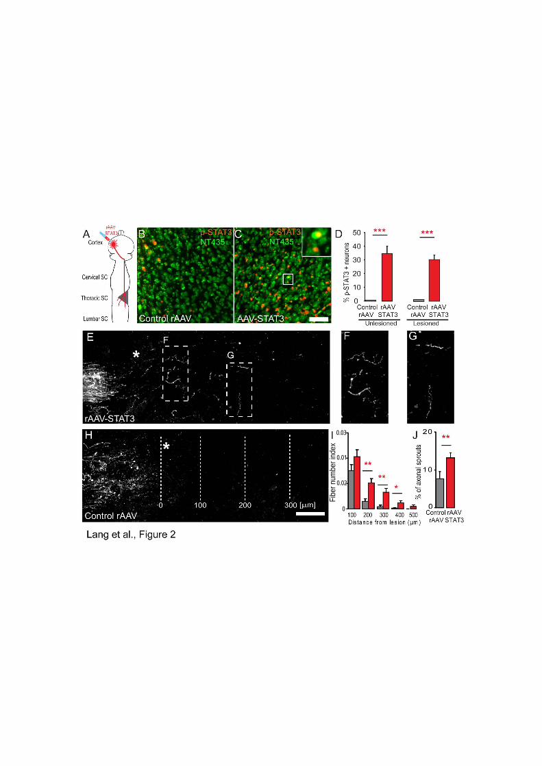
I 0.03
0
0.01
0.02
Fibe
r num
ber i
ndex


Chapter Four
77
Discussion

Discussion
78
4.1. Structural remodeling of corticospinal tract collaterals
4.1.1 Summary of key findings
Following a dorsal hemisection, the corticospinal tract (CST) sprouts newly formed
collaterals and remodels its axons rostral from the lesion site. The ability of the CST tract to
remodel its connections to form new intraspinal detour circuits is a key feature in enabling
spontaneous functional recovery following injury (Bareyre et al., 2004; Courtine et al., 2008).
The reconstruction of single CST collaterals at different time points after injury allowed us to
reveal that axonal remodeling occurs through three orchestrated phases (Figure 7; Lang et
al., 2012):
(1) A growth initiation phase, where there is growth of newly formed
collaterals that are simple in structure (10 days following injury),
(2) A collateral formation phase, where collaterals develop a complex
structure (occurs between 10 days-4 weeks after injury),
(3) A maturation phase where there is refinement and maturation of synaptic
connections onto spinal interneurons (occurs 3-12 weeks after injury).
Throughout these phases, newly formed collaterals undergo structural and synaptic
changes before reaching a mature projection pattern. Once they reach a mature pattern,
newly formed CST collaterals persist long-term for at least up to 6 months after injury. The
reconstruction of single collaterals originating from different CST components further
revealed that although the timing of the remodeling phases is similar between the main and
minor CST components, their structural complexity differed depending on their tract of origin.
In this study we were able to provide a detailed anatomical analysis and the time
course of CST remodeling following injury. This improved understanding of the
characteristics of the different phases will aid in the design and timing of therapeutic
interventions that specifically target the individual phases of the remodeling process.
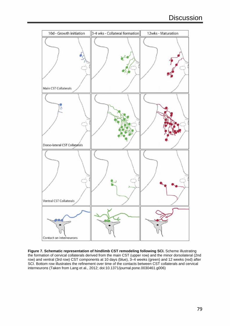
Discussion
79
Figure 7. Schematic representation of hindlimb CST remodeling following SCI. Scheme illustrating the formation of cervical collaterals derived from the main CST (upper row) and the minor dorsolateral (2nd row) and ventral (3rd row) CST components at 10 days (blue), 3–4 weeks (green) and 12 weeks (red) after SCI. Bottom row illustrates the refinement over time of the contacts between CST collaterals and cervical interneurons (Taken from Lang et al., 2012; doi:10.1371/journal.pone.0030461.g006)

Discussion
80
4.1.2 Stages of collateral remodeling
A key feature of CST remodeling that emerged from our analysis is that initially
extensive collaterals and contacts are established. However over time only some of these
contacts and collaterals are maintained while others are eliminated. This maturation and
refinement of the circuit following injury is reminiscent of similar mechanisms that occur
during development. For example, during development, in systems such as the visual
system (Sretavan and Shatz 1986), auditory system (Leake et al., 2002) and the neocortex
(Portera-Cailliau et al., 2005), there is an excess of immature axonal projections. This is then
followed by a refinement phase which eventually leads to a mature adult topographical
pattern. In the developing CST, aberrant sprouts are also formed though these are then later
pruned during maturation (Stanfield et al., 1982; Stanfield and O'Leary 1985; Chakrabarty
and Martin 2000; Li and Martin 2002), indicating that this process is a common feature
necessary for circuit formation. Molecules such as semphorins/plexins (Bagri et al., 2003;
Low et al., 2008) and ephrins (O'Leary and Wilkinson 1999) have been implicated to be
players in this refinement process. In our study we found that the injured CST undergoes a
similar process of sprouting followed by pruning, which raises the possibility that similar
molecular mechanisms may also regulate the refinement of neuronal circuits after injury.
Our work further shows that during CST remodeling that not only the number of CST
collaterals but also the CST contact pattern on interneurons evolves over time. Collaterals
formed 10 days post injury emerging from all tracts were short in length and had very few
boutons and branch points. These collaterals were immature in both structure and the
synaptic machinery present in their boutons (Lang et al., 2012). Comparative observations
from our population study revealed that at 10 days post injury, approximately 10% of newly
formed collaterals formed more than 3 contacts onto one interneuronal cell body, with
around 38% observed to form only one contact. At a late time point when a mature circuit is
established, 12 weeks post injury, 80% of collaterals were found to make one contact onto a
single neuronal cell body (Figure 7), while the remaining 20% were observed to form a
maximum of 2 contacts. A majority of the CST collaterals boutons at this time point
expressed the mature synaptic marker synapsin I (Lang et al., 2012). We considered this a
“mature” contact pattern as a comparable synaptic marker expression was observed in the
lumbar cord of unlesioned mice. Monosynaptic input appears to be the typical form of
innervation as this is reminiscent of the innervation pattern at the neuromuscular junction.
Here in this system, initially multiple axons innervate one junction. Competition between the

Discussion
81
axons then occurs and in the end only a single input remains (Balice-Gordon and Lichtman
1993; Balice-Gordon et al., 1993).
The mechanisms that determine which inputs are maintained and which are removed
during CST remodeling after injury are currently only partly understood. In the developing
nervous systems, where initial connections made by invading axons are often inaccurate,
neural activity has emerged as the deciding factor in the formation of a precise neural circuit
(Shatz 1996; Chakrabarty and Martin 2010; Kerschensteiner et al., 2009; Morgan et al.,
2011). In the case of the CST, it has been shown that inhibition of activity with an
intramuscular injection of botulinum toxin A, which causes muscle paralysis, results in the
development of immature CST axons (Martin et al., 2004). At maturity, CST axons are
known to be morphologically complex when compared to the immature CST axons seen in
development (Chakrabarty and Martin 2000; Li and Martin 2002). The complex structure of
the mature CST axons, mainly due to an increase in the number of branches, has been
noted to coincide with the establishment of the cortical motor map (Li and Martin 2002). To
produce the mature projection pattern that is required for skilled movements, incorrect CST
connections are eliminated and essential functional connections are maintained. In the CST
system, motor experiences are known to be important for the development of CST
terminations and function (Martin et al., 2004; Chakrabarty et al., 2009; Chakrabarty and
Martin 2010).
In general, synapse formation and elimination appears to be a recurrent occurrence
throughout the lifetime of an animal as even the adult system, in response to injury can
undergo cell type-specific plasticity and rearrangements. The observed parallels between
post-injury remodeling and developmental circuit formation suggest that a combination of
guidance cues and activity-dependent mechanisms governs the formation and refinement of
specific connections during corticospinal remodeling (Martin et al., 1999; Martin et al., 2000;
Li and Martin 2002). Collectively, the combination influences the permanency of a collateral,
its structure, a synapse, and synaptic strength (Colicos et al., 2001; Nikonenko et al., 2003;
De Paola et al., 2003; De Paola et al., 2006).

Discussion
82
4.1.3 Contribution of main and minor CST components
At maturation, the newly formed collaterals emerging from the different components
of the CST, the main dorsal and the minor dorsolateral and ventral components, have
distinct structures (Lang et al., 2012). Collaterals sprouting from different white matter
locations displayed differences in branching and bouton number, suggesting that their
contacts and their role in the de novo neural circuit may differ (Lang et al., 2012). Although
the signals that govern the distinct structure of CST collaterals are unknown, one
explanation is that structure is related to function. Newly sprouted collaterals emerging from
different anatomical locations would have access to distinct interneuronal pools. Dorsolateral
and main CST collaterals with their complex branching patterns are able to project into a
larger area of the spinal grey matter, mainly into the intermediate and ventral horn near the
vicinity of propriospinal interneurons and motorneurons (Steward et al., 2004). In contrast,
the projection field of the simple-in-structure ventral CST collaterals is more confined to the
region where the motorneurons are located. Given their extensive projection pattern, newly
formed main and dorsolateral CST collaterals after lesion are likely to have a more
prominent role in the formation of the detour circuit. In particular, the contribution from the
main component is more significant as this component sprouts the majority of fibers,
followed by the dorsolateral component. As a result we found that collaterals emerging from
the main and to a lesser extent from the dorsolateral CST, were responsible for the majority
of contacts onto spinal interneurons. The complex structure of dorsolateral and main CST
collaterals could be a requirement for creating strong functional connections and for the
integration of different interneuronal pools to create an operational communicative neural
circuit to enable the CST to regain control of skilled limb movement.
This raises questions about the simple ventral CST collaterals and their role in the
new neural circuit. While their simple structure might suggest an insignificant role, the
following findings suggest that ventral CST collaterals also play an important role during the
remodeling process. First, despite the fact that the ventral CST fibers are unlesioned in our
model system they sprout, relative to the tract size, a high number of new collaterals.
Second, previous work in rats has shown that the collaterals that sprout from this tract are
important for functional recovery. Weidner et al., (2001) found that when the ventral CST
component was lesioned there was no sprouting or functional recovery. Furthermore, lesion
of the ventral component (at cervical level C2) 5 weeks after a dorsal column transection (at
cervical level C3), eliminated the functional recovery that was previously observed with only

Discussion
83
a dorsal lesion. In addition it was reported that the proportion of fibers remaining in the
ventral or lateral funiculus after a T8 lesion in rats was proportional to the open field
locomotor score (Schucht et al., 2002).
4.1.4 Axonal remodeling following SCI and the implications for functional recovery and therapy
The ability of newly formed CST collaterals for spontaneous axonal remodeling is a
key mechanism that mediates functional recovery after spinal cord injury. Exploiting the
natural tendency for collaterals to form compensatory detour circuits is an attractive strategy
in terms of enhancing recovery following CNS lesions. Bareyre et al., (2004), illustrated that
following a dorsal thoracic lesion, CST collaterals remodel their connections rostral from the
lesion site. Contacts onto interneuronal pools that are not beneficial were lost while
functional contacts were strengthened. Comprehensive behavioral and electrophysiological
assessments validated that the CST remodeling, observed in this study, is a crucial step for
the recovery of CST function (Bareyre et al., 2004).
The CST is not the only tract that is known to spontaneously remodel following injury.
Studies have observed that other tract systems such as the rubrospinal and reticulospinal
tract and the serotonergic circuit respond to injury. For example, the rubrospinal tract that
cooperates with the corticospinal tract in controlling fine skilled motor movements (Whishaw
et al,. 1998) has been observed to sprout in the spinal cord following a CST transection at
the brainstem (Raineteau et al., 2001). The reorganization of reticulospinal axons and the re-
establishment of serotonergic activity can also contribute to functional recovery after injury.
The reticulospinal tract (RST) has its axons running in the lateral and ventral white matter
and is often spared in a contusion lesion (Hill et al., 2001). The RST tract, which is involved
in the initiation of walking, has been observed to sprout below the injury site (Ballermann and
Fouad 2006). Spinal motor neurons require neuromodulators such as serotonin (5-HT) for
the generation of rhythmic movement (Jordan et al., 2008; Fouad et al., 2010). Constitutive
serotonergic activity following SCI can be beneficial in terms of enabling motorneuron
recovery and hence walking. However there are detrimental effects should there be no
cortical regulation as constant expression of serotonergic activity can lead to spasms
(Murray et al., 2010).

Discussion
84
Remodeling processes are likely not limited to descending tract systems as circuit
refinement can also happen at the level of the spinal interneurons. Propriospinal
interneurons are part of the central pattern generator (CPG). The CPG is a self-sustained
spinal motor pattern able to produce neural activity that is modulated by sensory afferent and
supraspinal commands (Grillner and Wallen 1985). Courtine et al., (2008) found that
elimination of propriospinal neurons causes a loss of functional recovery after injury. Using a
staggered hemisection model they could show that when the propriospinal circuit was
destroyed all recovered movement was abolished. Additional studies show that the CPG as
a whole can spontaneously adapt and reorganize its connections following injury (Lovely et
al., 1989; Rossignol et al., 1999; de Leon et al., 1999; Edgerton et al., 2004). For example in
cats, when the supraspinal input is cut and the muscles are paralyzed, thereby depriving the
cord of sensory cues, the animals can still be trained to walk on a treadmill. Continuous
locomotor training further improves the stepping pattern. Cats with extensive treadmill
training for 12 weeks are capable of full weight support and efficient stepping (Hodgson et
al., 1994), demonstrating that the CPG circuits responds to task related plasticity (Raineteau
and Schwab 2001). However without constant training the cats have poor locomotor
performance, though with retraining the stepping ability could be relearned (Hodgson et al.,
1994). In relation to humans, functional MRI studies have revealed that the supraspinal
locomotor control between humans and quadruped animals is similar (Jahn et al., 2008; Filli
et al., 2011). CPG circuits are also known to exist in humans, and can be therapeutically
targeted to improve the recovery of SCI injured patients (Dietz et al., 1994; Barrière et al.,
2008; van Hedel and Dietz 2010).
Extensive reorganization following SCI not only occurs in the spinal cord, it
additionally transpires in the cortex. Intact areas of the cortex have been found to expand
into de-afferented regions with various methods including intracortical microstimulation
(Fouad et al., 2001; Emerick et al., 2003; Martinez et al., 2009), electrophysiology (Aguilar et
al., 2010), trans-synaptic tracing (Bareyre et al., 2004), functional magnetic resonance
imaging (fMRI) (Endo et al., 2007; Ghosh et al., 2009; Nishimura and Isa 2009; Ghosh et al.,
2010), voltage-sensitive dye imaging (VSD) (Ghosh et al., 2009; Ghosh et al., 2010) and
retrograde tracing (Ghosh et al., 2010). For example, Ghosh et al., (2010) found through
VSD and BOLDMRI imaging techniques that following a thoracic lesion there is expansion of
the forelimb sensory representation into the affected hindlimb sensorimotor area. Input from
the forelimb region could be a form of compensation or due to extensive reliance on the
forelimb after injury. Even the human brain has been reported to undergo such

Discussion
85
reorganization, utilizing these newly formed connections to regain functional recovery
(Freund et al., 2011; Lu et al., 2011).
Taken together, extensive axonal remodeling processes can be observed on multiple
anatomical levels and likely form the basis for the spontaneous improvement in motor,
sensory or other neurological function that can be observed in approximately 40% of SCI
patients (Bracken et al., 1992; Frankel 1998; Weidner et al., 2001; Freund et al., 2011).
Despite the fact that there is some spontaneous remodeling, in most cases functional
impairments still persist following CNS injures. The further support of axonal remodeling
processes is thus an attractive target for therapeutic strategies. The improved understanding
of the remodeling process can now help to guide the development of new therapeutic
strategies. Our investigations revealed that remodeling occurs in timely phases and these
phases offer windows of opportunities to influence the remodeling process. For example, to
improve growth, one would target treatment within the first 10 days following lesion.
Alternatively interventions that aim to modulate connectivity would be effective 3 weeks
following injury.

Discussion
86
4.2. Enhancing and modulating axonal outgrowth and remodeling
4.2.1 Summary of key findings
In the first study we investigated the role of STAT3 during the regeneration of
lesioned peripheral and central DRG branches (Bareyre et al., 2011). The main findings of
this study were:
• Sustained STAT3 expression is induced by a peripheral but not a central
lesion. Selective deletion of STAT3 impairs the regeneration of the peripheral
branch following injury. While viral gene transfer of STAT3 promoted axonal
outgrowth and sprouting of the DRG central branch.
• In vivo time-lapse imaging revealed that STAT3 is a phase-specific regulator
of the intrinsic growth program and that it is essential for the initiation of
axonal regeneration but does not affect axonal elongation.
In a second study we then examined whether and how the induction of STAT3
expression can be used to improve axonal regeneration, remodeling and functional recovery
after spinal cord injury (Lang et al., Chapter 3 submitted). The main findings of this study
were:
• Following spinal cord injury, STAT3 expression in cortical projection neurons
is only transiently induced and the conditional deletion of this expression
affects neither spontaneous regeneration nor remodeling of CST axons.
• Sustained expression and activation of STAT3 in cortical projection neurons
can be induced by viral gene transfer leading to improved regeneration and
remodeling of lesioned CST axons.
• The forced upregulation of STAT3 can recruit unlesioned CST fibers to the
remodeling process. In a unilateral CST lesion (pyramidotomy) paradigm,
sustained STAT3 expression triggers the formation of new collaterals that
emerge from the unlesioned tract. These collaterals were observed to cross
the spinal midline and contact interneurons and motorneurons on the
denervated side of the spinal cord (Figure 8). Moreover, behavioral and

Discussion
87
electrophysiological assessments validated that a new intraspinal circuit is
formed that contributes to the improved recovery of the impaired forelimb.
In summary, the results from these studies identify STAT3 as a key regulator of axonal
growth initiation and demonstrate that the induction of sustained STAT3 expression, for
example through viral gene transfer, is a promising strategy to enhance axonal regeneration,
remodeling and functional recovery after spinal cord injury.
Figure 8. Schematic representation of the pyramidotomy paradigm used in this experiment in conjunction with treatment using a control rAAV or a rAAV-STAT3. STAT3 overexpression induces compensatory midline sprouting of fibers from the unlesioned tract following a pyramidotomy lesion. (adapted from Lang et al., Chapter Three, manuscript submitted)

Discussion
88
4.2.2 STAT3 as an initiator of the intrinsic neuronal growth program
Peripheral and central injuries can trigger differential changes in gene expression,
hence explaining the difference in regenerative responses (Broude et al., 1997, Herdegen et
al., 1997, Herdegen and Leah 1998, Schwaiger et al., 2000, Sheu et al., 2000, Snider et al.,
2002, Kruse et al., 2011, Mason et al., 2011). Regenerative associated genes (RAGs) and
transcription factors are found to be upregulated in regenerating PNS neurons after a
peripheral nerve injury (Richardson and Verge 1987, Mason et al., 2002). STAT3 is a
regenerative associated transcription factor part of the intrinsic growth program present in a
neuron. Injury to the peripheral branch of the DRG sensory neuron results in the increased
expression of activated STAT3, even at two weeks post injury, and the activation of the
regeneration program (Neumann and Woolf 1999; Qiu et al., 2005; Bareyre et al., 2011).
The conditional deletion of STAT3 in the peripheral branch was found to only delay the
regenerative response, thereby verifying the role of this transcription factor in the intrinsic
neuronal growth program as an initiator (Bareyre et al., 2011). STAT3 was revealed to have
a significant regulatory role in the PNS following injury, as the endogenous expression and
activation of the transcription factor can influence the initiation of axonal growth.
In the CNS, the intrinsic growth machinery has been shown to be activated in CNS
neurons after axotomy (Richardson and Verge 1987; Benowitz and Routtenberg 1997; Smith
et al., 2011), and in CNS neurons that have axons regenerating through a peripheral nerve
graft (Benfey et al., 1985). However due to inhibitory factors and feedback pathways, the
effect is only transient (Sun and He 2010; Kruse et al., 2011). In upper cortical neurons,
following lesion, there is only a transient upregulation of activated STAT3 with return to
baseline levels one week following injury (Lang et al., Chapter Three submitted). This is due
to the fact that STAT3 activation is tightly regulated and controlled by SOCS3, which itself is
also upregulated after injury (Croker et al., 2008; Smith etc al., 2009). The lack of sustained
activation of factors in this program in injured mature CNS neurons is one of the main
reasons why regeneration in the PNS is successful, while in the CNS there is failure. This is
in accordance with not only our studies but with others who have found impaired growth and
regeneration when STAT3 levels were indirectly negatively affected via pharmacological
means. Inhibition of the pathway with a Janus kinase 2 (JAK2) inhibitor AG490 (Qiu et al.,
2005) or by blockade of IL6 signaling, in essence inhibiting activation of the Jak/STAT3
pathway (Zhong et al., 1999; Caffetry et al., 2001) resulted in regeneration failure of
‘conditioned’ dorsal column axons, which usually have regenerative capabilities.
Alternatively, a number of studies, ours included, have found that sustained activation of

Discussion
89
STAT3 either by genetic deletion of its inhibitor SOCS3 (Smith et al., 2009; Sun et al., 2011)
or through an rAAV-STAT3 (Bareyre et al., 2011; Lang et al., Chapter Three submitted) can
promote axonal growth and regeneration after a CNS lesion.
The upregulation of STAT3 following injury indicates that it is required for inducing
axonal growth, but its deletion will not affect the spontaneous recovery system that the
injured CST has in place in the form of spontaneous axonal remodeling and the formation of
de novo detour circuits. The presence of axonal growth in both the PNS and CNS when
STAT3 is deleted (Bareyre et al., 2011; Lang et al., Chapter Three submitted) indicates that
there are other systems able to compensate for its deletion. Therefore there are likely to be
substitute candidate factors that are able to initiate the growth response in the absence of
STAT3. Members of the PTEN/mTOR pathway (Park et al., 2010; Liu et al., 2010) are
potential candidates as they have been reported to influence the intrinsic growth response.
Another such potential candidate is the cAMP signaling pathway, which is important for
neuronal growth (McQuarrie et al., 1977; Qiu et al., 2000; Cai et al., 2001; Qiu et al., 2002).
cAMP levels have been found to be up-regulated after sciatic nerve transection and
administration of cAMP into the DRG can promote the regeneration of dorsal column axons
(Neumann et al., 2002; Qiu et al., 2002).
Additionally, the identification of STAT3 as a phase-specific regulator that only affects
growth initiation implies that there are distinct phases of axonal growth modulated by
different factors. What are the possible regulators that will switch the axon from a growing
state to an elongation state? Axonal elongation, for example, could be modulated by the
interaction with Schwann cells, which following injury align to form tubes and are able to
guide axons to their target cells (Höke 2006). Potentially, the Schwann cells are responsible
for the shift from the growth phase to the elongation phase. In a crush injury, which leaves
Schwann cell guidance intact, the absence of STAT3 has no effect on the elongation of
injured PNS axons. This suggests that once axons enter the axonal elongation phase,
STAT3 is no longer required. Nevertheless, sustained activation of pathways that enhance
and promote growth, such as STAT3, in selective neuronal populations is a promising
strategy that can further promote functional recovery. Indeed our studies (Bareyre et al.,
2011, Lang et al., Chapter Three manuscript submitted) found that direct overexpression of
STAT3 through gene therapy is a strong therapeutic strategy that can trigger further growth,
remodeling, regeneration compensatory sprouting and functional recovery following CNS
lesions.
Discussion
90
4.2.3 Integration of STAT3 into the signaling pathways that regulate neuronal growth
How STAT3 initiates the neuronal growth program is an interesting question that is so
far still only incompletely understood. However, as STAT3 is a transcription factor it is likely
that the downstream effects are mediated by induction of gene expression. A large number
of genes that are affected by STAT3 have already been identified. The small proline rich
protein 1a (SPRR1A), the cell cycle inhibitor P21/Cip1/ and the regulatory factor 1 (IRF1) are
known to be downstream targets of STAT3 (Coqueret and Gascan 200; Pradervand et al.,
2004; Smith et al., 2011). All of these molecules have also been implicated as regulators of
axonal regeneration (Bonilla et al., 2002; Tanaka et al., 2004; Smith et al., 2011). For
example, in cultured cerebellar neurons, IRF1 was found to increase neuronal outgrowth
(Smith et al., 2011). In addition to these downstream targets, STAT3 also increases the
expression of immediate early gene c-Jun. C-Jun expression has been previously identified
as a key step in the induction of axonal regeneration and mice that lack c-Jun expression in
the nervous system have impaired regeneration of motorneurons after injury (Raivich et al.,
2004).
Another important question is which factors are responsible for the activation of
STAT3 after injury. Referring to the Jak/STAT pathway, we know that the pathway is
activated by cytokines that include CNTF (Rajan et al., 1996), IL-6 (Zhong et al., 1994) and
LIF (Kunisada et al., 1996). Following injury the expression of these neuropoetic cytokines
factors are increased at the lesion site and can influence STAT3 signaling (Bourde et al.,
1996; Kurek et al., 1996). At the lesion site, the secretion of cytokines would lead to the
activation of STAT3, which is then transported retrogradely along the axon (Curtis et al.,
1994; Lee et al, 2004; Ben-Yaakov et al., 2012). Preventing expression of these classical
inducers of the Jak/STAT pathway, results in impaired axonal regeneration and functional
recovery (Zhong et al., 1994; Cafferty et al., 2001; Cafferty et al., 2004). Overall these
studies indicate that the activation of STAT3 by cytokines is a key step in activating the
intrinsic growth program and thus facilitating axonal regeneration.
Furthermore it appears that STAT3 expression can also be affected by classical
neurotrophin signaling. In particular the interaction between STAT3 and BDNF has been
reported (McAllister et al., 1999; Bramham and Messaoudi 2005; Ng et al., 2006). Knockout
of STAT3 by siRNA in hippocampal cells decreases the neurite outgrowth effect that BDNF
Discussion
91
can elicit (Bramham and Messaoudi 2005). In PC12 cells, crosstalk between the BDNF and
STAT3 signaling pathways was observed with STAT3 acting as a signal transducer for
tyrosine kinase A (TrkA). STAT3 activation is downstream from TrkA activation, with TrkA
able to phosphorylate STAT3 at Ser-727 thereby enhancing transcription (Ng et al., 2006).
In addition it is likely that other intracellular signaling pathways of the intrinsic
neuronal growth program can interact with STAT3. As mentioned previously, Liu et al.,
(2010) and Yip, et al., (2010) illustrated that through genetic manipulation that leads to the
inactivation of PTEN (Liu et al., 2010) or the overexpression of NCS1 (Yip et al., 2010), it is
possible to induce axonal growth and regeneration. These experiments identified PTEN and
NCS1 as key components of the intrinsic neuronal growth response. Inactivation of PTEN is
known to activate Akt and mTOR signaling and inhibit signaling molecules such as GSK-3
(Zhou et al., 2004; Ma and Blenis 2009; Park et al., 2010; Liu et al., 2010) and PIP3 (Zhao et
al., 2006). The Akt pathway provides a link between PTEN signaling and NCS1 as it has
been shown that overexpression of NCS1 increases the levels of phosphorylated Akt,
thereby activating the pathway (Yip et al., 2010; Figure 8). The activation of the PI3K/Akt
pathway is associated with enhanced neurite outgrowth in the DRG, perinatal cortical
neurons (Markus et al., 2002; Ozdinler and Macklis 2006) and embryonic cortical neurons
(Nakamura et al., 2006). Notably, STAT3 has also been reported to interact with the Akt
pathway via PI3K (Figure 8; Park et al., 2006; Lu et al., 2008; Blando et al., 2011). In cell
culture experiments, when STAT3 is deleted there is reduced expression of Akt (Park et al.,
2006; Lu et al., 2008). Further, in cancer cells interaction between the Jak/STAT3 and PTEN
signaling pathways has been observed (Sun and Steinberg 2002; Yang et al., 2003; Saxena
et al., 2007; Zhou et al., 2007; Blando et al., 2011). The Jak/STAT3 pathway was found to
be positively regulated by PI3K/mTOR signaling, whereas PTEN served as a negative
regulator of both Jak/STAT3 and mTOR signaling (Zhou et al., 2007). Sun et al., (2011)
found that PTEN and SOCS3 independently regulate two different pathways that can act
synergistically to promote axonal regeneration. Double deletion of the STAT3 inhibitor
pathway, SOCS3, and PTEN resulted in enhanced and sustained axonal regeneration in the
optic nerve, compared to a single deletion of either factor (Figure 8).

Discussion
92
Figure 9. Simplified scheme of selected pathways that have been identified to be important for enhancing axonal growth following injury. Overexpression of neuronal calcium sensor 1 (NCS1) increases the levels of phosphorylated Akt, a serine/threonine protein kinase. Activation of the Akt signaling pathway is known to increase neurite outgrowth. Inhibition of PTEN, which functions both as a dual specificity protein phosphatase and an inositol phospholipid, activates the Akt, and mTOR signaling pathway. PTEN is also known to negatively regulate the Jak/STAT3 signaling pathway. Deletion of both SOCS3 (an inhibitor of the Jak/STAT3 pathway) and PTEN was found to enhance axonal regeneration and growth in the optic nerve. While a single knockout of either will still result in axonal growth and regeneration following injury, the effect is not as great. Insert is of a layer 5 pyramidal cortical neuron.
4.2.4 STAT3 as a therapeutic target
Our results identify STAT3 as an attractive target for therapeutic strategies that aim
at improving functional recovery after spinal cord injury. One resulting action of sustained
STAT3 expression is that there is the induction of sprouting of lesioned CST axons at the
lesion site, as well as regeneration up to 400µm into the lesion site (Lang et al., Chapter
Three submitted). The finding that STAT3 can induce regeneration of corticospinal fibers is
important as the CST projection has a reputation of being one of the difficult axonal systems
to experimentally elicit regeneration (Blesch and Tuszynski 2009). Factors and proteins that
elicit regeneration in other tracts may not have an effect on the CST. For example, insulin-
like growth factor-1 (IGF-1) or BDNF are able to promote regeneration of raphespinal axons
or rubrospinal axons respectively but not of corticospinal axons (Kobayashi et al., 1997; Lu
et al., 2001; Jin et al., 2002; Liu et al., 2002; Kwon et al., 2002; Hollis et al., 2009). However

Discussion
93
it should be noted that although the treatment of STAT3 resulted in the regeneration of CST
fibers up to 400µm from the site of the lesion, long-distance regeneration that extends
substantially past the lesion site was not observed. This indicates that even though the
stimulation of growth can push CST neurons into a regenerative state, alone it is still not
enough to enable successful regeneration across the lesion site. The failure of successful
long-distance regeneration is due in part to the inhibitory environment formed after injury. It
is difficult to override the inhibitory environment with the presence of growth inhibitory
molecules that include chondroitin sulphate proteoglycans (CSPGs) (Fawcett and Asher
1999; Silver and Miller 2004) and myelin-associated neurite growth inhibitors (McGee and
Strittmatter 2003; Filbin 2003). In combination with the down-regulation of the CNS
regenerative response (Schwab and Bartholdi 1996; Neumann and Woolf 1999), and the
lack of trophic factor support (Widenfalk et al., 2001; Jones et al., 2001) it is not a surprise
that it is difficult for axons to fully extend past the lesion site without additional assistance.
For long-distance regeneration to be successful it is thus important to combine the induction
of the intrinsic growth program with strategies that target extrinsic growth inhibition. Such
strategies that target extrinsic inhibitors in the glial scar and myelin have been developed in
the recent years. For example, the removal or neutralization of myelin can improve axonal
regeneration. When an antibody aimed against neutralizing inhibitory factors like Nogo-A
(IN-1 antigen) is used, regenerative sprouts and long distance elongation is seen (Schnell
and Schwab 1990; Brosamle et al., 2000), as well as growth and sprouting of damaged and
intact fibers (Schnell and Schwab 1990; Liebscher et al., 2005; Freund et al., 2007; Müllner
et al., 2008; Maier et al., 2009; Gonzenbach et al., 2010). Chondroitinase ABC is a bacterial
enzyme that is often used in SCI research due to its property to digest extracellular
chondroitin sulfate proteoglycans (CSPGs) and the perineuronal nets (Carulli et al., 2010).
Following injury it has been found to enhance axonal regeneration (Zuo et al., 1998; Yick et
al., 2000; Bradbury et al., 2002) and reactivate plasticity in adult CNS by promoting sprouting
and an increase in bouton number (Pizzorusso et al., 2002; Wang et al., 2011). In theory, the
selective initiation of neuronal growth by sustained STAT3 expression is an ideal therapeutic
complement to strategies that then counteract the growth-inhibitory signals in the inhibitory
lesion environment that the regenerating axons encounters. In line with this concept we
could show synergistic effects on axonal outgrowth of central sensory connections when we
combined STAT3 induction with the neutralization of growth-inhibitory CSPGs (Bareyre et
al., 2011). Combined treatments that include STAT3 induction may thus be promising
strategies that might eventually enable successful regeneration of CST axons.
Discussion
94
At least equally important as the initiation of axon regeneration, are the effects of
sustained STAT3 expression on the remodeling of CST fibers. Our laboratory and other
groups have previously shown that lesioned CST fibers can spontaneously remodel after
injury and that the formation of detour circuits using intraspinal relay neurons is a key
mechanism that mediates recovery of CST function (Bareyre et al., 2004; Courtine et al.,
2008). We can show that sustained STAT3 expression can further increase the number of
cervical CST collaterals that form after lesion. However increased collateral formation does
not result in a significant increase of the CST connections to the long propriospinal relay
neurons. This might indicate that the endogenous growth response of lesioned CST
projection neurons is sufficient to promote the formation of detour circuits at an “optimal” rate
and that is not improved further by the presence of additional CST collaterals. In line with
this assumption, it was previously shown that the application of antibodies directed against
the growth inhibitor NogoA does not further enhance detour circuit formation (Bareyre et al.,
2004).
In contrast to lesioned fibers, unlesioned fibers mostly do not remodel after injury.
Our results now indicate that sustained STAT3 expression can also recruit unlesioned fibers
to the remodeling process. Through the use of the pyramidotomy model, we were able to
investigate the effects of STAT3 on unlesioned fibers. This model is advantageous as it
allows for a specific unilateral lesion of the CST, interrupting the direct cortical input
(Thallmair et al., 1998). Pyramidotomy is known to affect voluntary movement of the
forepaws (Steward et al., 2004; Lacroix et al., 2004), locomotion (Metz et al., 1998;
Fanardjian et al., 2001), somatosensory sensation (Thallmair et al., 1998), fine-skilled paw
reaching (Whishaw et al., 1998; Thallmair et al., 1998; Z’Graggen et al., 1998; Weidner et
al., 2001), and cause asymmetrical limb use (Thallmair et al., 1998; Z’Graggen et al., 1998;
Starkey et al., 2005). When STAT3 is overexpressed in cortical projection neurons, we found
that the unlesioned tract sprouted more collaterals and sends these collaterals across the
midline to the denervated side of the spinal cord, where they were found to contact both
SPSNs and motorneurons responsible for forelimb movement. Our results thus show that
after STAT3 overexpression the unlesioned tract can be recruited to the remodeling process
and compensate for the loss of innervation by forming a new intraspinal circuit (Figure 10)
that leads to functional recovery assessed electrophysiologically and behaviorally.
It should be noted that sustained STAT3 expression is not the only way to improve
recovery in the pyramidotomy model. For example, sprouting across the midline from the

Discussion
95
intact CST can be induced by neurotrophic factors (Zhou and Shine 2003), inhibition of the
neurite outgrowth inhibitor NOGO (Thallmair et al., 1998; Bareyre et al., 2002; Wiessner et
al., 2003), and even electrical stimulation (Brus-Ramer et al., 2007). There are also other
factors that if manipulated can also induce midline sprouting in the pyramidotomy model. Yip
et al., (2010) demonstrated that overexpression of the protein neuronal calcium sensor1
(NCS1) promotes midline sprouting and supports functional recovery. Thus, over the recent
years a number of therapeutic targets and tools have been identified that can, in principle,
improve axonal remodeling and functional recovery after spinal cord injury. STAT3 is a
particularly attractive target as it can induce axonal regeneration and remodeling of spared
fibers and due to its phase specific action it can selectively induce the initiation of axonal
outgrowth.
Figure 10. The effect of overexpression of STAT3 in layer 5 cortical neurons. (A) Overexpression of STAT3 promotes growth and regeneration of lesioned hindlimb corticospinal tract (hCST) fibers 3 weeks following a thoracic dorsal hemisection. (B) In the pyramidotomy paradigm, the overexpression of STAT3 induces compensatory sprouting and remodeling of forelimb CST (fCST) fibers from the unlesioned tract. Fibers from the intact fCST tract cross the midline into the denervated side of the cord, where they are found to contact both SPSNs and motorneurons responsible for forelimb movement. A new intraspinal circuit is formed enabling substantial functional recovery of the forelimb. (C) Image of pyramidotomy lesion performed at the level of the medulla and (D) of a mouse performing the pellet grasping test which assesses fine forelimb grasping ability. Scale bar equals 0.5mm in (C) and 5mm (D).
Discussion
96
The use of pharmacological inhibitors to manipulate STAT3 expression is not ideal as
STAT3 signaling is not limited to selective neuronal populations, thereby presenting a risk of
activating STAT3 action in other cell types. STAT3 signaling plays an essential part in many
biological processes such as regulating the immune response (Shuai and Liu 2003) and
astrogliosis (Okada et al., 2006; Herrmann et al., 2008). Additionally, sustained STAT3
expression can cause uncontrolled cell growth and is linked to diseases such as cancer
(Turkson and Jove 2000; Silver and Montell 2001; Sano et al., 2005). Therefore the use of
viral gene therapy to specifically target STAT3 to particular neuronal cell population is a
more suitable strategy. However with persistent expression the risk of tumor induction
remains, hence in the long run it will be important to develop strategies to limit the duration
of STAT3 overexpression. From our investigations, we have seen that through efficient
genetic delivery of STAT3 to specific neuronal populations it is possible to manipulate axonal
remodeling and regeneration. In terms of clinical implications, success with gene therapy
has been seen in leukemia where T-cells were genetically engineered with lentiviruses to
recognize and destroy leukemia tumor cells (Porter et al., 2011; Kalos et al., 2011).
Recombinant adeno-associated viral vectors (rAAV) are well characterized in terms of safety
profiles and are also used for gene therapy and clinical trials for neurodegenerative diseases
such as Alzheimer's disease and Parkinson (Kaspar et al., 2002; Kaplitt et al., 2007; Lim et
al., 2010). The vectors ability to infect CNS neurons and provide long term expression of the
gene of interest without causing an immune response makes them attractive tools (Papale et
al., 2009; Hutson et al., 2011; McCown 20011).
4.3. Outlook and future directions
The crucial step in the establishment of detour circuits is the formation of contacts
between cortical projection neurons and intraspinal relay neurons (Bareyre et al., 2005, Lang
et al., 2012). Newly formed dlCST and mCST collaterals display a complexity suggesting
they are capable of contacting different population of spinal relay neurons. The spinal cord is
full of excitatory and inhibitory interneuronal populations that could in principle be targeted by
the newly formed CST collaterals. From our study (Lang et al., 2012), we observed contacts
onto PSNs, an excitatory population of interneurons, and then wondered whether the
targeting of newly sprouted CST collaterals was equal on all interneuronal populations. To
address this question, we examined whether CST collaterals also formed contacts onto
glycinergic neurons. To selectively label glycinergic neurons we used the GlyT2-GFP mouse

Discussion
97
line that expresses enhanced green fluorescent protein (eGFP) in neurons under the Glycine
Transporter 2 promoter (Zeilhofer et al., 2005). Surprisingly, we found that compared to
excitatory interneurons such as long and short propriospinal neurons, only a very small
percentage of GlyT2 interneurons were contacted by newly formed CST collaterals. This
indicates that not all interneuronal populations are equally targeted by CST collaterals
(Figure 11).
This raises the question in what are the factors that determine which interneuronal
populations are contacted by growing CST collaterals. This is an important question as the
formation of functionally “meaningful” connections is key for functional recovery (Bareyre et
al., 2004) and the formation of “incorrect” connections can lead to undesired symptoms that
include neuropathic or phantom pain (Kaufmann and Moser 2000). The fact that
propriospinal interneurons are preferentially contacted following injury suggests that this
population of neurons express factors that attract the collaterals. Adhesive and repulsive
interactions have been shown to guide cell-target recognition (Tessier-Lavigne and
Goodman 1996; Sanes and Yamagata 1999; Matsuoka et al., 2011). Understanding the
molecules at play in guiding these new CST sprouts onto their target will be an important
future challenge that will help us to better understand and therapeutically enhancing post-
injury circuit formation.
Figure 11. Differential targeting of newly formed hCST collaterals following a dorsal thoracic hemisection. Newly formed hCST preferentially contact target short and long propriospinal interneurons (SPSN and LPSN), while only a small percentage of glycinergic (GlyT2) neurons were found to be contacted (insert).
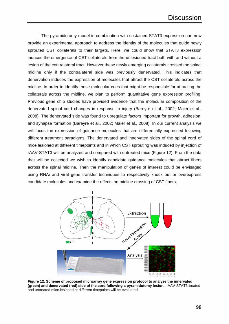
Discussion
98
The pyramidotomy model in combination with sustained STAT3 expression can now
provide an experimental approach to address the identity of the molecules that guide newly
sprouted CST collaterals to their targets. Here, we could show that STAT3 expression
induces the emergence of CST collaterals from the unlesioned tract both with and without a
lesion of the contralateral tract. However these newly emerging collaterals crossed the spinal
midline only if the contralateral side was previously denervated. This indicates that
denervation induces the expression of molecules that attract the CST collaterals across the
midline. In order to identify these molecular cues that might be responsible for attracting the
collaterals across the midline, we plan to perform quantitative gene expression profiling.
Previous gene chip studies have provided evidence that the molecular composition of the
denervated spinal cord changes in response to injury (Bareyre et al., 2002; Maier et al.,
2008). The denervated side was found to upregulate factors important for growth, adhesion,
and synapse formation (Bareyre et al., 2002; Maier et al., 2008). In our current analysis we
will focus the expression of guidance molecules that are differentially expressed following
different treatment paradigms. The denervated and innervated sides of the spinal cord of
mice lesioned at different timepoints and in which CST sprouting was induced by injection of
rAAV-STAT3 will be analyzed and compared with untreated mice (Figure 12). From the data
that will be collected we wish to identify candidate guidance molecules that attract fibers
across the spinal midline. Then the manipulation of genes of interest could be envisaged
using RNAi and viral gene transfer techniques to respectively knock out or overexpress
candidate molecules and examine the effects on midline crossing of CST fibers.
Figure 12. Scheme of proposed microarray gene expression protocol to analyze the innervated (green) and denervated (red) side of the cord following a pyramidotomy lesion. rAAV-STAT3-treated and untreated mice lesioned at different timepoints will be evaluated.

Discussion
99
4.4. Conclusions
Spontaneous remodeling after injury has been illustrated to be an important feature
that contributes to functional recovery after CNS injuries (Fouad et al., 2001; Bareyre et al.,
2004; Courtine et al., 2008). Incomplete spinal cord injuries are susceptible to such
remodeling because the cortical, subcortical and some of the local spinal cord circuitry
remains largely intact and still partially interconnected by unlesioned fibers. In SCI,
remodeling is characterized by three phases that consists of a growth initiation phase, which
is then followed by a collateral formation phase, and finally there is a refinement period. In
the process of anatomical remodeling, new circuits are established through the sprouting of
axonal branches and dendrites, and reorganization of connections. A better understanding
of this temporal sequence can help to guide future studies targeting the molecular regulation
and therapeutic support of axonal remodeling.
Despite the fact that there is spontaneous remodeling, functional impairments still
persist following CNS injures. Studies have indicated that supporting growth in the CNS is an
attractive strategy to enhance axonal remodeling (Zhou et al., 2003; Liu et al., 2010; Yip et
al., 2010). Therefore targeting the initial growth phase is an attractive and direct approach. In
our studies we have identified that the regenerative-associated transcription factor STAT3 is
a key phase-specific regulator of the initial phase and is necessary for the initiation of
regeneration in the PNS (Bareyre et al., 2011). Further investigations revealed that
sustained activation of STAT3 after CNS injuries not only enhances axonal regeneration, it
improves axonal growth and remodeling of both lesioned and unlesioned fibers, and
functional recovery. This approach to enhance axonal remodeling by stimulating growth
through the direct manipulation of the intrinsic growth program is a promising strategy for the
future. Nevertheless, from the published research it can be inferred that a successful
strategy would be expected to utilize a combination of therapies to overcome both the
intrinsic and extrinsic barriers. In particular, intrinsic growth induction can be ideally
combined with already established strategies to neutralize the extrinsic growth-inhibitory
environment of the lesioned CNS.
In the future, it is hoped that studies such as ours will lead to better understandings
of the mechanisms that underlie axonal remodeling after injury and help to devise effective
therapies to prevent the devastating consequences of spinal injuries. Recent groundbreaking

Discussion
100
work showed that improving functional recovery after spinal cord injury might not be an
unrealistic therapeutic goal. In 2011, Harkema et al. (2011) revealed that it is possible for a
human after spinal cord injury to achieve full weight-bearing standing, take steps on a
treadmill and regain other key functions. The human subject was a 23 year old man with
paraplegia from a C7–T1. A 16-electrode device was implanted in his lower back and in
combination with intensive training the patient was able to walk on a treadmill as long as his
cord was stimulated. The field of SCI research is continually evolving and over the last few
years encouraging results have been published and with them bringing hopes to long
suffering patients. The range of techniques available that include in vivo imaging, transgenic
mice, RNAi, electrophysiology, and optogenetics are further advancing the field, making it an
exciting time for scientific research and enabling us to further investigate, manipulate and
understand the complicated vast intriguing system that is the CNS network.

Bibliography
101
Bibliography Aaronson DS, Horvath CM. 2002. A Road Map for Those Who Don't Know JAK‐STAT. Science
296:1653‐5 Abdelkarim G, Gertz K, Harms C, Katchanov J, Dirnagl U, et al. 2001 Protective effects of PJ34, a
novel, potent inhibitor of poly(ADP‐ribose) polymerase (PARP) in in vitro and in vivo models of stroke. Int J Mol Med. 7:255‐60.
Agrawal G, Kerr C, Thakor NV, All AH. 2010. Characterization of Graded Multicenter Animal Spinal Cord Injury Study Contusion Spinal Cord Injury Using Somatosensory‐Evoked Potentials. Spine 35:1122‐7
Aguilar J, Humanes‐Valera D, Alonso‐Calviño E, Yague JG, Moxon KA, et al. 2010. Spinal Cord Injury Immediately Changes the State of the Brain. The Journal of Neuroscience 30:7528‐37
Akintunde A, Buxton D. 1992. Origins and collateralization of corticospinal, corticopontine, corticorubral and corticostriatal tracts: a multiple retrograde fluorescent tracing study. Brain Res. 586:208‐18
Akira S. 2000 Roles of STAT3 defined by tissue‐specific gene targeting.. Oncogene. 19:2607‐11. Alexander JK, Popovich PG. 2009. Neuroinflammation in spinal cord injury: therapeutic targets for
neuroprotection and regeneration. Progress in Brain Research, pp. 125‐37: Elsevier Alilain WJ, Horn KP, Hu H, Dick TE, Silver J. 2011. Functional regeneration of respiratory pathways
after spinal cord injury. Nature 475:196‐200 Allen AR. 1911 Surgery of experimental lesions of spinal cord equivalent to crush injury of fracture
dislocation. Preliminary report. J.Am. Med. Assoc. 57: 878‐880, 1911. Alstermark T, Pettersson L, Sasaki S. 2007. The C3‐C4 propriospinal system in the cat and monkey: a
spinal pre‐motoneuronal centre for voluntary motor control. . Acta Physiol. 189 123‐40 American Spinal Injury Association (ASIA). 2000. International Standards for Neurological
Classifications of Spinal Cord Injury. Chicago, Ill: American Spinal Injury Association;. 1‐23 pp. Bagri A, Cheng H‐J, Yaron A, Pleasure SJ, Tessier‐Lavigne M. 2003. Stereotyped Pruning of Long
Hippocampal Axon Branches Triggered by Retraction Inducers of the Semaphorin Family. Cell 113:285‐99
Balentine J. 1978 Pathology of experimental spinal cord trauma. II. Ultrastructure of axons and myelin. Lab Invest. 39:254‐66
Balice‐Gordon R, Lichtman J. 1993. In vivo observations of pre‐ and postsynaptic changes during the transition from multiple to single innervation at developing neuromuscular junctions. The Journal of Neuroscience 13:834‐55
Balice‐Gordon RJ, Chua CK, Nelson CC, Lichtman JW. 1993. Gradual loss of synaptic cartels precedes axon withdrawal at developing neuromuscular junctions. Neuron 11:801‐15
Ballermann M, Fouad K. 2006. Spontaneous locomotor recovery in spinal cord injured rats is accompanied by anatomical plasticity of reticulospinal fibers. European Journal of Neuroscience 23:1988‐96
Bamber NI, Li H, Lu X, Oudega M, Aebischer P, Xu XM. 2001. Neurotrophins BDNF and NT‐3 promote axonal re‐entry into the distal host spinal cord through Schwann cell‐seeded mini‐channels. European Journal of Neuroscience 13:257‐68
Bibliography
102
Bareyre FM, Haudenschild B, Schwab ME. 2002. Long‐Lasting Sprouting and Gene Expression Changes Induced by the Monoclonal Antibody IN‐1 in the Adult Spinal Cord. The Journal of Neuroscience 22:7097‐110
Bareyre FM, Schwab ME. 2003. Inflammation, degeneration and regeneration in the injured spinal cord: insights from DNA microarrays. Trends in Neurosciences 26:555‐63
Bareyre FM, Kerschensteiner M, Raineteau O, Mettenleiter TC, Weinmann O, Schwab ME. 2004. The injured spinal cord spontaneously forms a new intraspinal circuit in adult rats. Nat Neurosci 7:269‐77
Bareyre FM, Kerschensteiner M, Misgeld T, Sanes JR. 2005. Transgenic labeling of the corticospinal tract for monitoring axonal responses to spinal cord injury. Nat Med 11:1355‐60
Bareyre FM, Garzorz N, Lang C, Misgeld T, Büning H, Kerschensteiner M. 2011. In vivo imaging reveals a phase‐specific role of STAT3 during central and peripheral nervous system axon regeneration. Proceedings of the National Academy of Sciences 108:6282‐7
Barrière G, Leblond H, Provencher J, Rossignol S. 2008. Prominent Role of the Spinal Central Pattern Generator in the Recovery of Locomotion after Partial Spinal Cord Injuries. The Journal of Neuroscience 28:3976‐87
Barritt AW, Davies M, Marchand F, Hartley R, Grist J, et al. 2006. Chondroitinase ABC Promotes Sprouting of Intact and Injured Spinal Systems after Spinal Cord Injury. The Journal of Neuroscience 26:10856‐67
Benfey M, Bünger U, Vidal‐Sanz M, Bray G, Aguayo A. 1985. Axonal regeneration from GABAergic neurons in the adult rat thalamus. J Neurocytol. 14:279‐96.
Benowitz LI, Routtenberg A. 1997. GAP‐43: an intrinsic determinant of neuronal development and plasticity. Trends in Neurosciences 20:84‐91
Ben‐Yaakov K, Dagan SY, Segal‐Ruder Y, Shalem O, Vuppalanchi D, et al. 2012. Axonal transcription factors signal retrogradely in lesioned peripheral nerve. EMBO J 31:1350‐63
Bernstein D, Stelzner D. 1983. Plasticity of the corticospinal tract following midthoracic spinal injury in the postnatal rat. . J Comp Neurol 221:382‐400.
Bethea, J.R., and Dietrich, D.W. 2002. Targeting the host inflammatory response in traumatic spinal cord injury. Current Opinion in Neurology 15:355‐360.
Blando J, Carbajal S, Abel E, Beltran L, Conti C, et al. 2011. Cooperation between Stat3 and Akt signaling leads to prostate tumor development in transgenic mice. Neoplasia. 13:254‐65
Blesch A, Tuszynski MH. 2009. Spinal cord injury: plasticity, regeneration and the challenge of translational drug development. Trends in Neurosciences 32:41‐7
Blight A. 1983. Cellular morphology of chronic spinal cord injury in the cat: analysis of myelinated axons by line‐sampling. Neuroscience. 10:521‐43.
Blight A. 1992. Macrophages and inflammatory damage in spinal cord injury. J Neurotrauma. 9 S83‐91.
Blight A. 1993. Remyelination, revascularization, and recovery of function in experimental spinal cord injury. Adv Neurol 59:91‐104.
Blight AR, Decrescito V. 1986. Morphometric analysis of experimental spinal cord injury in the cat: The relation of injury intensity to survival of myelinated axons. Neuroscience 19:321‐41
Bonilla IE, Tanabe K, Strittmatter SM. 2002. Small Proline‐Rich Repeat Protein 1A Is Expressed by Axotomized Neurons and Promotes Axonal Outgrowth. The Journal of Neuroscience 22:1303‐15
Bibliography
103
Bourde O, Kiefer R, Toyka K, Hartung H. 1996 Quantification of interleukin‐6 mRNA in wallerian degeneration by competitive reverse transcription polymerase chain reaction. J Neuroimmunol. 69:135‐40.
Bracken MB. 2002. Steroids for acute spinal cord injury. UK: John Wiley & Sons Ltd Bracken MB, Shepard MJ, Hellenbrand KG, Collins WF, Leo LS, et al. 1985. Methylprednisolone and
neurological function 1 year after spinal cord injury. Journal of Neurosurgery 63:704‐13 Bracken MB, Shepard MJ, Collins WF, Holford TR, Baskin DS, et al. 1992. Methylprednisolone or
naloxone treatment after acute spinal cord injury: 1‐year follow‐up data. Journal of Neurosurgery 76:23‐31
Bracken MB, Shepard MJ, Holford TR, Leo‐Summers L, Aldrich EF, et al. 1997. Administration of Methylprednisolone for 24 or 48 Hours or Tirilazad Mesylate for 48 Hours in the Treatment of Acute Spinal Cord Injury. JAMA: The Journal of the American Medical Association 277:1597‐604
Bracken M, Holford T. 2002. Neurological and functional status 1 year after acute spinal cord injury: estimates of functional recovery in National Acute Spinal Cord Injury Study II from results modeled in National Acute Spinal Cord Injury Study III. J Neurosurg. 96:259‐66.
Bradbury EJ, McMahon SB. 2006. Spinal cord repair strategies: why do they work? Nat Rev Neurosci 7:644‐53
Bradbury EJ, Moon LDF, Popat RJ, King VR, Bennett GS, et al. 2002. Chondroitinase ABC promotes functional recovery after spinal cord injury. Nature 416:636‐40
Bramham CR, Messaoudi E. 2005. BDNF function in adult synaptic plasticity: The synaptic consolidation hypothesis. Progress in Neurobiology 76:99‐125
Bregman B, Kunkel‐Bagden E, McAtee M, O’Neill A. 1989. Extension of the critical period for developmental plasticity of the corticospinal pathway. . J. Comp. Neurol., 282:355‐70.
Bregman BS, Kunkel‐Bagden E, Schnell L, Dai HN, Gao D, Schwab ME. 1995. Recovery from spinal cord injury mediated by antibodies to neurite growth inhibitors. Nature 378:498‐501
Brösamle C, Schwab ME. 1997. Cells of origin, course, and termination patterns of the ventral, uncrossed component of the mature rat corticospinal tract. The Journal of Comparative Neurology 386:293‐303
Brösamle C, Huber AB, Fiedler M, Skerra A, Schwab ME. 2000. Regeneration of Lesioned Corticospinal Tract Fibers in the Adult Rat Induced by a Recombinant, Humanized IN‐1 Antibody Fragment. The Journal of Neuroscience 20:8061‐8
Broude E, McAtee M, Kelley MS, Bregman BS. 1997. c‐Jun Expression in Adult Rat Dorsal Root Ganglion Neurons: Differential Response after Central or Peripheral Axotomy. Experimental Neurology 148:367‐77
Brus‐Ramer M, Carmel JB, Chakrabarty S, Martin JH. 2007. Electrical Stimulation of Spared Corticospinal Axons Augments Connections with Ipsilateral Spinal Motor Circuits after Injury. The Journal of Neuroscience 27:13793‐801
Budh C, Osteråker A. 2007. Life satisfaction in individuals with a spinal cord injury and pain. . Clin Rehabil 21:89‐96
Bulsara, K.R., Iskandar, B.J., Villavicencio, A.T., and Skene, J.H.P. 2002. A New Millenium for Spinal Cord Regeneration: Growth‐Associated Genes. Spine 27:1946‐1949.
Bundesen LQ, Scheel TA, Bregman BS, Kromer LF. 2003. Ephrin‐B2 and EphB2 Regulation of Astrocyte‐Meningeal Fibroblast Interactions in Response to Spinal Cord Lesions in Adult Rats. The Journal of Neuroscience 23:7789‐800

Bibliography
104
Bunge R, Puckett W, Becerra J, Marcillo A, Quencer R. 1993. Observations on the pathology of human spinal cord injury. A review and classification of 22 new cases with details from a case of chronic cord compression with extensive focal demyelination. Adv Neurol 59:75‐89
Burns SP, Golding DG, Rolle Jr WA, Graziani V, Ditunno Jr JF. 1997. Recovery of ambulation in motor‐incomplete tetraplegia. Archives of physical medicine and rehabilitation 78:1169‐72
Cafferty WBJ, Gardiner NJ, Gavazzi I, Powell J, McMahon SB, et al. 2001. Leukemia Inhibitory Factor Determines the Growth Status of Injured Adult Sensory Neurons. The Journal of Neuroscience 21:7161‐70
Cafferty WBJ, Gardiner NJ, Das P, Qiu J, McMahon SB, Thompson SWN. 2004. Conditioning Injury‐Induced Spinal Axon Regeneration Fails in Interleukin‐6 Knock‐Out Mice. The Journal of Neuroscience 24:4432‐43
Cafferty WBJ, McGee AW, Strittmatter SM. 2008. Axonal growth therapeutics: regeneration or sprouting or plasticity? Trends in Neurosciences 31:215‐20
Caggiano A, Zimber M, Ganguly A, Blight A, Gruskin E. 2005. Chondroitinase ABCI improves locomotion and bladder function following contusion injury of the rat spinal cord. J Neurotrauma. 22:226‐39.
Cai D, Qiu J, Cao Z, McAtee M, Bregman BS, Filbin MT. 2001. Neuronal Cyclic AMP Controls the Developmental Loss in Ability of Axons to Regenerate. The Journal of Neuroscience 21:4731‐9
Cao Q, Xu X‐M, DeVries WH, Enzmann GU, Ping P, et al. 2005. Functional Recovery in Traumatic Spinal Cord Injury after Transplantation of Multineurotrophin‐Expressing Glial‐Restricted Precursor Cells. The Journal of Neuroscience 25:6947‐57
Carulli D, Pizzorusso T, Kwok JCF, Putignano E, Poli A, et al. 2010. Animals lacking link protein have attenuated perineuronal nets and persistent plasticity. Brain 133:2331‐47
Ceresa BP, Pessin JE. 1996. Insulin Stimulates the Serine Phosphorylation of the Signal Transducer and Activator of Transcription (STAT3) Isoform. Journal of Biological Chemistry 271:12121‐4
Chakrabarty S, Martin JH. 2000. Postnatal Development of the Motor Representation in Primary Motor Cortex. Journal of Neurophysiology 84:2582‐94
Chakrabarty S, Martin J. 2010. Postnatal development of a segmental switch enables corticospinal tract transmission to spinal forelimb motor circuits. J Neurosci. 30:2277‐88.
Chakrabarty S, Friel KM, Martin JH. 2009. Activity‐Dependent Plasticity Improves M1 Motor Representation and Corticospinal Tract Connectivity. Journal of Neurophysiology 101:1283‐93
Chen M, Huber A, van der Haar M, Frank M, Schnell L, et al. 2000. Nogo‐A is a myelin‐associated neurite outgrowth inhibitor and an antigen for monoclonal antibody IN‐1. Nature 403:434‐9.
Choi J, Kim S, Cha J, Choi Y, Sung K, et al. 2003. Upregulation of gp130 and STAT3 activation in the rat hippocampus following transient forebrain ischemia. Glia. 41:237‐46.
Chong M, Reynolds M, Irwin N, Coggeshall R, Emson P, et al. 1994. GAP‐43 expression in primary sensory neurons following central axotomy. The Journal of Neuroscience 14:4375‐84
Choo AM, Liu J, Lam CK, Dvorak M, Tetzlaff W, Oxland TR. 2007. Contusion, dislocation, and distraction: primary hemorrhage and membrane permeability in distinct mechanisms of spinal cord injury. Journal of Neurosurgery: Spine 6:255‐66
Colicos MA, Collins BE, Sailor MJ, Goda Y. 2001. Remodeling of Synaptic Actin Induced by Photoconductive Stimulation. Cell 107:605‐16
Conta A, Stelzner D. 2009. The propriospinal system: Academic Press, London. 180–90 pp.
Bibliography
105
Coqueret O, Gascan H. 2000. Functional Interaction of STAT3 Transcription Factor with the Cell Cycle Inhibitor p21 WAF1/CIP1/SDI1. Journal of Biological Chemistry 275:18794‐800
Courtine G, Song B, Roy RR, Zhong H, Herrmann JE, et al. 2008. Recovery of supraspinal control of stepping via indirect propriospinal relay connections after spinal cord injury. Nat Med 14:69‐74
Cowley K, Zaporozhets E, Schmidt B. 2010. Propriospinal transmission of the locomotor command signal in the neonatal rat. Ann. N. Y Acad. Sci., :42‐53
Cripps RA, Lee BB, Wing P, Weerts E, Mackay J, Brown D. 2011. A global map for traumatic spinal cord injury epidemiology: towards a living data repository for injury prevention. Spinal Cord 49:493‐501
Croker BA, Kiu H, Nicholson SE. 2008. SOCS regulation of the JAK/STAT signalling pathway. Developmental Biology 19:414‐22
Curtis R, Scherer SS, Somogyi R, Adryan KM, Ip NY, et al. 1994. Retrograde axonal transport of LIF is increased by peripheral nerve injury: Correlation with increased LIF expression in distal nerve. Neuron 12:191‐204
David S, Kroner A. 2011. Repertoire of microglial and macrophage responses after spinal cord injury. Nat Rev Neurosci 12:388‐99
de Leon RD, Hodgson JA, Roy RR, Edgerton VR. 1998. Locomotor Capacity Attributable to Step Training Versus Spontaneous Recovery After Spinalization in Adult Cats. Journal of Neurophysiology 79:1329‐40
De Paola V, Arber S, Caroni P. 2003. AMPA receptors regulate dynamic equilibrium of presynaptic terminals in mature hippocampal networks. Nat Neurosci. 5:491‐500.
De Paola V, Holtmaat A, Knott G, Song S, Wilbrecht L, et al. 2006. Cell Type‐Specific Structural Plasticity of Axonal Branches and Boutons in the Adult Neocortex. Neuron 49:861‐75
Demjen D, Klussmann S, Kleber S, Zuliani C, Stieltjes B, et al. 2004. Neutralization of CD95 ligand promotes regeneration and functional recovery after spinal cord injury. Nat Med 10:389‐95
Dietz V. 2002. Proprioception and locomotor disorders. Nat Rev Neurosci 3:781‐90 Dietz V, Colombo G, Jensen L. 1994. Locomotor activity in spinal man. The Lancet 344:1260‐3 Dietz V, Wirz M, Colombo G, Curt A. 1998. Locomotor capacity and recovery of spinal cord function
in paraplegic patients: a clinical and electrophysiological evaluation. Electroencephalogr Clin Neurophysiol. 109:140‐53.
Ditunno JF, Young W, Donovan WH, Creasey G. 1994. The International Standards Booklet for Neurological and Functional Classification of Spinal Cord Injury. Paraplegia 32:70‐80
Ditunno JF, Little JW, Tessler A, Burns AS. 2004. Spinal shock revisited: a four‐phase model. Spinal Cord 42:383‐95
Dottori M, Hartley L, Galea M, Paxinos G, Polizzotto M, et al. 1998. EphA4 (Sek1) receptor tyrosine kinase is required for the development of the corticospinal tract PNAS 95:13248‐53
Dusart I, Schwab ME. 1994. Secondary Cell Death and the Inflammatory Reaction After Dorsal Hemisection of the Rat Spinal Cord. European Journal of Neuroscience 6:712‐24
Edgerton V, Tillakaratne N, Bigbee A, de Leon R, Roy R, . 2004. Plasticity of the spinal neural circuitry after injury. Annu Rev Neurosci. 27:145‐67.
Edgerton V, Kim S, Ichiyama R, Gerasimenko Y, Roy R. 2006. Rehabilitative therapies after spinal cord injury. J Neurotrauma. 23:560‐70.

Bibliography
106
Emerick AJ, Neafsey EJ, Schwab ME, Kartje GL. 2003. Functional Reorganization of the Motor Cortex in Adult Rats after Cortical Lesion and Treatment with Monoclonal Antibody IN‐1. The Journal of Neuroscience 23:4826‐30
Endo T, Spenger C, Tominaga T, Brené S, Olson L. 2007. Cortical sensory map rearrangement after spinal cord injury: fMRI responses linked to Nogo signalling. Brain 130:2951‐61
Fanardjian VV, Gevorkyan OV, Mallina RK, Melik‐Moussian AB, Meliksetyan IB. 2001. Time‐dependent effects of pyramidotomy in the operantly conditioned rats. Behavioural Brain Research 122:139‐43
Fawcett JW, Asher RA. 1999. The glial scar and central nervous system repair. Brain Research Bulletin 49:377‐91
Fehlings MG, Hawryluk GWJ. 2010. Editorial. Journal of Neurosurgery: Spine 13:165‐7 Feldblum S, Arnaud S, Simon M, Rabin O, D’Arbigny P. 2000. Efficacy of a new neuroprotective agent,
gacyclidine, in a model of rat spinal cord injury. J Neurotrauma. 17:1079‐93 Ferguson IA, Koide T, Rush RA. 2001. Stimulation of corticospinal tract regeneration in the
chronically injured spinal cord. European Journal of Neuroscience 13:1059‐64 Filbin MT. 2003. Myelin‐associated inhibitors of axonal regeneration in the adult mammalian CNS.
Nat Rev Neurosci 4:703‐13 Filli L, Zörner B, Weinmann O, Schwab ME. 2011. Motor deficits and recovery in rats with unilateral
spinal cord hemisection mimic the Brown‐Séquard syndrome. Brain 134:2261‐73 Fitch MT, Silver J. 1997. Activated Macrophages and the Blood–Brain Barrier: Inflammation after CNS
Injury Leads to Increases in Putative Inhibitory Molecules. Experimental Neurology 148:587‐603
Fitch MT, Doller C, Combs CK, Landreth GE, Silver J. 1999. Cellular and Molecular Mechanisms of Glial Scarring and Progressive Cavitation: In Vivo and In VitroAnalysis of Inflammation‐Induced Secondary Injury after CNS Trauma. The Journal of Neuroscience 19:8182‐98
Fitch M, Silver J. 2001. Astrocytes are Dynamic Participants in Central Nervous System Development and Injury Responses. London: Oxford University Press. 263‐9. pp.
Fitch MT, Silver J. 2008. CNS injury, glial scars, and inflammation: Inhibitory extracellular matrices and regeneration failure. Experimental Neurology 209:294‐301
Flynn J, Graham B, Galea M, Callister R. 2011. The role of propriospinal interneurons in recovery from spinal cord injury. Neuropharmacology. 60:809‐22
Foreman R. 2000. Integration of viscerosomatic sensory input at the spinal level. Prog. Brain Res., 122:209‐21.
Fouad K, Pedersen V, Schwab ME, Brösamle C. 2001. Cervical sprouting of corticospinal fibers after thoracic spinal cord injury accompanies shifts in evoked motor responses. Current biology : CB 11:1766‐70
Fouad K, Schnell L, Bunge MB, Schwab ME, Liebscher T, Pearse DD. 2005. Combining Schwann Cell Bridges and Olfactory‐Ensheathing Glia Grafts with Chondroitinase Promotes Locomotor Recovery after Complete Transection of the Spinal Cord. The Journal of Neuroscience 25:1169‐78
Fouad K, Rank MM, Vavrek R, Murray KC, Sanelli L, Bennett DJ. 2010. Locomotion After Spinal Cord Injury Depends on Constitutive Activity in Serotonin Receptors. Journal of Neurophysiology 104:2975‐84
Fouad K, Krajacic A, Tetzlaff W. 2011. Spinal cord injury and plasticity: Opportunities and challenges. Brain Research Bulletin 84:337‐42

Bibliography
107
Fournier, A.E., and McKerracher, L. 1997. Expression of Specific Tubulin Isotypes Increases during Regeneration of Injured CNS Neurons, But Not after the Application of Brain‐Derived Neurotrophic Factor (BDNF). The Journal of Neuroscience 17:4623‐4632.
Frankel H, Coll J, Charlifue S, Whiteneck G, Gardner B, et al. 1998. Long‐term survival in spinal cord injury: a fifty year investigation. Aylesbury, Buckinghamshire, UK.: Stoke Mandeville Hospital. 266‐74. pp.
Freund P, Wannier T, Schmidlin E, Bloch J, Mir A, et al. 2007. Anti‐Nogo‐A antibody treatment enhances sprouting of corticospinal axons rostral to a unilateral cervical spinal cord lesion in adult macaque monkey. The Journal of Comparative Neurology 502:644‐59
Freund P, Weiskopf N, Ward NS, Hutton C, Gall A, et al. 2011. Disability, atrophy and cortical reorganization following spinal cord injury. Brain
Ghosh A, Sydekum E, Haiss F, Peduzzi S, Zörner B, et al. 2009. Functional and Anatomical Reorganization of the Sensory‐Motor Cortex after Incomplete Spinal Cord Injury in Adult Rats. The Journal of Neuroscience 29:12210‐9
Ghosh A, Haiss F, Sydekum E, Schneider R, Gullo M, et al. 2010. Rewiring of hindlimb corticospinal neurons after spinal cord injury. Nat Neurosci 13:97‐104
Giger RJ, Hollis ER, Tuszynski MH. 2010. Guidance Molecules in Axon Regeneration. Cold Spring Harbor Perspectives in Biology 2
Gledhill R, McDonald W. 1977. Morphological characteristics of central demyelination and remyelination: a single‐fiber study. Ann Neurol. 1:552‐60.
Gonzenbach R, Gasser P, Zorner B, Hochreutener E, Dietz V, Schwab ME. 2010. Nogo‐A antibodies and training reduce muscle spasms in spinal cord‐injured rats. . Ann. Neurol., 68:48‐57
Göritz C, Dias DO, Tomilin N, Barbacid M, Shupliakov O, Frisén J. 2011. A Pericyte Origin of Spinal Cord Scar Tissue. Science 333:238‐42
Grillner S, Wallen P. 1985. Central pattern generators for locomotion, with special reference to vertebrates. Annu Rev Neurosci 8:233‐61.
Gruner J. 1992 A monitored contusion model of spinal cord injury in the rat. Neurotrauma. 9:123‐6 Guest JD, Hiester ED, Bunge RP. 2005. Demyelination and Schwann cell responses adjacent to injury
epicenter cavities following chronic human spinal cord injury. Experimental Neurology 192:384‐93
Habas C, Kamdar N, Nguyen D, Prater K, Beckmann CF, et al. 2009. Distinct Cerebellar Contributions to Intrinsic Connectivity Networks. The Journal of Neuroscience 29:8586‐94
Hagino S, Iseki K, Mori T, Zhang Y, Hikake T, et al. 2003. Slit and glypican‐1 mRNAs are coexpressed in the reactive astrocytes of the injured adult brain. Glia 42:130‐8
Harel NY, Strittmatter SM. 2006. Can regenerating axons recapitulate developmental guidance during recovery from spinal cord injury? Nat Rev Neurosci 7:603‐16
Harkema S, Gerasimenko Y, Hodes J, Burdick J, Angeli C, et al. 2011. Effect of epidural stimulation of the lumbosacral spinal cord on voluntary movement, standing, and assisted stepping after motor complete paraplegia: a case study. The Lancet 377:1938‐47
Harrison B, McDonald W. 1977 Remyelination after transient experimental compression of the spinal cord. Ann Neurol. 1:542‐51.
Herdegen T, Leah JD. 1998. Inducible and constitutive transcription factors in the mammalian nervous system: control of gene expression by Jun, Fos and Krox, and CREB/ATF proteins. Brain Research Reviews 28:370‐490

Bibliography
108
Herdegen T, Skene P, Bähr M. 1997. The c‐Jun transcription factor – bipotential mediator of neuronal death, survival and regeneration. Trends in Neurosciences 20:227‐31
Herrmann JE, Imura T, Song B, Qi J, Ao Y, et al. 2008. STAT3 is a Critical Regulator of Astrogliosis and Scar Formation after Spinal Cord Injury. The Journal of Neuroscience 28:7231‐43
Hiersemenzel L‐P, Curt A, Dietz V. 2000. From spinal shock to spasticity. Neurology 54:1574‐82 Hill CE, Beattie MS, Bresnahan JC. 2001. Degeneration and Sprouting of Identified Descending
Supraspinal Axons after Contusive Spinal Cord Injury in the Rat. Experimental Neurology 171:153‐69
Hodgson J, Roy R, de Leon R, Dobkin B, Edgerton V. 1994. Can the mammalian lumbar spinal cord learn a motor task? Med Sci Sports Exerc. 26:1491‐7.
Höke A. 2006. Mechanisms of Disease: what factors limit the success of peripheral nerve regeneration in humans? Nat Clin Pract Neuro 2:448‐54
Hollis Ii ER, Lu P, Blesch A, Tuszynski MH. 2009. IGF‐I gene delivery promotes corticospinal neuronal survival but not regeneration after adult CNS injury. Experimental Neurology 215:53‐9
Houle JD, Tom VJ, Mayes D, Wagoner G, Phillips N, Silver J. 2006. Combining an Autologous Peripheral Nervous System "Bridge" and Matrix Modification by Chondroitinase Allows Robust, Functional Regeneration beyond a Hemisection Lesion of the Adult Rat Spinal Cord. The Journal of Neuroscience 26:7405‐15
Hughes J. 1974. Pathology of spinal cord damage in spinal injuries. .New York: Springer Hurlbert R, Hamilton M. 2008. Methylprednisolone for acute spinal cord injury: 5‐year practice
reversal. . Can J Neurol Sci 35:41‐5. Hutson T, Verhaagen J, Yáñez‐Muñoz R, Moon L. 2011 Corticospinal tract transduction: a comparison
of seven adeno‐associated viral vector serotypes and a non‐integrating lentiviral vector. Gene Ther.
International Campaign for Cures of Spinal Cord Injury Paralysis [ICCP]. http://www.campaignforcure.org/iccp/index.php?option=com_content&task=view&id=13&Itemid=28
Jahn K, Deutschländer A, Stephan T, Kalla R, Hüfner K, et al. 2008. Supraspinal locomotor control in quadrupeds and humans. In Progress in Brain Research, ed. K Christopher, RJ Leigh, pp. 353‐62: Elsevier
Jankowska E. 1992. Interneuronal relay in spinal pathways from proprioceptors. Prog. Neurobiol. 38:335‐78
Jessen KR, Richardson WD. 2001. Astrocytes are dynamic participants in central nervous system development and injury responses. London: Oxford University Press. 263–9. pp.
Jin Y, Fischer I, Tessler A, Houle JD. 2002. Transplants of Fibroblasts Genetically Modified to Express BDNF Promote Axonal Regeneration from Supraspinal Neurons Following Chronic Spinal Cord Injury. Experimental Neurology 177:265‐75
Jones LL, Oudega M, Bunge MB, Tuszynski MH. 2001. Neurotrophic factors, cellular bridges and gene therapy for spinal cord injury. The Journal of Physiology 533:83‐9
Jones LL, Margolis RU, Tuszynski MH. 2003. The chondroitin sulfate proteoglycans neurocan, brevican, phosphacan, and versican are differentially regulated following spinal cord injury. Experimental Neurology 182:399‐411
Joosten E, Schuitman R, Vermelis M, Dederen P. 1992. Postnatal development of the ipsilateral corticospinal component in rat spinal cord: a light and electron microscopic anterograde HRP study. J Comp Neurol. 326:133‐46

Bibliography
109
Jordan LM, Liu J, Hedlund PB, Akay T, Pearson KG. 2008. Descending command systems for the initiation of locomotion in mammals. Brain Research Reviews 57:183‐91
Justicia C, Gabriel C, Planas AM. 2000. Activation of the JAK/STAT pathway following transient focal cerebral ischemia: Signaling through Jak1 and Stat3 in astrocytes. Glia 30:253‐70
Kakulas B. 1984. Pathology of spinal injuries. Cent Nerv Syst Trauma. 1:117‐29. Kalb R, Strittmatter S. 2010. Neurobiology of Spinal Cord Injury. New Jersey: Humana Press Kalos M, Levine BL, Porter DL, Katz S, Grupp SA, et al. 2011. T Cells with Chimeric Antigen Receptors
Have Potent Antitumor Effects and Can Establish Memory in Patients with Advanced Leukemia. Science Translational Medicine 3:95ra73
Kapfhammer J, Schwab M. 1994 (a). Inverse patterns of myelination and GAP‐43 expression in the adult CNS: neurite growth inhibitors as regulators of neuronal plasticity? J Comp Neurol. 340:194‐206.
Kapfhammer J, Schwab M. 1994 (b). Increased expression of the growth‐associated protein GAP‐43 in the myelin‐free rat spinal cord. Eur J Neurosci. 6:403‐11.
Kaplitt MG, Feigin A, Tang C, Fitzsimons HL, Mattis P, et al. 2007. Safety and tolerability of gene therapy with an adeno‐associated virus (AAV) borne GAD gene for Parkinson's disease: an open label, phase I trial. The Lancet 369:2097‐105
Kartje‐Tillotson G, O'Donoghue DL, Dauzvardis MF, Castro AJ. 1987. Pyramidotomy abolishes the abnormal movements evoked by intracortical microstimulation in adult rats that sustained neonatal cortical lesions. Brain Research 415:172‐7
Kaufmann WE, Moser HW. 2000. Dendritic Anomalies in Disorders Associated with Mental Retardation. Cerebral Cortex 10:981‐91
Kerschensteiner D, Morgan JL, Parker ED, Lewis RM, Wong ROL. 2009. Neurotransmission selectively regulates synapse formation in parallel circuits in vivo. Nature 460:1016‐20
Kerschensteiner M, Bareyre FM, Buddeberg BS, Merkler D, Stadelmann C, et al. 2004. Remodeling of Axonal Connections Contributes to Recovery in an Animal Model of Multiple Sclerosis. The Journal of Experimental Medicine 200:1027‐38
Kerschensteiner M, Schwab ME, Lichtman JW, Misgeld T. 2005. In vivo imaging of axonal degeneration and regeneration in the injured spinal cord. Nature Medicien 11:572‐77
Kobayashi NR, Fan D‐P, Giehl KM, Bedard AM, Wiegand SJ, Tetzlaff W. 1997. BDNF and NT‐4/5 Prevent Atrophy of Rat Rubrospinal Neurons after Cervical Axotomy, Stimulate GAP‐43 and Tα1‐Tubulin mRNA Expression, and Promote Axonal Regeneration. The Journal of Neuroscience 17:9583‐95
Kostyuk P, Vasilenko D. 1979 Spinal interneurons. . Annu. Rev. Physiol. 41:115‐26 Kraus K. 1996. The pathophysiology of spinal cord injury and its clinical implications. J Neurosurg
Spine. 15:238‐51 Krause JS. 2003. Years to employment after spinal cord injury1 1 No commercial party having a
direct financial interest in the results of the research supporting this article has or will confer a benefit on the authors or any organization with which the authors are associated. Archives of physical medicine and rehabilitation 84:1282‐9
Kruse F, Bosse F, Vogelaar CF, Brazda N, K?ry P, et al. 2011. Cortical gene expression in spinal cord injury and repair: insight into the functional complexity of the neural regeneration program. Frontiers in Molecular Neuroscience 4
Kuang R, Kalil K. 1990 Specificity of corticospinal axon arbors sprouting into denervated contralateral spinal cord. J Comp Neurol. 302:461‐72.
Bibliography
110
Kühn R, Löhler J, Rennick D, Rajewsky K, Müller W. 1993. Interleukin‐10‐deficient mice develop chronic enterocolitis. Cell 75:263‐74
Kunisada K, Hirota H, Fujio Y, Matsui H, Tani Y, et al. 1996. Activation of JAK‐STAT and MAP Kinases by Leukemia Inhibitory Factor Through gp130 in Cardiac Myocytes. Circulation 94:2626‐32
Kurek JB, Austin L, Cheema SS, Bartlett PF, Murphy M. 1996. Up‐regulation of leukaemia inhibitory factor and interleukin‐6 in transected sciatic nerve and muscle following denervation. Neuromuscular Disorders 6:105‐14
Kwon BK, Liu J, Messerer C, Kobayashi NR, McGraw J, et al. 2002. Survival and regeneration of rubrospinal neurons 1 year after spinal cord injury. Proceedings of the National Academy of Sciences 99:3246‐51
Lacroix S, Havton LA, McKay H, Yang H, Brant A, et al. 2004. Bilateral corticospinal projections arise from each motor cortex in the macaque monkey: A quantitative study. The Journal of Comparative Neurology 473:147‐61
Lang C, Guo X, Kerschensteiner M, Bareyre FM. 2012. Single Collateral Reconstructions Reveal Distinct Phases of Corticospinal Remodeling after Spinal Cord Injury. PLoS ONE 7:e30461
Leake PA, Snyder RL, Hradek GT. 2002. Postnatal refinement of auditory nerve projections to the cochlear nucleus in cats. The Journal of Comparative Neurology 448:6‐27
Leal‐Filho MB. 2011. Spinal cord injury: From inflammation to glial scar. Surg Neurol Int 2::12 Lee N, Neitzel KL, Devlin BK, MacLennan AJ. 2004. STAT3 phosphorylation in injured axons before
sensory and motor neuron nuclei: Potential role for STAT3 as a retrograde signaling transcription factor. The Journal of Comparative Neurology 474:535‐45
Levy DE, Darnell JE. 2002. STATs: transcriptional control and biological impact. Nat Rev Mol Cell Biol 3:651‐62
Li Q, Martin JH. 2002. Postnatal development of connectional specificity of corticospinal terminals in the cat. The Journal of Comparative Neurology 447:57‐71
Li Y, Raisman G. 1995. Sprouts from Cut Corticospinal Axons Persist in the Presence of Astrocytic Scarring in Long‐Term Lesions of the Adult Rat Spinal Cord. Experimental Neurology 134:102‐11
Li Y, Field PM, Raisman G. 1997. Repair of Adult Rat Corticospinal Tract by Transplants of Olfactory Ensheathing Cells. Science 277:2000‐2
Li Y, Field PM, Raisman G. 1998. Regeneration of Adult Rat Corticospinal Axons Induced by Transplanted Olfactory Ensheathing Cells. The Journal of Neuroscience 18:10514‐24
Li Y, Decherchi P, Raisman G. 2003. Transplantation of Olfactory Ensheathing Cells into Spinal Cord Lesions Restores Breathing and Climbing. The Journal of Neuroscience 23:727‐31
Liebscher T, Schnell L, Schnell D, Scholl J, Schneider R, et al. 2005. Nogo‐A antibody improves regeneration and locomotion of spinal cord‐injured rats. Ann. Neurol., 58:706‐19
Lim CP, Cao X. 1999. Serine Phosphorylation and Negative Regulation of Stat3 by JNK. Journal of Biological Chemistry 274:31055‐61
Lim ST, Airavaara M, Harvey BK. 2010. Viral vectors for neurotrophic factor delivery: A gene therapy approach for neurodegenerative diseases of the CNS. Pharmacological Research 61:14‐26
Lindsay RM. 1986. Reactive gliosis. In: Astrocytes: Cell Biology and Pathology of Astrocytes. Orlando, FL: Academic. 231‐62 pp.
Liu K, Lu Y, Lee JK, Samara R, Willenberg R, et al. 2010. PTEN deletion enhances the regenerative ability of adult corticospinal neurons. Nat Neurosci 13:1075‐81

Bibliography
111
Liu K, Tedeschi A, Park KK, He Z. 2011. Neuronal Intrinsic Mechanisms of Axon Regeneration. Annu. Rev. Neurosci. 34:131‐52
Liu Y, Himes BT, Murray M, Tessler A, Fischer I. 2002. Grafts of BDNF‐Producing Fibroblasts Rescue Axotomized Rubrospinal Neurons and Prevent Their Atrophy. Experimental Neurology 178:150‐64
Livet J, Weissman TA, Kang H, Draft RW, Lu J, et al. 2007. Transgenic strategies for combinatorial expression of fluorescent proteins in the nervous system. Nature 450:56‐62
Lovely RG, Gregor RJ, Roy RR, Edgerton VR. 1989. Weight‐bearing hindlimb stepping in treadmill‐exercised adult spinal cats. Brain Research 514:206‐18
Low LK, Liu X‐B, Faulkner RL, Coble J, Cheng H‐J. 2008. Plexin signaling selectively regulates the stereotyped pruning of corticospinal axons from visual cortex. Proceedings of the National Academy of Sciences 105:8136‐41
Lu J, Liu H, Zhang M, Wang D, Cao Y, et al. 2011. Focal Pontine Lesions Provide Evidence That Intrinsic Functional Connectivity Reflects Polysynaptic Anatomical Pathways. The Journal of Neuroscience 31:15065‐71
Lu P, Blesch A, Tuszynski M. 2001 Neurotrophism without neurotropism: BDNF promotes survival but not growth of lesioned corticospinal neurons. J Comp Neurol. 436:456‐70.
Lu Y, Zhou J, Xu C, Lin H, Xiao J, et al. 2008. JAK/STAT and PI3K/AKT Pathways Form a Mutual Transactivation Loop and Afford Resistance to Oxidative Stress‐Induced Apoptosis in Cardiomyocytes. Cellular Physiology and Biochemistry 21:305‐14
Ma XM, Blenis J. 2009. Molecular mechanisms of mTOR‐mediated translational control. Nat Rev Mol Cell Biol 10:307‐18
MacGillavry HD, Stam FJ, Sassen MM, Kegel L, Hendriks WTJ, et al. 2009. NFIL3 and cAMP Response Element‐Binding Protein Form a Transcriptional Feedforward Loop that Controls Neuronal Regeneration‐Associated Gene Expression. The Journal of Neuroscience 29:15542‐50
Maier IC, Baumann K, Thallmair M, Weinmann O, Scholl J, Schwab ME. 2008. Constraint‐Induced Movement Therapy in the Adult Rat after Unilateral Corticospinal Tract Injury. The Journal of Neuroscience 28:9386‐403
Maier IC, Ichiyama RM, Courtine G, Schnell L, Lavrov I, et al. 2009. Differential effects of anti‐Nogo‐A antibody treatment and treadmill training in rats with incomplete spinal cord injury. Brain 132:1426‐40
Markandaya, M., Stein, D., and Menaker, J. 2012. Acute Treatment Options for Spinal Cord Injury. Current Treatment Options in Neurology:1‐13.
Markus A, Zhong J, Snider WD. 2002. Raf and Akt Mediate Distinct Aspects of Sensory Axon Growth. Neuron 35:65‐76
Martin JH, Lee SJ. 1999. Activity‐dependent competition between developing corticospinal terminations. NeuroReport 10:2277‐82
Martin JH, Kably B, Hacking A. 1999. Activity‐dependent development of cortical axon terminations in the spinal cord and brain stem. Experimental Brain Research 125:184‐99
Martin JH, Donarummo L, Hacking A. 2000. Impairments in Prehension Produced by Early Postnatal Sensory Motor Cortex Activity Blockade. Journal of Neurophysiology 83:895‐906
Martin JH, Choy M, Pullman S, Meng Z. 2004. Corticospinal System Development Depends on Motor Experience. The Journal of Neuroscience 24:2122‐32

Bibliography
112
Martinez M, Brezun J‐M, Zennou‐Azogui Yi, Baril N, Xerri C. 2009. Sensorimotor training promotes functional recovery and somatosensory cortical map reactivation following cervical spinal cord injury. European Journal of Neuroscience 30:2356‐67
Martirosyan NL, Feuerstein JS, Theodore N, Cavalcanti DD, Spetzler RF, Preul MC. 2011. Blood supply and vascular reactivity of the spinal cord under normal and pathological conditions. Journal of Neurosurgery: Spine 15:238‐51
Mason M, Tannemaat M, Malessy M, Verhaagen J. 2011. Gene therapy for the peripheral nervous system: a strategy to repair the injured nerve? Curr Gene Ther. 11:75‐89.
Mason MRJ, Lieberman AR, Grenningloh G, Anderson PN. 2002. Transcriptional Upregulation of SCG10 and CAP‐23 Is Correlated with Regeneration of the Axons of Peripheral and Central Neurons in Vivo. Molecular and Cellular Neuroscience 20:595‐615
Matsuoka RL, Nguyen‐Ba‐Charvet KT, Parray A, Badea TC, Chedotal A, Kolodkin AL. 2011. Transmembrane semaphorin signalling controls laminar stratification in the mammalian retina. Nature 470:259‐63
Mazzone G, Nistri A. 2011. Effect of the PARP‐1 Inhibitor PJ 34 on Excitotoxic Damage Evoked by Kainate on Rat Spinal Cord Organotypic Slices. Cellular and Molecular Neurobiology 31:469‐78
McAllister AK, Katz LC, Lo DC. 1999. Neurotrophins and synaptic plasticity. Annu. Rev. Neurosci. 22:295‐318
McCown T. 2011. Adeno‐Associated Virus (AAV) Vectors in the CNS. Curr Gene Ther. 11:181‐8. McGee AW, Strittmatter SM. 2003. The Nogo‐66 receptor: focusing myelin inhibition of axon
regeneration. Trends in Neurosciences 26:193‐8 McKeon RJ, Höke A, Silver J. 1995. Injury‐Induced Proteoglycans Inhibit the Potential for Laminin‐
Mediated Axon Growth on Astrocytic Scars. Experimental Neurology 136:32‐43 McQuarrie IG, Grafstein B, Gershon MD. 1977. Axonal regeneration in the rat sciatic nerve: Effect of
a conditioning lesion and of dbcAMP. Brain Research 132:443‐53 Mehrholz J, Kugler J, Pohl M. 2008 Locomotor training for walking after spinal cord injury. Cochrane
Database Syst Rev. 2 Menei P, Montero‐Menei C, Whittemore SR, Bunge RP, Bunge MB. 1998. Schwann cells genetically
modified to secrete human BDNF promote enhanced axonal regrowth across transected adult rat spinal cord. European Journal of Neuroscience 10:607‐21
Metz G, Dietz V, Schwab M, van de MH. 1998 The effects of unilateral pyramidal tract section on hindlimb motor performance in the rat. Behav Brain Res. 96:37‐46
Mikucki, S, and Oblinger, M. 1991. Corticospinal neurons exhibit a novel pattern of cytoskeletal gene expression after injury. J Neurosci Res. 30:213‐225.
Misgeld T, Kerschensteiner M, Bareyre FM, Burgess RW, Lichtman JW. 2007. Imaging axonal transport of mitochondria in vivo. Nat Meth 4:559‐61
Morgan Joshua L, Soto F, Wong Rachel OL, Kerschensteiner D. 2011. Development of Cell Type‐Specific Connectivity Patterns of Converging Excitatory Axons in the Retina. Neuron 71:1014‐21
Müllner A, Gonzenbach RR, Weinmann O, Schnell L, Liebscher T, Schwab ME. 2008. Lamina‐specific restoration of serotonergic projections after Nogo‐A antibody treatment of spinal cord injury in rats. European Journal of Neuroscience 27:326‐33

Bibliography
113
Murray KC, Nakae A, Stephens MJ, Rank M, D'Amico J, et al. 2010. Recovery of motoneuron and locomotor function after spinal cord injury depends on constitutive activity in 5‐HT2C receptors. Nat Med 16:694‐700
Nakamura TY, Jeromin A, Smith G, Kurushima H, Koga H, et al. 2006. Novel role of neuronal Ca2+ sensor‐1 as a survival factor up‐regulated in injured neurons. The Journal of Cell Biology 172:1081‐91
National Spinal Cord Injury Statistical Center B, Alabama. 2011. Spinal Cord Injury Facts and Figures at a Glance. Washington, DC: Office of Special Education and Rehabilitative Services, U.S. Department of Education,
Nesathurai S. 1998. Steroids and Spinal Cord Injury: Revisiting the NASCIS 2 and NASCIS 3 Trials. The Journal of Trauma 45:1088‐93
Nesic O, Xu G, McAdoo D, High K, Hulsebosch C, Perez‐Pol R. 2001 IL‐1 receptor antagonist prevents apoptosis and caspase‐3 activation after spinal cord injury. J Neurotrauma. 18:947‐56.
Neumann S, Woolf CJ. 1999. Regeneration of Dorsal Column Fibers into and beyond the Lesion Site following Adult Spinal Cord Injury. Neuron 23:83‐91
Neumann S, Bradke F, Tessier‐Lavigne M, Basbaum AI. 2002. Regeneration of Sensory Axons within the Injured Spinal Cord Induced by Intraganglionic cAMP Elevation. Neuron 34:885‐93
Ng YP, Cheung ZH, Ip NY. 2006. STAT3 as a Downstream Mediator of Trk Signaling and Functions. Journal of Biological Chemistry 281:15636‐44
Nicholson DW. 2000. From bench to clinic with apoptosis‐based therapeutic agents. Nature 407:810‐6
Nicolas C, Peineau S, Amici M, Csaba Z, Fafouri A, Javalet C, Collett VJ, Hildebrandt L., Seaton G, Choi SL, et al. 2012. The JAK/STAT Pathway Is Involved in Synaptic Plasticity. Neuron 73:374‐390.
Nicolas G, Marchand‐Pauvert V, Burke D, Pierrot‐Deseilligny E. 2001. Corticospinal excitation of presumed cervical propriospinal neurones and its reversal to inhibition in humans. The Journal of Physiology 533:903‐19
Nikonenko I, Jourdain P, Muller D. 2003. Presynaptic Remodeling Contributes to Activity‐Dependent Synaptogenesis. The Journal of Neuroscience 23:8498‐505
Nishimura Y, Isa T. 2009. Compensatory Changes at the Cerebral Cortical Level after Spinal Cord Injury. The Neuroscientist 15:436‐44
Nudo R, Masterton R. 1988. Descending pathways to the spinal cord: a comparative study of 22 mammals. J Comp Neurol. 277:53‐79
Nudo R, Masterton R. 1990. Descending pathways to the spinal cord, III: Sites of origin of the corticospinal tract. J Comp Neurol 296:559‐83
O'Leary DDM, Wilkinson DG. 1999. Eph receptors and ephrins in neural development. Current Opinion in Neurobiology 9:65‐73
Okada S, Nakamura M, Katoh H, Miyao T, Shimazaki T, et al. 2006. Conditional ablation of Stat3 or Socs3 discloses a dual role for reactive astrocytes after spinal cord injury. Nat Med 12:829‐34
Onifer S, Smith G, Fouad K. 2011. Plasticity After Spinal Cord Injury: Relevance to Recovery and Approaches to Facilitate It. Neurotherapeutics 8:283‐93
Ozdinler PH, Macklis JD. 2006. IGF‐I specifically enhances axon outgrowth of corticospinal motor neurons. Nat Neurosci 9:1371‐81
Pajoohesh‐Ganji A, Byrnes K. 2011. Novel Neuroinflammatory Targets in the Chronically Injured Spinal Cord. Neurotherapeutics 8:195‐205

Bibliography
114
Pan B, Kim E, Schramm L. 2005. Increased close appositions between corticospinal tract axons and spinal sympathetic neurons after spinal cord injury in rats. J Neurotrauma. 22:1399‐410.
Papale A, Cerovic M, Brambilla R. 2009. Viral vector approaches to modify gene expression in the brain. Journal of Neuroscience Methods 185:1‐14
Park E, Velumian A, Fehlings M. 2004 The Role of Excitotoxicity in Secondary Mechanisms of Spinal Cord Injury: A Review with an Emphasis on the Implications for White Matter Degeneration. J Neurotrauma. 21:754‐74.
Park KK, Liu K, Hu Y, Kanter JL, He Z. 2010. PTEN/mTOR and axon regeneration. Experimental Neurology 223:45‐50
Park S, Kaneko S, Kim D, Nicosia SV, Yu H, et al. 2006. Cross‐talk between Akt and Stat3 cascades. AACR Meeting Abstracts 2006:407‐c‐
Pasterkamp RJ, Anderson PN, and Verhaagen J. 2001. Peripheral nerve injury fails to induce growth of lesioned ascending dorsal column axons into spinal cord scar tissue expressing the axon repellent Semaphorin3A. European Journal of Neuroscience 13:457‐471.
Pasterkamp RJ, Verhaagen J. 2006. Semaphorins in axon regeneration: developmental guidance molecules gone wrong? Philosophical Transactions of the Royal Society B: Biological Sciences 361:1499‐511
Peng S‐P, Kügler S, Ma Z‐K, Shen Y‐Q, Schachner M. 2011. Comparison of AAV2 and AAV5 in gene transfer in the injured spinal cord of mice. NeuroReport 22:565‐9 10.1097/WNR.0b013e328348bff5
Pierrot‐Deseilligny E, D B. 2005. Propriospinal Relay for Descending Motor Commands. The Circuitry of the Human Spinal Cord, . New York: Cambridge University Press. 452‐510. pp.
Pizzorusso T, Medini P, Berardi N, Chierzi S, Fawcett JW, Maffei L. 2002. Reactivation of Ocular Dominance Plasticity in the Adult Visual Cortex. Science 298:1248‐51
Popovich PG, Guan Z, Wei P, Huitinga I, van Rooijen N, Stokes BT. 1999. Depletion of Hematogenous Macrophages Promotes Partial Hindlimb Recovery and Neuroanatomical Repair after Experimental Spinal Cord Injury. Experimental Neurology 158:351‐65
Porter DL, Levine BL, Kalos M, Bagg A, June CH. 2011. Chimeric Antigen Receptor–Modified T Cells in Chronic Lymphoid Leukemia. New England Journal of Medicine 365:725‐33
Portera‐Cailliau C, Weimer RM, De Paola V, Caroni P, Svoboda K. 2005. Diverse Modes of Axon Elaboration in the Developing Neocortex. PLoS Biol 3:e272
Pradervand S, Yasukawa H, Muller OG, Kjekshus H, Nakamura T, et al. 2004. Small proline‐rich protein 1A is a gp130 pathway‐ and stress‐inducible cardioprotective protein. EMBO J 23:4517‐25
Qiu J, Cai D, Filbin MT. 2000. Glial inhibition of nerve regeneration in the mature mammalian CNS. Glia 29:166‐74
Qiu J, Cai D, Dai H, McAtee M, Hoffman PN, et al. 2002. Spinal Axon Regeneration Induced by Elevation of Cyclic AMP. Neuron 34:895‐903
Qiu J, Cafferty WBJ, McMahon SB, Thompson SWN. 2005. Conditioning Injury‐Induced Spinal Axon Regeneration Requires Signal Transducer and Activator of Transcription 3 Activation. The Journal of Neuroscience 25:1645‐53
Raineteau O, Schwab ME. 2001. Plasticity of motor systems after incomplete spinal cord injury. Nat Rev Neurosci 2:263‐73

Bibliography
115
Raineteau O, Fouad K, Noth P, Thallmair M, Schwab ME. 2001. Functional switch between motor tracts in the presence of the mAb IN‐1 in the adult rat. Proceedings of the National Academy of Sciences 98:6929‐34
Raivich G, Bohatschek M, Da Costa C, Iwata O, Galiano M, et al. 2004. The AP‐1 Transcription Factor c‐Jun Is Required for Efficient Axonal Regeneration. Neuron 43:57‐67
Rajan P, Symes A, Fink J. 1996. STAT proteins are activated by ciliary neurotrophic factor in cells of central nervous system origin. J Neurosci Res. 43:403‐11.
Ramón‐Cueto A, Cordero MI, Santos‐Benito FF, Avila J. 2000. Functional Recovery of Paraplegic Rats and Motor Axon Regeneration in Their Spinal Cords by Olfactory Ensheathing Glia. Neuron 25:425‐35
Reier P, Houle J. 1988. The glial scar: its bearing on axonal elongation and transplantation approaches to CNS repair. In: Functional Recovery in Neurological Disease. New York: Raven. 87‐138. pp.
Rhee P, Kuncir EJ, Johnson L, Brown C, Velmahos G, et al. 2006. Cervical Spine Injury is Highly Dependent on the Mechanism of Injury Following Blunt and Penetrating Assault. The Journal of Trauma 61:1166‐70 10.097/01.ta.0000188163.52226.97
Rhodes KE, Fawcett JW. 2004. Chondroitin sulphate proteoglycans: preventing plasticity or protecting the CNS? Journal of Anatomy 204:33‐48
Richardson P, Issa V. 1984. Peripheral injury enhances central regeneration of primary sensory neurones. Nature. 309:791‐3.
Richardson PM, Verge VMK. 1987. Axonal regeneration in dorsal spinal roots is accelerated by peripheral axonal transection. Brain Research 411:406‐8
Rosenzweig ES, Courtine G, Jindrich DL, Brock JH, Ferguson AR, et al. 2010. Extensive spontaneous plasticity of corticospinal projections after primate spinal cord injury. Nat Neurosci 13:1505‐10
Rossignol S, Drew T, Brustein E, Jiang W. 1999. Locomotor performance and adaptation after partial or complete spinal cord lesions in the cat. Prog Brain Res. 123:349‐65.
Sanes JR, Yamagata M. 1999. Formation of lamina‐specific synaptic connections. Current Opinion in Neurobiology 9:79‐87
Sano S, Chan KS, Carbajal S, Clifford J, Peavey M, et al. 2005. Stat3 links activated keratinocytes and immunocytes required for development of psoriasis in a novel transgenic mouse model. Nat Med 11:43‐9
Saxena NK, Sharma D, Ding X, Lin S, Marra F, et al. 2007. Concomitant Activation of the JAK/STAT, PI3K/AKT, and ERK Signaling Is Involved in Leptin‐Mediated Promotion of Invasion and Migration of Hepatocellular Carcinoma Cells. Cancer Research 67:2497‐507
Sayer FT, Kronvall E, Nilsson OG. 2006. Methylprednisolone treatment in acute spinal cord injury: the myth challenged through a structured analysis of published literature. The Spine Journal 6:335‐43
Schnell L, Schwab ME. 1990. Axonal regeneration in the rat spinal cord produced by an antibody against myelin‐associated neurite growth inhibitors. Nature 343:269‐72
Schnell L, Schneider R, Kolbeck R, Barde Y‐A, Schwab ME. 1994. Neurotrophin‐3 enhances sprouting of corticospinal tract during development and after adult spinal cord lesion. Nature 367:170‐3

Bibliography
116
Schnell L, Fearn S, Klassen H, Schwab ME, Perry VH. 1999. Acute inflammatory responses to mechanical lesions in the CNS: differences between brain and spinal cord. European Journal of Neuroscience 11:3648‐58
Schnell L, Hunanyan AS, Bowers WJ, Horner PJ, Federoff HJ, et al. 2011. Combined delivery of Nogo‐A antibody, neurotrophin‐3 and the NMDA‐NR2d subunit establishes a functional ‘detour’ in the hemisected spinal cord. European Journal of Neuroscience 34:1256‐67
Schucht P, Raineteau O, Schwab ME, Fouad K. 2002. Anatomical Correlates of Locomotor Recovery Following Dorsal and Ventral Lesions of the Rat Spinal Cord. Experimental Neurology 176:143‐53
Schwab ME, Bartholdi D. 1996. Degeneration and regeneration of axons in the lesioned spinal cord. Physiological Reviews 76:319‐70
Schwaiger FW, Schmitt GHB, Horvat A, Hager G, Streif R, et al. 2000. Peripheral but not central axotomy induces changes in Janus kinases (JAK) and signal transducers and activators of transcription (STAT). European Journal of Neuroscience 12:1165‐76
Schwartz ED, Flanders AE. 2006. Spinal Trauma: Imaging, Diagnosis, and Management Schweizer U, Gunnersen J, Karch C, Wiese S, Holtmann B, et al. 2002. Conditional gene ablation of
Stat3 reveals differential signaling requirements for survival of motoneurons during development and after nerve injury in the adult. The Journal of Cell Biology 156:287‐98
Sekhon LHS, Fehlings MG. 2001. Epidemiology, Demographics, and Pathophysiology of Acute Spinal Cord Injury. Spine 26:S2‐S12
Serafini T, Colamarino SA, Leonardo ED, Wang H, Beddington R, et al. 1996. Netrin‐1 Is Required for Commissural Axon Guidance in the Developing Vertebrate Nervous System. Cell 87:1001‐14
Shatz C, Sretavan D. 1986. Interactions between retinal ganglion cells during the development of the mammalian visual system. Annu Rev Neurosci. 9:171‐207.
Shatz CJ. 1996. Emergence of order in visual system development. Proceedings of the National Academy of Sciences 93:602‐8
Sherrington C, Laslett E. 1902. Note upon descending intrinsic spinal tracts in the mammalian cord. Proc. R Soc. Lond, 71:115‐21
Sherrington C, Laslett E. 1903. Observations on some spinal reflexes and the interconnection of spinal segments. . J. Physiol.:58‐96
Sheu JY, Kulhanek DJ, Eckenstein FP. 2000. Differential Patterns of ERK and STAT3 Phosphorylation after Sciatic Nerve Transection in the Rat. Experimental Neurology 166:392‐402
Shuai K, Liu B. 2003. Regulation of JAK‐STAT signalling in the immune system. Nat Rev Immunol 3:900‐11
Siebert J, Middleton F, Stelzner D. 2010. Long descending cervical propriospinal neurons differ from thoracic propriospinal neurons in response to low thoracic spinal injury. BMC Neurosci. 11:148.
Silver DL, Montell DJ. 2001. Paracrine Signaling through the JAK/STAT Pathway Activates Invasive Behavior of Ovarian Epithelial Cells in Drosophila. Cell 107:831‐41
Silver J, Miller JH. 2004. Regeneration beyond the glial scar. Nat Rev Neurosci 5:146‐56 Sjöberg J, Kanje M. 1990. The initial period of peripheral nerve regeneration and the importance of
the local environment for the conditioning lesion effect. Brain Research 529:79‐84 Smith PD, Sun F, Park KK, Cai B, Wang C, et al. 2009. SOCS3 Deletion Promotes Optic Nerve
Regeneration In Vivo. Neuron 64:617‐23

Bibliography
117
Smith RP, Lerch‐Haner JK, Pardinas JR, Buchser WJ, Bixby JL, Lemmon VP. 2011. Transcriptional profiling of intrinsic PNS factors in the postnatal mouse. Molecular and Cellular Neuroscience 46:32‐44
Snider WD, Zhou F‐Q, Zhong J, Markus A. 2002. Signaling the Pathway to Regeneration. Neuron 35:13‐6
Spinal injury network. http://www.spinal‐injury.net/incomplete‐spinal‐cord‐injury.htmStam FJ, MacGillavry HD, Armstrong NJ, De Gunst MCM, Zhang Y, et al. 2007. Identification of
candidate transcriptional modulators involved in successful regeneration after nerve injury. European Journal of Neuroscience 25:3629‐37
Stanfield B, O'Leary D. 1985. Fetal occipital cortical neurones transplanted to the rostral cortex can extend and maintain a pyramidal tract axon. Nature. 18:135‐7.
Stanfield B, O'Leary D, Fricks C. 1982. Selective collateral elimination in early postnatal development restricts cortical distribution of rat pyramidal tract neurones. Nature. 298:371‐3.
Starkey ML, Barritt AW, Yip PK, Davies M, Hamers FPT, et al. 2005. Assessing behavioural function following a pyramidotomy lesion of the corticospinal tract in adult mice. Experimental Neurology 195:524‐39
Starkey ML, Schwab ME. 2011. Anti‐Nogo‐A and training: Can one plus one equal three? Experimental Neurology
Steeves J, Keirstead H, Ethell D, Hasan S, Muir G, et al. 1994. Permissive and restrictive periods for brainstem‐spinal regeneration in the chick. Prog Brain Res. 103:243‐62.
Stepanyants A, Hof PR, Chklovskii DB. 2002. Geometry and Structural Plasticity of Synaptic Connectivity. Neuron 34:275‐88
Steward O, Zheng B, Ho C, Anderson K, Tessier‐Lavigne M. 2004. The dorsolateral corticospinal tract in mice: An alternative route for corticospinal input to caudal segments following dorsal column lesions. The Journal of Comparative Neurology 472:463‐77
Storer PD, Dolbeare D, and Houle JD. 2003. Treatment of chronically injured spinal cord with neurotrophic factors stimulates βII‐tubulin and GAP‐43 expression in rubrospinal tract neurons. Journal of Neuroscience Research 74:502‐511.
Sun F, He Z. 2010. Neuronal intrinsic barriers for axon regeneration in the adult CNS. Current Opinion in Neurobiology 20:510‐8
Sun F, Park KK, Belin S, Wang D, Lu T, et al. 2011. Sustained axon regeneration induced by co‐deletion of PTEN and SOCS3. Nature advance online publication
Sun S, Steinberg BM. 2002. PTEN is a negative regulator of STAT3 activation in human papillomavirus‐infected cells. Journal of General Virology 83:1651‐8
Takami T, Oudega M, Bates ML, Wood PM, Kleitman N, Bunge MB. 2002. Schwann Cell But Not Olfactory Ensheathing Glia Transplants Improve Hindlimb Locomotor Performance in the Moderately Contused Adult Rat Thoracic Spinal Cord. The Journal of Neuroscience 22:6670‐81
Takeda K, Noguchi K, Shi W, Tanaka T, Matsumoto M, et al. 1997. Targeted disruption of the mouse
Stat3 gene leads to early embryonic lethality. Proceedings of the National Academy of
Sciences 94:3801‐4 Tanaka H, Yamashita T, Yachi K, Fujiwara T, Yoshikawa H, Tohyama M. 2004. Cytoplasmic
p21Cip1/WAF1 enhances axonal regeneration and functional recovery after spinal cord injury in rats. Neuroscience 127:155‐64

Bibliography
118
Taoka Y, Okajima K. 1998. Spinal cord injury in the rat. Progress in Neurobiology 56:341‐58 Tator CH. 1995 Update on the pathophysiology and pathology of acute spinal cord injury. Brain
Pathol. 5:407‐413. Tator CH. 1998. Biology of Neurological Recovery and Functional Restoration after Spinal Cord Injury.
Neurosurgery 42:696‐707 Terashima T. 1995. Anatomy, development and lesion‐induced plasticity of rodent corticospinal
tract. Neuroscience Research 22:139‐61 Tessier‐Lavigne M, Goodman CS. 1996. The Molecular Biology of Axon Guidance. Science 274:1123‐
33 Tetzlaff W, Kobayashi NR, Giehl KM, Tsui BJ, Cassar SL, and AM., B. 1994. Response of rubrospinal
and corticospinal neurons to injury and neurotrophins. Prog Brain Res. 103:271‐286. Thallmair M, Metz GAS, Z'Graggen WJ, Raineteau O, Kartje GL, Schwab ME. 1998. Neurite growth
inhibitors restrict plasticity and functional recovery following corticospinal tract lesions. Nat Neurosci 1:124‐31
The University of Alabama National Spinal Cord Injury Statistical Center. https://www.nscisc.uab.edu Thuret S, Moon LDF, Gage FH. 2006. Therapeutic interventions after spinal cord injury. Nat Rev
Neurosci 7:628‐43 Totoiu MO, Keirstead HS. 2005. Spinal cord injury is accompanied by chronic progressive
demyelination. The Journal of Comparative Neurology 486:373‐83 Turkson J, Jove R. 2000. STAT proteins: novel molecular targets for cancer drug discovery. Oncogene.
19 Vahlsing H, Feringa E. 1980. A ventral uncrossed corticospinal tract in the rat. Exp Neurol. 70:282‐7. van Hedel H, Dietz V. 2010. Rehabilitation of locomotion after spinal cord injury. Restor Neurol
Neurosci 28:123‐34. Vavrek R, Girgis J, Tetzlaff W, Hiebert GW, Fouad K. 2006. BDNF promotes connections of
corticospinal neurons onto spared descending interneurons in spinal cord injured rats. Brain 129:1534‐45
Wang D, Ichiyama RM, Zhao R, Andrews MR, Fawcett JW. 2011. Chondroitinase Combined with Rehabilitation Promotes Recovery of Forelimb Function in Rats with Chronic Spinal Cord Injury. The Journal of Neuroscience 31:9332‐44
Waxman S. 1989 Demyelination in spinal cord injury. J Neurol Sci. 91:1‐14. Waxman S. 1992. Demyelination in spinal cord injury and multiple sclerosis: what can we do to
enhance functional recovery? J Neurotrauma. 9:S105‐17. Weidner N, Ner A, Salimi N, Tuszynski MH. 2001. Spontaneous corticospinal axonal plasticity and
functional recovery after adult central nervous system injury. Proceedings of the National Academy of Sciences 98:3513‐8
Westgren N, Levi R. 1998. Quality of life and traumatic spinal cord injury. Archives of physical medicine and rehabilitation 79:1433‐9
Whishaw I, Kolb B. 1988. Sparing of skilled forelimb reaching and corticospinal projections after neonatal motor cortex removal or hemidecortication in the rat: support for the Kennard doctrine. Brain Res 451:97‐114.
Whishaw I, Gorny B, Sarna J. 1998. Paw and limb use in skilled and spontaneous reaching after pyramidal tract, red nucleus and combined lesions in the rat: behavioral and anatomical dissociations. Behav Brain Res. 93:167‐83

Bibliography
119
Widenfalk J, Lundströmer K, Jubran M, Brené S, Olson L. 2001. Neurotrophic Factors and Receptors in the Immature and Adult Spinal Cord after Mechanical Injury or Kainic Acid. The Journal of Neuroscience 21:3457‐75
Wiessner C, Bareyre F, Allegrini P, Mir A, Frentzel S, et al. 2003. Anti‐Nogo‐A antibody infusion 24 hours after experimental stroke improved behavioral outcome and corticospinal plasticity in normotensive and spontaneously hypertensive rats. J Cereb Blood Flow Metab. 23:154‐65.
Xia XG, Hofmann H‐D, Deller T, Kirsch M. 2002. Induction of STAT3 Signaling in Activated Astrocytes and Sprouting Septal Neurons Following Entorhinal Cortex Lesion in Adult Rats. Molecular and Cellular Neuroscience 21:379‐92
Xu X, Warrington AE, Bieber AJ, Rodriguez M. 2011. Enhancing CNS Repair in Neurological Disease: Challenges Arising from Neurodegeneration and Rewiring of the Network. CNS Drugs 25:555‐73 10.2165/11587830‐000000000‐00000
Xu XM, Guénard V, Kleitman N, Bunge MB. 1995. Axonal regeneration into Schwann cell‐seeded guidance channels grafted into transected adult rat spinal cord. The Journal of Comparative Neurology 351:145‐60
Yamauchi K, Osuka K, Takayasu M, Usuda N, Nakazawa A, et al. 2006. Activation of JAK/STAT signalling in neurons following spinal cord injury in mice. Journal of Neurochemistry 96:1060‐70
Yang L, Wang L, Lin H‐K, Kan P‐Y, Xie S, et al. 2003. Interleukin‐6 differentially regulates androgen receptor transactivation via PI3K‐Akt, STAT3, and MAPK, three distinct signal pathways in prostate cancer cells. Biochemical and Biophysical Research Communications 305:462‐9
Yang P, Yang Z. 2011. Enhancing intrinsic growth capacity promotes adult CNS regeneration. Journal of the Neurological Sciences 312:1‐6
Yick L‐W, Wu W, So K‐F, Yip HK, Shum DK‐Y. 2000. Chondroitinase ABC promotes axonal regeneration of Clarke's neurons after spinal cord injury. NeuroReport 11:1063‐7
Yick L‐W, Cheung P‐T, So K‐F, Wu W. 2003. Axonal regeneration of Clarke’s neurons beyond the spinal cord injury scar after treatment with chondroitinase ABC. Experimental Neurology 182:160‐8
Yip PK, Wong L‐F, Sears TA, Yáñez‐Muñoz RJ, McMahon SB. 2010. Cortical Overexpression of Neuronal Calcium Sensor‐1 Induces Functional Plasticity in Spinal Cord Following Unilateral Pyramidal Tract Injury in Rat. PLoS Biol 8:e1000399
Z’Graggen WJ, Metz GAS, Kartje GL, Thallmair M, Schwab ME. 1998. Functional Recovery and Enhanced Corticofugal Plasticity after Unilateral Pyramidal Tract Lesion and Blockade of Myelin‐Associated Neurite Growth Inhibitors in Adult Rats. The Journal of Neuroscience 18:4744‐57
Z'Graggen WJ, Fouad K, Raineteau O, Metz GAS, Schwab ME, Kartje GL. 2000. Compensatory Sprouting and Impulse Rerouting after Unilateral Pyramidal Tract Lesion in Neonatal Rats. The Journal of Neuroscience 20:6561‐9
Zeilhofer H, Studler B, Arabadzisz D, Schweizer C, Ahmadi S, et al. 2005. Glycinergic neurons expressing enhanced green fluorescent protein in bacterial artificial chromosome transgenic mice. . J Comp Neurol 482:123‐41
Zhang Z, Krebs CJ, Guth L. 1997. Experimental Analysis of Progressive Necrosis after Spinal Cord Trauma in the Rat: Etiological Role of the Inflammatory Response. Experimental Neurology 143:141‐52
Zhao M, Song B, Pu J, Wada T, Reid B, et al. 2006. Electrical signals control wound healing through phosphatidylinositol‐3‐OH kinase‐[gamma] and PTEN. Nature 442:457‐60

Bibliography
120
Zheng B, Lee JK, Xie F. 2006. Genetic mouse models for studying inhibitors of spinal axon regeneration. Trends in Neurosciences 29:640‐6
Zhong J, Dietzel ID, Wahle P, Kopf M, Heumann R. 1999. Sensory Impairments and Delayed Regeneration of Sensory Axons in Interleukin‐6‐Deficient Mice. The Journal of Neuroscience 19:4305‐13
Zhong Z, Wen Z, Darnell J. 1994. Stat3: A STAT Family Member Activated by Tyrosine Phosphorylation in Response to Epidermal Growth Factor and Interleukin‐6. Science 264:95‐8
Zhou FQ, Zhou J, Dedhar S, Wu Y‐H, Snider WD. 2004. NGF‐Induced Axon Growth Is Mediated by Localized Inactivation of GSK‐3² and Functions of the Microtubule Plus End Binding Protein APC. Neuron 42:897‐912
Zhou J, Wulfkuhle J, Zhang H, Gu P, Yang Y, et al. 2007. Activation of the PTEN/mTOR/STAT3 pathway in breast cancer stem‐like cells is required for viability and maintenance. Proceedings of the National Academy of Sciences 104:16158‐63
Zhou L, Shine HD. 2003. Neurotrophic factors expressed in both cortex and spinal cord induce axonal plasticity after spinal cord injury. Journal of Neuroscience Research 74:221‐6
Zhou L, Baumgartner BJ, Hill‐Felberg SJ, McGowen LR, Shine HD. 2003. Neurotrophin‐3 Expressed In Situ Induces Axonal Plasticity in the Adult Injured Spinal Cord. The Journal of Neuroscience 23:1424‐31
Zuo J, Neubauer D, Dyess K, Ferguson TA, Muir D. 1998. Degradation of Chondroitin Sulfate Proteoglycan Enhances the Neurite‐Promoting Potential of Spinal Cord Tissue. Experimental Neurology 154:654‐62
Acknowledgements
Acknowledgements There are many people that I would like to thank for their support throughout my thesis journey. Over the past few years, which have flown by so quickly, I have been extremely fortunate to be supported from people who are both old and new to my life.
First and foremost I would like to express my gratitude towards my supervisors Florence and Martin. This wouldn’t have been possible without the two of you. I was given a fantastic opportunity to learn and be guided from such talented scientists whose scientific enthusiasm created a working environment filled with exciting projects. Martin, thank you for the time you spent teaching me and additionally for sharing your valuable insights during times of discussions. Florence, I consider you an exceptional mentor and role model. I deeply appreciated your positive energy, continuous support, guidance, encouragement, advice and the amazing opportunities you've given me! Your door was always open and you took great interest not only in my development as a scientist but also in my well-being. Thank you for it all! All I can say is that you were the perfect supervisor for me.
I would like to also thank my supervisor at the biology faculty, Professor Hans Straka, who showed interest in my work and enthusiastically took the time to help me in my doctoral studies.
Next on the list that I would like to thank are my fellow lab members, a unique bunch (of misfits) who helped to create a memorable working environment. We all seemed to have this crazy sense of humor and love of sweets, which was valuable when times were tough. The constructive lab meetings, lab excursions and the general good times made living and working in Munich a great experience. No doubt about it, our Grosshadern group is exceptional. Over the last few years I have spent so much time with you all in the lab and of course in 007. Lili who painfully traced collaterals for hours on end for the structural plasticity project, thank you for your help. Bernie, Anja and Geraldine, our organized technicians who made sure we had the supplies we needed, therefore allowing us to concentrate on our projects. Dana for making sure our mice were happy and healthy. Now to our postdocs: Nasos, my office neighbor who helped me out as well as educated me in areas foreign to me. I now know much more about diverse topics such as the value of malt and distillation then I ever did. Peter who always came up with new methods for us to test out, your creativeness was always impressive. Thank you also for your help with setting up and performing all the electrophysiology experiments for the STAT3 project. The med students who have come and gone; Minh, Stephi, Franz, Anne, Fabian and Christoph. There was always the chance to have a wide range of interesting discussions on diverse topics with each of you, as well as some fun experiences. Finally I mention my fellow PhD students, Cathy and Anne, who were there to share the trials & tribulations. In particular Anne, one of the main organizers in our lab and occasional room buddy, you were always willing to help with anything. It was nice to have someone else to share the experiences with. To everyone in the GH lab, I am thankful for the camaraderie and for being able to work with a fantastic bunch of people. I would also like to mention the Biederstein group, in particular Thomas, Leanne, and Petar (an honorary GH member back when our numbers were sparse). Our joint lab meetings provided valuable discussions, lessons in critical thinking, support in problem solving and exposure to an assortment of ideas.
121

Acknowledgements
122
My friends, old and new, I am happy to have had you all in my life. Special mention goes out to Ivana and Elisa (+Marco), who were good friends during my PhD. I’ll remember the late night food runs with confused delivery men fondly. Angie, you were always encouraging and offered your assistance whenever I needed it. For my friend who goes by many names, your friendship has greatly helped me through this. The laughs, your ability to take all my moments (including the crazy ones), the advice to take it easy, and not to forget the ‘awesome’ times. Without them, life wouldn’t have been as sweet. Natalie, thank you for the lively scientific and non-scientific discussions, the priceless support, the experiences, and care you’ve given me. You have been an amazing friend to me. Sandi (equally as great and appreciated), though you were far you always stayed close. I knew you were always by and on my side. I hope you all know how grateful I am for your help and support throughout the years.
To my dear sisters Christine and Charlene and my family members, in particular my Aunt Edith and Uncle Klaus, for your constant care and support I give my sincere gratitude. Finally my parents, who throughout my life provided me with all they could offer. I am forever grateful and appreciative for all you have given me. For those dear to me and hold me dear.... You shared and celebrated my good times, helped me through my tough times and provided me with shoulders to lean on. Thank you!
只想让你知道, 放不下也忘不掉, 你的笑你的好, 是我温暖的依靠

Curriculum Vitae
Claudia Lang
Education 11/2007– present Doctoral studies at the Institute of Clinical Neuroimmunology, Ludwig-Maximilian-University, Munich, Germany. Supervisors: Dr. Florence Bareyre and Prof. Dr. Martin Kerschensteiner
2004- 2007 Masters in Science, second class honors, division I: Major Molecular and Cellular Biology, University of Canterbury, Christchurch, New Zealand. Supervisors: Associate Prof. Frank Sin and Dr. Ellen Podivinsky
2001–2003 Bachelor of Science: Major Biology, University of Canterbury, Christchurch, New Zealand.
Professional Experience 2004- 2007 Casual Respiratory Scientist at the Respiratory Physiology Lab, Christchurch Hospital, New Zealand
2005–2006 Lab Demonstrator for undergraduate Molecular Genetic Lab courses, University of Canterbury, Christchurch, New Zealand
2005–2006 Molecular Biology tutor, University of Canterbury, Christchurch, New Zealand
Publications
Lang C*, Guo, X*, Kerschensteiner, M., and Bareyre, F.M. (2012). Single Collateral Reconstructions Reveal Distinct Phases of Corticospinal Remodeling after Spinal Cord Injury. PLoS ONE. 7:e30461.
123

124
Bareyre FM, Garzorz N, Lang C, Misgeld T, Büning H, Kerschensteiner M. (2011) In vivo imaging reveals a phase-specific role of STAT3 during central and peripheral nervous system axon regeneration. Proc Natl Acad Sci U S A.108(15):6282-7. Taylor JS, Thomson BM, Lang CN, Sin FY, Podivinsky E. (2010) Estrogenic pyrethroid pesticides regulate expression of estrogen receptor transcripts in mouse Sertoli cells differently from 17beta-estradiol. J Toxicol Environ Health A. 73(16):1075-89.
Presentations
November 2011/ Oral presentation for the BMBF meeting, Jena, Germany Talk Title: Modulation of CST axonal outgrowth and plasticity following SCI
April 2011/ Poster presentation at Munich PhD <interact> symposium, Munich, Germany November 2010/ Oral and Poster presentation for the BMBF meeting, Hamburg, Germany
Talk Title: Structural plasticity of corticospinal axon collaterals and boutons following spinal cord injury in mice
July 2010/ Poster presentation at the FENS (Federation of European Neuroscience Societies) Meeting, July 3-7, 2010, (Amsterdam, The Netherlands)

126
![Exercise – evidence of a benefit and biological actions3. Anti-apoptopic effects on chondrocytes reducing cartilage degeneration [Shen]. 4. Modulation metalloproteinases - remodeling](https://img.pdfslide.us/doc/110x75/5f9e513a79dfeb15207b3972/exercise-a-evidence-of-a-benefit-and-biological-3-anti-apoptopic-effects-on-chondrocytes.jpg)