Embed Size (px)
Citation preview
Journal of Inorganic Biochemistry 131 (2014) 115–122
Contents lists available at ScienceDirect
Journal of Inorganic Biochemistry
j ourna l homepage: www.e lsev ie r .com/ locate / j inorgb io
Therapeutic properties of VO(dmpp)2 as assessed by in vitro and in vivostudies in type 2 diabetic GK rats
N. Domingues a,c,1, J. Pelletier c,1, C.-G. Ostenson c,⁎, M.M.C.A. Castro a,b,⁎⁎a Department of Life Sciences, Faculty of Sciences and Technology, University of Coimbra, Portugalb Coimbra Chemistry Centre, Rua Larga, University of Coimbra, 3004-535 Coimbra, Portugalc Department of Molecular Medicine and Surgery, Karolinska Institutet, Stockholm, Sweden
⁎ Correspondence to: C.-G. Ostenson, Department of MKarolinska Institutet, Karolinska University Hospital SolnSweden.⁎⁎ Correspondence to: M.M.C.A. Castro, Dept. of Life ScTechnology, Coimbra Chemistry Center, University of CoiCoimbra, Portugal. Tel.: +351 239 853292; fax: +351 23
E-mail address: [email protected] (M.M.C.A. Castro).1 These authors contributed equally to this work.
0162-0134/$ – see front matter © 2013 Elsevier Inc. All rihttp://dx.doi.org/10.1016/j.jinorgbio.2013.11.005
a b s t r a c t
a r t i c l e i n f oArticle history:Received 7 August 2013Received in revised form 15 November 2013Accepted 17 November 2013Available online 22 November 2013
Keywords:Type 2 diabetesVO(dmpp)2Goto-Kakizaki ratsGlucose uptake by adipocytesGlucose toleranceInsulin signaling pathway
The bis(1,2-dimethyl-3-hydroxy-4-pyridinonato)oxidovanadium(IV), VO(dmpp)2, has shown anti-diabeticeffects by in vitro studies in Wistar (W) rat adipocytes and in vivo in obese Zucker rats. The aim of this work isto confirm the therapeutic properties of VO(dmpp)2 in non-obese type 2 diabetic Goto-Kakizaki (GK) rats. Anin vivo study was carried out, treating W and GK rats during 21 days with a daily dose of VO(dmpp)2(44 μmol/kg). It was shown that VO(dmpp)2 doesn't affect the normal increase of body weight of both W andGK rats, after 8 days of treatment ameliorates glycemia in GK rats (8.4 ± 0.3 vs 10.1 ± 0.2 mM in GK control,P b 0.001) but doesn't interfere with glucose levels in W rats and, after 21 days of treatment, improves theglucose intolerant profile of GK rats (13.1 ± 0.5 vs 20.6 ± 0.7 mM/min in GK control, P b 0.001), despite no in-crease of plasma insulin levels during glucose tolerance test. Additionally, it was demonstrated that VO(dmpp)2significantly enhances [3-3H]-glucose uptake by W and GK rat adipocytes (non-toxic concentration of 100 μM:respectively 193 ± 20 and 254 ± 21%, P b 0.001, relative to the basal value) showing an efficacy similar to insu-lin 1.72 nMand better than the same concentration of BMOV (P b 0.01).Western blotting revealed that inWandGK rats VO(dmpp)2 significantly promotes IRS2 (P b 0.05) and p-AKT expression (P b 0.001 and P b 0.05,respectively, relative to the respective controls) and in GK animals reduces the increase of PTP1β expression(P b 0.001, relative to GK control).
© 2013 Elsevier Inc. All rights reserved.
1. Introduction
Type 2 diabetes (T2D) is characterized by hyperglycemia due to acombination of reduced insulin sensitivity in liver and extra hepatic tis-sues such as muscle and fat cells and impaired insulin secretion by thepancreatic β-cells [1–5]. Several oral anti-diabetic drugs are available,but suffer from long-term inadequate efficacy and a number of adverseeffects [6,7]. Therefore, T2D still remains amajor health concern all overthe world, and it is of utmost importance to find new therapeuticapproaches to treat diabetes. Intensive research has been carried outto synthesize new, more efficient and less toxic drugs and understandtheir mechanism of action to ameliorate diabetic features.
In the last two decades vanadium compounds (VCs) have attractedmuch interest due to their demonstrated pharmacological proper
olecular Medicine and Surgery,a, D2:04, SE-171 76 Stockholm,
iences, Faculty of Sciences andmbra, P.O. Box 3046, 3001-4019 853607.
ghts reserved.
ties [8,9]. In particular, their potential insulin mimetic capacity has beenextensively investigated [10–16]. It has been shown that VCs can beused to mitigate insufficient insulin response in DM thus presentinginsulin-mimetic properties in vitro and [17–20] in vivo [21–24], and twoof them were tested in clinical trials [25]. To date, the main limitationfor the clinical use of VCs in the treatment of diabetes has been theirpotential toxicity [26]. Most recently, renal changes detected in a three-month preclinical safety program required discontinuation of a VC devel-opment program that had already proceeded to early Phase IIa clinicaltesting [http://www.medicalnewstoday.com/releases/136363.php].However, studies in this area continue to be pursued in an attempt tominimize the likelihood of toxic effects [27].
A large number of vanadium (IV and V) complexes have been synthe-sized, structurally characterized [28–31] and their biological activity test-ed using adequate cellular models by measuring glucose uptake levels[32,33], inhibition of free fatty acid release [33,34] and in vivo studieswith diabetic rat models to test their capacity to reverse diabetic features[23,24,35,36]. Among the many vanadium compounds reported in theliterature, emphasis is given to the bis(matolato)oxidovanadium(IV)(BMOV) [11,14,37] and the similar one bis(ethylmaltolato) oxido-vanadium(IV) (BEOV) [25], the only vanadium compounds tested inclinical trials to date, but other compounds like bis(1,2-dimethyl-
116 N. Domingues et al. / Journal of Inorganic Biochemistry 131 (2014) 115–122
3-hydroxy-4-pyridinonato)oxidovanadium(IV), [VO(dmpp)2][31,33,36–41], bis(picolinato)oxidovanadium(IV) [VO(pic)2] [24,42,43]and bis(allixinato)oxidovanadium(IV) [18,44,45] have also attractedmuch interest due to the positive results which have been obtainedthrough in vitro and in vivo studies.
In an attempt to interpret the insulinmimetic activity of VCs, in par-ticular to investigate how they interfere in the insulin pathway and inglucose and lipid metabolism, studies have been carried out to findmo-lecular targets for these compounds [16,29,46–48]. Nevertheless, theexact cellularmechanism of action of VCs appears to involve a combina-tion of several post-receptor events in the insulin-signaling cascade. Ithas been demonstrated that protein tyrosine phosphatases (PTPases)play a complex role in the regulation of glucose-induced insulin secre-tion and increased expression and/or activity of a specific PTPase maycontribute to impaired insulin sensitivity in several biological systems[49–51]. Vanadium salts and VCs have been shown to inhibit theseenzymes [49,50,52], particularly some VCs inhibit phosphotyrosinephosphatase 1β (PTP1β) [50,52] within the insulin signaling cascadethus maintaining this enzyme in the phosphorylated state [27]. Themechanism of the insulin-mimetic action of VCs has been investigatedin detail and some results have ruled out stimulation of phosphotyrosinekinases, important proteins of the insulin cascade [50,53–55]. Stimulationof glucose uptake via GLUT4 transporter has also been demonstrated forBMOV [56].
The VO(dmpp)2 compound has been extensively studied for astructural characterization [30,31] and to investigate its therapeuticproperties using adequate cellular models [33,37–40]. Recently, itwas shown that it is able to restore normal glucose and lipid metab-olism in an obese pre-diabetic animal model, the Zucker fatty rats[36]. These results demonstrated promising anti-diabetic and anti-obesity activity of this vanadium compound which was shown tobe more effective than BMOV [33]. The aim of the present work isto study the therapeutic properties of this vanadium compound inan animal model of T2D, the Goto-Kakizaki (GK) rats, to validateprevious data and to provide some insights into the potential molec-ular mechanisms of its anti-diabetic action. The GK rat is a non-obese substrain of Wistar (W) rat origin, developing T2D early inlife. Glucose intolerance is most likely primarily due to impairedβ-cell function in the background of a polygenic inheritance. In ad-dition, secondary to chronic hyperglycemia impaired insulin actionmay superimpose [57,58]. Adipocytes isolated from GK rats wereused to characterize the effects of VO(dmpp)2 on glucose cell up-take. The obtained results were compared with those with BMOV,using W rat adipocytes as a control. In addition, a chronic treatmentof GK rats with VO(dmpp)2 during 21 days was carried out to investi-gate the effects on glycemia, plasma insulin levels and glucose tolerance.Furthermore, Western blotting was used to clarify the mechanism ofaction at the molecular level, looking for VO(dmpp)2 targets in theinsulin signaling pathway.
2. Materials and methods
2.1. Animals
Male type 2 diabetic GK rats were bred at the Department ofMolecular Medicine and Surgery (Karolinska Institutet, Stockholm,Sweden). Normal male W rats were purchased from a commercialbreeder (Charles Rivers, Sulzfed, Germany) and used as non-diabeticcontrols. All animals were kept at 22 °C on 12/12-hour light/darkcycle with food and water available ad libitum. For the in vitro study,10–12-week-old rats were used and for the in vivo study, the treatmentwas initiated in 6-week-old rats.
The present investigation was performed in accordance with theguiding principles in the care and use of animals (Laboratory AnimalEthics Committee of the Karolinska Institutet, N499/11).
2.2. Glucose uptake studies
The experiments were performed with adipocytes isolated fromrat epididymal fat, digested during 120 min at 37 °C with 0.25 mg/mLof type II collagenase (Sigma-Aldrich) in a Krebs-Ringer medium(139 mM NaCl, 5.4 mM KCl, 1 mM NaH2PO4, 1 mM MgSO4, 2.2 mMCaCl2, pH 7.4) buffered with 20 mM Hepes, containing 2% BSA (bovineserum albumin) with 7 mMglucose. Isolated cells were obtained by fil-tration through a coarse nylon mesh (250 μm) before being washedtwice with a 2% BSA buffer. After isolation, 1% adipocyte suspensionwas incubated for 2 h at 37 °C with [3-3H]-glucose (1 μCi/mL, PerkinElmer), 1 mM D-glucose solution (Sigma Aldrich) and VO(dmpp)2 orBMOV in a range of concentrations from 10 μM to 750 μM. After theincubation, the vials containing the cell suspensions were transferredinto ice to stop the reactions and 3 mL of scintillation cocktail (2 MPPO (2,5-diphenyloxazole) and 0.02 M POPOP (1,4-Bis(4-methyl-5-phenyl-2-oxazolylbenzene))), dissolved in toluene, Sigma Aldrich)was added to each vial at room temperature. The glucose uptakewas determined by measuring the radioactivity of 3H-glucose incor-porated in the de novo synthesized lipids, which is proportional to3H-glucose taken up by the cells, with the Liquid Scintillator Analyz-er (Tri-Carb 1900TR, Packard) [60]. The same experiment was car-ried out with insulin concentrations ranging from 0.1 to 172 nMused as control of glucose uptake, as well as with 1.72 nM of insulinand 25 μM or 250 μMVO(dmpp)2, to check the insulin enhancementeffect of VO(dmpp)2.
2.3. In vivo VO(dmpp)2 treatment
The compound bis(1,2-dimethyl-3-hydroxy-4-pyridinonato)oxovanadium(IV), VO(dmpp)2, was synthesized according to a pub-lished procedure [31]. Its purity and structure was confirmed by ele-mental analysis and spectroscopic data. A 3 mM VO(dmpp)2 salinesolution (0.9% NaCl) at pH 7.4 was prepared. This solution was fil-tered through a 0.2 μm membrane and stored at 4 °C. In the chronictreatment, both GK and W rats were treated during 21 days withVO(dmpp)2 by a once daily intraperitoneal (i.p.) injection at a doseof 44 μmol/kg body weight [18]. For the acute treatment a singleVO(dmpp)2 dose of 44 μmol/kg was administered to each animal30 min before the experiment. A saline solution (0.9%NaCl)was injectedas placebo in both GK and W rats to be used as control.
2.4. Glycemia and body weight
Blood samples for determination of glucose were taken after smalltail incisions. Blood glucose levels weremonitored by the glucose de-hydrogenase method (Accu-Check Aviva, Roche Diagnostics) [61]every 2 days before injection of VO(dmpp)2 or placebo. During theexperimental period, the body weight of all animals was measureddaily.
2.5. OGTT (oral glucose tolerance test)
After 21 days of chronic treatment (at day 21), as well as 30 minafter the acute treatment, OGTTs were performed in overnight fastedrats. Tail vein blood samples were collected at 0, 15, 30, 60, 90, 120and 150 min after an oral gavage of glucose (3 mg/g body weight,D-glucose, Sigma-Aldrich) and blood glucose levels were immediatelymeasured by the glucose dehydrogenase method [61]. In addition,blood samples collected at 0, 30 and 120 min after the glucose loadwere placed into ice-cold heparinized tubes, plasma was immediatelyseparated by centrifugation (8000 ×g, 10 min, 4 °C) and plasma insulinwas quantified using a radioimmunoassay [62]. Glucose homeostasiswas assessed by calculating the area under the curve (AUC) of plasmaglucose levels.
117N. Domingues et al. / Journal of Inorganic Biochemistry 131 (2014) 115–122
2.6. Western blotting
Proteins were extracted from 300 mg of frozen adipose tissue col-lected from the animals under study, using a RIPA lysis buffer containing1 mg/ml phenylmethylsulfonyl fluoride (PMSF), 1 mM Na3VO4, 1 mMNaF (Sigma-Aldrich), 1× protease inhibitors cocktail (Roche Diag-nostics), 1× phosphatase inhibitor cocktail (Roche Diagnostics).Adipose tissue lyses were performed in a homogenizer tissue(PT-2000, Polytron, Kinematica AG). Denaturizing samples wereseparated on SDS-PAGE and blotted onto nitrocellulose membranes(Sigma-Aldrich). After blocking with 5% fat-free milk, membraneswere probed for IRS-2, AKT2, phospho-Thr308-AKT, PTP1B andGAPDH protein detection using appropriate antibodies: anti-IRS-2(sc-8299, Santa Cruz), anti-AKT-2 (3063, Cell Signaling), anti-phospho-Thr308-AKT (4056, Cell Signaling), anti-PTP1B (sc-1718, Santa Cruz)and anti-GAPDH (3683, Cell Signaling). Appropriate horseradish peroxi-dase (HRP)-conjugated secondary antibody was used for detection:HRP-conjugated anti-rabbit (7074, Cell Signaling). Proteins werevisualized using an enhanced chemiluminescence procedure (34080,SuperSignal West Pito Cheluminescent Substrate, Thermo Scientific)or (p90720, Immoblion Western, Chemulinescent HRP substrate,Millipore). Quantification was carried out using Luminescent ImageAnalyzer (Image Reader LAS-100 Pro v1.0, Fujifilm) and ImageJsoftware (v1.47b, National Institute of Health).
2.7. Statistical analysis
Data are expressed as means ± SEM. Student's paired t-test wasused to evaluate the significance of changes of glucose uptake within agroup. Two-way analysis of variance was followed by the Student–Bonferroni multiple-range test to estimate the significance of differ-ences between groups for the body weight, glycemia during treatment
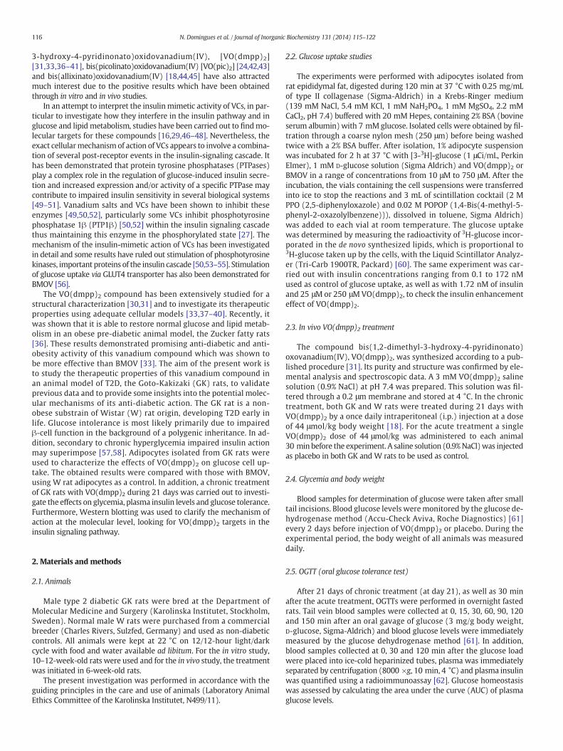
Fig. 1. Glucose uptake induced by insulin, VO(dmpp)2 and BMOV onWistar and GK adipocyte(○, n = 9) and GK (■, n = 11) rats for (a) insulin (b) VO(dmpp)2 and (c) BMOV stimulationGK (n = 11) adipocytes induced by different concentrations of insulin, VO(dmpp)2 and BMOVbasal value; §P b 0.05, §§P b 0.01, §§§P b 0.001 vs respective 1.72 nM of insulin; †P b 0.05, ††P b
concentration of VO(dmpp)2.
and OGTT. One-way analysis of variance was followed by the Student–Bonferroni multiple-range test to estimate the significance of differ-ences between groups for area under curve (AUC) of blood glucoselevels during OGTT. One-way analysis of variance was followed by theStudent–Newman–Keuls multiple-range test to estimate the signifi-cance of differences between groups for Western Blot analysis. A valueof P b 0.05 was considered as statistically significant. Data were ana-lyzed using GraphPad Prism (v5.0, GraphPad Software).
3. Results and discussion
The anti-diabetic properties of VO(dmpp)2 were evaluated byin vitro studies with primary GK rat adipocytes and in vivo studies inGK rats. The effect of this compound on some abnormal parameters in-dicative of diabetes was tested. Its capacity to improve glucose uptakeby adipocytes was measured. Body weight and blood glucose levels ofGK rats subjected to a daily administration of this compound during21 days were periodically determined and at the end of the chronictreatment (day 21), an OGTT was also carried out as well as 30 minafter the acute treatment.
3.1. In vitro studies with VO(dmpp)2 in isolated rat adipocytes
The GK rats used in this study had significantly lower body weightthan the W rats (286 ± 15 vs 336 ± 11 g, P b 0.001), in agreementwith published data which reported that although an increase in thenumber of adipocytes occurs before weaning, decreased body weightand leanmass are observed in the GK rats [63] and displayed hypergly-cemia (7.0 ± 0.2 vs 5.1 ± 0.2 mM, P b 0.001). Moreover glucoseuptake was significantly impaired in adipocytes from GK rats whencompared with W rats (81 ± 4 vs 121 ± 7 DPM/h, P b 0.001, DPM =disintegration per minute). The improvement of glucose uptake by
s. (A) Dose response curves of glucose uptake (in DPM/h, disintegration per min/h) on W. (B–C) Percentage of glucose uptake (relative to basal value of 100%) by W (n = 9) and. Data are shown as mean values ± SEM. *P b 0.05, **P b 0.01, ***P b 0.001 vs respective0.01, †††P b 0.001 vs respective 17.2 nM of insulin; ##P b 0.01, ###P b 0.001 vs the same
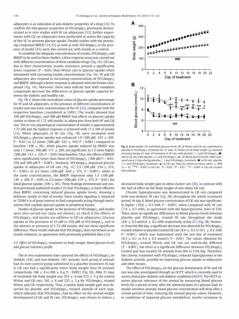
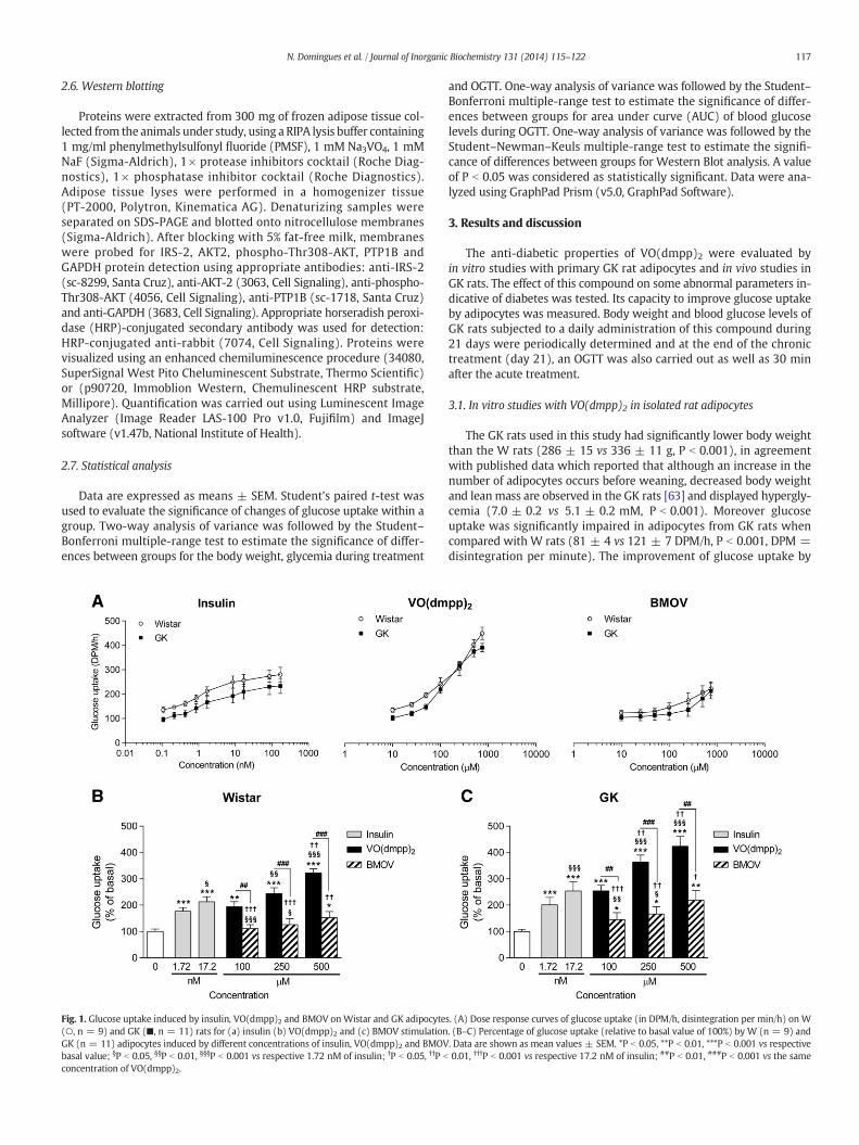
Fig. 2. Body weight (A) and blood glucose levels (B) of Wistar and GK rats submitted toplacebo or VO(dmpp)2 treatment for 21 days. A) Values of rat body weight (g) assessedeveryday during 21 days of treatment in W rats with placebo (□) and VO(dmpp)2 (■)and in GK rats with placebo (△) and VO(dmpp)2 (▲); B) Blood glucose levels (mM)mea-sured every 2 days during placebo (□) and VO(dmpp)2 treatment (■) inW rats; placebo(△) and VO(dmpp)2 treatment (▲) in GK rats. Data are shown as mean values ± SEM(n = 8). ###P b 0.001 vs GK rats; *P b 0.05, **P b 0.01, ***P b 0.001 vs GK rats treatedwith placebo.
118 N. Domingues et al. / Journal of Inorganic Biochemistry 131 (2014) 115–122
adipocytes is an indication of anti-diabetic properties of a drug [33]. Toconfirm the therapeutic properties of VO(dmpp)2 previously demon-strated in in vitro studies with W rat adipocytes [33], further experi-ments with GK rat adipocytes were performed to assess the capacityof this VC to promote glucose uptake. Parallel studies with the promis-ing compound BMOV [14,33], as well as with VO(dmpp)2 in the pres-ence of insulin [33], were also carried out, with insulin as a control.
To establish the adequate concentrations of insulin, VO(dmpp)2 andBMOV to be used in these studies, a dose response assaywas carried outwith different concentrations of these candidate drugs (Fig. 1A). GK rats,due to their characteristic insulin resistance, present a significantlylower response (P b 0.05) than Wistar rats to glucose uptake whenstimulated with increasing insulin concentrations (Fig. 1A). W and GKadipocytes also respond to increasing concentrations of VO(dmpp)2and BMOV, although a better response is obtainedwith the former com-pound (Fig. 1A). Moreover, these data indicate that both vanadiumcompounds decrease the differences in glucose uptake capacity be-tween the diabetic and healthy rats.
Fig. 1B–C shows the normalized values of glucose uptake percentagefor W and GK adipocytes, in the presence of different concentrations ofinsulin and non-toxic concentrations of the VC [33], compared with therespective baselines (considered as 100%). The results showed that100 μM VO(dmpp)2 and 500 μM BMOV had effects on glucose uptakesimilar to those of 1.72 nM insulin, in adipocytes from both W and GKrats. The in vivo physiological concentration of insulin is approximately1.72 nM and the highest response is achieved with 17.2 nM of insulin[33]. When adipocytes of W rats (Fig. 1B) were incubated withVO(dmpp)2, glucose uptake was enhanced 1.9 (100 μM: 193 ± 20%,P b 0.01) to 3.2 times (500 μM: 322 ± 16%, P b 0.001) compared tobaseline (100 ± 9%), while glucose uptake induced by BMOV wasonly 1.1 times (100 μM: 111 ± 20%, not significant) to 1.5 times higher(500 μM: 153 ± 23%, P b 0.05) than baseline. Thus, the effects of BMOVwere significantly lower than those of VO(dmpp)2 (100 μM P b 0.01;250 and 500 μM P b 0.001). Similarly, VO(dmpp)2 improved glucoseuptake in adipocytes of GK rats (Fig. 1C) 2.5 (100 μM: 254 ± 21%,P b 0.001) to 4.2 times (500 μM: 424 ± 37%, P b 0.001), while inthe same concentrations, the BMOV improved only 1.5 (100 μM:145 ± 26%, P b 0.05) to 2.2 times (500 μM: 219 ± 37%, P b 0.01) thebasal glucose uptake (100 ± 8%). These findings demonstrate and con-firm previously published results [33] that VO(dmpp)2 is more effectivethan BMOV, concerning induced glucose uptake levels, showing abehavior similar to that of insulin. Since insulin signaling is impairedin T2DM it is of great interest to find compounds acting through mech-anisms that regulate glucose uptake in peripheral tissues.
Studies of glucose uptake in the presence of VO(dmpp)2 and insulinwere also carried out (data not shown), to check if the effects ofVO(dmpp)2 and insulin are additive in GK rat adipocytes. Glucoseuptake in the presence of 25 μM or 250 μM of VO(dmpp)2, and inthe absence or presence of 1.72 nM insulin, did not show significantdifference. These results indicate that VO(dmpp)2 does not behave as aninsulin enhancer, in agreement with previously published data [33].
3.2. Effect of VO(dmpp)2 treatment on body weight, blood glucose levelsand glucose tolerance profile
The in vivo experiments have assessed the effects of VO(dmpp)2 ondiabetic (GK) and non-diabetic (W) animals. Each group of animalshad its own control group submitted to a placebo treatment At day0, GK rats had a significantly lower body weight than W animals(respectively, 148 ± 4 vs 200 ± 6 g, P b 0.001) (Fig. 2A). After 21 daysof treatment the body weight was 353 ± 4 and 233 ± 5 g for controlWistar and GK rats; 342 ± 6 and 225 ± 3 g for VO(dmpp)2 treatedWistar and GK, respectively. Thus, a similar body weight gain was ob-served for placebo and VO(dmpp)2 treated animals of each type,which indicates that VO(dmpp)2 had no effect on the normal weightdevelopment of GK and W rats. VO(dmpp)2 was shown to induce a
decreased body weight gain in obese Zucker rats [36], in contrast withthe lack of effect on the body weight of non-obese GK rats.
Chronic hyperglycemia was demonstrated in GK rats comparedwith non-diabetic W rats (Fig. 2B) throughout the whole treatmentperiod. At day 0, blood glucose concentration of GK rats was significant-ly higher (10.0 ± 0.3 mM, P b 0.001) when compared with W rats(7.9 ± 0.3 mM), in agreement with the hyperglycemic state of T2D.There were no significant differences in blood glucose levels betweenplacebo and VO(dmpp)2 treated W rats throughout the study(6.8 ± 0.2 and6.6 ± 0.2 mM, respectively, at day 21) (Fig. 2B). Howev-er, from the 8th day, a significant decreasewas observed for VO(dmpp)2treated relative to placebo treatedGK rats (8.4 ± 0.3 vs10.1 ± 0.2 mM,P b 0.001), which was maintained until the last day of treatment(8.3 ± 0.1 vs 9.4 ± 0.2 mmol/l, P b 0.05). The values obtained forVO(dmpp)2 treated Wistar and GK rats are statistically different(P b 0.001), but there is a significant difference between VO(dmpp)2treated and non-treated GK animals from 8th to 21th day. Therefore,this chronic treatment with VO(dmpp)2 reduced hyperglycemia in thediabetic animals, possibly by improving glucose uptake in adipocytesand other tissues.
The effect of VO(dmpp)2 on the glucose homeostasis of W and GKrats was also investigated through an OGTT, which is currently used toassess clinical pre-diabetic anddiabetic conditions [64,65]. TheOGTT as-sesses glucose tolerance of the animal by measuring blood glucoselevels for a period of time after the administration of a glucose load. Ininsulin sensitive animals, blood glucose concentration will drop after acertain period of time, restoring the normal glucose values. However,in conditions of impaired glucose metabolism, insulin resistance or
119N. Domingues et al. / Journal of Inorganic Biochemistry 131 (2014) 115–122
T2DM, insulin signaling does not function properly and blood glucoseconcentration remains high for a longer period of time [66–68]. TheOGTT results demonstrated that glucosewas significantly better tolerat-ed in W than in GK rats (Fig. 3A, P b 0.001) and that the 21-day treat-ment with VO(dmpp)2 improved significantly glucose tolerance in GKrats (Fig. 3A, P b 0.05 or less; values of area under curve 20.6 ± 0.7 vs13.1 ± 0.5 mM/min in placebo andVO(dmpp)2 treatedGK rats, respec-tively, P b 0.001, Fig. 3B). Interestingly, this glucose-lowering effect byVO(dmpp)2 was not seen in the W rats (8.7 ± 0.2 mM/min in placebotreated W rats and 8.0 ± 0.4 mM/min in VO(dmpp)2 treated W rats)(Fig. 3B), indicating that the compounddoes not appear to induce hypo-glycemia in non-diabetic rats, but may interact specifically with patho-logical mechanisms in diabetes.
To further assess the effects of VO(dmpp)2 on glucose homeostasis,plasma insulin levels in OGTT were evaluated, before glucose load(0 min), at the maximum effect of the overload (30 min) and 120 minafter glucose load, to test the ability of the animal to respond to arapid glucose challenge (Fig. 3C–D). In W rats, in which VO(dmpp)2treatment did not affect glucose tolerance (Fig. 3A–B), plasma insulinlevels were significantly lower than in animals treated with placebothroughout the test (respectively, at 0 min: 7.1 ± 1.8 vs 20.7 ±3.1 μU/mL, P b 0.01; at 30 min: 29.8 ± 3.8 vs 50.9 ± 3.7 μU/mL,P b 0.05; at 120 min: 7.6 ± 1.0 vs 21.2 ± 5.5 μU/mL, P b 0.05)(Fig. 3C). This discrepancy can be explained by the increased glucoseuptake induced by VO(dmpp)2. However, the kinetics of plasma insulinduring theOGTT remained the same. After 30 min, plasma insulin levelsin W rats treated with placebo or VO(dmpp)2 were significantly higherthan their respective groups before the glucose challenge (20.7 ± 3.1 vs50.9 ± 3.7 μU/mL, P b 0.01; 7.1 ± 1.8 vs 29.8 ± 3.8 μU/mL, P b 0.001,respectively) (Fig. 3C). Plasma insulin concentrations decreased after120 min compared to 30 min (50.9 ± 3.7 vs 21.2 ± 5.5 μU/mL,P b 0.01; 29.8 ± 3.8 vs 7.6 ± 1.0 μU/mL, P b 0.001, respectively) but
Fig. 3.Glucose tolerance profile (A–B) and plasma insulin levels (C–D) after 21 days of treatmenor VO(dmpp)2. A) Blood glucose values (mM) during anOGTT after 21 days of treatment inW r(▲); B) Values of area under curve (AUC), expressed as mM/min, obtained from the curves ofplacebo or VO(dmpp)2, shown in Fig. 3A; C) Plasma insulin levels during an OGTT inW rats treatwith placebo or VO(dmpp)2; E) Blood glucose values (mM) during an OGTT for the control (△,istered 30 min before the glucose load, n = 7) effects of VO(dmpp)2 inGK rats. Data are shownvs GK rats treated with VO(dmpp)2; #P b 0.05, ##P b 0.01, ###P b 0.001 vsW rats (Panel A); ****P b 0.001 vs 0 min of the respective group; ##P b 0.01, ###P b 0.001 vs 30 min of the respeand ***P b 0.001 vs GK control; $P b 0.05 and $$P b 0.01 vs GK rats under an acute treatment (
were not significantly different from basal plasma insulin levels. Indeed,although blood glucose profiles during OGTT were similar in W ratstreated with VO(dmpp)2 or placebo, plasma insulin levels were lowerin the former than in the latter group of animals. These results suggestan improvement in glucose uptake which could be by changing signal-ing pathway of insulin at the storage tissue.
In GK rats, VO(dmpp)2 did not significantly decrease plasma insulinlevels either at basal conditions and after 30 min of the glucose load(Fig. 3D), compared with GK rats treated with placebo. A significantdecrease in plasma insulin levels was found only at 120 min (Fig. 3D,45.0 ± 5.1 vs 26.9 ± 4.2 μU/mL, P b 0.05), suggesting an improvementin glucose tolerance observed during the OGTT (Fig. 3A). As for W rats,the glucose overload induced, 30 min later, a significant increase inplasma insulin levels (Fig. 3D) in placebo treated (37.8 ± 4.1 vs53.5 ± 5.9 μU/mL, P b 0.05) and VO(dmpp)2 treated (25.5 ± 3.8 vs49.2 ± 2.4 μU/mL, P b 0.001) GK rats (Fig. 3D). After 120 min of theglucose load, plasma insulin levels were not statistically different fromthe basal ones at 0 min, but the group treated with VO(dmpp)2showed a significant decreasewhen compared to 30 min. Important-ly, VO(dmpp)2 treated GK rats have shown plasma insulin levelssimilar to placebo treated W rats, demonstrating the efficiency ofVO(dmpp)2 to improve the glucose tolerance with less insulin-resistance in diabetic GK rats [58,59]. These results show that GKrats treated with VO(dmpp)2 recover from a glucose load with a pro-file similar to W rats, presenting blood glucose values significantlylower than those from GK control. This provides clear evidence thatVO(dmpp)2 ameliorates the glucose intolerant profile characteristicof GK rats and this effect occurs without any significant change inplasma insulin concentrations at 30 min of OGTT [69,70].
To evaluate the acute affects of VO(dmpp)2 treatment in GK rats, anOGTTwas carried out in animals whichwere submitted to a single doseof 44 μmol/kg (animal body weight) of VO(dmpp)2 administered
t and glucose tolerance profile after an acute treatment (E) ofW and GK rats with placeboatswith placebo (□) and VO(dmpp)2 (■) and inGK ratswith placebo (△) and VO(dmpp)2blood glucose concentration vs time after the glucose load in W and GK rats treated withedwith placebo or VO(dmpp)2; D) Plasma insulin levels during anOGTT inGK rats treatedcontrol, n = 8), chronic (21 days,▲, n = 8) and acute (single dose of 44 μmol/kg admin-asmean values ± SEM (n = 8). ***P b 0.001 vsWrats; §P b 0.05, §§P b 0.01, §§§P b 0.001**P b 0.001 vs W rats; ###P b 0.001 vs respective control (Panel B); *P b 0.05, **P b 0.01,ctive group; §P b 0.05, §§P b 0.01 vs respective control (Panel C–D); * P b 0.05, **P b 0.01Panel E).

120 N. Domingues et al. / Journal of Inorganic Biochemistry 131 (2014) 115–122
30 min before the glucose load and the results were compared with GKrats control and those under a chronic treatment. The obtained data areshown in Fig. 3E and demonstrate that a chronic treatment withVO(dmpp)2, is more efficient than an acute one concerning recoveryfrom a glucose load. The values of blood glucose levels in GK rats controlare statistically different from those submitted to a VO(dmpp)2 treat-ment (P b 0.001) and there is a significant difference between GKrats under an acute and chronic treatment (P b 0.001 at 60 min andP b 0.05 between 60 and 120 min after glucose load) although, at150 min, these values are similar. Several hypothesis can be formulatedto explain this finding: the improvement of pancreas function and con-sequently an increase in blood insulin concentration, as a response to afeeding state, after a long term treatmentwith VO(dmpp)2; the increaseof glucose uptake and regulation of glucose metabolism in adipocytes,muscle and others peripheral tissues, due to an effect on gene expres-sion of key proteins involved in glucose homeostasis.
3.3. Effect of VO(dmpp)2 treatment on insulin signaling pathway ofadipose tissue
The decrease in plasma insulin levels at 120 min in GK rats treatedwith VO(dmpp)2 suggests a more efficient and faster glucose uptakethan in GK animals treated with placebo. During hyperglycemia, thebody regulates glucose homeostasis by increasing insulin secretionto improve glucose storage in the target tissues. In T2D, however,insulin-sensitive cells may be resistant to insulin. As shown in thisstudy, GK rats have impaired glucose tolerance associatedwith the inef-fectiveness of insulin stimulated pathway and, thus, decreased glucoseinternalization [18]. The treatment with VO(dmpp)2 did not increase
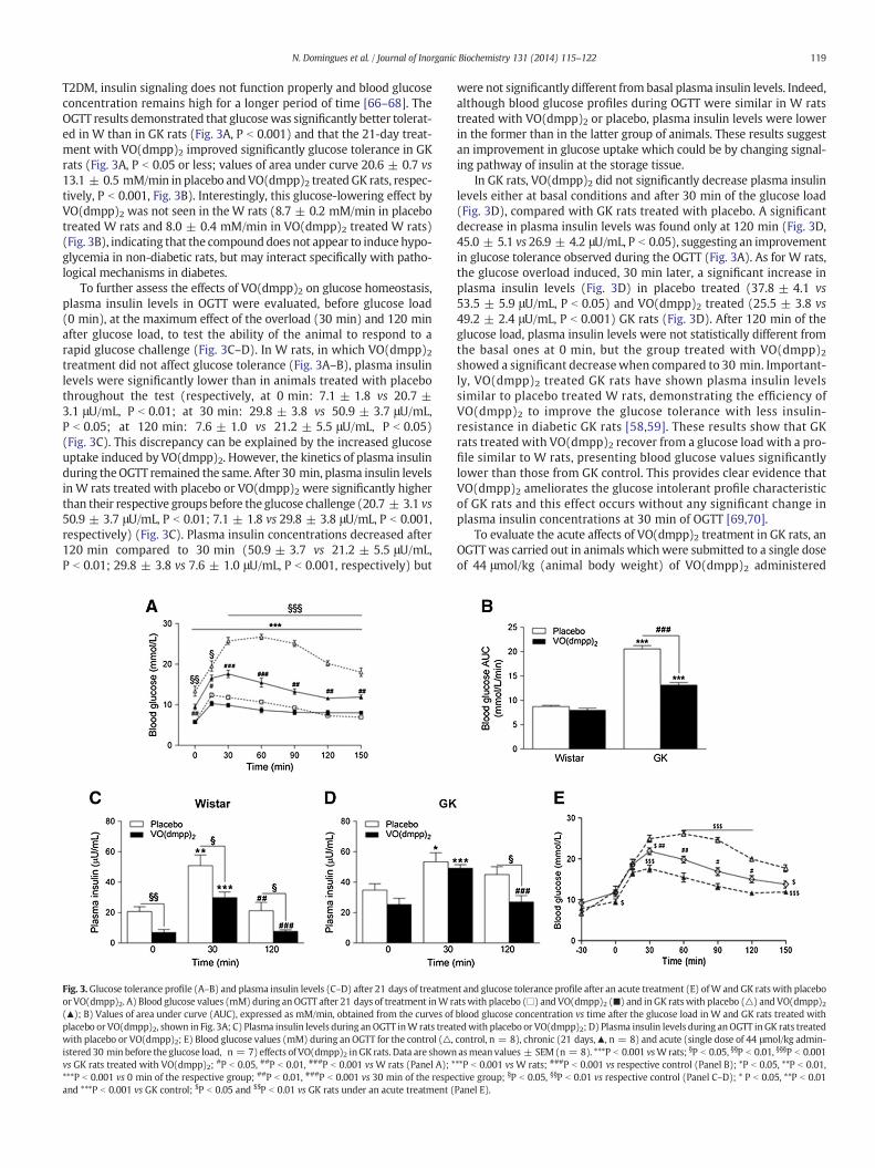
Fig. 4.Effects of VO(dmpp)2 treatment on the insulin signalingpathway in adipose tissue ofWaAKT-2 (C) and p-AKT (D) protein expression in adipose tissue fromWandGK rats treatedwith p(n = 8). *P b 0.05 vs W rats treated with placebo; §§P b 0.01, §§§P b 0.001 vs W rats treated w
plasma insulin levels in GK rats in parallel with improvement of glucosetolerance. In VO(dmpp)2 treatedW rats a normal glucose tolerance pro-file was observed despite the significant decrease in plasma insulinlevels, suggesting that VO(dmpp)2 improved glucose uptake at the insu-lin signaling pathway level.
One of themost importantmetabolic actions of insulin is to promoteglucose uptake into adipocytes, skeletal muscle and others peripheraltissue. This is accomplished via activation of the PI3K/AKT signalingpathway and subsequent translocation of GLUT4 from intracellular stor-age vesicles to the plasma membrane [46,47,53,56,71–73]. Westernblotting was used to assess the effect of VO(dmpp)2 on the insulin sig-naling cascade in adipocytes, more specifically on four target proteinsinvolved in glucose uptake. This will allow the identification of keypoints of this cascade which are deregulated in insulin resistant cells[71] and may be affected by VO(dmpp)2.
Insulin receptor substrate 2 (IRS2) is a protein directly involvedin insulin cell stimulation, the first event beyond insulin receptoractivation by tyrosine kinase that unleashes the transmission of intra-cellular insulin signals [73,74]. Chronic treatment of W and GK ratswith VO(dmpp)2 increased significantly the IRS2 expression comparedwith their respective controls treated with placebo (0.72 ± 0.07 vs0.51 ± 0.04, for W rats; P b 0.05 and 0.66 ± 0.05 vs 0.43 ± 0.03, inGK rats, P b 0.05) (Fig. 4A). Therefore, the increased IRS2 proteinexpression can explain the decrease of chronic hyperglycemia and im-provement of glucose tolerance. At first glance, these results suggest ahigher sensitivity of the insulin receptor resulting in increased proteinexpression in adipose tissue. The protein tyrosine phosphatase PTP1βinhibits the insulin receptor, preventing the activation of the insulinsignaling pathway. Moreover, PTP1β is known to be overexpressed in
ndGK rats. Representative immunoblots and densitometry analysis of IRS-2 (A), PTP1β (B),lacebo or VO(dmpp)2. Data fromdensitometry analysis are shown asmean values ± SEMith VO(dmpp)2; #P b 0.05, ###P b 0.001 vs respective control.
121N. Domingues et al. / Journal of Inorganic Biochemistry 131 (2014) 115–122
insulin resistant tissues and it is inhibited by different vanadiumcompounds, including BMOV [52]. In GK rats treated with placebo, thelevel of PTP1β expression was significantly higher compared to W ratstreated with placebo (0.62 ± 0.03 vs 0.47 ± 0.05, P b 0.05) (Fig. 4B),characteristics shared with other rodent models of insulin resistance[74–77]. However, in the GK rats treated with VO(dmpp)2, the PTP1βexpression decreased significantly compared to GK rats treatedwith placebo (0.34 ± 0.06 vs 0.62 ± 0.03, P b 0.001) andW rats treat-ed with VO(dmpp)2 (0.47 ± 0.05, P b 0.01) (Fig. 4B). Therefore, theVO(dmpp)2 like other vanadium compounds [33,76] inhibited thePTP1β expression, thereby removing the inhibition of the insulin recep-tor and promoting the activation of the insulin signaling pathway. Thusthe reduction of PTP1β expression may have contributed to improvedinsulin sensitivity in GK rats treated with VO(dmpp)2. The ability ofVO(dmpp)2 to inhibit PTPase activity may be one of the mechanismsby which it exerts its insulino-mimetic action [75].
Another target of VO(dmpp)2 can be the AKT activity [33]. Toaddress the role of the AKT pathway in relation to alternate pathways,it is essential to show that activation of AKT alone is sufficient tomimic the effects of insulin [78] associated with VO(dmpp)2. In thisstudy, the modifications in the protein expression involved in the insu-lin signaling pathway did not affect the expression of AKT2. Indeed, thelevels of AKT2 expression did not change after the in vivo VO(dmpp)2treatment and were not significantly different between W and GK rats(Fig. 4C). The phosphorylated-AKT2 (p-AKT) has been implicated ininsulin-regulated glucose uptake into fat cells by promoting the translo-cation of GLUT4 to the cell surface [56,78]. For this purpose, a Westernblot analysis of the phosphorylated protein was assessed. A significantincrease in the p-AKT expression was observed in W and GK ratstreated with VO(dmpp)2 when compared to their respective controlstreated with placebo (2.54 ± 0.16 vs 1.55 ± 0.08, P b 0.001 in W rats;1.81 ± 0.14 vs 1.40 ± 0.07, P b 0.05 in GK rats) (Fig. 4D). However,the expression levels of this protein were significantly lower in GKrats treated with placebo (1.40 ± 0.07, P b 0.001) or VO(dmpp)2(1.81 ± 0.14, P b 0.001) compared to W rats treated with VO(dmpp)2(2.54 ± 0.16) (Fig. 4D).
According to these data, VO(dmpp)2 treatment in GK rats improvedglucose tolerance and decreased the chronic hyperglycemia after 8 daysof treatment. These effects may be explained by the increase of the IRS2expression, thus improving the insulin sensitivity in adipocytes, by theinhibition of the PTP1β expression which exerts an inhibitory actionon IRS2 and by the increase of p-AKT expression thus promoting glucoseuptake by GLUT4 [71,72,78].
4. Conclusions
This work clearly demonstrates the in vitro and in vivo insulin mi-metic effects of VO(dmpp)2 in the GK rats, a T2D animal model. It isshown by the in vitro study that VO(dmpp)2 improves glucose uptakein adipocytes, more effectively than BMOV. A chronic in vivo treatmentwith VO(dmpp)2 decreases hyperglycemia and improves glucose toler-ance significantly, although a complete normalization during OGTTwasnot observed inGK rats.Moreover, there is a normal gain of bodyweightof both diabetic and control VO(dmpp)2 treated rats, and thiscompound does not induce hypoglycemia in non-diabetic W rats. Inaddition, a better behavior is observed in GK rats submitted to a chronictreatment than an acute one. All these effects can be explained by thedirect action of VO(dmpp)2 on key proteins of the insulin signalingcascade, by specifically increasing IRS2 expression and AKT phosphory-lation and inhibiting PTP1β expression. The combined results here pre-sented demonstrate the beneficial effects of VO(dmpp)2 in GK rats,corroborating previously published data and showing the insulinmimetic activity of this compound. Thus, taking into account the prob-lems of toxicity associated with vanadium compounds, which shouldbe minimized, VO(dmpp)2 could be a good alternative for the T2Dtherapy.
Acknowledgments
The authors thank Prof. Dr. João Costa Pessoa and his Post-docstudent Somnath Roy at the Technical University of Lisbon, Portugal,for the synthesis of the VO(dmpp)2 compound.
This work was supported by the Swedish Research Council,the Swedish Diabetes Association and the Karolinska Institutet.Julien Pelletier was supported by a grant from the Föreningen fördiabetesforskningens främjande. Neuza Domingues was supportedby an Erasmus grant during her stay in Karolinska Institutet.
References
[1] Y. Lin, Z. Sun, J. Endocrinol. 204 (2010) 1–11.[2] P. Raskin, A. Mohan, Expert Opin. Emerg. Drugs 15 (2010) 225–236.[3] K.D. Bruce, C.D. Byrne, Postgrad. Med. J. 85 (2009) 614–621.[4] T.M. Frayling, Nat. Rev. Genet. 8 (2007) 657–662.[5] C.J. Schofield, C. Sutherland, Diabet. Med. 29 (2012) 972–979.[6] A.J. Krentz, C.J. Bailey, Drugs 65 (2005) 385–411.[7] D.M. Nathan, J.B. Buse, M.B. Davidson, E. Ferrannini, R.R. Holman, R. Sherwin, B.
Zinman, Diabetes Care 32 (2009) 193–203.[8] In: A.S. Tracey, D.C. Crans (Eds.), Vanadium compounds, Chemistry, Biochemistry
and Therapeutic Applications, vol. 711, American Chemical Society, 1998.[9] D. Rehder, Inorg. Chem. Commun. 6 (2003) 604–617.
[10] K.H. Thompson, J.H. McNeill, C. Orvig, Chem. Rev. 99 (1999) 2561–2571.[11] K.H. Thompson, C. Orvig, J. Inorg. Biochem. 100 (2006) 1925–1935.[12] D.C. Crans, J. Inorg. Biochem. 80 (2000) 123–131.[13] D.C. Crans, J.J. Smee, E. Gaidamauskas, L. Yang, Chem. Rev. 104 (2004) 849–902.[14] K.H. Thompson, C. Orvig, Met. Ions Biol. Syst. 41 (2004) 221–252.[15] D.C. Crans, Pure Appl. Chem. 77 (2005) 1497–1527.[16] H. Sakurai, Y. Yoshikawa, H. Yasui, Chem. Soc. Rev. 37 (2008) 2383–2392.[17] K. Kawabe, Y. Yoshikawa, Y. Adachi, H. Sakurai, Life Sci. 78 (2006) 2860–2866.[18] H. Yasui, Y. Adachi, A. Katoh, H. Sakurai, J. Biol. Inorg. Chem. 12 (2007) 843–853.[19] T. Kiss, T. Jakusch, D. Hollender, Á. Dörnyei, É.A. Enyedy, J. Costa Pessoa, H. Sakurai, A.
Sanz-Medel, Coord. Chem. Rev. 252 (2008) 1153–1162.[20] P.W. Winter, A. Al-Qatati, A. Wolf-Ringwall, S. Schoeberl, A.K. Van Orden, B.G.
Barisas, D.A. Roess, D.C. Crans, Dalton Trans. 41 (2012) 6419–6430.[21] H. Sakurai, K. Fujii, H. Watanabe, H. Tamura, Biochem. Biophys. Res. Commun. 214
(1995) 1095–1101.[22] H. Sakurai, H. Sano, T. Takino, H. Yasui, J. Inorg. Biochem. 80 (2000) 99–105.[23] G.R. Willsky, L. Chi, M.E. Godzalla III, P.J. Kostyniak, J.J. Smee, A.M. Trujillo, J.A. Alfano,
W. Ding, Z. Hu, D.C. Crans, Coord. Chem. Rev. 19–20 (2011) 2258–2269.[24] M. Li, W. Ding, J.J. Smee, B. Baruah, G.R. Willsky, D.C. Crans, BioMetals 103 (2009)
585–905.[25] K.H. Thompson, J. Lichter, C. LeBel, M.C. Scaife, J.H. McNeill, C. Orvig, J. Inorg.
Biochem. 103 (2009) 554–558.[26] K.H. Thompson, M. Battell, J.H. McNeill, in: J.O. Nriagu (Ed.), Vanadium in the Envi-
ronment. Part 2: Health Effects, John Wiley & Sons, 1998, pp. 21–37.[27] Y.Wei, C. Zhang, P. Zhao, X. Yang, K.Wang, J. Inorg. Biochem. 105 (2011) 1081–1085.[28] J. Costa Pessoa, I. Cavaco, I. Correia, I. Tomaz, P. Adão, I. Vale, V. Ribeiro, M.M.C.A.
Castro, C.C.F.G. Geraldes, in: K. Kustin, J. Costa Pessoa, D.C. Crans (Eds.), Vanadium:the versatile metal, Vanadium Schiff Base Complexes: Chemistry, Properties andConcerns about Possible Therapeutic Applications, American Chemical SocietySymposium Series, vol. 974, 2007, pp. 340–351.
[29] D. Redher, Bioinorganic Vanadium Chemistry, Wiley, UK, 2008.[30] M.M. Castro, C.F. Geraldes, P. Gameiro, E. Pereira, B. Castro, M. Rangel, J. Inorg.
Biochem. 80 (2000) 177–179.[31] M.M.C.A. Castro, F. Avecilla, C.F.G.C. Geraldes, B. de Castro, M. Rangel, Inorg. Chim.
Acta 356 (2003) 142–154.[32] W.M. Mueller, K.L. Stanhope, F. Gregoire, J.L. Evans, P.J. Havel, Obes. Res. 8 (2000)
530–539.[33] M. Passadouro, A.M. Metelo, A.S. Melao, J.R. Pedro, H. Faneca, E. Carvalho, M.M.
Castro, J. Inorg. Biochem. 104 (2010) 987–992.[34] Y. Adachi, H. Sakurai, Chem. Pharm. Bull. 52 (2004) 428–433.[35] W. Basuki, M. Hiromura, Y. Adachi, K. Tayama, M. Hattori, H. Sakurai, Biochem.
Biophys. Res. Commun. 349 (2006) 1163–1170.[36] A.M. Metelo, R. Perez-Carro, M.M. Castro, P. Lopez-Larrubia, J. Inorg. Biochem. 115
(2012) 44–49.[37] G.R. Willsky, A.B. Goldfine, P.J. Kostyniak, J.H. McNeill, L. Yang, A.R. Khan, D.C. Crans,
J. Inorg. Biochem. 85 (2001) 33–42.[38] D. Rehder, J.C. Pessoa, C.F.G.C. Geraldes, M.M.C.A. Castro, T. Kabanos, T. Kiss, B. Meier,
G. Micera, L. Pettersson, M. Rangel, A. Salifoglou, I. Turel, D. Wang, J. Biol. Inorg.Chem. 7 (2002) 384–396.
[39] T.C. Delgado, M.M. Castro, C.F. Geraldes, J.G. Jones, Magn. Reson. Med. 51 (2004)1283–1286.
[40] T.C. Delgado, I. Correia, J.C. Pessoa, J.G. Jones, C.F.G.C. Geraldes, M.M.C.A. Castro,J. Inorg. Biochem. 99 (2005) 2328–2339.
[41] H. Faneca, V.A. Figueiredo, I. Tomaz, G. Gonçalves, F. Avecilla, M.C. Pedroso de Lima,C.F.G.C. Geraldes, J.C. Pessoa, M.M.C.A. Castro, J. Inorg. Biochem. 103 (2009)601–608.
[42] H. Esbak, E.A. Enyedy, T. Kiss, Y. Yoshikawa, H. Sakurai, E. Garribba, D. Rehder,J. Inorg. Biochem. 103 (2009) 590–600.
122 N. Domingues et al. / Journal of Inorganic Biochemistry 131 (2014) 115–122
[43] M. Li, J.J. Smee, W. Ding, D.C. Crans, J. Inorg. Biochem. 103 (2009) 585–589.[44] H. Sakurai, A. Katoh, T. Kiss, T. Jakusch, M. Hattori, Metallomics 2 (2010) 670–682.[45] Y. Adachi, Y. Yoshikawa, J. Yoshida, Y. Kodera, A. Katoh, J. Takada, H. Sakurai,
Biochem. Biophys. Res. Commun. 345 (2006) 945–950.[46] D.B. Savage, K.F. Petersen, G.I. Shulman, Physiol. Rev. 87 (2007) 507–520.[47] A.R. Saltiel, C.R. Kahn, Nature 414 (2001) 799–806.[48] M. Hiromura, H. Sakurai, Pure Appl. Chem. 80 (2008) 2727–2733.[49] S. Gogg, J. Chen, S. Efendic, U. Smith, C. Ostensson, Biochem. Biophys. Res. Commun.
280 (2001) 1161–1168.[50] J. Chen, C.G. Ostenson, Biochem. Biophys. Res. Commun. 325 (2004) 555–560.[51] C.G. Ostenson, A.C. Sandberg-Nordqvist, J. Chen, M. Hallbrink, D. Rotin, U. Langel, S.
Efendic, Biochem. Biophys. Res. Commun. 291 (2002) 945–950.[52] M. Li,W.Ding, B. Baruah,D.C. Crans, R.Wang, J. Inorg. Biochem. 102 (2008) 1846–1853.[53] P.G. Drake, A. Balbis, J. Wu, J.J. Bergeron, B.I. Posner, Am. J. Physiol. Endocrinol.
Metab. 279 (2000) E266–E274.[54] R.J. Comi, G. Grunberger, P. Gorden, J. Clin. Invest. 79 (1987) 453–462.[55] A. Hundberg, A. Vaag, J. Vinten, H. Beck-Nielson, Diabetologia 36 (1993) 668–674.[56] M. Hiromura, A. Nakayama, Y. Adachi, M. Doi, H. Sakurai, J. Biol. Inorg. Chem. 12
(2007) 1275–1287.[57] S.J. Hughes, K. Suzuki, Y. Goto, Diabetologia 37 (1994) 863–870.[58] C.G. Ostenson, A. Khan, S.M. Abdel-Halim, A. Guenifi, K. Suzuki, Y. Goto, S. Efendic,
Diabetologia 36 (1993) 3–8.[59] C.G. Ostenson, S. Efendic, Diabetes Obes. Metab. 9 (2007) 180–186.[60] A. Green, Biochem. J. 238 (1986) 663–669.
[61] S. Aziz, Y.H. Hsiang, Diabetes Care 6 (1983) 529–532.[62] V. Herbert, K.S. Lau, C.W. Gottlieb, S.J. Bleicher, J. Clin. Endocrinol. Metab. 25 (1965)
1375–1384.[63] J. Movassat, D. Bailbé, C. Lubrano-Berthelier, F. Picarel-Blanchot, E. Bertin, J. Mourot,
B. Portha, Am. J. Physiol. Endocrinol. Metab. 294 (2008) E168–E175.[64] E. Bartoli, G.P. Fra, G.P. Carnevale Schianca, Eur. J. Intern. Med. 22 (2011) 8–12.[65] M. Buysschaert, M. Bergman, Med. Clin. North Am. 95 (2011) 289–297.[66] H. Gin, V. Rigalleau, Diabetes Metab. 26 (2000) 265–272.[67] K.F. Petersen, S. Dufour, D.B. Savage, S. Bilz, G. Solomon, S. Yonemitsu, G.W. Cline, D.
Befroy, L. Zemany, B.B. Kahn, X. Papademetris, D.L. Rothman, G.I. Shulman, Proc.Natl. Acad. Sci. U. S. A. 104 (2007) 12587–12594.
[68] R.A. Rizza, Diabetes 59 (2010) 2697–2707.[69] K. Ueta, T. Ishihara, Y. Matsumoto, A. Oku, M. Nawano, T. Fujita, A. Saito, K. Arakawa,
Life Sci. 76 (2005) 2655–2668.[70] H. Kanno, M. Iwai, S. Inaba, I. Senba, H. Nakaoka, H. Sone, H. Sone, M. Mogi, M.
Horiuchi, Biol. Pharm. Bull. 28 (2005) 2092–2095.[71] A.F. Rowland, D.J. Fazakerley, D.E. James, Traffic 12 (2011) 672–681.[72] R.T. Watson, J.E. Pessin, Recent Prog. Horm. Res. 56 (2001) 175–193.[73] A.C. Thirone, C. Huang, A. Klip, Trends Endocrinol. Metab. 17 (2006) 72–78.[74] A.M. Valverde, A. Gonzalez-Rodriguez, Arch. Physiol. Biochem. 117 (2011) 105–115.[75] A.K. Srivastava, M.Z. Mehdi, Diabet. Med. 22 (2005) 2–13.[76] A. Mohammad, J. Wang, J.H. McNeill, Mol. Cell. Biochem. 229 (2002) 125–128.[77] B.J. Goldstein, J. Clin. Endocrinol. Metab. 87 (2002) 2474–2480.[78] Y. Ng, G. Ram, D.E. James, Cell Metab. 7 (2008) 348–356.