Embed Size (px)
Citation preview
Contents lists available at ScienceDirect
Radiation Measurements
journal homepage: www.elsevier.com/locate/radmeas
Theoretical calculation and measurement accuracy of Cerenkov optic-fiberdosimeter under electron and photon radiation therapies
Xudong Zhanga, Xiaobin Tanga,b,∗, Diyun Shua, Chunhui Gonga, Changran Genga,b, Yao Aia,Haiyan Yua, Wencheng Shaoa
a Department of Nuclear Science and Engineering, Nanjing University of Aeronautics and Astronautics, Nanjing, 210016, Chinab Collaborative Innovation Center of Radiation Medicine of Jiangsu Higher Education Institutions, Nanjing, 210016, China
A R T I C L E I N F O
Keywords:Cerenkov optic-fiber dosimeterRadiotherapyResponse differenceMeasurement accuracy
A B S T R A C T
This work aimed to study the mechanism and understand the influencing factors of measurement accuracy forthe Cerenkov optic-fiber dosimeter under electron and photon radiation therapies. Through the Geant4 calcu-lation, we determined responses between Cerenkov photon numbers recorded by the measurement device andthe dose deposited in the fiber and found responses differed for electron and photon beam irradiations. Forelectron beams, the relative Cerenkov photon number recorded by the device agreed well with the relative dosefor the depths after the maximum dose depth, but differed before that depth. For the photon beams, the relativeCerenkov photon number showed good agreement with the relative dose for all depths. Considering thetransmission efficiency of the Cerenkov radiation in the fiber, energy spectra and angular distributions ofelectrons were analyzed to explain the response difference. For the photon beams, energy spectra and angulardistributions of electrons changed less in various phantom depth than electron beams, therefore, better responsewas observed for photon beams. Besides, the influences of fiber parameters (i.e. diameter, refractive index, andmaterial) were investigated. With the increase of the core diameter, the transmission efficiency of Cerenkovradiation changed slightly but the Cerenkov photon numbers produced in the fiber increased by square. Withsmaller ratio of cladding/core refractive index or using PMMA as the core materials, Cerenkov photon numbersrecorded by the device could also be increased. This study could provide better understanding of the Cerenkovoptic-fiber dosimeter and promote its development.
1. Introduction
Dose measurement technology in advanced radiotherapy is im-portant to ensure the accuracy of dose delivery and is an effectivemethod of quality assurance and control. Although the ionizationchamber and thermoluminescence dosimeter have been available forcommercial application, certain problems still exist in dose measure-ments for radiotherapy (Kron et al., 1996; Stenstrom and Marvin,1946). The scintillating fiber-optic dosimeter (SFOD) is recently de-veloped with several advantages, such as good tissue equivalence, highspatial resolution, and remote measurement capability. However, thequenching effect of scintillator and the Cerenkov light noise limit theuse of SFOD in clinical situations (Mouatassim et al., 1995). In recentyears, researchers have suggested the new Cerenkov optic-fiber dosi-meter (CFOD), which removes the scintillator of the SFOD and directlydetects the Cerenkov radiation to characterize the radiation dose (Janget al., 2012, 2013; Lee et al., 2013; Yoo et al., 2013a, 2013b).
Cerenkov radiation is an electromagnetic radiation produced by acharged particle traveling through a transparent medium at a velocitygreater than that of light in the same medium (Beddar et al., 1992).Although fluorescence photons and Cerenkov photons can be bothgenerated during x-ray and electron irradiation, light generated in theoptical fiber were mostly composed of light from the Cerenkov effect(94%–100% contribution) for high-energy modalities (e.g. radiationtherapy) (Therriault-Proulx et al., 2013). As a light signal generated inthe dosimeter through radiation interaction, Cerenkov radiation has acertain relationship with the energy deposition of particles and has thepotential to be used for dose measurement (Shu et al., 2016a, 2016b).… The CFOD uses the Cerenkov radiation generated in the fiber tocharacterize the dose by Cerenkov light intensity, as shown in Fig. 1.Researchers (Jang et al., 2013; Yoo et al., 2013b) carried out con-siderable experimental verification of the CFOD under different radia-tions, such as electron and photon radiations. They have demonstratedthe potential and viability of the dosimeter for radiotherapy dose
https://doi.org/10.1016/j.radmeas.2018.01.001Received 27 May 2017; Received in revised form 19 October 2017; Accepted 13 January 2018
∗ Corresponding author. Department of Nuclear Science and Engineering, Nanjing University of Aeronautics and Astronautics, Nanjing, 210016, China.E-mail address: [email protected] (X. Tang).
Radiation Measurements 110 (2018) 1–6
Available online 31 January 20181350-4487/ © 2018 Elsevier Ltd. All rights reserved.
T

measurement.Although much verification has been conducted, the intrinsic phy-
sical mechanism of the CFOD and the factors that affect the dosemeasurement still need to be further studied. Using the Monte Carlotoolkit Geant4, a CFOD model and the radiation transportation processwere simulated (Agostinelli et al., 2003). The relationships between theCerenkov photon numbers (CPNs) and the deposited dose were ana-lyzed. The intrinsic physical mechanism of the dosimeter was studied.The influences of fiber parameters, such as core diameter, ratio ofcladding/core refractive index, and core material, on measurementwere investigated, thereby providing a basis for improving the dosemeasurement accuracy.
2. Materials and methods
Geant4 is a public toolkit which can be used to accurately simulatethe passage of particles through matter. A three-dimensional waterphantom and a CFOD model were constructed in Geant4 (version10.01.p02), as shown in Fig. 2. The size of the phantom was25 cm×25 cm×25 cm. Step-index plastic optical fibers were used togenerate and transmit Cerenkov radiation with a core/cladding struc-ture. The outer diameter of the plastic optical fiber was 1.0mm, and thethickness of cladding was 0.01mm. The refractive indices of the coreand the cladding were 1.492 and 1.402, respectively. The materials of
the core and the cladding were polymethyl methacrylate (PMMA) andfluorinated polymer, respectively. The fiber was covered by a poly-ethylene (PE)-based black jacket to intercept external light noise (Yooet al., 2013b).
Generally, the subtraction method can be used to measure the signaldifference of two fibers (Clift et al., 2002). To apply the subtractionmethod, two fibers with different lengths were placed closely and thelength difference L was considered the measurement region. CPNs re-corded by the device were obtained by subtracting the signal of theshorter fiber at the measurement device from the longer one. The de-posited dose and the CPNs generated in the fiber were directly obtainedthrough Geant4 simulation.
When the beam irradiated on the fibers, the Cerenkov radiationgenerated in the fibers was transmitted to the measurement devicethrough a long optical fiber. For remote measurement, the fiber had alength of 25m between the measurement device and the phantom. Theenergy spectra and the angular distributions of electrons at differentdepths of phantom were also calculated.
Electron and photon beams are commonly used in radiotherapy. Thetwo beams with energies of 6, 9, and 15MeV were simulated in thecurrent study. All beams we used were set to be non-clinical mono-energetic to study the mechanism under relatively simplified condi-tions. The field size of the planer source was maintained at10 cm×10 cm. The distance between the source and the phantomsurface was 80 cm. The beams perpendicularly irradiated the phantomalong the central axis. Therefore, the size of irradiation field was thesame as that of the source.
3. Results and discussion
3.1. Relationship between CPNs and dose
It was necessary to determine the length difference of two fibersfirst. Fig. 3 illustrates the relationship between CPNs generated in thefiber per irradiated particle and the length difference of two fibers. TheCPNs are linearly correlated to the length difference. When the lengthdifference is small, the spatial resolution of dose measurement is high,thereby resulting in less CPNs produced in the fiber. A balance existsbetween the precision and accuracy of dose measurement in terms ofthe length difference. After comprehensive consideration, the lengthdifference was set to 10mm.
Fig. 4 shows the generated and detected CPNs curves and the per-cent depth dose (PDD) curves under the electron beam at irradiationenergies of 6, 9, and 15MeV. The two CPNs curves were normalized totheir largest values and compared with the PDD curve deposited in thefiber. The CPNs curves generated in the fiber agree well with the PDD
Fig. 1. Measurement diagram of the Cerenkov optic-fiber dosimeter.
Fig. 2. Schematic of calculation model.
X. Zhang et al. Radiation Measurements 110 (2018) 1–6
2
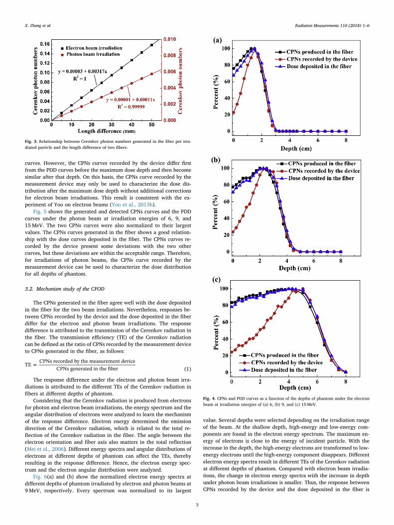
curves. However, the CPNs curves recorded by the device differ firstfrom the PDD curves before the maximum dose depth and then becomesimilar after that depth. On this basis, the CPNs curve recorded by themeasurement device may only be used to characterize the dose dis-tribution after the maximum dose depth without additional correctionsfor electron beam irradiations. This result is consistent with the ex-periment of Yoo on electron beams (Yoo et al., 2013b).
Fig. 5 shows the generated and detected CPNs curves and the PDDcurves under the photon beam at irradiation energies of 6, 9, and15MeV. The two CPNs curves were also normalized to their largestvalues. The CPNs curves generated in the fiber shows a good relation-ship with the dose curves deposited in the fiber. The CPNs curves re-corded by the device present some deviations with the two othercurves, but these deviations are within the acceptable range. Therefore,for irradiations of photon beams, the CPNs curve recorded by themeasurement device can be used to characterize the dose distributionfor all depths of phantom.
3.2. Mechanism study of the CFOD
The CPNs generated in the fiber agree well with the dose depositedin the fiber for the two beam irradiations. Nevertheless, responses be-tween CPNs recorded by the device and the dose deposited in the fiberdiffer for the electron and photon beam irradiations. The responsedifference is attributed to the transmission of the Cerenkov radiation inthe fiber. The transmission efficiency (TE) of the Cerenkov radiationcan be defined as the ratio of CPNs recorded by the measurement deviceto CPNs generated in the fiber, as follows:
=TECPNs recorded by the measurement device
CPNs generated in the fiber (1)
The response difference under the electron and photon beam irra-diations is attributed to the different TEs of the Cerenkov radiation infibers at different depths of phantom.
Considering that the Cerenkov radiation is produced from electronsfor photon and electron beam irradiations, the energy spectrum and theangular distribution of electrons were analyzed to learn the mechanismof the response difference. Electron energy determined the emissiondirection of the Cerenkov radiation, which is related to the total re-flection of the Cerenkov radiation in the fiber. The angle between theelectron orientation and fiber axis also matters in the total reflection(Mei et al., 2006). Different energy spectra and angular distributions ofelectrons at different depths of phantom can affect the TEs, therebyresulting in the response difference. Hence, the electron energy spec-trum and the electron angular distribution were analyzed.
Fig. 6(a) and (b) show the normalized electron energy spectra atdifferent depths of phantom irradiated by electron and photon beams at9MeV, respectively. Every spectrum was normalized to its largest
value. Several depths were selected depending on the irradiation rangeof the beam. At the shallow depth, high-energy and low-energy com-ponents are found in the electron energy spectrum. The maximum en-ergy of electrons is close to the energy of incident particle. With theincrease in the depth, the high-energy electrons are transformed to low-energy electrons until the high-energy component disappears. Differentelectron energy spectra result in different TEs of the Cerenkov radiationat different depths of phantom. Compared with electron beam irradia-tions, the change in electron energy spectra with the increase in depthunder photon beam irradiations is smaller. Thus, the response betweenCPNs recorded by the device and the dose deposited in the fiber is
Fig. 3. Relationship between Cerenkov photon numbers generated in the fiber per irra-diated particle and the length difference of two fibers.
Fig. 4. CPNs and PDD curves as a function of the depths of phantom under the electronbeam at irradiation energies of (a) 6, (b) 9, and (c) 15MeV.
X. Zhang et al. Radiation Measurements 110 (2018) 1–6
3
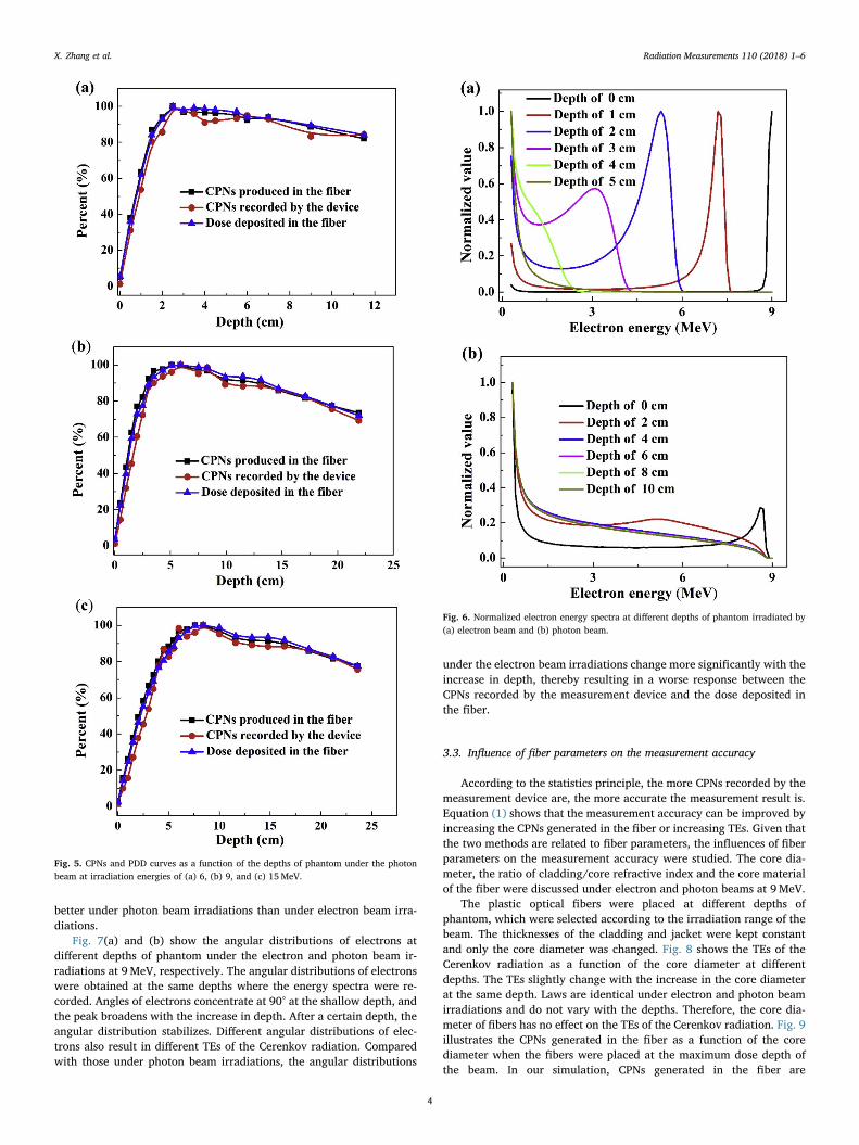
better under photon beam irradiations than under electron beam irra-diations.
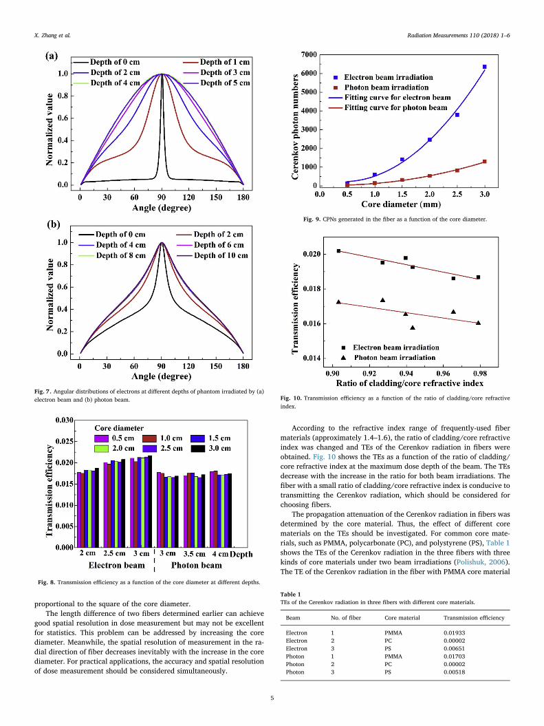
Fig. 7(a) and (b) show the angular distributions of electrons atdifferent depths of phantom under the electron and photon beam ir-radiations at 9MeV, respectively. The angular distributions of electronswere obtained at the same depths where the energy spectra were re-corded. Angles of electrons concentrate at 90° at the shallow depth, andthe peak broadens with the increase in depth. After a certain depth, theangular distribution stabilizes. Different angular distributions of elec-trons also result in different TEs of the Cerenkov radiation. Comparedwith those under photon beam irradiations, the angular distributions
under the electron beam irradiations change more significantly with theincrease in depth, thereby resulting in a worse response between theCPNs recorded by the measurement device and the dose deposited inthe fiber.
3.3. Influence of fiber parameters on the measurement accuracy
According to the statistics principle, the more CPNs recorded by themeasurement device are, the more accurate the measurement result is.Equation (1) shows that the measurement accuracy can be improved byincreasing the CPNs generated in the fiber or increasing TEs. Given thatthe two methods are related to fiber parameters, the influences of fiberparameters on the measurement accuracy were studied. The core dia-meter, the ratio of cladding/core refractive index and the core materialof the fiber were discussed under electron and photon beams at 9MeV.
The plastic optical fibers were placed at different depths ofphantom, which were selected according to the irradiation range of thebeam. The thicknesses of the cladding and jacket were kept constantand only the core diameter was changed. Fig. 8 shows the TEs of theCerenkov radiation as a function of the core diameter at differentdepths. The TEs slightly change with the increase in the core diameterat the same depth. Laws are identical under electron and photon beamirradiations and do not vary with the depths. Therefore, the core dia-meter of fibers has no effect on the TEs of the Cerenkov radiation. Fig. 9illustrates the CPNs generated in the fiber as a function of the corediameter when the fibers were placed at the maximum dose depth ofthe beam. In our simulation, CPNs generated in the fiber are
Fig. 5. CPNs and PDD curves as a function of the depths of phantom under the photonbeam at irradiation energies of (a) 6, (b) 9, and (c) 15MeV.
Fig. 6. Normalized electron energy spectra at different depths of phantom irradiated by(a) electron beam and (b) photon beam.
X. Zhang et al. Radiation Measurements 110 (2018) 1–6
4
proportional to the square of the core diameter.The length difference of two fibers determined earlier can achieve
good spatial resolution in dose measurement but may not be excellentfor statistics. This problem can be addressed by increasing the corediameter. Meanwhile, the spatial resolution of measurement in the ra-dial direction of fiber decreases inevitably with the increase in the corediameter. For practical applications, the accuracy and spatial resolutionof dose measurement should be considered simultaneously.
According to the refractive index range of frequently-used fibermaterials (approximately 1.4–1.6), the ratio of cladding/core refractiveindex was changed and TEs of the Cerenkov radiation in fibers wereobtained. Fig. 10 shows the TEs as a function of the ratio of cladding/core refractive index at the maximum dose depth of the beam. The TEsdecrease with the increase in the ratio for both beam irradiations. Thefiber with a small ratio of cladding/core refractive index is conducive totransmitting the Cerenkov radiation, which should be considered forchoosing fibers.
The propagation attenuation of the Cerenkov radiation in fibers wasdetermined by the core material. Thus, the effect of different corematerials on the TEs should be investigated. For common core mate-rials, such as PMMA, polycarbonate (PC), and polystyrene (PS), Table 1shows the TEs of the Cerenkov radiation in the three fibers with threekinds of core materials under two beam irradiations (Polishuk, 2006).The TE of the Cerenkov radiation in the fiber with PMMA core material
Fig. 7. Angular distributions of electrons at different depths of phantom irradiated by (a)electron beam and (b) photon beam.
Fig. 8. Transmission efficiency as a function of the core diameter at different depths.
Fig. 9. CPNs generated in the fiber as a function of the core diameter.
Fig. 10. Transmission efficiency as a function of the ratio of cladding/core refractiveindex.
Table 1TEs of the Cerenkov radiation in three fibers with different core materials.
Beam No. of fiber Core material Transmission efficiency
Electron 1 PMMA 0.01933Electron 2 PC 0.00002Electron 3 PS 0.00651Photon 1 PMMA 0.01703Photon 2 PC 0.00002Photon 3 PS 0.00518
X. Zhang et al. Radiation Measurements 110 (2018) 1–6
5
is largest among the three fibers with different core materials. PMMAmaterial is suitable to be used as the core material of the fiber fortransmitting the Cerenkov radiation.
4. Conclusion
In this study, a CFOD model and a water phantom were constructedusing the Monte Carlo toolkit Geant4. Irradiations of electron andphoton beams were simulated. The CPNs recorded by the measurementdevice, the CPNs generated in the fiber, and the dose deposited in thefiber were analyzed.
For the irradiations of electron or photon beams, the CPNs gener-ated in the fiber present a certain response relationship with the dosedeposited in the fiber. However, responses between the CPNS recordedby the device and the dose deposited in the fiber differ for electron andphoton beam irradiations. Characterizing the dose by CPNs recorded bythe device is useful for all depths under photon beam irradiations, butuseful only after the maximum dose depth under electron beam irra-diations. The energy spectra and the angular distributions of electronswere analyzed and various degrees of change of the two aspects withthe increase in depth under the two beam irradiations are the mainreasons for the response difference. The influences of fiber parameters,such as core diameter, ratio of cladding/core refractive index, and corematerial, on the CPNs recorded by the measurement device were dis-cussed. These fiber parameters influence the dose measurement accu-racy and are essential in selecting the appropriate parameters based onactual needs. The study can provide better understanding of and pro-mote the research on the Cerenkov optic-fiber dosimeter.
Acknowledgments
This work was supported by the National Natural ScienceFoundation of China [Grant No. 11475087]; the National Key Researchand Development Program [Grant No. 2016YFE0103600]; the NationalKey Research and Development Program (Grant No.2017YFC0107700); the Foundation of Graduate Innovation Center inNUAA (Grant No. kfjj20170617) and the Priority Academic ProgramDevelopment of Jiangsu Higher Education Institutions.
References
Agostinelli, S., Allison, J., Amako, K., Apostolakis, J., Araujo, H., Asai, M., Axen, D.,
Banerjee, S., Barrand, G., Behner, F., et al., 2003. GEANT4—a simulation toolkit.Nucl. Instrum. Meth. Phys. Res. A 506, 250–303. http://doi.org/10.1016/S0168-9002(03)01368-8.
Beddar, A.S., Mackie, T.R., Attix, F.H., 1992. Cerenkov light generated in optical fibresand other light pipes irradiated by electron beams. Phys. Med. Biol. 37, 925.
Clift, M.A., Johnston, P.N., Webb, D.V., 2002. A temporal method of avoiding theCerenkov radiation generated in organic scintillator dosimeters by pulsed mega-voltage electron and photon beams. Phys. Med. Biol. 47, 1421. http://dx.doi.org/10.1088/0031-9155/47/8/313.
Jang, K.W., Yagi, T., Pyeon, C.H., Yoo, W.J., Shin, S.H., Jeong, C., Min, B.J., Shin, D.,Misawa, T., Lee, B., 2013. Application of Cerenkov radiation generated in plasticoptical fibers for therapeutic photon beam dosimetry. J. Biomed. Optic. 18, 027001.https://doi.org/10.1117/1.JBO.18.2.027001.
Jang, K.W., Yoo, W.J., Shin, S.H., Shin, D., Lee, B., 2012. Fiber-optic Cerenkov radiationsensor for proton therapy dosimetry. Optic Express 20, 13907–13914. https://doi.org/10.1364/OE.20.013907.
Kron, T., Butson, M., Hunt, F., Denham, J., 1996. TLD extrapolation for skin dose de-termination in vivo. Radiother. Oncol. 41, 119–123. https://doi.org/10.1016/S0167-8140(96)01795-1.
Lee, B., Jang, K.W., Yoo, W.J., Shin, S.H., Moon, J., Han, K.T., Jeon, D., 2013.Measurements of Cerenkov lights using optical fibers. IEEE Trans. Nucl. Sci. 60,932–936. http://dx.doi.org/10.1109/TNS.2013.2252623.
Mei, X., Rowlands, J.A., Pang, G., 2006. Electronic portal imaging based on Cerenkovradiation: a new approach and its feasibility. Med. Phys. 33, 4258–4270. http://dx.doi.org/10.1118/1.2362875.
Mouatassim, S., Costa, G.J., Guillaume, G., Heusch, B., Huck, A., Moszyński, M., 1995.The light yield response of NE213 organic scintillators to charged particles resultingfrom neutron interactions. Nucl. Instrum. Meth. Phys. Res. A 359, 530–536. https://doi.org/10.1016/0168-9002(95)00020-8.
Polishuk, P., 2006. Plastic optical fibers branch out. IEEE Commun. Mag. 44, 140–148.http://dx.doi.org/10.1109/MCOM.2006.1705991.
Shu, D., Tang, X., Geng, C., Gong, C., Chen, D., 2016a. Determination of the relationshipbetween dose deposition and Cerenkov photons in homogeneous and heterogeneousphantoms during radiotherapy using Monte Carlo method. J. Radioanal. Nucl. Chem.308, 187–193. http://dx.doi.org/10.1007/s10967-015-4316-x.
Shu, D., Tang, X., Guan, F., Geng, C., Yu, H., Gong, C., Zhang, X., Chen, D., 2016b.Analysis of the relationship between neutron dose and Cerenkov photons underneutron irradiation through Monte Carlo method. Radiat. Meas. 93, 35–40. http://doi.org/10.1016/j.radmeas.2016.07.001.
Stenstrom, K.W., Marvin, J.F., 1946. Ionization measurements with bone chambers andtheir application to radiation therapy. Am. J. Roentgenol. 56, 759–770.
Therriault-Proulx, F., Beaulieu, L., Archambault, L., Beddar, S., 2013. On the nature of thelight produced within PMMA optical light guides in scintillation fiber-optic dosi-metry. Phys. Med. Biol. 58, 2073–2084. http://dx.doi.org/10.1088/0031-9155/58/7/2073.
Yoo, W.J., Han, K.T., Shin, S.H., Seo, J.K., Jeon, D., Lee, B., 2013a. Development of aCerenkov radiation sensor to detect low-energy beta-particles. Appl. Radiat. Isot. 81,196–200. http://doi.org/10.1016/j.apradiso.2013.03.075.
Yoo, W.J., Shin, S.H., Jeon, D., Hong, S., Kim, S.G., Sim, H.I., Jang, K.W., Cho, S., Lee, B.,2013b. Simultaneous measurements of pure scintillation and Cerenkov signals in anintegrated fiber-optic dosimeter for electron beam therapy dosimetry. Optic Express21, 27770–27779. https://doi.org/10.1364/OE.21.027770.
X. Zhang et al. Radiation Measurements 110 (2018) 1–6
6


![angularity distributions at NNLL accuracy · s) calculation from [34] rep-resented the highest accuracy achieved. However, a recent calculation of the two-loop angularity soft function](https://img.pdfslide.us/doc/110x75/5eade4f79fdb6e18a16c3ee4/angularity-distributions-at-nnll-accuracy-s-calculation-from-34-rep-resented.jpg)