Embed Size (px)
Citation preview

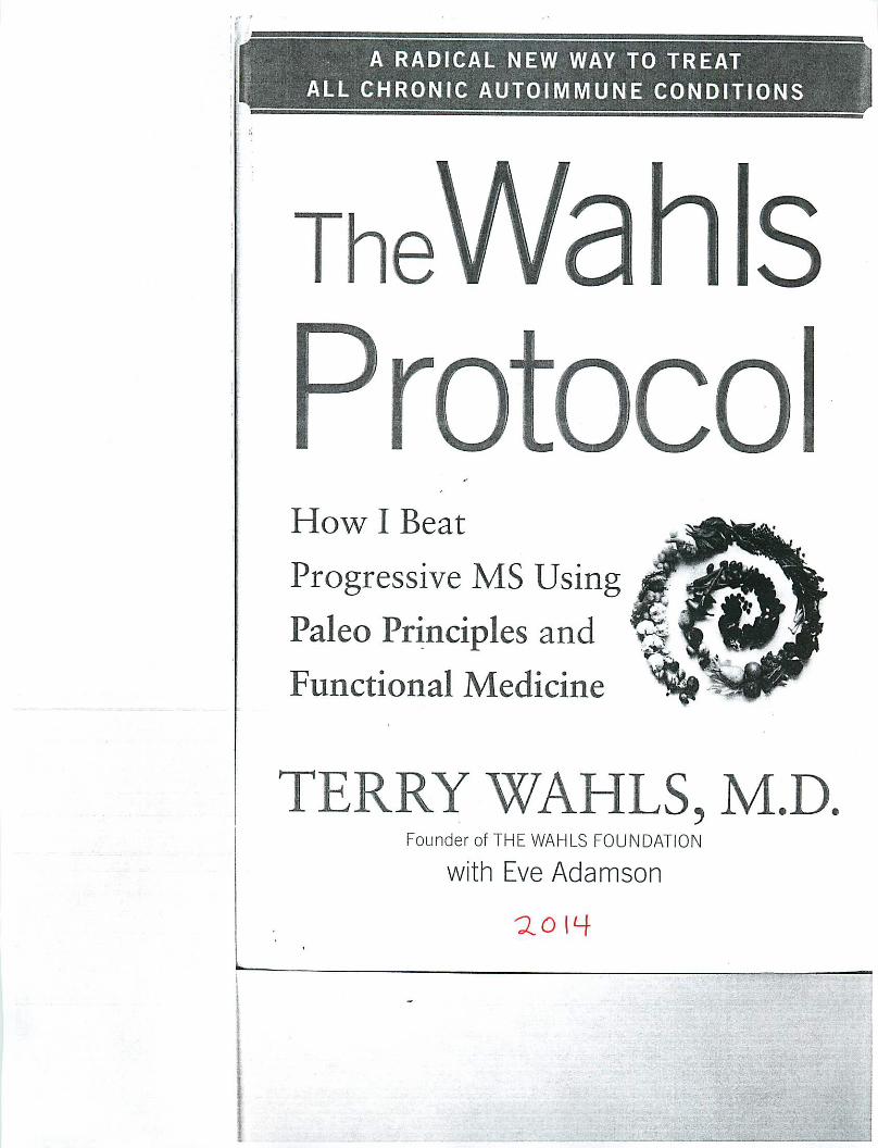
The Wave
2014
NeuroImmunology
Dan Murphy, DC

Recent Books


1 The Key To Preventing Spinal Degeneration, Dysfunction, Pain
Understanding Levers
First Class Lever: load fulcrum effort
Load Effort
Fulcrum 10 lbs. Load Effort
Fulcrum 10 in. 10 in. 100 lbs. 100 lbs. 200 lbs. The effective load (EF) at the fulcrum is the actual load (AL), multiplied by the lever arm (LA), plus the counterbalancing effort (CBE):
EL = 10 lbs. (AL) X 10 in. (LA) + 100 lbs. (CBE) = 200 lbs. (EL)

4 Weight
Muscle Contraction
Fulcrum Rene Cailliet, MD, also uses the first class lever example in his 1996 book Soft Tissue Pain and Disability pertaining to the forward head syndrome (17). The patient has an unbalanced forward head posture. Dr. Cailliet assigns the head a weight of 10 lbs. and displaces the head’s center of gravity forward by 3 inches. The required counter balancing muscle contraction on the opposite side of the fulcrum (the vertebrae) would be 30 lbs. (10 lbs. X 3 inches):

5
The constant muscle contraction required to balance postural distortions creates muscle fatigue and myofascial pain syndromes. Rene Cailliet, MD states “This increase [in muscle tension] not only is fatiguing, but acts as a compressive force on the soft tissues, including the disk.” Dr. Cailliet explains how the constant contraction in the counterbalancing muscles creates a cascade that leads to muscle fatigue, inflammation, fibrosis, and eventually to chronic musculoskeletal pain syndromes:
Chronic Muscle Tension
Internal Retained Tissue Metabolites Ischemia
Inflammation
Fibrosis Reaction
Limited Muscle Elongation Restricted Joint Movement Tendon Function Limitation
Fascial Shortening
Functional Disability


Inflammation
Fibrosis
Stiffness
Reduced Mechanoreception
Increased Sympathetic Tone
Constricted Blood Vessel Diameter
Reduced Delivery of
O2 + G
Reduced ATP

10/31/13 5:55 PMSpinal manipulation in the treatment of lo... [Can Fam Physician. 1985] - PubMed - NCBI
Page 1 of 1http://www.ncbi.nlm.nih.gov/pubmed/21274223
Can Fam Physician. 1985 Mar;31:535-40.
Spinal manipulation in the treatment of low-back pain.Kirkaldy-Willis WH, Cassidy JD.
AbstractSpinal manipulation, one of the oldest forms of therapy for back pain, has mostly been practicedoutside of the medical profession. Over the past decade, there has been an escalation of clinical andbasic science research on manipulative therapy, which has shown that there is a scientific basis forthe treatment of back pain by manipulation. Most family practitioners have neither the time norinclination to master the art of manipulation and will wish to refer their patients to a skilled practitionerof this therapy. Results of spinal manipulation in 283 patients with low back pain are presented. Thephysician who makes use of this resource will provide relief for many patients.
PMID: 21274223 [PubMed] PMCID: PMC2327983 Free PMC Article
Display Settings: Abstract
LinkOut - more resources
PubMed


3/25/14 7:13 PMThe spinal cord as organizer of disease... [J Am Osteopath Assoc. 1979] - PubMed - NCBI
Page 1 of 1http://www.ncbi.nlm.nih.gov/pubmed/583147
How to join PubMed Commons
PubMed Commons home
J Am Osteopath Assoc. 1979 Dec;79(4):232-7.
The spinal cord as organizer of disease processes: III. Hyperactivity ofsympathetic innervation as a common factor in disease.
Korr IM.
PMID: 583147 [PubMed - indexed for MEDLINE]
PubMed Commons
0 comments
Display Settings: Abstract
MeSH Terms
LinkOut - more resources
PubMed


The Sympathetic Nerve—An Integrative Interfacebetween Two Supersystems: The Brain and the
Immune SystemILIA J. ELENKOV, RONALD L. WILDER, GEORGE P. CHROUSOS, AND E. SYLVESTER VIZI1
Inflammatory Joint Diseases Section, Arthritis and Rheumatism Branch, National Institute of Arthritis and Musculoskeletal and SkinDiseases, National Institutes of Health, Bethesda, Maryland (I.J.E., R.L.W.); Pediatric Endocrinology Section, Developmental
Endocrinology Branch, National Institute of Child Health and Human Development, National Institutes of Health, Bethesda, Maryland(I.J.E., G.P.C.); Department of Pharmacology, Institute of Experimental Medicine, Hungarian Academy of Sciences, Budapest, Hungary
(E.S.V.); and Department of Pharmacology and Pharmacotherapy, Semmelweis University, Budapest, Hungary (E.S.V.)
This paper is available online at http://www.pharmrev.org
Abstract . . . . . . . . . . . . . . . . . . . . . . . . . . . . . . . . . . . . . . . . . . . . . . . . . . . . . . . . . . . . . . . . . . . . . . . . . . . . . . 596I. Introduction . . . . . . . . . . . . . . . . . . . . . . . . . . . . . . . . . . . . . . . . . . . . . . . . . . . . . . . . . . . . . . . . . . . . . . . . . . . 597
A. Overview . . . . . . . . . . . . . . . . . . . . . . . . . . . . . . . . . . . . . . . . . . . . . . . . . . . . . . . . . . . . . . . . . . . . . . . . . . 597B. Historical perspectives . . . . . . . . . . . . . . . . . . . . . . . . . . . . . . . . . . . . . . . . . . . . . . . . . . . . . . . . . . . . . . 597
II. Anatomy and physiology of the autonomic nervous system . . . . . . . . . . . . . . . . . . . . . . . . . . . . . . . . . 598A. Organization of the autonomic/sympathetic nervous system . . . . . . . . . . . . . . . . . . . . . . . . . . . . . 598B. Role of sympathetic nervous system and hypothalamo-pituitary-adrenal axis in maintaining
basal and stress-related homeostasis . . . . . . . . . . . . . . . . . . . . . . . . . . . . . . . . . . . . . . . . . . . . . . . . . 599III. Autonomic/sympathetic innervation of lymphoid organs: nonsynaptic communication . . . . . . . . . 599
A. Innervation of the thymus . . . . . . . . . . . . . . . . . . . . . . . . . . . . . . . . . . . . . . . . . . . . . . . . . . . . . . . . . . . 601B. Innervation of the spleen . . . . . . . . . . . . . . . . . . . . . . . . . . . . . . . . . . . . . . . . . . . . . . . . . . . . . . . . . . . . 601C. Innervation of lymph nodes and tonsils . . . . . . . . . . . . . . . . . . . . . . . . . . . . . . . . . . . . . . . . . . . . . . . 601D. Innervation of the bone marrow . . . . . . . . . . . . . . . . . . . . . . . . . . . . . . . . . . . . . . . . . . . . . . . . . . . . . . 601E. Innervation of mucosa-associated lymphoid tissues. . . . . . . . . . . . . . . . . . . . . . . . . . . . . . . . . . . . . 602F. Coexistence patterns . . . . . . . . . . . . . . . . . . . . . . . . . . . . . . . . . . . . . . . . . . . . . . . . . . . . . . . . . . . . . . . . 602G. General pattern of the autonomic/sympathetic innervation of lymphoid organs. . . . . . . . . . . . 602H. Spatial relationships with peptidergic innervation . . . . . . . . . . . . . . . . . . . . . . . . . . . . . . . . . . . . . 603I. Neuroimmune connection in nonorganized lymphoid compartments . . . . . . . . . . . . . . . . . . . . . . 603
IV. Nonsynaptic release of norepinephrine in lymphoid organs: presynaptic modulation and effect ofdrugs . . . . . . . . . . . . . . . . . . . . . . . . . . . . . . . . . . . . . . . . . . . . . . . . . . . . . . . . . . . . . . . . . . . . . . . . . . . . . . . . . 603A. Evidence for neural release of norepinephrine (and dopamine) in lymphoid organs . . . . . . . . 603B. Norepinephrine is released and affects immune cells nonsynaptically . . . . . . . . . . . . . . . . . . . . 604C. Presynaptic modulation of norepinephrine release in lymphoid organs: effect of drugs . . . . . 605D. Release of neuropeptide Y and its action on immune cells. . . . . . . . . . . . . . . . . . . . . . . . . . . . . . . 606
V. Systemic and local effects of cytokines on sympathetic nervous system activity. . . . . . . . . . . . . . . 606A. Systemic effects: long feedback loop between the immune system and the brain. . . . . . . . . . . 606B. Local effects of tumor necrosis factor-! and interleukin-1 . . . . . . . . . . . . . . . . . . . . . . . . . . . . . . . 607
VI. Expression of adrenoreceptors on lymphoid cells: signal transduction . . . . . . . . . . . . . . . . . . . . . . . 608A. Expression and distribution of adrenoreceptors on lymphoid cells. . . . . . . . . . . . . . . . . . . . . . . . 608B. Signal pathways and molecular aspects of catecholamines actions . . . . . . . . . . . . . . . . . . . . . . . 609
1. Cyclic adenosine 5!-monophosphate . . . . . . . . . . . . . . . . . . . . . . . . . . . . . . . . . . . . . . . . . . . . . . . 6092. Intracellular Ca2" . . . . . . . . . . . . . . . . . . . . . . . . . . . . . . . . . . . . . . . . . . . . . . . . . . . . . . . . . . . . . . . 610
VII. Role of sympathetic innervation in immune system development and hematopoiesis . . . . . . . . . . 611A. Immune system development . . . . . . . . . . . . . . . . . . . . . . . . . . . . . . . . . . . . . . . . . . . . . . . . . . . . . . . . 611B. Hematopoiesis. . . . . . . . . . . . . . . . . . . . . . . . . . . . . . . . . . . . . . . . . . . . . . . . . . . . . . . . . . . . . . . . . . . . . . 611C. Thymocyte development . . . . . . . . . . . . . . . . . . . . . . . . . . . . . . . . . . . . . . . . . . . . . . . . . . . . . . . . . . . . . 612
VIII. Sympathetic control of lymphocyte traffic and circulation . . . . . . . . . . . . . . . . . . . . . . . . . . . . . . . . . . 612
1 Address for correspondence: Dr. E. Sylvester Vizi, Department of Pharmacology, Institute of Experimental Medicine, HungarianAcademy of Sciences, H-1450 Budapest, P.O. Box 67, Hungary. E-mail: [email protected]
0031-6997/00/5204-0595$03.00/0PHARMACOLOGICAL REVIEWS Vol. 52, No. 4U.S. Government work not protected by U.S. copyright 41/865371Pharmacol Rev 52:595–638, 2000 Printed in U.S.A
595

11/29/13 3:22 PMAutonomic innervation and regulation of th... [Brain Behav Immun. 2007] - PubMed - NCBI
Page 1 of 2http://www.ncbi.nlm.nih.gov/pubmed/17467231
Brain Behav Immun. 2007 Aug;21(6):736-45. Epub 2007 Apr 27.
Autonomic innervation and regulation of the immune system (1987-2007).Nance DM, Sanders VM.Susan Samueli Center for Integrative Medicine, University of California Irvine, Orange, CA 92868-4283, [email protected]
AbstractSince 1987, only a few neuroanatomical studies have been conducted to identify the origin ofinnervation for the immune system. These studies demonstrated that all primary and secondaryimmune organs receive a substantial sympathetic innervation from sympathetic postganglionicneurons. Neither the thymus nor spleen receive any sensory neural innervation; however, there isevidence that lymph nodes and bone marrow may be innervated by sensory neurons located indorsal root ganglia. There is no neuroanatomical evidence for a parasympathetic or vagal nervesupply to any immune organ. Thus, the primary pathway for the neural regulation of immune functionis provided by the sympathetic nervous system (SNS) and its main neurotransmitter, norepinephrine(NE). Activation of the SNS primarily inhibits the activity of cells associated with the innate immunesystem, while it either enhances or inhibits the activity of cells associated with the acquired/adaptiveimmune system. Innate immune cells express both alpha and beta-adrenergic receptor subtypes,while T and B lymphocytes express adrenergic receptors of the beta2 subtype exclusively, except formurine Th2 cells that lack expression of any subtype. Via these adrenergic receptors, NE is able toregulate the level of immune cell activity by initiating a change in the level of cellular activity, whichoften involves a change in the level of gene expression for cytokines and antibodies.
PMID: 17467231 [PubMed - indexed for MEDLINE] PMCID: PMC1986730 Free PMC Article
Display Settings: Abstract
Images from this publication. See all images (1) Freetext
Publication Types, MeSH Terms, Grant Support
PubMed

5/28/13 4:15 PMPubMed Central, FIGURE 1: Brain Behav Immun. 2007 August; 21(6): 736–745. Published online 2007 April 27. doi: 10.1016/j.bbi.2007.03.008
Page 1 of 2http://www.ncbi.nlm.nih.gov/pmc/articles/PMC1986730/figure/F1/?report=objectonly
FIGURE 1
All primary and secondary immune organs receive a substantial sympathetic innervation from sympatheticpostganglionic neurons. There is no neuroanatomical evidence for a parasympathetic or vagal nerve supply to anyimmune organ. Input to the brain comes from sensory, e.g., dorsal root ganglia, or immune stimuli, e.g., cytokines.The primary pathway for the neural regulation of immune function is provided by the sympathetic nervous systemand its main neurotransmitter, norepinephrine. Activation of the SNS primarily inhibits the activity of cellsassociated with the innate immune system, while it either enhances or inhibits the activity of cells associated with


“The Innate Immune System”
Chapter 2
How The immune System Works
By Lauren Sompayrac, PhD
Department of Molecular, Cellular, and
Developmental Biology University of Colorado, Boulder
Blackwell Science
1999 “Until recently, most immunologists didn’t pay much attention to the innate system, perhaps because the adaptive system seemed more exciting. However, studies of the adaptive immune system have led to a new appreciation of the role that the innate system plays, not only as a second line of defense (if we count physical barriers as our first defense), but also as an activator and a controller of the adaptive response.” p. 17


Mechanoreceptors
- - Sympathetic Tone Pain Firing Catecholamines Gate Theory Epinephrine Norepinephrine Alter Epigenetic Methylation Shorten Telomeres Cause Vasoconstriction Reduces ATP Reduces healing Reduced pumps Na++ glutamate Immunosuppressive Increase Oxidative Stress

Whole Spine Quote
“We tend to divide the examination of the spine intoregions: cervical, thoracic, and lumbar spine clinical
studies.
This is a mistake.
The three units are closely interrelated structurallyand functionally – a whole person with a whole spine.
The cervical spine may be symptomatic because of athoracic or lumbar spine abnormality, and vice versa!
Sometimes treating a lumbar spine will relieve acervical spine syndrome, or proper management of
cervical spine will relieve low backache.”
Disorders of the Cervical Spine
John Bland, MD
Professor of Medicine, University of Vermont Collegeof Medicine
WB Saunders Company1987
Page 84

A hypothesis of chronic back pain: Ligament subfailure injuries lead to muscle control dysfunction
European Spine Journal
May 2006
Manohar Panjabi Yale University School of Medicine

4/30/14, 4:57 PMCerebral metabolic changes in... [Altern Ther Health Med. 2011 Nov-Dec] - PubMed - NCBI
Page 1 of 2http://www.ncbi.nlm.nih.gov/pubmed/22314714
BACKGROUND:
OBJECTIVE:
METHODS:
RESULTS:
Altern Ther Health Med. 2011 Nov-Dec;17(6):12-7.
Cerebral metabolic changes in men after chiropractic spinal manipulation forneck pain.Ogura T , Tashiro M, Masud M, Watanuki S, Shibuya K, Yamaguchi K, Itoh M, Fukuda H, Yanai K.
AbstractChiropractic spinal manipulation (CSM) is an alternative treatment for back pain.
The autonomic nervous system is often involved in spinal dysfunction. Although studies on the effectsof CSM have been performed, no chiropractic study has examined regional cerebral metabolismusing positron emission tomography (PET).
The aim of the present study was to investigate the effects of CSM on brain responsesin terms of cerebral glucose metabolic changes measured by [18F]fluorodeoxyglucose positronemission tomography (FDG-PET).
Twelve male volunteers were recruited. Brain PET scanning was performed twice oneach participant, at resting and after CSM. Questionnaires were used for subjective evaluations. Avisual analogue scale (VAS) was rated by participants before and after chiropractic treatment, andmuscle tone and salivary amylase were measured.
Increased glucose metabolism was observed in the inferior prefrontal cortex, anteriorcingulated cortex, and middle temporal gyrus, and decreased glucose metabolism was found in thecerebellar vermis and visual association cortex, in the treatment condition (P < .001). Comparisons ofquestionnaires indicated a lower stress level and better quality of life in the treatment condition. Asignificantly lower VAS was noted after CSM. Cervical muscle tone and salivary amylase weredecreased after CSM. Conclusion The results of this study suggest that CSM affects regional cerebralglucose metabolism related to sympathetic relaxation and pain reduction.
PMID: 22314714 [PubMed - indexed for MEDLINE]
Display Settings: Abstract
1
Author information
Publication Types, MeSH Terms, Substances
LinkOut - more resources
PubMed


1 Sensory and Autonomic Innervation of the Cervical Intervertebral Disc
The Pathomechanics of Chronic Discogenic Neck Pain
Spine July 15, 2012 Volume 37, Number 16, pp. 1357–1362
Kazuki Fujimoto, MD; Masayuki Miyagi, MD; Tetsuhiro Ishikawa, MD; Gen Inoue, MD, PhD; and 16 more; Primary authors from the Department of Orthopaedic Surgery, Graduate School of Medicine, Chiba University, Chiba, Japan KEY POINTS FROM DAN MURPHY 1) This study used rats. The authors note that prior studies have shown that rats and humans have similar spinal innervation patterns. However, they also note that these findings should be reaffirmed on human specimens. 2) This study is an immunohistological analysis of the cervical intervertebral disc (IVD) to investigate its sensory and autonomic innervation. The authors used the neuro-tracer Fluoro-gold (FG) to stain ten C5–C6 IVDs. They stained for the following neurons: • Dorsal root ganglions (DRG) from level C1–C8 [sensory {afferents} cell bodies; this would include pain afferents] • Stellate ganglion (SG) [inferior cervical sympathetic efferent ganglion] • Nodose ganglion (NG) [Vagus nerve parasympathetic sensory ganglion to the nucleus tractus solitarius]. 3) Several studies have reported that sympathetic nerves participate in chronic pain; sympathectomy procedures can effectively treat chronic pain. The DRGs, SG, and NG neurons were immune-stained for: • Calcitonin gene-related peptide (CGRP); found in inflammatory pain neurons. • Isolectin B4 (IB4); found in neuropathic (nerve injury) neurons. 4) Findings for the innervation of the C5-C6 IVD: • The neurons innervating the C5–C6 IVD were derived from the C2–C8 DRGs, but not from the C1 DRG. • 3% of the neurons were for neuropathic (nerve injury) pain:
Isolectin B4 (IB4); [therefore pain afferents] • 21% of the neurons were for inflammatory pain:
Calcitonin gene-related peptide (CGRP); [therefore pain afferents]

1
Is Elevated Noradrenaline [Norepinephrine] an Aetiological Factor in a
Number of Diseases?
Autonomic & Autacoid Pharmacology
October 2009, Vol. 29, No 4.; pp. 143-156
P. J. Fitzgerald (From the Department of Neuroscience, Johns Hopkins University)
Noradrenaline (NA) = Norepinephrine; Sympathetic Nervous System (SNS)
This is a theoretical study with 153 references to support the biological plausibilityof how increased sympathetic nervous system tone and release of NA is a
etiological factor in a broad range of diseases, including, cancer, bipolar disorder,
excessive body weight, hypertension, type I autoimmune diabetes, type II diabetes,
glaucoma, osteoarthritis, rheumatoid arthritis, asthma, and immune systemfunction.
FROM ABSTRACT:
1) Noradrenaline (NA) is a signaling molecule in the brain and sympatheticnervous system (SNS).
2) Elevated NA is a factor in various types of cancer.
3) Elevated sympathetic tone (elevated NA levels) predisposes a large number of
individuals to a broad range of diseases.
4) Psychological stress is associated with increased release of NA which maycause or exacerbate diseases.
THIS AUTHOR ALSO NOTES:
5) “Noradrenaline (NA) is the principle signaling molecule used at the output ofthe sympathetic nervous system (SNS), and thereby plays a critical role in the
body’s ‘fight or flight’ response to environmental stressors.”
6) The parasympathetic nervous system, whose principle signaling molecule isacetylcholine, “is more responsible for maintenance processes” know as ‘rest and
digest’ functions.
7) The function of the SNS between individuals may be significant as aconsequence of genetic differences; this includes the genetic ability to produce NA
as well as the number of receptors (adrenoreceptors) NA uses to influence
physiology.
8) There are genetic differences in NA sympathetic tone. [tone]

2
9) Different organs have different sympathetic innervation patterns and thus
different sympathetic tones that can influence diseases in these organs.
10) Also, different diseases may share comorbidity because of shared patterns of
sympathetic innervation.
11) Some individuals may have elevated sympathetic tone with “continuously
elevated ‘fight or flight’ mode wherein the body is not devoting sufficient resources
toward maintenance or homeostatic processes. These individuals may also have a
deficiency of parasympathetic ‘rest and digest’ tone.”
12) There are multiple lines of evidence showing that elevated NA levels from the
SNS and adrenaline from the adrenal glands are etiological factors in diabetes
mellitus, glaucoma, osteoarthritis, rheumatoid arthritis, and asthma.
13) The SNS innervates the lymphoid (immune) organs and thus modulates the
immune response.
14) SNS NA may cause autoimmune diseases, including the “autoimmunedestruction of the insulin secreting pancreatic beta cells,” and therefore type I
diabetes.
15) SNS NA also regulates the release of insulin from the pancreatic beta cells(type II diabetes).
16) Elevated intraocular pressure damages the optic nerve. Increased SNS NA
increases the secretions of the aqueous humour and also decreases the drainage,thereby elevating intraocular pressure (glaucoma).
17) The radial muscles of the eye are innervated by the SNS, and they dilate the
pupil. Increased sympathetic tone causes radial muscle hypertrophy, which is also
associated with glaucoma.
18) Elevated SNS tone may also cause glaucoma by altering the tone of the blood
vessels of the eye.
19) SNS fibers innervate the joints. Elevated SNS tone increase inflammation
from a number of mechanism, including increased production of inflammatory
cytokines, resulting in degeneration (osteoarthritis).
20) Elevated SNS tone causes vasoconstriction, inflammation, and degeneration
(osteoarthritis).
21) SNS catecholamine production of pro-inflammatory cytokines can perpetuatethe inflammation of rheumatoid arthritis.

3
22) SNS fibers innervate human airways and can cause chronic inflammation
and asthma.
23) Elevated SNS NA and increased sympathetic tone should be viewed in two
categories:
Phasic: A transient burst of NA
Tonic: The steady output of the SNS
[During exercise there is elevated sympathetic tone {phasic} and release of NA, but
in the periods between exercise the sympathetic tone is reduced {tonic} because of
that same exercise.]
[An adjustment may spike the sympathetic tone {phasic} and release of NA, butthe improvement of biomechanical function that occurred as a consequence of that
adjustment may reduce the steady output from the SNS {tonic}.]
24) “Tonic NA, through its sustained presence, is the more important aetiological
factor in … diseases than phasic NA.” [Important]Genetics Environment
Increased Sympathetic Tone
Increased Release of Noradrenaline
Increased Adrenoreceptor Activation
Increased Second Messenger Activation
Insulin Immune Joint Airway Increased
Dysregulation System Inflammation Inflammation Intraocular
Dysregualtion Pressure
Diabetes Autoimmune Osteoarthritis Asthma Glaucoma
Diseases Rheumatoid
Arthritis
[InfectionProblems]
COMMENTS FROM DAN MURPHY
Since the time of Korr (1979) there has been biological plausibility that the
subluxation complex results in increased sustained sympathetic tone (tonic),
becoming an etiological factor in visceral disease, and that improvement of
mechanical integrity (the adjustment) inhibits this adverse tonic release of NA.
This article adds to that biological plausibility.

Behavioral/Systems/Cognitive
The Neurochemically Diverse Intermedius Nucleus of theMedulla as a Source of Excitatory and Inhibitory SynapticInput to the Nucleus Tractus Solitarii
Ian J. Edwards,1* Mark L. Dallas,1* Sarah L. Poole,1 Carol J. Milligan,1 Yuchio Yanagawa,2 Gabor Szabo,3
Ferenc Erdelyi,3 Susan A. Deuchars,1 and Jim Deuchars1
1Institute of Membrane and Systems Biology, University of Leeds, Leeds LS2 9JT, United Kingdom, 2Department of Genetic and Behavioral Neuroscience,Gunma University Graduate School of Medicine, and Solution Oriented Research for Science and Technology, Japan Science and Technology Agency,Maebashi 371-8511, Japan, and 3Department of Gene Technology and Developmental Neurobiology, Institute of Experimental Medicine, H-1450 Budapest,Hungary
Sensory afferent signals from neck muscles have been postulated to influence central cardiorespiratory control as components ofpostural reflexes, but neuronal pathways for this action have not been identified. The intermedius nucleus of the medulla (InM) is a targetof neck muscle spindle afferents and is ideally located to influence such reflexes but is poorly investigated. To aid identification of thenucleus, we initially produced three-dimensional reconstructions of the InM in both mouse and rat. Neurochemical analysis includingtransgenic reporter mice expressing green fluorescent protein in GABA-synthesizing neurons, immunohistochemistry, and in situ hy-bridization revealed that the InM is neurochemically diverse, containing GABAegric and glutamatergic neurons with some degree ofcolocalization with parvalbumin, neuronal nitric oxide synthase, and calretinin. Projections from the InM to the nucleus tractus solitarius(NTS) were studied electrophysiologically in rat brainstem slices. Electrical stimulation of the NTS resulted in antidromically activatedaction potentials within InM neurons. In addition, electrical stimulation of the InM resulted in EPSPs that were mediated by excitatoryamino acids and IPSPs mediated solely by GABAA receptors or by GABAA and glycine receptors. Chemical stimulation of the InM resultedin (1) a depolarization of NTS neurons that were blocked by NBQX (2,3-dioxo-6-nitro-1,2,3,4-tetrahydrobenzo[f ]quinoxaline-7-sulfonoamide) or kynurenic acid and (2) a hyperpolarization of NTS neurons that were blocked by bicuculline. Thus, the InM containsneurochemically diverse neurons and sends both excitatory and inhibitory projections to the NTS. These data provide a novel pathwaythat may underlie possible reflex changes in autonomic variables after neck muscle spindle afferent activation.
Key words: posture; neck; cardiovascular; respiration; medulla oblongata; autonomic
IntroductionReflex changes in cardiorespiratory variables during body move-ments rely on interactions between the somatic and autonomicnervous systems. A prime example of such interaction is the so-matosympathetic reflex, in which stimulation of thinly myelin-ated group III (A�) and unmyelinated group IV (C-fiber) limbmuscle afferent fibers can reflexly increase cardiorespiratory out-put (Potts et al., 2000, 2003; Wilson, 2000). These reflexes aremediated via sensory afferent input to the spinal cord, which isthen relayed to the nucleus tractus solitarius (NTS), a brainstem
site for cardiorespiratory integration (Potts et al., 2003). Cardiore-spiratory changes can also be evoked by stimulation of neck muscleafferents (Bolton et al., 1998; Bolton and Ray, 2000), proposed tocontribute to alterations in cardiorespiratory outflow in preparationfor a change in posture (Bolton and Ray, 2000). In contrast to limbafferents, the sensory signals from these muscles appear to be medi-ated by group IA muscle spindle afferents (Bolton et al., 1998). How-ever, the neural pathways that link these afferent signals to cardiore-spiratory control are completely unknown.
One target for sensory information from neck muscles is thecervical spinal cord where terminations can be found in the dor-sal horn (although sparse) and the central cervical nucleus (CCN)(Bakker et al., 1984; Pfaller and Arvidsson, 1988; Prihoda et al.,1991). The CCN projection is generally considered to underliespinal somatic reflex circuits, such as those for the tonic neckreflex involved in postural control (Wilson et al., 1984; Brink etal., 1985; Hongo et al., 1988; Popova et al., 1995). There is also astrong direct neck muscle afferent projection to the medulla ob-longata where fibers terminate in the external cuneate nucleusand a nucleus located at the lateral edges of the dorsal aspect ofthe hypoglossal motor nucleus (XII), referred to either as the
Received Feb. 13, 2007; revised May 25, 2007; accepted June 20, 2007.This work was supported in part by the Wellcome Trust (C.J.M. and J.D.) and Grants-in-Aid for Scientific Research
from the Ministry of Education, Culture, Sports, Science and Technology and the Ministry of Health, Labor, andWelfare, Japan (Y.Y.). I.J.E. was supported by the Biotechnology and Biological Sciences Research Council. Weacknowledge the contribution of Gareth Dobson, who was an undergraduate project student, to this work.
*I.J.E. and M.L.D. contributed equally and significantly to this work.Correspondence should be addressed to either Jim Deuchars or Susan A. Deuchars, Institute of Membrane and
Systems Biology, Garstang Building, University of Leeds, Leeds LS2 9JT, UK. E-mail: [email protected] [email protected].
DOI:10.1523/JNEUROSCI.0638-07.2007Copyright © 2007 Society for Neuroscience 0270-6474/07/278324-10$15.00/0
8324 • The Journal of Neuroscience, August 1, 2007 • 27(31):8324 – 8333

The Neurochemically Diverse Intermedius Nucleus of the Medulla as a Source of Excitatory andInhibitory Synaptic Input to the Nucleus Tractus Solitarii
The Journal of NeuroscienceAugust 1, 2007
Dorsal MotorNucleusof theVagus
ParasympatheticEfferents
Heart
Lungs
Stomach
Intestines
Etc.
NucleusTractus
Solitarius
IntegratedAutonomicAndCardiorespiratoryCircuits
ParasympatheticAfferents
FromThoracic
AndAbdominal
Viscera
Cerebellum
ExternalCuneateNucleus
NucleusIntermedius
CentralCervicalNucleus
TonicPosturalReflexes
UpperCervical
MechanoreceptorsFrom
ChiropracticUpper
CervicalAdjustments
Review
The intermedius nucleus of the medulla: A potential site for the integration ofcervical information and the generation of autonomic responses
Ian J. Edwards, Susan A. Deuchars, Jim Deuchars *
Institute of Membrane and Systems Biology, Garstang Building, University of Leeds, Leeds, LS2 9JT, United Kingdom
Contents
1. Introduction . . . . . . . . . . . . . . . . . . . . . . . . . . . . . . . . . . . . . . . . . . . . . . . . . . . . . . . . . . . . . . . . . . . . . . . . . . . . . . . . . . . . . . . . . . . . . . . . . . . . . 167
1.1. Nomenclature . . . . . . . . . . . . . . . . . . . . . . . . . . . . . . . . . . . . . . . . . . . . . . . . . . . . . . . . . . . . . . . . . . . . . . . . . . . . . . . . . . . . . . . . . . . . . . 167
1.2. Insights into function . . . . . . . . . . . . . . . . . . . . . . . . . . . . . . . . . . . . . . . . . . . . . . . . . . . . . . . . . . . . . . . . . . . . . . . . . . . . . . . . . . . . . . . . 167
2. Neurotransmitters . . . . . . . . . . . . . . . . . . . . . . . . . . . . . . . . . . . . . . . . . . . . . . . . . . . . . . . . . . . . . . . . . . . . . . . . . . . . . . . . . . . . . . . . . . . . . . . . 168
2.1. Amino acids . . . . . . . . . . . . . . . . . . . . . . . . . . . . . . . . . . . . . . . . . . . . . . . . . . . . . . . . . . . . . . . . . . . . . . . . . . . . . . . . . . . . . . . . . . . . . . . 168
2.2. NOS . . . . . . . . . . . . . . . . . . . . . . . . . . . . . . . . . . . . . . . . . . . . . . . . . . . . . . . . . . . . . . . . . . . . . . . . . . . . . . . . . . . . . . . . . . . . . . . . . . . . . . 170
2.3. Peptide transmitters. . . . . . . . . . . . . . . . . . . . . . . . . . . . . . . . . . . . . . . . . . . . . . . . . . . . . . . . . . . . . . . . . . . . . . . . . . . . . . . . . . . . . . . . . 170
3. Calcium binding proteins . . . . . . . . . . . . . . . . . . . . . . . . . . . . . . . . . . . . . . . . . . . . . . . . . . . . . . . . . . . . . . . . . . . . . . . . . . . . . . . . . . . . . . . . . . 171
3.1. Parvalbumin is predominantly found in inhibitory neurones . . . . . . . . . . . . . . . . . . . . . . . . . . . . . . . . . . . . . . . . . . . . . . . . . . . . . . . . 171
3.2. Calretinin is found within inhibitory and excitatory neurones in the InM . . . . . . . . . . . . . . . . . . . . . . . . . . . . . . . . . . . . . . . . . . . . . . 171
4. Receptors . . . . . . . . . . . . . . . . . . . . . . . . . . . . . . . . . . . . . . . . . . . . . . . . . . . . . . . . . . . . . . . . . . . . . . . . . . . . . . . . . . . . . . . . . . . . . . . . . . . . . . . 171
4.1. Glutamate receptors. . . . . . . . . . . . . . . . . . . . . . . . . . . . . . . . . . . . . . . . . . . . . . . . . . . . . . . . . . . . . . . . . . . . . . . . . . . . . . . . . . . . . . . . . 171
Journal of Chemical Neuroanatomy 38 (2009) 166–175
A R T I C L E I N F O
Article history:
Received 24 September 2008
Received in revised form 6 January 2009
Accepted 6 January 2009
Available online 14 January 2009
Keywords:
Autonomic
Proprioception
Perihypoglossal
Brainstem
A B S T R A C T
The intermedius nucleus of the medulla (InM) is a small perihypoglossal brainstem nucleus, which
receives afferent information from the neck musculature and also descending inputs from the vestibular
nuclei, the gustatory portion of the nucleus of the solitary tract (NTS) and cortical areas involved in
movements of the tongue. The InM sends monosynaptic projections to both the NTS and the hypoglossal
nucleus. It is likely that the InM acts to integrate information from the head and neck and relays this
information on to the NTS where suitable autonomic responses can be generated, and also to the
hypoglossal nucleus to influence movements of the tongue and upper airways.
Central to the integratory role of the InM is its neurochemical diversity. Neurones within the InM
utilise the amino acid transmitters glutamate, GABA and glycine. A proportion of these excitatory and
inhibitory neurones also use nitric oxide as a neurotransmitter. Peptidergic transmitters have also been
found within InM neurones, although as yet the extent of the pattern of co-localisation between
peptidergic and amino acid transmitters in neurones has not been established.
The calcium binding proteins calretinin and parvalbumin are found within the InM in partially
overlapping populations. Parvalbumin and calretinin appear to have complementary distributions
within the InM, with parvalbumin being predominantly found within GABAergic neurones and calretinin
being predominantly found within glutamatergic neurones.
Neurones in the InM receive inputs from glutamatergic sensory afferents. This glutamatergic
transmission is conducted through both NMDA and AMPA ionotropic glutamate receptors.
In summary the InM contains a mixed pool of neurones including glutamatergic and GABAergic in
addition to peptidergic neurones. Neurones within the InM receive inputs from the upper cervical region,
descending inputs from brain regions involved in tongue movements and those involved in the co-
ordination of the autonomic nervous system. Outputs from the InM to the NTS and hypoglossal nucleus
suggest a possible role in the co-ordination of tongue movements and autonomic responses to changes in
posture.
� 2009 Elsevier B.V. All rights reserved.
* Corresponding author. Tel.: +44 113 343 4249.
E-mail address: [email protected] (J. Deuchars).
Contents lists available at ScienceDirect
Journal of Chemical Neuroanatomy
journa l homepage: www.e lsev ier .com/ locate / jchemneu
0891-0618/$ – see front matter � 2009 Elsevier B.V. All rights reserved.
doi:10.1016/j.jchemneu.2009.01.001

7/23/14, 9:04 AMNeck muscle afferents influence oromotor ... [Brain Struct Funct. 2014] - PubMed - NCBI
Page 1 of 2http://www.ncbi.nlm.nih.gov/pubmed/24595534
Brain Struct Funct. 2014 Mar 5. [Epub ahead of print]
Neck muscle afferents influence oromotor and cardiorespiratory brainstemneural circuits.Edwards IJ , Lall VK, Paton JF, Yanagawa Y, Szabo G, Deuchars SA, Deuchars J.
School of Biomedical Sciences, University of Leeds, Leeds, LS2 9JT, UK, [email protected].
AbstractSensory information arising from the upper neck is important in the reflex control of posture and eyeposition. It has also been linked to the autonomic control of the cardiovascular and respiratorysystems. Whiplash associated disorders (WAD) and cervical dystonia, which involve disturbance tothe neck region, can often present with abnormalities to the oromotor, respiratory and cardiovascularsystems. We investigated the potential neural pathways underlying such symptoms. Simulating neckafferent activity by electrical stimulation of the second cervical nerve in a working heart brainstempreparation (WHBP) altered the pattern of central respiratory drive and increased perfusion pressure.Tracing central targets of these sensory afferents revealed projections to the intermedius nucleus ofthe medulla (InM). These anterogradely labelled afferents co-localised with parvalbumin and vesicularglutamate transporter 1 indicating that they are proprioceptive. Anterograde tracing from the InMidentified projections to brain regions involved in respiratory, cardiovascular, postural and oro-facialbehaviours-the neighbouring hypoglossal nucleus, facial and motor trigeminal nuclei, parabrachialnuclei, rostral and caudal ventrolateral medulla and nucleus ambiguus. In brain slices, electricalstimulation of afferent fibre tracts lateral to the cuneate nucleus monosynaptically excited InMneurones. Direct stimulation of the InM in the WHBP mimicked the response of second cervical nervestimulation. These results provide evidence of pathways linking upper cervical sensory afferents withCNS areas involved in autonomic and oromotor control, via the InM. Disruption of these neuronalpathways could, therefore, explain the dysphagic and cardiorespiratory abnormalities which mayaccompany cervical dystonia and WAD.
PMID: 24595534 [PubMed - as supplied by publisher]
Display Settings: Abstract
1
Author information1
LinkOut - more resources
PubMed


5 [an important
role in cardiorespiratory
control]
Oromotor Control
[a pontine viscerosensory
relay]
Orofacial Control
To Phrenic Nerve for
Inspiratory Activity
[fovea, clarity of vision]
Pontine Parabrachial
Nucleus
CN V
CN VII
C4—C5—C6 Motor
Neurons
Eye Position
Hypoglossal Nucleus
NUCLEUS INTERMEDIUS
C1—C3 MECHANOS
Vestibular Nucleus
Tongue, Swallowing,
Airway Patency
Splanchnic Sympathetic
Nerves
Nucleus Ambiguus
Caudal Ventrolateral
Medulla
Nucleus Tractus
Solitarius
Autonomic Innervation to and From the
Viscera
Posture
Muscles of the Soft Palate,
Pharynx, Larynx
Inhibits Sympathetic
Tone and Blood Pressure
The Integratory
Center
Most of the Sympathetic Nerves in the
Body are Splanchnic
Regulation of Reflex
Cardiovascular Activity and Modulate
Respiratory Functions
Respiratory and
Cardiovascular Behaviors


From The Wall Street Journal
WWI: A CENTURY LATER
World War I: The War That Changed Everything World War I began 100 years ago this month, and in many ways, writes historian Margaret MacMillan, it remains the defining conflict of the modern era.
By MARGARET MACMILLAN Updated June 20, 2014 10:54 p.m. ET The cold numbers capture much of the war's horror: more than 9 million men dead and twice as many again wounded—a loss of sons, husbands and fathers but also of skills and talents. Graves in the north of France and Belgium and war memorials across the U.S. bear witness to the 53,000 American soldiers who died. Thousands of civilians died, too, during the war itself, whether of hunger, disease or violence. And then, as the guns were falling silent, a new pestilence struck humanity in the shape of a virulent influenza. As troops returned home, they unwittingly helped carry the disease around the world. It has been estimated that 50 million died.


1The Official History Of Chiropractic in Texas
By Walter R Rhodes, DCPublished by the Texas Chiropractic Association
1978
CHAPTER VI:THE THREE GREAT SURVIVAL FACTORS
[Excerpts by Dan Murphy, DC]
“The 1917 - 1918 influenza epidemic swept silently across the world bringingdeath and fear to homes in every land. Disease and pestilence, especially theepidemics, are little understood even now and many of the factors that spread themare still mysterious shadows, but in 1917-1918 almost nothing was known aboutprevention, protection, treatment or cure of influenza. The whole world stood at itsmercy, or lack of it.”
“But out of that particular epidemic, the young science of chiropractic grewinto a new measure of safety. While many struggles would lie ahead this successfulpassage of the profession into early maturity assured its immediate survival andmade the eventual outcome of chiropractic a matter for optimism. If there had beenany lack of enthusiasm among the doctors of chiropractic, or a depleting of thesources of students then the epidemic took care of them too. These chiropracticsurvivors of the flu epidemic were sure, assured, determined, and ready to fightany battle that came up. The effect of the epidemic becomes evident in interviewsmade with old-timers practicing in those years. The refrain comes repeatedly,”
‘I was about to go out of business when the flu epidemic came - but when it was over, I was firmly established in practice.’
“Why?The answer is reasonably simple. Chiropractors got fantastic results from
influenza patients while those under medical care died like flies all around.”
“Statistics reflect a most amazing, almost miraculous state of affairs. Themedical profession was practically helpless with the flu victims but chiropractorsseemed able to do no wrong.”
“In Davenport, Iowa, 50 medical doctors treated 4,953 cases, with 274deaths. In the same city, 150 chiropractors including students and faculty of thePalmer School of Chiropractic, treated 1,635 cases with only one death.”
“In the state of Iowa, medical doctors treated 93,590 patients, with 6,116deaths - a loss of one patient out of every 15. In the same state, excludingDavenport, 4,735 patients were treated by chiropractors with a loss of only 6 cases- a loss of one patient out of every 789.”

Psychological stress and susceptibility to the common cold.
N Engl J Med 1991 Aug 29;325(9):606-612, 654 Cohen S, Tyrrell DA, Smith AP. Department of Psychology, Carnegie Mellon University, Pittsburgh These authors prospectively studied the relation between psychological stress and the frequency of documented clinical colds among subjects intentionally exposed to respiratory viruses. After completing questionnaires assessing degrees of psychological stress, 394 healthy subjects were given nasal drops containing one of five respiratory viruses. The subjects were then quarantined and monitored for the development of evidence of infection and symptoms. The rates of both respiratory infection and clinical colds increased in a dose-response manner with increases in the degree of psychological stress. Infection rates ranged from approximately 74 percent to approximately 90 percent, according to levels of psychological stress, and the incidence of clinical colds ranged from approximately 27 percent to 47 percent. CONCLUSIONS: Psychological stress was associated in a dose-response manner with an increased risk of acute infectious respiratory illness. ALSO NOTED IN ARTICLE AND ITS FOLLOW-UP: “Psychological stress is thought to influence immune function through autonomic nerves innervating lymphoid tissue, or hormone-mediated alteration of immune cells.” “In biomedical terms, ‘stress’ refers to any adverse physical, mental, or emotional stimulus (stressor) that upsets the organism’s homeostasis.” “At the physiological level, stress has been the subject of study going back to Walter B. Cannon’s description of the ‘fight or flight’ response involving actions of the sympathetic nervous system and the adrenal medulla.”

Nonsynaptic noradrenaline release in neuro-immune responses.
Acta Biol Hung. 2002;53(1-2):229-44.
Vizi ES, Elenkov IJ. Evidence has recently been obtained that the branches of the autonomic nervous system, mainly, the sympathetic, regulate cytokine production. Not only the primary (thymus, bone marrow) and secondary (spleen, tonsils, and lymph nodes) lymphoid organs, but also many other tissues are involved in immune responses and are heavily influenced by noradrenaline (NA) derived from varicose axon terminals of the sympathetic nervous system. Besides NA released from nonsynaptic varicosities of noradrenergic terminals, circulating catecholamines (adrenaline, dopamine, NA) are also able to influence immune responses, the production of pro- and anti-inflammatory cytokines by different immune cells. The sympathetic nervous system (catecholamines) and the hypothalamic-pituitary-adrenal (HPA) axis (cortisol) are the major integrative and regulatory components of different immune responses. NA released non-synaptically from sympathetic axon terminals is able to inhibit production of pro-inflammatory (TNF-alpha, IFN-gamma, IL-12, IL-1) and increase anti-inflammatory cytokines (IL-10) in response. The effects are mediated via beta2-adrenoceptors expressed on immune cells and coupled to cAMP levels.


1
Sympathetic Segmental Disturbances
The Evidences of the Association, in Dissected Cadavers, of Visceral
Disease with Vertebral Deformities of the Same Sympathetic Segments
Medical Times, November 1921, pp. 1-7
Henry Winsor, MD
THIS AUTHOR NOTES:
“The object of these necropsies was to determine whether any connection existed
between minor curvatures of the spine, on the one hand, and diseased organs on
the other.”
This author used 50 cadavers from the University of Pennsylvania.
49 of the 50 cadavers displayed minor curvatures of the spine, and 1 cadaver
displayed the normal “slight smooth lateral curve in the thoracic spine.”
This 1 cadaver still showed “very minor visceral pathology in the segments
immediately above and below the reported curve,” at “segments which should form
compensatory curves.”
“All [other] curves and deformities of the spine were rigid, apparently of long
duration; irreducible by ordinary manual force: extension, counter-extension,
rotation, even strong lateral movements failed to remove them or even cause them
to change their relative positions.”
Importantly, minor spinal curvatures “their association with disease of organs
belonging to the same sympathetic segment is more frequent than with gross
curves.”
Also importantly, in the 4 spines with gross curvatures “diseased organs were not
found to belong to the same sympathetic segments as the gross curves, but were
[found at] the same sympathetic segments as the minor compensatory curvatures
above and below the greater curves.”

4
5) “The organs were in many instances affected by acute disease, while the
deformed vertebrae proved that the curvatures preceded the organic diseases…” [EXTREMELY IMPORTANT]
6) “…though theoretically, reflexes through muscle spasm may reverse the order
of precedence.” [WOW!]
The author notes that spondylosis is a process, “the last stage being fixation of
segments, immobilization of painful joints being one of nature’s later efforts to
check disease.” “The disease [process then] going to the point of least resistance, in this instance to
the minor curvatures of the spine.”
The author describe the spondylosis process as follows: A “sacro-iliac subluxation, an apparent shortening of the leg, comparative elevation
of the posterior superior iliac spine of the ilium, combined with lateral curve in the
lumbar region, lumbar curve and sacro-iliac subluxation (rotation of the
innominate) appear to be interdependent.”
[He even uses subluxation in the same context as a chiropractor].
“The stages of the process appears to be:
1) Minor curves, or so-called sacroiliac subluxations;
2) The muscles are converted into ligaments, ligaments to bone. 3) Finally true bony ankylosis occurs.”
[This perfectly describes the phases of subluxation degeneration from
Renaissance from the 1970s by Feleesia and Riekeman].
“The disease appears to precede old age and to cause it. The spine becomes stiff
first and old age follows. Therefore, we may say a man is as old as his spine, the
arteries becoming hardened later from constant vaso-motor spasm, following
sympathetic irritation.” [Wow, can you believe this?]
The author notes that the sympathetic nerves can become entrapped extraspinally,
peripherally. “When the lungs were pulled out of the cadavers [of pleurisy patients
with pleural adhesions], the adhesions were sufficiently strong to pull the
intercostals vessels and nerves” including the sympathetic nerves. This “irritation of the sympathetic nerves causes reflex spasm of the vaso-motors deranging the
blood-supply of the organs supplied by the sympathetic segment in curve.” The
results are an increase in lung disease, heart disease, and pneumonia [infection].
“Of three cadavers with inguinal disturbances (bilateral hernia, hydrocele, idiopathic
bubo or cancer, which had been excised in an old woman), all showed rotation of
the twelfth dorsal vertebrae; the connection links being the ilio-inguinal and genito-
crural nerves.” [WOW!]
“Skin diseases: two cadavers with warts exhibited minor curvatures in the region
from which the affected skin derived its nerve supply.” [WOW!]

3/26/14 6:41 PM"Prespondylosis" and some pain... [Spine (Phila Pa 1976). 1980 Mar-Apr] - PubMed - NCBI
Page 1 of 2http://www.ncbi.nlm.nih.gov/pubmed/6247768
How to join PubMed Commons
PubMed Commons home
Spine (Phila Pa 1976). 1980 Mar-Apr;5(2):185-92.
"Prespondylosis" and some pain syndromes following denervationsupersensitivity.
Gunn CC.
Abstract
Pain is determined by the neurologic properties of receptor organs, neurons, and their
interconnections. These may become supersensitive or hyperreactive following denervation
(Cannon's Law). A common cause of denervation in the peripheral nervous system is neuropathy or
radiculopathy as a sequel to spondylosis. Spondylosis in its early stage may be "asymptomatic" or
painless and hency unsuspected, because small-diameter pain fibers may not initially be involved
despite the attenuation of the other component fibers of the nerve. The term "prespondylosis" is
introduced here to describe this presently unrecognized phase of insidious attrition to the other
functions of the nerve, especially the trophic aspect. It is postulated that many diverse pain
syndromes of apparently unrelated causation may be attributed to abnormal noxious input into the
central nervous system from supersensitive receptor organs (nociceptors) and hyperreactive control
systems at internuncial pools. Furthermore, trauma to a healthy nerve is usually painless or only
briefly painful, unless there is preexisting neuropathy. Some pain syndromes in muscle (eg, trigger
points and myofascial pain syndromes) and nerve (eg, causalgia and diabetic neuropathy) that may
be related to denervation are discussed.
PMID: 6247768 [PubMed - indexed for MEDLINE]
PubMed Commons
0 comments
Display Settings: Abstract
MeSH Terms
LinkOut - more resources
PubMed


1
The Gunn Approach to the Treatment of Chronic Pain
Intramuscular Stimulation for Myofascial Pain of Radiculopthic Origin
C. Chan Gunn, MD
Clinical Professor, Multidisciplinary Pain Center
University of Washington Medical School, Seattle, WA, USA
Churchill Livingston, 1996
NEUROPHYSIOLOGY
What is not well known is that “when a nerve is below par and is not functioning properly, it becomes supersensitive and will behave erratically. This principle is
fundamental and universal, yet is not at all well known or credited.” This is known
as Cannon’s and Rosenblueth’s:
Law Of Denervation Supersensitivity
“Of all the structures that develop supersensitivity, the most common and
significant is striated muscle.” [Key Point]
Striated muscles become supersensitive to stretch and pressure, increasing
tenderness and pain.
Supersensitive nerve roots and/or muscles cause muscle shortening.
Muscle shortening can cause a large variety of pain syndromes by its relentless pull
on various structures.
Muscle shortening is the key to myofascial pain. “Stated differently, pain cannot exist in the absence of muscle shortening—no shortening, no pain.”
“Muscle shortening is a fundamental feature of musculoskeletal pain.”
“Muscle shortening is an inherent component of persistent musculoskeletal pain,
and its release is central to treatment.”
“An important source of pain in musculoskeletal pain syndromes is from muscle shortening that mechanically stresses muscle attachments, causing conditions such
as bicipital tendonitis and lateral epicondylitis.”
“Shortening in muscles acting across a joint can upset alignment, and can precipitate pain in the joint, i.e. arthralgia.” [alignment]
“Muscle shortening can eventually bring about joint degeneration—osteoarthritis.”

1
Osteopathic Methods and the Great Flu Pandemic of 1917-1918
JAOA - Vol. 100 - No 5 - May 2000 - 309-328
Michael Patterson, PhD, JAOA Associate Editor
Dr. Patterson notes:
The great influenza pandemic of 1917-1918 has been legend in osteopathic lore. It
killed almost 1.5 times as many people worldwide (10 million) in 6 months as did
the entire World War I in more than 4 years (7.5 million). Some sources put the death toll of the pandemic at closer to 20 million.
“The osteopathic medical community treated patients with influenza and its more
potent sequela, pneumonia, with various forms of manipulative treatment, rest, and hydration. After the death sweep had abated, the leaders of the profession
surveyed osteopathic practitioners nationwide regarding their experiences with
treatment.”
The results showed that patients treated by osteopathic physicians had a death rate of 0.5%, whereas medically treated patients had an average 6% death rate (up to
27% in Boston).
“Patients with pneumonia under osteopathic care had a death rate of less than 10%, as opposed to 33% of medically treated cases.”
“It is apparent that osteopathic methods were highly effective in the epidemic.”
Patterson quotes a 1919 study indicating that “people receiving routine osteopathic
care seemed to have contracted that influenza at a much lower rate than did the
untreated population.”
He discusses a 1937 article that indicated that drugs used to treat influenza, pneumonia, and other diseases by the medical profession were actually harmful to
those receiving them. [It is noteworthy that the same drugs and classes of
drugs are being used today.]
Lastly, he notes that “the best defense against disease and infection remains
health. Optimal health is the result of the optimization of the function of each
individual. Osteopathic care that includes intelligently applied manipulative
treatment is an excellent preventative treatment.” __________________________________________________________
Following this, the JAOA reprinted 4 articles from their archives.
This first is an editorial by Dr. CP McConnell. It was originally printed October 1918.

1
Avian influenza: an osteopathic component to treatment
Osteopathic Medicine and Primary Care
July 9, 2007
Raymond J Hruby and Keasha N Hoffman
These authors are from the Department of Osteopathic Manipulative Medicine,
Western University of Health Sciences, College of Osteopathic Medicine of the
Pacific, Pomona, CA.
KEY POINTS FROM DAN MURPHY
1) There is a fear that an Avian influenza pandemic could result in the kind of
mortality that was seen with the Spanish influenza pandemic of 1918–1919, where
the number of deaths was estimated to be as high as 40 million people.
2) During the 1918–1919 influenza pandemic “osteopathic physicians (DOs), using their distinctive osteopathic manipulative treatment (OMT) methods,
observed significantly lower morbidity and mortality among their patients as
compared to those treated by allopathic physicians (MDs) with standard medical
care available at the time.”
3) “The known data regarding the success of DOs treating influenza were
gathered from the 1918 Spanish influenza pandemic and was first presented by R.
Kendric Smith, MD, in a paper in which he described the ‘osteopathic conquest of disease in which medicine has failed’.”
4) “Doctor Smith reported that the mortality rate for a total of 110,120 patients
with influenza treated by 2,445 DOs was 0.25%. Mortality due to influenza in
patients receiving traditional medical care, however, was estimated to be 5% to 6%.”
5) “Patients with pneumonia treated with standard medical care had a mortality
rate estimated at 33% overall, and as high as between 68% and 78% in some large cities. Of 6,258 patients cared for by osteopathic physicians the death rate due to
pneumonia was 10%.”
6) “Certain OMT procedures can have a positive stimulating effect on the immune system, possibly allowing the patient to avoid the complications of, and
eventually recover from, such illnesses as influenza.”
7) “Based on the results (discussed above) of the use of OMT during the 1918 Spanish influenza pandemic, we propose that OMT be included as a part of the
overall treatment plan for patients with influenza.”

1
Neural regulation of innate immunity: a coordinated nonspecific host
response to pathogens
Nature Reviews Immunology
6, April 2006, pp 318-328
Esther M. Sternberg
This article has 140 references
KEY POINTS FROM DAN MURPHY
1) The central nervous system regulates innate immune responses through
hormonal and neuronal routes.
2) Our first line of defense from infections locally is by the nervous system
release of neuropeptides that cause inflammation, which controls infection. [Key]
3) The innate immune system provides the first line of defense against invading
pathogens.
4) The neuroendocrine stress response, and both the sympathetic and
parasympathetic nervous systems generally inhibit innate immune responses.
5) The innate immune system’s initial response to pathogens is inflammation
that both contains and eliminates the pathogens. The triggers for this inflammatory
response are neurological in origin. [Very Important]
6) Cytokines released by the innate immune system “activate neural responses
that both amplify local immune responses to clear pathogens and trigger systemic
neuroendocrine and regional neural responses that eventually return the system to
a resting state.”
7) An increased or prolonged sympathetic nervous system response following
infection results in uncontrolled infection. [Very Important]
8) A reduced sympathetic nervous system response following infection results in excess inflammation, allergies (atopic disorders) and autoimmune disorders.
[Very Important]
9) The sympathetic (adrenergic) nervous system, and the parasympathetic (cholinergic) nervous system innervate the immune organs, inhibit inflammation,
and reduce the initial immunological response against pathogens.
10) Immunological neuroendocrine responses systemically reduce inflammation through the hypothalamic–pituitary–adrenal axis by stimulating the release of anti-
inflammatory glucocorticoids from the adrenal cortex.

2
11) The nervous system also influences immune-cell maturation.
[Very important for our pediatric patients]
12) Increased activity of the HPA-axis (from chronic stress, as an example),
elevates glucocorticoids levels, which increases susceptibility to viral infections,
prolongs wound healing and decreases antibody production. [This means that chronic stress is immunosuppressive]
13) Glucocorticoids are anti-inflammatory, which decreases the innate and
adaptive immune responses.
14) Glucocorticoids, promote death of macrophages, dendritic cells (DCs) and T
cells, leading to inhibition of immune responses.
15) “The SNS includes a neuronal component that regulates immunity at a
regional level through the innervation of immune organs and the release of
noradrenaline, and a hormonal component that regulates immunity systemically
through the release of adrenaline from the medulla of the adrenal glands.”
[Extremely Important For Chiropractors]
16) Increased activity of the SNS activity is immunosuppressive.
17) Adrenaline (from the adrenal cortex) decreases circulating numbers of monocytes, B and T cells, and NK cells systemically, and therefore inhibits the
innate immune response systemically.
18) Most of the systemic effects of the SNS are anti-inflammatory and therefore immunosuppressive.
19) “The parasympathetic nervous system modulates immune responses at a
regional level through both the efferent and afferent fibres of the vagus nerve.”
20) The parasympathetic release of acetylcholine from efferent vagus nerve fibers
are also anti-inflammatory and therefore immunosuppressive.
21) Glucocorticoids, the SNS, and the parasympathetic systems are activated by cytokines released during activation of the innate immune system, and then the
nervous system in turn provides a negative-feedback anti-inflammation control of
innate immune responses to restore homeostasis. [This means that the nervous
system turns off the immune system response so that we do not get allergies or autoimmune diseases]
22) The central and peripheral nervous systems regulate immunity and have
important physiological roles in both health and disease. [Very Important]
23) The CNS is as integral to the physiological responses to pathogens as is the
innate immune system.


1
Relationship Between Vertebral Deformities And Allergic Diseases
The Internet Journal of Orthopedic Surgery
2004; Volume 2; Number 1
Yasuhiko Takeda and Shouji Arai
KEY POINTS FROM DAN MURPHY
1) Chiropractors and other medical practitioners have presented evidence that
there is a relationship between vertebral deformities and sympathetic segmental
disturbances secondary to visceral disease and immune dysfunction.
2) These authors report on the positive results of a controlled study of the
correction of vertebral misalignments in patients with atopic dermatitis and bronchial
asthma.
3) These authors claim there is a relationship between visceral and immune dysfunction and chronic vertebral misalignments.
4) Correction of spinal misalignments improved the itching symptoms of chronic
atopic dermatitis patients by 88%.
5) Correction of spinal misalignments improved the skin appearance of chronic
atopic dermatitis patients by 72%.
6) These improvements were only observed in patients that were treated daily for 3 – 6 months.
7) Among atopic dermatitis patients who did not receive spinal correction
treatments every day, there was no treatment improvement.
8) Allergy symptoms improved in over 70% of patients who received spinal
misalignment treatment.
9) “Vertebral misalignment is a common and characteristic finding in patients with atopic dermatitis and bronchial asthma.”
10) “According to the results of this study chronic nerve compression secondary to
vertebral deformity in the thoracic region had a significant effect on the immune function of atopic dermatitis and bronchial asthma patients.”
11) “The adrenal cortex functions of these allergy patients may be in the chronic
decline condition with this chronic nerve compression.”
12) Patients with allergic diseases, atopic dermatitis and bronchial asthma, hay
fever, etc., have a high ratio of “chronic vertebral misalignments.” [Important]
3
19) Atopic dermatitis and bronchial asthma patients are often medically treated
with corticosteroids, antihistamines, and immunosuppressants, which can actually aggravate their symptoms in the long run.
20) “It can be said that the fundamental treatment of these diseases [atopic
dermatitis and bronchial asthma] is the improvement of the chronic narrowing of the intervertebral foramina secondary to vertebral distortion.” [Important]
21) The patient must “improve the muscles supporting the vertebral column and to
engage in sufficient sleep and rest, active stress reduction, and nutrition to improve basic physical strength (immunity and resistance) for the recovery from the disease.”
22) The method of correction of the chronic vertebral misalignments and
improvements in the chronic narrowing of the intervertebral foramina used in this study were developed at the University of Tokai (Tokyo, Japan), Graduate School of
Engineering, Department of Human Engineering. It involved both segmental and
postural corrections.
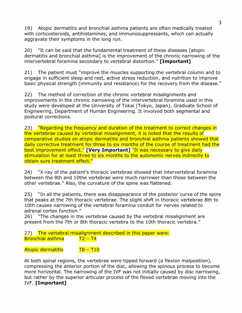
23) “Regarding the frequency and duration of the treatment to correct changes in the vertebrae caused by vertebral misalignment, it is noted that the results of
comparative studies on atopic dermatitis and bronchial asthma patients showed that
daily corrective treatment for three to six months of the course of treatment had the
best improvement effect.” [Very Important] “It was necessary to give daily stimulation for at least three to six months to the autonomic nerves indirectly to
obtain sure treatment effect.”
24) “X-ray of the patient’s thoracic vertebrae showed that intervertebral foramina between the 8th and 10the vertebrae were much narrower than those between the
other vertebrae.” Also, the curvature of the spine was flattened.
25) “In all the patients, there was disappearance of the posterior curve of the spine
that peaks at the 7th thoracic vertebrae. The slight shift in thoracic vertebrae 8th to 10th causes narrowing of the vertebral foramina conduit for nerves related to
adrenal cortex function.”
26) “The changes in the vertebrae caused by the vertebral misalignment are
present from the 7th or 8th thoracic vertebra to the 10th thoracic vertebra.”
27) The vertebral misalignment described in this paper were:
Bronchial asthma T2 – T4
Atopic dermatitis T8 – T10
At both spinal regions, the vertebrae were tipped forward (a flexion malposition),
compressing the anterior portion of the disc, allowing the spinous process to become more horizontal. The narrowing of the IVF was not initially caused by disc narrowing,
but rather by the superior articular process of the flexed vertebrae moving into the
IVF. [Important]

4
28) This study “confirmed that over 98% of allergy patients had the vertebral misalignment.”
29) The chronic vertebral misalignments were found only in the thoracic vertebra
region that corresponded to the innervation of the adrenal glands (T8-T10).
30) These authors believed that the improved symptoms with the correction of the
vertebral misalignments were due to altered function of the autonomic sympathetic
nerves. “On the occasion of correction of vertebral misalignment, we must consider the nature and the functions of the autonomous nerves.”
[Very Important]
31) “As a result of this multi-faceted study investigation, we re-confirmed that these vertebral deformities and the allergic diseases linked together strongly.”
32) “Based on the test results, we can state that the only treatment that can
demonstrate fundamental effects on allergies such as atopic dermatitis, bronchial
asthma, and pollinosis will have the potential to treat spinal curvature disappearance. In other words, we can state that a treatment that cannot
fundamentally treat spinal problems cannot fundamentally improve conditions such
as atopic dermatitis, bronchial asthma, pollinosis, and allergic coryza.”
33) “There is a high possibility that allergic disease relates to the innervation of
organs that relate to the immune function which are affected by changes in the
vertebrae caused by the chronic vertebral misalignment.”
34) “There is an expectation of alleviation, and prevention of development of
symptoms by correcting the changes in the vertebrae caused by chronic vertebral
misalignment, which is common in allergic disease patients.”

1
Innocuous mechanical stimulation of the neck and alterations in heart-rate variability in healthy young adults.
Auton Neurosci. 2001 Aug 13;91(1-2):96-9.
Budgell B, Hirano F.
FROM ABSTRACT:
The present study examined the effects of cervical spinal manipulation, a widely
applied form of physical therapy, which involves innocuous mechanical stimulation, on heart rate and heart-rate variability, in a cohort of healthy young adults.
Using a cross-over treatment design, with a one-week washout period and, in
contrast to a sham procedure, the authentic manipulation produced significant
alterations in both heart rate and measures of heart-rate variability calculated from power spectrum analysis.
In particular, results may reflect a shift in balance between sympathetic and
parasympathetic output to the heart.
THESE AUTHORS ALSO NOTE:
It is established that noxious physical and psychological stimulation effect autonomic and cardiovascular function in humans.
Also, innocuous mechanical stimulation of the neck via spinal manipulation, is
capable of eliciting changes in heart rate and blood pressure.
In this study, these authors used a cohort of 25 healthy young adults without neck
or shoulder pain.
“The mechanical stimuli were applied to the first and second (C1 and C2) cervical levels.”
Both sham and authentic manipulations were performed by a licensed doctor of
chiropractic.
The manipulations were done supine with limited rotation and no extension, and an
audible was achieved. The manipulation was described as a “supine cervical rotary
adjustment.” It took no more than 5 seconds.
The sham manipulation used the same set-up, but there was no thrust and no
audible.

1Heart rate changes in response to mild mechanical irritation of
the high cervical spinal cord region in infants
Forensic Science InternationalVolume 128, Issue 3, August 28, 2002, Pages 168-176
L. E. Koch, H. Koch, S. Graumann-Brunt, D. Stoll, J. M. Ramirez and K. S. Saternus
FROM ABSTRACT:
Alterations in the heart rate were monitored before, during and after the applicationof a unilateral mechanical impulse to the high cervical spinal cord region which wasadministered strictly in connection with the so called manual therapy(diagnosis=KISS).
The investigation is based on a survey of 695 infants between the ages of 1 and 12months.
A notable change in the heart rate was evident in 47.2% of all examined infants.
In 40.1% of these infants, the change in heart rate was characterized by heart ratedecrease of 15-83% compared to control conditions.
Infants in their first 3 months of life responded more often with a severebradycardia (50-83% decrease), older infants (7-12 months) more often with amild bradycardia (15-49.9% decrease).
This comparison revealed a significantly increased occurrence of severe bradycardiain the younger age group compared to the group of children >3 months.
In 12.1% (n=84) of the infants, the bradycardia was accompanied by an apnea.
We discuss the hypothesis that mechanical irritation of the high-cervical regionserves as a trigger that may be involved in sudden infant death (SID).
THESE AUTHORS ALSO NOTE:
“In first world countries, sudden infant death (SID) is the most common cause ofdeath during the first 12 months of postnatal life.”
“One of the major risks is the prone position of the sleeping baby, which enhancesthe death rate by (odd ratio) 3.5 per thousand.”
“There is increasing evidence that SIDs-victims exhibit a pronounced bradycardiabefore dying.”

1
Impact of Osteopathic Manipulative Treatment on Secretory Immunoglobulin A Levels in a Stressed Population
Journal of the American Osteopathic Association
March 2011, Vol. 111, No. 3, pp. 143-147 Gregory Saggio, DO; Salvatore Docimo, DO; Jennifer Pile, DO; Jennifer Norton, DO, RN; and Wolfgang Gilliar, DO FROM ABSTRACT: Context: High levels of human secretory immunoglobulin A (sIgA) have been shown to decrease the incidence of acquiring upper respiratory tract infections. Osteopathic manipulative treatment (OMT) has been shown to improve cardiac indices, increase lymph flow rates through the thoracic duct, and decrease sympathetic tone in postoperative patients and those in intensive care. Therefore, we hypothesized that OMT may also increase sIgA levels in people under high levels of emotional and psychological stress, thereby enhancing immunity and potentially preventing subsequent infections. Objective: To determine if OMT increases sIgA levels in highly stressed individuals. Methods: Twenty-five second-year osteopathic medical students were randomly assigned to an experimental group (n=12) or a control group (n=13). All participants were scheduled to take their national board examination within 2 to 3 weeks after the experiment. After each participant submitted a saliva sample for a baseline sIgA level assessment, the experimental group received 20 minutes of OMT while the control group sat quietly and relaxed in a separate area for 20 minutes. Participants in both groups rested quietly for 1 hour after the 20-minute session and then submitted a second saliva sample. Results: A 2 X 2 repeated measures analysis of variance revealed that the experimental group displayed a statistically significant greater increase in post-intervention sIgA levels than the control group [by 139% increase]. Conclusion: This study demonstrates the positive effect of OMT on sIgA levels in persons experiencing high stress. Results suggest that OMT may then have therapeutic preventive and protective effects on both healthy and hospitalized patients, especially those experiencing high levels of emotional or physiological stress and those at higher risk of acquiring upper respiratory tract infections. KEY POINTS FROM THIS STUDY: 1) Secretory immunoglobulin A (sIgA) is also referred to as salivary IgA.

2
2) “Secretory IgA provides protection from pathogenic organisms by preventing attachment, replication, and colonization of such organisms.” 3) Stress (perceived or physical) can cause a decrease in mucosal sIgA levels. 4) Levels of sIgA in humans indicate immune system potency. 5) High levels of sIgA decrease the incidence of upper respiratory tract infections. Low levels of sIgA are associated with increased incidence of infections. 6) Studies have reported a statistically significant enhanced immunologic response in subjects who received osteopathic manipulative therapy as compared to a control group. 7) An increase in sIgA among patients receiving osteopathic manipulation suggests that manipulation “could be used to increase immunity in vulnerable populations.” 8) Both controls and patients submitted a baseline saliva sample and their sIgA were measured. The controls were reassessed 1 hour after sitting quietly. The patients were reassessed 1 hour after osteopathic manipulation. 9) The sIgA level in the manipulation group increased by an average 139%, more than 100% greater than the control group. 10) “Decreases in sIgA levels have shown to have deleterious effects on the immune system, which leads to an increased risk of infection. Such findings demonstrate the importance of discovering osteopathic clinical practices that increase the level of patients' sIgA.” 11) Osteopathic manipulation increases lymphatic flow, increases peripheral circulation, improves cardiac indices, and decreases sympathetic tone. 12) This study demonstrates that osteopathic manipulative therapy “significantly increases the sIgA level.” “Participants who received osteopathic manipulative therapy experienced an average increase of 139% in their level of sIgA post intervention.” 13) High levels of human sIgA “have been shown to benefit the immune system by decreasing the risk of acquiring upper respiratory tract infections.” 14) Simple osteopathic manipulative techniques may increase immunity and lower the incidence of infections. 15) “Our findings demonstrate OMTs ability to increase sIgA and to potentially improve immune system function.”

Osteopathic Manipulative Therapy Induces Early PlasmaCytokine Release and Mobilization of a Population ofBlood Dendritic CellsStevan Walkowski1., Manindra Singh2, Juan Puertas3, Michelle Pate3, Kenneth Goodrum3.,
Fabian Benencia2,3,4,5*.
1 Osteopathic Manipulative Medicine Department, Heritage College of Osteopathic Medicine, Ohio University, Athens, Ohio, United States of America, 2 Molecular and
Cellular Biology Program, Ohio University, Athens, Ohio, United States of America, 3 Department of Biomedical Sciences, Heritage College of Osteopathic Medicine, Ohio
University, Athens, Ohio, United States of America, 4 Biomedical Engineering Program, Russ College of Engineering and Technology, Ohio University, Athens, Ohio, United
States of America, 5 Diabetes Institute, Ohio University, Athens, Ohio, United States of America
Abstract
It has been claimed that osteopathic manipulative treatment (OMT) is able to enhance the immune response of individuals.In particular, it has been reported that OMT has the capability to increase antibody titers, enhance the efficacy ofvaccination, and upregulate the numbers of circulating leukocytes. Recently, it has been shown in human patients sufferingchronic low back pain, that OMT is able to modify the levels of cytokines such as IL-6 and TNF-a in blood upon repeatedtreatment. Further, experimental animal models show that lymphatic pump techniques can induce a transient increase ofcytokines in the lymphatic circulation. Taking into account all these data, we decided to investigate in healthy individualsthe capacity of OMT to induce a rapid modification of the levels of cytokines and leukocytes in circulation. Humanvolunteers were subjected to a mixture of lymphatic and thoracic OMT, and shortly after the levels of several cytokines wereevaluated by protein array technology and ELISA multiplex analysis, while the profile and activation status of circulatingleukocytes was extensively evaluated by multicolor flow cytometry. In addition, the levels of nitric oxide and C-reactiveprotein (CRP) in plasma were determined. In this study, our results show that OMT was not able to induce a rapidmodification in the levels of plasma nitrites or CRP or in the proportion or activation status of central memory, effectormemory or naıve CD4 and CD8 T cells. A significant decrease in the proportion of a subpopulation of blood dendritic cellswas detected in OMT patients. Significant differences were also detected in the levels of immune molecules such as IL-8,MCP-1, MIP-1a and most notably, G-CSF. Thus, OMT is able to induce a rapid change in the immunological profile ofparticular circulating cytokines and leukocytes.
Citation: Walkowski S, Singh M, Puertas J, Pate M, Goodrum K, et al. (2014) Osteopathic Manipulative Therapy Induces Early Plasma Cytokine Release andMobilization of a Population of Blood Dendritic Cells. PLoS ONE 9(3): e90132. doi:10.1371/journal.pone.0090132
Editor: Derya Unutmaz, New York University, United States of America
Received September 26, 2013; Accepted January 27, 2014; Published March 10, 2014
Copyright: � 2014 Walkowski et al. This is an open-access article distributed under the terms of the Creative Commons Attribution License, which permitsunrestricted use, distribution, and reproduction in any medium, provided the original author and source are credited.
Funding: This work was funded by a Research & Scholarly Affairs Committee grant from the Heritage College of Osteopathic Medicine, Ohio University (http://www.oucom.ohiou.edu/) and a research grant (10-08-606) from the American Osteopathic Association (www.osteopathic.org). The funders had no role in studydesign, data collection and analysis, decision to publish, or preparation of the manuscript.
Competing Interests: The authors have declared that no competing interests exist.
* E-mail: [email protected]
. These authors contributed equally to this work.
Introduction
Osteopathic medicine is based on the premise that the primary
role of the physician is to facilitate the body’s inherent ability to
heal itself. Osteopathic physicians view diseases as a disruption of
the normal interactions of anatomy, physiology and behavior. One
unique aspect of osteopathic medicine is treatment by manually
applied procedures, often referred to as manipulative therapies.
These therapies have been successfully used by osteopathic
physicians for more than a hundred years in order to treat
dysfunctions of the neuromusculoskeletal, lymphatic, or vascular
tissue. As described in Foundations for osteopathic medicine [1]
some of the techniques relevant to osteopathy include: 1) soft-tissue
techniques that increase muscle relaxation and circulation of body
fluids; and 2) isometric and isotonic techniques that focus on
restoring physiological movements and altered joint mechanisms.
Manipulative therapies aimed to increase lymphatic flow, such as
thoracic or abdominal lymphatic pump, have been extensively
used in osteopathic medicine [2,3]. In particular, these techniques
are proposed to treat patients with asthma, edema and certain
pulmonary infections since an increase in the lymphatic flow may
enhance filtering and removal of fluid, inflammatory mediators,
and waste products from interstitial spaces [4]. Interestingly, it has
been claimed that osteopathic treatment decreased mortality rates
associated with the 1918 influenza epidemic in the United States
and may be relevant in the case of an avian influenza pandemic
[5,6]. Various reports of beneficial clinical responses to lymphatic
pump treatments may be related to increased lymph flow
[7,8,9,10,11]. In addition, it has been proposed that lymphatic
and splenic pump treatments at the time of vaccination were
correlated with a faster rise in antibody titers in human subjects
receiving the hepatitis B vaccine [7]. In another series of studies,
patients receiving thoracic pump treatments showed statistically
PLOS ONE | www.plosone.org 1 March 2014 | Volume 9 | Issue 3 | e90132


Dan Murphy, DC
Copyright 2012 by Daniel J. Murphy, DC and Michael L. Underhill, DC, PC. All Rights Reserved.
Complete Set of 4 DVD’s for $199 + $5.50 S&H
Featuring on-trend, up-to-date topics
for Doctors and Patients.
Disc 1 Disc 2
Disc 3 Disc 4
-Environment and Toxins
-Fibrosis of Repair
-The Sunshine Vitamin: D3
Name Phone Email
Address City State Zip
Credit Card # Exp. Date CCV Code
Signature Date Representative
-Pain Medications
-Fish Oils & Omega 3’s
-Excitotoxins
-Statin Drugs
-Whiplash
-Kids & Chiropractic
-Posture & Neurology
-Laser Therapy
-Interview with Dr. Michael Underhill
-Environment and Toxins
-Fibrosis of Repair
-The Sunshine Vitamin: D3Q&A
Vitam
in
D3
Michelle Schaer
613 S. Mesa Dr.
Mesa, AZ 85210
Email: [email protected]
Office: 480.644.1227
Fax: 480.644.7737

Did You Know?
• Spinal stiffness was linked to visceral pathology with nearly 100% accuracy based upon sympathetic innervation.(Medical Times, 1921)
• 1,000 capsules of Tylenol in a lifetime doubles the risk of end stage renal disease. (New England Journal of Medicine, 1994)
• The average time for a whiplash-injured patient to achieve maximum improvement is 7 months 1 week. (Spine, 1994)
• 93% of patients with chronic whiplash pain who have failed medical and physical therapy care improve with chiropractic adjustments. (Injury, 1996)
• Taking the correct drug for the correct diagnoses in the correct dose will kill about 106,000 Americans per year, making it the4th most common cause of death in the US. (Journal of the American Medical Association, 1998)
• Nonsteroidal anti-inflammatory drugs for rheumatoid and/or osteoarthritis conservatively cause 16,500 Americans to bleed to death each year, making that the 15th most common cause of death in the US. (New England Journal of Medicine, 1999)
• Glutamate and aspartame can cause chronic pain sensitization, and removing them from the diet for 4 consecutive months caneliminate all chronic pain symptoms. (Annals of Pharmacotherapy, 2002)
• Chiropractic spinal adjusting has been shown to be better than 5 times more effective than the NSAIDs pain drugs Celebrex and Vioxx in the treatment of chronic neck and low back pain. (Spine, 2003)
• In patients suffering from chronic pain subsequent to degenerative spinal disease, 59% can eliminate the need for pain drugs by consuming adequate levels of omega-3 essential fatty acids. (Surgical Neurology, 2006)
• Chiropractic adjustments have been shown to significantly lower blood pressure. (Journal of Human Hypertension, 2007)
• The estimated incidence of chronic pain from whiplash trauma is 15-40%. (Jour of the Am Academy of Ortho Surg, 2007)
• Meniere’s Disease has been linked to a disorder of the upper cervical spine facet joints. (International Tinnitus Jour, 2007)
• Supplementing with vitamin D3 has the potential to reduce cancer deaths in America by 75%. (Ann of Epidemiology, 2009)
• Potentially, the largest exposure of Americans to the neurotoxin mercury is through the consumption of products containing High Fructose Corn Syrup. (Environmental Health, 2009)
• Those who consumed the highest amounts of nonsteroidal anti-inflammatory pain drugs increased their risk of dementia, including Alzheimer’s dementia, by 66%. (Neurology, 2009)
• The newest estimate for the incidence of autism is 1 in 91 US children. (Pediatrics, 2009)
These published facts and hundreds more are available through my Article Review Service, now in its 11th year. Reviews are detailed,thorough, timely and cutting-edge, with KEY POINTS summary and chiropractic practical applications. The Reviews are in PDFformat for easy printing. They are excellent for educating the chiropractor, staff, patients and for lecture preparation.Easy sign-up through the website with a credit card using PayPal. $100.00 per year. The Archives (past years) are also available.
Website: www.danmurphydc.com Assistant: Phyllis (925) 420-6564
SUBSCRIBER COMMENTSDr. Dan,Any chiropractor that truly cares about his patients and not about just making a buck needs to be subscribing to your Article ReviewUpdates. I certainly am going to do my part to see that each chiro I come in contact with knows what an absolutely invaluableresource it is. I sat in amazement at the last two articles you sent regarding antibiotic overuse and atopic disorders. What crucialinformation to pass on to my practice members. Thanks and keep up the awesome work. Dr. G.M.; August 1, 2002Dear Dan,I hope you can continue providing this information for many years to come. !I have been in practice for 18 years and find thesecitations to be the most informative, chiropractically relevant information that I have received in my career. !I would be willing to paymore for this information to make sure that it keeps coming. !Again, thank you!! JR, DC; January 8, 2005