Embed Size (px)
Citation preview
The visual systemThe visual system
Part IPart I

In general, our visual system In general, our visual system represents the world:represents the world:
a)a) ImperfectlyImperfectly
b)b) AccuratelyAccurately
c)c) Better than realityBetter than reality
Distinction between:-- transduction-- coding
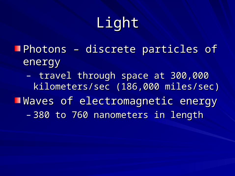
LightLight
Photons – discrete particles of energyPhotons – discrete particles of energy– travel through space at 300,000 travel through space at 300,000
kilometers/sec (186,000 miles/sec)kilometers/sec (186,000 miles/sec)
Waves of electromagnetic energyWaves of electromagnetic energy– 380 to 760 nanometers in length380 to 760 nanometers in length
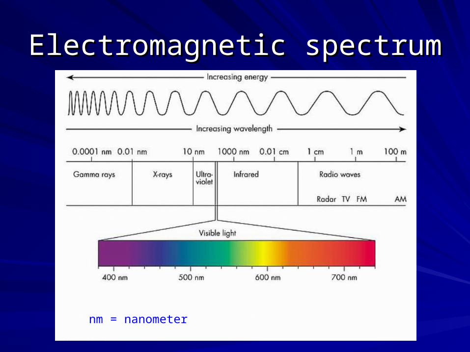
Electromagnetic spectrumElectromagnetic spectrum
nm = nanometer

What other animals see…What other animals see…
Honeybees can see Ultraviolet light Rattlesnakes can see infrared light

Rats can see ultraviolet lightRats can see ultraviolet light

Properties of light and perceptionProperties of light and perception
In general:In general:
Wavelength – color (hue) perceptionWavelength – color (hue) perception
Intensity – brightness perceptionIntensity – brightness perception
Saturation – purity perceptionSaturation – purity perception

Light enters the eyeLight enters the eye
through the pupilthrough the pupil
size of the pupilsize of the pupil
is regulated by the irisis regulated by the iris
The lens focuses lightThe lens focuses light
on the retina on the retina
Note: that the Note: that the
retinal image retinal image
is upside down. is upside down.

Pupil sizePupil sizeAdjusted in response to changes in illumination, Adjusted in response to changes in illumination, which is a tradeoff between:which is a tradeoff between:
– SensitivitySensitivity – ability to detect the presence of dimly lit – ability to detect the presence of dimly lit objectsobjects
– AcuityAcuity – ability to see the details of objects – ability to see the details of objects
– When illumination is high, pupils are constricted When illumination is high, pupils are constricted allowing a greater depth of focus of the image falling allowing a greater depth of focus of the image falling on the retinaon the retina
– When illumination is low, pupils dilate in response to When illumination is low, pupils dilate in response to low activation of receptors allowing more light to enter low activation of receptors allowing more light to enter the eye but sacrificing acuity and depth of focusthe eye but sacrificing acuity and depth of focus

Anatomy of the eyeAnatomy of the eye
Ligament
Ones to know
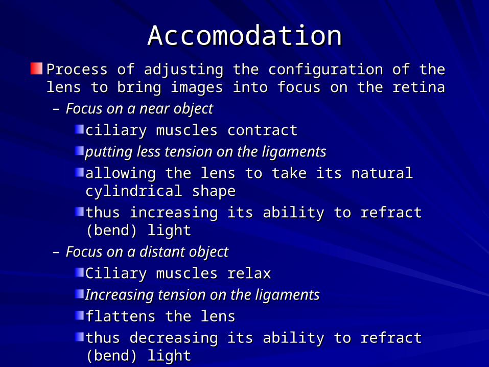
AccomodationAccomodationProcess of adjusting the configuration of the lens to bring Process of adjusting the configuration of the lens to bring images into focus on the retinaimages into focus on the retina– Focus on a near objectFocus on a near object
ciliary muscles contractciliary muscles contract
putting less tension on the ligamentsputting less tension on the ligaments
allowing the lens to take its natural cylindrical allowing the lens to take its natural cylindrical shapeshape
thus increasing its ability to refract (bend) lightthus increasing its ability to refract (bend) light– Focus on a distant objectFocus on a distant object
Ciliary muscles relaxCiliary muscles relax
Increasing tension on the ligamentsIncreasing tension on the ligaments
flattens the lensflattens the lens
thus decreasing its ability to refract (bend) lightthus decreasing its ability to refract (bend) light

Binocular disparityBinocular disparity
The difference in the positions of the same The difference in the positions of the same image on the two retinasimage on the two retinas– Is greater for close objects (eyes must Is greater for close objects (eyes must
converge or turn slightly inward)converge or turn slightly inward)– The degree of binocular disparity enables the The degree of binocular disparity enables the
visual system to construct 3-D perception visual system to construct 3-D perception from two 2-D retinal imagesfrom two 2-D retinal images

The retinaThe retina
Composed of 5 layers of neurons Composed of 5 layers of neurons – Receptors (photoreceptors) Receptors (photoreceptors)
1 rod1 rod
3 cones3 cones
– Horizontal cells (2 subtypes)Horizontal cells (2 subtypes)– Bipolar cells (10 subtypes)Bipolar cells (10 subtypes)– Amacrine cells (25-30 subtypes)Amacrine cells (25-30 subtypes)– Ganglion cells (10-15 subtypes)Ganglion cells (10-15 subtypes)
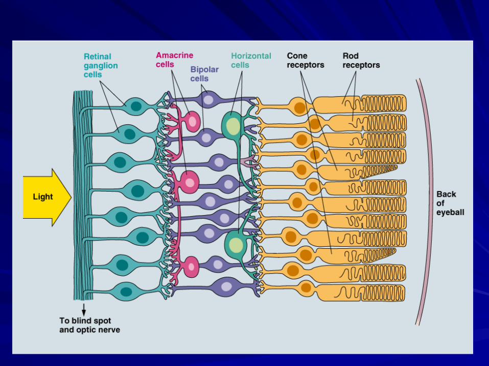
The cellular structure of the retinaThe cellular structure of the retina
Appears to be inside-outAppears to be inside-out– Light passes through the 4 cell layers before Light passes through the 4 cell layers before
reaching the receptorsreaching the receptors– After receptor activation, signals are After receptor activation, signals are
transmitted back out to the ganglion cells transmitted back out to the ganglion cells whose axons project across inside surface of whose axons project across inside surface of the retina, gathering at the optic disk where the retina, gathering at the optic disk where the optic nerve begins as the ganglion cell the optic nerve begins as the ganglion cell axons leave the eye. axons leave the eye.
Two visual problemsTwo visual problems
result from the inside out arrangement:result from the inside out arrangement:
1.1. Incoming light is distorted as it passes Incoming light is distorted as it passes through the cell layersthrough the cell layers
2.2. There is a blind spot (no receptors or There is a blind spot (no receptors or cells) at the optic disk where the axons cells) at the optic disk where the axons gather to exit the eyegather to exit the eye
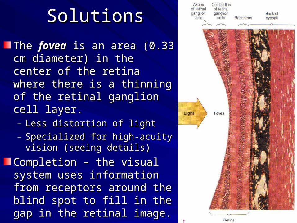
SolutionsSolutions
The The foveafovea is an area (0.33 cm is an area (0.33 cm diameter) in the center of the diameter) in the center of the retina where there is a thinning retina where there is a thinning of the retinal ganglion cell of the retinal ganglion cell layer.layer.– Less distortion of lightLess distortion of light– Specialized for high-acuity vision Specialized for high-acuity vision
(seeing details)(seeing details)
Completion – the visual system Completion – the visual system uses information from uses information from receptors around the blind spot receptors around the blind spot to fill in the gap in the retinal to fill in the gap in the retinal image.image.

photopic and scotopic visionphotopic and scotopic vision
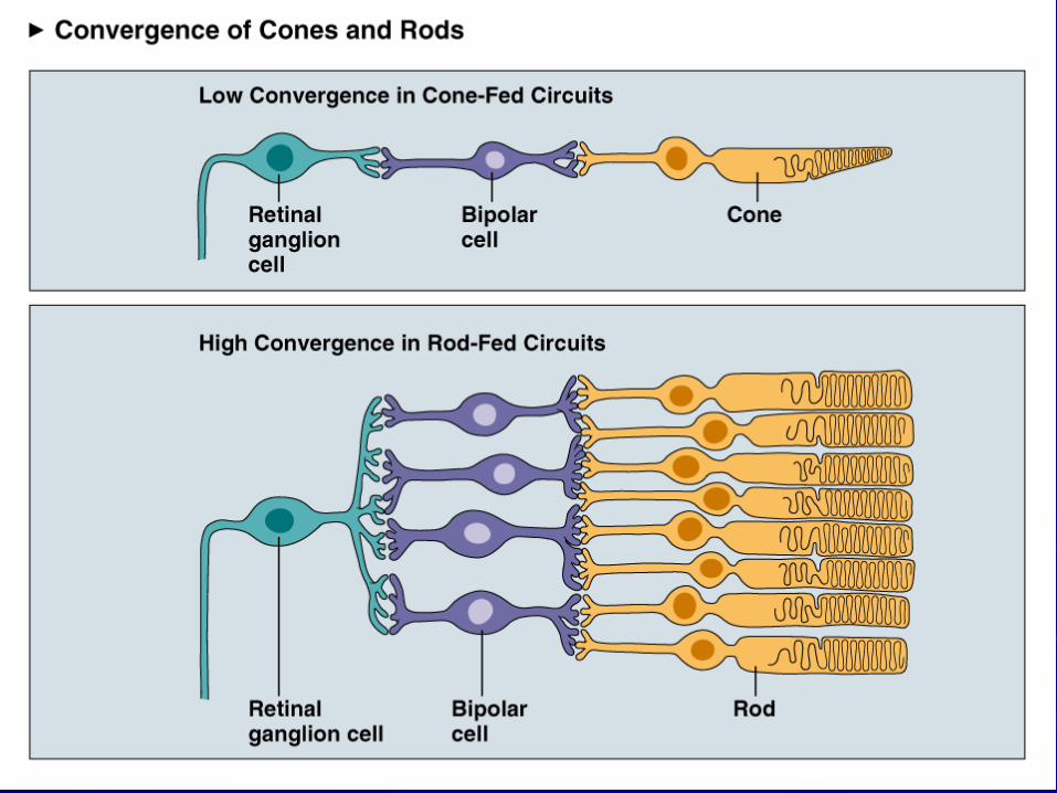
The two systems are “wired” differentlyThe two systems are “wired” differently
Cones – low degree of convergence (a Cones – low degree of convergence (a single ganglion cell receives signals from a single ganglion cell receives signals from a few cones).few cones).
Rods – high degree of convergence (a Rods – high degree of convergence (a single ganglion cell receives signals from single ganglion cell receives signals from hundreds of rods).hundreds of rods).

photopic and scotopic visionphotopic and scotopic vision
Cones are concentrated in the fovea, which contains no rods.Cones are concentrated in the fovea, which contains no rods.Rods are concentrated 20 degrees from the fovea and in the Rods are concentrated 20 degrees from the fovea and in the nasal hemiretina (retina half of both eyes near the nose). nasal hemiretina (retina half of both eyes near the nose).

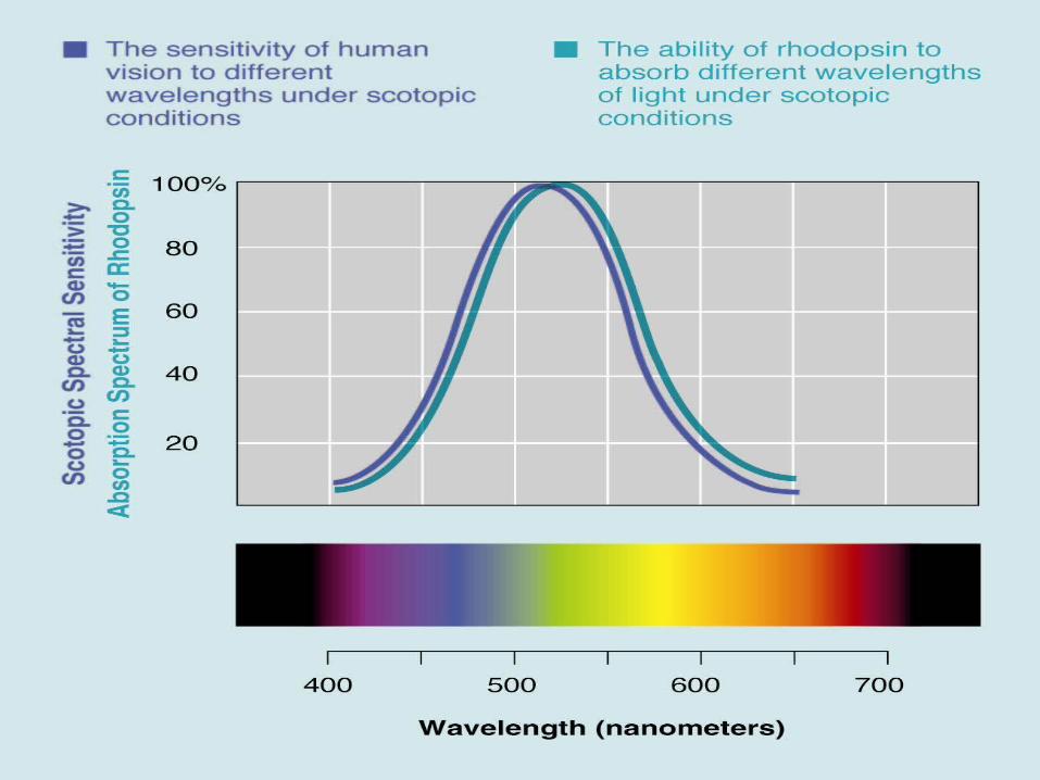
Spectral sensitivity curveSpectral sensitivity curveIn general, more intense light appears In general, more intense light appears brighter. However, wavelength also has brighter. However, wavelength also has an effect on the perception of brightness.an effect on the perception of brightness.
A graph of the relative A graph of the relative brightness of lights of brightness of lights of the same intensity but the same intensity but at different wavelengths at different wavelengths is called a is called a spectral spectral sensitivity curvesensitivity curve (see (see Pinel p. 138). Pinel p. 138).

Spectral Spectral sensitivity sensitivity
curvescurves
There are two spectral sensitivity curves.There are two spectral sensitivity curves.– The photopic spectral sensitivity curve has a The photopic spectral sensitivity curve has a
peak brightness at 555 nm (yellow-green)peak brightness at 555 nm (yellow-green)– The scotopic spectral sensitivity curve has a The scotopic spectral sensitivity curve has a
peak brightness at 507 nm (green-blue)peak brightness at 507 nm (green-blue)
The Purkinje effect – walking through his garden, The Purkinje effect – walking through his garden, Purkinje noticed that his yellow and red flowers Purkinje noticed that his yellow and red flowers were brighter than the blues ones just before dusk; were brighter than the blues ones just before dusk; just a few minutes later the trend was reversed just a few minutes later the trend was reversed (blue flowers appeared as brighter greys).(blue flowers appeared as brighter greys).

TransductionTransduction- Conversion of one form of - Conversion of one form of
energy to another. energy to another.
Visual transduction – Visual transduction – conversion of light to neural conversion of light to neural signals. signals.
Rhodopsin – the red pigment in Rhodopsin – the red pigment in rods becomes bleached when rods becomes bleached when exposed to light. exposed to light.
It is a G-protein-linked receptor It is a G-protein-linked receptor that responds to light.that responds to light.
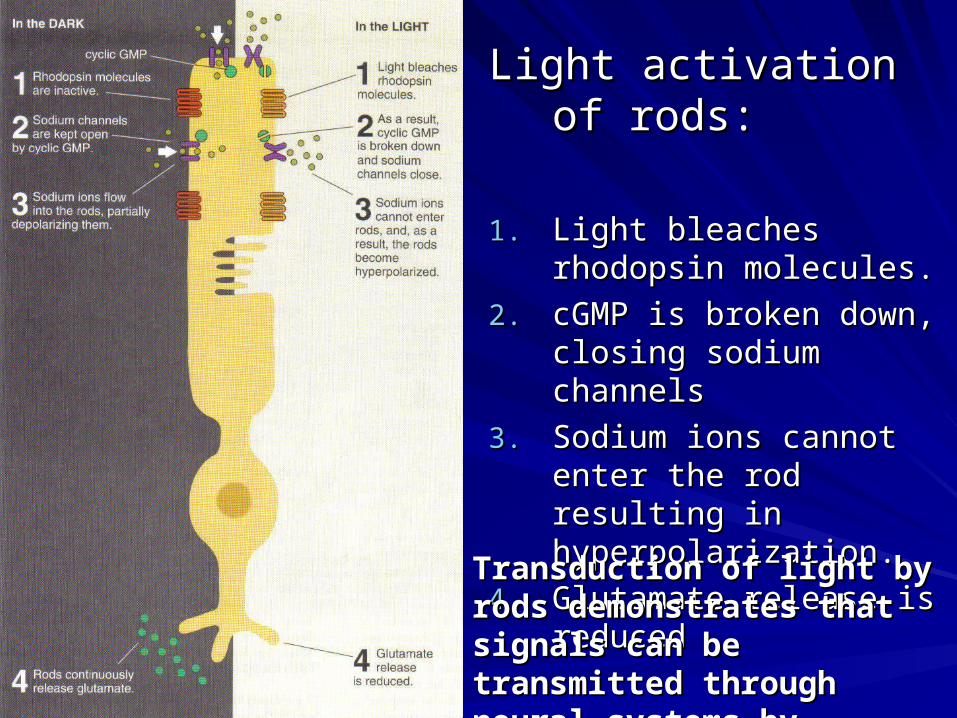
Light activation of rods:Light activation of rods:
1.1. Light bleaches rhodopsin Light bleaches rhodopsin molecules. molecules.
2.2. cGMP is broken down, cGMP is broken down, closing sodium channelsclosing sodium channels
3.3. Sodium ions cannot enter Sodium ions cannot enter the rod resulting in the rod resulting in hyperpolarization.hyperpolarization.
4.4. Glutamate release is Glutamate release is reducedreduced
Transduction of light by rods Transduction of light by rods demonstrates that signals can demonstrates that signals can be transmitted through neural be transmitted through neural systems by inhibition.systems by inhibition.

signal transductionsignal transduction1) Light bleaches rhodopsin2) Opsin separates from retinal molecule3) Transducin (G-protein) activated4) α-subunit breaks away and activates PDE65) PDE breaks down cGMP6) With cGMP broken, the Na+ channel closes

From retina to primary visual cortexFrom retina to primary visual cortex
Pathway: retina Pathway: retina lateral geniculate nucleus lateral geniculate nucleus (LGN) (LGN) primary visual cortex primary visual cortex~90% of axons of retinal ganglion cells make up ~90% of axons of retinal ganglion cells make up this pathwaythis pathwayLGN channelsLGN channels– Parvocellular (P layers) run through top 4 layers of Parvocellular (P layers) run through top 4 layers of
LGN – responsive to color and fine detail (input from LGN – responsive to color and fine detail (input from cones)cones)
– Magnocellular (M layers) run through bottom 2 layers Magnocellular (M layers) run through bottom 2 layers of LGN – responsive to movement (input from rods)of LGN – responsive to movement (input from rods)
Most LGN neurons that project to primary visual Most LGN neurons that project to primary visual cortex (V1, striate cortex) terminate in the lower cortex (V1, striate cortex) terminate in the lower part of cortical layer IVpart of cortical layer IV

Temporal hemiretinaTemporal hemiretinadoes not crossdoes not cross
Nasal hemiretina Nasal hemiretina crossescrosses
Right visual fields ofRight visual fields ofBoth eyesBoth eyes
Left visual fields ofLeft visual fields ofBoth eyesBoth eyes
Top of visual fieldTop of visual fieldto ventral cortexto ventral cortex
Bottom of visual fieldBottom of visual fieldto dorsal cortexto dorsal cortex
Is retinotopic – each level is organized like a Is retinotopic – each level is organized like a map of the retinamap of the retina
Note that 25% of V1 is dedicated to foveaNote that 25% of V1 is dedicated to fovea

Seeing EdgesSeeing Edges
A visual edge is A visual edge is “nothing” “nothing” Where two Where two different areas of different areas of an image meet.an image meet.A perception of A perception of contrast between contrast between two adjacent two adjacent areas of the areas of the visual fieldvisual field

Mach bandsMach bands
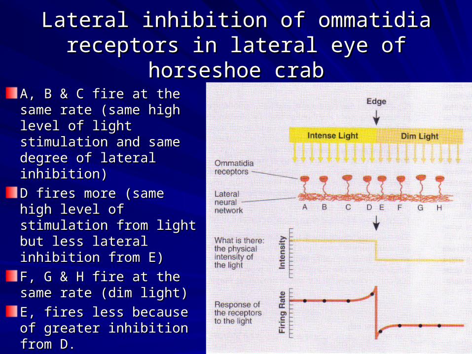
Lateral inhibition of ommatidia receptors in Lateral inhibition of ommatidia receptors in lateral eye of horseshoe crablateral eye of horseshoe crab
A, B & C fire at the same A, B & C fire at the same rate (same high level of rate (same high level of light stimulation and light stimulation and same degree of lateral same degree of lateral inhibition)inhibition)
D fires more (same high D fires more (same high level of stimulation from level of stimulation from light but less lateral light but less lateral inhibition from E)inhibition from E)
F, G & H fire at the same F, G & H fire at the same rate (dim light)rate (dim light)
E, fires less because of E, fires less because of greater inhibition from D.greater inhibition from D.
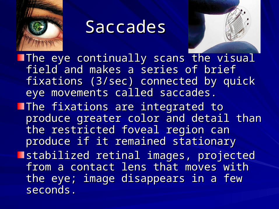
SaccadesSaccades
The eye continually scans the visual field The eye continually scans the visual field and makes a series of brief fixations and makes a series of brief fixations (3/sec) connected by quick eye (3/sec) connected by quick eye movements called saccades. movements called saccades. The fixations are integrated to produce The fixations are integrated to produce greater color and detail than the restricted greater color and detail than the restricted foveal region can produce if it remained foveal region can produce if it remained stationarystationarystabilized retinal images, projected from a stabilized retinal images, projected from a contact lens that moves with the eye; contact lens that moves with the eye; image disappears in a few seconds.image disappears in a few seconds.

Brief fixations Brief fixations associated with associated with
saccades while a saccades while a person views person views
different picturesdifferent pictures
Making visual saccades to items of interest is a function of the superior colliculus
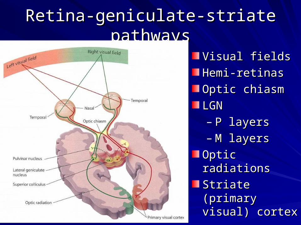
Retina-geniculate-striate pathwaysRetina-geniculate-striate pathways
Visual fieldsVisual fields
Hemi-retinasHemi-retinas
Optic chiasmOptic chiasm
LGNLGN– P layersP layers– M layersM layers
Optic radiationsOptic radiations
Striate (primary Striate (primary visual) cortexvisual) cortex

LGNLGN