Embed Size (px)
Citation preview

The views expressed in this thesis are those of the author and do not reflect the official policy or position of the United States Air Force, Department of Defense, or the United
States Government. This material is declared a work of the U.S. Government and is not subject to copyright protection in the United States.
AFIT-ENP-MS-15-M-089
EVIDENCE FOR INHIBITION OF DENGUE VIRUS BINDING IN THE
PRESENCE OF SILVER NANOPARTICLES
THESIS
Presented to the Faculty
Department of Engineering Physics
Graduate School of Engineering and Management
Air Force Institute of Technology
Air University
Air Education and Training Command
In Partial Fulfillment of the Requirements for the
Degree of Master of Science in Combating Weapons of Mass Destruction
Kelley J. Williams, MS
Captain, USA
March 2015
DISTRIBUTION STATEMENT A.
APPROVED FOR PUBLIC RELEASE; DISTRIBUTION UNLIMITED.
AFIT-ENP-MS-15-M-089
EVIDENCE FOR INHIBITION OF DENGUE VIRUS BINDING IN THE
PRESENCE OF SILVER NANOPARTICLES
Kelley J. Williams, MS
Captain, USA
Committee Membership:
LTC Douglas R. Lewis, USA, PhD
Chairman
Col Christian R. Benjamin, M.D., USAF Member
Dr. Saber Hussain
Member
ii
AFIT-ENP-MS-15-M-089
ABSTRACT
Dengue is an emerging hemorrhagic fever virus and widely considered the most
important arbovirus in the world [1]. The CDC and the World Health Organization
estimates Dengue virus (DENV) infects 50-400 million people annually in the tropical
and subtropical regions of the world [2]. More than 500 thousand of these will develop
severe infection and approximately 22 thousand will lead to death [3]. DENV is a
positive-sense RNA virus that exists in 4, potentially 5, antigenic serotypes. An
immunological phenomenon called antibody-dependent enhancement (ADE) leaves a
DENV victim vulnerable to increased risk of subsequent infections. Secondary infections
with DENV are known to increase in severity from Dengue Fever to Dengue
Hemorrhagic Fever or Dengue Shock Syndrome. Currently, no vaccines or treatments
are approved for DENV infections. Unsuccessful vaccine trials open the door for non-
traditional treatments such as silver nanoparticles. Silver nanoparticles (AgNP) are
known to inhibit viral replication of numerous viruses but have never before been tested
for inhibition of dengue virus type 2 (DENV2, ATCC® VR-1584™). For the first time,
this research presents up to a 96% reduction in DENV2 binding to Vero cells following
pretreatment with AgNP (6-10nm, 25µg/mL). These results suggest that similarly to
other viruses, DENV2 can be inhibited at the first stage of the virus replication cycle,
binding & entry.
iii
ACKNOWLEDGEMENTS
I would like to express my sincere appreciation to my faculty advisor, LTC Doug Lewis,
and committee members Col Christian Benjamin and Dr. Saber Hussain for their
guidance and support throughout the course of this research. Your guidance and trust
were certainly appreciated. I would, also, like to thank my wife and children for enduring
my academic endeavors.
Great thanks are also due to Dr. Nancy Bigley, Dr. Iona Pavel-Sizemore, Kevin Dorney,
Arathi Paluri, Nagarjuna Reddy Cheemarla, and Beth Maurer for their contributions and
insight with this research.
Kelley J. Williams
iv
HYPOTHESIS
Null Hypothesis: Silver nanoparticles do not inhibit the binding step of the dengue virus
serotype 2 (DENV2) replication cycle in Vero 76 cells, inferred through similar
fluorescence intensity visualization.
Alternate Hypothesis: Uncoated silver nanoparticles (8nm) inhibit the binding step of
dengue virus serotype 2 (DENV2) replication cycle in Vero 76 cells, inferred through
reduced fluorescence intensity visualization.
v
LIST OF FIGURES
Figure Page
Figure 1. Symptoms of dengue fever. Public domain image, via Wikimedia Commons. . 2 Figure 2. Vero cell 96-hour growth curve......................................................................... 17
Figure 3. Schematic depicting a typical TFF set-up for the filtration of colloidal AgNPs. Gray and blue spheres represent AgNPs larger and smaller than the filter
membrane pores, respectively. ......................................................................... 21 Figure 4. UV-Vis spectra for the AgNP colloid utilized in binding inhibition assay. ...... 22 Figure 5. Work flow for AgNP characterization............................................................... 23
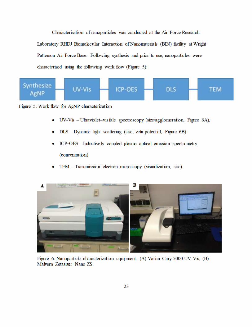
Figure 6. Nanoparticle characterization equipment. (A) Varian Cary 5000 UV-Vis, (B) Malvern Zetasizer Nano ZS.............................................................................. 23
Figure 7. Absorbance spectrum of formazan in dimethyl sulfoxide................................. 25 Figure 8. CytoViva Dual Mode Fluorescence System, AFRL, 711th HPW. ................... 28 Figure 9. Image processing and analysis overview .......................................................... 30
Figure 10. Sample background subtraction in ImageJ. A) Original greyscale image and histogram, B) Image with background subtracted and associated changes to the
histogram. ......................................................................................................... 31 Figure 11. Identification of exclusion regions for black and artifact intensity. ................ 32 Figure 12. Viability of DENV infected Vero cells. .......................................................... 34
Figure 13. Cytotoxicity of silver nanoparticles (AgNP) versus silver ions (Ag+) at in concentrations of 100µg/mL, 50µg/mL, and 10µg/mL.................................... 35
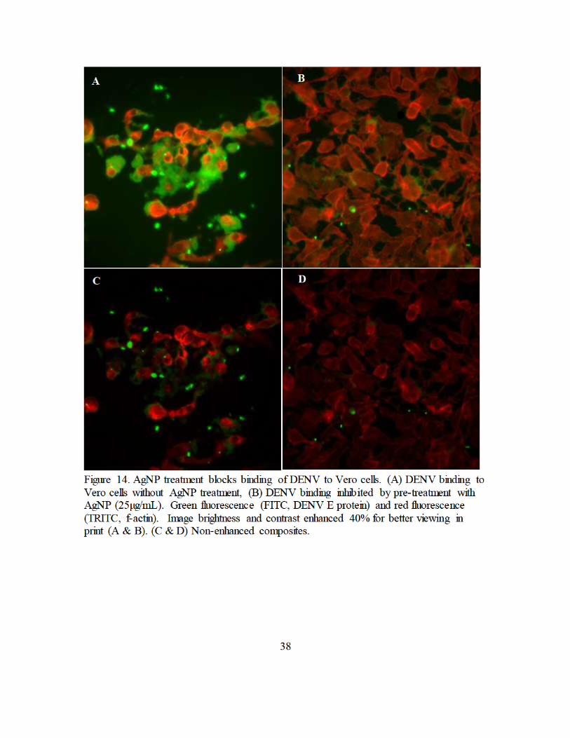
Figure 14. AgNP treatment blocks binding of DENV to Vero cells. (A) DENV binding to
Vero cells without AgNP treatment, (B) DENV binding inhibited by pre-treatment with AgNP (25µg/mL). Green fluorescence (FITC, DENV E protein)
and red fluorescence (TRITC, f-actin). Image brightness and contrast enhanced 40% for better viewing in print (A & B). (C & D) Non-enhanced composites. ....................................................................................................... 38
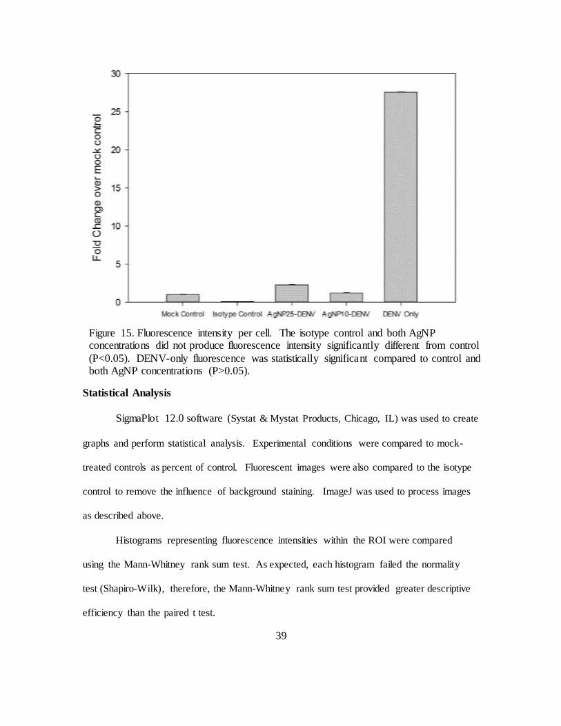
Figure 15. Fluorescence intensity per cell. The isotype control and both AgNP concentrations did not produce fluorescence intensity significantly different
from control (P<0.05). DENV-only fluorescence was statistically significant compared to control and both AgNP concentrations (P>0.05). ........................ 39
Figure 16. ICP-OES Calibration curve ............................................................................ 48
Figure 17. AgNP size distribution by intensity, Z-avg 23.5nm +0.95nm......................... 48
vi
LIST OF ABBREVIATIONS
μg microgram
μL microliter ADE antibody-dependent enhancement Ag silver
AgNP silver nanoparticle(s) DLS dynamic light scattering
DNA deoxyribonucleic acid FBS fetal bovine serum mg milligram
mL milliliter MTT 3-(4,5-Dimethylthiazol-2-yl)-2,5-diphenyltetrazolium bromide
NHP non-human primate nm nanometer NP nanoparticles
PBS phosphate buffered saline PdI polydispersity index
PEG polyethylene glycol SD standard deviation ss(+)RNA single-strand positive sense ribonucleic acid
SEM scanning electron microscopy TEM transmission electron microscopy UV-Vis ultraviolet to visible absorption spectroscopy
Z-Ave Z- average diameter
vii
TABLE OF CONTENTS
ABSTRACT......................................................................................................................... ii
ACKNOWLEDGEMENTS ................................................................................................ iii
HYPOTHESIS .................................................................................................................... iv
LIST OF FIGURES ............................................................................................................. v
LIST OF ABBREVIATIONS ............................................................................................. vi
TABLE OF CONTENTS................................................................................................... vii
INTRODUCTION ...............................................................................................................1
Dengue as a Global Threat ...........................................................................................1
Dengue Pathology ........................................................................................................1
Dengue Threatens DOD Operations.............................................................................4
Dengue as a Potential Bioterrorism Agent ...................................................................5
Challenges to Dengue Therapeutics .............................................................................5
Nanoparticles as an Antiviral Treatment ......................................................................6
LITERATURE REVIEW ....................................................................................................7
Dengue Virus Summary ...............................................................................................7
Dengue Virus Binding and Entry .................................................................................9
Silver Nanoparticles as Antiviral Agents ...................................................................10
Silver Ions...................................................................................................................14
Silver Nanoparticle Selection .....................................................................................15
MATERIALS AND METHODS.......................................................................................16
Overview ....................................................................................................................16
viii
Vero Cells ...................................................................................................................16
Cell Line Maintenance ...............................................................................................17
Virus Propagation .......................................................................................................18
Virus Quantification by Flow Cytometry ...................................................................19
Silver Nanoparticle Synthesis and Filtration ..............................................................20
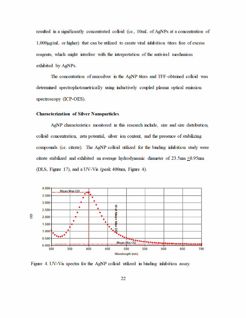
Characterization of Silver Nanoparticles....................................................................22
AgNP-DENV2 Treatment ..........................................................................................24
Trypan Blue Exclusion Test (Hemacytometer method) .............................................24
MTT Assay .................................................................................................................25
Biocompatibility of AgNPs in Vero cells...................................................................26
Virus Binding Study ...................................................................................................26
Blocking Non-Specific Binding During Immunofluorescence ..................................29
Image Processing and Analysis ..................................................................................29
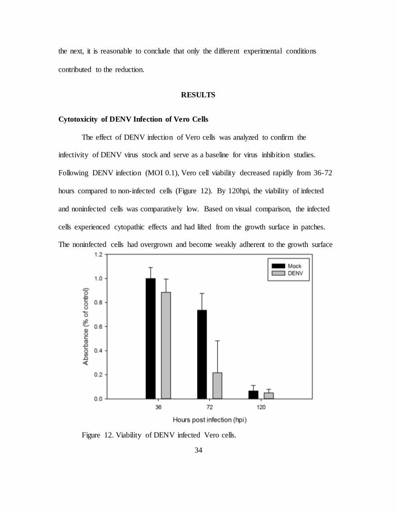
RESULTS ..........................................................................................................................34
Cytotoxicity of DENV Infection of Vero Cells..........................................................34
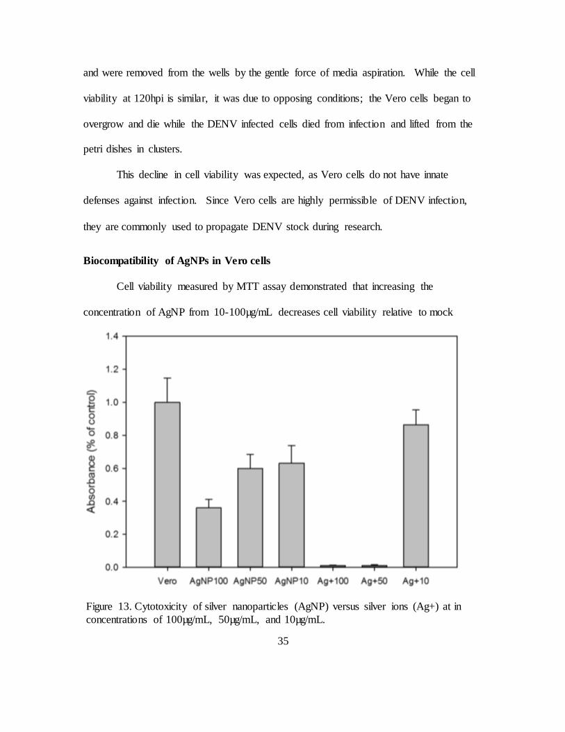
Biocompatibility of AgNPs in Vero cells...................................................................35
AgNP Inhibition of DENV Binding ...........................................................................36
Statistical Analysis .....................................................................................................39
CONCLUSION ..................................................................................................................40
REFERENCES ..................................................................................................................42
APPENDIX A – AgNP Characterization ...................................................................48
1
INTRODUCTION
Dengue as a Global Threat
Dengue is a disease caused by four closely related viruses known as dengue virus
-1 through -4 (DENV1-DENV4). A fifth serotype (DENV5) was discovered from a
serological sample from 2007 but is not published. Dengue fever is a disease that became
prevalent in tropics during the great shipping industry expansion in the 18th and 19th
centuries [4]. The principle arthropod vectors of dengue virus, the Aedes aegypti and
Aedes albopictus mosquitoes, determine the geographic distribution of the disease.
Approximately 40% of the world’s population (2.5 billion people) live in a region at risk
of dengue [2]. Dengue is typically spread throughout the tropical and subtropical
latitudes, but has demonstrated the ability to spread to new areas, such as Europe,
Croatia, the United States, and Portuguese islands [2]. Based on the morbidity of dengue
infection and the widespread presence of its vectors, DENV is considered the most
important arbovirus (arthropod-borne virus) in the world [1]–[3]. Vector control is the
primary means of stopping an outbreak and ending the disease transmission cycle.
Dengue Pathology
Dengue is transmitted through the bite of an infected female mosquito during its
blood meal [5]. Humans are a dead-end host and the infection is not transmissible
between humans. A noninfected Aedes mosquito can become infected through a blood
meal on an infected human. After 4-10 days post-infection, a mosquito is an infectious
vector for the remainder of its life [2]. The severity of an individual’s infection depends
largely on the history of infection and is characterized by the three clinical categories –
2
dengue fever, dengue hemorrhagic fever (DHF) or dengue shock syndrome (DSS).
Primary infection may be asymptomatic or lead to classic dengue fever. Secondary
infections may lead to the serious conditions of DHF or DSS [1].
The World Health Organization estimates that 50-100 million people are infected
with dengue each year. Due to the lack in accurate and timely reporting and the absence
of adequate medical facilities in endemic regions, other sources, such as the Centers for
Disease Control (CDC) place this estimate as high as 400 million annual infections [3].
Classic dengue fever is the predominant syndrome. DHF accounts for over 500 thousand
reported cases and there are over 22 thousand deaths annually [2].
Classic dengue fever is also known as “breakbone fever”, and was first described
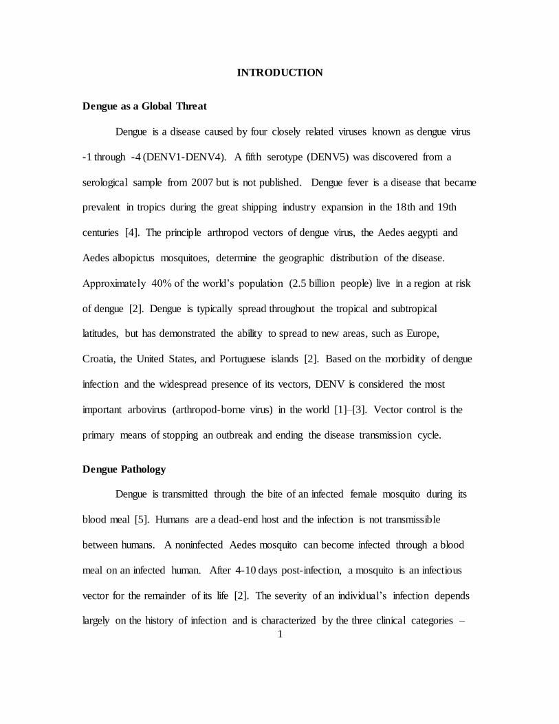
by Benjamin Rush during an outbreak in Philadelphia in 1780 [5]. Dengue fever begins
after a 5-8 day incubation period with the abrupt onset of fever (103-1060F) and headache
(Figure 1). In the first day or two of fever,
the face flushes and a generalized macular
rash becomes evident. Between days 4-6,
symptoms include nausea, vomiting,
anorexia, and swollen lymph nodes
(lymphadenopathy). The fever typically
lasts 4-6 days and often culminates with a
secondary maculopapular rash. The level
of viremia generally coincides with fever.
Figure 1. Symptoms of dengue fever. Public domain image, via Wikimedia Commons.
3
Dengue hemorrhagic fever (DHF) and dengue shock syndrome (DSS) are severe
forms of dengue infection that can lead to death. DHF and DSS are common in areas
with multiple endemic DENV serotypes. These serious illnesses begin with typical
dengue fever. Severe symptoms follow 2-5 days later with petechiae, bruising, and large
spontaneous bruising (ecchymosis).
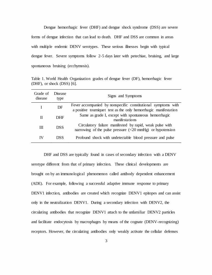
Table 1. World Health Organization grades of dengue fever (DF), hemorrhagic fever
(DHF), or shock (DSS) [6].
Grade of disease
Disease type
Signs and Symptoms
I DF Fever accompanied by nonspecific constitutional symptoms with
a positive tourniquet test as the only hemorrhagic manifestation
II DHF Same as grade I, except with spontaneous hemorrhagic
manifestations
III DSS Circulatory failure manifested by rapid, weak pulse with
narrowing of the pulse pressure (<20 mmHg) or hypotension
IV DSS Profound shock with undetectable blood pressure and pulse
DHF and DSS are typically found in cases of secondary infection with a DENV
serotype different from that of primary infection. These clinical developments are
brought on by an immunological phenomenon called antibody dependent enhancement
(ADE). For example, following a successful adaptive immune response to primary
DENV1 infection, antibodies are created which recognize DENV1 epitopes and can assist
only in the neutralization DENV1. During a secondary infection with DENV2, the
circulating antibodies that recognize DENV1 attach to the unfamiliar DENV2 particles
and facilitate endocytosis by macrophages by means of the cognate (DENV-recognizing)
receptors. However, the circulating antibodies only weakly activate the cellular defenses
4
and the virus will still undergo replication. Additionally, the DENV1 antibodies bound to
DENV2 particles will be able to bind and enter cells through the Fc (constant) receptors
which are ubiquitously expressed. This additional infectious pathway greatly contributes
to the progressive symptoms of severe dengue (DHF/DSS).
Dengue Threatens DOD Operations
The United States has military and civilian personnel stationed in many countries
around the world. Some of these locations are in regions where Dengue is endemic and a
growing threat to the human population [3]. Although dengue fever is uncommon among
US citizens, dengue is a top priority for health and medical services in endemic areas.
Dengue has caused illness in US military personnel in every major conflict since the
Spanish American War, excluding the recent Iraq and Afghanistan campaigns [7]. From
the 1960s and through the 1990s, dengue infections were seen often in US
servicemembers in Vietnam, the Philippines, Somalia, and Haiti. In 1942, a dengue
outbreak effected 80% of a single unit stationed in the Australian North Territory [7].
Although the total number of cases in this outbreak was not documented, it casts a dark
shadow in the potential for entire military units to be rendered incapable of performing
their mission. In 1944, over 24 thousand troops in New Guinea and approximately 20
thousand in the Marshall Islands were confirmed to have dengue. Throughout WWII,
seventeen US bases were impacted by dengue [7].
Dengue is a historic problem within the continental United States. Between 1977
and 1987, 1655 suspected cases of dengue were observed in the southern states, 345 of
which were confirmed by serological tests. These cases occurred long after dengue was
5
eliminated from the southern US in the 1920s. This example shows that the potential for
reintroduction of dengue still exists. The most recent outbreaks of DENV occurred in
Hawaii (2001), Texas (2005), and Florida (2014) [3].
Dengue as a Potential Bioterrorism Agent
DENV is classified as a Biosafety Level 2 (BL-2) virus. Some fear that biological
agents could be used to contaminate water supplies. DENV is not found naturally in
water supplies and must infect living cells to replicate. DENV is expected to survive in
untreated water for short periods but not is not expected to survive common water
treatment methods [8]. In this suspended form, DENV would not be infectious and
requires parenteral transmission. Dengue is only infectious through parenteral
transmission which greatly limits the potential for non-vector dissemination. Dengue
does not exhibit rapid or morbid common in biowarfare agents and is not lethal unless
severe shock or other complications are involved.
Challenges to Dengue Therapeutics
The design and implementation of a dengue vaccine is complicated by numerous
factors. First, the candidate vaccine must provide equal and lasting immune responses
against all DENV serotypes. This requires a tetravalent vaccine to address DENV1-4,
and a pentavalent vaccine if the novel DENV5 is considered. This vaccine would also
need to solicit neutralizing antibodies regardless of the individuals existing immune
status to DENV [6]. A second challenge is that there is not a validated correlate of
protection. Since the mechanism of DENV infection is not completely understood, the
required antibody titers and impact of additional immunological pathways require further

6
study [9]. Third, an animal model does not exist which recapitulates both the disease
outcome and a predictive immune response in humans. Currently, mouse and non-human
primate (NHP) models are used to evaluate vaccine candidates. The mouse model
requires modification of the viral genome to present clinical disease and may not be
indicative of efficacy in humans. The NHP model does not present clinical disease but
does demonstrate viremia. Possibly due to these factors, discordance is occasionally
observed between preclinical and clinical trials [10].
Vaccines development is complicated by small mutations in DENV serotypes.
One common mechanism of each DENV serotype and flaviviruses in general is the action
of RNA-dependent RNA polymerase (RdRp). This enzyme catalyzes the replication of
RNA from the ss(+)RNA strand [11], [12]. Novartis developed an adenosine nucleoside
inhibitor (NITD008) which directly inhibits the RdRp activity of DENV in vitro and in
vivo. NITD008 is an adenosine analog with several substitutions; a carbon for N-7 of the
purine and an acetylene at the 2’ position of ribose [12]. Potential may exist for AgNP to
inhibit DENV by binding to exposed disulfide motifs within DENV RdRp [11].
Nanoparticles as an Antiviral Treatment
Nanoparticle therapeutics are in their infancy and there are currently no FDA
approved nanotherapudics [13]. Several studies demonstrate the systemic effects of
silver nanoparticles through various routes of exposure [14]–[17]. In a mouse model,
inhalational and gastrointestinal exposure (1-3 months) to AgNP (20nm, citrate
stabilized) did not show severe systemic toxicity but intravenous exposure has potential
to suppress the functional immune system in a dose-dependent manner (0.0082-6 mg/mL)
7
[15]. Research also suggests that NP have potential to penetrate the blood-brain barrier
(BBB) through intravenous and intraperitoneal exposure at high doses (25-100 mg/kg)
[14].
These and similar studies offer unique insight to in vivo actions of AgNP but they
do not offer broad applicability outside the structure of the research. Published research
involving AgNP does not consistently provide full characterization of the administered
silver with respect to silver ion content, particle agglomeration, particle size distribution,
surface coatings, or presence of synthesis byproducts. Since these additional factors play
a critical role in AgNP biocompatibility and in vivo response, such omissions reduce the
transferability of those data. Above certain concentrations (often >100µg/mL), AgNPs
may exhibit genotoxic and cytotoxic effects in human tissue culture. The possibility that
this effect is due to higher concentration of silver ions must be considered. This can lead
to DNA damage, chromosomal aberrations, and cell death [18]. If AgNP are to be
utilized in clinical settings, the applied concentration must be well characterized under in
vivo applications and remain under the cytotoxic levels.
LITERATURE REVIEW
Dengue Virus Summary
DENV is a member of the Flavivirus family, as is the yellow fever virus (the
family’s prototype), West Nile, Japanese encephalitis virus, and many others. DENV is
also categorized as a Group IV ss(+)RNA virus under the Baltimore Classification.
Based on serological studies, five serotypes of DENV (DENV1-5) are known to exist
[19], [20]. While DENV1-4 are in common circulation, DENV5 is known only to exist
8
in a single outbreak in Malaysia, presents challenges for the future of DENV vaccines
and treatments. Many scientists are skeptical about the serological independence of
DENV5 and feel more research is necessary to determine if it is not a mutant of another
DENV serotype [21]. These distinct serotypes present conserved structural and non-
structural proteins although the genome identity between serotypes ranges from 23-73%
[22], [23]. DENV virions exist in one of four stages of maturation. Only one of the four
stages is infectious and considered mature. The mature virions exist in the extracellular
environment while the three immature virion forms are primarily found intracellularly.
A mature DENV virion is composed of envelope proteins surrounding a
nucleocapsid core which contains the viral genome. The genome consists of a single,
positive-sense strand of RNA, approximately 10.7kb in length. The vRNA is translated
into a single polyprotein. Viral and host proteases cleave this into three structural (C,
prM, E) and seven non-structural (NS1-NS2A-NS2B-NS3-NS4A-NS4B-NS5) proteins.
The genome is surrounded by an icosahedral nucleocapsid C (capsid) protein
approximately 30nm in diameter [5]. The nucleocapsid is disordered and associated with
the vRNA in an overlapping region [24]. This core is surrounded by a lipid bilayer then a
well-organized icosahedral envelope which consists of E-prM protein heterodimeric
complexes [5], [25].
DENV successfully grows in many cells lines from vertebrate and invertebrate
hosts, but the most common are the C6/36 (ATCC® CRL-1660™) derived from the
Aedes albopictus mosquito and the Cercopithecus aethiops (African green monkey)
kidney epithelial (Vero) cell lineage [5], [26]. Selection of the C6/36 cells for DENV
9
research is often preferable since they originate from the virus host. The titer of DENV
isolated from C6/36 cell propagation tends to higher and express less mutations than
isolates from Vero cells propagation [26]. However, as an invertebrate cell line,
the utilization of C6/36 cells requires growth at 28oC. Since this was a condition not
supported at the researcher’s laboratory facility, Vero cells were selected as a suitable cell
line for virus propagation and additional assays.
Dengue Virus Binding and Entry
Dengue virus binds to mammalian cells through a wide range of cell-surface
receptors and co-receptors. Among the known DENV receptors, heparin sulfate (Vero
cells), DC-SIGN, and the mannose receptor (macrophages) appear to play a critical role
[27]–[29]. In the event of antibody-dependent enhancement, the ubiquitous Fc receptor
enhances DENV infection by binding anti-DENV antibodies and providing another entry
pathway. The DENV E protein (envelope glycoprotein) is the central epitope during
binding. Once bound to the cell, DENV is trafficked in a diffusive manner along the cell
surface until reaching a pre-existing clathrin-coated pit [30]. Dengue entry into
mammalian cells may occur through multiple mechanisms, dominated by clathrin-
mediated endocytosis and macropinocytosis [31], [32].
In the dominant case of clathrin-mediated endocytosis, DENV is internalized
when the clathrin-coated pit invaginates and surrounds the particle. Now considered an
endosome, the pH is lowered through action of proton pumps. The acidic pH causes a
conformational change in the E protein below pH 6.2, causing it to become spiked in
10
appearance. The hydrophobic tips of the spiked envelope proteins penetrate and fuse
with the endosomal membrane, releasing the DENV genome into the cytoplasm.
Silver Nanoparticles as Antiviral Agents
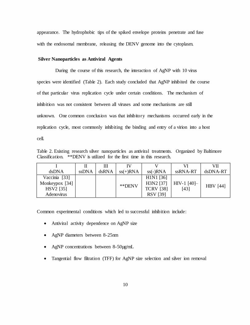
During the course of this research, the interaction of AgNP with 10 virus
species were identified (Table 2). Each study concluded that AgNP inhibited the course
of that particular virus replication cycle under certain conditions. The mechanism of
inhibition was not consistent between all viruses and some mechanisms are still
unknown. One common conclusion was that inhibitory mechanisms occurred early in the
replication cycle, most commonly inhibiting the binding and entry of a virion into a host
cell.
Common experimental conditions which led to successful inhibition include:
Antiviral activity dependence on AgNP size
AgNP diameters between 8-25nm
AgNP concentrations between 8-50µg/mL
Tangential flow filtration (TFF) for AgNP size selection and silver ion removal
I dsDNA
II ssDNA
III dsRNA
IV ss(+)RNA
V ss(-)RNA
VI ssRNA-RT
VII dsDNA-RT
Vaccinia [33]
Monkeypox [34] HSV2 [35] Adenovirus
**DENV
H1N1 [36]
H3N2 [37] TCRV [38] RSV [39]
HIV-1 [40]–[43]
HBV [44]
Table 2. Existing research silver nanoparticles as antiviral treatments. Organized by Baltimore Classification. **DENV is utilized for the first time in this research.
11
AgNP which are naked or capped or stabilized with citrate, polyethylene glycol
(PEG), or polyvinylpyrrolidone
Vaccinia virus (VACV)
Silver Nanoparticles demonstrated cytoprotective and virucidal effects during
VACV infection of Vero cells [33], [43]. Adsorption of VACV was not prevented when
Vero cells were pretreated with AgNP (11nm, 8-128µg/mL), as determined by plaque
assay. The entry of vaccinia virus into Vero cells is inhibited by AgNP at values above
48µg/mL as determined by a β-gal (beta galactosidase) assay under the control of a
constitutively active promoter. Through confocal microscopy quantification, VACV
entry was inhibited when pretreated > 16µg/mL. VACV is an enveloped dsDNA virus
that replicated entirely in the cytoplasm. VACV is in the family Poxviridae and contains
both a large genome (~190kbp) and large virion (~300nm). VACV enters host cells
through macropinocytosis and direct fusion.
Monkeypox virus (MPV)
MPV infection of Vero cells was inhibited by 10nm polysaccharide-coated AgNP
[34]. AgNP in diameters of 10-80nm, concentrations from 12.5-100µg/mL, and either
polysaccharide coated or naked were tested against MPV in plaque reduction assays.
Only the 10nm PS-coated AgNP reduced mean PFU below control (P < 0.05). This
result is similar to HIV-1 studies with AgNP where smaller particles (1-10nm) provided
the best antiviral activity. Rogers, et al. suggest the small particles effectively bind to
viral epitopes and block the binding complex between virus and cell surface receptors
and co-receptors. Another conclusion of this research was that the mechanism of
12
inhibition is unknown but may “involve blocking of MPV to host cells, disruption of host
cell biochemical pathways, or both” [34]. MPV is similar to VACV in size and
replication cycle.
Herpes simplex virus (HSV)
AgNP of unspecified diameter and concentration reportedly inhibit HBV2 in Vero
cells. Sopova et al. described in the Russian journal (not translated to English) that
“silver nanopowders” significantly reduced viral progeny in vitro. HSV is an enveloped
dsDNA virus (152kbp) that enters host cells through fusion.
Adenovirus type 3 (ADV3)
Adenoviruses are non-enveloped dsDNA viruses with a diameter of 70–90 nm.
ADV3 infection of HeLa cells was inhibited by AgNP (11.4+6.2nm) in concentrations
from 3.125-50µg/mL [45]. Cytopathic effects were observed in in HeLa cells when
AgNP concentrations exceeded 50µg/mL as seen in similar experiments. Unlike most
AgNP antiviral experiments which treat cells and virus for 1 hour, Chen et al. treated for
2 hours. ADV3 that was treated with 50µg/mL AgNP for 30 minutes experienced
conformational changes in the virion structure. Treatment for 90 and 120 minutes created
dramatic damage to the virion. Unfortunately, the researchers did not mention whether
their AgNP colloid contained significant levels of silver ions, the most likely cause of
ADV3 damage.
Influenza A (H1N1 and H3N2 IFV)
H1N1 and H3N2 influenza A infection of Madin-Darby Canine Kidney epithelial
(MDCK) cells was inhibited by non-coated AgNP [36], [37]. AgNP in these studies were
13
characterized in the range of 5-120nm (10nm mean). This study found that when H1N1
or H3N2 IFV were pretreated for 2 hours with AgNP concentrations of 12.5-50µg/mL,
the viability of MDCK cells was significantly increased (P<0.01). When virus was
incubated with AgNP (50µg/mL) for 30-120 minutes, the IFV experienced structural
damage. Damage was observed even when there did not appear to be AgNP bound to
virions, indicating damage through silver ions. These studies recognized the potential
significance of silver ions in an antiviral mechanism but did not present data on ion
content in their colloid or any methods used to remove silver ions during
experimentation.
Tacaribe virus (TCRV)
Tacaribe virus infection of Vero cells was inhibited by AgNP of varying size and
coating [38]. AgNP (10nm and 25nm) that were either naked or polysaccharide-coated
significantly inhibited viral progeny (P<0.05). AgNP concentrations above 50µg/mL
were determined to exhibit cytopathic effect. Unlike other AgNP antiviral studies, TCRV
experienced enhanced uptake into Vero cells when in complex with 10nm non-coated
AgNP (50µg/mL). Replication was inhibited after entry to the cell by an unknown
mechanism.
Respiratory syncytial virus (RSV)
RSV infection of HEp-2 cells was reduced by 44% after RSV pretreatment with
poly(N-vinyl-2-pyrrolidone) (PVP) coated AgNP. These functionalized AgNP exhibited
low cytotoxicity at low concentrations.
14
Human immunodeficiency virus (HIV-1)
Several studies have determined that HIV-1 (or HIV-1 pseudotyped virus [43])
binding and entry to host cells can be inhibited by AgNP pretreatment [40]–[43].
Common conclusions indicate that AgNP (<25nm) bind to the glycoprotein 120 (gp120)
knobs of the HIV-1 envelope but a 1-10nm diameter is optimal [42]. This binding event
blocks the formation of the gp120-CD4 complex and inhibits cell fusion. One study
determined that the use of AgNP and neutralizing antibodies had an additive inhibitory
effect against cell-associated HIV-1 [41].
Hepatitis B (HBV)
AgNP (10nm & 50nm, uncoated) inhibit hepatitis B virus replication in HepAD38
cells [44]. The suggested mechanism of inhibition is the direct interaction between
AgNP and the HBV dsDNA or virion. Lu et al. determined through TEM that the 10nm
particles were able to bind directly to the HBV virion. AgNP had little effect on HBV
circular DNA but inhibited the formation of HBV intracellular RNA. HBV is an
enveloped dsDNA virus with a reverse transcription intermediate (dsDNA-RT). HBV is
generally spherical and 42nm in diameter (similar to DENV, 50nm) but pleomorphic
forms exist including filamentous and spheres without a nucleocapsid core.
Silver Ions
It is important for AgNP research to differentiate between effects of silver
nanoparticles and silver ions. AgNP are widely viewed as antimicrobial, but it may
actually be the silver ions present in the colloid that impart protective effects. Silver ions
(Ag+) are known to possess antibacterial effects by the mechanism is not fully
15
understood. Silver ions are also cytotoxic at much lower concentrations than ion-free
AgNP colloids. Potential in-vivo treatments must utilize well-characterized silver
colloids to ensure ion levels are safe. Silver ions interact with thiol groups in enzymes
and proteins, altering their intended conformation and function [46]. Ions released from
silver nanoparticle colloids is known to inactivate enterobacter aerogenes-specific
bacteriophage (UZ1). Silver ions in the form of silver nitrate (AgNO3) also inactivated
the MS2 and T2 phages and implicated in the inactivation of numerous other viruses [34],
[47], [48]. The bactericidal effect of silver ions are well known and provide the
antibacterial properties of silver used in medical dressings, surgical tools, and many
commercial products [49].
Silver Nanoparticle Selection
The increased attention of potential AgNP antiviral properties led to numerous studies
regarding the influence of AgNP size, concentration, surface modification, and
morphology on their viralcidal or cytoprotective effects [34]–[43]. Several reports
indicate that uncoated, spherical AgNPs of approximately 10-20nm in diameter are the
most effective at preventing viral infection in a variety of cell lines while avoiding
significant host cell cytotoxicity [34], [38], [42]. Such particles commonly exhibit non-
cytotoxic antiviral effects at concentrations <50µg/mL.
Stabilizing AgNP with citrate (1%) prevents aggregation and improves particle
suspension in a colloid. Citrate stabilizes the AgNP by creating an electrostatic barrier in
weak complex with the particles. Citrate is easily displaced which permits unhindered
binding between AgNP and ligands such as sulfides on viral envelopes [50].
16
MATERIALS AND METHODS
Overview
Research was conducted in three phases: characterization, fluorescence, and
analysis. First, the key components of the research were characterized (Vero cells,
AgNP, and DENV2). Vero cells were passaged numerous times to ensure they were free
of foreign contamination and their growth curve was determined at 24 hour intervals.
The silver nanoparticles used in this research were analyzed for size distribution, colloid
concentration, and aggregation. DENV2 was propagated in Vero cells and titrated by
plaque assay and flow cytometry. Second, the effect of AgNP, DENV, and DENV-
AgNP were explored through immunofluorescence binding assays. The impact of AgNP
on DENV binding to Vero cells constituted the endpoint assay of this research. Third,
the fluorescent images were processed and analyzed to determine the effect of AgNP on
DENV binding to Vero cells.
Vero Cells
Vero 76 cells (CRL-1587, ATCC) are African green monkey kidney epithelial
cells. Vero cells are commonly used in microbiology, molecular and cell biology
research when a continuous mammalian cell line is required [51]. Vero cells are
anchorage-dependent and susceptible to a wide range of viral infection, thus permitting
the study of viral infection through plaque reduction assays and other common analytical
techniques [51]. Vero cells have been used extensively with DENV and are a proven
model for DENV experimentation [5], [26], [52].
17
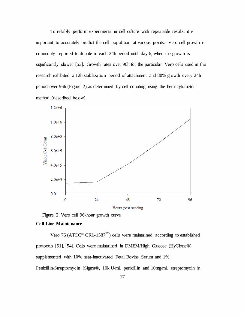
To reliably perform experiments in cell culture with repeatable results, it is
important to accurately predict the cell population at various points. Vero cell growth is
commonly reported to double in each 24h period until day 6, when the growth is
significantly slower [53]. Growth rates over 96h for the particular Vero cells used in this
research exhibited a 12h stabilization period of attachment and 80% growth every 24h
period over 96h (Figure 2) as determined by cell counting using the hemacytometer
method (described below).
Cell Line Maintenance
Vero 76 (ATCC® CRL-1587TM
) cells were maintained according to established
protocols [51], [54]. Cells were maintained in DMEM/High Glucose (HyClone®)
supplemented with 10% heat-inactivated Fetal Bovine Serum and 1%
Penicillin/Streptomycin (Sigma®, 10k U/mL penicillin and 10mg/mL streptomycin in
Figure 2. Vero cell 96-hour growth curve
18
0.9% NaCl). During subculture procedures, cell count and viability was performed by
the Trypan Blue exclusion test using a hemacytometer, described below. This permitted
a high consistency in cell growth timelines and cell survival. A cell growth curve (Figure
2) was established as a baseline for experiments influencing cellular growth rates and to
provide greater consistency and predictability when preparing for experiments.
Five 60mm petri dishes were seeded with Vero cells at a density of 15x104 per
dish, as determined by the hemacytometer method of cell counting. At 24 hour intervals,
one culture was removed from incubation and processed for cell counting as described
below. The slow growth in the first 24 hours is typical since the cells take approximately
12 hours to stabilize in the media, and undergo the process of adhesion to the dish prior to
entering the phase of growth where they double approximately every 24 hours [53].
Virus Propagation
Propagation of Dengue virus serotype 2 (DENV2-NGC) (Dr. Eric M. Vela,
Battelle Research Center) was performed in Vero 76 cells. The titer of virus stock was
approximately 106 PFU/mL. Vero 76 cells were seeded at 2x10⁶ in 100 mm petri dishes
in DMEM/High Glucose (HyClone®) supplemented with 10% heat-inactivated Fetal
Bovine Serum and 1% Penicillin/Streptomycin (Sigma®, 10k U/mL penicillin and
10mg/mL streptomycin in 0.9% NaCl). The cells were incubated at 37oC and 5% CO2
until they reached a confluency of approximately 90% then infected with DENV2 for 5-6
days, which was prior to significant cytopathic effects as observed through a light
microscope. Previous research has demonstrated that the presence of serum in the growth
media during DENV propagation does not inhibit the titer yield but is necessary for
19
proper cell maintenance in culture [55]. The cells were then detached from the growth
surface with Trypsin and centrifuged at 3200 rpm (2136x g) at 4oC for 10 minutes to
eliminate cell debris. Immature (non-infectious) isoforms of DENV are primarily
intracellular and captured in the pellet following centrifugation. The virus recovered
from the supernatant contain primarily mature (infectious) DENV although the various
isoforms can never be isolated with 100% efficiency. The supernatant was aliquoted and
stored at −80°C until use. Virus stock was filtered before use using 0.22µm filter
membranes (EMD Millipore).
Virus Quantification by Flow Cytometry
Dengue virus titers were determined by plaque assay in confluent monolayers of
Vero 76 cells grown in 6-well plates. Vero 76 cells were inoculated with ten-fold serial
dilutions when monolayers reached 80 to 90% confluence. After 2 hours of viral
adsorption, the monolayers were overlaid with 2 ml of Opti-MEM GlutaMAX (Life
Technologies Inc.) containing 2% Methyl-cellulose (Acros Organics) and 0.5% fetal
bovine serum. The cultures were incubated at 37 °C for 5-6 days and then counted for
plaque formation after fixation with 4% paraformaldehyde and staining with 1% crystal
violet solution (Sigma). Additionally viral titers were also determined using flow
cytometry as described by Medina et al [26]. Briefly, Vero cells grown in 24-well plates
at a density of 1.25x10⁵ per well 24 hours prior to infection were infected with ten-fold
serial dilutions of DENV2 diluted in Opti-MEM. After incubating one hour with gentle
rocking every 15 minutes, the supernatant was aspirated and overlaid with 1ml of 2%
methyl cellulose and incubated at 37 °C for 24 hours. Next day, infected monolayers
20
were trypsinized, fixed, blocked for unspecific binding and then stained with DENV2
monoclonal antibody 4G2 (Millipore). These antibodies are directed to the E protein of
the dengue virus and are capable of specifically neutralizing DENV2 serotype. Because
these antibodies are not fluorescent tagged, the cells were stained with fluorescein
isothiocyanate (FITC) conjugated secondary antibodies (Biolegend) and analyzed using
Accuri C6 flow cytometer. Final viral titers were calculated as infectious units (IU)/ml.
Silver Nanoparticle Synthesis and Filtration
AgNPs were synthesized by the reduction of 0.4% w/v (~0.024 M) silver nitrate
(AgNO3, MW=169.87, Sigma-Aldrich) with 0.5% w/v (~0.132 M) sodium borohydride
(NaBH4, MW=37.83, Sigma-Aldrich) in an aqueous matrix, stabilized by 1.0% w/v (~0.034
M) trisodium citrate dihydrate (Na3C6H5O7·2H2O, MW=294.10, Fluka). In a dark chemical
fume hood, 50mL of 4°C de-ionized water and 1.25mL of 0.4% silver nitrate were
combined and mixed with a magnetic stirrer for 1 minute. 500uL of 0.5% sodium
borohydride was rapidly added to the sidewall of the solution while mixing vigorously
for 1 minute. After 1 minute, 200uL of 1% sodium citrate was added and continuously
stirred for an additional 1 minute. The solution was placed in a foil covered 50 mL
conical tube and kept stored at 4°C until use.
This synthesis results in a colloid containing spherical AgNPs with a mean
diameter of 8nm. This method also results in trace amounts of non-reduced silver ions
following the formation of AgNPs. Silver ions were removed through tangential flow
filtration.
21
Tangential flow filtration (TFF) was employed to size-select AgNPs for viral
inhibition studies. Typically reserved for biological separations, TFF operates on the
continuous recirculation of a solution containing an analyte of interest across a porous
membrane (Figure 3). As a solution or colloid is passed over the membrane, larger
components are retained by the filter membrane and kept within the recirculation tubing
(e.g. gray spheres in Figure 3) while constituents smaller than the filter pores are passed
through the membrane and collected in a dilute filtrate (e.g. blue spheres in Figure 3).
By employing filters of varying pore size, AgNPs were size-selected for diameters
of approximately 5-20nm (8nm average) and concentrated by removing excess solvent
using membranes with ultrafine pore sizes (e.g., 5nm in diameter). The TFF process
Figure 3. Schematic depicting a typical TFF set-up for the filtration of colloidal AgNPs. Gray and blue spheres represent AgNPs larger and smaller than the filter
membrane pores, respectively.
24
AgNP-DENV2 Treatment
Silver nanoparticles were incubated with DENV2 at room temperature in various
concentrations (10, 25, 50, 100µg/mL) for one hour prior to Vero cell infection. This
pre-treatment timeline was chosen in order to allow stable binding between DENV2 and
the AgNP.
Trypan Blue Exclusion Test (Hemacytometer method)
Trypan Blue staining was performed during cell culture to ensure continued
viability of the Vero cells between passages. Trypan Blue staining was also performed to
support the results of MTT assays (cell viability). After centrifugation, cells were
suspended in DMEM and titurated thoroughly. A hemacytometer was cleaned with 70%
ethanol and a 20mm glass coverslip was placed over the viewing grid. 50µL of the cell
suspension was added to 100µL Trypan Blue stain in a 0.5mL centrifuge tube and
titurated thoroughly. This 50:100 ratio provided a dilution factor of 3. The cell-stain
mixture was pipetted into the loading channel until the viewing grid was full
(approximately 10µL). Viable cells exclude the stain and appear white while dead cells
take up the stain and appear blue. Eight grids were counted (living/dead) to provide cell
viability as a percentage of total cells. Estimated cell count was determined by the
formula:
𝑉𝑖𝑎𝑏𝑙𝑒 𝑐𝑒𝑙𝑙𝑠
𝑚𝐿=
# 𝑣𝑖𝑎𝑏𝑙𝑒 𝑐𝑒𝑙𝑙𝑠
# 𝑔𝑟𝑖𝑑𝑠 𝑐𝑜𝑢𝑛𝑡𝑒𝑑∗ 𝑑𝑖𝑙𝑢𝑡𝑖𝑜𝑛 𝑓𝑎𝑐𝑡𝑜𝑟 ∗ 104
26
hours at 37oC and 5% CO2. Following incubation and visual confirmation of the
formation of purple formazan crystals in mock treated wells, the MTT/HEPES solution
was carefully aspirated and the crystals were solubilized with 100µL dimethyl sulfoxide.
After 15 minutes, the absorbance of each well was read at 570nm (Figure 7) with a
background of 620nm in a spectrophotometer with 3 reads per well following 30 seconds
of slow orbital rotation and 10 seconds of settling time. Quantitative results are averages
of four measurements under the same conditions.
Biocompatibility of AgNPs in Vero cells
The biocompatibility of silver nanoparticles versus silver ions were determined
through MTT assay. Vero cells were seeded at 2x10⁴ cells/well in a 96-well plate in
DMEM-10 and incubated at 37oC and 5% CO2. The cells were allowed to stabilize for 24
hours under these conditions prior to treatment. After 24 hours, the cells were exposed to
AgNP or silver nitrate (AgNO3) in concentrations of 100µg/mL, 50µg/mL, and 10µg/mL,
for 24 hours. MTT assay was performed as described above.
Virus Binding Study
To assess the influence of AgNP in the binding of DENV to target cells, a cold-
binding study was conducted. The first steps in the viral life cycle are binding to a target
cells and entry into that cell by virus-dependent mechanisms. DENV is able to undergo
cell-binding events at 4oC but entry (by clatherin-mediated endocytosis) is not permitted
at this low temperature.
27
Vero cells were seeded at 3x10⁴ cells/well in a 12-well μ-slide (Ibidi(R)) for 24
hours. Experimental treatments (AgNP-DENV, DENV, and AgNP) were prepared at
room temperature for one hour. The AgNP-treated DENV treatment was co-incubated
for one hour at room temperature to permit binding of AgNP to DENV. Volumes of
AgNP and DENV used in the co-incubation were calculated to provide the desired AgNP
concentration and DENV multiplicity of infection (MOI). The required virus inoculum
was calculated using the formula:
(𝑐𝑒𝑙𝑙𝑠 𝑝𝑒𝑟 𝑤𝑒𝑙𝑙)(𝑀𝑂𝐼)
(𝑣𝑖𝑟𝑢𝑠 𝑡𝑖𝑡𝑒𝑟, 𝑝𝑓𝑢)∗ 1000µ𝐿 = (𝑖𝑛𝑛𝑜𝑐𝑢𝑙𝑢𝑚)µ𝐿
Cells and treatments were cooled to 4oC for 15 minutes. The treatments were incubated
with cells for one hour at 4oC. Slides were gently rocked every 15 minutes during
treatment. Following cold incubation, cells were gently washed three times with PBS
(4oC) to remove unbound DENV and/or AgNP. Control cells were also washed in the
same manner. Treatments and washes were removed by pouring onto an absorbent
towel.1 Cells were fixed with chilled 4% paraformaldehyde to prevent the internalization
of DENV and AgNP. Indirect fluorescence (without Triton-X permeabilization) was
performed to visualize the effects of AgNP on DENV binding to target cells.
Cells were washed three times with PBS. Primary antibody was diluted 1:300 per
manufacturer specifications and incubated with cells for one hour at 37oC or overnight at
4oC. The primary antibody used was the Anti-Dengue Virus Serotype 2 Antibody, clone
3H5-1 of mouse origin (MAB8702 Chemicon, Hampshire, United Kingdom). As an
1 Vacuum aspiration repeatedly removed cells in early trials
28
isotype control, normal mouse IgG (NI03, Calbiochem®) was added in place of the 3H5
in one well.
Following incubation, the primary antibody was removed and cells were washed
three times with chilled PBS. Secondary antibody was diluted and added to cells for one
hour at 37oC or overnight at 4oC. The secondary antibody was rabbit anti-mouse IgG-
FITC. The secondary antibody was removed and cells washed three times with chilled
PBS. Cells were counter-stained with Texas Red®-X Phalloidin for 10 minutes to
visualize the polymerized F-actin cytoskeleton of the cells. Cells were washed three
times with chilled PBS and a cover slip was mounted using Vectashield® H-1500 which
contains DAPI (4',6-diamidino-2-phenylindole, blue) for nuclear visualization.
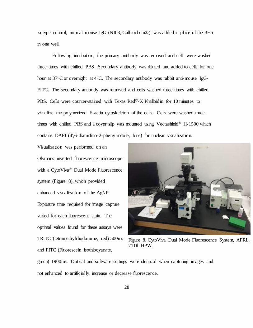
Visualization was performed on an
Olympus inverted fluorescence microscope
with a CytoViva® Dual Mode Fluorescence
system (Figure 8), which provided
enhanced visualization of the AgNP.
Exposure time required for image capture
varied for each fluorescent stain. The
optimal values found for these assays were
TRITC (tetramethylrhodamine, red) 500ms
and FITC (Fluorescein isothiocyanate,
green) 1900ms. Optical and software settings were identical when capturing images and
not enhanced to artificially increase or decrease fluorescence.
Figure 8. CytoViva Dual Mode Fluorescence System, AFRL, 711th HPW.
29
Blocking Non-Specific Binding During Immunofluorescence
During indirect immunofluorescence procedures, the decision was made to
exclude the common blocking step. Traditionally, cells are incubated with bovine serum
albumin (BSA) and serum matching the host of the secondary antibody to block the non-
specific binding of antibodies to cell Fc receptors. According to research by Igor
Buchwalow, et al, there were no significant differences between various samples that
received or did not receive blocking treatment [23]. Buchwalow demonstrated that the
standard fixation step removes the ability of endogenous Fc receptors to bind the Fc
portion of antibodies. This result was confirmed during early fluorescence trials where
blocked and non-blocked samples were compared under the same experimental
conditions. In the absence of blocking, background staining was never observed. This
decision saved time and lab resources and add further credibility to the suggestion that
protein blocking steps, although commonplace in laboratories, are unnecessary.
Image Processing and Analysis
For purposes of this research and the virus binding study, the green (FITC)
fluorescent intensity represents the level of DENV attachment to the surface of Vero
cells. Image processing methods were used to isolate the fluorescent region of interest
(ROI) and remove the background and artifacts. Image analysis methods were used to
quantify the differences between ROI. The relative difference in fluorescence intensity
between images provided quantifiable differences between the level of DENV binding to
cells. Immunofluorescent images were processed and analyzed in ImageJ (National
Institutes of Health, http://imagej.nih.gov/ij/). Detailed screenshots of the image
30
processing and analysis steps are contained in Appendix B. Image processing and
analysis was conducted using procedures established for the quantification of
fluorescence which are accepted within the microscopy community.
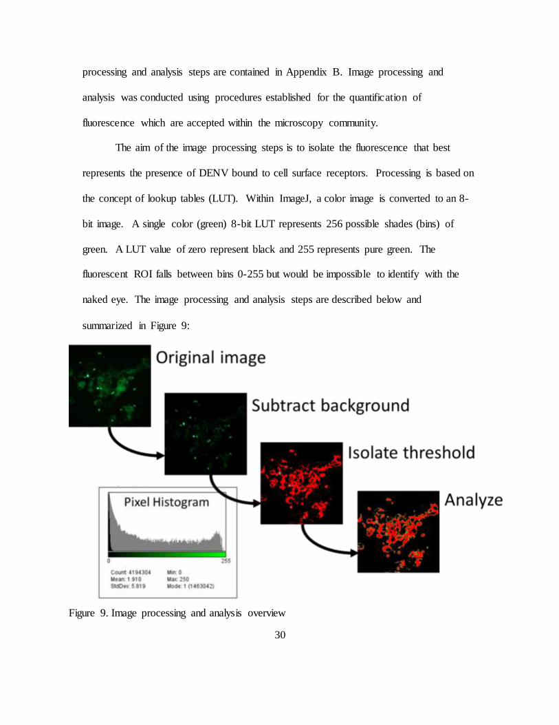
The aim of the image processing steps is to isolate the fluorescence that best
represents the presence of DENV bound to cell surface receptors. Processing is based on
the concept of lookup tables (LUT). Within ImageJ, a color image is converted to an 8-
bit image. A single color (green) 8-bit LUT represents 256 possible shades (bins) of
green. A LUT value of zero represent black and 255 represents pure green. The
fluorescent ROI falls between bins 0-255 but would be impossible to identify with the
naked eye. The image processing and analysis steps are described below and
summarized in Figure 9:
Figure 9. Image processing and analysis overview
31
1. Convert single-color image to 8-bit. While other image depths are available (i.e.
24-bit, with range 0-4095), the additional resolution is not required for this analysis.
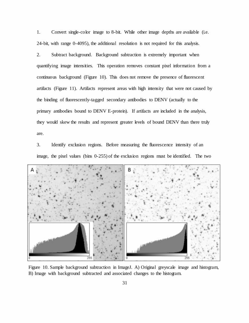
2. Subtract background. Background subtraction is extremely important when
quantifying image intensities. This operation removes constant pixel information from a
continuous background (Figure 10). This does not remove the presence of fluorescent
artifacts (Figure 11). Artifacts represent areas with high intensity that were not caused by
the binding of fluorescently-tagged secondary antibodies to DENV (actually to the
primary antibodies bound to DENV E-protein). If artifacts are included in the analysis,
they would skew the results and represent greater levels of bound DENV than there truly
are.
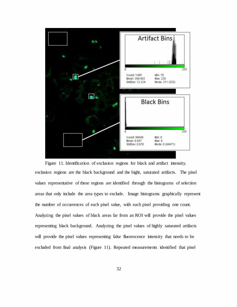
3. Identify exclusion regions. Before measuring the fluorescence intensity of an
image, the pixel values (bins 0-255) of the exclusion regions must be identified. The two
Figure 10. Sample background subtraction in ImageJ. A) Original greyscale image and histogram,
B) Image with background subtracted and associated changes to the histogram.
32
exclusion regions are the black background and the bight, saturated artifacts. The pixel
values representative of these regions are identified through the histograms of selection
areas that only include the area types to exclude. Image histograms graphically represent
the number of occurrences of each pixel value, with each pixel providing one count.
Analyzing the pixel values of black areas far from an ROI will provide the pixel values
representing black background. Analyzing the pixel values of highly saturated artifacts
will provide the pixel values representing false fluorescence intensity that needs to be
excluded from final analysis (Figure 11). Repeated measurements identified that pixel
Figure 11. Identification of exclusion regions for black and artifact intensity.
33
values from 0-8 represent black and values from 198-255 are present only in the artifacts.
In each image, the final range of pixel values representing the ROI were bins 9-197.
4. Isolate the threshold (ROI) using the limits of black maximum and artifact
minimum. Within the ImageJ Threshold feature, the pixel values are set to the 9-197
range mentioned above. Thresholding the image to this range creates a binary image
where excluded pixels are given a value of 0 and included pixels are given a value of 255.
At this point, the image is composed of area that will or will not be included in analysis.
This technique does not differentiate between strong fluorescence and weak fluorescence
directly, but is used to determine the visible surface area that contains fluorescent values
(from original image) determined to be valid intensities. The qualifying area represents
the ROI. The thresholding function creates closed objects within which is a portion of
the total ROI. Image analysis techniques are next used to measure the total area
contained within these ROI “islands” and other properties related to size and distribution.
5. Analyze images. The target metrics are total ROI area (sq. pixels) and cell count.
The Measure Particles function within ImageJ calculates the area of individual ROI
islands and provides an image total. Knowing the total area in the image that contained
qualifying pixel intensity is required, but does not present the whole story. If the pixel
intensity is held constant, an image with more cells should have greater area of ROI.
Normalizing an image’s ROI area with cell count provides a more accurate estimate of
DENV binding per cell. The Cell Counter tool was utilized to count the number of cells
in each image. If the normalized region of interest (nROI) is reduced from one image to
34
the next, it is reasonable to conclude that only the different experimental conditions
contributed to the reduction.
RESULTS
Cytotoxicity of DENV Infection of Vero Cells
The effect of DENV infection of Vero cells was analyzed to confirm the
infectivity of DENV virus stock and serve as a baseline for virus inhibition studies.
Following DENV infection (MOI 0.1), Vero cell viability decreased rapidly from 36-72
hours compared to non-infected cells (Figure 12). By 120hpi, the viability of infected
and noninfected cells was comparatively low. Based on visual comparison, the infected
cells experienced cytopathic effects and had lifted from the growth surface in patches.
The noninfected cells had overgrown and become weakly adherent to the growth surface
Figure 12. Viability of DENV infected Vero cells.
35
and were removed from the wells by the gentle force of media aspiration. While the cell
viability at 120hpi is similar, it was due to opposing conditions; the Vero cells began to
overgrow and die while the DENV infected cells died from infection and lifted from the
petri dishes in clusters.
This decline in cell viability was expected, as Vero cells do not have innate
defenses against infection. Since Vero cells are highly permissible of DENV infection,
they are commonly used to propagate DENV stock during research.
Biocompatibility of AgNPs in Vero cells
Cell viability measured by MTT assay demonstrated that increasing the
concentration of AgNP from 10-100µg/mL decreases cell viability relative to mock
Figure 13. Cytotoxicity of silver nanoparticles (AgNP) versus silver ions (Ag+) at in
concentrations of 100µg/mL, 50µg/mL, and 10µg/mL.
36
treated cells. Positive controls (silver ions, Ag+) in the same concentrations yielded much
greater cytotoxicity than AgNP, with the exception of Ag+ (10µg/mL), which had higher
viability than AgNP. The results suggest that AgNP stock colloid does not have a high
concentration of silver ions and that AgNP concentrations should be selected to be as low
as possible to minimize cytotoxic effects while providing antiviral or cytoprotective
effect from viral infection.
AgNP Inhibition of DENV Binding
Vero cells were incubated with treatments (AgNP-DENV, DENV only, AgNP
only) in 4oC to permit binding but inhibit internalization of AgNP and DENV. Based on
immunofluorescence analysis, DENV (MOI 40) pretreated with AgNP (25µg/mL),
resulted in far fewer bound virion compared to DENV infection without AgNP treatment
(positive control) and negative controls (data not shown). Untreated DENV presents
greater green fluorescence than after AgNP treatment (Figure 14). Fluorescence images
were captured at identical optical and processing software settings.
The reduction in FITC intensity was evaluated in ImageJ as previously described.
DENV pretreatment with AgNP (10 and 25µg/mL) reduced fluorescence below the levels
of the mock treated and isotype controls (Figure 15). Compared to DENV-only,
pretreatment with 10µg/mL reduced DENV binding by a factor of 23.2. Pretreatment
with 25µg/mL reduced DENV binding by a factor of 12.2.
Interestingly, the higher concentration of AgNP (25µg/mL) resulted in
approximately twice the fluorescence intensity of the lower concentration (10µg/mL).
One possible reason this occurred is that higher AgNP concentrations contributed to
37
increased agglomeration. This could reduce the effective number of AgNP available to
bind with DENV particles and therefore lead to reduced DENV binding inhibition.
Regardless, of the cause of this effect, both AgNP treatments led to fluorescence
intensities that were not significantly different than the mock treated control (P<0.05).
Comparison of the normalized ROI (nROI) histograms revealed statistically
significant differences (P<0.05, ANOVA) in the fluorescence intensity of treated cells
and controls, and between the AgNP-treated DENV and DENV-only treatments.
While cold incubation does not reflect in vivo conditions during infection, this
condition is effective when attempting to isolate the step at which AgNP may inhibit the
DENV life cycle. These results are similar to research showing AgNP inhibition of
various viruses at the binding and entry phase of viral replication. However, this is the
first time a Group IV virus (ss(-)RNA) has been inhibited by AgNP.
39
Statistical Analysis
SigmaPlot 12.0 software (Systat & Mystat Products, Chicago, IL) was used to create
graphs and perform statistical analysis. Experimental conditions were compared to mock-
treated controls as percent of control. Fluorescent images were also compared to the isotype
control to remove the influence of background staining. ImageJ was used to process images
as described above.
Histograms representing fluorescence intensities within the ROI were compared
using the Mann-Whitney rank sum test. As expected, each histogram failed the normality
test (Shapiro-Wilk), therefore, the Mann-Whitney rank sum test provided greater descriptive
efficiency than the paired t test.
Figure 15. Fluorescence intensity per cell. The isotype control and both AgNP concentrations did not produce fluorescence intensity significantly different from control
(P<0.05). DENV-only fluorescence was statistically significant compared to control and both AgNP concentrations (P>0.05).
40
CONCLUSION
This research examined the ability of 8nm silver nanoparticles to inhibit the
binding event of dengue virus to Vero cells in vitro. During the course of this research,
silver nanoparticles were synthesized, purified, and characterized. Dengue virus type 2
was propagated in Vero cells, quantified and verified for virulence, and purified of
bacterial/fungal contaminants. The biocompatibility of AgNP and Vero cells were
measured and compared to silver nitrate. The influences of AgNP in the binding event of
DENV to Vero cells was visualized through indirect immunofluorescence and quantified
through histogram analysis.
The null hypothesis of this study was that silver nanoparticles do not inhibit the
binding step of the dengue virus serotype 2 (DENV2) replication cycle in Vero 76 cells,
inferred through similar fluorescence intensity visualization. Results indicate that AgNP-
pretreated DENV produced a statistically significant reduction in fluorescence intensity
compared to DENV infection without AgNP pretreatment (P<0.05). Therefore, the null
hypothesis is rejected.
This research presents, for the first time that DENV binding to Vero cells is
inhibited by 8nm AgNP (citrate stabilized). Fluorescence intensity was 96% (10 µg/mL
AgNP) and 92% (25µg/mL AgNP) in AgNP-treated DENV compared to untreated
DENV-only infection of Vero cells. A limitation of this study is that the mechanism of
binding inhibition is unknown. Existing knowledge on AgNP interaction with viral
envelopes suggests that AgNP bind to DENV disulfide regions and possibly others. This
could inhibit binding by preventing receptor-epitope covalent interactions. However, it is
41
possible that unbound AgNP in the AgNP-DENV colloid coat the cells and prevent
DENV binding in a cytoprotective manner, rather than an antiviral agent.
This serves as a stepping off point for further research into the interaction of
AgNP and DENV. Continued research could include:
Development of a technique to filter unbound AgNP from the AgNP-DENV colloid
to prevent unbound AgNP from binding to cells
Examining the mechanism of binding inhibition by confirming AgNP-DENV binding
through TEM/SEM
Measure cell viability throughout the course of DENV infection versus AgNP-
DENV, measured by MTT assay
Uses of AgNP to disrupt the disease cycle in Aedes mosquitoes
Progeny virus production following AgNP-treated DENV infection of Vero cells or
macrophages, measured by using supernatants from infected cultures to infect second
set of cell cultures, quantified by either flow cytometry or MTT assay
Changes in DENV protein production, measured through Western blot after DENV
infection
Impact of AgNP during DENV infection of macrophages (target cells)
Quantification of DENV infection after AgNP treatment, measured by flow
cytometry
42
REFERENCES
[1] A. P. Goncalvez, R. E. Engle, M. St Claire, R. H. Purcell, and C.-J. Lai,
“Monoclonal antibody-mediated enhancement of dengue virus infection in vitro and in vivo and strategies for prevention.,” Proc. Natl. Acad. Sci. U. S. A., vol. 104, no. 22, pp. 9422–7, May 2007.
[2] WHO, “Dengue and severe dengue,” 2014. [Online]. Available:
http://www.who.int/mediacentre/factsheets/fs117/en/.
[3] CDC, “Dengue Epidemiology,” 2014. [Online]. Available: http://www.cdc.gov/dengue/epidemiology/index.html. [Accessed: 09-Jan-2015].
[4] D. J. Gubler, “Epidemic dengue/dengue hemorrhagic fever as a public health,
social and economic problem in the 21st century.,” Trends Microbiol., vol. 10, no. 2, pp. 100–3, Mar. 2002.
[5] E. a Henchal and J. R. Putnak, “The dengue viruses.,” Clin. Microbiol. Rev., vol. 3,
no. 4, pp. 376–96, Oct. 1990.
[6] World Health Organization, “Dengue: guidelines for diagnosis, treatment, prevention, and control,” Spec. Program. Res. Train. Trop. Dis., pp. x, 147, 2009.
[7] R. V Gibbons, M. Streitz, T. Babina, and J. R. Fried, “Dengue and US military
operations from the Spanish-American War through today.,” Emerg. Infect. Dis., vol. 18, no. 4, pp. 623–30, Apr. 2012.
[8] M. M. Wade, A. E. Chambers, J. M. Insalaco, and A. W. Zulich, “Survival of viral biowarfare agents in disinfected waters.,” Int. J. Microbiol., vol. 2010, p. 412694,
Jan. 2010.
[9] J. Hombach, M. Jane Cardosa, A. Sabchareon, D. W. Vaughn, and A. D. T. Barrett, “Scientific consultation on immunological correlates of protection induced
by dengue vaccines. Report from a meeting held at the World Health Organization 17-18 November 2005,” in Vaccine, 2007, vol. 25, pp. 4130–4139.
[10] B. L. Innis, K. H. Eckels, E. Kraiselburd, D. R. Dubois, G. F. Meadors, D. J. Gubler, D. S. Burke, and W. H. Bancroft, “Virulence of a live dengue virus
vaccine candidate: a possible new marker of dengue virus attenuation.,” J. Infect. Dis., vol. 158, pp. 876–880, 1988.
[11] T. L. Yap, T. Xu, Y.-L. Chen, H. Malet, M.-P. Egloff, B. Canard, S. G.
Vasudevan, and J. Lescar, “Crystal structure of the dengue virus RNA-dependent
43
RNA polymerase catalytic domain at 1.85-angstrom resolution.,” J. Virol., vol. 81, no. 9, pp. 4753–65, May 2007.
[12] Z. Yin, Y. Chen, W. Schul, Q. Wang, F. Gu, J. Duraiswamy, W. Liu, B. Liu, J. Y.
H. Lim, C. Young, M. Qing, C. Chin, A. Yip, G. Wang, W. Ling, H. Pen, K. Lin, B. Zhang, G. Zou, K. A. Bernard, C. Garrett, K. Beltz, M. Dong, M. Weaver, H.
He, A. Pichota, V. Dartois, T. H. Keller, and P. Shi, “An adenosine nucleoside inhibitor of dengue virus,” 2009.
[13] A. C. Eifler and C. S. Thaxton, “Nanoparticle therapeutics: FDA approval, clinical
trials, regulatory pathways, and case study.,” Methods Mol. Biol., vol. 726, pp. 325–38, Jan. 2011.
[14] A. M. Schrand, M. F. Rahman, S. M. Hussain, J. J. Schlager, D. a Smith, and A. F. Syed, “Metal-based nanoparticles and their toxicity assessment.,” Wiley
Interdiscip. Rev. Nanomed. Nanobiotechnol., vol. 2, no. 5, pp. 544–68, 2010.
[15] R. J. Vandebriel, E. C. Tonk, L. J. de la Fonteyne-Blankestijn, E. R. Gremmer, H. W. Verharen, L. T. van der Ven, H. van Loveren, and W. H. de Jong,
“Immunotoxicity of silver nanoparticles in an intravenous 28-day repeated-dose toxicity study in rats.,” Part. Fibre Toxicol., vol. 11, no. 1, p. 21, Jan. 2014.
[16] J. S. Kim, J. H. Sung, J. H. Ji, K. S. Song, J. H. Lee, C. S. Kang, and I. J. Yu, “In
vivo Genotoxicity of Silver Nanoparticles after 90-day Silver Nanoparticle Inhalation Exposure.,” Saf. Health Work, vol. 2, no. 1, pp. 34–8, Mar. 2011.
[17] J. H. Lee, Y. S. Kim, K. S. Song, H. R. Ryu, J. H. Sung, J. D. Park, H. M. Park, N. W. Song, B. S. Shin, D. Marshak, K. Ahn, J. E. Lee, and I. J. Yu, “Biopersistence
of silver nanoparticles in tissues from Sprague-Dawley rats.,” Part. Fibre Toxicol., vol. 10, p. 36, Jan. 2013.
[18] I. Papageorgiou, C. Brown, R. Schins, S. Singh, R. Newson, S. Davis, J. Fisher, E.
Ingham, and C. P. Case, “The effect of nano- and micron-sized particles of cobalt-chromium alloy on human fibroblasts in vitro.,” Biomaterials, vol. 28, no. 19, pp. 2946–58, Jul. 2007.
[19] L. Schnirring, “Researchers identify fifth dengue serotype,” CIDRAP News, 2013.
[Online]. Available: http://www.cidrap.umn.edu/news-perspective/2013/10/researchers- identify-fifth-dengue-subtype.
[20] C. Moore, “UTMB Galveston Researchers Discover First New Dengue Fever
Serotype In 50 Years,” BioNews Texas, 2013. [Online]. Available: http://bionews-tx.com/news/2013/10/25/utmb-galveston-researchers-discover-first-new-dengue-
fever-serotype-in-50-years/.
44
[21] J. M. da Silva Voorham, “A possible fifth dengue virus serotype,” Ned. Tijdschr. Geneeskd., vol. 158, p. A7946, Jan. 2014.
[22] M. G. Guzmán and G. Kourí, “Dengue: an update.,” Lancet. Infect. Dis., vol. 2, no.
1, pp. 33–42, Jan. 2002.
[23] J. Blok, “Genetic Relationships of the Dengue Virus Serotypes,” pp. 1323–1325, 1985.
[24] R. J. Kuhn, W. Zhang, M. G. Rossmann, S. V Pletnev, J. Corver, E. Lenches, C. T.
Jones, S. Mukhopadhyay, P. R. Chipman, E. G. Strauss, T. S. Baker, and J. H. Strauss, “Structure of dengue virus: implications for flavivirus organization,
maturation, and fusion.,” Cell, vol. 108, no. 5, pp. 717–25, Mar. 2002.
[25] Y. Modis, S. Ogata, D. Clements, and S. C. Harrison, “A ligand-binding pocket in the dengue virus envelope glycoprotein.,” Proc. Natl. Acad. Sci. U. S. A., vol. 100, no. 12, pp. 6986–91, Jun. 2003.
[26] F. Medina, J. F. Medina, C. Colón, E. Vergne, G. A. Santiago, and J. L. Muñoz-Jordán, “Dengue virus: isolation, propagation, quantification, and storage.,” Curr. Protoc. Microbiol., vol. Chapter 15, no. November, p. Unit 15D.2., Nov. 2012.
[27] K. I. P. J. Hidari and T. Suzuki, “Dengue virus receptor.,” Trop. Med. Health, vol.
39, no. 4 Suppl, pp. 37–43, Dec. 2011.
[28] J. L. Miller, B. J. de Wet, L. Martinez-Pomares, C. M. Radcliffe, R. a Dwek, P. M. Rudd, and S. Gordon, “The mannose receptor mediates dengue virus infection of
macrophages,” PLoS Pathog, vol. 4, no. 2, p. e17, Feb. 2008.
[29] J. D. J. Marti, R. M. Angel, and S. Marti, “Identification of a Putative Coreceptor on Vero Cells That Participates in Dengue 4 Virus Infection Identification of a Putative Coreceptor on Vero Cells That Participates in Dengue 4 Virus Infection,”
vol. 75, no. 17, 2001.
[30] H. M. van der Schaar, M. J. Rust, C. Chen, H. van der Ende-Metselaar, J. Wilschut, X. Zhuang, and J. M. Smit, “Dissecting the cell entry pathway of dengue
virus by single-particle tracking in living cells.,” PLoS Pathog., vol. 4, no. 12, p. e1000244, Dec. 2008.
[31] L. Suksanpaisan, T. Susantad, and D. R. Smith, “Characterization of dengue virus
entry into HepG2 cells.,” J. Biomed. Sci., vol. 16, no. strain 16007, p. 17, Jan. 2009.
45
[32] M. N. Krishnan, B. Sukumaran, U. Pal, H. Agaisse, J. L. Murray, T. W. Hodge, and E. Fikrig, “Rab 5 is required for the cellular entry of dengue and West Nile
viruses.,” J. Virol., vol. 81, no. 9, pp. 4881–5, May 2007.
[33] J. C. Trefry, “The development of silver nanoparticles as antiviral agents,” Dayton, 2011.
[34] J. V. Rogers, C. V. Parkinson, Y. W. Choi, J. L. Speshock, and S. M. Hussain, “A
Preliminary Assessment of Silver Nanoparticle Inhibition of Monkeypox Virus Plaque Formation,” Nanoscale Res. Lett., vol. 3, no. 4, pp. 129–133, Apr. 2008.
[35] E. A. Sopova, V. I. Baranov, O. A. Gankovskaia, V. F. Lavrov, and V. V Zverev,
“Effects of silver and silicon dioxide nanopowders on the development of herpesvirus infection in vitro,” Gig. Sanit., pp. 89–91.
[36] D. Xiang, Q. Chen, L. Pang, and C. Zheng, “Inhibitory effects of silver nanoparticles on H1N1 influenza A virus in vitro.,” J. Virol. Methods, vol. 178, no.
1–2, pp. 137–42, Dec. 2011.
[37] D. Xiang, Y. Zheng, W. Duan, X. Li, J. Yin, S. Shigdar, M. L. O’Connor, M. Marappan, X. Zhao, Y. Miao, B. Xiang, and C. Zheng, “Inhibition of
A/Human/Hubei/3/2005 (H3N2) influenza virus infection by silver nanoparticles in vitro and in vivo,” Int. J. Nanomedicine, vol. 8, pp. 4103–4114, 2013.
[38] J. L. Speshock, R. C. Murdock, L. K. Braydich-Stolle, A. M. Schrand, and S. M.
Hussain, “Interaction of silver nanoparticles with Tacaribe virus.,” J. Nanobiotechnology, vol. 8, p. 19, Jan. 2010.
[39] L. Sun, A. K. Singh, K. Vig, S. R. Pillai, and S. R. Singh, “Silver nanoparticles inhibit replication of respiratory syncytial virus,” J. Biomed. Nanotechnol., vol. 4,
no. 2, pp. 149–158, 2008.
[40] H. H. Lara, N. V Ayala-Nuñez, L. Ixtepan-Turrent, and C. Rodriguez-Padilla, “Mode of antiviral action of silver nanoparticles against HIV-1.,” J.
Nanobiotechnology, vol. 8, p. 1, Jan. 2010.
[41] H. H. Lara, L. Ixtepan-Turrent, E. N. Garza Treviño, and D. K. Singh, “Use of silver nanoparticles increased inhibition of cell-associated HIV-1 infection by
neutralizing antibodies developed against HIV-1 envelope proteins.,” J. Nanobiotechnology, vol. 9, no. 1, p. 38, Jan. 2011.
[42] J. L. Elechiguerra, J. L. Burt, J. R. Morones, A. Camacho-Bragado, X. Gao, H. H. Lara, and M. J. Yacaman, “Interaction of silver nanoparticles with HIV-1.,” J.
Nanobiotechnology, vol. 3, p. 6, Jan. 2005.
46
[43] J. C. Trefry and D. P. Wooley, “Rapid assessment of antiviral activity and cytotoxicity of silver nanoparticles using a novel application of the tetrazolium-
based colorimetric assay.,” J. Virol. Methods, vol. 183, no. 1, pp. 19–24, Jul. 2012.
[44] L. Lu, R. W.-Y. Sun, R. Chen, C.-K. Hui, C.-M. Ho, J. M. Luk, G. K. K. Lau, and C.-M. Che, “Silver nanoparticles inhibit hepatitis B virus replication.,” Antivir.
Ther., vol. 13, no. 2, pp. 253–62, Jan. 2008.
[45] N. Chen, Y. Zheng, J. Yin, X. Li, and C. Zheng, “Inhibitory effects of silver nanoparticles against adenovirus type 3 in vitro,” J. Virol. Methods, vol. 193, no.
2, pp. 470–477, 2013.
[46] J. R. Furr, A. D. Russell, T. D. Turner, and A. Andrews, “Antibacterial activity of Actisorb Plus, Actisorb and silver nitrate,” J. Hosp. Infect., vol. 27, no. 3, pp. 201–208, Jul. 1994.
[47] J. Y. Kim, C. Lee, M. Cho, and J. Yoon, “Enhanced inactivation of E. coli and
MS-2 phage by silver ions combined with UV-A and visible light irradiation,” Water Res., vol. 42, pp. 356–362, 2008.
[48] R. M. Richards, “Antimicrobial action of silver nitrate.,” Microbios, vol. 31, pp.
83–91, 1981.
[49] P. Jena, S. Mohanty, R. Mallick, B. Jacob, and A. Sonawane, “Toxicity and antibacterial assessment of chitosan-coated silver nanoparticles on human
pathogens and macrophage cells.,” Int. J. Nanomedicine, vol. 7, pp. 1805–18, Jan. 2012.
[50] C. Levard, B. C. Reinsch, F. M. Michel, C. Oumahi, G. V. Lowry, and G. E. Brown, “Sulfidation processes of PVP-coated silver nanoparticles in aqueous
solution: Impact on dissolution rate,” Environ. Sci. Technol., vol. 45, pp. 5260–5266, 2011.
[51] N. C. Ammerman, M. Beier-Sexton, and A. F. Azad, “Growth and Maintenance of
Vero Cell Lines,” Curr. Protoc. Microbiol., no. 11, pp. 1–10, 2008.
[52] T. Matsumura, V. Stollar, and R. W. Schlesinger, “Studies on the nature of dengue viruses,” Virology, vol. 46, no. 2, pp. 344–355, Nov. 1971.
[53] a T. Nahapetian, J. N. Thomas, and W. G. Thilly, “Optimization of environment
for high density Vero cell culture: effect of dissolved oxygen and nutrient supply on cell growth and changes in metabolites.,” J. Cell Sci., vol. 81, pp. 65–103, Mar. 1986.
47
[54] ATCC, “RAW 264.7 (ATCC ® TIB-71 TM ) Product Sheet.” American Type Culture Collection, 2013.
[55] C.-C. Liu, S.-C. Lee, M. Butler, and S.-C. Wu, “High genetic stability of dengue
virus propagated in MRC-5 cells as compared to the virus propagated in vero cells.,” PLoS One, vol. 3, no. 3, p. e1810, Jan. 2008.
[56] M. I. Patel, R. Tuckerman, and Q. Dong, “A Pitfall of the 3-(4,5-dimethylthiazol-
2-yl)-5(3-carboxymethonyphenol)-2-(4-sulfophenyl)-2H-tetrazolium (MTS) assay due to evaporation in wells on the edge of a 96 well plate.,” Biotechnol. Lett., vol.
27, no. 11, pp. 805–8, Jun. 2005.
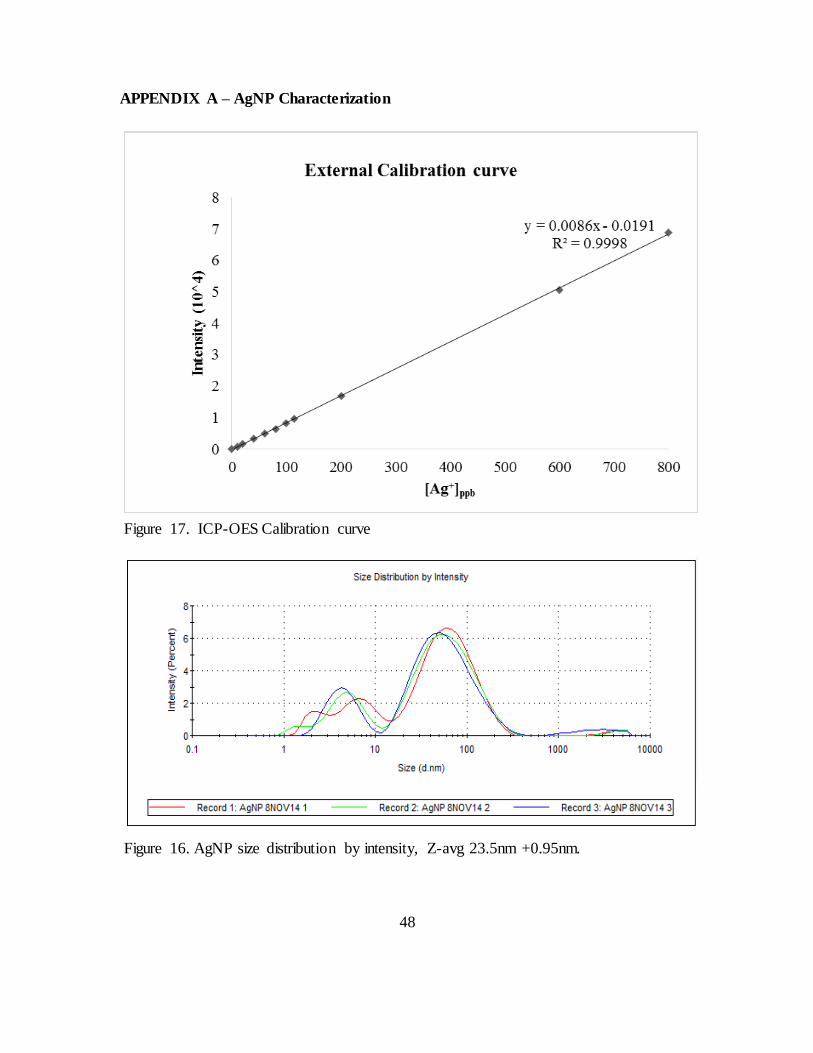
48
APPENDIX A – AgNP Characterization
Figure 16. AgNP size distribution by intensity, Z-avg 23.5nm +0.95nm.
Figure 17. ICP-OES Calibration curve
49
REPORT DOCUMENTATION PAGE Form Approved OMB No. 074-0188
The public reporting burden for this collection of information is estimated to average 1 hour per response, including the time for reviewing instructions, searching existing data sources, gathering and maintaining the data needed, and completing and reviewing the collection of information. Send comments regarding this burden estimate or any other aspect of the collection of information, including suggestions for reducing this burden to Department of Defense, Washington Headquarters Services, Directorate for Information Operations and Reports (0704-0188), 1215 Jefferson Davis Highway, Suite 1204, Arlington, VA 22202-4302. Respondents should be aware that notwithstanding any other provision of law, no person shall be subject to an penalty for failing to comply with a collection of information i f it does not display a currently valid OMB control
number.
PLEASE DO NOT RETURN YOUR FORM TO THE ABOVE ADDRESS. 1. REPORT DATE (DD-MM-YYYY)
26-03-2015
2. REPORT TYPE
Master’s Thesis
3. DATES COVERED (From – To)
March 2012 – March 2015
TITLE AND SUBTITLE
Evidence for the Inhibition of Dengue Virus Binding in the
Presence of Silver Nanoparticles
5a. CONTRACT NUMBER
5b. GRANT NUMBER
5c. PROGRAM ELEMENT NUMBER
6. AUTHOR(S)
Williams, Kelley J., Captain, USA
5d. PROJECT NUMBER
5e. TASK NUMBER
5f. WORK UNIT NUMBER
7. PERFORMING ORGANIZATION NAMES(S) AND ADDRESS(S)
Air Force Institute of Technology
Graduate School of Engineering and Physics (AFIT/ENP)
2950 Hobson Way, Building 640
WPAFB OH 45433-8865
8. PERFORMING ORGANIZATION REPORT NUMBER
AFIT-ENP-MS-15-M-089
9. SPONSORING/MONITORING AGENCY NAME(S) AND ADDRESS(ES)
AFRL/711th Human Performance Wing
2729 R St, Area B, Bldg 837
Dayton, OH 45433
(937) 904-9570
10. SPONSOR/MONITOR’S ACRONYM(S)
AFRL/BIN
11. SPONSOR/MONITOR’S REPORT NUMBER(S)
12. DISTRIBUTION/AVAILABILITY STATEMENT
DISTRUBTION STATEMENT A. APPROVED FOR PUBLIC RELEASE; DISTRIBUTION UNLIMITED.
13. SUPPLEMENTARY NOTES This material is declared a work of the U.S. Government and is not subject to copyright protection in the
United States.
14. ABSTRACT
Dengue is an emerging hemorrhagic fever virus and widely considered the most important arbovirus in the world. Dengue virus (DENV) is a positive-sense RNA virus that exists in 4, potentially 5, antigenic
serotypes. Currently, no vaccines or treatments are approved for DENV infections. Unsuccessful vaccine trials open the door for non-traditional treatments such as silver nanoparticles. Silver nanoparticles (AgNP) are known to inhibit viral replication of numerous viruses but have never before
been tested for inhibition of dengue virus type 2 (DENV2, ATCC® VR-1584™). For the first time, this research presents up to a 96% reduction in DENV2 binding to Vero cells following pretreatment with AgNP (6-10nm, 25µg/mL). These results suggest that similarly to other viruses, DENV2 can be
inhibited at the first stage of the virus replication cycle, binding & entry. 15. SUBJECT TERMS
Dengue, silver nanoparticle, antiviral, virus, flavivirus
16. SECURITY CLASSIFICATION
OF: 17. LIMITATION
OF ABSTRACT
UU
18. NUMBER
OF PAGES
60
19a. NAME OF RESPONSIBLE PERSON
Douglas R. Lewis, AFIT/ENP a. REPORT
U
b. ABSTRACT
U
c. THIS PAGE
U
19b. TELEPHONE NUMBER (Include area code)
(937) 255-3636, ext 4569
Standard Form 298 (Rev. 8-98) Prescribed by ANSI Std. Z39-18