Embed Size (px)
Citation preview

The value of leaf cuticle characteristics in the identification and classification of Iberian Mediterranean members of the genus Pinus
SALVIA GARCIA ALVAREZ , IGNACIO GARCIA-AMORENA, JUAN M. RUBIALES and CARLOS MORLA
Unidad Docente de Botdnica, Escuela Tecnica Superior de Ingenieros de Montes, Universidad Politecnica de Madrid, Ciudad Universitaria sin, 28040 Madrid, Spain
This study reports the value of leaf cuticle characteristics in the identification and classification of Iberian Mediterranean species of the genus Pinus (P. nigra subsp. salzmannii, P. pinaster, P. pinea and P. halepensis), with the aim of using these characters to identify isolated cuticles and stomata in palynology slides. Preparations were made of the cuticles of pine needles belonging to one natural Iberian population of each of the above species. A number of epidermal morphological characteristics were then recorded with the aim of distinguishing these species from one another. The structure of the stomatal complex (the shape and arrangement of the subsidiary cells) was different in each species. The aperture of the epistomatal chamber was significantly smaller in P. pinea than in the other species examined, and the variables recorded for the thickening of the guard cells provided relationships that clearly distinguished all four taxa. The width and length of the stomata and the upper woody lamellae, the central distance between the external limits of the medial lamellae borders and the length of the stem were the most useful variables in this respect. The present results contribute to the ongoing discussion regarding the taxonomic classification of the members of Pinus, and provide valuable clues for the identification of Iberian Mediterranean pine species from small pine needle fragments or isolated stomata. After validation of the present results for multiple populations, these results could also be used to help identify fossil leaf macroremains and the scattered/ isolated stomata commonly observed in palaeopalynological samples.
ADDITIONAL KEYWORDS: Iberian Peninsula - macrofossils - palaeobiogeography - palaeobotany - Pinus halepensis - Pinus nigra - Pinus pinaster - Pinus pinea - stomata.
I N T R O D U C T I O N
Improving our species-level knowledge of the less studied vegetative par ts of plants, such as their cuticular and stomatal features, could provide information of great taxonomic and even palaeobotanical interest (Barclay etal., 2007). This is t rue even for well-known taxa, such as members of the genus Pinus L.
The Iberian Peninsula is currently the natural home of six species of Pinus (Gaussen, Heywood & Chater, 1964). Pinus sylvestris L. and P. uncinata
Ramond ex DC, both of typically Eurosiberian distribution, have been the subject of morphological studies at the level of the leaf epidermis, and, in some cases, the results have allowed the distinction of these species (Boratynska & Bobowicz, 2001; Struzkova, 2002; Garcia Alvarez etal., 2009). However, little information is available for most of the Pinus species with Mediterranean distributions: P. nigra J.F.Arnold subsp. salzmannii (Dunal) Franco (Yoshie & Sakai, 1985), P. pinaster Alton (Yoshie & Sakai, 1985) and P. halepensis Mill (Boddi, Bonzi & Calamassi, 2002). In addition, morphological details of the leaf cuticle of P. pinea L. remain unstudied.
In this article, we examine the differences and similarities of the cuticles and stomata of these four Iberian Mediterranean taxa. Morphological
differences in these features could allow the identification of these species when only fragments of pine needles are available, for instance, in the analysis of herbivore gut contents (e.g. Stewart, 1967). Epidermal information could also be used as a tool to identify palaeobotanical material . The resistance to degradation demonstrated by cutin allows cuticles to become fossilized (Kerp, 1990). The identification of these fragments would help reveal the role played by different forest species during the evolution of historical landscapes (Theobald, Krahulik & Rollins, 1979; Barron & Buades, 2002). Finally, epidermal differences may also be important in systematic studies, as different authors have classified the six Iberian species of Pinus in different ways (Shaw, 1914, 1924; Pilger, 1926; Little & Critchfield, 1969; Price, Liston & Strauss, 1998; Liston et al., 1999; Wang et al., 1999; Gernandt etal., 2005). Any taxonomic differences shown by the cuticles could provide new information for determining the phylogenetic relationships between them.
Stomatal analysis is commonly used in the examination of the dispersed stomata observed in pollen preparations (Hansen & Engstrom, 1996; Birks & Birks, 2000; Hicks, 2006). The assumption of the local presence of taxa based on their pollen record could be controversial in some instances, but the finding of stomata in a fossil pollen sample allows the local presence of a taxon to be confirmed (Dunwiddie, 1987; Ammann & Wick, 1993). The taxonomic identification of pine stomata has been successful at the genus level (Hansen, 1995; Sweeney, 2004), but, to date, the classification of these dispersed stomata has been possible at the species' level only in contexts in which a single known species is thought to have been present. For example, stomata belonging to P. sylvestris L. have thus been identified in material from recent Quaternary Scottish and Scandinavian settings (Gervais etal., 2002; Froyd, 2005). The ability to identify Iberian Mediterranean pines properly via the remains of their s tomata and cuticles would also be of great help in determining the influence of each species on the Quaternary evolution of Iberian Pinus forests. Currently, this is well understood at the genus level, but only a few works have contributed to the history of Pinus spp. (Franco Mujica et al. 2000; Garcia-Amorena etal., 2007; Rubiales etal., 2007, 2009).
In this article, we report the taxonomic value of different leaf epidermal characteristics in single populations of the four Iberian Mediterranean Pinus species. Three of these taxa, which are native to south-western Europe, have been included in previous leaf epidermal studies (Yoshie & Sakai, 1985; Boddi et al., 2002), but P. pinea has not been examined in this way.
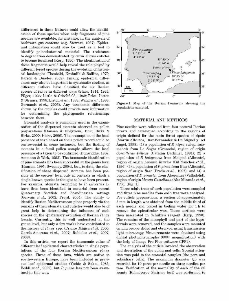
Figure 1. Map of the Iberian Peninsula showing the populations sampled.
MATERIAL A N D M E T H O D S
Pine needles were collected from four na tura l Iberian forests and catalogued according to the regions of origin defined for the main forest species of Spain (Martin Albertos, Diaz-Fernandez & De Miguel y Del Angel, 1998): (1) a population of P. nigra subsp. salz-mannii from La Sagra (Granada), region of origin Cordilleras Beticas (Catalan Bachiller, 1991); (2) a population of P. halepensis from Maigmo (Alicante), region of origin Levante Interior (Gil Sanchez etal., 1996); (3) a population of P. pinea from Biar (Alicante), region of origin Biar (Prada etal., 1997); and (4) a population of P. pinaster from Ataquines (Valladolid), region of origin Meseta Castellana (Alia Miranda et al., 1996) (Fig. 1).
Three adult trees of each population were sampled and three pine needles from each tree were analysed. For cuticle preparations, a section of approximately 5 mm in length was obtained from the middle third of each needle and placed in boiling water for 1 h to remove the epicuticular wax. These sections were then macerated in Schulze's reagent (Kerp, 1990). The remains of the mesophyll and par t of the hypo-dermis were removed, and the samples were mounted on microscope slides and observed using transmission light microscopy. Measurements were obtained using digital photomicrographs (600x magnification) with the help of Image Pro Plus software (IPP4).
The analysis of the cuticle involved the observation and description of the epidermal cells. Special attention was paid to the stomatal complex (the pore and subsidiary cells). The maximum diameter (p) was recorded for 10 pores per needle on three needles per tree. Verification of the normality of each of the 30 counts (Kolmogorov-Smirnov test) was performed to
'
1 Lb La « J
V 1
f Ab *
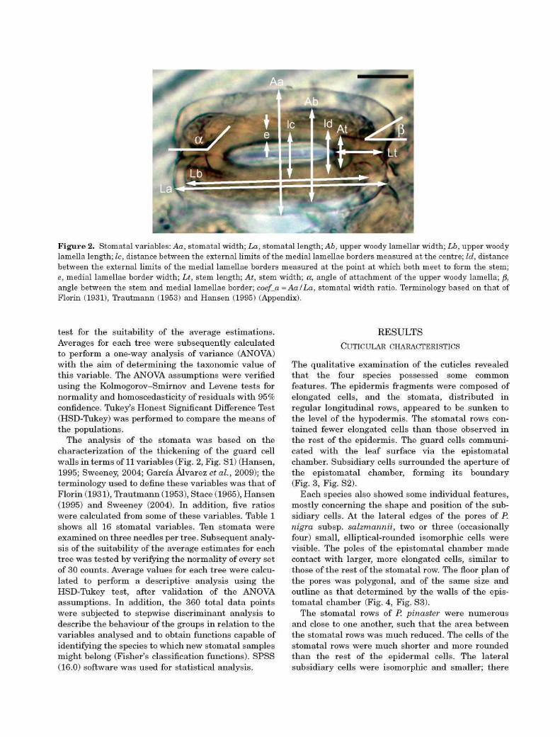
£ Figure 2. Stomatal variables: Aa, stomatal width; La, stomatal length; Ab, upper woody lamellar width; Lb, upper woody lamella length; Ic, distance between the external limits of the medial lamellae borders measured at the centre; Id, distance between the external limits of the medial lamellae borders measured at the point at which both meet to form the stem; e, medial lamellae border width; Lt, stem length; At, stem width; a, angle of attachment of the upper woody lamella; /}, angle between the stem and medial lamellae border; coef_a =AalLa, stomatal width ratio. Terminology based on that of Florin (1931), Trautmann (1953) and Hansen (1995) (Appendix).
test for the suitability of the average estimations. Averages for each tree were subsequently calculated to perform a one-way analysis of variance (ANOVA) with the aim of determining the taxonomic value of this variable. The ANOVA assumptions were verified using the Kolmogorov-Smirnov and Levene tests for normality and homoscedasticity of residuals with 95% confidence. Tukey's Honest Significant Difference Test (HSD-Tukey) was performed to compare the means of the populations.
The analysis of the s tomata was based on the characterization of the thickening of the guard cell walls in terms of 11 variables (Fig. 2, Fig. SI) (Hansen, 1995; Sweeney, 2004; Garcia Alvarez etal., 2009); the terminology used to define these variables was tha t of Florin (1931), Trautmann (1953), Stace (1965), Hansen (1995) and Sweeney (2004). In addition, five ratios were calculated from some of these variables. Table 1 shows all 16 stomatal variables. Ten stomata were examined on three needles per tree. Subsequent analysis of the suitability of the average estimates for each tree was tested by verifying the normality of every set of 30 counts. Average values for each tree were calculated to perform a descriptive analysis using the HSD-Tukey test, after validation of the ANOVA assumptions. In addition, the 360 total data points were subjected to stepwise discriminant analysis to describe the behaviour of the groups in relation to the variables analysed and to obtain functions capable of identifying the species to which new stomatal samples might belong (Fisher's classification functions). SPSS (16.0) software was used for statistical analysis.
RESULTS CUTICULAR CHARACTERISTICS
The qualitative examination of the cuticles revealed that the four species possessed some common features. The epidermis fragments were composed of elongated cells, and the stomata, distributed in regular longitudinal rows, appeared to be sunken to the level of the hypodermis. The stomatal rows contained fewer elongated cells than those observed in the rest of the epidermis. The guard cells communicated with the leaf surface via the epistomatal chamber. Subsidiary cells surrounded the aperture of the epistomatal chamber, forming its boundary (Fig. 3, Fig. S2).
Each species also showed some individual features, mostly concerning the shape and position of the subsidiary cells. At the lateral edges of the pores of P. nigra subsp. salzmannii, two or three (occasionally four) small, elliptical-rounded isomorphic cells were visible. The poles of the epistomatal chamber made contact with larger, more elongated cells, similar to those of the rest of the stomatal row. The floor plan of the pores was polygonal, and of the same size and outline as tha t determined by the walls of the epistomatal chamber (Fig. 4, Fig. S3).
The stomatal rows of P. pinaster were numerous and close to one another, such tha t the area between the stomatal rows was much reduced. The cells of the stomatal rows were much shorter and more rounded than the rest of the epidermal cells. The lateral subsidiary cells were isomorphic and smaller; there

Table 1. Measured characters describing the variation in size and shape of stomatal cuticular thickenings
Variable
Mentioned in previous studies besides Garcia Alvarez et al. (2009)
Stomatal width Stomatal length Upper woody lamellar width
Upper woody lamellar length
Distance between the external limits of the medial lamellae borders measured at their centre
Distance between the external limits of the medial lamellae borders measured at the point at which both meet to form the stem (see Appendix for the use of this term)
Medial lamellae border width Stem length Stem width
Angle of attachment of upper woody lamella Angle between the stem and medial lamella border Stomatal width ratio*f Upper woody lamellar width ratio*! Coefficient associated with the shape of the medial
lamellae border* Coefficient associated with the relative size of the medial
lamellae border width of a guard cell with respect to the distance between the external limits of the medial lamellae border*
Stem width ratio*§
Aa La Ab
Lb
Ic
Id
Trautmann (1953) Trautmann (1953), Sweeney (2004) Trautmann (1953), Hansen (1995),
Sweeney (2004) Trautmann (1953), Hansen (1995),
Sweeney (2004) Yu (1997)
e Lt At
a
P coef_a =Aa/La*f coefb=AblLb*% coef_c = lclld*
Sweeney (2004), Yu (1997) Hansen (1995), Yu (1997) Hansen (1995), Yu (1997),
Sweeney (2004) Hansen (1995), Sweeney (2004) Sweeney (2004)
coef_e = lc/e*
coef_T = At/Lt*§
*Recalculated variables. fCoefScient of stomatal slimness according to Garcia Alvarez et al. (2009). ^Coefficient of slimness of the upper woody lamella according to Garcia Alvarez et al. (2009). fCoefScient of slimness of the stem according to Garcia Alvarez et al. (2009)
tended to be two (sometimes one or three) on each side. Commonly, two contiguous epistomatal chambers shared the same polar subsidiary cell. The outline of the pore coincided with the outline determined by the epistomatal chamber walls, which was similar in size and shape to the contiguous cells. This gave the stomatal rows a homogeneous appearance.
Pinus pinea showed two (occasionally one or three) isomorphic cells at the lateral sides of the epistomatal chamber. It was common for the polar subsidiary cells of the stomatal complex to be somewhat distinct from the lateral ones. The pore was small compared with the floor plan dimensions of the epistomatal chamber. The difference between the optimum focusing planes for each element showed the pore to be more elevated than the subsidiary cells (Fig. 5, Fig. S4).
Pinus halepensis showed a pa t tern similar to that described for P. nigra subsp. salzmannii. The main difference was the number of lateral subsidiary cells
(three to four) and a slight widening of the stomatal rows in these areas, a consequence of the large number of subsidiary cells flanking the epistomatal chambers (Fig. 4, Fig. S3).
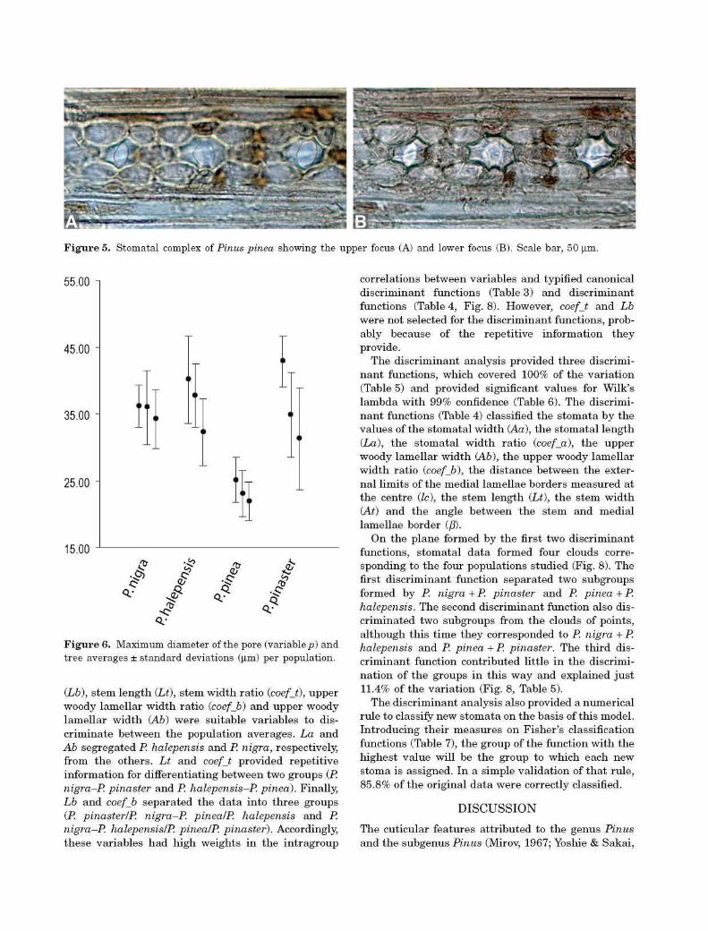
The maximum diameter of the pore (variable p) also showed differences between populations. Means and s tandard deviations of the variable p for each tree shown in Figure 6 could suggest smaller values for the P. pinea population than the others. In line with this intuitive approach, the ANOVA results for the factor 'species' rejected the hypothesis of equality between the means of the groups (99% confidence level), and the HSD-Tukey test identified the P. pinea population as the significantly different population. Examination of the homogeneous tree groups revealed similar values for the populations of Pinus nigra subsp. salzmannii (35.44 ± 1.06 |J.m), P. halepensis (36.73 ± 4.00 |im) and P. pinaster (36.30 ± 5.95 |J.m), whereas P. pinea had significantly

Figure 3. Stomatal rows of Pinus nigra subsp. salzmannii.
smaller pore diameters (23.37 ± 1.65 urn). The 95% confidence levels (mean ± two s tandard deviations) for the two groups showed no overlap between 26.64 |im and 28.81 |im. Thus, with a probability of 95%, values ofp tha t are less than 26.64 |im can be attributed to P. pinea.
STOMATAL FEATURES
Several stomatal features were common to all the taxa studied: the lower woody lamella covered the entire lower periclinal wall of the guard cells, the upper woody lamella was thicker and smaller than the lower, the stems joining the poles of the guard cells were the thickest elements, the medial lamellae border ran longitudinally from stem to stem, and a less thickened zone was visible in the central region of the pair of guard cells (Fig. 7, Fig. S5). Although no qualitative differences were observed between the stomata of the four species, significant differences were detected during statistical analysis of the stomatal variables.
The HSD-Tukey test (Table 2) revealed that the distance between the external limits of the medial lamellae borders measured near the stem (Id), the coefficient associated with the shape of the medial lamellae border (coef_c), the medial lamellae border width (e), the stem width (At) and the angle of attachment of the upper woody lamella (a) were unable to discriminate between population averages. The stomatal length (La), upper woody lamellar length
Figure 4. Stomatal complex of Pinus nigra subsp. salzmannii (A), Pinus pinaster (B), Pinus pinea (C) and Pinus halepensis (D). Scale bar, 50 |im.
'">'
Figure 5. Stomatal complex of Pinus pinea showing the upper focus (A) and lower focus (B). Scale bar, 50 |im.
55.00
45.00
35.00
25.00
15.00
3*
Figure 6. Maximum diameter of the pore (variable p) and tree averages ± standard deviations (|im) per population.
{Lb), stem length (Lt), stem width ratio (coefjt), upper woody lamellar width ratio (coefjb) and upper woody lamellar width (Ab) were suitable variables to discriminate between the population averages. La and Ab segregated P. halepensis and P. nigra, respectively from the others. Lt and coefjt provided repetitive information for differentiating between two groups (P. nigra-P pinaster and P. halepensis-P pinea). Finally Lb and coefjb separated the da ta into three groups (P. pinasterlP. nigra-P. pinealP. halepensis and P. nigra-P. halepensislP. pinealP. pinaster). Accordingly these variables had high weights in the intragroup
correlations between variables and typified canonical discriminant functions (Table 3) and discriminant functions (Table 4, Fig. 8). However, coefjt and Lb were not selected for the discriminant functions, probably because of the repetitive information they provide.
The discriminant analysis provided three discriminant functions, which covered 100% of the variation (Table 5) and provided significant values for Wilk's lambda with 99% confidence (Table 6). The discriminant functions (Table 4) classified the s tomata by the values of the stomatal width (Aa), the stomatal length (La), the stomatal width ratio (coef_a), the upper woody lamellar width (Ab), the upper woody lamellar width ratio (coefjb), the distance between the external limits of the medial lamellae borders measured at the centre (Ic), the stem length (Lt), the stem width (At) and the angle between the stem and medial lamellae border (/?).
On the plane formed by the first two discriminant functions, stomatal data formed four clouds corresponding to the four populations studied (Fig. 8). The first discriminant function separated two subgroups formed by P. nigra + P. pinaster and P. pinea + P. halepensis. The second discriminant function also discriminated two subgroups from the clouds of points, although this t ime they corresponded to P. nigra + P. halepensis and P. pinea + P. pinaster. The third discriminant function contributed little in the discrimination of the groups in this way and explained just 11.4% of the variation (Fig. 8, Table 5).
The discriminant analysis also provided a numerical rule to classify new stomata on the basis of this model. Introducing their measures on Fisher's classification functions (Table 7), the group of the function with the highest value will be the group to which each new stoma is assigned. In a simple validation of that rule, 85.8% of the original data were correctly classified.
D I S C U S S I O N
The cuticular features attr ibuted to the genus Pinus and the subgenus Pinus (Mirov, 1967; Yoshie & Sakai,
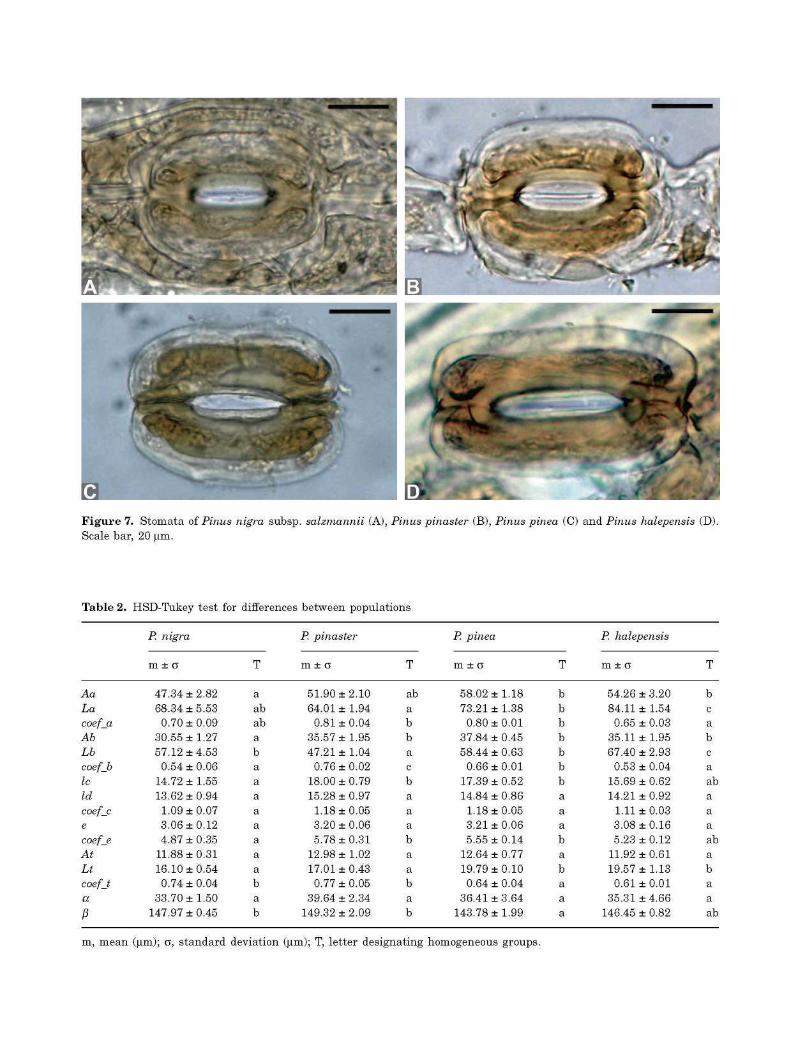
Figure 7. Stomata of Pinus nigra subsp. salzmannii (A), Pinus pinaster (B), Pinus pinea (C) and Pinus halepensis (D) Scale bar, 20 |im.
Table 2. HSD-Tukey test for differences between populations
P. nigra P. pinaster P. pinea P. halepensis
m ± o T m ± o T m ± o T m ± o T
Aa 47.34 ±2.82 a 51.90 ±2.10 ab 58.02 ± 1.18 b 54.26 ± 3.20 b La 68.34 ±5.53 ab 64.01 ± 1.94 a 73.21 ± 1.38 b 84.11 ± 1.54 c coef_a 0.70 ±0.09 ab 0.81 ± 0.04 b 0.80 ± 0.01 b 0.65 ± 0.03 a Ab 30.55 ±1.27 a 35.57 ± 1.95 b 37.84 ± 0.45 b 35.11 ± 1.95 b Lb 57.12 ±4.53 b 47.21 ± 1.04 a 58.44 ± 0.63 b 67.40 ± 2.93 c coefb 0.54 ±0.06 a 0.76 ± 0.02 c 0.66 ± 0.01 b 0.53 ± 0.04 a Ic 14.72 ±1.55 a 18.00 ± 0.79 b 17.39 ± 0.52 b 15.69 ± 0.62 ab Id 13.62 ±0.94 a 15.28 ± 0.97 a 14.84 ± 0.86 a 14.21 ± 0.92 a coef_c 1.09 ±0.07 a 1.18 ±0.05 a 1.18 ±0.05 a 1.11 ± 0.03 a e 3.06 ±0.12 a 3.20 ± 0.06 a 3.21 ± 0.06 a 3.08 ± 0.16 a coef_e 4.87 ±0.35 a 5.78 ± 0.31 b 5.55 ± 0.14 b 5.23 ± 0.12 ab At 11.88 ±0.31 a 12.98 ± 1.02 a 12.64 ± 0.77 a 11.92 ± 0.61 a Lt 16.10 ±0.54 a 17.01 ± 0.43 a 19.79 ±0.10 b 19.57 ± 1.13 b coefj 0.74 ±0.04 b 0.77 ± 0.05 b 0.64 ± 0.04 a 0.61 ± 0.01 a a 33.70 ± 1 . 5 0 a 39.64 ± 2 . 3 4 a 36.41 ± 3.64 a 35.31 ± 4 . 6 6 a j3 147.97 ± 0.45 b 149.32 ± 2.09 b 143.78 ± 1.99 a 146.45 ± 0.82 ab
m, mean (|im); o, standard deviation (|im); T, letter designating homogeneous groups.
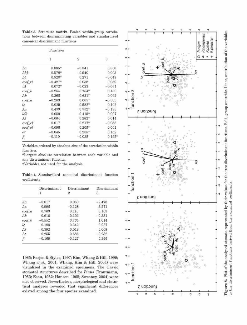
Table 3. Structure matrix. Pooled within-group correlations between discriminating variables and standardized canonical discriminant functions
La
Lb-\
Lt
coef_t\
of coefjb
Ab
coef_a Ic
Aa
ld-\ At
coef_c\
coef_c\
et P
Function
1
0.685* 0.578* 0.520*
-0.427* 0.072*
-0.264 0.268
-0.203 -0.029
0.433 0.059
-0.064 0.017
-0.098 -0.045 -0.110
2
-0 .341 -0.540
0.271 0.038
-0 .023 0.754* 0.621* 0.605* 0.582* 0.552* 0.415* 0.282* 0.217* 0.205* 0.205*
-0 .038
3
0.368 0.003
-0 .047 0.059
-0 .061 0.150 0.002
-0 .303 0.102
-0 .193 0.097 0.014
-0 .058 0.001 0.152 0.195*
Variables ordered by absolute size of the correlation within function. *Largest absolute correlation between each variable and any discriminant function. ^Variables not used for the analysis.
Table 4. Standardized canonical discriminant function coefficients
Aa
La
coef_a Ab
coefjb
Ic
At Lt
P
Discriminant 1
-0.017 0.996 0.763 0.610
-0.552 0.109
-0.392 0.255
-0.169
Discriminant 2
0.060 -0.128
0.113 -0.106
0.734 0.342 0.018 0.585
-0.127
Discriminant 3
-2.478 3.271 3.103
-0 .381 1.014 0.267
-0.008 -0.232
0.356
1985; Farjon & Styles, 1997; Kim, Whang & Hill, 1999; Whang etal., 2001; Whang, Kim & Hill, 2004) were visualized in the examined specimens. The classic stomatal structures described for Pinus (Trautmann, 1953; Esau, 1982; Hansen, 1995; Sweeney, 2004) were also observed. Nevertheless, morphological and statistical analyses revealed tha t significant differences existed among the four species examined.
V
-71
•TN
-
"7
••y
7
•?
T
M
C%J
M
n
T
. CM c o
c 3
. 4 -
- c o +J
4 -
c o - *J
c
•} m <M T- o «- *N
i i i i i i r
+ + + * *
< *
•3 +s [ S t * c • orSi^
<?
- r
B .a> ^ a :
Ifl
& 5
•C
1 1
g c o,5, •
CUCL a;
O <i 1
3 § ^
^fes - .\\?°.%°
1 1 1 1 1 1 1 1
* * 4
, a , j * J - 4 < « *r *
•*• A ^ i ^ B f V * + 1 + * o o
* • ; * • * o
£ uojpunj
• * >Jfo
. s O
+ + * + + + ° o o
+
£ uoipun.) i i i t i i i i
* + .
-
•
"
•
-
1
"
-
-
•
-
•
•
*
Table 5. Eigenvalues of the discriminant analysis
Function Eigenvalue % of variance % cumulative
Canonical correlation
2.771f 1.350f 0.531f
59.6 29.0 11.4
59.6 88.6
100.0
0.857 0.758 0.589
fThe first three canonical discriminant functions were used in the analysis.
Table 6. Wilks' lambda of the canonical discriminant functions
Test of functions Wilks' lambda Chi-square d.f. Sig.
1 to 3 2 to 3 3
0.074 0.278 0.653
919.237 451.351 150.207
27 16 7
0.000 0.000 0.000
Table 7. Fisher's classification function coefficients
Aa La coef_a Ab coef_b Ic At Lt
P (Constant)
' nigra
-52.955 42.270
3923.718 1.271
77.339 0.984 4.140 3.015 3.059
•1888.298
P. halepensis
-53.735 43.499
4001.781 1.775
77.770 1.105 3.137 3.635 3.030
-2008.477
' pinea
-52.658 42.421
3935.383 1.759
77.936 1.230 3.407 4.329 2.952
•1943.387
P. pinaster
-53.622 42.776
3966.123 0.982
111.619 1.639 4.255 3.725 3.072
-1959.405
CUTICULAR FEATURES The shape and arrangement of the subsidiary cells of the stomatal complex appear to be valid features for the differentiation of the four species in the studied populations. They are therefore potentially useful when discussing the general taxonomy of Pinus.
The circular structure around the pore observed in many species of Pinus, the Florin ring (Appendix) (Florin, 1931; Farjon & Styles, 1997; Whang etal., 2004), has been recorded in different populations of P. sylvestris L. (Yoshie & Sakai, 1985; Struzkova, 2002; Garcia Alvarez etal., 2009), but was not seen in any of the taxa studied in the present work. Yoshie & Sakai (1985), who studied two of the present species by scanning electron microscopy, only reported small variations in the cuticle surface: P. nigra Arnold was described as having a type A Florin ring (absent or barely visible), and P. pinaster Ait. was described as having a type B Florin ring
(slightly visible). The unremarkable na ture of the Florin ring in these species could be caused by the fact tha t they are not supported by any cellular structure of circular shape.
In P. pinaster, the homogeneity observed for all elements of the stomatal row agrees with the results of anatomical studies analysing cross-sections of pine needles. The shape and size of the pore are similar to those of the cells of the stomatal row, and the perpendicular na ture of the anticlinal walls of these cells and the epistomatal chamber is noticeable (Fieschi, 1932).
Pinus pinea showed the most anatomical differences among the species studied. The pore size (p) in this species was noticeably smaller than in the other taxa, allowing its numerical differentiation. Furthermore, the pore did not correspond, either in shape or in size, to the outline of the epistomatal chamber floor plan, and it was present in a different focal plane.
These features indicate a unique form of stomatal complex for this species. The substantial difference between P. pinea and the other three species supports the segregation of this taxon into a different group, as established by Price et al. (1998) - subgenus Pinus section Pinus subsection Pineae.
STOMATAL CHARACTERISTICS
The statistical analyses performed using the stomatal variables highlighted differences among the four populations studied. This opens up the possibility of making taxonomic differentiations despite the apparent morphological similarity of the stomata of these taxa.
The participation of the coefficients, not jus t the direct measurements , in the stepwise discriminant analysis is notable. The stomatal and upper woody lamellar width ratios (coef_a and coefjb) were variables with great weight in the first discriminant function, which is associated with 59.6% of the variation. It could be argued that , as these coefficients reflect ratios of perpendicular direct measurements , they are less dependent on stomatal size and therefore less dependent on the influence of environmental conditions (Ticha, 1982; Jones, 1992; Garcia-Amorena et al., 2006).
The angle of a t tachment of the upper woody lamella (angle a) displayed similar values in all of the studied populations. This is a reflection of its stability within Pinus, as indicated by other authors (Florin, 1931; Trautmann, 1953; Hansen, 1995).
The classification of s tomata into two subgroups, suggested by the first discriminant function, supports the older infrageneric classifications that position P. nigra and P. pinaster in the same section or subsection and leave P. pinea and P. halepensis in different groups, as suggested by Little & Critchfield (1969) and Price et al. (1998). However, in the light of modern phylogenetic studies, P. pinaster seems to be more closely related to P. pinea and P. halepensis, all in Pinus section Pinus subsection Pinaster, than to P. nigra, in Pinus section Pinus subsection Pinus (Gernandt etal., 2005). The second discriminant function grouped the species into two different pairs: P. nigra + P. halepensis and P. pinaster + P. pinea. Although P. pinea has been found to be closely related to P. pinaster in some phylogenetic studies (Liston etal., 1999; Wang etal., 1999), the grouping of P. nigra and P. halepensis is not reflected in any current systematic classification. Therefore, this function is essential for the statistical separation of the four clouds of points, but has no systematic interpretation. Rather, it appears to respond to morphological differences with no phylogenetic importance.
C O N C L U S I O N S
The differences found in the arrangement of the stomatal complex subsidiary cells and pore size highlight the diagnostic capacity and potential taxonomic use of cuticular analysis in Iberian Mediterranean pines. The shape and arrangement of the subsidiary cells, their comparison with those of the rest of the stomatal row cells and the pore size allow the taxonomic differentiation of the studied populations. These features may therefore be useful in the development of a taxonomic key to distinguish between Iberian Mediterranean pines.
The stomatal complex of the P. pinea samples displays strong differences compared with the other individuals analysed, such as a narrower pore and the characteristic elevation of this opening. Fur ther investigations of this poorly studied taxon will be useful for confirming the presence of the unique form of the P. pinea stomatal complex.
Despite the apparent morphological similarity of the stomata of the Pinus species, the present stomatal analysis detected significant differences between them. The variables related to the length and width of the stomata (Aa, La, coef_a) and woody lamellae (Ab, Lb, coefjb), the distance between the external limits of the medial lamellae borders measured at the centre (Ic) and the stem length (Lt) had the greatest taxonomic weight. These findings will facilitate new studies that might establish the classification of dispersed Pinus s tomata seen in fossil pollen preparations in an Iberian Mediterranean context. The generalization of the present results from individual populations to the species' level through the study of multiple populations is the necessary first step to achieve this goal.
A C K N O W L E D G E M E N T S
This research was performed with the kind help of the personnel of the 'History and Dynamics of the Vegetal Landscape' Research Group of the Univer-sidad Politecnica de Madrid, Spain. We also thank Miguel Angel Casado for statistical advice during the revision process. Nature Publishing Group Language Editing and Adrian Burton corrected the English. The study was partially supported by the Ministerio de Ciencia e Innovacion (projects CGL-2006-02956/BOS and CGL2008-06005/BOS). We also thank the anonymous reviewers of this article for their constructive input.
R E F E R E N C E S
Alia Miranda R, Martin Albertos S, De Miguel y Del Angel J, Galera Peral R, Agundez Leal D, Gordo

Alonso J, Salvador Nemoz L, Catalan Bachil ler G, Gil Sanchez L. 1996. Las regiones de procedendo, de Pinus pinaster Alton. Madrid: Organismo Autonomo. Parques Nacionales.
Ammann B, Wick L. 1993. Analysis of fossil stomata of conifers as indicators of the alpine tree line fluctuations during the Holocene. In: Prenzel B, ed. Oscillations of the alpine and polar tree limits in the Holocene. Palaoklimafor-schung 9. Stuttgart/Jena/New York: Gustav Fischer, 175-186.
Barclay R, McElwain JC, Dilcher D, Sageman B. 2007. The Cuticle Database: developing an interactive tool for taxonomic and paleoenvironmental study of the fossil cuticle record. Courier Forschung Institut Senckenburg 258: 39-55.
Barron E, Buades A. 2002. Aportaciones al estudio de la epidermis foliares en las especies vivientes de la familia Taxodiaceae (Coniferales, Coniferophyta). Boletin de la Real Sociedad Espahola de Historia Natural (Seccion Biologica) 97: 1-4.
Birks HH, Birks HJB. 2000. Future uses of pollen analysis must include plant macrofossils. Journal of Biogeography 27: 31-35.
Boddi S, Bonzi LM, Calamassi R. 2002. Structure and ultrastructure of Pinus halepensis primary needles. Flora -Morphology, Distribution, Functional Ecology of Plants 197: 10-23.
Boratynska K, Bobowicz MA. 2001. Pinus uncinata Ramond taxonomy based on needle characters. Plant Sys-tematics and Evolution 227: 183-194.
Catalan Bachil ler G. 1991. Las regiones de procedencia de Pinus sylvestris L. y Pinus nigra Am. subsp. salzmannii (Dunal) Franco en Espaha. Madrid: Dpto. de Sistemas Forestales del INIA, Servicio de Material Genetico del ICONA, Ministerio de Agricultura, Pesca y Alimentacion.
Dunwiddie PW. 1987. Macrofossil and pollen representation of coniferous trees in modern sediments from Washington. Ecology 68: 1-11.
Esau K. 1982. Anatomia de las plantas con semilla. Buenos Aires: Ed. Hemisferio Sur S.A.
Farjon A, Styles BT. 1997. Pinus (Pinaceae). Flora neotro-pica, 75. New York: Organization for Flora Neotropica.
Fieschi MV. 1932. Anatomie de la feuille chez les pins maritimes. Bulletin de la Societe d'Histoire Naturelle de Toulouse 64: article XVIII, 1-18.
Florin R. 1931. Untersuchungen zur Stammesgeschichte der Coniferales und Cordiatales. Kungliger Svenska Vetenskap-sakademien Handlingar 10: 109-111, 208-209, 340.
Franco Miijica F, Gomez Manzaneque F, Maldonado J, Morla Juarist i C, Post igo Mijarra JM. 2000. El papel de los pinares en la vegetacion holocena de la peninsula Iberica. Ecologia 14: 61-77.
Froyd CA. 2005. Fossil stomata reveal early pine presence in Scotland: implications for colonization analyses. Ecology 86: 579-586.
Garcia Alvarez S, Morla C, Solana Gutierrez J, Garcia-Amorena I. 2009. Taxonomic differences between Pinus sylvestris and P. uncinata revealed in the stomata and
cuticle characters for use in the study of fossil material. Review of Palaeobotany and Palynology 155: 61-68.
Garcia-Amorena I, Gomez Manzaneque F, Rubiales JM, Granja HM, de Carvalho GS, Morla C. 2007. The Late Quaternary coastal forests of western Iberia: a study of their macroremains. Palaeogeography, Palaeoclimatology, Palaeoecology 254: 448-461.
Garcia-Amorena I, Wagner F, van Hoof T, Gomez Manzaneque F. 2006. Stomatal responses in deciduous oaks from southern Europe to the anthropogenic atmospheric CO2 increase; refining the stomatal-based CO2 proxy. Review of Palaeobotany and Palynology 141: 303-312.
Gaussen H, Heywood VH, Chater AO. 1964. Pinus. In: Tutin TG, Heywood VH, Burges NA, Valentine DH, Walter SM, Webb DA, eds. Flora Europaea. Cambridge: Cambridge University Press, 29-35.
Gernandt DS, Geada Lopez G, Ortiz Garcia S, Liston A. 2005. Phylogeny and classification of Pinus. Taxon 54: 29-42.
Gervais BR, Macdonald GM, Snyder JA, Kremenetski CV. 2002. Pinus sylvestris treeline development and movement on the Kola Peninsula of Russia: pollen and stomate evidence. Journal of Ecology 90: 627-638.
Gil Sanchez L, Diaz-Fernandez PM, J imenez Sancho MP, Roldan Moreno M, Alia Miranda R, Agundez Leal D, De Miguel y Del Angel J, Martin Albertos S, De Tuero y Reina M. 1996. Las regiones de procedencia de Pinus halepensis Mill. En Espaha. Madrid: Organismo Autonomo Parques Nacionales.
Hansen BCS. 1995. Conifer stomata analysis as a paleoeco-logical tool: an example from the Hudson Bay Lowlands. Canadian Journal of Botany 73: 244-252.
Hansen BCS, Engstrom DR. 1996. Vegetation history of Pleasant Island, southeastern Alaska, since 13,000 yr B.P. Quaternary Research 46: 161-175.
Hicks S. 2006. When no pollen does not mean no trees. Vegetation History and Archaeobotany 15: 253-261.
Jones H. 1992. Plants and microclimate. Cambridge: Cambridge University Press.
Kerp H. 1990. The study of fossil gymnosperms by means of cuticular analysis. Palaios 5: 548-569.
Kim K, Whang SS, Hill RS. 1999. Cuticle micromorphology of leaves of Pinus (Pinaceae) in east and south-east Asia. Botanical Journal of the Linnean Society 129: 55-74.
Liston A, Robinson WA, Pinero D, Alvarez-Buylla ER. 1999. Phylogenetics of Pinus (Pinaceae) based on nuclear ribosomal DNA internal transcribed spacer region sequences. Molecular Phylogenetics and Evolution 11: 95-109.
Little EL, Critchfield WB. 1969. Subdivisions of the genus Pinus. USDAForest Service Miscellaneous Publication 1144.
Martin Albertos S, Diaz-Fernandez PM, De Miguel y Del Angel J. 1998. Regiones de procedencia de las especies forestales espaholas. Generos Abies, Fagus, Pinus y Quercus. Madrid: Servicio de Material Genetico, Publica-ciones del Organismo Autonomo Parques Nacionales.
Mirov NT. 1967. The genus Pinus. New York: Ronald Press. Pilger R. 1926. Genus Pinus. In: Engler A, Prantl K, eds.
Die natilrlichen Pflanzenfamilien. Vol. XIII, Gymnospermae. Leipzig: Wilhelm Engelmann, 121-403.
Prada A, Gordo Alonso J, De Miguel y Del Angel J, Mutke S, Catalan Bachil ler G, Iglesias S, Gil Sanchez L. 1997. Las regiones de procedendo, de Pinus pinea L. en Espana. Madrid: Organismo Autonomo Parques Nacionales.
Price RA, Liston A, Strauss SH. 1998. Phylogeny and systematics of Pinus. In: Richardson DM, ed. Ecology and biogeography of Pinus. Cambridge: Cambridge University Press, 49-68.
Rubiales JM, Garcia-Amorena I, Garcia Alvarez S, Morla C. 2009. Anthracological evidence suggests naturalness of Pinus pinaster in inland southwestern Iberia. Plant Ecology 200: 155-160.
Rubiales JM, Garcia-Amorena I, Genova M, Gomez Manzaneque F, Morla C. 2007. The Holocene history of highland pine forests in a submediterranean mountain: the case of Credos mountain range (Iberian Central Range, Spain). Quaternary Sciences Reviews 26: 1759-1770.
Shaw GR. 1914. The genus Pinus. Boston, MA: Arnold Arboretum Publications.
Shaw GR. 1924. Notes on the genus Pinus. Journal of the Arnold Arboretum 5: 225-227.
Stace CA. 1965. Cuticular studies as an aid to plant taxonomy. The Bulletin of the British Museum (Natural History) Botany Series 4: 3-78.
Stewart DRM. 1967. Analysis of plant epidermis in faeces: a technique for studying the food preferences of grazing herbivores. Journal of Applied Ecology 4: 83-111.
Struzkova D. 2002. The cuticular analysis - a method to distinguish the needles of Pinus sylvestris L (Scots pine) from those of Pinus mugo Turra s str (mountain pine). Vegetation History and Archaeobotany 11: 241-246.
Sweeney CA. 2004. A key for the identification of stomata of the native conifers of Scandinavia. Review of Palaeobotany and Palynology 128: 281-290.
Theobald WL, Krahulik JL, Roll ins RC. 1979. Trichome description and classification. In: Metcalfe CR, Chalk L, eds. Anatomy of the dicotyledons V. I. Systematic anatomy of leaf and stem, with a brief history of the subject. Oxford: Clarendon, 40-53.
Ticha I. 1982. Photosynthetic characteristics during ontogenesis of leaves: 7. Stomata density and sizes. Photosyn-thetica 16: 375-471.
Trautmann W. 1953. Zur Unterscheidung fossiler Splatoff-nungen der mitteleuropaischen Coniferen. Flora 140: 523-533.
Wang XR, Tsumura Y, Yoshimaru H, Nagasaka K, Szmidt AE. 1999. Phylogenetic relationships of Eurasian pines (Pinus, Pinaceae) based on chloroplast rbcL, matK, rpl20-rpslS spacer, and trnV intron sequences. American Journal of Botany 86: 1742-1753.
Whang SS, Kim K, Hill RS. 2004. Cuticle micromorphology
of leaves of Pinus (Pinaceae) from North America. Botanical Journal of the Linnean Society 144: 303-320.
Whang SS, Pak JH, Hill RS, Kim K. 2001. Cuticle micro-morphology of leaves of Pinus (Pinaceae) from Mexico and Central America. Botanical Journal of the Linnean Society 135: 349-373.
Yoshie F, Sakai A. 1985. Types of Florin rings, distributional patterns of epicuticular wax, and their relationships in the genus Pinus. Canadian Journal of Botany 63: 2150-2158.
Yu Z. 1997. Late Quaternary paleoecology of Thuja and Juni-perus (Cupressaceae) at Crawford Lake, Ontario, Canada: pollen, stomata and macrofossils. Review of Palaeobotany and Palynology 96: 241-254.
APPENDIX
GLOSSARY OF MORPHOLOGICAL TERMS BASED
ON THE TERMINOLOGY OF FLORIN (1931) ,
TRAUTMANN (1953) , STACE (1965) ,
H A N S E N (1995) A N D S W E E N E Y (2004)
Florin ring: A circular th icken ing formed by the cells surrounding the s t o m a t a of p ine needles , first described by Florin (1931). S ix different types of Florin r ings h a v e b e e n described for the g e n u s Pinus, four of w h i c h (types A, B, C and D) are s e e n in subgenus Pinus (Yoshie & Sakai , 1985; Farjon & Styles , 1997).
Lamella (woody lamella): Lignified portions of the upper and lower wal l of the guard cel ls . The upper lamel la i s often thicker t h a n the lower. The lower woody lamel la is not often preserved in fossil pol len samples . In Pinus, the out l ine of the guard cells coincides w i t h the shape of the lower woody lamel la; the latter complete ly covers the lower wa l l of the cell.
Medial lamellae border: P o r t i o n of t h e l a m e l l a e
bordering the s toma, often thickened; close to a l ine drawn through the s t ems .
Pore: The aperture of the ep i s tomata l chamber. In m a n y conifers, the guard cel ls are deeply s u n k e n and are overarched by the subs id iary cel ls , such that , in a surface view, their posi t ion is marked by a r ing of subsidiary cells around a near ly circular hole.
Stem: The portion of the lamel lae borders beg inn ing at their junct ion and ex tend ing towards the poles away from the s toma.