Embed Size (px)
Citation preview

The VA Parkinson Report
Department of Veterans Affairs
Department of Veterans Affairs Volume 10 No. 1, Summer 2013
Parkinson’s Action Net-
work is thrilled to be honor-
ing Secretary Eric K.
Shinseki with the Morris K.
Udall Award for Public Ser-
vice for his tireless efforts
advocating for Veterans
health issues and recogniz-
ing Parkinson’s disease
among the illnesses that
affect thousands of our
brave men and women.
In 2010, under Secretary Shinseki’s leadership, the U.S. De-
partment of Veterans Affairs (VA) recognized Parkinson’s
disease on the list of illnesses associated with exposure to
the herbicide known as Agent Orange. Using evidence from
a July 2009 National Academy of Science's Institute of Medi-
cine report, Veterans and Agent Orange, this decision simpli-
fied and accelerated the benefits application process for
Vietnam veterans with Parkinson's disease.
Secretary Shinseki has also been a supporter of the VA’s
Parkinson’s Disease Research, Education and Clinical Cen-
ters, which treat Veterans with various movement disorders
in addition to Parkinson’s disease, such as essential tremor,
restless leg syndrome, dystonia, atypical parkinsonian disor-
ders, or "parkinson plus" syndromes. There are currently
about 80,000 veterans affected by Parkinson’s disease and
the VA centers provide them with critical patient care in sev-
eral cities around the country. We, along with the rest of the
Parkinson’s community, appreciate the attention and fund-
ing given to these centers and the people they serve.
Most recently, the VA issued a proposed rule to make it easi-
er for Veterans to receive health care and compensation for
certain illnesses, including parkinsonism, dementia, and de-
pression, which have been linked to moderate to severe trau-
matic brain injury. This is an example of how the VA, under
the Secretary’s leadership, ensures that its benefits pro-
grams keep up with the latest science.
Prior to his time at the VA, Secretary Shinseki served as the
chief of staff in the U.S. Army. During his long military career,
he also received two Purple Hearts for injuries in Vi-
etnam. Because of his injuries, including losing part of his
foot, Shinseki knows first-hand the challenges of a disabled
Veteran.
His list of accomplishments should make it clear why he will
be honored with the Morris K. Udall Award for Public Service
at this year’s Morris K. Udall Awards Dinner.
New Research Publication...2
EMST………………...........3
DaTscan…………...……....6
Dystonia…………….…....8
Telehealth……...………...10
RLS Relief……..………..12
PADRECC News……….13
Inside this Issue:
Secretary Shinseki Receives
Udall Award for Support of Veterans with
Parkinson's and Commitment to Public Service

The public service award is named for Morris “Mo” K. Udall, who served in the U.S. House of Representatives for 30
years and was a candidate for the Democratic Presidential nomination in 1976. During Udall’s distinguished career, he
infused American politics with his singular style of humor, grace, and dignity. Many of today’s leaders – both Demo-
crats and Republicans – have cited Udall as an inspiration and role model. As a partisan Democrat, he was a much-
feared debate opponent, but he was fair and always decent. It is this spirit that the Morris K. Udall Award for Public
Service is presented to Secretary Shinseki, who has made important contributions to public policy with humor, grace,
and dignity.
Alan Oates, director of legislative affairs, research and special projects at the U.S. Military Veterans with Parkinson's
(USMVP), had the following to say about Secretary Shinseki.
"Veterans respect him for his military service and for standing his ground. After his decision to add Parkinson's
and two other diseases as service related for exposure to Agent Orange, General Shinseki met with members
of our group. In response to our thanks, he replied, 'It was the only decision I could make, it was the right deci-
sion.' General Shinseki is a courageous leader who has earned the respect of those he serves. He is my kind
of soldier."
Shinseki is the first Asian-American four-star general in U.S. history. He was born in Hawaii to Japanese-American par-
ents and is married with two children. (article retrieved from www.parkinsonsaction.org/news)
The VA Parkinson Report, Vol 10 No 1 Page 2
New PADRECC Research Publication
Secondary Data Analysis of PADRECC CSP Study
Submitted by Daniel Weintraub, MD, Philadelphia PADRECC
Be on the lookout for a newly published article using a secondary data analysis of the PADRECC’s CSP Study, entitled
Suicide Ideation and Behaviors after STN and GPi DBS Surgery for Parkinson's Disease: Results from a Randomized,
Controlled Trial. Drs. Weintraub, Duda, Luo, Sagher, Stern, Follett, Reda, Weaver and Ms. Carlson will soon have an
article in the Journal of Neurology, Neurosurgery & Psychiatry (JNNP).
The research team looked at the risk of suicide behaviors post-deep brain stimulation (DBS) surgery in Parkinson's
disease (PD). They assessed if suicide ideation and behaviors were more common in PD patients randomized to DBS
surgery versus best medical therapy (BMT); and for those randomized to subthalamic nucleus (STN) versus globus palli-
dus interna (GPi) DBS surgery.
In Phase 1 of the Veterans Affairs CSP 468 study, 255 PD patients were randomized to DBS surgery (n=121) or 6
months of BMT (n=134). For Phase 2, a total of 299 patients were randomized to STN (n=147) or GPi (n=152) DBS
surgery. Patients were assessed serially with the Unified Parkinson’s Disease Rating Scale Part I depression item,
which queries for suicide ideation; additionally, both suicide behavior adverse event data and proxy symptoms of in-
creased suicide risk from the Parkinson’s Disease Questionnaire (PDQ-39) and the Short Form Health Survey (SF-36)
were collected.
Study outcomes for Phase I reported no suicide behaviors and rare new-onset suicide ideation (1.9% for DBS vs. 0.9%
for BMT; Fisher’s exact p=0.61). Rates of suicide ideation at 6 months were similar for patients randomized to STN vs.
GPi DBS (1.5% vs. 0.7%; Fisher’s exact p=0.61), but several proxy symptoms were worse in the STN group. The investi-
gators concluded that DBS surgery study in PD patients did not support a direct association between DBS surgery and
an increased risk for suicide ideation and behaviors.
Udall Award (continued from front page)
The EMST technique. Over a decade of work from our labora-
tory describes the use of a technique called expiratory mus-
cle strength training or EMST. EMST is enabled by the use of
a portable, handheld device, known as a pressure threshold
device. EMST improves maximum expiratory pressure (MEP),
cough effectiveness, and swallowing function in case series,
single group and randomized clinical trials (see Troche et al.,
20101 for a complete list of references which include our
previous work as well as Sapienza et al., 20112). EMST is
simply a behavioral treatment option. It is a tool for rehabili-
tation specialists to assist with improving certain skeletal
muscles’ ability to increase force by exposing them to a load
or a “weight” (See Figure 1).
Figure 1. Photographs of expiratory muscle strength and placement in mouth.
Drs. McConnell and Romer contributed an article called
“Respiratory muscle strength training (RMST) in healthy hu-
mans: resolving the controversy”.3 In this article the rationale
for specific respiratory muscle training which includes EMST
and inspiratory muscle strength training or IMST, were re-
viewed. There are several techniques used to accomplish
RMST, such as resistive loading and pressure threshold load-
ing. The conclusions from this literature were inspiring as it
supported RMST as a treatment for respiratory muscle fa-
tigue and improved exercise performance. With the use of
the appropriate methodologies and trial design, as well as
selection of valid and sensitive outcome measures, RMST
has been transferred to patient populations including those
with Parkinson’s disease, COPD, spinal cord injury, multiple
sclerosis, sedentary elderly, and to others.1,2,4-8
Our research group at the University of Florida has specifical-
ly focused on the EMST technique for its effects on breathing
with new applications of it to functions such as swallowing
and cough.1,8,9 Additionally, EMST has been incorporated as
preventative exercise for elderly10 and as a mechanism for
strengthening expiratory muscles for song and wind instru-
ment performance (e.g. Sapienza, Davenport & Martin).2
Motor exercise protocols like EMST, include intensity and/or
duration regimens. This means that a clinician or user is pre-
scribing the treatment duration. The variation of the intensity
and duration are not random choices. They are based on
knowledge adapted from exercise physiology literature, indi-
cating the importance of the amount of exercise performed
over time as it relates to both muscular, or myogenic, chang-
es, and changes occurring within the nervous system. Thus,
these treatments are often delivered over a time period rang-
ing from 4 – 8 weeks, between 3 - 5 days per week, and 1 –
3 sessions per day. Within a daily session, 25 – 30 repeti-
tions are typically completed.11,12
The EMST treatment protocol we have incorporated clinically
as well as within our research design incorporates intensity
levels targeting muscle strength and thus targeted muscle
groups may benefit from improved force-generating capabil-
ity. It is the improved force generating capacity which lends
itself to hypotheses regarding changes to function. Use of
the EMST technique for affecting swallow function relies on
cross training of the submental musculature as discussed by
Wheeler et al (2008).13 EMST effects on swallow function are
covered in detail in Pitts et al.9; Troche, et al.1; Wheeler et
al.13 The Troche at al.1, work is the most comprehensive
work to date on Parkinson’s disease detailing the outcomes
of a randomized clinical trial and its positive effects of swal-
What is Expiratory Muscle Strength Training (EMST)? Christine Sapienza, PhD & Erin Silverman, PhD
Brain Rehabilitation Research Center, Malcom Randall VA
Department of Speech, Language, and Hearing Sciences
University of Florida
The VA Parkinson Report, Vol 10 No 1 Page 3

of a randomized clinical trial and its positive effects of swallow timing, swallow movements and airway protection.
Common to all potential strength training treatments is the fact that treatment must be sustained over time in order to result
in physiologic or functional gains with regard to these improved functions. Based on study findings of EMST specifically, it can
be hypothesized that no less than 2 weeks of treatment, delivered 3-5 times per week can be recommended with reasonable
expectation for improvements. Development of a maintenance program will be necessary to prevent detraining effects com-
mon to the cessation of strength training protocols. 14-16
Why use it for persons with Parkinson’s disease?
Many individuals with Parkinson’s disease (PD) suffer from obstructive or restrictive pulmonary disease.17-20 This phenomenon
is thought to be influenced by reduced respiratory muscle strength and by increased chest wall rigidity.18 Given that persons
with PD often succumb to pulmonary sequelae and pulmonary dysfunction has been identified at all stages of disease. Based
on that, management of pulmonary compromise is a top priority throughout the course of the disease.18,21-32 There is mounting
evidence suggesting that EMST improves ventilatory function in persons with neurodegenerative disease (e.g. Chiara, et al.4,
Kim, et al.10). As skeletal muscles, the respiratory muscles seem to respond well to strength training.4,7,10,33,34 EMST improves
respiratory muscle pumping force capacity which is important in ventilation. In fact, pilot testing revealed a 158% improve-
ment in MEP with EMST training in PD,35,36 suggesting that EMST is a viable treatment option targeting expiratory muscles and
could also result in improvement in pulmonary function – a function that is intricately related to ventilation of the lung.
What is the EMST device?
The EMST150TM device (Aspire Products, LLC) is a hand held device that provides a consistent pressure load on expiration.
The training results by simply blowing into the trainer to overcome the threshold load which is easily adjusted by a calibrated
spring inside of the device. The pressure threshold maximum is 150 cmH20 (Figure 1). Users of the device can hear and feel
the release of the spring loaded valve once the threshold load set on the device is met or exceeded during the expiratory phase of
breath.
Common Training Protocol
Place a nose clip provided on your nose to prevent air loss through the nose during high effort blowing
Turn the knob on the pressure threshold EMST150 device until the small metal screw on the bottom lines up with the
number 30.
Take a deep breath in; insert the EMST150 mouthpiece in your mouth, behind the teeth, making a tight lip seal around
the mouthpiece.
Don’t breathe any air out until the mouthpiece is securely in place. Use the hand that is not holding the device to help se-
cure the lips around the mouthpiece, if needed.
Next, blow hard and fast through the device until air rushes through, and then stop.
If able to accomplish Steps 1-4 easily, turn the knob clockwise ¼ turn and repeat. If unable to move air through the de-
vice, turn the knob ¼ turn counterclockwise and continue to do that until able to move air through the device.
**Continue turning the knob clockwise until you are unable to move air through the device easily. This stopping point will be
the MAXIMUM pressure you are able to create. The best analogy that we have used to help patients understand the concept of
strength training is that of the pin in the weight machine at the gym. Place the pin in the machine; find the weight that makes
force generation difficult but not so difficult that overstraining occurs.
Ease of Training
EMST training can be done in the home or with the aid of rehabilitation specialist. It represents a short-term treatment that
can be quantified and translated into functional outcomes that may directly improve functions related to breathing, cough and
swallow. The impact is high because of its high cost-effectiveness, ability to minimize direct therapist time required to rehabili-
tate the deficits, reduced need for clinical resources and patient travel time and because it can be developed into a home-
based therapy program.
The VA Parkinson Report, Vol 10 No 1 Page 4
Expiratory Muscle Strength Training (continued from page 3)

Acknowledgments. Appreciation is extended to the patients of the University of Florida Movement Disorders Center for their involvement in our projects and to Drs.
Michael Okun, Rodriguez, Malaty and Janet Romrell, PA as well the collaborative support of the Speech Language Pathology department at the Malcom Randall VA,
Gainesville and Nan Musson, MA, CCC-SLP. Portions of this work were supported by the Veterans Affairs RR & D Merit B3721 R award, NIH/NIDCHD
HD046903—01A112/0 R21 and the MJ Fox Foundation, Clinical Discovery Award. A special thanks to Drs. Michelle Troche, Karen Wheeler- Hegland, Teresa Pitts,
Paul Davenport and Donald Bolser for their collaborative research support.
References
Troche M.S., Okun M.S., Rosenbek J.C., Musson N., Fernandez H.H., Rodriguez R., Romrell .J, Pitts T., Wheeler-Hegland K.M., &Sapienza C.M. (2010). Aspiration and swallowing in Parkinson’s disease and rehabilita-
tion with EMST: A randomized trial. Neurology, 75, 21, 1912-1919.
Sapienza, C.M., Davenport, P., & Martin, A.D. (2002). Expiratory muscle training increases pressure support in high school band students. Journal of Voice, 18, 4, 495-501.
McConnell A.K.,& Romer L.M. (2004). Respiratory muscle training in healthy humans: resolving the controversy. Int J Sports Med, 25, 4, 284-293.
Chiara, T., Martin, D., & Sapienza, C. (2007). Expiratory muscle strength training: speech production outcomes in patients with multiple sclerosis. Neurorehabil Neural Repair, 21, 3, 239-249.
Fitsimones, L. B., Sapienza, C. M., & Davenport, P. (2004). Expiratory muscle strength training in low cervical/high thoracic spinal cord injury. Paper presented at the American Thoracic Society 100th International
Conference, Orlando, FL.
Huang, C.H., Yang, G.G., Wu, Y.T., & Lee, C.W. (2011). Comparison of inspiratory muscle strength training effects between older subjects with and without chronic obstructive pulmonary disease. J. Formos Med. Assoc,
110, 8, 518-526.
Kim, J., & Sapienza, C. M. (2005). Implications of expiratory muscle strength training for rehabilitation of the elderly: Tutorial. J Rehabil Res Dev, 42, 2, 211-224.
Pitts, T., Bolser, D., Rosenbek, J. C., Troche, M. S., Okun, M. S., & Sapienza, C. M. (2009). Impact of expiratory muscle strength training on voluntary cough and swallow function in Parkinson's disease. Chest, 13, 5,
1301-1308.
Pitts, T., Bolser, D., & Sapienza, C. M. ( 2009). Effects of expiratory muscle strength training (EMST) on voluntary cough and swallowing in PD. Paper presented at the 4th International Cough Symposium, London,
England.
Kim, J., Davenport, P., & Sapienza, C. (2008). Effect of expiratory muscle strength training on elderly cough function. Arch Gerontol Geriatr. doi: S0167-4943(08)00065-4 [pii]10.1016/j.archger.2008.03.006
Anand, S., Bashiti, N., & Sapienza, C.M (2012). Effect of training frequency on maximum expiratory pressure, Am. J. Speech Lang Pathol, 21,4, 380-386.
Ransmayr, G.. (2011). Physical, occupational, speech, and swallowing therapies and physical exericse in Parkinson’s disease. J. neural Trasn, 118, 5, 773-781.
Wheeler, K., Chiara, T., & Sapienza, C. (2008). Surface electromyographic activity of the submental muscles during swallow and expiratory pressure threshold training tasks. Dysphagia, 22, 2, 108-116.
Baker, S., Davenport, P., & Sapienza, C. (2005). Examination of strength training and detraining effects in expiratory muscles. J Speech Lang Hear Res, 48, 6, 1325-1333.
Ogasawara R., Kobayashi K., Tsutaki A., Lee K., Abe T., Fujita S., Nakazato K., Ishii N. (2013). Motor signaling response to resistance exercise is latered by chronic resistance training and detraining in skeletal muscle.
Journal of Applied Physiology, 14, 7, 934-940.
Sherk, K.A., Bemben, D.A., Brickman, S.E., & Bemben, M.G. (2012). Effects of resistance training duration on muscular strength retention 6-month posttraining in older men and women. J. Geriatr. Phys, 351, 1, 20-27.
Sabate, M., Rodriguez, M., Mendez, E., Enriquez, E., & Gonzalez, I. (1996). Obstructive and restrictive pulmonary dysfunction increases disability in Parkinson disease. Arch Phys Med Rehabil, 77, 1, 29-34.
Sathyaprabha, T. N., Kapavarapu, P. K., Pall, P. K., Thennarasu, K., & Raju, T. R. (2005). Pulmonary functions in Parkinson's disease. Indian J Chest Dis Allied Sci, 47, 4, 251-257.
Schiermeier, S., Schafer, D., Schafer, T., Greulich, W., & Schlafke, M. E. (2001). Breathing and locomotion in patients with Parkinson's disease. Pflugers Arch, 443, 1, 67-71.
Shill, H., & Stacy, M. (1998). Respiratory function in Parkinson's disease. Clin Neurosci, 5, 2, 131-135.
Bogaard, J. M., Hovestadt, A., Meerwaldt, J., vd Meche, F. G., & Stigt, J. (1989). Maximal expiratory and inspiratory flow-volume curves in Parkinson's disease. Am Rev Respir Dis, 139, 3, 610-614.
Canning, C. G., Alison, J. A., Allen, N. E., & Groeller, H. (1997). Parkinson's disease: an investigation of exercise capacity, respiratory function, and gait. Arch Phys Med Rehabil, 78(2), 199-207. doi: S0003-9993
(97)90264-1 [pii]
deBruin PF, de Bruin VM, Lees AJ, Pride NB. Effects of treatment on airway dynamics and respiratory muscle strength in Parkinson’s disease. Am Rev Respir Dis. 1993 Dec:148(6Pt1):1576-80. PubMed PMID: 8256904.
Herer, B., Arnulf, I., & Housset, B. (2001). Effects of levodopa on pulmonary function in Parkinson's disease. Chest, 119, 2, 387-393.
Hovestadt, A., Bogaard, J. M., Meerwaldt, J. D., van der Meche, F. G., & Stigt, J. (1989). Pulmonary function in Parkinson's disease. J Neurol Neurosurg Psychiatry, 52, 3, 329-333.
MacIntosh DJ. Respiratory dysfunction in Parkinson’s disease. Prim Care. 1977 Sep:4(3):441-5. PubMed PMID: 264221.
Nakano KK, Bass H, Tyler HR. Levodopa in Parkinson’s disease: effect on pulmonary function. Arch Intern Med. 1972Sep:130(3):346-8. PubMed PMID: 4560177.
Obenour, W. H., Stevens, P. M., Cohen, A. A., & McCutchen, J. J. (1972). The causes of abnormal pulmonary function in Parkinson's disease. Am Rev Respir Dis, 105, 3, 382-387.
Polatli M, Akyol A, Cildag O, Bayulkem K. Pulmonary function tests in Parkinson’s disease. Eur J Neurol. 2001Jul:8(4):341-5. PubMed PMID: 11422431.
Sabate, M., Gonzalez, I., Ruperez, F., & Rodriguez, M. (1996). Obstructive and restrictive pulmonary dysfunctions in Parkinson's disease. J Neurol Sci, 138 (1-2), 114-119.
Tzelepis, G. E., McCool, F. D., Friedman, J. H., & Hoppin, F. G., Jr. (1988). Respiratory muscle dysfunction in Parkinson's disease. Am Rev Respir Dis, 138, 2, 266-271.
Weiner, P., Inzelberg, R., Davidovich, A., Nisipeanu, P., Magadle, R., Berar-Yanay, N., et al. (2002). Respiratory muscle performance and the Perception of dyspnea in Parkinson's disease. Can J Neurol Sci, 29, 1, 68-72.
Sapienza, C. M., & Wheeler, K. (2006). Respiratory muscle strength training: functional outcomes versus plasticity. Semin Speech Lang, 27, 4, 236-244.
Powers, S. K., & Howley, E. T. (2004). Exercise physiology : theory and applications to fitness and performance (5th ed.). Boston, Mass. ; Toronto: McGraw-Hill
Saleem, A. F., Sapienza, C. M., & Okun, M. S. (2005). Respiratory muscle strength training: treatment and response duration in a patient with early idiopathic Parkinson's disease. NeuroRehabilitation, 20, 4, 323-333.
Silverman, E. P., Sapienza, C. M., Saleem, A., Carmichael, C., Davenport, P. W., Hoffman-Ruddy, B., et al. (2006). Tutorial on maximum inspiratory and expiratory mouth pressures in individuals with idiopathic Parkin-
son disease (IPD) and the preliminary results of an expiratory muscle strength training program. NeuroRehabilitation, 21, 1, 71-79.
The VA Parkinson Report, Vol 10 No 1
Page 5
Expiratory Muscle Strength Training (continued from page 4)

Although radiotracers that target presynaptic dopamine transport-
ers have been used on a research basis for many years, (123I ) Io-
flupane single-photon (emission tomography, (DaTscan SPECT, GE)
was the first to be approved by the FDA as a tool to assist in the
evaluation of adult patients with suspected Parkinsonian syn-
dromes, and to distinguish these syndromes from essential trem-
or1. In Parkinson’s disease, loss of nigrostriatal dopaminergic pro-
jections results in reduced uptake of radiotracer in the striatum,
especially the posterior putamen contralateral to the clinically most
affected limbs. Since some free123I may be present in the injected
radiopharmaceutical, patients who undergo DaTscan should have
“thyroid blockade” prior to the procedure with potassium iodide oral
solution (SSKI) drops. Medications such as stimulants, bupropion,
and benztropine, which have high
affinity for dopamine transporters,
should be held for at least five plasma
half-lives prior to DaT scanning; other
medications with small effects on DaT
uptake can probably be continued
(Table 1).2,3 In each case, the desire to optimize scan quality
should be weighed against the risks of holding therapeutic medica-
tions.
Since Parkinson’s disease is a clinical diagnosis, for which the best
premorbid gold standard is longitudinal follow-up, the question of
when DaTscan is useful in clinical practice arises. A recent analysis
of the studies performed to obtain FDA approval suggested that the
clinical examination has equivalent sensitivity and specificity to a
DaTscan.4 However, other investigators have contended that infor-
mation from the scan does alter diagnostic confidence and stream-
line treatment decisions.5
For a biomarker such as DaTscan to improve clinical management
it must extend diagnostic accuracy, predict disease onset or rate of
progression, or track treatment-based changes in long-term out-
come. Studies that have investigated the relationship of dopamine
transporter imaging to delayed (6-48 months) clinical diagnoses
have suggested that dopamine transporter imaging has approxi-
mately 80% sensitivity and greater than 90% specificity for a longi-
tudinal clinical diagnosis of Parkinsonism.6 Relatively small neuro-
pathological studies have suggested sensitivity and specificity of 85
The VA Parkinson Report, Vol 10 No 1
Page 2
A Brief Overview of DaTscan
Catherine Gallagher, William S. Middleton VA, Madison, WI &
John Duda, MD, Director, Philadelphia PADRECC
Table 1
Common Pharmacologic Agents Known to Affect DaT Uptake
Significant effect (>20%) Recommended discontinuation (days)
Cocaine 2
Fentanyl (IV) 1
Methylphenidate 1-2
Methylamfetamine 2
Modafinil 3
Amphetamine, Dexamphetamine 7
Benzatropine 5
Bupropion 8
Minor effect (<15%) Optional discontinuation (days)
Venlafaxine, duloxetine 3
Fluvoxamine, Paroxetine, Memantine 5
Amantadine, imipramine, ephedrine, sertraline 6
Citalopram, Escitalopram 8
Clomipramine 21
Pimozide 28
Fluoxetine 45
Levodopa little effect
Selegiline little effect
Pramipexole little effect
Table 1 was adapted from:
Kagi G, Bhatia KP, Tolosa E. The
role of DAT-SPECT in movement
disorders. J Neurol Neurosurg
Psychiatry 2010;81:5-12.
and
Cummings JL, Henchcliffe C,
Schaier S, Simuni T, Waxman A,
Kemp P. The role of dopaminer-
gic imaging in patients with
symptoms of dopaminergic
system neurodegeneration.
Brain 2011;134:3146-66.
Page 6

and 86%, respectively, for pathologi-
cally confirmed Parkinsonism
(DaTscan briefing document for
Peripheral and Central Nervous
System Advisory Committee Meet-
ing Aug 11, 2009; available at
www.fda.gov). This means that one
can be fairly certain that a person
with an abnormal DaTscan has Par-
kinsonism. However, the negative
predictive value is less certain. In
several clinical trials, 15-21% of
subjects with clinical symptoms of
Parkinson’s disease had normal
positron emission tomography or
SPECT with dopamine synthesis or
transporter radiotracers. Longitudi-
nal follow-up of patients and “scans
without evidence of dopaminergic
deficit” (SWEDDs) has shown stria-
tal radiotracer uptake to remain
relatively stable over time (in con-
trast to the expected 6-13% annual
decline expected in Parkinson’s
disease).7,8 Some SWEDD patients
were later diagnosed with dystonic tremor.
From a clinical point of view, another situation in which DaTscan can
be useful is in the distinction of medication-induced from neuro-
degenerative Parkinsonism. Many patients treated with neuroleptic
medications have motor signs of Parkinsonism; however, these pa-
tients are frequently psychiatrically fragile such that trials of medica-
tion withdrawal may result in hospitalization. Some studies have
indicated dopamine transporter imaging to be normal in 90% of cas-
es of medication-induced Parkinsonism, while other studies are less
definitive9. Cases of neuroleptic-induced Parkinsonism with abnor-
mal DaTscans may represent “subclinical” Parkinson’s disease that
was unmasked by use of the neuroleptic. Other potential uses of
DaTscan include distinguishing Lewy body dementia from other forms
of dementia such as Alzheimer’s disease, and distinguishing essen-
tial tremor from tremor-predominant Parkinson’s disease. Medica-
tion trials are not infallible in distinguishing these disorders, since
both Parkinsonian tremor and essen-
tial tremor may be refractory to
“correct” therapy. In idiopathic Park-
inson’s disease, relative preservation
of radiotracer uptake in the caudate
nucleus compared with putamen,
and asymmetry of tracer uptake be-
tween hemispheres, may differenti-
ate Parkinson’s disease from other
Parkinsonian syndromes; however,
DaTscan is not approved for this use.
Factors that reduce the attractive-
ness of DaTscan as a diagnostic tool
include potential variability in inter-
pretation between centers, possible
lower sensitivity in early and preclini-
cal disease stages, and cost. Howev-
er, used selectively, this test offers a
new diagnostic tool that can guide
patient management. With these
considerations in mind, PADRECC
Leadership developed Clinical Guide-
lines for use of dopaminergic imaging
(See Table 2) that will certainly be
refined as we move forward.
References
1. Benamer TS, Patterson J, Grosset DG, et al. Accurate differentiation of parkinsonism and essential
tremor using visual assessment of [123I]-FP-CIT SPECT imaging: the [123I]-FP-CIT study group. Mov
Disord 2000;15:503-10.
2. Kagi G, Bhatia KP, Tolosa E. The role of DAT-SPECT in movement disorders. J Neurol Neurosurg
Psychiatry 2010;81:5-12.
3. Booij J, Kemp P. Dopamine transporter imaging with [(123)I]FP-CIT SPECT: potential effects of drugs.
Eur J Nucl Med Mol Imaging 2008;35:424-38.
4. de la Fuente-Fernandez R. Role of DaTSCAN and clinical diagnosis in Parkinson disease. Neurology
2012;78:696-701.
5. Kupsch AR, Bajaj N, Weiland F, et al. Impact of DaTscan SPECT imaging on clinical management,
diagnosis, confidence of diagnosis, quality of life, health resource use and safety in patients with clini-
cally uncertain parkinsonian syndromes: a prospective 1-year follow-up of an open-label controlled
study. J Neurol Neurosurg Psychiatry 2012;83:620-8.
6. Cummings JL, Henchcliffe C, Schaier S, Simuni T, Waxman A, Kemp P. The role of dopaminergic
imaging in patients with symptoms of dopaminergic system neurodegeneration. Brain 2011;134:3146-
66.
7. Marshall VL, Reininger CB, Marquardt M, et al. Parkinson's disease is overdiagnosed clinically at
baseline in diagnostically uncertain cases: a 3-year European multicenter study with repeat [123I]FP-CIT
SPECT. Mov Disord 2009;24:500-8.
8. Seibyl J, Jennings D, Tabamo R, Marek K. Neuroimaging trials of Parkinson's disease progression. J
Neurol 2004;251 Suppl 7:vII9-13.
9. Diaz-Corrales FJ, Sanz-Viedma S, Garcia-Solis D, Escobar-Delgado T, Mir P. Clinical features and 123I-
FP-CIT SPECT imaging in drug-induced parkinsonism and Parkinson's disease. Eur J Nucl Med Mol
Imaging 2010;37:556-64.
The VA Parkinson Report, Vol 10 No 1
Table 2. Clinical Indications for the Use of
Dopaminergic Functional Imaging
Dopaminergic functional imaging has been shown to be a
useful adjunct to the clinical diagnosis of movement disor-
ders in some settings. In general, the risk of functional imag-
ing is justified when the outcome of the examination will help
to dictate clinical management. Accurate diagnosis can pre-
vent exposure to inefficacious treatments, improve prognos-
tic abilities and improve cost efficiency. Dopaminergic func-
tional imaging has not been shown to be helpful in differenti-
ating between different Parkinsonian syndromes (e.g. Parkin-
son’s disease, progressive supranuclear palsy, multiple sys-
tem atrophy, corticobasal degeneration). Decisions on wheth-
er or not to conduct scans for a given clinical scenario should
also be guided by relevant sensitivity and specificity data as
well as the recognition that there is some inter-rater variabil-
ity in the interpretation of scans. Scenarios in which the re-
sult of dopaminergic functional imaging may prove helpful in
determining therapeutic interventions include:
Patients with tremor that is not clearly differentiated into
either essential tremor or Parkinsonian tremor.
Patients with tremor or other features of Parkinsonism in
the context of treatment with dopamine-blocking medica-
tions known to induce Parkinsonism to determine if the
Parkinsonism is likely to be purely drug-induced or if
there is an underlying neurodegenerative condition.
Patients with possible psychogenic Parkinsonism.
Patients with tremor of unclear etiology that is not re-
sponsive to dopaminergic replacement therapies and
Page 7
DaTscan (continued from page 6)
The VA Parkinson Report, Vol 10 No 1
Page 8
Primary Dystonia Misinterpreted as Parkinson Disease:
A Case Presentation and Practical Clues
Olga Klepitskaya, MD; Alexander J. Neuwelt, MD; Tam Nguyen, MD; Maureen Leehey, MD
Department of Neurology, University of Colorado Denver and Denver VA Medical Center
Introduction
Dystonia is a movement disorder characterized by sustained mus-
cle contractions frequently causing twisting repetitive movements
and abnormal postures.1 It can present at any age with a variety of
clinical features that may overlap with other movement disorders,
making the diagnosis challenging. Patients with dystonia are com-
monly misdiagnosed.2 Dystonic tremor is a significant source of
erroneous diagnoses of Parkinson disease (PD) and Essential Trem-
or (ET).2-4 We report an illustrative case of a patient with DYT1 dys-
tonia that was originally misdiagnosed as PD.
Case History
A 46-year-old veteran with a five-year history of tremor and a diag-
nosis of PD was referred to the movement disorders clinic. His ini-
tial symptom was left (dominant) hand tremor at rest and with ac-
tion, followed by head tremor. He had no family history of any move-
ment disorder. Brain MRI was normal. Treatment with trihex-
yphenidyl, propranolol, and primidone was not helpful. Pramipexole
and later, ropinirole, resulted in suboptimal symptom control and
excessive sedation. Carbidopa/levodopa provided some improve-
ment. Examination showed slight rotation of his head to the left and
tilt to the right, slight hypertrophy of the right sternocleidomastoid
muscle, and irregular head tremor that changed direction, frequen-
cy and amplitude with changing of head position. Holding his chin
with his hand temporarily decreased the tremor. He had a slight
intermittent resting tremor of his left thumb, a low amplitude fast
tremor of his outstretched arms that worsened with hand inversion
and fine movements, handwriting induced dystonic posturing with
left arm extension, elbow elevation, wrist hyperextension, and an
irregular tremor. The patient reported that he had this “writer’s
cramp” since age 18. No rigidity, bradykinesia, nor postural instabil-
ity was noted. Search for the etiology of his dystonia revealed a
GAG946 deletion the DYT1 gene. Botulinum toxin injections in the
cervical muscles, along with carbidopa/levodopa and propranolol,
improved the tremor control but the patient remained relatively
disabled. Deep Brain Stimulation (DBS) surgery was thus per-
formed, resulting in complete resolution of his head tremor, and
improvement of his hand tremor and posturing.
Discussion
Several features of this patient’s dystonia led to the initial misdiag-
nosis. First, parkinsonian tremor is a true resting tremor seen
when the extremity is completely supported against gravity.5 In
contrast, the hand tremor in our patient was position specific,
worse with handwriting, and satisfied criteria of dystonic tremor,
which is focal tremor in the body part affected by dystonia, postur-
al and kinetic; irregular amplitude and variable frequencies, not
seen during complete rest, frequently reduced by “gestes antago-
nists” (sensory cues).5 Presence of head tremor argues against the
diagnosis of PD and raises a consideration of ET or dystonia.4 His
head tremor was typical for dystonic tremor: increasing amplitude
and frequency in certain directions, sensory cue (touching chin
temporarily decreased tremor), and partial null point (certain posi-
tion almost or completely resolved tremor).6, 7 This patient had mini-
mal dystonic posturing in his neck, but had a disabling head trem-
or.4 Such disproportion between subtle posturing and severe tremor
in the same part of the body is not unusual in dystonia,3 especially
in the cervical region where the tremor can be the predominant
feature.
Second, his left hand dystonia was task specific and not initially
noted. The patient himself did not recognize the relationship be-
tween his long standing “writer’s cramp” and the head tremor, and
his first neurologist did not observe it. As a result, the onset of his
symptoms was considered to be at age 39, not 18 when the
“writer’s cramp” started. This emphasizes the importance of observ-
ing handwriting in patients with dystonia and atypical tremor. DYT1
dystonia usually manifests before the third decade,8 and genetic
testing is indicated if the onset of symptoms is before the age 26 or
the patient with late onset has an affected relative with early onset
dystonia.6, 9 After the actual age of onset was determined in our
patient it became evident that he met criteria for DYT1 testing.

Third, as seen in some types of dystonia, our patient had a positive
response to levodopa.10 The mutant DYT1 gene encodes an ATP
binding protein, torsin that is expressed most intensely in the dopa-
minergic neurons of the substantia nigra and striatum, causing alter-
ations in dopaminergic transmission. This may explain the partial
responsiveness to levodopa in patients with DYT1 dystonia.
Finally, this patient did not have any rigidity or bradykinesia, neces-
sary for diagnosis of PD. According to United Kingdom Brain Bank
criteria, bradykinesia, is an obligatory feature of PD. 11 Strict applica-
tion of these criteria could have prevented the misdiagnosis.4
Conclusion
The diagnosis of dystonia is primarily clinical, and heavily depends on
the physician’s awareness of the condition and recognition of subtle
clinical signs. The incidence of dystonia in VA patients is expected to
be lower because many types of dystonia present in childhood, and
therefore, patients usually cannot be qualified for military service.
Potentially that can further decrease awareness of VA physicians of
this condition. Due to clinical heterogeneity and overlapping features
with other disorders, dystonia is significantly under-recognized and
frequently misdiagnosed.2, 7 Our case demonstrates that tremor may
be a predominant feature of dystonia and can be confused with ET or
PD. However, careful history taking and observation of handwriting
may reveal dystonia, resulting in the correct diagnosis. Misdiagnosis
of dystonia for PD has significant social, prognostic and therapeutic
implications, and financial. It is especially true in VA where PD might
be considered a service-connected condition for certain patients. Our
case illustrates typical challenges in the recognition and diagnosis of
dystonia, and serves to increase clinicians’ awareness of this disa-
bling, but sometimes, treatable, condition.
References
1. Fahn S. Concept and classification of dystonia. Advances in Neurology 1988;50:1-8.
2. Lalli S, Albanese A. The diagnostic challenge of primary dystonia: evidence from misdiagnosis.
MovDisord 2010;25:1619-1626.
3. Quinn NP, Schneider SA, Schwingenschuh P, Bhatia KP. Tremor--some controversial aspects. Mov
Disord 2011;26:18-23.
4. Schwingenschuh P, Ruge D, Edwards MJ, et al. Distinguishing SWEDDs patients with asymmetric
resting tremor from Parkinson's disease: a clinical and electrophysiological study. Mov Disord
2010;25:560-569.
5. Deuschl G, Bain P, Brin M. Consensus statement of the Movement Disorder Society on Tremor. Ad
Hoc Scientific Committee. Mov Disord 1998;13 Suppl 3:2-23.
6. Bressman SB. Dystonia update. Clinical Neuropharmacology 2000;23:239-251.
7. Kupsch A, Kuehn A, Klaffke S, et al. Deep brain stimulation in dystonia. J Neurology 2003;250
Suppl 1:I47-52.
8. Schwarz CS, Bressman SB. Genetics and treatment of dystonia. Neurologic Clinics 2009;27:697-
718, vi.
9. Bressman SB, Sabatti C, Raymond D, et al. The DYT1 phenotype and guidelines for diagnostic
testing. Neurology 2000;54:1746-1752.
10. de Carvalho Aguiar PM, Ozelius LJ. Classification and genetics of dystonia. Lancet Neurology
2002;1:316-325.
11. Gibb WR, Lees AJ. The relevance of the Lewy body to the pathogenesis of idiopathic Parkinson's
disease. J Neurol Neurosurg Psychiatry 1988;51:745-752.
Page 9
The VA Parkinson Report, Vol 10 No 1
Advanced Care Planning
Advanced care planning (ACP) is a portion of Palliative Care
(PC) in which discussion of desires about future medical care
and end of life decisions should be communicated by the patient
and their family to the health care providers.1 It is a specific
plan of care that should include at a minimum:
Establishing a living will and/or power of attorney
Preferences of care about specific symptoms that may
occur such as if you experience psychiatric, physical and
cognitive complications which may include dementia or
depression, sleep disruption, pain and discomfort or
difficulty swallowing and speaking 2
Appointing someone to speak for you when you are no
longer able to express your desires of care and deter-
mining who will communicate your personal preferences
and advanced decision to refuse care or treatment (end
of life decisions)
Where you want to be at the end of your life, your de-
sired place of death
The plan should be reviewed regularly and documented with an
updated version provided to everyone who is involved in ensur-
ing the plan is implemented as you desire. ACP has proven to
decrease anxiety and stress of persons with PD and their fami-
lies, decrease in use of life sustaining interventions and in-
creased use of hospice which can aid in providing dignity during
death .1
Coping Strategies for Family Members
Set jointly agreed upon realistic goals with the person
with PD and the PC team with the flexibility to adjust
the goals as needed
Pursue periods of rest by utilizing respite care for time
to care for yourself and for emergency needs such as if
the family caregiver is hospitalized
References
Walker RW Palliative Care and End of Life Planning in Parkinson's Disease.
J Neural Transm. 2013 Apr;120(4):635-8.Lokk J Delbari A Clinical Aspects of Palliative Care in
Advanced Parkinson's Disease. BMC Palliative Care 2012, 11:20.
Dystonia (continued from page 8)

The VA Parkinson Report, Vol 10 No 1
Page 10
Serving Veterans with Parkinson’s Disease Through Telehealth Technology
Virginia Janovsky, MN,MS,RN-BC; Miriam Hirsch, MS,RN,CCRC; Lynn Klanchar, MSN, RN; Susan Heath, MSN, RN
Veterans with Parkinson’s Disease in the Veterans
Health Administration System
Approximately 65,000 Veterans with Parkinson’s disease (PD) re-
ceive care from the Veterans Health Administration (VHA) system.
PD is the second most common neurodegenerative disease after
Alzheimer’s disease.1 It is more prominent in persons over 65 years
of age, and 45% of Veterans are in this age group.2 About 41% or
3.3 million Veterans live in rural areas of the country. Veterans who
live in rural settings tend to be poorer, have higher disease bur-
dens, and have worse health outcomes than their urban counter-
parts.3,4 Despite greater health care needs, rural Veterans are less
likely to access VHA health services or have alternative health cov-
erage.2,5 The burden of travel to VHA facilities, compromised health
and/or limited financial resources may be additional barriers to
health care for older Veterans.
The Cost of Parkinson’s Disease
PD typically involves a progressive deterioration in function over a
10 to 25 year period. The associated economic burden to persons
with PD and to society is significant. It is estimated that more than
$25 billion in health care costs and $25,000 per person with PD is
spent per year, including both direct health care costs (for drugs,
physician services, and hospitalization) and indirect costs such as
lost worker productivity.6 People with PD tend to have higher health
care expenses and higher utilization of medical care and long-term
care than Medicare beneficiaries without PD.7
PADRECC Responds to Veterans through Technology
The PADRECCs, (Parkinson’s Disease Research, Education, and
Clinical Centers), six regional centers within VHA, have implement-
ed supportive approaches to provide clinical care, psychosocial
support, information and education, research, national outreach
and advocacy to Veterans and their caregivers.
Providing expert clinical care to Veterans with PD can be challeng-
ing due to limited expertise in some VHA facilities. Veterans may
need to travel long distances to receive the specialty care they
need. Telehealth care, specifically Tele-Neurology within VHA, has
helped bridge some of the gaps in health care by clinical video tele-
health. Tele-Neurology, which includes Movement Disor-
ders/PADRECC, provides an opportunity for consultative clinical
care and collaboration with referral site providers, follow-up care,
visits with other providers, and care coordination. The number of
encounters has rapidly increased in the past two years with a high
degree of Veteran satisfaction. In addition, tele-education support
programs have shown effectiveness in supplementing clinical care
and offering opportunities to those in remote areas.
Internet technology use is encouraged within VHA so that Veterans
can access their health care records and benefit from health infor-
mation and resources. An example is VHA’s MyHealtheVet website,
designed to provide Veterans access to wellness and personal
health information as well as Secure Messaging with their health
care providers. Secure Messaging through MyHealtheVet is a web-
based message system that allows participating Veterans and VA
health care teams to communicate non-urgent, health related infor-
mation in a private and safe computer environment. It has proven
to be helpful in allowing an alternative communication means to
the telephone that is convenient and flexible. In addition, PADRECC
staff have also used the internet as a vehicle for educating the
public and community. For example, an overview of PD, treatment,
and caregiver resources is sponsored on the website of Family
Caregiver Alliance, a national center on caregiving.8 The National
PADRECC website at www.parkinsons.va.gov is designed to be Vet-
eran-focused and friendly. Many helpful resources can be found
under the major section “For Veterans and Families.”
Support groups are also an important resource for Veterans with
PD and their caregivers. Traditionally, support groups meet in per-
son, however, participation barriers include health challenges, trav-
el or time resources, and travel distance. To address such challeng-
es, the PADRECC instituted a monthly interactive support group by
teleconference that is accessible nationwide to Veterans with PD
and their caregivers. Smith and Toseland found that telephone
support decreased the amount of strain and depression on the
adult caregivers.9 In a study by Colantonio, et al., caregivers ex-
pressed a preference for telephone support over in-person
group settings to meet their psychosocial needs.10 At some of the
PADRECCs, video telehealth technology is used at remote commu-
nity based outpatient clinics. This allows Veterans to join and partic-
ipate in support group meetings remotely.
Care coordination, an important component of broad quality im-
provement strategies, is an alternative approach to health care in

The VA Parkinson Report, Vol 10 No 1
Page 14
Page 11
which one main point of contact, the care coordinator, helps the
Veteran navigate among multiple healthcare providers and sys-
tems, manage chronic conditions and avoid preventable emergency
visits. The care coordinator identifies the Veteran’s strengths and
health care issues and aims for the most appropriate treatment
while ensuring gaps in care or duplications of care do not inadvert-
ently occur. A Veteran with PD may benefit from care coordination,
improving the quality of care as well as patient satisfaction. Care
coordination can be administered by telephone and also by tele-
health technology.
Currently, a comprehensive care management research interven-
tion, Care Coordination for Health Promotion and Activities in Park-
inson’s Disease, is being implemented in VISN 22. Nurse special-
ists conduct structured assessments and proactively identify prob-
lems and unmet needs. Identified issues trigger protocols that will
be coordinated with the goal of delivering evidence-based, PD treat-
ment guidelines in concert with Veterans’ priorities. The primary
study outcome is adherence to evidence-based practice guidelines
that encompass both motor and non-motor manifestations of PD;
secondary outcomes are Veteran self-efficacy, health-related quality
of life, and perceptions of PD care quality.
Measures of Success
The success of telehealth information technology can be evaluated
by using such measures as:
Veteran-Centered
Increased time effectiveness of Veteran, caregiver and
provider
Improved collaborative efforts with PADRECC personnel
and/or other health providers
Increased interaction between visits to improve manage-
ment of PD
Increased Veteran and caregiver satisfaction
Processes and Outcomes
Increased provider and staff productivity
Improved access to care including the rural areas and
Veterans with varying socioeconomic needs
Reduced travel cost for the VA and its related logistics for
the Veteran and caregiver
Reduced health care costs, preventable emergency room
and hospital admissions
Increased engagement by the Veteran and caregiver
Improved clinical health indicators and quality of life
Improved self-management by enhancing compliance,
which in turn will improve quality of life
Improved patient-reported and care process indicators
Improved staff and provider satisfaction
Many Veterans with PD and their caregivers face multiple challeng-
es in accessing health care. The PADRECC’s use of telehealth tech-
nology has helped bridge the gap in providing expert clinical care
and support services to Veterans with PD and related disorders.
References
1. Department of Veterans Affairs ProClarity Analytics Server Decision Support System, Retrieved
October 21, 2011 from https://vaww.fcdm.med.va.gov/pas/en/src/Proclarity.asp
2. 2010 Survey of Veteran Enrollees’ Health and Reliance Upon VA With Selected Comparisons To
The 1999-2008 Surveys. Retrieved April 11, 2012 from
www.va.gov/healthpolicyplanning/reports1.asp
3. VHA Office of Rural Health NationalPrioritiesProject. (2011, November). ORH Fact Sheet, 1(12).
Retrieved December 12, 2011 from www.ruralhealth.va.gov/index.asp
4. Buzza C, Ono S, Turvey C, Wittrock S, Noble M, Reddy G, Kaboli P, and Reisinger H. (2011).
Distance is Relative: Unpacking a Principal Barrier in Rural Healthcare. Journal of General Internal
Medicine, 26(2), 648-54.
5. 2008 Survey of Enrollees, Health and Reliance Upon VA, Office of the ADUSH for Policy and
Planning; Strategic Plan Refresh Fiscal Year 2012-2014, Retrieved April 11, 2012 from
www.ruralhealth.va.gov/docs/ORH_StrategicPlanRefresh_FY2012-2014.pdf from VHA Office of
Rural Health.
6. Scheife, R.T., Schumock, G. T., Burstein, A., Gottwald, M. D. and Luer, M. S. (2000). Impact of
Parkinson’s Disease and Its Pharmacologic Treatment on Quality of Life and Economic Outcomes.
American Journal of Health-Systems Pharmacists, 57(10): 953-962.
7. Noyes, K., Liu, H., Li, Y., Holloway, R., and Dick, A.W. (2006). Economic Burden Associated With
Parkinson’s Disease on Elderly Medicare Beneficiaries. Movement Disorders, 21(3), 362-372.
8. Family Caregiver Alliance, Heath, S. & Lanier, E. (2012). Parkinson’s Disease and Caregiving.
Available from Family Caregiver Alliance® website,
www.caregiver.org/caregiver/jsp/print_friendly.jsp?nodeid=577
9. Smith, T. L, and Toseland, R. W. (2006). The effectiveness of a telephone support program for
caregivers of frail older adults. Gerontologist, 46(5): 620-9.
10. Colantonio, A., Cohen, C., Corlett, S. (1998). Support needs of elderly caregivers of persons with
dementia. Canadian Journal of Aging, 17(3): 330-45.
Check out the PADRECC
Education Materials!
“My Parkinson’s Story” was produced in conjunction
with the Veterans Health Administration Employee
Education System. These films provide information
about common concerns related to PD. The 14 seg-
ments explore specific issues from the perspective of
the patient, his or her family and the health care
team. Each segment is approximately 8 minutes long.
Formats include: DVD, online at
www.parkinsons.va.gov, and now on YouTube http://
www.youtube.com/playlist?list=PL3AQ_JVoBEyxd5tkfQG-
S3p_SDYBFtJ6c
Telehealth (continued from page 10)
The VA Parkinson Report, Vol 10, No 1
Page 12
Restless Leg Syndrome Relief
Lisa Cebrun, BSN, RN, Graduate Student Prairie View A&M University, College of Nursing
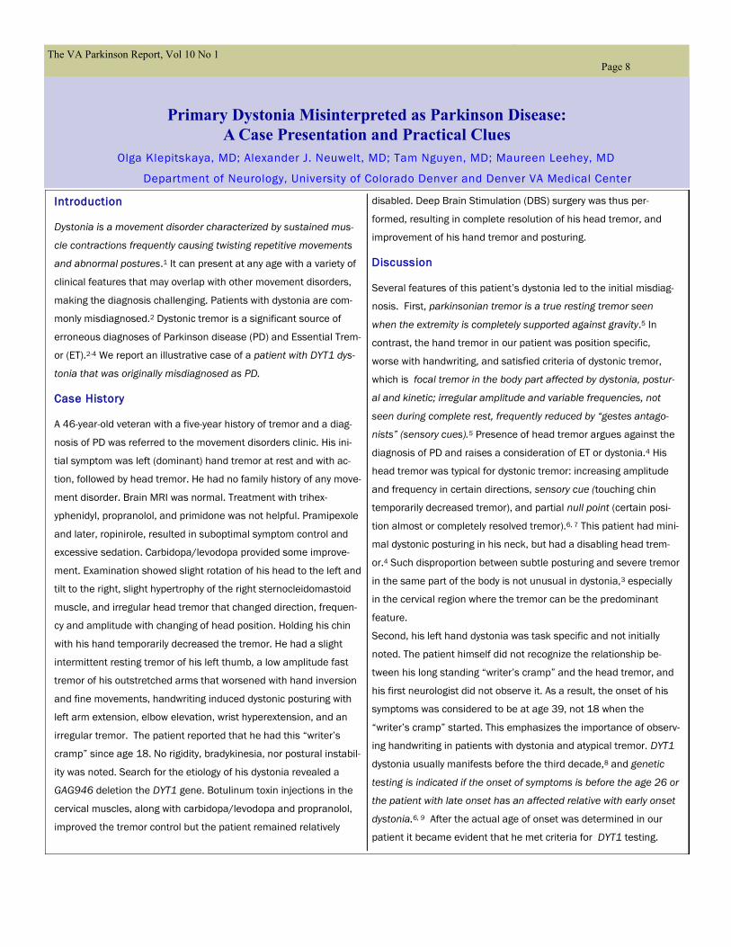
Restless Leg Syndrome (RLS) is describes as the periodic need to move your legs in an attempt to relieve unpleasant or uncomfortable
sensations. About 15% of the population has symptoms of RLS. Persons with Parkinson’s disease may also experience RLS.
What are the signs and symptoms of RLS?
Symptoms can range from mild to severe:
Overwhelming urge to moves your legs
Sensations of tingling, jittery, a “creepy crawly” feeling in the legs
Feelings of itching or pulling in the legs
A “fizzy soda” running through the veins
Feeling are more intense with sitting or at bedtime
Symptoms are relieved by walking or moving your legs
Alternative treatments to try:
Yoga
Acupuncture
Traction Straight Leg Exercises
Massages
Aerobic exercise and walking
Sleeping with a pillow between your legs
Wearing compression stockings
Applying cold or hot packs to legs 10 minutes several times a day
Meditation
Lifestyle changes that may offer some relief:
Taking hot baths
Limit caffeine
Avoid alcohol

2013 Report from Southeast/Richmond PADRECC
Lynn Klanchar, RN, MS, Associate Director of Education, PADRECC Southeast
The VA Parkinson Report, Vol 10, No 1
Page 13
The PADRECC at Hunter Holmes McGuire Veterans Affairs Medical
Center (VAMC) in Richmond, Virginia is making a difference for
Veterans and their families with Parkinson’s Disease (PD) and other
movement disorders. Serving the Southeast region of the United
States including VISNs 5, 6, 7, 8 and 9, we provide state-of-the-art
clinical and surgical care for Veterans with movement disorders,
along with research, education, and community outreach.
Clinical News
Integrated Neurology Center of Excellence Pilot Project: In FY2013,
Southeast PADRECC began participating in this national Veterans
Healthcare Administration (VHA) initiative. The project will continue
through FY2014 with the goal of increasing Clinical Video
Telehealth (CVT) in specialty care services. CVT in Richmond has
been successful at bringing quality care to Veterans at distant and
rural Southeast region VAMCs and Community Based Outpatient
Clinics (CBOCs) and minimizing the burden of travel to Richmond.
Deep Brain Stimulation (DBS) surgery at the Richmond PADRECC
and Virginia Commonwealth University (VCU) uses the frameless
method. This disposable guidance device replaces the traditional
heavy frame used in DBS and enables greater patient comfort and
participation during surgery. Dr. Kathryn Holloway, Director of
PADRECC Neurosurgical Services helped develop the frameless
device and also incorporates an intraoperative scanner, the O-arm,
into the procedure. She recently worked with Medtronic, Inc., on a
film project to train more neurosurgeons in these newer techniques.
Research News
Eye Movements A large study is researching specific eye
movements in various movement disorders. Preliminary results
suggest the ability to accurately differentiate typical movement
disorders. “Pervasive Ocular Tremor in Patients with Parkinson’s
Disease” (Gitchel G, Baron M) was published in the Archives of
Neurology in August 2012. The research garnered media interest
and will be featured in an upcoming issue of Movement Disorders.
Repetitive Transcranial Magnetic Stimulation (rTMS) This research
is examining the effectiveness of rTMS on speech in people with PD.
TMS uses strong magnets to stimulate brain cells. Dr. Kathryn
Holloway leads the research team that includes a speech
pathologist.
O-arm Accuracy This research determined O-arm accuracy,
specifically how reliable and accurate the O-arm portable,
computerized tomography (CT) scanner images were compared to
traditional CT images. Study results found efficient, accurate
registration and assessment of DBS lead location before the
conclusion of surgery. “A quantitative assessment of the accuracy
and reliability of O-arm images for deep brain stimulation
surgery” (Holloway K) was published in Operative Neurosurgery in
March 2013.
Education News
Parkinson’s Education via Telehealth – each month, a one hour
education session on a topic related to PD is now broadcast to
CBOCs in Charlottesville, Fredericksburg and Emporia using
Telehealth technology. This education is a component of the
PADRECC Education & Support Group meeting held monthly in
Richmond at McGuire VAMC.
Dr. Mark Baron, Southeast PADRECC Director along with Dr. Steven
Schreiber from VA Long Beach Parkinson’s Consortium Center,
presented “Tele-Provider Hot Topics and Best Practices for Tele-
Neurology”. This monthly series, presented by VHA Telehealth
Services and the Employee Education System demonstrates
successful Telehealth clinical models in specialty care.
PADRECC Southeast Partnerships with other Parkinson’s
organizations or those with similar interests continues to grow. An
annual PD Community Education Day is sponsored by PADRECC, the
local Richmond Metro Chapter of American Parkinson Disease
Association (APDA), and the University of Virginia. PADRECC and
VCU Parkinson’s disease and Movement Disorders Center
collaborate to capitalize on shared resources, subject matter
experts, and collective creativity. We continue to identify gaps in
services and are jointly able to offer more education programs to
the movement disorder community. In 2013, PADRECC Southeast
and VCU connected with organizations such as Central Virginia
Chapter of National Multiple Sclerosis Society, the Richmond
Chapter of the International Essential Tremor Foundation, and the
American Red Cross. Together we presented a Family Caregiving
course in March, an Essential Tremor conference in April, and
Support Group Leader Training in May. The Annual PD Community
Education Day is slated for October 12, 2013.

The VA Parkinson Report, Vol 10, No 1 Page 14
New Resource Review
Lynn Klanchar, RN, MS, PADRECC Southeast
The Dance for PD® program has released a much anticipated At Home DVD. For individuals that
want to supplement their dancing at home, or for those who don’t have a class in their area, this
DVD will give them something close to the joyful experience of being in a live class with inspiration-
al and motivating music. The DVD is over 2 hours long with enough material and options ranging
from practice classes that will challenge the beginner, to the expedited “through class” that may
appeal to an experienced student. Movements were specially designed for people with Parkinson’s
and can be done seated or standing. The dance phrases draw from tap, ballet, jazz, modern dance
and improvisation.
This expertly produced resource is in keeping with the high quality of classes, training, and ser-
vices offered by Dance for PD® ®which was developed through a collaboration of Mark Morris
Dance Group and Brooklyn Parkinson Group in New York over twelve years ago. The Dance for PD®
program is built on one fundamental premise that professionally-trained dances are movement
experts whose knowledge is useful to persons with PD. The DVD can be ordered through
www.danceforparkinsons.org .
Our logo was developed by Eugene C. Lai, MD, PhD, who at the time was the Director of the
Houston PADRECC. This logo conveys a visual image of a patient constrained by Parkinson’s
disease moving from slowness, stiffness, and low spirits, to agility, balance, and joy!
Parkinson’s Disease Research, Education, and Clinical Centers are part of the nationwide Net-
work of Care for Veterans with movement disorders.
About the PADRECC Logo

The VA Parkinson Report, Vol 10, No 1 Page 15
We welcome Dr. Jackson to the Houston PADRECC!
Dr. George Jackson, is Houston PADRECC’s new Associate Director of Research.
Dr. Jackson received the baccalaureate and master’s degrees at the University
of Texas at Austin and the M.D and Ph.D. degrees from the University of Texas
Medical Branch (UTMB) at Galveston. He completed an internship in medicine
and residency in neurology at the University of California, Los Angeles and a fel-
lowship in movement disorders with Dr. Jeff Bronstein (now director of the South-
west PADRECC) at UCLA along with research training in neurogenetics at UCLA.
Dr. Jackson was faculty in the Department of Neurology and the Semel Institute
for Neuroscience and Human Behavior at UCLA until 2008. From 2009-2013 he
was professor and held the John Sealy Chair for Parkinson’s Disease at UTMB,
where he was Director of the George P. & Cynthia Woods Mitchell Center for Neu-
rodegenerative Diseases. He was recently recruited as the Associate Director for
Research in the Houston PADRECC at the Michael E. DeBakey VA Medical Center
and the Department of Neurology at Baylor College of Medicine.
We look forward to your leadership in PD research.
Houston PADRECC
The American Association of Nurse Practitioners is the largest national organization of
nurse practitioners of all specialties in the United States.
As a Fellow of the AANP, Dr. Willson was inducted as a nurse practitioner (NP) leader who
has made outstanding contributions to NP education, policy, clinical practice or research,
and developing NP leaders of the future. She is an adjunct professor at Prairie View A&M
University College of Nursing where she teaches in the Family Nurse Practitioner Program.
She is a Co-Investigator for a Baylor College of Medicine Interprofessional Geriatric Con-
sortium 5 million dollar grant to increase NP and Resident geriatric clinical knowledge and
skills. At the Michael E. DeBakey VA Medical Center she is the Advance Practice Regis-
tered Nurse (APRN) Council Chair and has led the development and implementation of
their new Scope of Practice.
Pamela Willson, PhD, RN, FNP-BC, CNE, FAANP, Associate Director of
Education for Houston PADRECC, has been recognized as a Fellow of
the American Association of Nurse Practitioners.
Congratulations Dr. Pamela Wilson

Page 16
The VA Parkinson Report, Vol 10, No 1
Consortium Coordinating Center
Rebecca Martine, APRN, CS, BC
Chairperson
215-823-5934
Dawn McHale, Coordinator
215-823-5800 x 2238
Consortium Center Referral Line
800-949-1001 x 2749
Newsletter Editor
Pamela Willson, PhD, RN, FNP-BC, CNE
Newsletter Editor
Houston PADRECC
PADRECC Centers:
Center Medical Center City, State Director Telephone
Houston Michael E. DeBakey
VAMC Houston, TX Dr. Aliya Sarwar 713-794-7841
Southwest West Los Angeles
VAMC Los Angeles, CA Dr. Jeff Bronstein 310-478-3711 ext. 48001
Northwest
Portland VAMC
VA Puget Sound Health
Care System
Portland, OR
Seattle, WA Dr. Joe Quinn
Portland: 503-721-1091
Seattle: 206-277-4560
Philadelphia Philadelphia VAMC Philadelphia, PA Dr. John Duda 215-823-5934 or toll free
888-959-2323
Southeast Hunter Holmes McGuire
VAMC Richmond, VA Dr. Mark Baron
804-675-5931 or toll free
800-784-8381 ext 5931
San Francisco San Francisco VAMC San Francisco, CA Dr. Jill Ostrem (Interim) 415-379-5530