Embed Size (px)
Citation preview
The utility of Ki-67 and BrdU as proliferative markers of adultneurogenesis
N. Kee 1, S. Sivalingam 1, R. Boonstra, J.M. Wojtowicz *
Department of Physiology, Medical Sciences Building, University of Toronto, Toronto, Ont., Canada M5S 1A8
Received 20 August 2001; received in revised form 4 January 2002; accepted 4 January 2002
Abstract
Adult animals continue to produce new neurons in the dentate gyrus of hippocampus. Until now, the principal method of
studying neurogenesis has been to inject either tritiated thymidine or 5?-Bromo-2-deoxyuridine (BrdU) intraperitoneally followed by
autoradiographic or immunohistochemical detection methods respectively. However, such exogenous markers may produce toxic
effects. Our objective was to determine whether Ki-67, a nuclear protein expressed in all phases of the cell cycle except the resting
phase, can be used as an alternative, endogenous marker. Using immunohistochemistry, we examined Ki-67 and BrdU expression
pattern in rats. Ki-67 was expressed within the proliferative zone of the dentate gyrus and its expression pattern mimicked that of
BrdU when examined soon after exogenous BrdU administration. Quantitative comparison of BrdU and Ki-67-positive cells
showed 50% higher numbers of the latter when examined 24 h after the BrdU injection. This was expected, since BrdU can be
incorporated into DNA only during the S-phase of the mitotic process, whereas Ki-67 is expressed for its whole duration.
Experimental increases (by ischemia) or reductions (by radiation) in the number of mitotic cells produced parallel changes in BrdU
and Ki-67 signals. Thus, Ki-67 is an effective mitotic marker and has most of the benefits of BrdU and none of the costs. This study
provides evidence for Ki-67 to be used as a marker of proliferation in the initial phase of adult neurogenesis. # 2002 Elsevier
Science B.V. All rights reserved.
Keywords: Neurogenesis; Cell proliferation; Hippocampus; Dentate gyrus; Mitotic indicator; 5-Bromo-2-deoxyuridine; Ki-67;
Immunohistochemistry
1. Introduction
5-Bromo-2-deoxyuridine (BrdU) has been a principal
marker for mitotic cells in studies of adult neurogenesis
(Gratzner, 1982). The method consists of a pulse
injection of BrdU into the intraperitoneal cavity fol-
lowed by a variable survival time allowing for tracking
the fate of divided cells and their progeny. At the end of
the experiment, the animal is sacrificed and the tissues
fixed with a standard paraformaldehyde-based fixative.
BrdU is detected in the tissue using specific primary
antibodies. The primary antibodies are then labeled with
a secondary antibody tagged with a fluorescent com-
pound or with a substrate for diaminobenzidine (DAB)
(Seki and Arai, 1993; Scott et al., 2000). The substitution
of an endogenous DNA base, Thymidine, with the
BrdU analogue ensures specific labeling of only the
dividing cells. This specificity of BrdU combined with
the high sensitivity of fluorescence microscopy, includ-
ing confocal microscopy has led to the great popularity
of the method. It is believed that the application of this
BrdU method contributed to the current acceptance of
neurogenesis as a natural process in the brain of adult
mammals (Gross, 2000). One useful feature of BrdU is
its long-term retention in divided cells and its passage to
their daughter cells. This feature can be used to trace the
cell lineage and cell survival. A spectacular example of
such use was a study by Eriksson and colleagues who
injected patients with BrdU and then examined brain
tissue postmortem up to 2 years later. BrdU was
detected in the brains of these patients and double
labeling confirmed that the surviving cells were neurons
(Eriksson et al., 1998).
* Corresponding author. Tel.: �1-416-978-2899; fax: �1-416-978-
4940.
E-mail address: [email protected] (J.M. Wojtowicz).1 Equal contribution.
Journal of Neuroscience Methods 115 (2002) 97�/105
www.elsevier.com/locate/jneumeth
0165-0270/02/$ - see front matter # 2002 Elsevier Science B.V. All rights reserved.
PII: S 0 1 6 5 - 0 2 7 0 ( 0 2 ) 0 0 0 0 7 - 9

However, the undeniable usefulness of the BrdU
method goes hand in hand with its limitations and
drawbacks. The possibility of BrdU producing mutated
cells and the consequent severe abnormalities of thedeveloping tissues has been reported (Kolb et al., 1999).
These side effects aside, the use of BrdU in whole-animal
experiments is difficult due to uncertainty of diffusion of
this substance among the various tissues of the body
following intraperitoneal injection. For example, the
blood brain barrier may prevent the BrdU from freely
penetrating the brain tissue, especially in old animals
(Cameron and McKay, 2001). In certain species ofrodents, the effective incorporation of exogenous tri-
tiated thymidine (3HThy) in DNA synthesis in some
tissues was lacking (Adelstein et al., 1964). Since both
BrdU and 3HThy may use the same transport mechan-
isms (Cameron and McKay, 2001), a comparative study
of mitosis in different tissues and/or species, using BrdU
as a mitotic marker, could be difficult. Stress is known
to inhibit neurogenesis (Cameron and Gould, 1994;Gould et al., 1997) and therefore the handling and the
BrdU injection procedure is likely to have an unintended
effect on the rate of neurogenesis.
Given these drawbacks, an alternative method is
highly desirable. Here we report on the use of Ki-67, a
nuclear protein, expressed in dividing cells for the entire
duration of their mitotic process (Scholzen and Gerdes,
2000). Like BrdU, Ki-67 can be detected with immuno-histochemistry. Unlike BrdU, Ki-67 is an endogenous
marker that does not have any adverse effects on living
cells. Although the function of Ki-67 is not known, it is
a reliable marker of mitosis because it is expressed,
albeit at different levels, during mitosis and its half-life is
very short. Moreover, studies reported thus far show
that Ki-67 is expressed during mitosis in all mammalian
species from rodents to humans (Scholzen and Gerdes,2000; Endl and Gerdes, 2000). We focused our attention
on the use of Ki-67 in the brain tissue, especially in the
hippocampal dentate gyrus, because of the current
interest in the possible consequences of nerve cell
proliferation on cognition and behaviour (Boonstra et
al., 2001a). One example of such use of Ki-67 is already
available in the study by Tanapat et al. (1999). In that
study a difference between the rate of adult neurogen-esis, as indicated by BrdU labeling, between male and
female rats was found. Yet the numbers of Ki-67 labeled
cells were the same in males and females. They
concluded that perhaps the slower cell cycle, that would
not be detectable by Ki-67, was responsible for the
discrepancy although other interpretations are also
possible. In the present study we show a comparison
of BrdU and Ki-67 labeling in various experiments andcombine their use with the neuronal markers neuronal
nuclear protein (NeuN) (Mullen et al., 1992), and
collapsin response mediator protein-4 (CRMP-4)
(Quinn et al., 1999). Our results will clarify the pros
and cons of Ki-67 as a mitotic marker and make its use
routine and easier to interpret in future studies of adult
neurogenesis.
2. Methods
2.1. BrdU administration
The thymidine analogue BrdU (Sigma, St. Louis,
MO) was administered intraperitoneally in pulse injec-
tions of the stock solution. BrdU stock was prepared in
phosphate buffered saline (PBS), (pH 7.2, 0.1 M) with
0.1 N NaOH at 20 mg/ml.
2.2. Animal groups
In experiment 1 (effects of intraperitoneal injections)
the subjects were young adult (35�/40 days) male Wistar
rats (Charles River). A pulse injection of 300 mg/kg was
given to one group of animals (n�/3). To control for
possible toxic or stress-related effects due to the injec-
tion of BrdU, we compared the BrdU-injected group
with a second group (n�/3) injected with the equivalent
amount of saline, and with a third control (n�/3) groupthat was not injected. Animals in all groups were
sacrificed 24 h after the injections.
In experiment 2 (effects of gamma irradiation) young
adult (35�/60 days) male Wistar rats were used. The
procedure and the handling of animals were significantly
different from those described by us previously (Snyder
et al., 2001) so we give more details. In each case the
animal was anesthetized with Somnotol (60 mg/kg). Thehead portion of the experimental rats (n�/2) was
exposed to focused beam gamma irradiation (Theratron,60Co source) for a dose of 10 Gy, while the sham rats
(n�/4) were not exposed to radiation. Four sham
animals were sacrificed 3 days, 1, 2 and 3 weeks after
the procedure respectively. Two irradiated animals were
sacrificed 3 weeks after the procedure. Each animal was
injected with a single dose of 300 mg/kg BrdU andperfused 2 h afterwards to assess cell proliferation in the
dentate gyrus.
In experiment 3 (effects of ischemia) male adult (�/3
months old) Sprague�/Dawley rats (n�/3 for ischemic
and n�/3 for sham controls) were used. In this group we
gave 3 injections of BrdU (100 mg/kg total) in 12 h to
each rat. This dosage was used for consistency with our
previous studies of neurogenesis and ischemia (Snyder etal., 2001; Kee et al., 2001). Other procedures for the
ischemia experiment were exactly the same as described
before (Kee et al., 2001).
N. Kee et al. / Journal of Neuroscience Methods 115 (2002) 97�/10598

2.3. Tissue preparation for immunohistochemistry
Animals were anaesthetized with Somnotol (60 mg/
kg; MTC pharmaceuticals, Ontario). They were per-fused transcardially with ice-cold PBS, followed by ice-
cold 4% paraformaldehyde in 0.1 M phosphate buffer
(PB, pH 7.4), after which the brains were extracted. The
hippocampus was removed and postfixed overnight in
the paraformaldehyde fixative. It was then mounted and
sectioned transversally on a vibratome at 30 mm and the
slices were stored in PBS containing 0.1% sodium azide
at 4 8C until use.
2.4. BrdU immunohistochemistry
Tissue to be labeled was washed with PBS to remove
any sodium azide prior to immunolabeling. Sections
were incubated in wells (48 well Nunc Multidish)
containing the desired antibody solutions. For immu-
nohistochemical detection of BrdU-incorporating nu-
clei, DNA was first denatured to expose the antigen by
incubating the tissue sections in 1N HCl for 45 min at
45 8C as described by Kee et al. (2001). The sectionswere rinsed three times for 5 min each in PBS, thenincubated with primary antibody to BrdU (rat mono-clonal; Accurate Chemical, 1:200 in 0.3% Triton-Xsolution) for 48 h at 4 8C. Next, the labeled sectionswere washed with PBS (3 times at 5 min each) andsecondary incubation was carried out in the dark for 2 hat room temperature. For the secondary incubation,fluorochrome-conjugated secondary antibody (Alexa488 goat anti-rat; Chemicon) was diluted at 1:200 in0.3% Triton-X solution. After 2 h the tissue was washedin PBS, rinsed with double-distilled water and mountedonto slides using an anti-fade mounting medium (Per-mafluor, Molec. Probes). The slides were allowed to airdry in the dark at room temperature and then stored at4 8C until used for analysis.
2.5. Ki-67 immunohistochemistry
Sections were washed with PBS to remove any sodium
azide and then incubated with Ki-67 antibody. The Ki-
67 antibody (NCL-Ki-67-MM1; NovoCastra Labs Ltd.)
was diluted at a ratio of 1:200 in 0.3% Triton-X in PBS
solution and sections to be labeled were allowed to
incubate for 48 h at 4 8C with gentle shaking. Thesections were then washed three times at 5 min each inPBS, followed by a 2 h incubation with fluorescent-conjugated secondary antibody (Alexa 568/488 goatanti-mouse, 1:200 in 0.3% Triton-X solution; Chemi-con) at room temperature. Sections were then rinsed andmounted as described above.
2.6. Ki-67�/BrdU co-labeling
Ki-67 labeling was performed as described above.
Once the sections were washed after the secondaryantibody incubation and observed under a wet mount,
they were post-fixed in 8% paraformaldehyde in 0.1 M
phosphate buffer (PB, pH 7.4) for 15 min. This was
necessary to protect the Ki-67 signals from the HCl
treatment (Palmer et al., 2000). Sections were then
washed three times at 5 min each and processed for
BrdU labeling as described above.
2.7. Ki-67�/CRMP-4 co-labeling
Ki-67 labeling was performed as described above. For
CRMP-4 co-labeling, the Ki-67 labeled sections were
washed with PBS, then incubated in primary antibody
to CRMP-4 (1:500 in 0.3% Triton-X solution, Anti-
rabbit; Chemicon) for 72 h at 4 8C with gentle shaking.The sections were washed three times at 5 min each inPBS. The resulting sections were incubated for 2 h in thedark at room temperature in fluorescent-conjugatedsecondary antibody (Alexa 488, goat anti-rabbit; Che-micon). Sections were washed again thoroughly in PBSand finally mounted onto slides with Permafluor.
2.8. Ki-67�/NeuN co-labeling
Ki-67 labeling was performed as described above,
however, the secondary incubation with the fluro-
chrome-conjugated antibody (Alexa 488 goat anti-
mouse) was left overnight. After the secondary incuba-
tion was completed, tissue sections were washed three
times to ensure that only bound Alexa 488 antibodies
remain. This was necessary to avoid nonspecific binding.Next, an Alexa 568 anti-mouse antibody (1:200 in 0.3%
Triton-X) was added and incubated for 2 h. Slices were
washed with PBS and incubated for 24 h at 4 8C withgentle shaking with anti-NeuN antibody (MsX NeuN;Chemicon), diluted at 1:1000 in 0.3% Triton-X in PBS.The sections were then washed in PBS and subsequentlyincubated with Alexa 568 goat anti-mouse (1:200 in0.3% Triton-X solution) for 2 h in the dark. Sectionswere washed thoroughly and mounted.
2.9. Quantification of labeled cells
A stereological counting method (optical dissector)
using the confocal microscope (Scott et al., 2000) was
used to determine the number of positively labeled cellsin six random sections of the dentate gyrus taken from
each animal. The results from all sections were pooled
and the mean from all sections is taken as n�/1.
N. Kee et al. / Journal of Neuroscience Methods 115 (2002) 97�/105 99
2.10. Microscopy
Representative images were taken using a Nikon
Optiphot-2 fluorescence microscope with either 20�/
dry lens or 100�/ oil immersion lens. The signal for
Alexa 488 labeling (green) was detected using an Omega
XF22 filter, and the Alexa 568 labeling (red) was
detected using a Nikon BA590 filter. Images were
digitized by a Sensicam CCD camera. Fluorescent
signals for quantification were detected using a confocal
microscope (Microsystems, Zeiss LSM 410) with 40�/
dry objective. A single wavelength laser line of 488 nm(ArKr laser) with LP515 filter was used to quantify the
number of Ki-67/BrdU positive cells.
3. Results
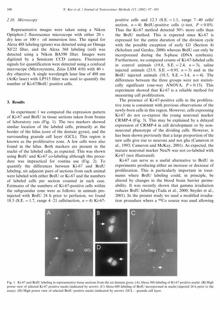
In experiment 1 we compared the expression pattern
of Ki-67 and BrdU in tissue sections taken from brains
of laboratory rats (Fig. 1). The two markers showed
similar location of the labeled cells, primarily at theborder of the hilus (core of the dentate gyrus), and the
surrounding granule cell layer (GCL). This region is
known as the proliferative zone. A few cells were also
found in the hilus. Both markers are present in the
nuclei of the labeled cells, as expected. This was shown
using BrdU and Ki-67 co-labeling although this proce-
dure was impractical for routine use (Fig. 2). To
quantify the differences between Ki-67 and BrdUlabeling, six adjacent pairs of sections from each animal
were labeled with either BrdU or Ki-67 and the numbers
of labeled cells per section counted in each case.
Estimates of the numbers of Ki-67-positive cells within
the subgranular zone were as follows: in animals pre-
injected with BrdU 24 h prior to perfusion, we found
18.5 (S.E.�/1.7, range 4�/21 cells/section, n�/4) Ki-67-
positive cells and 12.3 (S.E.�/1.1, range 7�/40 cells/
section, n�/4) BrdU-positive cells (t -test, P B/0.05).
Thus the Ki-67 method detected 50% more cells than
the BrdU method. This is expected since Ki-67 is
expressed for the entire duration of the division cycle
with the possible exception of early G1 (Section 4)
(Scholzen and Gerdes, 2000) whereas BrdU can only be
incorporated during the S-phase (DNA synthesis).
Furthermore, we compared counts of Ki-67-labeled cells
in control animals (19.8, S.E.�/2.4, n�/3), saline
injected animals (23.9, S.E.�/0.95, n�/3) and in the
BrdU injected animals (18.5, S.E.�/3.4, n�/4). The
differences between the three groups were not statisti-
cally significant (one-way ANOVA, P�/0.15). This
experiment showed that Ki-67 is a reliable method for
measuring cell proliferation.
The presence of Ki-67-positive cells in the prolifera-
tive zone is consistent with previous observations of the
newly-born cells in this region. However, cells expressing
Ki-67 do not co-express the young neuronal marker
CRMP-4 (Fig. 3). This may be explained by a delayed
expression of CRMP-4 in cell development or by non-
neuronal phenotype of the dividing cells. However, it
has been shown previously that a large proportion of the
new cells give rise to neurons and not glia (Cameron et
al., 1993; Cameron and McKay, 2001). As expected, the
mature neuronal marker NeuN was not co-labeled with
Ki-67 (not illustrated).
Ki-67 can serve as a useful alternative to BrdU in
experiments producing either an increase or decrease of
proliferation. This is particularly important in treat-
ments where BrdU labeling could, in principle, be
altered by changes in the blood brain barrier perme-
ability. It was recently shown that gamma irradiation
reduces BrdU labeling (Tada et al., 2000; Snyder et al.,
2001). In the present study we used a modified irradia-
tion procedure where a 60Co source was used allowing
Fig. 1. Ki-67 and BrdU labeling in representative tissue sections from the rat dentate gyrus. (A) Alexa-568 labeling of Ki-67 positive nuclei. (B) High
power view of selected Ki-67 positive nuclei (indicated by arrow). (C) Alexa-488 labeling of BrdU incorporated in nuclei (injected 24 h prior to the
assay). (D) High power view of selected BrdU positive nuclei (indicated by arrow). GCL*/granule cell layer.
N. Kee et al. / Journal of Neuroscience Methods 115 (2002) 97�/105100

accurate focusing of the radiation beam on the head of
the animal. In two irradiated animals, Ki-67 labeling
was reduced by an average of 79% (Fig. 4), and that of
BrdU by 60%. The densities of the Ki-67 positive cells in
the sham control group were 25.2 cells/section (S.E.�/
2.7, n�/4). The densities of Ki-67 positive cells in the
irradiated group were 5.1 cells/section (S.E.�/0.85, n�/
2). The corresponding densities of the BrdU positive
cells were 9.6 cells/section (S.E.�/1.65, n�/4) before and
3.8 cells/section (S.E.�/0.02, n�/2) after the irradiation.
It should be noted that in this experiment, in contrast to
experiment 1, BrdU was injected 2 h and not 24 h prior
to sacrificing the animals (Section 4).
Neurogenesis can be enhanced by a variety of stimuli
including the transient ischemia produced by occlusion
of the carotid arteries. The ischemia procedure leads to
an approximately 100% increase in the density of the
BrdU labeled cells (Kee et al., 2001). One possible
explanation of this finding is that the increased labeling
is caused by increased influx of BrdU into the brain due
to a breakdown of the blood brain barrier after ischemia
(Preston et al., 1993). To counter this argument we
performed experiment 3 in which we used Ki-67 labeling
in parallel with BrdU. We show that 10 days after
ischemia similar proportional increases in the density of
Ki-67 and of BrdU-positive cells were seen (Fig. 5). The
densities of Ki-67 positive cells increased from 9.9 cells/
section (S.D.�/0.6, n�/3) in sham controls to 22 cells/
section (S.D.�/0.75, n�/3) in ischemic animals. The
densities of the BrdU positive cells increased from 10.6
cells/section (S.D.�/0.75) to 21.1 cells/section (S.D.�/
4.0, n�/3). We conclude that the observed enhancement
Fig. 2. Ki-67 (red) and BrdU (green) co-labeling in a representative tissue section from the rat dentate gyrus. (A) Alexa-568 labeling of Ki-67 shows
six identified nuclei (arrows). (B) Alexa-488 labeling of BrdU injected 24 h prior to the assay shows three nuclei. (C) Superimposed images A and B
show that three out of six nuclei were labeled with both markers. GCL*/granule cell layer.
Fig. 3. CRMP-4 (green) and Ki-67 (red) co-labeling in a tissue section from the rat dentate gyrus. (A) Alexa-488 labeling shows typical round cell
bodies of CRMP-4 positive young neurons aligned along the proliferative zone. (B) Elongated Ki-67-positive nucleus in the same field. (C) Combined
image illustrates that CRMP-4 and Ki-67 were present in different cells.
N. Kee et al. / Journal of Neuroscience Methods 115 (2002) 97�/105 101

of cell labeling seen after ischemia is not an artifact of
BrdU method but due to increased cell proliferation.
Perhaps the most beneficial feature of Ki-67 is that it
can be readily used in species other than laboratory
Fig. 4. Ki-67 immunoreactivity in tissue sections from control and experimental rats after gamma irradiation. Note the decrease in proliferative
activity after radiation treatment. GCL*/granule cell layer.
Fig. 5. Ki-67 and BrdU immunoreactivity in tissue sections from control and experimental rats 10 days after transient global ischemia. Note, both
markers indicate an increase in proliferative activity after ischemia although the absolute levels are difficult to compare since BrdU was injected 3
days prior to the examination and Ki-67 shows a snap shot of the proliferation at the time of death. GCL*/granule cell layer.
N. Kee et al. / Journal of Neuroscience Methods 115 (2002) 97�/105102

rodents without the need for capturing, handling and
injecting such as that required with BrdU. We have
tested the use of Ki-67 on brain tissue of the arctic
ground squirrel (Spermophilus parryii ), a rodent foundin the tundra and alpine areas of mainland North
America. Application of BrdU to these wild animals is
not practical since their capture produces a strong stress
response (Boonstra et al., 2001b) that in turn is known
to suppress cell proliferation in the dentate gyrus in all
species examined (Gould et al., 1998). Ki-67 may be a
method of choice in such cases and it does indeed show
proliferative activity in the dentate gyrus of the groundsquirrel (Fig. 6).
4. Discussion
Neurogenesis in the adult dentate gyrus has been
observed in all mammalian species examined to date,
including humans (Eriksson et al., 1998). To establish a
functional significance of the new neurons, one needs to
determine that the proliferating cells respond to physio-
logical stimuli, become integrated in the neuronal
circuitry and are playing a significant role. Since thedentate gyrus of the hippocampus is one of the hot spots
of neurogenesis, it is tempting to speculate that the new
cells play a role in cognition and spatial learning, the
established functions of this structure. Indeed the
experiments on rats have shown that some types of
hippocampal-dependent learning are critically influ-
enced by the presence of the newly-born neurons (Gould
et al., 1999; Shors et al., 2001). In other experiments the
rate of cell proliferation was altered by physiological
stimuli such as stress (Gould et al., 1997) or running
(van Praag et al., 1999). However, until now BrdU has
been used as a principal mitotic marker in such studies
and it is prone to various difficulties. The main difficulty
is that the administration of BrdU before or after the
learning experiment is a potentially stressful procedure
and stress can alter the BrdU incorporation into the
dividing neurons (Gould et al., 1997, 1998). Numerous
other side effects of BrdU, including a direct effect on
adrenal gluocorticoid secretion, have been reported
(Anisimov, 1994; Malendowicz et al., 1997) (Table 1).
We addressed this problem in experiment 1 by examin-
ing the cell proliferation using Ki-67 in three groups of
animals: one injected with BrdU 24 h prior to labeling,
second injected with saline 24 h prior to labeling and
third not injected at all. Contrary to the expectations
there were no significant differences between the three
groups suggesting that neither the injection procedure
(presumed stress) nor BrdU itself had an inhibitory
effect on the rate of cell division.
In experiment 2 we examined cell proliferation in the
dentate gyrus before and after gamma irradiation
applied through a 60Co source to the head of the animal.
In this experiment, unlike in experiment 1, we injected
BrdU 2 h prior to killing the animals. Assuming that
there was no effect of stress due to the injection
procedure, this experiment should give an accurate
Fig. 6. Ki-67/NeuN immunoreactivity in tissue sections from the Arctic ground squirrel. (A) NeuN labeled section of the dentate gyrus of a juvenile
male arctic ground squirrel. (B) Ki-67 labeled section in the same animal shows characteristic immunoreactive nuclei of dividing cells in the
subgranular proliferative zone (arrows).
N. Kee et al. / Journal of Neuroscience Methods 115 (2002) 97�/105 103

comparison of BrdU and Ki-67 labeling. In sham
control animals we found that the ratio of BrdU/Ki-67
labeled cells was 0.38 consistent with the expected BrdU
incorporation during the S-phase of the cell cycle only.
In fact, approximate doubling of the BrdU labeled cells
could be expected from 2 to 24 h survival as reported by
Cameron and McKay (2001) due to mitotic divisions of
the labeled cells. Although we did see a trend (28%
increase) towards the increased BrdU labeling in experi-
ment 2 as compared to experiment 1, the difference was
not significant (t-test, P�/0.23). This observation
doesn’t conflict with the experiments of Cameron and
McKay (2001) since a strict comparison between
experiments 1 and 2 in our study is not possible due to
significant differences in the procedures. For example,
the ages of control animals were slightly different in the
two experiments. Also, the sham procedures involved in
the irradiation procedure were not trivial since they
included transportation to and from the irradiation
facility and anaesthesisa with pentobarbital. Our results
with the irradiation suggest that the BrdU/Ki-67 ratio
can change dramatically (from 0.38 before to 0.75 after
irradiation) although more experiments will be needed
to assess this change quantitatively and determine its
cause.
The data from experiment 3 show considerably lower
Ki-67 and BrdU counts in sham animals as compared to
experiments 1 and 2. The lower rates of cell proliferation
are expected due to the considerable age (Seki and Arai,
1995) and possibly strain differences (Wistar vs
Sprague�/Dawley) of rats used in these experiments
(Section 2). Furthermore, in this experiment BrdU was
injected 3 days prior the perfusions so the ratio of BrdU/
Ki-67 could have been changed by possible secondary
divisions and cell death of the BrdU labeled cells in the
three day period.
Another possible direction in such studies of neuro-
genesis will be to establish the effects of various stimuli
on cell proliferation in other species, particularly those
living in natural conditions and participating in natural
behaviours such as finding and storing of food, finding
mates etc. (Boonstra et al., 2001a). A non-invasive
marker of neuronal proliferation such as Ki-67 will be
a valuable addition to the arsenal of experimental tools
necessary for such studies. In Table 1 we compared the
features of BrdU and Ki-67. It shows that in experi-
ments that involve tracing of the progeny and measuring
the survival rates of the divided cells, BrdU is still the
method of choice. Although this persistent presence of
BrdU in the cells under study may produce long-term
developmental abnormalities due to possible mutagenic
activity (Anisimov, 1994; Kolb et al., 1999). The
probable reason for such mutagenic effects is the
substitution of CG by AT base pairs in the DNA and
ineffectiveness of repair enzymes in case of BrdU
induced DNA damage (Anisimov, 1994). No such
effects have been reported in the case of adult neurogen-
esis so far (Cameron and McKay, 2001) but it should be
realized that the assessment of cellular structure and
function in these and other studies of adult neurogenesis
has been quite limited and extended only over a period
of several weeks. The blood-brain barrier and other
metabolic factors (Adelstein et al., 1964; Cameron and
McKay, 2001) may reduce the accessibility of BrdU in
the brain in certain species while Ki-67 is not prone to
such difficulties. Thus, a comparative study of neuronal
proliferation among different species or among different
brain regions within a species should involve the use of
Ki-67.
Our results from experiment 1 suggest that in
laboratory rats, the presumed stress due to the injection
of BrdU has no effect on the number of mitotically
active cells in the dentate gyrus as shown by Ki-67
immunoreactivity. However, in other species, particu-
larly in wild populations, the effect of captivity, hand-
ling, injections of BrdU and re-capture of animals could
be significant (Boonstra et al., 2001b). In experiments
that do not involve tracing of cells over long periods or
those that must be done on wild animals or on human
subjects where the invasiveness of BrdU may be a
problem, Ki-67 is a welcome alternative. Although Ki-
67 is only a marker of cell proliferation it can be
Table 1
Comparison of BrdU and Ki-67 as proliferative markers
BrdU Ki-67
Selectivity S-phase only (Gratzner, 1982) All phases of cell cycle except G0
(Endl and Gerdes, 2000;
Scholzen and Gerdes, 2000)
Delivery and incorpora-
tion into tissue
Dependent on dose, bioavailability and diffusion barriers
(Cameron and McKay, 2001)
Not dependant on dose, bioavailability or diffusion
Expression across species Variable expression due to incorporation issues mentioned
above (Adelstein et al., 1964)
Endogenous expression in all species examined thus far
(Endl and Gerdes, 2000; Scholzen and Gerdes, 2000)
Use in cell fate tracking Feasible but with possible toxic effects during long
exposures (Anisimov, 1994; Kolb et al., 1999)
Not applicable
Side effects Stress during injection and mutagenesis following incor-
poration (Anisimov, 1994; Malendowicz et al., 1997)
Not applicable (intrinsic expression)
Relevant references are given in parentheses.
N. Kee et al. / Journal of Neuroscience Methods 115 (2002) 97�/105104

supplemented by other endogenous markers of young
cells such as CRMP-4 (Kee et al., 2001).
The advantages of Ki-67 notwithstanding its applica-
tion as a proliferative marker are not without someproblems. For example it is not certain whether Ki-67
signal is strong enough to be detectable by immunohis-
tochemistry during early G1, the initial phase of the cell
cycle (Endl and Gerdes, 2000). Thus any changes in cell
cycle during G1 may be undetectable.
Nevertheless Ki-67 is a useful marker of cell prolif-
eration, the initial step in the fascinating process leading
to production of new neurons in the adult brain. Thusfuture studies of neurogenesis should include both BrdU
and Ki-67 in combination with neuronal markers such
as NeuN and CRMP-4, taking their advantages and
limitations into account.
Acknowledgements
This research was supported by CIHR and the Heart
and Stroke Foundation grants to JMW, and NSERC
grant to RB. The assistance of Jason Snyder with the
irradiation experiments is greatly appreciated.
References
Adelstein SJ, Lyman CP, O’Brien RC. Variations in the incorporation
of thymidine into the DNA of some rodent species. Comp Biochem
Physiol 1964;12:223�/31.
Anisimov VN. The sole DNA damage induced by Bromodeoxyuridine
is sufficient for initiation of both aging and carcinogenesis. Ann
NY Acad Sci 1994;719:494�/501.
Boonstra RA, Galea LAM, Matthews S, Wojtowicz JM. Hippocampal
neurogenesis in natural populations. Can J Physiol Pharmacol
2001a;79:297�/302.
Boonstra RA, Hubbs H, Lacey EA, McColl CJ. Seasonal changes in
glucocorticoids and testosterone in free-living arctic ground
squirrels from the boreal forest of the Yukon. Can J Zool
2001b;79:49�/58.
Cameron H, Gould E. Adult neurogenesis is regulated by adrenal
steroids in the dentate gyrus. Neuroscience 1994;61:203�/9.
Cameron HA, McKay RDG. Adult neurogenesis produces a large
pool of new granule cells in the dentate gyrus. J Comp Neurol
2001;435:406�/17.
Cameron HA, Woolley CS, McEwen BS, Gould E. Differentiation of
newly born neurons and glia in the dentate gyrus of the adult rat.
Neuroscience 1993;56:337�/44.
Endl E, Gerdes J. The Ki-67 Protein: fascinating forms and an
unknown function. Exp Cell Res 2000;257:231�/7.
Eriksson PS, Perfilieva E, Bjork-Eriksson T, Alborn AM, Nordborg C,
Peterson DA, et al. Neurogenesis in the adult human hippocampus.
Nat Med 1998;4:1313�/7.
Gould E, Beylin A, Tanapat P, Reeves AJ, Shors TJ. Learning
enhances adult neurogenesis in the hippocampal formation. Nat
Neurosci 1999;2:260�/5.
Gould E, McEwen BS, Tanapat P, Galea LAM, Fuchs E. Neurogen-
esis in the dentate gyrus of the adult tree shrew is regulated by
psychosocial stress and NMDA receptor activation. J Neurosci
1997;17:2492�/8.
Gould E, Tanapat P, McEwen BS, Fluggge G, Fuchs E. Proliferation
of granule cell precursors in the dentate gyrus of adult monkeys is
diminished by stress. Proc Natl Acad Sci USA 1998;95:3168�/71.
Gratzner HG. Monoclonal antibody to 5-Bromo- and 5-iododeoxyur-
idine: a new reagent for detection of DNA replication. Science
1982;218:474�/5.
Gross CG. Neurogenesis in the adult brain: death of a dogma. Nat Rev
2000;1:67�/73.
Kee N, Preston E, Wojtowicz JM. Enhanced neurogenesis after
transient ischemia in the dentate gyrus of the rat. Exp Brain Res
2001;136:313�/20.
Kolb B, Pedersen B, Ballermann M, Gibb R, Whishaw IQ. Embryonic
and postnatal injections of bromodeoxyuridine produce age-
dependent morphological and behavioral abnormalities. J Neurosci
1999;19:2337�/46.
Malendowicz LK, Nussdorfer GG, Trejer M. Effect of 5-Bromo-2-
deoxyuridine on the proliferative activity of thymus and regenerat-
ing adrenal cortex. Life Sci 1997;61:641�/3.
Mullen RJ, Buck CR, Smith A. NeuN, a neuronal specific nuclear
protein in vertebrates. Development 1992;116:201�/11.
Palmer TD, Willhoite AR, Gage FG. Vascular niche for adult
hippocampal neurogenesis. J Comp Neurol 2000;425:479�/94.
Preston E, Sutherland G, Finsten A. Three openings of the blood-
brain barrier produced by forebrain ischemia in the rat. Neurosci
Lett 1993;149:75�/8.
Quinn CC, Gray GE, Hockfield S. A family of proteins implicated in
axon guidance and outgrowth. J Neurobiol 1999;41:158�/64.
Scholzen T, Gerdes J. The Ki-67 Protein: from the known and the
unknown. J Cell Physiol 2000;182:311�/22.
Scott BW, Wojtowicz JM, Burnham WM. Neurogenesis in the dentate
gyrus of the rat following electroconvulsive shock seizures. Exp
Neurol 2000;165:231�/6.
Seki T, Arai Y. Highly polysialylated neural cell adhesion molecule
(NCAM-H) is expressed by newly generated granule cells in the
dentate gyrus of the adult rat. J Neurosci 1993;13:2351�/8.
Seki T, Arai Y. Age-related production of new granule cells in the
adult dentate gyrus. Neuroreport 1995;6:2479�/82.
Shors TJ, Miesegaes G, Beylin A, Zhao M, Rydel T, Gould E.
Neurogenesis in the adult is involved in the formation of trace
memories. Nature 2001;410:372�/6.
Snyder JS, Kee N, Wojtowicz JM. Effects of adult neurogenesis on
synaptic plasticity in the rat dentate gyrus. J Neurophysiol
2001;85:2423�/31.
Tada E, Parent JM, Lowenstein DH, Fike JR. X-irradiation causes a
prolonged reduction in cell proliferation in the dentate gyrus of
adult rats. Neuroscience 2000;99:33�/41.
Tanapat P, Hastings NB, Reeves AJ, Gould E. Estrogen stimulates a
transient increase in the number of new neurons in the dentate
gyrus of the adult female rat. J Neurosci 1999;19:5792�/801.
van Praag H, Kempermann G, Gage FH. Running increases cell
proliferation and neurogenesis in the adult mouse dentate gyrus.
Nat Neurosci 1999;2:266�/70.
N. Kee et al. / Journal of Neuroscience Methods 115 (2002) 97�/105 105