Embed Size (px)
Citation preview
This is a repository copy of The use of quantitative sensory testing in cancer pain assessment: A systematic review.
White Rose Research Online URL for this paper:http://eprints.whiterose.ac.uk/154689/
Version: Accepted Version
Article:
Martland, ME, Rashidi, AS, Bennett, MI orcid.org/0000-0002-8369-8349 et al. (4 more authors) (2020) The use of quantitative sensory testing in cancer pain assessment: A systematic review. European Journal of Pain, 24 (4). pp. 669-684. ISSN 1090-3801
https://doi.org/10.1002/ejp.1520
© 2019 European Pain Federation ‐ EFIC®. This is the peer reviewed version of the following article: Martland, ME, Rashidi, AS, Bennett, MI, et al. The use of quantitative sensory testing in cancer pain assessment: A systematic review. Eur J Pain. 2020; 24: 669– 684. https://doi.org/10.1002/ejp.1520, which has been published in final form at https://doi.org/10.1002/ejp.1520. This article may be used for non-commercial purposes in accordance with Wiley Terms and Conditions for Use of Self-Archived Versions. Uploaded in accordance with the publisher's self-archiving policy.
[email protected]://eprints.whiterose.ac.uk/
Reuse
Items deposited in White Rose Research Online are protected by copyright, with all rights reserved unless indicated otherwise. They may be downloaded and/or printed for private study, or other acts as permitted by national copyright laws. The publisher or other rights holders may allow further reproduction and re-use of the full text version. This is indicated by the licence information on the White Rose Research Online record for the item.
Takedown
If you consider content in White Rose Research Online to be in breach of UK law, please notify us by emailing [email protected] including the URL of the record and the reason for the withdrawal request.
Title:
The use of Quantitative Sensory Testing (QST) in cancer pain assessment: a systematic review
Running header
Use of QST in cancer pain assessment
Authors:
M.E. Martland, 1†
A.S. Rashidi,2†
M.I Bennett,1 M. Fallon,3 C. Jones,1 R. Rolke,
4 M.R. Mulvey
1
†M.E. Martland and A.S. Rashidi are joint first authors and contributed equally to this
systematic review.
Affiliations:
1St Gemma’s Academic Unit of Palliative Care, Leeds Institute of Health Science, University of
Leeds, Leeds, UK
2Southern University of Denmark
3Edinburgh Cancer Research Centre, IGMM, University of Edinburgh, Edinburgh, UK;
4Department of Palliative Medicine, Medical Faculty RWTH Aachen University, Germany
Corresponding author
M.R. Mulvey
Academic Unit of Palliative Care, Leeds Institute of Health
Sciences, School of Medicine, University of Leeds
Worsley Building, Clarendon Way, Leeds LS2 9NL, UK Acc
epte
d A
rtic
le
This article has been accepted for publication and undergone full peer review but has not been
through the copyediting, typesetting, pagination and proofreading process, which may lead to
differences between this version and the Version of Record. Please cite this article as doi:
10.1002/EJP.1520
This article is protected by copyright. All rights reserved

This article is protected by copyright. All rights reserved
Email: [email protected]
Category – Review
Funding Statement
M.E. Martland is funding by a PhD scholarship from the University of Leeds. M.R. Mulvey is
funded by a fellowship from Yorkshire Cancer Research.
Conflict of interest
All authors declare no conflicts of interest with this manuscript and its content
Significance
This systematic review found that pain in cancer patients is associated with abnormal sensory
responses to thermal, mechanical and pinprick stimuli. However, these findings are based
primarily on studies of chemotherapy-induced peripheral neuropathy and data on tumour-
related pain are lacking, warranting further research.
Key words
Cancer, Pain, Quantitative Sensory Testing, QST, Assessment, Systematic Review
Acc
epte
d A
rtic
le
This article is protected by copyright. All rights reserved
Abstract
Objective: To summarises the literature on the use of quantitative sensory testing (QST) in the
assessment of pain in people with cancer and to describe which QST parameters consistently
demonstrate abnormal sensory processing in patients with cancer pain.
Databases and Data Treatment: Medline, EMBASE, AMED, CINAHL, SCOPUS and CENTRAL were
searched for observational or experimental studies using QST in patients with a cancer diagnosis and
reporting pain. Search strategies were based on the terms ‘quantitative sensory testing’, ‘cancer’,
‘pain’, ‘cancer pain’ and ‘assessment’. Databases were search from inception to January 2019. Data
were extracted and synthesised narratively, structured around the different QST modalities and sub-
grouped by cancer pain aetiology (tumour- or treatment-related pain)
Results: Searches identified 286 records of which 18 met the eligibility criteria for inclusion. Three
studies included patients with tumour-related pain, and 15 studies included patients with pain from
chemotherapy induced peripheral neuropathy (CIPN). Across all studies, 50% (9/18) reported sensory
abnormities using thermal detection thresholds (cool and warm), 44% (8/18) reported abnormal
mechanical detection thresholds using von-Frey filaments and 39% (7/18) found abnormal pin-prick
thresholds. Abnormal vibration and thermal pain (heat/cold) thresholds were each reported in a third
of included studies.
Conclusion: This systematic review highlights the lack of published data characterising the sensory
phenotype of tumour-related cancer pain. This has implications for our understanding of the
underlying pathophysiological mechanisms of cancer pain. Understanding the multiple mechanisms
driving cancer pain will help to move towards rational individualised analgesic treatment choices.
Acc
epte
d A
rtic
le
This article is protected by copyright. All rights reserved
Introduction
A third of patients receiving anticancer treatment report moderate to severe pain and over
50% of patients with advanced cancer report moderate to severe pain. Forty percent of
patients with cancer are affected by neuropathic pain which is associated with higher levels
of pain and reduced quality of life (van den Beuken-van Everdingen et al. 2007, Breivik et al.
2009, Bennett et al. 2012, Rayment et al. 2012, ONS 2015). As survival rates for cancer rise
(Kumar 2011), the burden of living with chronic pain from cancer and its treatment is rapidly
becoming a global health problem. Furthermore, the reduction in cancer pain prevalence
seen in the second half of the 20th
century (following publication of the WHO analgesic ladder
in cancer pain) has stalled in the past decade (van den Beuken-van Everdingen et al. 2018).
Accurately diagnosing cancer pain based on aetiology and neurological mechanisms is
essential to provide targeted and effective treatments for cancer patients (Mulvey et al.
2014, Mulvey et al. 2017).
Approximately 76% of cancer pains are caused by the tumour itself (tumour-related cancer
pain) and is regarded as a mixed-mechanism pain (Grond et al. 1996). Tumour-related cancer
pain is difficult to treat as nociceptive, inflammatory and neuropathic mechanisms commonly
co-exist, particularly in bone (Grond et al. 1999, Caraceni et al. 2005, Piano et al. 2012, Pina
et al. 2014, Paice et al. 2016)metastases (Paley et al. 2011, Falk et al. 2014). As cancer
treatments become more sophisticated, exposure to anti-cancer therapies (such as
chemotherapy, surgery or radiotherapy) is recognised as an important cause of pain in
patients with cancer (Paice et al. 2016). Between 10-20% of cancer pains are caused by anti-
cancer therapies (treatment-related cancer pain) (Grond et al. 1996, Grond et al. 1999,
Bennett et al. 2012) which are considered more similar to classic peripheral neuropathic pain
in mechanism and character (Paice et al. 2016). The remainder of pains in cancer patients are
due to co-morbid diseases unrelated to cancer. Identifying the most predominate pain
mechanism(s) is essential to diagnosing cancer pain and tailoring treatment plans.
A 2012 systematic review estimated that 40% of cancer patients with pain have a dominance
of neuropathic mechanisms, which can be either tumour-related or treatment-related
(Bennett et al. 2012). Neuropathic cancer pain (NCP) is associated with greater analgesic
requirements and oncological treatments, as well as poorer quality of life and lower
performance status (Rayment et al. 2012). Patients with NCP experience chronic and acute
Acc
epte
d A
rtic
le
This article is protected by copyright. All rights reserved
exacerbations of pain (pain flares) that peak multiple times per day (Fallon 2013). These
spontaneous pain flares are usually located in areas where there is observed sensory
dysfunction (hypersensitivity and/or hyposensitivity) (Fallon 2013, Haanpaa 2013, Baron et al.
2017). Patients with NCP also experience other sensations such as dysesthesia, allodynia and
hyperalgesia (Fallon 2013, Haanpaa 2013, Paice et al. 2016).
Pain assessment and diagnostic guidelines propose the use of objective measurable tests
such as quantitative sensory testing (QST,
Table 1) to support the diagnosis of neuropathic pain (Cruccu et al. 2009, Cruccu et al. 2010,
Haanpaa et al. 2011, Piano et al. 2012, Piano et al. 2013, Finnerup et al. 2016). QST has been
used most frequently in the assessment of peripheral neuropathic pain syndromes to defined
thermal and mechanical stimuli (Rolke et al. 2006, Roldan et al. 2015). In cancer, QST has
primarily focused on the assessment of peripheral neuropathy associated with exposure to
chemotherapy (Lipton et al. 1987, Lipton et al. 1991, Augusto et al. 2008, Boyette-Davis et al.
2011, Boyette-Davis et al. 2012, Boyette-Davis et al. 2013, Vichaya et al. 2013). These data
show a consistent a pattern of loss in function of small and large sensory fibres associated
with painful CIPN symptoms, characterised by increased thresholds to vibration, light touch
(von Frey hair) and pinprick stimuli. These data demonstrate the potential for QST to link
patient reported symptoms with underlying pathophysiological mechanisms.
Nevertheless, the standardised QST protocol is labour intensive, requires expensive
equipment and highly trained operators to complete the tests and interpret the data (Cruz-
Almeida et al. 2014). Shorter QST protocols that are both clinically predictive and simple to
operate and interpret are likely to be more clinically useful (Cruz-Almeida et al. 2014). The
first step towards developing a clinically relevant bedside QST protocol is to systematically
review the literature to identify which QST parameters most frequently identify sensory
abnormalities in tumour-related and treatment-related cancer. This will indicate which QST
parameters to take forward into clinical trial testing.
Acc
epte
d A
rtic
le

This article is protected by copyright. All rights reserved
Aim Objectives
The aim of this systematic review is to describe QST parameters commonly used in the
assessment of cancer pain (tumour-related or treatment-related pain) and identify which
ones consistently demonstrate abnormal sensory processing in this patient population.
Methods
A systematic literature review was undertaken of studies which reported the use of QST in
the assessment of cancer pain. This review was conducted in accordance with Centre for
Reviews and Dissemination Guidelines (Centre for Reviews and Dissemination 2009) which
include the PRISMA guidance on reporting study selection, (Moher et al. 2009) and included
on the PROSPERO international register of systematic reviews (CRD42018090092).
Eligibility Criteria / Types of studies to be included
Studies were eligible for inclusion if they:
1. were original observational or experimental studies
2. included adults (≥ 16 years old) with a cancer diagnosis and reporting pain (tumour-
or treatment-related pain)
3. had reported using standardised quantitative sensory testing (QST) procedures
4. included patients from primary, secondary or community care settings
5. were written in English language
6. were available in full-text version
7. were published in a peer-reviewed journal
The following were excluded:
1. studies that had recruited patients with cancer and non-cancer pain aetiologies where
the QST data were not reported separately for cancer and non-cancer patients
2. case reports and systematic reviews
3. studies that had only include data on acute post-surgical pain outcomes
4. intervention studies where:
- baseline data were only reported on pain free participants
- only post-intervention data were available Acc
epte
d A
rtic
le

This article is protected by copyright. All rights reserved
Information sources and search strategy
Electronic databases Ovid Medline EMBASE, AMED, CINAHL, SCOPUS and CENTRAL were
searched from inception to March 2018. Searches were updated in January 2019. A search
strategy was developed for Ovid Medline based on primary search terms for ‘quantitative
sensory testing’, ‘cancer’, ‘pain’, ‘cancer pain’ and ‘assessment’ (Table 2). Additional search
terms which encompassed the main attributes of psychophysiology and pain classification
were also included in the searchers. Search terms were tailored to each subsequent
database, as required. Full search strategy is described in Table 2.
Study selection process
Three reviewers (ASR, MRM, MEM) independently applied the eligibility criteria to the search
results by examining the titles and abstracts. Full texts were retrieved for articles that met
the inclusion criteria or could not be excluded based on abstract or title. Full text of relevant
articles were assessed for inclusion independently by two authors with reasons for exclusion
documented. Disagreement on included studies was discussed with an independent reviewer
(MB) and consensus was reached.
Data collection and synthesis
Data was extracted on: study characteristics (publication year, aim, design and country);
participants (number, age, gender, cancer diagnosis, pain aetiology, control subjects); setting
(e.g. primary care, secondary care, research institution); outcome measures (QST, patient
reported outcome measures (PROMS)); results (QST, PROMS); authors conclusion. Where
data were not available, attempts were made to contact the authors for the missing
information.
It was intended that the data would be synthesised narratively. This synthesis was structured
around the different QST modalities and (if sufficient data were available) cancer pain
aetiology (i.e. tumour or treatment related pain). Where the data were available, the
proportion of studies that included participants with advanced cancer, as well as the
numbers and proportions of participants in each study who had advanced cancer was
calculated. Demographic and clinical variables (including cancer types) were also summarised
across all included studies. Acc
epte
d A
rtic
le

This article is protected by copyright. All rights reserved
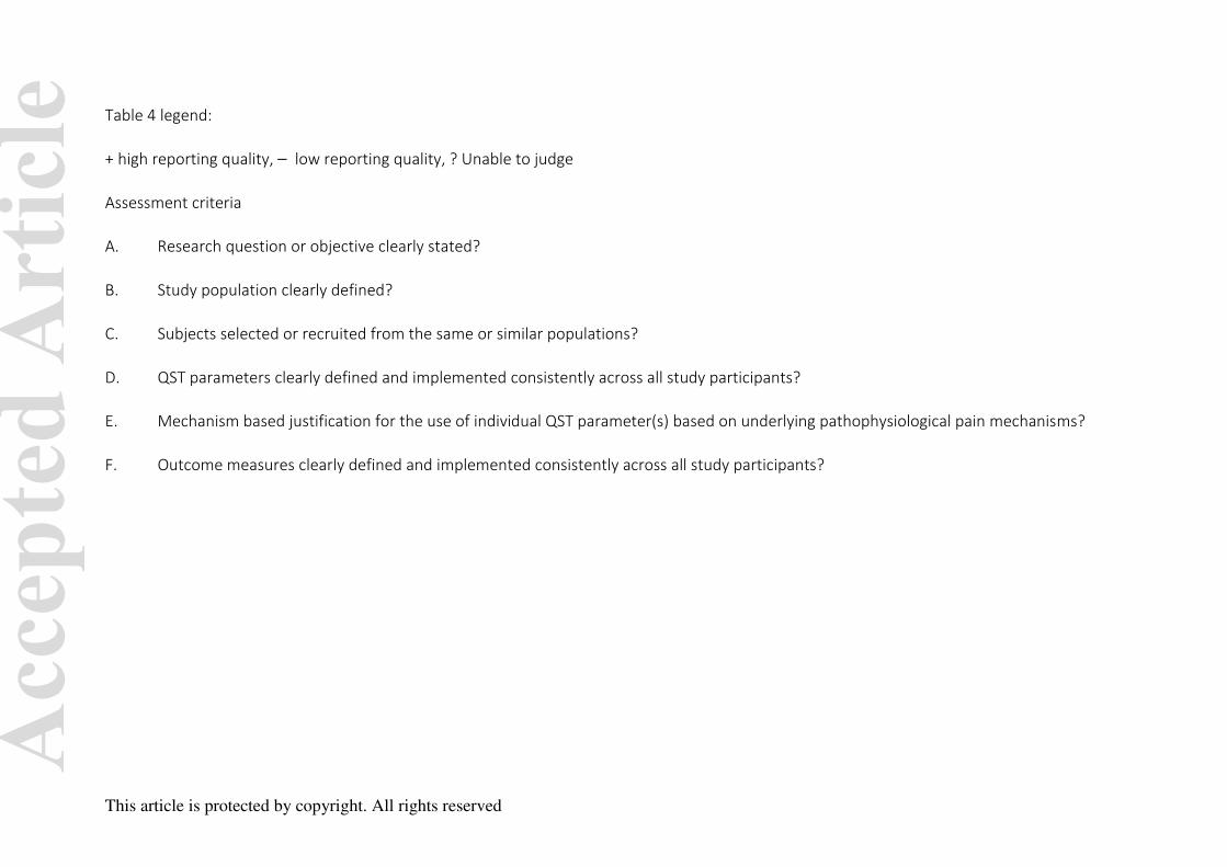
Quality of included studies
Methodological and reporting quality of each included study was assessed using a modified
version the National Heart, Lung and Blood institute Study Quality Assessment Tool for
Observational Cohort and Cross-Sectional Studies (U.S. Department of Health & Human
Services 2019,). The criteria assessed the quality of reporting of evidence upon which the
conclusions of this systematic review are drawn.
A. Research question or objective clearly stated?
B. Study population clearly defined?
C. Subjects selected or recruited from the same or similar populations?
D. QST parameters clearly defined and implemented consistently across all study
participants?
E. Mechanism based justification for the use of individual QST parameter(s) based
on underlying pathophysiological pain mechanisms?
F. Outcome measures clearly defined and implemented consistently across all study
participants?
Results
Included studies
Our searches identified 189 potentially relevant studies, of which 41 full text reports were
examined (Figure 1). A total of 18 studies met the eligibility criteria (Figure 1) (Lipton et al.
1991, Forsyth et al. 1997, Dougherty et al. 2004, Binder et al. 2007, Cata et al. 2007,
Dougherty et al. 2007, Caraceni et al. 2008, Attal et al. 2009, Boyette-Davis et al. 2011,
Hershman et al. 2011, Scott et al. 2012, Boyette-Davis et al. 2013, Krøigård et al. 2014, Fallon
et al. 2015, Velasco et al. 2015, Andersen et al. 2016, Andriamamonjy et al. 2017, Ventzel et
al. 2017). Three studies included patients with tumour-related cancer pain (Lipton et al. 1991,
Scott et al. 2012, Andersen et al. 2016). Fifteen studies included patients with chemotherapy
induced peripheral neuropathy (CIPN) (Forsyth et al. 1997, Dougherty et al. 2004, Binder et
al. 2007, Cata et al. 2007, Dougherty et al. 2007, Caraceni et al. 2008, Attal et al. 2009,
Boyette-Davis et al. 2011, Hershman et al. 2011, Boyette-Davis et al. 2013, Krøigård et al.
2014, Fallon et al. 2015, Velasco et al. 2015, Andriamamonjy et al. 2017, Ventzel et al. 2017).
Across all included studies 789 participants were included: 510 had cancer pain, 73 had non-
painful cancer (QST data not extracted) and 206 were pain-free control participants.
Acc
epte
d A
rtic
le

This article is protected by copyright. All rights reserved
Study characteristics
Eight studies were conducted in the USA and ten in Europe. Sixteen studies were
observational: eight cross-sectional (Lipton et al. 1991, Dougherty et al. 2004, Binder et al.
2007, Cata et al. 2007, Dougherty et al. 2007, Krøigård et al. 2014, Andriamamonjy et al.
2017, Ventzel et al. 2017), seven prospective (Forsyth et al. 1997, Caraceni et al. 2008, Attal
et al. 2009, Boyette-Davis et al. 2011, Boyette-Davis et al. 2013, Velasco et al. 2015, Andersen
et al. 2016), and one used cross-sectional and prospective groups (Hershman et al. 2011).
Two studies were experimental trials of analgesic interventions; only baseline (pre
intervention) QST data were extracted (Scott et al. 2012, Fallon et al. 2015). Three studies
also included cancer patients without pain (Binder et al. 2007, Krøigård et al. 2014, Andersen
et al. 2016). For each study, only QST data on patients who reported pain were extracted.
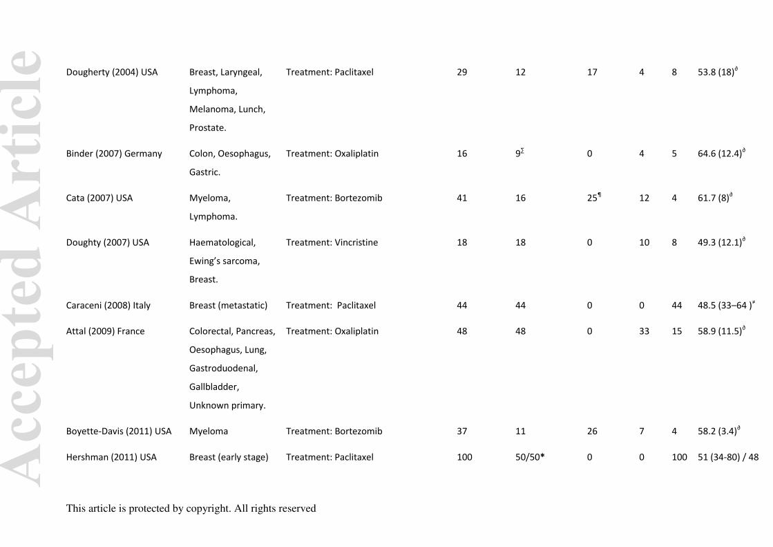
The most common cancers were colorectal and breast (Table 3).
Of the 15 CIPN studies, five included patients with acute pain symptoms (Forsyth et al. 1997,
Binder et al. 2007, Caraceni et al. 2008, Velasco et al. 2015, Andriamamonjy et al. 2017) and
10 included patients with chronic pain symptoms (Dougherty et al. 2004, Cata et al. 2007,
Dougherty et al. 2007, Attal et al. 2009, Boyette-Davis et al. 2011, Hershman et al. 2011,
Boyette-Davis et al. 2013, Krøigård et al. 2014, Fallon et al. 2015, Ventzel et al. 2017)
following chemotherapy administration. Data from the three tumour-related pain studies
could not be categorised as acute/chronic due to the ongoing nature of advancing disease
over an extended period of time. The three studies reporting data on tumour-related pain all
included patients with chronic pain (i.e. pain longer than 3 months). Across all studies, the
mean (SD, range) age was 58 (6.6, 48-73) and 63% of cancer pain participants were female.
Age and gender of non-pain participants were not extracted.
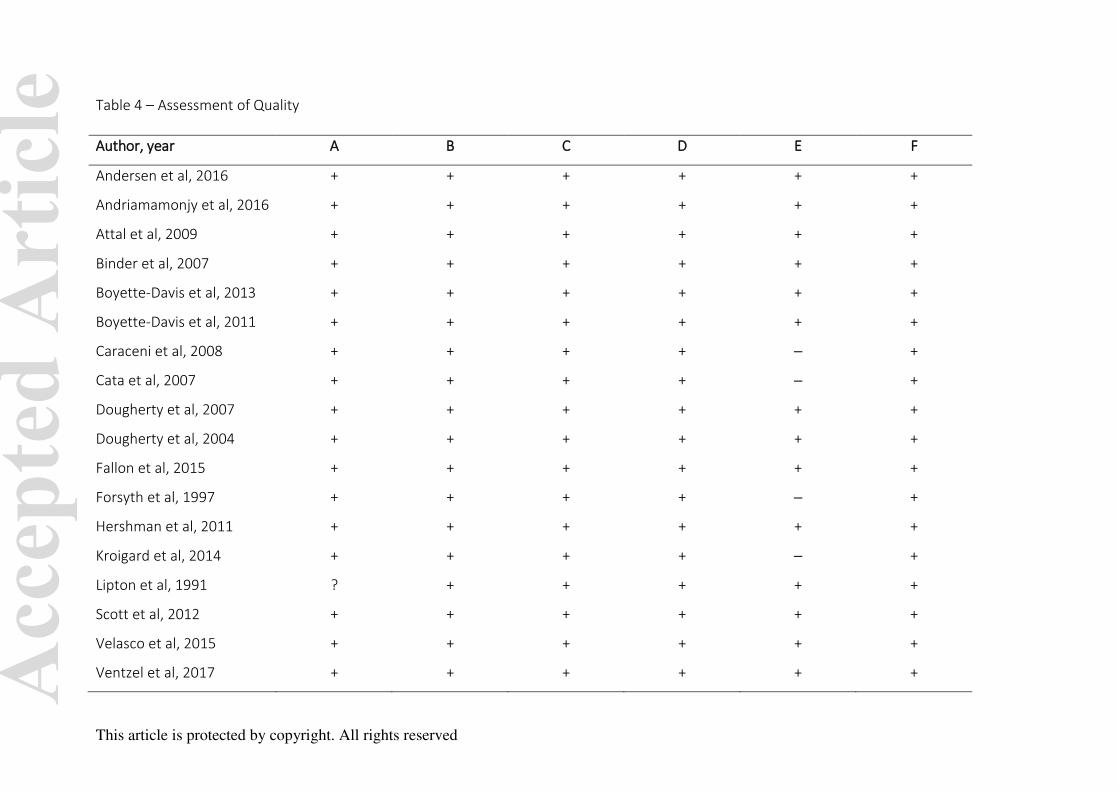
Quality assessment of included studies
A summary of the quality of included studies is reported in Table 4. All studies reported a
clearly stated research question and/or the objectives [A], except Lipton et al (Lipton et al.
1991) for whom the research objectives were not explicitly stated. The study populations
were clearly described in all studies [B] and recruited from the same or similar populations
[C]. All studies described the QST parameters that were employed, or indicated that the DNFS
QST protocol (Rolke et al. 2006) was adhered to [D]. In addition, all studies clearly defined the
outcome measures for each QST parameter that was implemented in the study [F].
Acc
epte
d A
rtic
le
This article is protected by copyright. All rights reserved
Studies varied in terms of the extent to which the choice of QST parameters was justified by a
priori theory of underlying pain mechanisms [E]. Few studies made direct links between pain
symptoms, the underlying neurobiological mechanism, and the selection of the appropriate
QST parameter. Four studies presented no justification for the selection of QST parameters
they used (Forsyth et al. 1997, Cata et al. 2007, Caraceni et al. 2008, Krøigård et al. 2014). Six
studies cited previous literature on sensory symptoms associated with tumour-related or
treatment-related cancer pain (Dougherty et al. 2007, Boyette-Davis et al. 2011, Hershman et
al. 2011, Boyette-Davis et al. 2013, Fallon et al. 2015, Andersen et al. 2016). Three studies
justified QST selection by linking individual symptoms to QST parameters and associated
underlying pathophysiological mechanisms. Andriamamonjy et al (Andriamamonjy et al.
2017) linked previously reported transient cold-induced distal allodynia with the assessment
of thermal detection and thermal pain thresholds in oxaliplatin induced neuropathy Attal et
al (Attal et al. 2009) justified the selection of QST parameters based on their ability to detect
sensory loss, hyperalgesia or allodynia which were previously reported to be associated with
oxaliplatin therapy. Dougherty et al (Dougherty et al. 2004) linked individual CIPN symptoms
to QST parameters and underlying pathophysiological mechanisms: sensory data on
mechanical, heat and cold perception pain to assess function in specific fibre types within
areas of sensory disturbance in patients with painful CIPN. In two studies sensory
abnormalities of CIPN were profiled using all 13 QST parameters described in the DFNS
protocol. The authors of these studies stated their intention was to characterise large and
small fibre function in CIPN patients and relate sensory profiles to underlying
pathophysiological mechanisms (Binder et al. 2007, Ventzel et al. 2017).
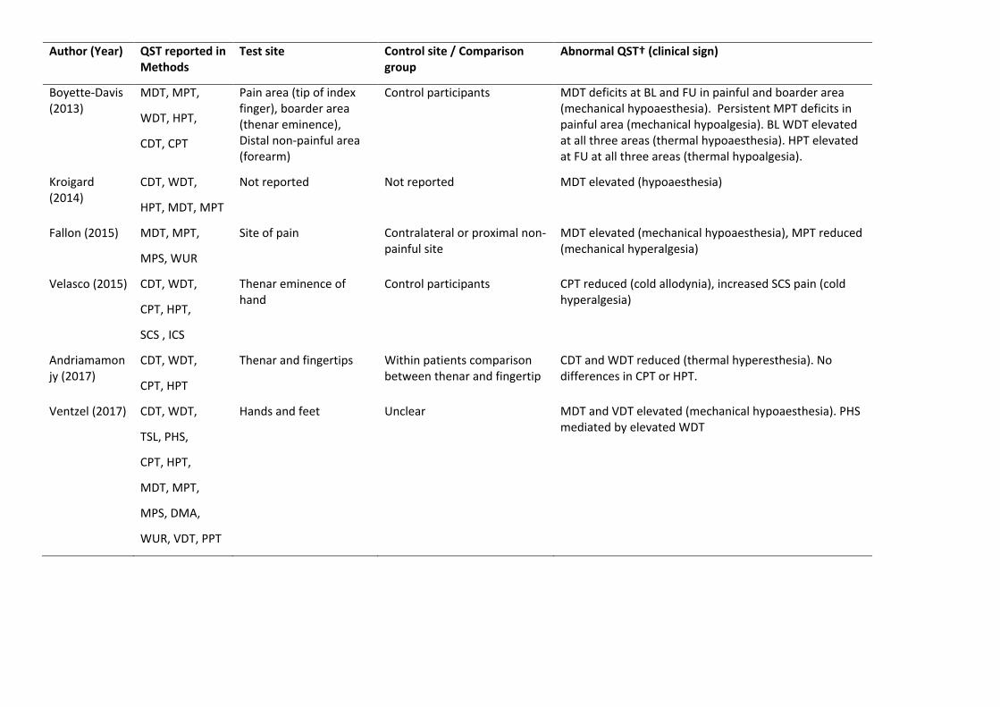
Thermal detection thresholds (CDT/WDT)
Cold (CDT) and warm (WDT) detection thresholds were the most commonly assessed QST
parameters in fifteen studies of which nine (60%) identified abnormal thermal detection
thresholds. Lipton et al (Lipton et al. 1991) found elevated thermal detection thresholds in
the feet of 50% of a heterogeneous cohort of 29 patients with tumour-related pain (Table 5).
Scott et al. observed a variety of hypo- and hyper-sensitivity to warm and cool stimuli (Scott
et al. 2012). Anderson et al. did not observe altered cool or warm perception (Andersen et al.
2016). Of the 12 CIPN studies that evaluated thermal detection thresholds, four reported
elevated WDT (Dougherty et al. 2007, Boyette-Davis et al. 2011, Boyette-Davis et al. 2013, Acc
epte
d A
rtic
le
This article is protected by copyright. All rights reserved
Ventzel et al. 2017), two reported elevated CDT (Forsyth et al. 1997, Dougherty et al. 2004),
and one reported that both CDT and WDT were elevated (Cata et al. 2007).
Thermal pain thresholds (CPT/HPT)
Cold (CPT) and heat (HPT) pain thresholds were reported in twelve studies of which six (50%)
reported abnormal thermal pain thresholds. Only Andersen et al. (Andersen et al. 2016)
evaluated thermal pain thresholds in tumour-related pain, but found no difference. Six of 11
CIPN studies that evaluated HPT and/or CPT identified abnormal thermal pain processing
(Table 5). Four studies observed a reduction in CPT (cold-hyperalgesia) in response to
Oxaliplatin (Binder et al. 2007, Attal et al. 2009, Velasco et al. 2015) and Bortezomib (Cata et
al. 2007). Three studies found elevated HPT (heat hypoalgesia) associated with exposure to
Bortezomib, Paclitaxel and Vincristine (Cata et al. 2007, Boyette-Davis et al. 2011, Boyette-
Davis et al. 2013). Attal et al. observed reduced HPT (heat hyperalgesia) in response to
Oxaliplatin (Attal et al. 2009). Velasco et al. and Attal et al. also observed increased pain
response induced by supra-threshold cold stimuli (cold hyperalgesia) (Attal et al. 2009,
Velasco et al. 2015).
Paradoxical heat sensations (PHS)
PHS were evaluated in two studies (Binder et al. 2007, Ventzel et al. 2017) one of which,
Ventzel et al., found 65% of colorectal cancer patients exposed to Oxaliplatin and 44% of
breast cancer patients exposed to Docetaxel experienced PHS mediated by elevated WDT.
Mechanical detection threshold (MDT)
MDT were reported in thirteen studies of which eight (62%) detected sensory abnormalities.
Two of three tumour-pain studies evaluated MDT (Table 5) (Scott et al. 2012, Andersen et al.
2016). Neither observed differences in MDT when comparing a painful region with a control
region in patients with tumour-related pain. Eleven CIPN studies assessed MDT at the site of
pain (finger tips or toes), of which eight (72%) found elevated MDT (Table 5). One study
found that reduced Aβ-fibre function preceded and predicted dysfunction in Aδ-fibres and C-
fibres (Dougherty et al. 2007). Three CIPN studies did not observed altered MDT (Binder et al.
2007, Attal et al. 2009, Hershman et al. 2011).
Acc
epte
d A
rtic
le
This article is protected by copyright. All rights reserved
Mechanical pain threshold (MPT)
MPT were reported in twelve studies of which seven (58%) identified altered pinprick
sensitivity. Two studies evaluated MPT in patients with tumour-related pain (Table 5) (Scott
et al. 2012, Andersen et al. 2016). Neither observed differences in MPT. Of the ten CIPN
studies that assessed MPT, seven (70%) reported altered pinprick sensations. Four studies
found elevated MPT (pinprick hypoalgesia) (Cata et al. 2007, Dougherty et al. 2007, Boyette-
Davis et al. 2011, Boyette-Davis et al. 2013). In contrast, three studies reported reduced MPT
(pinprick hyperalgesia) (Dougherty et al. 2004, Binder et al. 2007, Fallon et al. 2015).
Mechanical pain sensitivity (MPS)
Four studies evaluated MPS of which one (25%) found alerted pain response to supra-
threshold pinprick pain (Binder et al. 2007, Scott et al. 2012, Fallon et al. 2015, Ventzel et al.
2017). Scott et al. observed pinprick hyper- and hypoalgesia (45% and 9% respectively),
indicating mixed pathophysiology in cancer induced bone pain (CIBP) patients (Table 5) (Scott
et al. 2012). Fallon et al., Binder et al., and Ventzel et al. all observed no difference in MPS
(Table 5) (Binder et al. 2007, Fallon et al. 2015, Ventzel et al. 2017).
Dynamic mechanical allodynia (DMA)
DMA was assessed in four studies using light tactile stimuli e.g. soft burst of which one (25%)
identified altered light touch perception. Scott et al. observed DMA in 13% of CIBP patients
centrally augmented pain sensitisation present in a sub-set of CIBP patient (Table 5) (Scott et
al. 2012). The authors also observed tactile hypoaesthesia in response to dynamic soft brush
stimulus in 22% of CIPB patients. The other three studies that evaluated DMA found no
altered sensations (Table 5) (Binder et al. 2007, Attal et al. 2009, Ventzel et al. 2017).
Temporal pain summation (Wind-up ratio (WUR))
Augmented temporal pain summation was assessed using the pinprick wind-up ratio (WUR)
in three studies (Binder et al. 2007, Fallon et al. 2015, Ventzel et al. 2017). None (0%) of
these CIPN studies observed increased WUR in painful regions in comparison to normative
data sets (Binder et al. 2007), contralateral pain free region (Fallon et al. 2015) or between
hands and feet (Table 5) (Ventzel et al. 2017).
Acc
epte
d A
rtic
le

This article is protected by copyright. All rights reserved
Vibration detection threshold (VDT)
VDT were evaluated in seven studies of which six (86%) identified altered vibration
perception. Lipton et al. identified elevated VDT in the feet of tumour-related pain patients
(Table 5) (Lipton et al. 1991). Of the six CIPN studies that evaluated vibration perception, five
observed elevated VDT (Table 5) (Forsyth et al. 1997, Caraceni et al. 2008, Attal et al. 2009,
Hershman et al. 2011, Ventzel et al. 2017). In two prospective cohorts, cumulative Paclitaxel
(Caraceni et al. 2008) and Oxaliplatin (Attal et al. 2009) doses correlated with increasingly
elevated VDT. However, in another prospective cohort, Hershaman et al. observed elevated
VDT within a month of first Paclitaxel cycle which returned to baselined (pre-treatment)
levels by 12 months (Hershman et al. 2011). In a cross-sectional cohort, Ventzel et al. found
significantly elevated VDT in the hands (but not feet) of colorectal and breast cancer patients
exposed to Oxaliplatin or Docetaxel respectively (Ventzel et al. 2017). In another cross-
sectional cohort, Hershman et al. identified significant negative correlation between
increased sensations of numbness and pain in hands and feet and reduced vibration
perception (Hershman et al. 2011).
Pressure pain threshold (PPT)
Pressure pain thresholds (PPT) were assessed in two studies (Binder et al. 2007, Ventzel et al.
2017). Binder et al. found no difference in PPT between those with painful CIPN and pain free
patients (Binder et al. 2007). Ventzel et al. found PPT within normative data range for both
Oxaliplatin and Docetaxel treated patients (Ventzel et al. 2017).
Variation in QST procedures
Studies varied in terms of the different procedures used to evaluate the same or similar QST
parameters. Lipton et al. (Lipton et al. 1991) and Forsyth et al. (Forsyth et al. 1997) reported
measuring ‘thermal detection threshold’ (TDT) via a forced choice paradigm which most
closely mimics evaluating CDT in terms of thermal range used (i.e. 20-30oC). Scott et al. (Scott
et al. 2012) qualitatively determined presence or absence of thermal hyperaesthesia,
hypaesthesia or allodynia using cool and warm thermos rollers set at fixed temperatures
(25oC and 40
oC). In contrast, 12 studies quantitatively evaluated thermal detection and pain
thresholds using computer controlled devices via a Peltier thermode (Dougherty et al. 2004,
Binder et al. 2007, Cata et al. 2007, Dougherty et al. 2007, Attal et al. 2009, Boyette-Davis et Acc
epte
d A
rtic
le
This article is protected by copyright. All rights reserved
al. 2011, Boyette-Davis et al. 2013, Krøigård et al. 2014, Velasco et al. 2015, Andersen et al.
2016, Andriamamonjy et al. 2017, Ventzel et al. 2017).
Techniques to assess vibration perception also varied. Two studies evaluated VDT using a
forced choice paradigm to distinguish between two metal rods vibrating at increasingly
similar frequencies (Lipton et al. 1991, Forsyth et al. 1997). Two studies used a standardised
64Hz tuning fork which decreases in frequency until the vibration is no longer felt
(disappearance threshold) (Binder et al. 2007, Ventzel et al. 2017). Three studies used
electronic vibrameters which increase in frequency until the vibration is felt (perception
threshold) (Caraceni et al. 2008, Attal et al. 2009, Hershman et al. 2011). Studies that
evaluated MDT all used von Frey monofilaments in a method of limits paradigm. However,
punctate sensitivity was evaluated quantitatively using calibrated weighted needle pinprick
stimulators (Binder et al. 2007, Ventzel et al. 2017) or qualitatively single pinprick stimulus
(NeurotipsTM
Owen Mumford) (Scott et al. 2012, Fallon et al. 2015).
All of the four studies that included MPS in their methodology used noxious pin prick stimuli,
two of which (Scott et al. 2012, Fallon et al. 2015) specified the NeurotipsTM
Owen Mumford,
whilst the remaining two studies (Binder et al. 2007, Ventzel et al. 2017) documented that
they followed the QST-protocol of the German Research Network on Neuropathic Pain
(DNFS). Similarly, the three studies who included WUR in their methodology all used pin
prick stimuli (256mN) and compared a single stimulus to ten repeated stimuli with the
participant giving a pain rating from 0-10.
Techniques to measure dynamic allodynia varied slightly. Of the four studies that included an
assessment of dynamic allodynia, two studies (Binder et al. 2007, Ventzel et al. 2017)
followed the DNFS protocol which describes using three light tactile stimulators: a cotton
wisp (~3mN), a cotton wool tip fixed to an elastic strip (~100mN) and a standardised brush
(~200-400mN). The remaining two studies just used a standardised brush (Attal et al. 2009,
Scott et al. 2012).
Finally, for the two studies that measured PPT (Binder et al. 2007, Ventzel et al. 2017), both
studies followed the DNFS QST-protocol which uses a pressure gauge device (FDN200,
Wagner Instruments, USA). Acc
epte
d A
rtic
le
This article is protected by copyright. All rights reserved
Discussion
Across all studies, 50% (9/18) reported sensory abnormities using thermal detection
thresholds (cold and warm), 44% (8/18) reported abnormal mechanical detection thresholds
using von-Frey filaments and 39% (7/18) found abnormal pin-prick thresholds. Abnormal
vibration and thermal pain (heat/cold) thresholds were reported in a third of included studies
respectively.
This systematic review demonstrates a paucity of published data characterising the
phenotype of cancer pain using quantitative pain assessment techniques. Three studies were
identified that characterised tumour-related pain using QST, of which only two reported data
on sensory abnormalities (Lipton et al. 1991, Scott et al. 2012). Fifteen studies were identified
that characterised sensory abnormalities in CIPN using QST. These treatment-related pain
studies included a heterogeneous sample of patients with chronic and acute painful CPIN.
The in the number of QST studies profiling sensory abnormalities associated with CIPN has
increased in the past two decades. However, overall the number of QST studies profiling
cancer pain are small and the data should be interpreted with caution for this reason. There
are more recent emerging studies using QST to quantify pain in cancer patients; although
they didn’t meet the eligibility criteria for this systematic review, they are interesting for
reference (Kaunisto et al. 2013, Sipila et al. 2017, Griffiths et al. 2018).
Of the three tumour-related pain studies (Lipton et al. 1991, Scott et al. 2012, Andersen et al.
2016), Liption et al. and Scott et al. reported abnormalities in thermal detection and
mechanical or vibration detection, indicative of Aβ and Aδ fibre dysfunction. These limited
data suggest that systemic and localised loss in Aβ-fibre function, characterised by reduced
light touch sensation (hypoaesthesia) may be common in tumour-related cancer pain.
Andersen et al. found no sensory abnormalities; however, this study excluded patients with
any kind of peripheral nerve dysfunction and therefore were very unlikely to come across any
sensory abnormalities using QST.
The data presented in Table 5 indicate that CIPN is predominately characterised by increased
vibration and mechanical detection thresholds (reduced Aβ function), as well as increased
thermal detection and thermal pain thresholds (reduced Aδ and C-fibre function). Altered
pin-prick (mechanical pain) thresholds were also observed; however, whether this
manifested as a hypo- or hyper-sensitivity (loss or gain in Aδ fibre function) was treatment-
Acc
epte
d A
rtic
le
This article is protected by copyright. All rights reserved
dependent. Across all treatment-related studies, none identified altered perception of supra-
thresholds stimuli, such as mechanical pain sensitivity, dynamic mechanical allodynia,
pressure pain threshold or temporal summation (wind-up pain). However, the inclusion of
chronic and acute pain patients across the CIPN studies should also be considered. Patients
with chronic pain typically experience higher levels of pain intensity, reduced wellbeing and
poorer quality of life (Smith et al. 2007, Rayment et al. 2012). Chronic pain is also thought to
be associated with more central pathophysiological pain processes (Loeser 2019) which, in
the context of this systematic review, may influence QST findings based on peripheral
cutaneous examination techniques. None of the included CIPN studies explicitly stated using
QST to examine central pain mechanisms.
We acknowledge that a limitation of this systematic review is the lack of available data on
which to base conclusions, particularly for tumour-related pain. Despite a comprehensive
search strategy developed in line with international guidelines (Centre for Reviews and
Dissemination 2009, Moher et al. 2009) and broad eligibility criteria, very few studies were
identified. Due to the paucity of available quantitative data, it was not possible to undertake
a pooled analysis of QST data across all studies. Instead a narrative synthesis was undertaken
to summarise the main findings and identify commonalities in the patterns of sensory
abnormalities reported in tumour-related and treatment-related cancer pain.
The most commonly used QST parameters were thermal detection thresholds (CDT, WDT)
and mechanical detection. PPT and WUP were only included in 3 and 2 studies respectively
and revealed no sensory abnormalities. No studies reported finding DMA in tumour or
treatment related cancer pain, although this is contrary to anecdotal clinical experience of
bedside assessment in many patients. As DMA is a centrally mediated phenomenon, this
might suggest that tumour related pain is caused by peripheral sensitisation rather than
central sensitisation. However, these conclusions are speculative due to the lack of available
data.
The majority of CIPN studies did not make explicit statements linking the selection of
individual QST parameters to underlying pain mechanisms or patient reported symptoms.
Therefore, it remains unclear whether the data presented on treatment-related cancer pain
represents sensory abnormalities common to CIPN, or if it reflects the QST parameters that
were most frequently selected by the researchers. Although, the latter is unlikely as reduced
Acc
epte
d A
rtic
le

This article is protected by copyright. All rights reserved
tactile sensitivity has been widely reported as a common side effect of chemotherapy.
Nevertheless, future cancer pain research should consider profiling all 13 DNFS parameters
to gain a clearer understanding of which sensory abnormalities are most relevant to the
diagnosis and management of pain in patients with cancer.
The increasing abundance of QST evidence from CIPN studies (of a variety of
chemotherapeutic agents) indicates dysfunction in myelinated and unmyelinated cutaneous
sensory fibres. Similar quantitative sensory data in patients with tumour-related pain are
lacking; although evidence presented here suggests dysfunction in both myelinated and
unmyelinated cutaneous sensory fibres. Further research is required to map the sensory
phenotypes of tumour-related pain to improve our understanding of the underlying
pathophysiological mechanisms. Studies should include a heterogeneous population of
cancer types to better understand whether patterns of sensory dysfunction are disease
specific or unique to individual pain profiles which span cancer diagnostic groups.
Interestingly, none of the included studies evaluated descending pain control mechanisms.
Increasing evidence is emerging to support the theory that descending inhibitory (anti-
nociceptive) and facilitatory (pro-nociceptive) pathways are fundamental to our
understanding of chronic and neuropathic pain as seen in cancer patients. Data from animal
models of neuropathic pain reveals a multitude of descending pain control mechanisms
associated with hypersensitivity to noxious and non-noxious stimuli, as well as spontaneous
pain and central sensitization (Wang et al. 2013). These data demonstrate the complexities
associated with understanding the interactions between descending inhibitory pathways and
the clinical presentation of pain in patients with cancer (De Felice et al. 2011). QST may be a
potential biomarker for describing the function of these descending pain modulatory
pathways. For example, the conditioned pain modulation (CPM) paradigm has demonstrated
that reduced efficiency of descending control of pain is associated with the development of
chronic pain states in cancer and non-cancer populations (Yarnitsky et al. 2014, Yarnitsky
2015). Quantifying the function of descending pain modulatory pathways using QST would
further our understanding of the role that such neurobiological mechanisms have on the
clinical presentation of pain in cancer patients.
In conclusion, this systematic review highlights the lack of published studies characterising
the sensory phenotype of pain in cancer, with particularly few studies looking at tumour-
Acc
epte
d A
rtic
le
This article is protected by copyright. All rights reserved
related chronic or neuropathic pain. This limits our understanding of the underlying
pathophysiological mechanisms of cancer pain. Understanding the multiple mechanisms
driving cancer pain will enable rational individualised analgesic treatment choices (Vardeh et
al. 2016). Future studies should justify the selection of individual test parameters from
standardised QST protocols (Rolke et al. 2006) and consider incorporating CPM into their
protocols to evaluate the important role of descending pain mechanisms in cancer pain.
Acknowledgments
M.E. Martland is funded by a PhD scholarship from the University of Leeds. M.R. Mulvey is funded by
a fellowship from Yorkshire Cancer Research. All authors declare no conflicts of interest with this
manuscript and its content.
Acc
epte
d A
rtic
le
This article is protected by copyright. All rights reserved
Legends for figures and tables
Figure 1 - PRISMA flow diagram of searches, screening, eligibility and inclusion
Figure 1 legend
* hand searching reference lists of included studies
Table 1 – QST modalities and associated peripheral nerve fibres tested
Table 1 legend:
Summary of Quantitative Sensory Testing (QST) modalities, associated bedside equivalent
examinations and peripheral nerve fibres tested
Table 2 – Search Strategy
Acc
epte
d A
rtic
le

This article is protected by copyright. All rights reserved
Table 3 – Description of included studies
Table 3 legend:
† Gender of cancer pain participants only (gender of control participants not reported)
∆ decision made that this is tumour-related PN because patients with neurotoxic chemo-
therapeutic agents and other neurotoxic medications were excluded
∂ data are mean (SD)
§ data are median (range)
‡ 8 (15%) reported moderate-severe pre-treatment tumour pain. Therefore, only baseline
data (pre-intervention) extracted
- unable to extract age data for only pain patients
≠ data are mean (range)
∑ 9/16 participants had pain
¶ control subjects consisted of 20 health pain-free volunteers and 5 pain-free chemotherapy
naïve multiple myeloma ɸ data are mean (SD, min-max)
* Cross-sectional group / Prospectively cohort
± data presented for Docetaxel group of whom 80% reported pain. Oxaliplatin group 70%
reported no pain; therefore, data not extracted
∫ calculated by hand
ɤ data are presented for Oxaliplatin and Docetaxel groups respectively
ɸ data are mean (SD, min-max)
PN = Peripheral Neuropathy
CIPN = Chemotherapy Induced Peripheral Neuropathy
Table 4 – Assessment of Quality
Table 4 legend:
Acc
epte
d A
rtic
le

This article is protected by copyright. All rights reserved
+ high reporting quality, ⎯ low reporting quality, ? Unable to judge
Assessment criteria
A. Research question or objective clearly stated?
B. Study population clearly defined?
C. Subjects selected or recruited from the same or similar populations?
D. QST parameters clearly defined and implemented consistently across all study
participants?
E. Mechanism based justification for the use of individual QST parameter(s) based on
underlying pathophysiological pain mechanisms?
F. Outcome measures clearly defined and implemented consistently across all study
participants?
Acc
epte
d A
rtic
le
Table 5 – Quantitative Sensory Testing (QST) parameters used in cancer pain assessment
Table 5 legend
* Control participants referred to in
methods but not data are reported on them
in results.
† Mean data are summarised unless stated
otherwise
VT = Vibration Threshold
TDT = Thermal Detection Threshold
CDT = Cold Detection Threshold
WDT = Warm Detection Threshold
TSL = Thermal Sensory Limen
PHS = Paradoxical Heat Sensation
CPT = Cold Pain Threshold
HPT = Heat Pain Threshold
MDT = Mechanical Detection Threshold
MPT = Mechanical Pain Threshold
MPS = Mechanical Pain Sensitivity
DMA = Dynamic Mechanical Allodynia
WUR = Wind Up Ratio
VDT = Vibration Detection Threshold
PPT = Pressure Pain Threshold
SCS = Suprathreshold Cold Stimulus
ICS = Intense Cold Stimulus
This article is protected by copyright. All rights reserved
References
Andersen, K. G., H. M. Duriaud, E. K. Aasvang and H. Kehlet (2016). "Association between sensory
dysfunction and pain 1 week after breast cancer surgery: A psychophysical study." Acta
Anaesthesiologica Scandinavica 60(2): 259-269.
Andriamamonjy, M., J. B. Delmotte, F. Savinelli, H. Beaussier and F. Coudore (2017). "Quantification of
Chronic Oxaliplatin-Induced Hypesthesia in Two Areas of the Hand." Journal of Clinical
Neurophysiology 34(2): 126-131.
Attal, N., D. Bouhassira, M. Gautron, J. N. Vaillant, E. Mitry, C. Lepere, P. Rougier and F. Guirimand
(2009). "Thermal hyperalgesia as a marker of oxaliplatin neurotoxicity: A prospective quantified
sensory assessment study." Pain 144(3): 245-252.
Augusto, C., M. Pietro, M. Cinzia, C. Sergio, C. Sara, G. Luca and V. Scaioli (2008). "Peripheral
neuropathy due to paclitaxel: study of the temporal relationships between the therapeutic schedule
and the clinical quantitative score (QST) and comparison with neurophysiological findings." J
Neurooncol. 86(1): 89-99. doi: 10.1007/s11060-11007-19438-11068. Epub 12007 Jul 11065.
Baron, R., C. Maier, N. Attal, A. Binder, D. Bouhassira, G. Cruccu, N. B. Finnerup, M. Haanpaa, P.
Hansson, P. Hullemann, T. S. Jensen, R. Freynhagen, J. D. Kennedy, W. Magerl, T. Mainka, M. Reimer,
A. S. Rice, M. Segerdahl, J. Serra, S. Sindrup, C. Sommer, T. Tolle, J. Vollert and R. D. Treede (2017).
"Peripheral Neuropathic Pain: A mechanism-related organizing principle based on sensory profiles."
Pain 158(2): 261-272.
Bennett, M. I., C. Rayment, M. Hjermstad, N. Aass, A. Caraceni and S. Kaasa (2012). "Prevalence and
aetiology of neuropathic pain in cancer patients: a systematic review." Pain 153(2): 359-365.
Binder, A., M. Stengel, R. Maag, G. Wasner, R. Schoch, F. Moosig, B. Schommer and R. Baron (2007).
"Pain in oxaliplatin-induced neuropathy - Sensitisation in the peripheral and central nociceptive
system." European Journal of Cancer 43(18): 2658-2663.
Boyette-Davis, J. and P. M. Dougherty (2011). "Protection against oxaliplatin-induced mechanical
hyperalgesia and intraepidermal nerve fiber loss by minocycline." Exp Neurol 229(2): 353-357.
Boyette-Davis, J. A., J. P. Cata, L. C. Driver, D. M. Novy, B. M. Bruel, D. L. Mooring, G. Wendelschafer-
Crabb, W. R. Kennedy and P. M. Dougherty (2013). "Persistent chemoneuropathy in patients receiving
the plant alkaloids paclitaxel and vincristine." Cancer Chemotherapy and Pharmacology 71(3): 619-
626.
Boyette-Davis, J. A., J. P. Cata, H. Zhang, L. C. Driver, G. Wendelschafer-Crabb, W. R. Kennedy and P.
M. Dougherty (2011). "Follow-up psychophysical studies in bortezomib-related chemoneuropathy
patients." Journal of Pain 12(9): 1017-1024. Acc
epte
d A
rtic
le

This article is protected by copyright. All rights reserved
Boyette-Davis, J. A., C. Eng, X. S. Wang, C. S. Cleeland, G. Wendelschafer-Crabb, W. R. Kennedy, D. A.
Simone, H. Zhang and P. M. Dougherty (2012). "Subclinical peripheral neuropathy is a common finding
in colorectal cancer patients prior to chemotherapy." Clin Cancer Res 18(11): 3180-3187.
Breivik, H., N. Cherny, B. Collett, F. de Conno, M. Filbet, A. J. Foubert, R. Cohen and L. Dow (2009).
"Cancer-related pain: a pan-European survey of prevalence, treatment, and patient attitudes." Ann
Oncol 20(8): 1420-1433.
Caraceni, A., C. Brunelli, C. Martini, E. Zecca and F. De Conno (2005). "Cancer pain assessment in
clinical trials. A review of the literature (1999-2002)." J Pain Symptom Manage 29(5): 507-519.
Caraceni, A., M. Pietro, M. Cinzia, C. Sergio, C. Sara, G. Luca and V. Scaioli (2008). "Peripheral
neuropathy due to paclitaxel: Study of the temporal relationships between the therapeutic schedule
and the clinical quantitative score (QST) and comparison with neurophysiological findings." Journal of
Neuro-Oncology 86(1): 89-99.
Cata, J. P., H. R. Weng, A. W. Burton, H. Villareal, S. Giralt and P. M. Dougherty (2007). "Quantitative
sensory findings in patients with bortezomib-induced pain." J Pain 8(4): 296-306.
Centre for Reviews and Dissemination (2009). Guidance for undertaking reviews in health care. York,
University of York.
Cruccu, G., C. Sommer, P. Anand, N. Attal, R. Baron, L. Garcia-Larrea, M. Haanpaa, T. S. Jensen, J. Serra
and R. D. Treede (2010). "EFNS guidelines on neuropathic pain assessment: revised 2009." Eur J
Neurol 17(8): 1010-1018.
Cruccu, G. and A. Truini (2009). "Tools for assessing neuropathic pain." PLoS Med 6(4): e1000045.
Cruz-Almeida, Y. and R. B. Fillingim (2014). "Can quantitative sensory testing move us closer to
mechanism-based pain management?" Pain Med 15(1): 61-72.
De Felice, M., R. Sanoja, R. Wang, L. Vera-Portocarrero, J. Oyarzo, T. King, M. H. Ossipov, T. W.
Vanderah, J. Lai, G. O. Dussor, H. L. Fields, T. J. Price and F. Porreca (2011). "Engagement of
descending inhibition from the rostral ventromedial medulla protects against chronic neuropathic
pain." Pain 152(12): 2701-2709.
Dougherty, P. M., J. P. Cata, A. W. Burton, K. Vu and H. R. Weng (2007). "Dysfunction in Multiple
Primary Afferent Fiber Subtypes Revealed By Quantitative Sensory Testing in Patients with Chronic
Vincristine-Induced Pain." Journal of Pain and Symptom Management 33(2): 166-179.
Dougherty, P. M., J. P. Cata, J. V. Cordella, A. Burton and H. R. Weng (2004). "Taxol-induced sensory
disturbance is characterized by preferential impairment of myelinated fiber function in cancer
patients." Pain 109(1-2): 132-142.
Falk, S. and A. H. Dickenson (2014). "Pain and nociception: mechanisms of cancer-induced bone pain."
J Clin Oncol 32(16): 1647-1654. Acc
epte
d A
rtic
le

This article is protected by copyright. All rights reserved
Fallon, M. T. (2013). "Neuropathic pain in cancer." Br J Anaesth 111(1): 105-111.
Fallon, M. T., D. J. Storey, A. Krishan, C. J. Weir, R. Mitchell, S. M. Fleetwood-Walker, A. C. Scott and L.
A. Colvin (2015). "Cancer treatment-related neuropathic pain: proof of concept study with menthol-a
TRPM8 agonist." Supportive Care in Cancer 23(9): 2769-2777.
Finnerup, N. B., S. Haroutounian, P. Kamerman, R. Baron, D. L. H. Bennett, D. Bouhassira, G. Cruccu, R.
Freeman, P. Hansson, T. Nurmikko, S. N. Raja, A. S. C. Rice, J. Serra, B. H. Smith, R. D. Treede and T. S.
Jensen (2016). "Neuropathic pain: An updated grading system for research and clinical practice." PAIN
157(8): 1599-1606.
Forsyth, P. A., C. Balmaceda, K. Peterson, A. D. Seidman, P. Brasher and L. M. DeAngelis (1997).
"Prospective study of paclitaxel-induced peripheral neuropathy with quantitative sensory testing."
Journal of Neuro-Oncology 35(1): 47-53.
Griffiths, C., N. Kwon, J. L. Beaumont and J. A. Paice (2018). "Cold therapy to prevent paclitaxel-
induced peripheral neuropathy." Supportive Care in Cancer 26(10): 3461-3469.
Grond, S., L. Radbruch, T. Meuser, R. Sabatowski, G. Loick and K. A. Lehmann (1999). "Assessment and
treatment of neuropathic cancer pain following WHO guidelines." Pain 79(1): 15-20.
Grond, S., D. Zech, C. Diefenbach, L. Radbruch and K. A. Lehmann (1996). "Assessment of cancer pain:
a prospective evaluation in 2266 cancer patients referred to a pain service." Pain 64(1): 107-114.
Haanpaa, M. (2013). "The assessment of neuropathic pain patients." Pain Manag 3(1): 59-65.
Haanpaa, M., N. Attal, M. Backonja, R. Baron, M. Bennett, D. Bouhassira, G. Cruccu, P. Hansson, J. A.
Haythornthwaite, G. D. Iannetti, T. S. Jensen, T. Kauppila, T. J. Nurmikko, A. S. Rice, M. Rowbotham, J.
Serra, C. Sommer, B. H. Smith and R. D. Treede (2011). "NeuPSIG guidelines on neuropathic pain
assessment." PAIN 152(1): 14-27.
Hershman, D. L., L. H. Weimer, A. Wang, G. Kranwinkel, L. Brafman, D. Fuentes, D. Awad and K. D.
Crew (2011). "Association between patient reported outcomes and quantitative sensory tests for
measuring long-term neurotoxicity in breast cancer survivors treated with adjuvant paclitaxel
chemotherapy." Breast Cancer Research and Treatment 125(3): 767-774.
Kaunisto, M. A., R. Jokela, M. Tallgren, O. Kambur, E. Tikkanen, T. Tasmuth, R. Sipila, A. Palotie, A. M.
Estlander, M. Leidenius, S. Ripatti and E. A. Kalso (2013). "Pain in 1,000 women treated for breast
cancer: a prospective study of pain sensitivity and postoperative pain." Anesthesiology 119(6): 1410-
1421.
Krøigård, T., H. D. Schrøder, C. Qvortrup, L. Eckhoff, P. Pfeiffer, D. Gaist and S. H. Sindrup (2014).
"Characterization and diagnostic evaluation of chronic polyneuropathies induced by oxaliplatin and
docetaxel comparing skin biopsy to quantitative sensory testing and nerve conduction studies."
European Journal of Neurology 21(4): 623-629. Acc
epte
d A
rtic
le

This article is protected by copyright. All rights reserved
Kumar, S. P. (2011). "Cancer Pain: A Critical Review of Mechanism-based Classification and Physical
Therapy Management in Palliative Care." Indian J Palliat Care 17(2): 116-126.
Lipton, R. B., B. S. Galer, J. P. Dutcher, R. K. Portenoy, A. Berger, J. C. Arezzo, M. Mizruchi, P. H.
Wiernik and H. H. Schaumburg (1987). "Quantitative sensory testing demonstrates that subclinical
sensory neuropathy is prevalent in patients with cancer." Arch Neurol 44(9): 944-946.
Lipton, R. B., B. S. Galer, J. P. Dutcher, R. K. Portenoy, V. Pahmer, F. Meller, J. C. Arezzo and P. H.
Wiernik (1991). "Large and small fibre type sensory dysfunction in patients with cancer." J Neurol
Neurosurg Psychiatry 54(8): 706-709.
Loeser, J. D. (2019). "A new way of thinking about pain." Pain Manag 9(1): 5-7.
Moher, D., A. Liberati, J. Tetzlaff, D. G. Altman and P. Group (2009). "Preferred reporting items for
systematic reviews and meta-analyses: the PRISMA statement." PLoS Med 6(7): e1000097.
Mulvey, M. R., E. G. Boland, D. Bouhassira, R. Freynhagen, J. Hardy, M. J. Hjermstad, S. Mercadante, C.
Perez and M. I. Bennett (2017). "Neuropathic pain in cancer: systematic review, performance of
screening tools and analysis of symptom profiles." Br J Anaesth 119(4): 765-774.
Mulvey, M. R., R. Rolke, P. Klepstad, A. Caraceni, M. Fallon, L. Colvin, B. Laird, M. I. Bennett, I. C. P. S. I.
G. On behalf of the and E. R. N. the (2014). "Confirming neuropathic pain in cancer patients: Applying
the NeuPSIG grading system in clinical practice and clinical research." PAIN 155(5): 859-863.
ONS, O. f. N. S. (2015). "Office of Natioanl Statistics. National Survey of Bereaved People (VOICES):
2014."Retrieved July 2016, 2016, from
http://www.ons.gov.uk/peoplepopulationandcommunity/healthandsocialcare/healthcaresystem/bull
etins/nationalsurveyofbereavedpeoplevoices/2015-07-09.
Paice, J. A., M. Mulvey, M. Bennett, P. M. Dougherty, J. T. Farrar, P. W. Mantyh, C. Miaskowski, B.
Schmidt and T. J. Smith (2017). "AAPT Diagnostic Criteria for Chronic Cancer Pain Conditions." J Pain.
18(3): 223-246
Paley, C. A., M. I. Bennett and M. I. Johnson (2011). "Acupuncture for cancer-induced bone pain?"
Evid Based Complement Alternat Med 2011.
Piano, V., A. Schalkwijk, J. Burgers, S. Verhagen, H. Kress, Y. Hekster, M. Lanteri-Minet, Y. Engels and K.
Vissers (2012). "Guidelines for Neuropathic Pain Management in Patients with Cancer: A European
Survey and Comparison." Pain Pract. 13(5): 349-357
Piano, V., S. Verhagen, A. Schalkwijk, J. Burgers, H. Kress, R. D. Treede, Y. Hekster, M. Lanteri-Minet, Y.
Engels and K. Vissers (2013). "Diagnosing neuropathic pain in patients with cancer: comparative
analysis of recommendations in national guidelines from European countries." Pain Pract 13(6): 433-
439. Acc
epte
d A
rtic
le
This article is protected by copyright. All rights reserved
Pina, P. R., E. Sabri and P. Lawlor (2014). "Characteristics of cancer pain and predictors of pain
intensity." Psycho-Oncology Conference: IPOS 16th World Congress of Psycho-Oncology and
Psychosocial Academy. Lisbon Portugal. Conference Start: 20141020. Conference End: 20141024.
Conference Publication: (var.pagings). 20141023 (pp 20141040).
Rayment, C., M. J. Hjermstad, N. Aass, S. Kaasa, A. Caraceni, F. Strasser, E. Heitzer, R. Fainsinger and
M. I. Bennett (2012). "Neuropathic cancer pain: Prevalence, severity, analgesics and impact from the
European Palliative Care Research Collaborative-Computerised Symptom Assessment study." Palliat
Med. 27(8): 714-721
Roldan, C. J. and S. Abdi (2015). "Quantitative sensory testing in pain management." Pain Manag 5(6):
483-491.
Rolke, R., R. Baron, C. Maier, T. R. Tolle, R. D. Treede, A. Beyer, A. Binder, N. Birbaumer, F. Birklein, I.
C. Botefur, S. Braune, H. Flor, V. Huge, R. Klug, G. B. Landwehrmeyer, W. Magerl, C. Maihofner, C.
Rolko, C. Schaub, A. Scherens, T. Sprenger, M. Valet and B. Wasserka (2006). "Quantitative sensory
testing in the German Research Network on Neuropathic Pain (DFNS): standardized protocol and
reference values." Pain 123(3): 231-243.
Rolke, R., W. Magerl, K. A. Campbell, C. Schalber, S. Caspari, F. Birklein and R. D. Treede (2006).
"Quantitative sensory testing: a comprehensive protocol for clinical trials." Eur J Pain 10(1): 77-88.
Scott, A. C., S. McConnell, B. Laird, L. Colvin and M. Fallon (2012). "Quantitative Sensory Testing to
assess the sensory characteristics of cancer-induced bone pain after radiotherapy and potential
clinical biomarkers of response." European Journal of Pain 16: 123-133.
Scott, A. C., S. McConnell, B. Laird, L. Colvin and M. Fallon (2012). "Quantitative Sensory Testing to
assess the sensory characteristics of cancer-induced bone pain after radiotherapy and potential
clinical biomarkers of response." Eur J Pain 16(1): 123-133.
Sipila, R. M., L. Haasio, T. J. Meretoja, S. Ripatti, A. M. Estlander and E. A. Kalso (2017). "Does
expecting more pain make it more intense? Factors associated with the first week pain trajectories
after breast cancer surgery." Pain 158(5): 922-930.
Smith, B. H., N. Torrance, M. I. Bennett and A. J. Lee (2007). "Health and quality of life associated with
chronic pain of predominantly neuropathic origin in the community." Clinical Journal of Pain 23(2):
143-149.
U.S. Department of Health & Human Services. (2019,). "Study Quality Assessment Tools." from
https://www.nhlbi.nih.gov/health-topics/study-quality-assessment-tools.
van den Beuken-van Everdingen, M. H., J. M. de Rijke, A. G. Kessels, H. C. Schouten, M. van Kleef and
J. Patijn (2007). "Prevalence of pain in patients with cancer: a systematic review of the past 40 years."
Ann Oncol 18(9): 1437-1449. Acc
epte
d A
rtic
le

This article is protected by copyright. All rights reserved
van den Beuken-van Everdingen, M. H. J., S. M. J. van Kuijk, D. J. A. Janssen and E. A. J. Joosten (2018).
"Treatment of Pain in Cancer: Towards Personalised Medicine." Cancers (Basel) 10(12).
Vardeh, D., R. J. Mannion and C. J. Woolf (2016). "Toward a Mechanism-Based Approach to Pain
Diagnosis." J Pain 17(9 Suppl): T50-69.
Velasco, R., S. Videla, J. Villoria, E. Ortiz, X. Navarro and J. Bruna (2015). "Reliability and accuracy of
quantitative sensory testing for oxaliplatin-induced neurotoxicity." Acta Neurologica Scandinavica
131(5): 282-289.
Ventzel, L., C. S. Madsen, P. Karlsson, H. Tankisi, B. Isak, A. Fuglsang-Frederiksen, A. B. Jensen, A. R.
Jensen, T. S. Jensen and N. B. Finnerup (2017). "Chronic Pain and Neuropathy Following Adjuvant
Chemotherapy." Pain Medicine. 19(9): 1812-1824
Vichaya, E. G., X. S. Wang, J. A. Boyette-Davis, T. R. Mendoza, Z. He, S. K. Thomas, N. Shah, L. A.
Williams, C. S. Cleeland and P. M. Dougherty (2013). "Subclinical pretreatment sensory deficits appear
to predict the development of pain and numbness in patients with multiple myeloma undergoing
chemotherapy." Cancer Chemother Pharmacol 71(6): 1531-1540.
Wang, R., T. King, M. De Felice, W. Guo, M. H. Ossipov and F. Porreca (2013). "Descending facilitation
maintains long-term spontaneous neuropathic pain." J Pain 14(8): 845-853.
Yarnitsky, D. (2015). "Role of endogenous pain modulation in chronic pain mechanisms and
treatment." Pain 156 Suppl 1: S24-31.
Yarnitsky, D., M. Granot and Y. Granovsky (2014). "Pain modulation profile and pain therapy: between
pro- and antinociception." Pain 155(4): 663-665.
Acc
epte
d A
rtic
le

This article is protected by copyright. All rights reserved
Table 1 – QST modalities and associated peripheral nerve fibres tested
Table 1 legend: Summary of Quantitative Sensory Testing (QST) modalities, associated bedside equivalent examinations and peripheral nerve
fibres tested
Type of Stimulus Nerve Fibre Tested QST Method Bedside Examination
Mechanical Detection Threshold Aβ Von Frey Filaments Brush, cotton swab
Dynamic Mechanical Allodynia Aβ Brush, cotton wisp, wool tip Brush, cotton swab
Vibration Aβ Calibrated Tuning Fork Tuning fork
Cold Detection Threshold Aδ
Computer controlled
thermal testing device
Cold/ warm/ hot thermo rollers/
test tubes
Warm Detection Threshold C
Cold Pain Threshold Aδ, C
Heat Pain Threshold Aδ, C
Mechanical Pain Threshold Aδ
Needle Stimulators (pin
prick)
Toothpick, pin
Mechanical Pain Sensitivity Aδ Toothpick, pin
Wind-up (Temporal Summation) Aδ Toothpick, pin
Pressure Pain Threshold Aδ, C Algometer Examiner’s thumb
Acc
epte
d A
rtic
le

This article is protected by copyright. All rights reserved
Table 2 – Search Strategy
Quantitative sensory testing Pain
Cancer Assessment
quantitative sensory test*
AN
D
neuropath*
AN
D
cancer
AN
D
Assess*
QST pain malignancy identif*
psychophysical somatosensory radiotherapy categor*
psycho-physical somato-sensory chemotherapy phenotype
cold threshold sens* metastatic profile
warm threshold bone pain outcome
pain threshold tumour management
detection threshold neoplasm
thoracotomy
mastectomy
Combined with OR Combined with OR Combined with OR Combined with OR
Limits: Human, Adult
Acc
epte
d A
rtic
le

This article is protected by copyright. All rights reserved
Table 3 – Description of included studies
Author (Year) Country Cancer Type Type of pain Total study
sample
Cancer Pain
Subjects
Control
Subjects
Gender
M† F†
Age
Mean (range)
Tumour-related pain
Lipton (1991) USA Breast, Colon,
Myeloma,
Lymphoma,
Leukaemia, Gastric,
Prostate, Ovarian.
Tumour: Systemic PN in hands and
feet∆
129 29 100 14 15 56.8∂
Scott (2012) UK Prostate, Breast,
Lung, Renal,
Colorectal, Unknown
Primary.
Tumour: CIBP 23 23 0 13 10 73 (33–83)§
Anderson (2016) Denmark Breast Tumour: Breast 54 8‡ 0 0 8 -
Treatment-related pain - CIPN
Forsyth (1997) USA Breast (metastatic) Treatment: Paclitaxel 37 37 0 0 37 50.7 (25–69)≠
Acc
epte
d A
rtic
le
This article is protected by copyright. All rights reserved
Dougherty (2004) USA Breast, Laryngeal,
Lymphoma,
Melanoma, Lunch,
Prostate.
Treatment: Paclitaxel 29 12 17 4 8 53.8 (18)∂
Binder (2007) Germany Colon, Oesophagus,
Gastric.
Treatment: Oxaliplatin 16 9∑ 0 4 5 64.6 (12.4)
∂
Cata (2007) USA Myeloma,
Lymphoma.
Treatment: Bortezomib 41 16 25¶ 12 4 61.7 (8)
∂
Doughty (2007) USA Haematological,
Ewing’s sarcoma,
Breast.
Treatment: Vincristine 18 18 0 10 8 49.3 (12.1)∂
Caraceni (2008) Italy Breast (metastatic) Treatment: Paclitaxel 44 44 0 0 44 48.5 (33–64 )≠
Attal (2009) France Colorectal, Pancreas,
Oesophagus, Lung,
Gastroduodenal,
Gallbladder,
Unknown primary.
Treatment: Oxaliplatin 48 48 0 33 15 58.9 (11.5)∂
Boyette-Davis (2011) USA Myeloma Treatment: Bortezomib 37 11 26 7 4 58.2 (3.4)∂
Hershman (2011) USA Breast (early stage) Treatment: Paclitaxel 100 50/50* 0 0 100 51 (34-80) / 48
Acc
epte
d A
rtic
le
This article is protected by copyright. All rights reserved
(28-78)*§
Boyette-Davis (2013) USA Lung, Breast,
Haematological,
Ewing's Sarcoma.
Treatment: Paclitaxel & Vincristine 32 14 18 5 9 60.1 (2.3)∂
Kroigard (2014) Denmark Colorectal, Breast. Treatment: Docetaxel 40 20± 0 14 6 53.1 (8)
∫
Fallon (2015) UK Colorectal,
Myeloma, Lung,
Ovary, Breast.
Treatment: Oxaliplatin, Paclitaxel,
Taxotere, Bortezomib, Cisplatin,
Carboplatin
51 51 0 19 32 61 (20 -89)§
Velasco (2015) Spain Colorectal (stag not
reported).
Treatment: Oxaliplatin 40 20 20 13 7 59 (8.4)∂
Andriamamonjy (2017)
France
Gastrointestinal (any
stage).
Treatment: Oxaliplatin 12 12 0 7 5 64.5 (11.7)∂
Ventzel (2017) Denmark Colorectal, Breast. Treatment: Oxaliplatin & Docetaxel 38 38 0 11/9ɤ 0/1
8ɤ
66.5 (5.9), 56.0
(8.4)ɤ
Totals 789 510 181 161 320 58 (6.6, 48-73)ɸ
Table 3 legend:
† Gender of cancer pain participants only (gender of control participants not reported)
Acc
epte
d A
rtic
le
This article is protected by copyright. All rights reserved
∆ decision made that this is tumour-related PN because patients with neurotoxic chemo-therapeutic agents and other neurotoxic medications
were excluded
∂ data are mean (SD)
§ data are median (range)
‡ 8 (15%) reported moderate-severe pre-treatment tumour pain. Therefore, only baseline data (pre-intervention) extracted
- unable to extract age data for only pain patients
≠ data are mean (range)
∑ 9/16 participants had pain
¶ control subjects consisted of 20 health pain-free volunteers and 5 pain-free chemotherapy naïve multiple myeloma ɸ data are mean (SD, min-
max)
* Cross-sectional group / Prospectively cohort
± data presented for Docetaxel group of whom 80% reported pain. Oxaliplatin group 70% reported no pain; therefore, data not extracted
∫ calculated by hand
ɤ data are presented for Oxaliplatin and Docetaxel groups respectively
ɸ data are mean (SD, min-max)
PN = Peripheral Neuropathy
CIPN = Chemotherapy Induced Peripheral Neuropathy
Acc
epte
d A
rtic
le
This article is protected by copyright. All rights reserved
Table 4 – Assessment of Quality
Author, year A B C D E F
Andersen et al, 2016 + + + + + +
Andriamamonjy et al, 2016 + + + + + +
Attal et al, 2009 + + + + + +
Binder et al, 2007 + + + + + +
Boyette-Davis et al, 2013 + + + + + +
Boyette-Davis et al, 2011 + + + + + +
Caraceni et al, 2008 + + + + ⎯ +
Cata et al, 2007 + + + + ⎯ +
Dougherty et al, 2007 + + + + + +
Dougherty et al, 2004 + + + + + +
Fallon et al, 2015 + + + + + +
Forsyth et al, 1997 + + + + ⎯ +
Hershman et al, 2011 + + + + + +
Kroigard et al, 2014 + + + + ⎯ +
Lipton et al, 1991 ? + + + + +
Scott et al, 2012 + + + + + +
Velasco et al, 2015 + + + + + +
Ventzel et al, 2017 + + + + + + Acc
epte
d A
rtic
le
This article is protected by copyright. All rights reserved
Table 4 legend:
+ high reporting quality, ⎯ low reporting quality, ? Unable to judge
Assessment criteria
A. Research question or objective clearly stated?
B. Study population clearly defined?
C. Subjects selected or recruited from the same or similar populations?
D. QST parameters clearly defined and implemented consistently across all study participants?
E. Mechanism based justification for the use of individual QST parameter(s) based on underlying pathophysiological pain mechanisms?
F. Outcome measures clearly defined and implemented consistently across all study participants?
Acc
epte
d A
rtic
le

Table 5 – Quantitative Sensory Testing (QST) parameters used in cancer pain assessment
Author (Year) QST reported in
Methods
Test site Control site / Comparison
group
Abnormal QST† (clinical sign)
Tumour-related pain
Lipton (1991) VT, TDT Hands and feet Comparison between hand
and feet
37% of cases had elevated VT (mechanical
hypoaesthesia) in feet. 50% of cases had elevated TDT
(thermal hypoaesthesia) in feet. No sensory dysfunction
in hands.
Scott (2012) DMA, MDT,
MPT, MPS,
WDT (40oC),
CDT (25oC)
Skin overlying area of
CIBP
Contralateral or proximal non-
painful site
Brush allodynia 13%, brush hypoaesthesia in 22%,
pinprick hyperalgesia in 45%, pinprick hypoalgesia in 9%,
warm allodynia in 43%, cold allodynia in 35%, thermal
(warm or cool) hypoaesthesia in 26%.
Andersen
(2016)
MDT, MPT,
WDT, CDT, HPT,
Pathological side Contralateral or proximal non-
painful site
None
Treatment-related pain - Chemotherapy Induced Peripheral Neuropathy (CIPN)
Forsyth (1997) VT, TDT Hands and feet Control participants* VT 'abnormal' (hypo/hyper-aesthesia) in foot in 74% of
cases in feet. TDT elevated (thermal hypoaesthesia) in
43% of cases in feet and 12% of cases in hands.
Dougherty
(2004) USA
MDT, MPT,
CDT, CPT,
WDT, HPT
Pain area (tip of index
finger), broader area
(thenar eminence),
distal non-painful area
(forearm)
Control participants MDT elevated (mechanical hypoaesthesia) in all areas.
MPT reduced in pain and boarder areas. CDT reduced
(cold allodynia). No difference in WDT, HPT or CPT.
Binder (2007) CDT, WDT,
TSL, PHS,
CPT, HPT,
MDT, MPT,
MPS,
Dorsum right hand Normative data set
(data not reported)
CPT reduced (cold allodynia), MPT reduced (mechanical
hyperalgesia)

Author (Year) QST reported in
Methods
Test site Control site / Comparison
group
Abnormal QST† (clinical sign)
DMA, WUR,
VDT, PPT
Cata (2007)
USA
MDT, MPT,
CDT, CPT,
WDT, HPT
Pain area (tip of index
finger), boarder area
(thenar eminence),
distal non-painful area
(forearm)
Control participants MDT elevated in all areas. MPT elevated in pain area
only. WDT and HPT elevated in all areas. CPT reduced in
all areas. No differences in CDT.
Doughty
(2007)
MDT, MPT,
CDT, CPT,
WDT, HPT
Pain area (tip of index
finger), boarder area
(thenar eminence)
Distal non-painful area
(forearm)
MDT elevated (mechanical hypoaesthesia) at painful site
and proximally. Elevated MPT (mechanical hypoalgesia)
and WDT (thermal hypoaesthesia) at painful site.
Caraceni
(2008)
VDT Hands and feet Comparison between hand
and feet
Elevated VDT (mechanical hypoaesthesia) correlated
with cumulative paclitaxel dose. Deficits in foot greater
than hand.
Attal (2009) DMA, VDT,
MDT, MPT,
CDT, WDT,
CPT, HPT
Hands and feet Comparison between hand
and feet
VDT elevated (mechanical hypoaesthesia), CPT & HPT
decreased (cold/heat allodynia), increased
suprathreshold cold pain (cold hyperalgesia)
Boyette-Davis
(2011)
MDT, MPT,
WDT, HPT,
CDT, CPT
Pain area (tip of index
finger), boarder area
(thenar eminence),
distal non-painful area
(forearm)
Control participants MDT twice that of controls (mechanical hypoaesthesia)
in painful and boarder area at BL and 12 months. MPT
elevated (mechanical hypoalgesia) at fingertips only at BL
and FU. WDT and HPT elevated (thermal hypo-
aesthesia/algesia) in all areas at BL. At FU WDT and HPT
deficits remained in painful area
Hershman
(2011)
MDT, VDT Hands and feet Correlation with NP symptom
items; pre/post intervention
QST data comparison
Cross-sectional data: VDT negatively correlated with
numbness and discomfort in hands (mechanical
hypoesthesia). Prospective data: significantly elevated
VDT (mechanical hypoalgesia) one-month after Paclitaxel
normalised by 12. No change in MDT at any time point.
Author (Year) QST reported in
Methods
Test site Control site / Comparison
group
Abnormal QST† (clinical sign)
Boyette-Davis
(2013)
MDT, MPT,
WDT, HPT,
CDT, CPT
Pain area (tip of index
finger), boarder area
(thenar eminence),
Distal non-painful area
(forearm)
Control participants MDT deficits at BL and FU in painful and boarder area
(mechanical hypoaesthesia). Persistent MPT deficits in
painful area (mechanical hypoalgesia). BL WDT elevated
at all three areas (thermal hypoaesthesia). HPT elevated
at FU at all three areas (thermal hypoalgesia).
Kroigard
(2014)
CDT, WDT,
HPT, MDT, MPT
Not reported Not reported MDT elevated (hypoaesthesia)
Fallon (2015) MDT, MPT,
MPS, WUR
Site of pain Contralateral or proximal non-
painful site
MDT elevated (mechanical hypoaesthesia), MPT reduced
(mechanical hyperalgesia)
Velasco (2015) CDT, WDT,
CPT, HPT,
SCS , ICS
Thenar eminence of
hand
Control participants CPT reduced (cold allodynia), increased SCS pain (cold
hyperalgesia)
Andriamamon
jy (2017)
CDT, WDT,
CPT, HPT
Thenar and fingertips Within patients comparison
between thenar and fingertip
CDT and WDT reduced (thermal hyperesthesia). No
differences in CPT or HPT.
Ventzel (2017) CDT, WDT,
TSL, PHS,
CPT, HPT,
MDT, MPT,
MPS, DMA,
WUR, VDT, PPT
Hands and feet Unclear MDT and VDT elevated (mechanical hypoaesthesia). PHS
mediated by elevated WDT
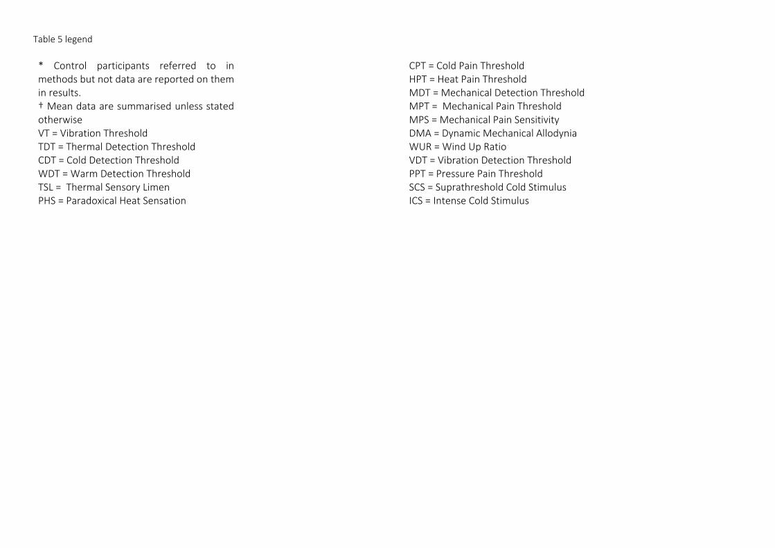
Table 5 legend
* Control participants referred to in
methods but not data are reported on them
in results.
† Mean data are summarised unless stated otherwise
VT = Vibration Threshold
TDT = Thermal Detection Threshold
CDT = Cold Detection Threshold
WDT = Warm Detection Threshold
TSL = Thermal Sensory Limen
PHS = Paradoxical Heat Sensation
CPT = Cold Pain Threshold
HPT = Heat Pain Threshold
MDT = Mechanical Detection Threshold
MPT = Mechanical Pain Threshold
MPS = Mechanical Pain Sensitivity
DMA = Dynamic Mechanical Allodynia
WUR = Wind Up Ratio
VDT = Vibration Detection Threshold
PPT = Pressure Pain Threshold
SCS = Suprathreshold Cold Stimulus
ICS = Intense Cold Stimulus
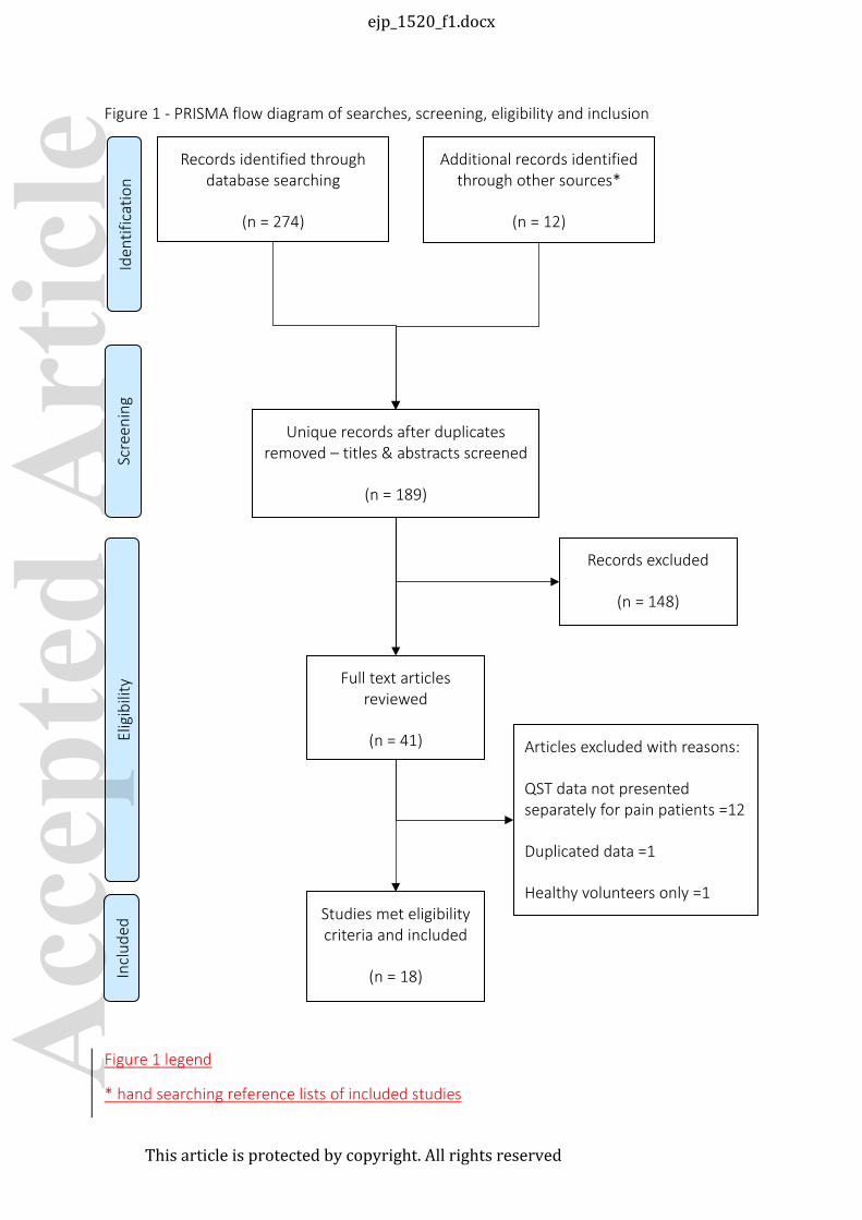
Figure 1 - PRISMA flow diagram of searches, screening, eligibility and inclusion
Figure 1 legend
* hand searching reference lists of included studies
Records identified through
database searching
(n = 274)
Scr
ee
nin
g
Incl
ud
ed
E
lig
ibilit
y
Ide
nti
fica
tio
n
Additional records identified
through other sources*
(n = 12)
Unique records after duplicates
removed – titles & abstracts screened
(n = 189)
Records excluded
(n = 148)
Full text articles
reviewed
(n = 41) Articles excluded with reasons:
QST data not presented
separately for pain patients =12
Duplicated data =1
Healthy volunteers only =1
Studies met eligibility
criteria and included
(n = 18)
ejp_1520_f1.docx
Thisarticleisprotectedbycopyright.Allrightsreserved
Acc
epte
d A
rtic
le


![Quantitative sensory testing in chronic pain patients with ... G et al_Clin J Pain_2016... · somatosensory deficits (NDSD) [2, 3]. NDSD have been reported in various chronic non-neuropathic](https://img.pdfslide.us/doc/110x75/5a89cc0c7f8b9a7f398b6265/quantitative-sensory-testing-in-chronic-pain-patients-with-g-et-alclin-j-pain2016somatosensory.jpg)