Embed Size (px)
Citation preview

Proc. NatL Acad. Sci. USAVol. 80, pp. 594-598, January 1983Neurobiology
Some rat sensory neurons in culture express characteristics ofdifferentiated pain sensory cells
(trigeminal ganglion neurons/capsaicin/bradykdnin/prostaglandin E2/substance P)
PAOLA I. BACCAGLINI AND PATRICK G. HOGANDepartment of Neurobiology, Harvard Medical School, 25 Shattuck Street, Boston, Massachusetts 02115
Communicated by Edwin J. Furshpan, October 8, 1982
ABSTRACT Sensory neurons were dissociated from trigemi-nal ganglia or from dorsal root ganglia of rats, grown in culture,and examined for expression of properties of pain sensory cells.Many sensory neurons in culture are excited by low concentrationsof capsaicin, reportedly a selective stimulus for pain sensory neu-rons. Many are excited by bradykinin, sensitized by prostaglandinE2, or specifically stained by an antiserum against substance P.These experiments provide a basis for the study of pain mecha-nisms in cell culture.
Pain sensory neurons have been identified as a distinct class ofsensory neurons in mammals (1-4). Pain sensory endings areactivated or sensitized by painful mechanical stimulation, bypainful heat, and by compounds that are released locally in dam-aged tissue (1, 5, 6). Bradykinin, prostaglandins, and amines areamong the compounds that have been shown to activate or sen-sitize pain endings and are thought to have a role in the painassociated with injury and inflammation (5, 6).The action of these compounds on sensory endings has been
studied in experimental animals (7-14), but the studies haveencountered technical limitations. A major limitation is that themechanisms that underlie excitation or sensitization ofpain sen-sory endings are not accessible to biophysical measurements.Other limitations are that the concentration of bradykinin, pros-taglandins, or amines at the sensory endings is not accuratelyknown; and that each of these compounds produces inflam-matory changes in the tissue as well as release of other media-tors, so that its actions are not evaluated in isolation. Thesedifficulties would be alleviated if differentiated pain sensoryneurons could be studied in cell culture. This alternative ap-proach would allow more detailed pharmacological, biophysi-cal, and biochemical studies of pain sensory neurons.
As a first step toward the study ofpain mechanisms in culture,we have tested whether some characteristics of pain sensoryneurons are expressed by sensory neurons in culture. We findthat sensory cells grown in the absence of other cells expresssensitivity to capsaicin (8-methyl-N-vanillyl-6-nonenamide), aproperty restricted to unmyelinated pain sensory fibers in adultanimals (15-18), and in addition express other properties (7, 8,10, 11, 19) of differentiated pain sensory neurons.
METHODSCell Culture. The portion of the trigeminal ganglion asso-
ciated with the mandibular nerve was dissected from newbornrats (CD strain; Charles River Breeding Laboratories) and thecells were dissociated by treatment with dispase (grade 2; Boeh-ringer Mannheim) and collagenase (type I; Worthington). Cellswere plated on islands of collagen less than 1 mm in diameter(20) and grown in a modified L-15-CO2 growth medium (21)
from which methocel and bovine serum albumin were omittedand in which glucose, penicillin, and streptomycin concentra-tions were reduced by half. Cultures were treated with 10 tkM1-f3-D-arabinofuranosylcytosine (cytosine arabinoside) duringthe 4 days after plating to minimize growth of non-neuronalcells.
Dorsal root ganglia from all spinal segments, and superiorcervical ganglia, were dissociated and cultures were preparedby a similar procedure.
For histological experiments, sensory neurons were grownon a collagen substrate about 8 mm in diameter.
Electrophysiology. Neurons were studied after growth inculture for 10-35 days. Cultures were placed on the stage ofa phase-contrast microscope and continuously perfused withHepes-buffered medium at 35-37TC. The composition of therecording medium was as described (22), except that bovineserum albumin and NaHCO3 were omitted; 5 mM Hepes, pen-icillin (100 units/ml), and streptomycin (100 tkg/ml) wereadded; and pH was adjusted to 7.4 with NaOH. Conventionalintracellular recording techniques were used.
Responses were displayed simultaneously on an oscilloscopeand on a chart recorder. The action potentials and fast depo-larizing potentials are attenuated in those traces reproducedfrom the chart record.Drug Application. Capsaicin (Sigma) was dried from a stock
solution in ethanol, redissolved at 10 ,4M in recording medium,and then further diluted. Capsaicin was applied from a micro-pipette by opening a solenoid valve connecting the micropipetteto a reservoir of nitrogen at 4 psi gauge pressure (23). To ensurethat capsaicin reached the cell soma and all the cell processes,micropipettes with tip outer diameters of 10-25 t.m were usedand capsaicin was delivered at several positions above the is-land. When responses to different concentrations were com-pared, in each case the capsaicin was applied from a micropi-pette with a tip outer diameter of about 20 lim.
Bradykinin triacetate (Sigma) in recording medium contain-ing 0.1% bovine serum albumin (fraction V, fatty acid-free;Miles) was applied as described for capsaicin. There was no re-sponse when recording medium containing 0.1% bovine serumalbumin was applied in this way.
Prostaglandin E2 (Sigma) was dried from a stock solution inethanol, redissolved at 10 ,uM in recording medium, and thenfurther diluted.
The composition ofthe recording medium containing 50mMK+ was the same as that of normal recording medium, exceptfor equimolar substitution of KCl for NaCl to increase the K+concentration.
Statistical Analysis. The mean number of action potentialselicited by the test stimulus in the presence of prostaglandinE2 was compared with the mean number of action potentialselicited in normal recording medium by using the t statistic ina one-tailed test; P < 0.05 was required for significance.
594
The publication costs ofthis article were defrayed in part by page chargepayment. This article must therefore be hereby marked "advertise-ment" in accordance with 18 U. S. C. §1734 solely to indicate this fact.

Proc. Natd Acad. Sci. USA 80 (1983) 595
Immunologic Staining. Cultures were fixed with 4% para-formaldehyde in 0.1 M sodium phosphate buffer at pH 7.3 andstained by using the unlabeled antibody peroxidase-antiperox-idase method (24, 25). Sera used were rabbit anti-substance P(Rd2 pooled; provided by S. Leeman), goat anti-rabbit IgG(Sternberger-Meyer), and rabbit peroxidase-antiperoxidasecomplex (Sternberger-Meyer). Details of the procedure havebeen described (26).
RESULTSNeurons were dissociated from sensory ganglia and grown oncollagen islands <1 mm in diameter, each island having froma few to a few dozen neurons. Intracellular recordings from theneurons were made with standard microelectrode techniques.Both trigeminal ganglion cells and dorsal root ganglia cells inculture usually were quiescent under our recording conditions.
Capsaicin. To determine whether capsaicin sensitivity wasexpressed in culture, we tested trigeminal ganglion neuronswith capsaicin at concentrations less than 0.1 1M. Capsaicindissolved in recording medium was applied from a micropipetteby a brief pulse of pressure. Many cells responded to capsaicinwith action potentials and a few cells responded with a slowdepolarization (Fig. 1). Frequently the responses also includedfast depolarizing potentials whose amplitudes ranged from <1mV to 30 mV (Fig. lb). Because experiments described below
c
'l
FIG. 1. Responses of three different neurons in cell cultures of rattrigeminal ganglion to application of 30 nM capsaicin. The upper tracein each panel is an intracellular recording from the neuron; the lowertrace monitors application of capsaicin. (a) Brief train of action po-tentials. Twenty days in culture; resting membrane potential, -58mV. (b) Train of action potentials and fast depolarizing potentials. Thecomplete response lasted 17 sec from application of capsaicin. Only theinitial portion is shown. Fifteen days in culture; resting membranepotential, -57 mV. (c) Slow depolarization. Thirteen days in culture;resting membrane potential, -61 mV. Each stimulus pulse in thelower trace of c is 200 msec in duration. Calibration: a and b, 20 mV,0.5 sec; c, 20 mV, 8 sec.
gave no evidence of synaptic interactions in these cultures, itis likely that the fast depolarizing potentials were action poten-tials arising in the processes that failed to propagate into the cellbody. Often the responses to capsaicin were mixtures of actionpotentials and fast depolarizing potentials (Fig. lb) or of actionpotentials, fast depolarizing potentials, and slow depolarization.Our observations are consistent with the presence of a singleclass of responsive cells that depolarize when exposed to lowconcentrations of capsaicin and in which action potentials mayarise as a result of depolarization in the cell body or in the pro-cesses.A high proportion (1,214/1,748) of trigeminal ganglion neu-
rons in culture were excited by 0.1 AM capsaicin. The propor-tion of cells responding would be greater than the proportionof cells sensitive to capsaicin ifsome cells were excited synapti-cally, but two lines of evidence suggest that most of the re-sponses to capsaicin were not synaptic potentials. We found thatthe excitatory responses to capsaicin persisted in recordingmedium in which the Ca2+ concentration was reduced to 0.28mM and the Mg2+ concentration was increased to 10 mM. Wealso tested directly 39 pairs of neurons for synaptic interactionsby eliciting short trains ofaction potentials at 0.5, 5, and 10 Hzin one ofthe cells ofeach pair. Action potentials, fast depolariz-ing potentials, and depolarization were never recorded in thesecond neuron, although 24 ofthese same neurons were excitedby 0.1 ,M capsaicin. Therefore, it seems likely that most ofthecells excited by capsaicin in trigeminal ganglion cultures wereexcited directly.
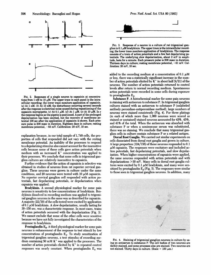
Higher concentrations of capsaicin did not elicit action po-tentials in a larger fraction of the cells. We tested 30 neuronswith increasing concentrations of capsaicin, from 1 nM to 10AM. Eighteen of these cells responded with action potentialsor fast depolarizing potentials, and all 18 responded at concen-trations of 0.1 AM or lower. Depolarization became increasing-ly prominent as the amount ofcapsaicin increased; in almost all(17/18) cells it exceeded 30 mV at high concentrations. A typicalresponse to low concentrations was a burst of action potentialsand fast depolarizing potentials; a typical response to high con-centrations was a burst of action potentials on the rising phaseof a prolonged depolarization (Fig. 2). Two neurons did not re-spond to capsaicin with action potentials but had relatively largeslow depolarizations which increased with the amount of cap-saicin applied. Because depolarizing responses to capsaicinwithout action potentials are common during the first week inculture, these may have been cells that were maturing moreslowly than most of the cells in the cultures.
Cells Insensitive to Capsaicin. The same experiment-test-ing neurons with capsaicin at concentrations from 1 nM to 10MM-also examined the behavior of cells that did not respondwith action potentials to low concentrations ofcapsaicin. Twelveof the 30 cells studied were insensitive by this test. Except forthe two cells already described, which may have been respon-sive to capsaicin but electrically immature, these cells were alsorelatively unresponsive to high concentrations of capsaicin.Three of the cells did not respond at any concentration of cap-saicin, and the other cells had only small slow depolarizing re-sponses which did not increase appreciably with increasing con-centration of capsaicin. All of these cells had depolarizations<4 mVwhen 10,M capsaicin was applied. The insensitive cellswere not desensitized to capsaicin, because the desensitizationwhich may occur after exposure to high concentrations of cap-saicin was avoided in this experiment by testing only one cellin each island.An apparent insensitivity could have resulted from damage
or from the inability of the processes to respond to depolarizingstimuli. Damage by the recording electrode seems an unlikely
Neurobiology: Baccaghni and Hogan
596 Neurobiology: Baccaglini and Hogan
a b d -j
e
FIG. 2. Responses of a single neuron to capsaicin at concentra-tions from 1 nM to 10 ,utM. The upper trace in each panel is the intra-cellular recording; the lower trace monitors application of capsaicin.(a) At 1 nM. (b) At 10 nM; the disturbance occurring several secondsafter the response is electrical noise caused during repositioning of thecapsaicin micropipette. (c) At 0.1 ,uM. (d) At 1 pM. (e) At 10 pM. In ethe response begins as the pipette is positioned. A part of the prolongeddepolarization has been omitted, but the recovery of membrane po-tential 100 sec after the application of capsaicin is shown. Each pres-sure pulse is 200 msec in duration. Eighteen days in culture; restingmembrane potential, -55 mV. Calibration: 20 mV, 10 sec.
explanation because, in our total sample of 1,748 cells, the pro-portion of cells that responded did not vary with the restingmembrane potential. An inability of the processes to respondto a depolarizing stimulus also cannot account for the insensitivecells because some of these cells gave action potentials whenmedium with an increased K+ concentration was applied totheir processes. We conclude that some cells in trigeminal gan-glion cultures are relatively insensitive to capsaicin.
Further evidence that the action ofcapsaicin is selective wasobtained in studies of neurons from rat superior cervical gan-glion. These neurons were grown in culture under the sameconditions, and 20 neurons were tested with 10 ,uM capsaicin.No superior cervical ganglion cell responded with action po-tentials, fast depolarizing potentials, or depolarization whencapsaicin was applied.
Bradykinin. A second physiological marker for some painneurons is sensitivity to low concentrations of bradykinin. Bra-dykinin dissolved in recording medium was applied to trigemi-nal ganglion neurons in the same way as described for capsaicin.A majority (22/39) ofthe cells tested were excited by applicationof 0.1 kuM bradykinin. A slow depolarization, usually lasting for30-150 sec, was a characteristic response. In most cases, trainsof action potentials occurred with the depolarization (Fig. 3).We cannot exclude that some of the other cells were sensitivebecause we have not fully investigated the characteristics of theresponse to bradykinin.
Prostaglandin E2. A third physiological marker for some painneurons is enhancement of the response to test stimuli by lowconcentrations of prostaglandin E2. To study sensitization oftrigeminal ganglion neurons, a test stimulus of recording me-dium containing 50 mM K+ was applied to the processes. Thenumber of action potentials elicited by K+ in repeated controlresponses was nearly constant. When prostaglandin E2 was
FIG. 3. Response of a neuron in a culture of rat trigeminal gan-glion to 0.1 p bradykinin. The upper trace is the intracellular record-ing; the lower trace monitors application of bradykinin. The responseconsists of a train of action potentials and a few fast depolarizing po-tentials. The underlying slow depolarization, about 2 mV in ampli-tude, lasts for a minute. Each pressure pulse is 200 msec in duration.Thirteen days in culture; resting membrane potential, -61 mV. Cal-ibration: 20 mV, 10 sec.
added to the recording medium at a concentration of 0.1 &Mor less, there was a statistically significant increase in the num-ber ofaction potentials elicited by K+ in about half (9/21) of theneurons. The number of action potentials returned to controllevels after return to normal recording medium. Spontaneousaction potentials were recorded in some cells during exposureto prostaglandin E2.
Substance P. A histochemical marker for some pain neuronsis staining with antiserum to substance P. In trigeminal ganglioncultures stained with an antiserum to substance P (unlabeledantibody peroxidase-antiperoxidase method), large numbers ofneurons were stained consistently (Fig. 4). For three platings(in each of which more than 1,000 neurons were scored asstained or unstained) stained neurons accounted for 43%, 45%,and 41% of the total. When the antiserum was absorbed withsubstance P or when a nonimmune serum was substituted,there was no staining. We conclude that many trigeminal gan-glion cells in culture contain substance P or a related antigen.
Dorsal Root Ganglia. We carried out similar experiments oncells dissociated from dorsal root ganglia and grown in culture.A large proportion (124/155) of these neurons responded to 0.1,uM capsaicin. The responses were excitatory and included ac-tion potentials, fast depolarizing potentials, and slow depolar-ization. When higher concentrations of capsaicin were applied,the same neurons responded with action potentials and withdepolarizations >30 mV. Many cells in dorsal root ganglia cul-tures were excited by 0.1 ,M bradykinin, and many were sen-sitized by prostaglandin E2 (Fig. 5). The responses were similarto those seen in trigeminal ganglion neurons. In addition, many
I
FIG. 4. Staining of trigeminal ganglion neurons in culture by us-ing an antiserum to substance P. The cell bodies of two neurons aredarkly stained, and some processes also are stained. Two neurons arenot stained. Thirty days in culture. (Scale = 100 ,tm.)
Proc. Nad Acad. Sci. USA 80 (1983)

Proc. Nati. Acad. Sci. USA 80 (1983) 597
a
AL".
b
iiiiiilllivl I III-lPIi t 1 1
il-iU 1
FIG. 5. Sensitization of a neuron in a culture of rat dorsal rootganglia during application of 0.1 uM prostaglandin E2. The uppertrace in each panel is the intracellular recording; the lower trace mon-itors application of the test stimulus. The test stimulus-recordingmedium containing 50 mM K+-was applied every 5 min during theexperiment. Only representative responses are shown. (a) Control re-sponse before application of prostaglandin E2. (b) Increased responseduring perfusion with medium containing 0.1 ;LM prostaglandin E2.(c) Control response after return to recording medium without pros-taglandin E2. Thirty-three days in culture; resting membrane poten-tial, -51 mV. Calibration: 20 mV, 0.4 sec.
cells in dorsal root ganglia cultures stained with antiserum tosubstance P. Thus, it seems that these sensory ganglion cellsdo not differ from trigeminal ganglion cells in expression of thecharacteristics we studied.
DISCUSSION
The present study shows that many sensory neurons culturedfrom rat trigeminal ganglion and dorsal root ganglia are excitedby low concentrations ofcapsaicin. A distinct smaller populationof cells does not respond to capsaicin. In addition, many cellsare excited by low concentrations of bradykinin, show en-
hanced responsiveness to stimuli in the presence ofprostaglan-din E2, or stain with an antiserum to substance P.
Previous studies have provided evidence that sensitivity tocapsaicin and to bradykinin, sensitization by prostaglandin E,and content of substance P are properties of pain sensory neu-rons. Capsaicin sensitivity is perhaps the most specific markerfor unmyelinated pain sensory fibers. In rat and cat saphenousnerve, capsaicin excites only unmyelinated fibers (15, 16). Cap-saicin applied to the skin excites only unmyelinated pain fibers,and not other unmyelinated fibers, in the rat saphenous nerve
(17). Capsaicin applied to the skin or injected into the saphenousartery acts selectively on unmyelinated pain fibers, and not on
unmyelinated mechanoreceptor fibers, in the cat saphenousnerve (16, 18).An excitatory action of capsaicin on sensory neurons in cul-
ture has not been reported. One report described a hyperpo-larization and a lengthening ofthe action potential plateau -when0.3 mM capsaicin was applied to rat and chicken dorsal rootganglia neurons in culture (27).
In contrast to the physiological studies, studies ofthe toxicityof capsaicin have led to the conclusion that it may not be usefulas a selective marker for pain sensory neurons. When high con-
centrations of capsaicin are administered to newborn animals,there is apermanent decrease in the number ofsensory ganglioncells, an almost complete loss of unmyelinated sensory -fibers,and a loss ofsome myelinated sensory fibers (28, 29). The extentof damage to sensory fibers in these experiments implies thatnot only pain sensory neurons but also sensory neurons withother physiological functions are affected. Thus, both a selectivephysiological action of capsaicin in adult animals and a less se-lective toxicity of capsaicin in newborn animals are clearly es-tablished. The two results can be reconciled if, early in devel-opment, most unmyelinated. sensory fibers are transientlysensitive to capsaicin and perhaps express other pain propertiesor if capsaicin toxicity and excitation by capsaicin involve sep-arate mechanisms.
Bradykinin activates unmyelinated and thinly myelinatedpain fibers in cutaneous nerves of the cat (7). It excites fiberswith similar properties, which are probably pain fibers, in mus-cle nerves of the cat and the dog (8, 9). Myelinated sensory fi-bers from hair follicles, pacinian corpuscles, muscle spindles,and Golgi tendon organs are not appreciably excited by bra-dykinin (7, 8). However, bradykinin is somewhat less selectivethan capsaicin because it activates some myelinated mechano-receptor fibers and some unmyelinated mechanoreceptor fibersin addition to pain fibers (7).
Prostaglandin E2 sensitizes some unmyelinated fibers in catplantar nerve to painful stimulation with. noxious heat, and itsensitizes some unmyelinated fibers in cat muscle nerves topainful stimulation with bradykinin (10, 11). Prostaglandin E1,which is structurally like prostaglandin E2, has similar effects(10). Prostaglandin El also sensitizes some thinly myelinatedfibers in rat saphenous nerve to moderate mechanical stimu-lation (12). Some fibers that are sensitized by prostaglandin Elare slowly adapting mechanoreceptor fibers (12, 13). Thus pros-taglandin E1 and prostaglandin.E2, although important in sen-sitization of pain fibers, are probably not more selective thanbradykinin.
Substance P has been proposed as a marker for some painsensory cells. Substance P or a similar compound is present insome neurons located in the dorsal root ganglia and in the tri-geminal ganglion (30). The processes of these neurons are un-myelinatedand have a distribution similar to that ofpain sensoryfibers in the spinal cord and in the brainstem (30-33). SubstanceP is released in the spinal cord by stimulation of sensory nerves(34, 35), and substance P applied iontophoretically increases theactivity ofneurons in the spinal cord and brainstem that receiveinput from pain sensory neurons (36-38). The evidence thatsubstance P is a transmitter for certain pain sensory neurons hasbeen reviewed (19).
Substance P has been identified previously in chicken dorsalroot ganglia cells in culture (39, 40).
Capsaicin sensitivity and content of substance P have beenproposed as specific markers for unmyelinated pain fibers,-butit is not known whether these markers are expressed togetherin all pain sensory cells. It can be estimated from fiber counts(41) and.from physiological studies (4, 17, 42) that 30-50% ofall sensory fibers in the rat are unmyelinated pain fibers andthat most or. all of these are sensitive to capsaicin (4, 17). Incontrast, only about 20% of sensory neurons stain for substanceP (30). In our cultures the fraction of cells sensitive to capsaicinalso is larger than the.fraction of cells staining for substance P.One interpretation of these results is that some unmyelinatedpain fibers do not contain substance P. Another possible inter-pretation is that all unmyelinated pain fibers contain substanceP but that immunologic staining is not sensitive enough to -de-tect all cells in sensory ganglia which contain substance P. Infavor of the second interpretation, all unmyelinated pain fibers
Neurobiology:. Baccaglini-and Hogan

598 Neurobiology: Baccaglini and Hogan
in the rat saphenous nerve release one or more compounds thatcan produce local inflammatory changes in the surrounding tis-sue (42). Substance P is currently considered the most likelymediator of this neurogenic inflammation (43-45). However,it remains possible that some unmyelinated pain fibers containand release other peptides, either alone or together with sub-stance P.The fraction of cells in culture that are excited by capsaicin
is larger than the fraction of sensory fibers in the rat that areexcited by capsaicin. Likewise, the fraction of cells in culturethat stain with antiserum to substance P is larger than the frac-tion that stain in the animal. This result suggests that a relativelylarge fraction of sensory neurons are able to express pain prop-erties early in development, if it is assumed that the cells sur-viving in culture are a representative sample of the cells in theganglion. An alternative possibility, however, is that pain sen-sory cells survive preferentially in our culture conditions.We set out to determine whether characteristics of pain sen-
sory neurons are expressed in culture. Previous studies indi-cated that capsaicin sensitivity is a marker for unmyelinated painfibers. We have found that many neurons in rat trigeminal gan-glion and dorsal root ganglia cultures are excited by low con-centrations of capsaicin. Furthermore, in common with somepain sensory neurons, many neurons in culture are sensitive tolow concentrations of bradykinin, are sensitized by prostaglan-din E2, or stain with an antiserum to substance P. These sensoryneurons in culture which express characteristics of differen-tiated pain sensory cells are likely to be useful in studying theneuronal mechanisms involved in pain.
We thank Doreen McDowell, Geraldine Spencer, and Allison Doupefor their contributions to this work and many other colleagues for theircomments on the manuscript. We are grateful to Dr. Susan Leemanfor the antiserum that was used in some of these experiments. This re-search was supported by National Institutes ofHealth Grants NS 11576,NS 03273, NS 02253, by Grant-in-Aid 78-964 from the American HeartAssociation, and by funds contributed in part by the American HeartAssociation Massachusetts Affiliate. P.I.B. was supported by the Whit-ney Foundation and by Training Grant T32NS07112.
1. Burgess, P. R. & Perl, E. R. (1973) in Handbook ofSensory Phys-iology, ed. Iggo, A. (Springer, Berlin), Vol. 2, pp. 29-78.
2. Kumazawa, T. & Perl, E. R. (1977)J. Physiol (Paris) 73, 287-304.3. Torebjork, H. E. (1974) Acta Physiol. Scand. 92, 374-390.4. Lynn, B. & Carpenter, S. E. (1982) Brain Res. 238, 29-43.5. Armstrong, D. (1970) in Handbook of Experimental Pharmacol-
ogy, ed. Erdos, E. G. (Springer, Berlin), Vol. 25, pp. 434-481.6. Ferreira, S. H. (1972) Nature (London) New Biol 240, 200-203.7. Beck, P. W. & Handwerker, H. 0. (1974) Pfluegers Arch. 347,
209-222.8. Mense, S. (1977)J. Physiol (London) 267, 75-88.9. Kumazawa, T. & Mizumura, K. (1976) Brain Res. 101, 589-593.
10. Handwerker, H. 0. (1976) in Advances in Pain Research andTherapy, eds. Bonica, J. J. & Albe-Fessard, D. (Raven, NewYork), Vol. 1, pp. 41-45.
11. Mense, S. (1981) Brain Res. 225, 95-105.12. Pateromichelakis, S. & Rood, J. P. (1982) Brain Res. 232, 89-96.13. Chahl, L. A. & Iggo, A. (1977) Br. J. PharmacoL 59, 343-347.14. Fjiallbrant, N. & Iggo, A. (1961) J. PhysioL (London) 156, 578-
590.15. PorszAsz, J. & Jancs6, N. (1959) Acta PhysioL Hung. 16, 299-306.16. SzolcsAnyi, J. (1977)J. PhysioL (Paris) 73, 251-259.17. Kenins, P. (1982) Neurosci. Lett. 29, 83-88.18. Foster, R. W. & Ramage, A. G. (1981) Neuropharmacology 20,
191-198.19. Nicoll, R. A., Schenker, C. & Leeman, S. E. (1980) Annu. Rev.
Neurosci. 3, 227-268.20. Furshpan, E. J., MacLeish, P. R., O'Lague, P. H. & Potter, D.
D. (1976) Proc. NatL Acad. Sci. USA 73, 4225-4229.21. Mains, R. E. & Patterson, P. H. (1973)J. Cell BioL 59, 329-345.22. O'Lague, P. H., Obata, K., Claude, P., Furshpan, E. J. & Pot-
ter, D. D. (1974) Proc. NatL Acad. Sci. USA 71, 3602-3606.23. Choi, D. W., Farb, D. H. & Fischbach, G. D. (1977) Nature
(London) 269, 342-344.24. Sternberger, L. A., Hardy, P. H., Jr., Cuculis, J. J. & Meyer, H.
G. (1970)J. Histochem. Cytochem. 18, 315-333.25. Sternberger, L. A. (1979) Immunocytochemistry (Wiley, New
York), 2nd Ed., pp. 104-169.26. Hogan, P. G. (1982) Dissertation (Harvard University, Cam-
bridge, MA).27. Godfraind, J. M., Jessell, T. M., Kelly, J. S., McBurney, R. N.,
Mudge, A. W. & Yamamoto, M. (1981)J. Physiol. (London) 312,32P-33P (abstr.).
28. Lawson, S. N. & Nickels, S. M. (1980) J. PhysioL (London) 303,12P (abstr.).
29. Nagy, J. I., Hunt, S. P., Iversen, L. L. & Emson, P. C. (1981)Neuroscience 6, 1923-1934.
30. Hbkfelt, T., Kellerth, J.-O., Nilsson, G. & Pernow, B. (1975) Sci-ence 190, 889-890.
31. Takahashi, T. & Otsuka, M. (1975) Brain Res. 87, 1-11.32. Hokfelt, T., Kellerth, J.-O., Nilsson, G. & Pernow, B. (1975)
Brain Res. 100, 235-252.33. Cuello, A. C., Del Fiacco, M. & Paxinos, G. (1978) Brain Res.
152, 499-509.34. Yaksh, T. L., Jessell, T. M., Gamse, R., Mudge, A. W. & Lee-
man, S. E. (1980) Nature (London) 286, 155-157.35. Otsuka, M. & Konishi, S. (1976) Nature (London) 264, 83-84.36. Henry, J. L. (1976) Brain Res. 114, 439-451.'37. Randic, M. & Miletic, V. (1977) Brain Res. 128, 164-169.38. Andersen, R. K., Lund, J. P. & Puil, E. (1978) Can. J. Physiol.
PharnacoL 56, 216-222.39. Mudge, A. W., Leeman, S. E. & Fischbach, G. D. (1979) Proc.
NatL Acad. Sci. USA 76, 526-530.40. Fischbach, G. D., Dunlap, K., Mudge, A. & Leeman, S. (1981)
in Neurosecretion and Brain Peptides, eds. Martin, J. B., Reich-lin, S. & Bick, K. L. (Raven, New York), pp. 175-188.
41. Langford, L. A. & Coggeshall, R. E. (1979)J. Comp. NeuroL 184,193-204.
42. Kenins, P. (1981) Neurosci. Lett. 25, 137-141.43. Lembeck, F. & Holzer, P. (1979) Naunyn-Schmiedebergs Arch.
Pharmakol. 310, 175-183.44. Lembeck, F., Folkers, K. & Donnerer, J. (1981) Biochem. Bio-
phys. Res. Commun. 103, 1318-1321.45. Rosell, S., Olgart, L., Gazelius, B., Panopoulos, P., Folkers, K.
& Horig, J. (1981) Acta PhysioL Scand. 111, 381-382.
Proc. Nad Acad. I-Sci. USA 80 (1983)