Embed Size (px)
Citation preview

TINS-1124; No. of Pages 12
The unsteady eye: an information-processing stage, not a bugMichele Rucci1,2 and Jonathan D. Victor3
1 Department of Psychological and Brain Sciences, Boston University, Boston, MA 02215, USA2 Graduate Program in Neuroscience, Boston University, Boston, MA 02215, USA3 Brain and Mind Research Institute, Weill Cornell Medical College, New York, NY 10065, USA
Opinion
Glossary
Brownian motion: a random-walk process that mimics the jiggling motion of a
particle in a fluid. Brownian motion provides a good approximation for the
inter-saccadic motion of the retinal image.
Diffusion coefficient: a parameter describing the speed of Brownian motion.
The diffusion constant gives the ratio between the expected value of the
square of the distance moved and the time elapsed since the onset of the
motion.
Fixation: the period in between saccades, in which visual information is
acquired and the image on the retina moves relatively little.
Fixational eye movements: small eye movements that incessantly occur during
fixation. They include occasional microsaccades, ocular drift, and tremor.
Fovea centralis (fovea, in brief): a depression in the surface of the retina with
diameter �1.5 mm used for high-acuity vision.
Foveola: the central region of the fovea (�0.2 mm in diameter, approximately
18 in visual angle) without rod photoreceptors and where cones are most
densely packed.
Image (or perceptual, or Troxler) fading: the progressive disappearance of the
visual percept experienced under retinal stabilization.
Microsaccade: a very small saccade, traditionally with amplitude smaller than
300 or less, which keeps the attended stimulus within the foveola.
Minute of arc (or arcmin, or minarc): a measurement unit of angle, corresponding
to 1/60th of 1 degree. It is usually indicated by the prime symbol (’).
Ocular drift: the relatively slow incessant motion of the eye during the inter-
saccadic interval. Here we use this term to also include tremor, a superimposed
very small high-frequency motion.
Power spectrum: a representation of how the power of a random signal is
distributed across the various frequencies composing the signal.
Retinal ganglion cells: the neurons in the output stage of the retina, which relay
information to the thalamus, midbrain, and other central areas.
Retinal stabilization: a laboratory procedure that completely eliminates the
How is space represented in the visual system? At firstglance, the answer to this fundamental questionappears straightforward: spatial information is directlyencoded in the locations of neurons within maps. Thisconcept has long dominated visual neuroscience, lead-ing to mainstream theories of how neurons encodeinformation. However, an accumulation of evidenceindicates that this purely spatial view is incompleteand that, even for static images, the representation isfundamentally spatiotemporal. The evidence for thisnew understanding centers on recent experimentalfindings concerning the functional role of fixationaleye movements, the tiny movements humans and otherspecies continually perform, even when attending toa single point. We review some of these findings anddiscuss their functional implications.
The unsteady eyeSensory perception and motor behavior are closely cou-pled. Most species are not passively exposed to the incom-ing flow of sensory data, but actively seek usefulinformation by coordinating sensory processing with mo-tor activity. A clear example of this interaction is given bythe role of eye movements in visual perception. In humans,as in many other species, acuity is not uniform throughoutthe visual field, but rapidly declines with increasing dis-tance from the foveola (see Glossary), the small rod-freeregion of the retina covering a visual area the size of thefull moon in the sky. As a consequence, humans acquirevisual information during brief periods of ‘fixation’ sepa-rated by saccades, rapid gaze-shifts that enable inspectionof the objects of interest with the high-acuity region(Figure 1A).
This coupling between visual sensation and eye move-ments goes beyond the fixational sequence enabled bysaccades. Close examination of gaze position in the inter-vals in between saccades reveals that the term ‘fixation’ ismisleading as the eyes are never at rest. Tiny eye move-ments – known as fixational eye movements – incessantlyoccur during the periods between saccades (Figure 1B).These movements are often labeled as microscopic, but
0166-2236/
� 2015 Published by Elsevier Ltd. http://dx.doi.org/10.1016/j.tins.2015.01.005
Corresponding author: Rucci, M. ([email protected]).Keywords: vision; eye movements; retina; neural encoding; ocular drift; microsaccades.
they shift the projection of the stimulus over many recep-tors on the retina. It is remarkable that we are normallynot aware of them because they yield motion signalswith speeds that would be immediately visible had theyoriginated from objects in the scene rather than from ourown eyes [1].
Fixational eye movements have been observed in a widevariety of species [2] including the owl [3], a predatorcommonly believed not to move its eyes. However, theyare often ignored by theoreticians and are regarded as anuisance by experimentalists. When they are taken intoaccount, they are frequently regarded as a problem thatthe visual system has to overcome to establish fine spatialrepresentations [4] and to avoid perceptual blurring of theimage [5].
physiological motion of the retinal image.
Saccade: a very rapid eye movement normally used to bring the retinal
projection of the object of interest onto the high-acuity fovea. Saccades
typically occur 2–3 times per second.
Whitening: a signal-processing operation that equalizes the power across all
frequencies. After whitening a signal, its power spectrum is flat. This process
removes pairwise correlations in the signal.
Trends in Neurosciences xx (2015) 1–12 1

Normal head/eye movements
Fixa�on
Saccade
0 20 40 600
50
100
150
200
250–2
–4
–6
–8
–10
Time (ms)Po
si�on
(arc
min
)
0 200 400 600 800 1000
–5
0
5
HorizontalKey:Ver�cal
Instantaneous speed (arcmin/s)
Prob
abili
ty
15 30 45 60 75 900
0.1
0.2
(A)
(F) (G) (H)
(B) (C)
(D)
(E)
0 20 40 600
50
100
150
200
250
Tim
e (m
s)
0 20 40 60
−8
−7
−6
−5
−4
−3
−2
Space (arcmin) Space (arcmin)
Tim
e (m
s)
Length (arcmin)
Prob
abili
ty
0 5 10 15 20 25 30 35 400
0.1
0.2
0.3
Pow
er (d
B)
10 100
−40
−30
−20
−10
0
10
Frequency (Hz)
Ocular dri� spectrum
Ocular dri� speed distribu�on
Head-fixed viewing
Inter-saccadic distance traveled
Head movements ignored
Log probability
Log probability
TRENDS in Neurosciences
Figure 1. Vision and eye movements. (A) As an observer looks at a static scene, rapid eye movements (saccades) separate the periods of ‘fixation’ in which visual
information is acquired. (B) High-resolution recording of oculomotor activity reveals that the eye moves also during these periods. The line of sight continually wanders
with a seemingly random trajectory (ocular drift) occasionally interrupted by saccades with small amplitudes (microsaccades; arrow). (C�F) Characteristics of ocular drift
when the head is immobilized, a standard practice to measure small eye movements. Subjects freely observed natural scenes [27]. (C) Power spectrum. The dashed line
represents the eye-tracker noise level. (D) Distribution of mean instantaneous speed. (E) Length of the inter-saccadic trajectory. (F) Probability (in natural logarithmic scale)
that the retinal image shifted by a given distance (horizontal scale) after a given time (vertical scale). (G) As in (F), but during normal head-free viewing [28]. (H) The same
data as in (G), but with head movements artificially eliminated in the reconstruction process. More accurate reconstructions of retinal image motion that also consider the
optics of the eye are given in [28].
Opinion Trends in Neurosciences xxx xxxx, Vol. xxx, No. x
TINS-1124; No. of Pages 12
All eye movements transform a static scene into a spa-tiotemporal input signal to the retina. This article buildsupon the proposal that this transformation constitutes afundamental step for visual perception: the visual systemtakes advantage of the resulting temporal modulations toencode spatial information in the joint space–time domain.Within this context, we focus here on the role of fixationaleye movements, but similar principles and considerationsextend to other types of eye movements, saccades in partic-ular. Furthermore, we restrict our focus to the consequencesof fixational eye movements for visual encoding and onlymarginally touch upon decoding mechanisms. For largermovements, a vast body of evidence indicates that oculomo-tor signals are taken into account in the interpretation of the
2
retinal input [6,7], and similar strategies may also hold atthe scale of fixational eye movements.
What is the function of fixational eye movements?
As with most scientific questions, several types of answersare available for why the eyes move incessantly, some moreinformative than others. Consider, for example, an unre-lated fundamental question: ‘what is the function ofbreathing?’. A possible answer could be that breathingprevents death by suffocation, but few scientists wouldfind this superficial level of explanation satisfactory. In-deed, if we had stopped at this level, we would have neverlearned that breathing oxygenates the blood and aboutthe associated chains of events. However, as explained

Opinion Trends in Neurosciences xxx xxxx, Vol. xxx, No. x
TINS-1124; No. of Pages 12
below, a similar plain answer seems to have satisfied visionresearchers for over half a century.
In the 1950s it became possible for the first time tocounteract the physiological motion of the eye to immobi-lize the image on the retina, a laboratory procedure knownas retinal stabilization. The outcome was striking: theimage would progressively lose contrast and eventuallyfade away [8,9]. Similar results were observed in laterstudies in which the eye muscles were paralyzed andsubjects even underwent total paralysis [10]. These find-ings have provided an explanation for the existence offixational eye movements that has survived to this day.Fixational eye movements serve the purpose of ‘refreshing’neuronal responses so to prevent the fading of a stationaryscene experienced when all motion is eliminated on theretina. Although popular among scientists and often citedin textbooks of vision, this answer is dangerously simplis-tic. It only provides an empirical description of the con-sequences of eliminating retinal image motion. It does notexplain why vision stops functioning nor identifies themechanisms by which fixational eye movements makevisual perception possible.
Although less well-known to most neuroscientists,theories at deeper levels of explanation have circulatedfor almost a century. Because the luminance modulationsresulting from fixational eye movements contain spatialinformation at individual retinal locations, it has longbeen postulated that fixational eye movements may be acrucial component of a dynamic strategy for encodingspace, an approach that converts spatial informationinto temporal structure [11–18]. While these theoriesdiffer in the specific mechanisms they propose, theyshare the common hypothesis that fixational instabilityplays a central role in ‘structuring’ – rather than merelyrefreshing – neural activity for establishing spatial repre-sentations.
Early theories lost momentum after experiments thateliminated retinal image motion found little or nochange in visual acuity [19,20]. However, these pioneer-ing experiments also suffered from several technologicaland methodological limitations which cast serious doubtson extrapolating their conclusions to more natural view-ing conditions [17,21,22]. Furthermore, the proposal ofan involvement of fixational eye movements in spatialperception has recently found new support from multiplesources including neurophysiological [16,23,24] and be-havioral investigations [22,25,26], statistical examina-tions of retinal input signals [27,28], and theoreticalanalyses of the impact of a continually moving retinalinput on neural responses [15,17,18,29,30].
In this article we examine the perceptual, computation-al, and neural consequences of using fixational eye move-ments to represent space through time and review themounting evidence supporting this idea. This emergingbody of evidence indicates that fixational eye movementsconstitute a crucial information-processing stage, not abug, and challenges established views of early visual pro-cessing at a fundamental level. Again, we remind thereader that many of these ideas also apply to larger eyemovements. Although saccades generally serve to bringthe high-acuity foveola onto the focus of attention, in doing
so they generate transients in the visual input to theretina, thereby also converting purely spatial informationinto a spatiotemporal format.
Types of fixational eye movementsThat the eye continues to move even during visual ‘fixation’has been known for a long time. One of the first scientificreports goes back to 1786 when Robert Darwin (CharlesDarwin’s father) noticed that color after-effects appear tomove because of fixational instability [31]. It is now knownthat fixational eye movements come in different varieties[2,31,32] (Figure 1B).
Most studies have focused on microsaccades – miniaturereplicas of the rapid movements by which humans volun-tarily shift gaze – which, among fixational eye movements,are the easiest to detect and measure. It has long beensuggested that microsaccades serve a special role in pre-venting perceptual fading [8,33], but this popular hypoth-esis has remained controversial among researchers[1,31,32,34–36], and visual impairments similar to thoseoccurring with larger saccades have been observed at thetime of microsaccades [37–40]. Recent studies have shownthat when observers are not required to maintain fixation –an unnatural laboratory condition – but are left free tonormally move their eyes, microsaccades precisely shiftgaze toward nearby interesting locations [41]. This strate-gy appears to take advantage of a very small preferredretinal locus of fixation that enhances performance in high-acuity tasks [7]. Together with a substantial body ofemerging evidence [23,39,40,42–45], these results suggestthat little difference exists between microsaccades andlarger saccades in terms of both control and function(see [46] for recent review).
In this article we focus on the motion of the eye in theperiods between saccades and microsaccades. The dynam-ics of this activity are well delineated by spectral analysis(Figure 1C) and can be subdivided into two main compo-nents. One component, which corresponds to a slow,meandering motion, occupies the frequency range from0 to 40 Hz, and is most prominent at low temporal frequen-cies. A second component, with a smaller amplitude and aspectral peak in the range 40–100 Hz, is known as oculartremor. We consider these components together and usethe term ocular drift (or, for brevity, ‘drift’) to refer to theinter-saccadic motion of the eye.
The eye is traditionally believed to move very little andat very low velocity in the periods between saccades.Classical studies typically reported amplitude valuesranging from about 1.50 to 40 (the prime symbol 0 representsminutes of arc), with median velocities around 40/s[8]. However, these numbers were measured from highlyexperienced observers as they attempted to maintainsteady fixation while moving as little as possible. Theyalso represent estimates obtained over relatively longintervals – typically, the average amplitude of the overalleye displacement between two successive saccades[8]. However, the eye moves more during normal inter-saccadic fixation (especially immediately after a saccade),and changes direction very frequently, such that param-eters measured in this way severely underestimate thereal drift displacement and speed. Figure 1D shows the
3

Opinion Trends in Neurosciences xxx xxxx, Vol. xxx, No. x
TINS-1124; No. of Pages 12
mean distribution of the instantaneous drift speed assubjects freely examined natural scenes. The resultingspeed (mean �500/s) is more than one order of magnitudelarger than previous coarse estimates reported in theliterature, covering a considerable length in visual space(Figure 1E).
Studies that examined ocular drift with the subject’shead immobilized – a standard procedure for resolvingvery small eye movements – have long observed that oculardrift appears to move in an erratic fashion [31,32,47].Figure 1F shows the probability distribution that theeye moves by any given amount in any given interval.The gaze position becomes progressively more dispersedas time goes by, and the variance of the spatial distributionincreases approximately linearly with time, a behaviorthat is characteristic of Brownian motion. Because thesubject’s head was immobilized, this distribution alsoapproximates the motion on the retina of a fixated point.A consequence of this behavior is that the standard devia-tion of the eye position increases at speed slower thanlinear (only as Ht), and the target’s projection remainswithin a relatively narrow retinal region during the natu-rally brief periods of intersaccadic fixation.
The notion that ocular drift resembles Brownian motionshould not be taken to imply a lack of oculomotor control.This motion could still be centrally generated, and controlcould be exerted in several ways; for example, by changingthe diffusion coefficient, the parameter that regulates thespeed of the Brownian process. Indeed, it has long beenknown that: (i) ocular drift becomes much faster (by afactor of four and more) when the subject no longerclenches on a bite-bar; and (ii) drift seems to partiallycompensate for tiny head movements that occur duringfixation, thus revealing a form of control [28].
Figure 1G shows the probability distribution of themotion on the retina of a fixated target during normalhead-free fixation. These data were obtained from onesubject by means of the revolving field monitor [34], toour knowledge the only eye-tracker with demonstratedprecision for resolving ocular drift during normal headmovements. Note the similarity between the distributionsin Figures 1F and G. This similarity is striking given thatdrift speed is significantly higher and that fixational headmovements also contribute to retinal image motion in G.For comparison, Figure 1H shows the distribution thatwould be obtained by ocular drift alone – in other words,without considering head movements in the reconstructionof retinal image motion. These data indicate that oculardrift is under motor control and is designed to yield retinalimage motion with specific characteristics. Furthermore,the compensation for head movements suggests that onesource of this control is the vestibulo-ocular reflex [48,49].
Transforming space into timeThe idea that the temporal modulations resulting from eyemovements might do more than simply ‘refresh’ theresponses of retinal neurons follows immediately fromexamination of the visual input impinging onto retinalreceptors during natural fixation. We will explain the mainideas by considering the visual input signals experiencedby a linear array of retinal receptors – the visual world of
4
the inhabitants of Abbott’s Flatland (Figure 2) – but ourconsiderations are general and directly extend to the 2Dvisual space.
In natural scenes, most objects are stationary. In theabsence of eye movements and any other motor activity,the visual signals from these objects would change little onthe retina: each retinal receptor would continue to beexposed to a similar level of luminance, as illustrated inthe top panel of Figure 2B. This visual input signal con-tains a great deal of information at low temporal frequen-cies. However, neurons in the retina and the early visualsystem are relatively insensitive to an unchanging input;they preferentially respond to a much higher band oftemporal frequencies, above 1–2 Hz. Thus, it is not sur-prising that the visual system functions poorly when allmotion signals are eliminated on the retina, as approxi-mated by the condition of retinal stabilization.
Luckily, the scenario of Figure 2B does not occur in reallife. Even during fixation of a stationary scene, the physio-logical motion of the eye causes retinal receptors to expe-rience continually varying input signals (Figure 2B,bottom panel). The resulting temporal modulations redis-tribute spatial information into the temporal domain. Asexplained below, the specific temporal frequencies result-ing from this motion depend on both the objects beingobserved and the characteristics of the eye movements.Regardless of these specifics, a first important consequenceof eye movements is to shift the low temporal frequencypower of a static scene into a range that the retina cansignal sensitively.
Crucially, this redistribution of input power to highertemporal frequencies cannot be regarded as a simple ‘re-freshing’ of the image. The amplitude of the fixationalmodulations is the results of an interaction between howthe eye moves and the spatial characteristics of the scene.This redistribution is a linear process in space, and we cantherefore analyze it by examining the effects on individualsinusoidal components. Figure 2C shows an example of themodulations given by the same drift trajectory when look-ing at stimuli at three different spatial frequencies. Themagnitude of the temporal modulations resulting frommoving the retina over an image amplifies high spatialfrequencies more than low spatial frequencies. An intuitiveunderstanding of why this happens can be gained byconsidering the luminance change experienced by a retinalreceptor during an infinitesimally brief interval: in thisperiod, ocular drift can be regarded as uniform motion (aconstant-speed translation), and the amplitude of themodulation is determined by the spatial gradient of image.For a sinusoidal pattern of luminance, such as those shownin Figure 2C, the gradient is proportional to the spatialfrequency of the stimulus.
For longer periods of time, ocular drift can no longer beapproximated by uniform motion, and its Brownian-likecharacter needs to be taken into account. The main impli-cation is that amplification will only occur at spatial fre-quencies for which drift covers a small fraction of theperiod. For sufficiently high spatial frequencies – for whichthe eyes move over a larger fraction of the period – therewill instead be attenuation. Figure 2C shows the actualspatial-frequency amplification resulting from ocular drift
Space
Re�n
al sp
ace
Space
Time Time
Inpu
t lum
inan
ce
(A)
(C) (D)
(E) (F)
(B)
Pow
er
Spa�al frequency (cycles/degree)1 10 20 30
1 10
–50
–30
–10
Spec
tral
den
sity
(dB)
No mo�on
Normal dri�Key:Brownian mo�on
Time
Inpu
t lum
inan
ce
Time
Inpu
t lum
inan
ce
SpaceSpa�al frequency (cycles/degree)
No dri�
Normal dri�
Visual scene Re�nal input
Visual scene Re�nal input
Re�nal input amplifica�on
Re�nal input power spectrum
TRENDS in Neurosciences
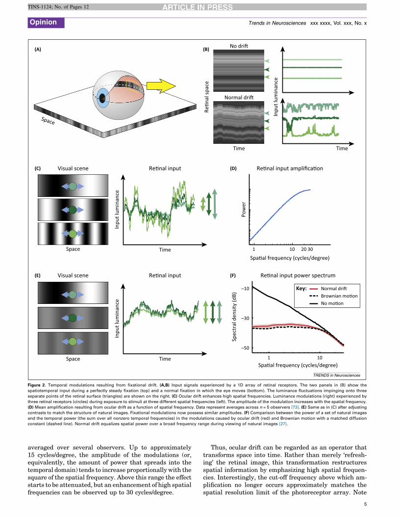
Figure 2. Temporal modulations resulting from fixational drift. (A,B) Input signals experienced by a 1D array of retinal receptors. The two panels in (B) show the
spatiotemporal input during a perfectly steady fixation (top) and a normal fixation in which the eye moves (bottom). The luminance fluctuations impinging onto three
separate points of the retinal surface (triangles) are shown on the right. (C) Ocular drift enhances high spatial frequencies. Luminance modulations (right) experienced by
three retinal receptors (circles) during exposure to stimuli at three different spatial frequencies (left). The amplitude of the modulation increases with the spatial frequency.
(D) Mean amplification resulting from ocular drift as a function of spatial frequency. Data represent averages across n = 5 observers [73]. (E) Same as in (C) after adjusting
contrasts to match the structure of natural images. Fixational modulations now possess similar amplitudes. (F) Comparison between the power of a set of natural images
and the temporal power (the sum over all nonzero temporal frequencies) in the modulations caused by ocular drift (red) and Brownian motion with a matched diffusion
constant (dashed line). Normal drift equalizes spatial power over a broad frequency range during viewing of natural images [27].
Opinion Trends in Neurosciences xxx xxxx, Vol. xxx, No. x
TINS-1124; No. of Pages 12
averaged over several observers. Up to approximately15 cycles/degree, the amplitude of the modulations (or,equivalently, the amount of power that spreads into thetemporal domain) tends to increase proportionally with thesquare of the spatial frequency. Above this range the effectstarts to be attenuated, but an enhancement of high spatialfrequencies can be observed up to 30 cycles/degree.
Thus, ocular drift can be regarded as an operator thattransforms space into time. Rather than merely ‘refresh-ing’ the retinal image, this transformation restructuresspatial information by emphasizing high spatial frequen-cies. Interestingly, the cut-off frequency above which am-plification no longer occurs approximately matches thespatial resolution limit of the photoreceptor array. Note
5
Opinion Trends in Neurosciences xxx xxxx, Vol. xxx, No. x
TINS-1124; No. of Pages 12
that our analysis applies to the temporally fluctuatingcomponents of the signals incident on the retina, not tothe mean level. This is in keeping with the overall dynam-ics of retinal processing from photoreceptor to ganglioncell: typical ganglion cells are severalfold more sensitive tochanges in temporal contrast than to the average DCluminance.
The enhancement of high spatial frequencies shown inFigure 2D may appear to contrast with the small effectsreported by classical retinal stabilization studies with briefstimulus exposures [19,20]. However, as observed above,these studies suffered from multiple limitations whichprevented analysis of the consequences of normal fixa-tional instability. Note that imperfect stabilization (i.e.,partial elimination of retinal image motion) leads to asmaller amplification in Figure 2D, but one that extendsto higher spatial frequencies, complicating interpretationof experimental results. Furthermore, to investigate tem-poral mechanisms of spatial encoding, special attentionneeds to be paid to the transients introduced merely by theonset and offset of the stimulus. The data summarized inBox 1 provide evidence that the human visual system issensitive to drift modulations and that the functional effectof this sensitivity is as predicted above.
Interactions with natural images
Natural scenes do not yield random images on the retina.Statistical regularities are present at multiple scales innatural environments, and it has long been argued thatsensory systems are tuned to these regularities and exploitthem in establishing neural representations [50–54]. As wedescribe below, this tuning begins even before neuralprocessing.
One of the most evident characteristics of a natural imageis its very specific spectral distribution: the power spectrumof natural scenes declines with spatial frequency in a waythat is approximately proportional to the square of thespatial frequency [55] (Figure 2F). A considerable amountof work has focused on the consequences of this inputspectrum on early visual representations [51,56]. However,because the eye is always in motion, a full account of theimpact of this statistical regularity inevitably needs to takeinto consideration not only the image but also the actualspatiotemporal input resulting from the interaction be-tween the image and eye movements. Based on our previousdiscussion, we can expect that oculomotor activity willtransform a stationary natural scene into temporal modula-tions on the retina, effectively redistributing the power ofthe image into the joint space–time domain.
Figure 2E analyzes a fixation in the ‘natural world’. Thesame three stimuli of Figure 2C (three sinusoids at differ-ent spatial frequencies) are again observed by a driftingeye, but their contrasts are now adjusted according to thepower spectrum of natural scenes. The well-known spec-tral distribution of a natural image [55] implies that thecontrast of each Fourier component decreases as its fre-quency increases. For this reason, the high-frequencystimulus in Figure 2E is displayed at a lower contrastthan the low-frequency stimulus. This attenuation of con-trast affects fixational modulations in a direction oppositeto the effect described in Figure 2C,D. Whereas natural
6
images emphasize low spatial frequencies, the motion ofthe eye enhances high spatial frequencies. Remarkably,the two effects counterbalance each other. As illustrated inFigure 2E, after scaling contrast to replicate naturalimages, looking at the three frequency components withthe same drift trajectory gives modulations of approxi-mately equal amplitudes.
The net effect of the interaction between normal inter-saccadic eye drift and natural images is summarized inFigure 2F. This graph compares the power spectrum of aset of images to the average temporal power made avail-able by ocular drift in the form of modulations, as humansfreely looked at them. As shown by these data, fixationalinstability yields temporal modulations with uniform spec-tral density over a broad range of spatial frequencies[27]. Very similar results were also obtained duringhead-free viewing, a condition in which eye and headmovements combine to form a retinal stimulus with almostidentical characteristics [28]. Thus, the signals impingingonto retinal receptors differ sharply from the images pre-sented on the display. However, behavioral and neurophys-iological investigations commonly take these images as theinput to the visual system.
In sum, ocular drift causes a very specific spatiotempo-ral reformatting of the retinal input when humans look atnatural scenes. Within the range of peak temporal sensi-tivity of retinal neurons, the normal fixational fluctuationsof luminance possess equalized power across spatial fre-quencies. This equalization of power – a transformationknown as ‘spectral whitening’ – depends on three factors:the spectral density of natural scenes, the statistics ofnormal ocular drift, and the temporally bandpass natureof retinal processing from photoreceptor to ganglion cell. Itthus reveals a form of matching between the characteris-tics of the natural world, normal eye movements, andretinal network dynamics.
Consequences for neural encodingBecause the fixational modulations of luminance covertemporal frequencies within the range of peak sensitivityof neurons in the retina and thalamus [57], they are likelyto profoundly influence neural responses. Many neurons inthe visual system are primarily sensitive to time-varyingstimuli. During fixation on a natural scene, these neuronswill effectively be driven by the equalized signal inFigure 2F. Other neurons with more sustained responsesmay also be sensitive to the average pattern of luminancecovered by their receptive fields – in other words, what isleft of the low-frequency power of the image at 0 Hz. Inthese neurons, eye movements are expected to inducephasic modulations superimposed on a tonic level of re-sponse. In both cases, the resulting synchronous modula-tions of activity are likely to elicit strong responses indownstream neurons in the cortex [58,59].
What are the implications of the fixational input refor-matting for the mechanisms of neural encoding? The firstconsideration is that, although this spatiotemporal trans-formation is linear in space, it is not linear in time. Eyemovements create power at temporal frequencies whichare not present in the external image: even a static visualenvironment results in temporal modulations of light on

Box 1. Are humans sensitive to fixational modulations?
The spatiotemporal reformatting resulting from ocular drift
(Figure 2D) suggests that fixational eye movements enhance vision
at high spatial frequencies. To better examine this prediction we
developed a new method of retinal stabilization that enables selective
isolation of the motion of the retinal image present during natural
fixation. This method relies on a hardware/software system specifi-
cally designed to process eye-movement signals in real time and
update the display according to the specifications of the experimenter
[74]. By moving the stimulus on the monitor to compensate for the
subject’s eye movements (Figure IA), this system accurately stabilizes
the stimulus on the retina with video-frame resolution up to 200 Hz,
while offering a degree of experimental flexibility far greater than that
of previous methods.
Figure I shows the results of an experiment in which we examined
the consequences of eliminating fixational modulations of luminance
during examination of fine spatial detail. In this task, subjects
reported whether a noisy grating displayed after a saccade was tilted
by 458 clockwise or counter-clockwise. Based on the way ocular drift
transforms spatial information into temporal modulations, we
examined the effect of stabilizing two different types of stimuli: one
in which the target (the grating) was at a higher spatial frequency than
the noise, and one in which the target was at a lower spatial
frequency. These two stimuli yield different predictions: fixational
modulations should enhance visibility of the high spatial-frequency
target, but not the low one.
Figure IC compares the average percentages of correct discrimina-
tion measured in the presence of the normal fixational modulations
to those reported when they were stabilized on the retina by
counteracting the effects of eye movements. Results confirm the
predictions of Figure 2D: eliminating retinal image motion drastically
impaired discrimination of high-frequency gratings, but had little
effect on the low spatial-frequency gratings. These findings conflict
with traditional views of the influence of fixational eye movements
on vision. The fading-prevention hypothesis would have predicted a
stronger impact of retinal stabilization with the low-frequency
gratings because low spatial frequencies are the range in which
fading is most pronounced (i.e., contrast sensitivity is most
attenuated) when stimuli are presented for unnaturally long periods
of time [1,22].
The bottom row of Figure I shows results from a separate
experiment in which retinal stabilization was restricted to a single
axis. In this experiment we selectively compensated for eye
movements on a given axis, either parallel or orthogonal to the
grating, leaving normal motion on the perpendicular axis
(Figure ID). In this way, fixational modulations were only driven
by the pattern of noise during motion parallel to the grating, but
provided information about the grating when motion occurred on
the orthogonal axis. Performance reflected the information content
of fixational modulations. Discrimination was impaired when retinal
image motion was restricted to the axis parallel to the grating but
was instead normal when motion occurred on the orthogonal axis
(Figure IE). These results support the proposal that humans take
advantage of the input luminance modulations caused by fixational
eye movements.
High fr.
Low fr.
85
80
75
70
65
80
6050
70
60
High
freq
uenc
yLo
w fr
eque
ncy
(A)
(D) (E)
(B) (C)
Normal Stabilized
Perc
enta
ge c
orre
ct
Parallel Orthogonal
∗∗
∗
∗∗
∗
∗∗
∗
∗∗
∗
Para
llel
Ort
hogo
nal
Time
Subject 2
Normal Orth. Par.
S�muli
Par�al stabiliza�on Subject 1
Stab.Normal Orth. Par.Stab.
TRENDS in Neurosciences
Figure I. Consequences of eliminating fixational modulations of luminance. (A) A modern retinal stabilization method. The position of the stimulus on a fast display is
continually updated according to the subject’s eye movements so as to eliminate retinal image motion. (B�E) Results of experiments in which subjects judged the
orientation (�458) of gratings embedded within noise fields with naturalistic spectral distributions [22]. (B) The spatial frequency of the grating was either higher or
lower than the band of the noise. (C) Comparison of performance during normal fixational instability and under retinal stabilization. To isolate the normal inter-saccadic
motion of the eye, stimuli were displayed at the onset of fixation after the subject performed a saccade toward a randomly cued location. Removal of fixational
modulations via retinal stabilization selectively impaired high spatial-frequency vision. Error bars represent 95% confidence intervals; fr., frequency. (D) Partial
stabilization restricting movement of the stimulus to a single axis, either parallel or orthogonal to the grating. Fixational modulations experienced by a retinal receptor
(circle) convey information about the grating when motion is restricted to the orthogonal, but not the parallel, axis. (E) Mean percentages of correct discrimination �SEM for two subjects. High-frequency discrimination is impaired when motion is restricted to the axis parallel to the grating and normal when motion is restricted to the
orthogonal axis. (*) and (**) indicate significant differences from complete retinal stabilization (Stab.) and from normal retinal image motion (Normal), respectively
(P <0.05; one-tailed z-tests). Abbreviations: Orth., orthogonal; Par., parallel.
Opinion Trends in Neurosciences xxx xxxx, Vol. xxx, No. x
TINS-1124; No. of Pages 12
7

Opinion Trends in Neurosciences xxx xxxx, Vol. xxx, No. x
TINS-1124; No. of Pages 12
the retina – simply because the retina moves. In thetemporal domain, this nonlinearity enables a match be-tween the low temporal frequency power of natural scenesand the higher-frequency sensitivity of retinal neurons. Inspace, however, eye movements can be regarded as afiltering stage. Because of the spatial processing describedin Figure 2D, presentation of visual stimulation in theabsence of eye movements is likely to misgauge neuronalsensitivity during natural vision. As shown in Figure 3A,B,during normally active fixation, models of retinal ganglioncells respond less to low spatial frequencies and more tohigh spatial frequencies than their contrast sensitivityfunctions measured with immobile retinas would suggest.
0 50 100 Tim
1 10
–10
0
Cont
rast
sens
i�vi
ty (d
B)
Cont
rast
sens
i�vi
ty (d
B)Co
ntra
st se
nsi�
vity
(dB)
Spa�al frequency (cycles/degree)
B1
B2
B3
A3
A2
A1
PassiveKey:Ac�ve
(A) (B)
(D(C)
(E) (F)
Key: B1 B2
Neu
ral r
espo
nses
Parvocellular ganglion cell
Normal dri�
Key: A1 A
Figure 3. Predicted consequences of normal active fixation for retinal responses. (A,B
ganglion cells. The contrast sensitivity functions of model neurons were measured w
replicate experimental data collected with immobile retinas [57,75]. (C�G) Modeling the
fields of an array of ON-center parvocellular neurons (six cells shown here; circles) mov
linear filters with the spatial and temporal contrast sensitivities of a cell recorded in th
moment in time. The mean instantaneous firing rate of every simulated neuron is plotte
(F) Time-course of the responses of the six neurons shown in (C). (G) Same as in (E) d
8
Further important consequences for neural representa-tions follow from the interaction between eye movementsand natural images shown in Figure 2F. It has long beensuggested that sensory systems remove what is predictablefrom the general statistics of the environment (i.e., redun-dant) so that they can focus their resources on what isactually informative [50,60], a strategy that enables effi-cient transmission of visual information. The center–sur-round organization of the receptive fields of ganglion cell iscommonly held to be responsible for discarding redundan-cy [61,62] and eliminating the strong broad correlationspresent in natural scenes [56]. However, experimentalevidence in support of these theories has been scarce,
1 10
150 200 250
e (s)
–10
0
–10
0
–10
0
1 10
Spa�al frequency (cycles/degree)
1 10Spa�al frequency (cycles/degree)
Temporal frequency (cycles/degree)
)
(G)
B3
Magnocellular ganglion cell
Model sensi�vity profiles
No dri�
PassiveKey:Ac�ve
2 A3
TRENDS in Neurosciences
) Changes in the spatial sensitivities of parvocellular (A) and magnocellular (B)
ith (Active) and without fixational eye movements (Passive). The latter functions
responses of retinal ganglion cells during natural stimulation [27]. (C) The receptive
ed according to recorded traces of ocular drift (arrow). (D) Response models were
e macaque [76]. (E) Greyscale representation of activity in the cell array at a given
d at the center location of the cell’s receptive field. Note the enhancement of edges.
uring static presentation of the image without fixational eye movements.

Opinion Trends in Neurosciences xxx xxxx, Vol. xxx, No. x
TINS-1124; No. of Pages 12
and broad correlations have been reported with presenta-tion of natural images in the absence of eye movements[63,64].
These previous proposals did not take into account theincessant motion of the retinal image and face a funda-mental problem raised by the fixational reformatting of thevisual input. Because the power spectrum is, by definition,the Fourier transform of the autocorrelation function, anequalization of power across spatial frequencies is equiva-lent to a removal of correlations in space (the autocorrela-tion becomes localized to a point). In other words, thespectral distribution in Figure 2F implies that pairs ofretinal receptors will experience uncorrelated fluctuationsin luminance during a normally active fixation. That is,because of eye movements, the decorrelation believed to bethe result of retinal circuitry has already occurred evenbefore light is transduced into chemical signals.
If decorrelation is already accomplished by eye move-ments, the subsequent spatial filtering carried out by theretinal network must have some other function. To inferthis function, we first recall the reason why decorrelation isuseful for efficient coding: under specific constraints, adecorrelated representation enables transmission of animage through a limited-capacity channel (the optic nerve)in a way that maximizes transmitted information[65]. Note that this well-known result from informationtheory treats all aspects of the image equally, regardless oftheir behavioral value. But intuition suggests that this isan oversimplification: contours (features such as edges andlines) are particularly important in extracting meaningfrom the image because they tend to form object bound-aries. The further amplification of high spatial frequenciesoperated by ganglion cells – beyond what is necessary toachieve decorrelation – supports the notion that not allinformation is treated equally by the retina: the luminancediscontinuities present at contours seem to be emphasized.That is, the process of feature extraction, that is commonlybelieved to take place at higher stages in the passive visualsystem, appears to begin in the retina during normal activefixation.
This feature enhancement is conveyed by the temporalstructure of neural responses in several ways. First, it is aconsequence of the specific filtering characteristics of neu-rons in the retina. Ganglion cells are tuned to non-zerotemporal frequencies, a range in which fixational eyemovements have already equalized spatial power. Thus,temporal changes in the response of each individual cellemphasize the high spatial-frequency content of the scene.Second, luminance discontinuities are also encoded in thetemporal structure of cell responses, particularly in theway pairs of neurons respond together. Synchronousresponses during drift are likely to encode contours. Third,because of the non-linearity inherent in spike generation, arobust and noise-insensitive neural code may emergewhich uses the motion of the eye as a form of stochasticresonance. The net result at the population level is a codethat takes advantage of the efficacy of temporally synchro-nous responses in propagating contours through neuralnetworks.
To clarify some of these ideas, Figure 3 shows an exam-ple of activity in a neural model. When the photoreceptor
array moves, response modulations are synchronous inganglion cells with receptive fields aligned with a contour(neuronal population A in Figure 3C). By contrast, gangli-on cells with receptive fields over more uniform regionsexhibit uncorrelated responses (neuronal population B).This edge enhancement occurs even though model neuronsare circularly symmetric and do not possess a preferencefor oriented stimuli. It is lost in the absence of fixationaleye movements (Figure 3G). Thus, eye movements encodeluminance discontinuities in correlated modulations,which are amplified by ganglion cells. Such synchronousactivity is likely to be highly effective in driving down-stream neurons but could be easily mistaken for noise inneurophysiological recordings unless careful experimentalmeasurement of fixational eye movements [23] is under-taken.
Seeing with an active eyeFixational eye movements are commonly regarded in anegative light: a source of uncertainty that needs to beovercome to establish unambiguous spatial maps, a way toavoid image fading, and an experimental nuisance difficultto control. These negative views fail to do justice to theimportance of eye movements in fine spatial vision. Fixa-tional movements are not a ‘bug’ but a ‘feature’: an integraland crucial stage of information processing, which enablesthe retina to represent space in a temporal fashion and tobegin the process of feature extraction.
We have argued that both the amount and the conse-quences of the incessant inter-saccadic motion of the eyehave been severely underestimated. This motion profound-ly reshapes the spatiotemporal input impinging onto reti-nal receptors matching the characteristics of the naturalworld to those of retinal neurons. It yields an effectiveinput signal for driving neural responses that discardspredictable input correlations, an outcome long advocatedto be an important goal of early visual processing [50,60],but until now attributed to neural processing. This redun-dancy reduction occurs in the temporal domain. Simulta-neously, the reshaping of the input pattern generatessynchronous modulations that convey image-specific infor-mative features, thus facilitating their extraction andencoding by the neural circuitry [16,27], and enhancingvision of fine spatial detail [22].
The phenomena described in this paper likely representonly some among the most evident consequences of ageneral sensorimotor strategy for representing space.While we have focused on fixational drift, many of ourconsiderations are general and extend to larger eye move-ments. Indeed, oculomotor activity always transforms spa-tial information into temporal structure, but movements asdistinct as saccades and ocular drift yield highly differentspatiotemporal distributions in the retinal input, suggest-ing complementary roles in temporally reformatting space[66].
Our proposal that the visual system uses oculomotorbehavior to represent space in time may appear at oddswith the observation that some aspects of the visual scenecan be extracted even with extremely brief stimulus expo-sures [67–70]. However, in these experiments the stimuluspresentation itself produces very sharp transients – an
9
Box 2. Outstanding questions
� How do modulations from different types of eye movements
contribute to encoding space?
Under natural viewing conditions, fixational periods of smooth
retinal motion alternate with saccadic transients. What are the
visual consequences of this natural alternation of input modula-
tions? How are spatial representations updated during the course
of post-saccadic fixation? One possibility is that this recurring
sequence of transients shapes the dynamics of spatial vision
within each fixation. To investigate this hypothesis, time–fre-
quency analyses of the spatiotemporal input to the retina need to
be coupled with measurements of contrast sensitivity at various
times during post-saccadic fixation. Gaze-contingent display
techniques will be necessary to precisely control retinal stimula-
tion.
� How is spatial information from oculomotor transients decoded?
During ocular drift, both synchronous and non-synchronous
modulations in the responses of retinal ganglion cells are likely to
contain spatial information. Does the brain only use synchronous
modulations, or does it use non-synchronous temporal modula-
tions as well? Furthermore, for larger eye movements it is well
established that spatial representation are updated on the basis of
neural copies of motor commands. Do similar strategies extend to
fixational eye movements? Recent results suggest the presence of
extraretinal signals both for microsaccades [23,40,77] and ocular
drift [78], but it remains unknown how these signals participate in
the decoding of spatial information during normal fixation.
Decoupling of retinal and extra-retinal signals is necessary to
examine this question.
� How are fixational eye movements controlled?
A surprising level of control has been found both in micro-
saccades [7,41] and ocular drift [28]. How is this control exerted?
Neurophysiological investigations have started to unveil the neural
mechanisms responsible for microsaccades [42], but little is
presently known about the control strategies and mechanisms of
the inter-saccadic fixational motion. Can ocular drift be adjusted
to tune the range of spatial frequency amplification in the retinal
input in a task-dependent manner? Further, how is the fixational
head/eye compensation accomplished?
� To what extent do disturbances in fixational eye movements
contribute to dysfunction in disease?
The encoding of space in time implies that some visual
impairments may have unrecognized motor origins, and, conver-
sely, that motor disturbances may have unsuspected sensory
consequences. For example, larger than normal ocular drift yields
a reduced range of whitening in the retinal input, which may lead to
a reduction in visual acuity. Consistent with this idea, models have
suggested that normal fixational eye movements are necessary
for refining cortical selectivity during visual development
[79,80]. Analysis of fixational eye movements in clinical populations
will be necessary to investigate this hypothesis.
Opinion Trends in Neurosciences xxx xxxx, Vol. xxx, No. x
TINS-1124; No. of Pages 12
extreme amplification of the modulations normally causedby saccades – which effectively redistribute spatial infor-mation across temporal frequencies. A visual systemdesigned to operate in the joint space–time domain cantake advantage of these rich transients in laboratory set-tings, but has to rely on the modulations caused by eyemovements, both large and small, under natural viewing.It is well known that visual functions are severely impairedin the absence of temporal transients [8,9], and that evenmajor changes in the scene are not perceived if they occurat sufficiently low temporal frequencies [71].
Our proposal that fixational eye movements are part of astrategy for representing space through time challengescurrent views on the mechanisms of early visual processingat the most fundamental level. It implies that widely ac-cepted encoding theories need to be revised to incorporatethe consequences of retinal image motion. It suggests that,in a continually moving eye, the process of edge extractionstarts in the retina rather than in the cortex, and that acrucial function of early visual processing is to begin toextract features, not merely discard redundancy. It impliesdifferent encoding/decoding mechanisms for spatial infor-mation than those commonly postulated, mechanisms rem-iniscent of somatosensation [15,72]. More generally, itreplaces the traditional notion of the retina as a passiveencoding stage – that optimizes overall information trans-mission – with that of an active system for feature extrac-tion, whose mechanisms are intrinsically sensorimotor.Given the growing body of evidence indicating that fixa-tional eye movements are under central control [28,41–43],our proposal also raises the hypothesis that representationscan be flexibly adapted to the task by means of behavior.These considerations suggest that, rather than seeking tounderstand visual processing within the framework of in-formation theory and a single optimal code, a broaderframework, which explicitly recognizes the importance oftask, dynamics, and flexible codes may be better suited.
10
Crucially, this shift of view has several practical con-sequences. It implies that eye movements are in partresponsible for fundamental properties of spatial visionthat, at present, are solely attributed to neural mecha-nisms. It raises the hypothesis that spatial vision impair-ments present in neurologic disorders may have anunrecognized motor component. Furthermore, it arguesthat the consequences of oculomotor activity need to beconsidered in the development of effective prostheses,especially those that bypass normal eye movements. Manyquestions (some of which are listed in Box 2) need to beanswered to understand the mechanisms of active vision.Nonetheless, it is time to abandon the simplistic idea thatfixational eye movements serve to prevent fading; we mustnow start asking how these movements enable us to see.
AcknowledgmentsThe authors thank Murat Aytekin and Martina Poletti for many helpfulcomments on the manuscript. This work was supported by NationalInstitutes of Health grants EY18363 (M.R.), EY09314 (J.V.), andEY07977 (J.V.) and National Science Foundation grants 1127216,1420212, and 0843304 (M.R.).
References1 Kowler, E. (2011) Eye movements: the past 25 years. Vision Res. 51,
1457–14832 Martinez-Conde, S. and Macknik, S.L. (2008) Fixational eye
movements across vertebrates: comparative dynamics, physiology,and perception. J. Vis. 8, 1–16
3 Steinbach, M.J. (2004) Owls’ eyes move. Br. J. Ophthalmol. 88, 11034 Burak, Y.U. et al. (2010) Bayesian model of dynamic image
stabilization in the visual system. Proc. Natl. Acad. Sci. U.S.A. 107,19525–19530
5 Packer, O. and Williams, D.R. (1992) Blurring by fixational eyemovements. Vision Res. 32, 1931–1939
6 Wurtz, R.H. et al. (2011) Neuronal mechanisms for visual stability:progress and problems. Philos. Trans. R. Soc. Lond. B: Biol. Sci. 366,492–503
7 Poletti, M. et al. (2013) Microscopic eye movements compensatefor nonhomogeneous vision within the fovea. Curr. Biol. 23,1691–1695
Opinion Trends in Neurosciences xxx xxxx, Vol. xxx, No. x
TINS-1124; No. of Pages 12
8 Ditchburn, R.W. (1973) Eye Movements and Visual Perception,Clarendon Press
9 Yarbus, A.L. (1967) Eye Movements and Vision, Plenum Press10 Stevens, J.K. et al. (1976) Paralysis of the awake human: Visual
perceptions. Vision Res. 16, 93–9811 Averill, H.I. and Weymouth, F.W. (1925) Visual perception and the
retinal mosaic. II. The influence of eye movements on the displacementthreshold. J. Comp. Psychol. 5, 147–176
12 Marshall, W.H. and Talbot, S.A. (1942) Recent evidence for neuralmechanisms in vision leading to a general theory of sensory acuity. InBiological Symposia – Visual Mechanisms (Kluver, H., ed.), pp. 117–164, Cattel
13 Arend, L.E. (1973) Spatial differential and integral operations inhuman vision: implications of stabilized retinal image fading.Psychol. Rev. 80, 374–395
14 Rucci, M. et al. (2000) Modeling LGN responses during free-viewing: apossible role of microscopic eye movements in the refinement of corticalorientation selectivity. J. Neurosci. 20, 4708–4720
15 Ahissar, E. and Arieli, A. (2001) Figuring space by time. Neuron 32,185–201
16 Greschner, M. et al. (2002) Retinal ganglion cell synchronization byfixational eye movements improves feature estimation. Nat. Neurosci.5, 341–347
17 Rucci, M. (2008) Fixational eye movements, natural image statistics,and fine spatial vision. Network 19, 253–285
18 Ahissar, E. and Arieli, A. (2012) Seeing via miniature eye movements: adynamic hypothesis for vision. Front. Comput. Neurosci. 6, 1–27
19 Riggs, L.A. et al. (1953) The disappearance of steadily fixated visualtest objects. J. Opt. Soc. Am. 43, 495–501
20 Tulunay-Keesey, U. and Jones, R.M. (1976) The effect ofmicromovements of the eye and exposure duration on contrastsensitivity. Vision Res. 16, 481–488
21 Steinman, R.M. and Levinson, J.Z. (1990) The role of eye movements inthe detection of contrast and spatial detail. In Eye Movements and theirRole in Visual and Cognitive Processes (Kolwer, E., ed.), pp. 115–212,Elsevier
22 Rucci, M. et al. (2007) Miniature eye movements enhance fine spatialdetail. Nature 447, 852–855
23 Kagan, I. et al. (2008) Saccades and drifts differentially modulateneuronal activity in V1: effects of retinal image motion, position,and extraretinal influences. J. Vis. 8, 1–25
24 Ennis, R. et al. (2014) Eye movements and the neural basis of contexteffects on visual sensitivity. J. Neurosci. 34, 8119–8129
25 Rucci, M. and Desbordes, G. (2003) Contributions of fixational eyemovements to the discrimination of briefly presented stimuli. J. Vis. 3,852–864
26 Chen, C.Y. and Hafed, Z.M. (2013) Post-microsaccadic enhancement ofslow eye movements. J. Neurosci. 33, 5375–5386
27 Kuang, X. et al. (2012) Temporal encoding of spatial information duringactive visual fixation. Curr. Biol. 20, 510–514
28 Aytekin, M. et al. (2014) The visual input to the retina during naturalhead-free fixation. J. Neurosci. 34, 12701–12715
29 Henning, M.H. and Worgotter, F. (2004) Eye micro-movementsimprove stimulus detection beyond the Nyquist limit in theperipheral retina. In Advances in Neural Information ProcessingSystems 16 (Thrun, S. et al., eds), pp. 1475–1482, MIT Press
30 Rucci, M. and Casile, A. (2005) Fixational instability and natural imagestatistics: Implications for early visual representations. Network 16,121–138
31 Rolfs, M. (2009) Microsaccades: small steps on a long way. Vision Res.49, 2415–2441
32 Collewijn, H. and Kowler, E. (2008) The significance of microsaccadesfor vision and oculomotor control. J. Vis. 8, 1–21
33 McCamy, M.B. et al. (2012) Microsaccadic efficacy and contribution tofoveal and peripheral vision. J. Neurosci. 32, 9194–9204
34 Steinman, R.M. (2003) Gaze control under natural conditions. In TheVisual Neurosciences (Chalupa, L.M. and Werner, J.S., eds), pp. 1339–1356, MIT Press
35 Poletti, M. and Rucci, M. (2010) Fixational eye movements undervarious conditions of image fading. J. Vis. 10, 1–18
36 Kagan, I. (2012) Microsaccades and image fading during natural vision.Electronic response to McCamy et al. Microsaccadic efficacy andcontribution to foveal and peripheral vision. J. Neurosci. 32, 9194–9204
37 Zuber, B.L. et al. (1964) Saccadic suppression associated withmicrosaccades. Q. Prog. Rep. 74, 244–249
38 Herrington, T.M. et al. (2009) The effect of microsaccades on thecorrelation between neural activity and behavior in middletemporal, ventral intraparietal, and lateral intraparietal areas. J.Neurosci. 29, 5793–57805
39 Hass, C.A. and Horwitz, G.D. (2011) Effects of microsaccades oncontrast detection and V1 responses in macaques. J. Vis. 11, 1–17
40 Hafed, Z.M. (2013) Alteration of visual perception prior tomicrosaccades. Neuron 77, 775–786
41 Ko, H-K. et al. (2010) Microsaccades precisely relocate gaze in a highvisual acuity task. Nat. Neurosci. 13, 1549–1553
42 Hafed, Z.M. et al. (2009) A neural mechanism for microsaccadegeneration in the primate superior colliculus. Science 323, 940–943
43 Goffart, L. et al. (2012) Visual fixation as equilibrium: evidence fromsuperior colliculus inactivation. J. Neurosci. 32, 10627–10636
44 Van Horn, M.R. and Cullen, K.E. (2012) Coding of microsaccades inthree-dimensional space by premotor saccadic neurons. J. Neurosci. 32,1974–1980
45 Munoz, D.P. and Corneil, B.D. (2014) Overt responses during covertorienting. Neuron 82, 1230–1243
46 Poletti, M. and Rucci, M. A compact field guide to the study ofmicrosaccades: challenges and functions. Vision Res. (in press)
47 Engbert, R. et al. (2011) An integrated model of fixational eye movementsand microsaccades. Proc. Natl. Acad. Sci. U.S.A. 108, 765–770
48 Steinman, R.M. and Collewijn, H. (1980) Binocular retinal imagemotion during active head rotation. Vision Res. 20, 415–429
49 Angelaki, D.E. and Cullen, K.E. (2008) Vestibular system: the manyfacets of a multimodal sense. Annu. Rev. Neurosci. 31, 125–150
50 Barlow, H.B. (1961) Possible principles underlying the transformationsof sensory messages. In Sensory Communication (Rosenblith, W.A.,ed.), pp. 217–234, MIT Press
51 Simoncelli, E. and Olshausen, B. (2001) Natural image statistics andneural representation. Annu. Rev. Neurosci. 24, 1193–1216
52 Hyvarinen, A. et al. (2009) Natural Image Statistics: A ProbabilisticApproach to Early Computational Vision, Springer-Verlag
53 Tkacik, G. et al. (2010) Local statistics in natural scenes predict thesaliency of synthetic textures. Proc. Natl. Acad. Sci. U.S.A. 107, 18149–18154
54 Nitzany, E.I. and Victor, J.D. (2010) The statistics of local motionsignals in naturalistic movies. J. Vis. 14, 1–15
55 Field, D.J. (1987) Relations between the statistics of natural imagesand the response properties of cortical cells. J. Opt. Soc. Am. A 4, 2379–2394
56 Atick, J.J. and Redlich, A. (1992) What does the retina know aboutnatural scenes? Neural Comput. 4, 196–210
57 Kaplan, E. and Benardete, E. (2001) The dynamics of primate retinalganglion cells. Prog. Brain Res. 134, 17–34
58 Dan, Y. et al. (1998) Coding of visual information by preciselycorrelated spikes in the lateral geniculate nucleus. Nat. Neurosci. 6,501–507
59 Bruno, R.M. and Sakmann, B. (2006) Cortex is driven by weak butsynchronously active thalamocortical synapses. Science 312, 1622–1627
60 Attneave, F. (1954) Some informational aspects of visual perception.Psychol. Rev. 61, 183–193
61 Srinivasan, M.V. et al. (1982) Predictive coding: a fresh view ofinhibition in the retina. Proc. R. Soc. Lond. B: Biol. Sci. 216, 427–459
62 van Hateren, J.H. (1992) A theory of maximizing sensory information.Biol. Cybern. 68, 23–29
63 Puchalla, J.L. et al. (2005) Redundancy in the population code of theretina. Neuron 46, 493–504
64 Ohshiro, T. and Weliky, M. (2007) Simple fall-off pattern of correlatedneural activity in the developing lateral geniculate nucleus. Nat.Neurosci. 9, 1541–1548
65 Dimitrov, A.G. et al. (2011) Information theory in neuroscience. J.Comp. Neurosci. 30, 1–5
66 Desbordes, G. and Rucci, M. (2007) A model of the dynamics of retinalactivity during natural visual fixation. Visual Neurosci. 24, 217–230
67 Bacon-Mace, N. et al. (2005) The time course of visual processing:backward masking and natural scene categorisation. Vision Res. 45,1459–1469
68 Greene, M.A. and Oliva, A. (2009) The briefest of glances: the timecourse of natural scene understanding. Psychol. Sci. 20, 464–472
11
Opinion Trends in Neurosciences xxx xxxx, Vol. xxx, No. x
TINS-1124; No. of Pages 12
69 Thurgood, A. et al. (2009) Towards a visual recognition threshold:new instrument shows humans identify animals with only 1 ms ofvisual exposure. Vision Res. 51, 1966–1971
70 Greene, E. and Ogden, R.T. (2012) Evaluating the contribution ofshape attributes to recognition using the minimal transient discretecue protocol. Behav. Brain Funt. 8, 1–14
71 Simons, D.J. et al. (2000) Change blindness in the absence of a visualdisruption. Perception 29, 1143–1154
72 Diamond, M.E. et al. (2008) ‘Where’ and ‘What’ in the whiskersensorimotor system. Nat. Rev. Neurosci. 9, 601–612
73 Mostofi, N. et al. (2014) Influence of microsaccades on contrastsensitivity: Theoretical analysis and experimental results. J. Vis. 14, 109
74 Santini, F. et al. (2007) EyeRIS: a general-purpose system for eyemovement contingent display control. Behav. Res. Methods 39, 350–364
12
75 Croner, L.J. and Kaplan, E. (1995) Receptive fields of P and M ganglioncells across the primate retina. Vision Res. 35, 7–24
76 Derrington, A.M. and Lennie, P. (1984) Spatial and temporal contrastsensitivities of neurons in lateral geniculate nucleus of macaque.J. Physiol. 357, 219–240
77 Havermann, K. et al. (2014) Fine-scale plasticity of microscopicsaccades. J. Neurosci. 34, 11665–11672
78 Arathorn, D.W. et al. (2013) How the unstable eye sees a stable andmoving world. J. Vis. 13, 1–19
79 Rucci, M. and Casile, A. (2004) Decorrelation of neural activity duringfixational instability: possible implications for the refinement of V1receptive fields. Visual Neurosci. 21, 725–738
80 Casile, A. and Rucci, M. (2009) A theory of the influence of eyemovements on the refinement of direction selectivity in the cat’sprimary visual cortex. Network 20, 197–232