Embed Size (px)
Citation preview
Published online 17 August 2007 Nucleic Acids Research, 2007, Vol. 35, No. 16 5581–5592doi:10.1093/nar/gkm578
The three transfer RNAs occupying the A, P andE sites on the ribosome are involved in viralprogrammed -1 ribosomal frameshiftMelissa Leger, Dominic Dulude, Sergey V. Steinberg and Lea Brakier-Gingras*
Departement de Biochimie, Universite de Montreal, Montreal, Quebec, Canada, H3T 1J4
Received May 30, 2007; Revised June 28, 2007; Accepted July 13, 2007
ABSTRACT
The -1 programmed ribosomal frameshifts (PRF),which are used by many viruses, occur at aheptanucleotide slippery sequence and are cur-rently thought to involve the tRNAs interacting withthe ribosomal P- and A-site codons. We investigatedhere whether the tRNA occupying the ribosomal Esite that precedes a slippery site influences -1 PRF.Using the human immunodeficiency virus type 1(HIV-1) frameshift region, we found that mutating theE-site codon altered the -1 PRF efficiency. When theHIV-1 slippery sequence was replaced with otherviral slippery sequences, mutating the E-site codonalso altered the -1 PRF efficiency. Because HIV-1 -1PRF can be recapitulated in bacteria, we used abacterial ribosome system to select, by randommutagenesis, 16S ribosomal RNA (rRNA) mutationsthat modify the expression of a reporter requiringHIV-1 -1 PRF. Three mutants were isolated, whichare located in helices 21 and 22 of 16S rRNA, aregion involved in translocation and E-site tRNAbinding. We propose a novel model where -1 PRF istriggered by an incomplete translocation anddepends not only on the tRNAs interacting with theP- and A-site codons, but also on the tRNAoccupying the E site.
INTRODUCTION
The -1 programmed ribosomal frameshift (PRF) is a non-conventional translation phenomenon that pertains to aparticular change in the reading frame of the messengerRNA (mRNA) induced by a stimulatory signal. Thisstrategy is mainly used by viruses to synthesize theprecursor of their enzymes and to maintain a specificratio between structural and enzymatic proteins (1). Inaddition, -1 PRF is used during the translation of some
prokaryotic and eukaryotic mRNAs (2). One of the best-known examples of -1 PRF occurs when ribosomestranslate the full-length mRNA of the human immuno-deficiency virus type 1 (HIV-1) (3–5). A -1 PRF is inducedby two cis-elements within the mRNA: a slipperyheptanucleotide X XXY YYZ (X is any nucleotide, Y iseither A or U and Z is not a G in eukaryotes; spacesindicate the initial reading frame), where the -1 PRFoccurs, and a following specific RNA secondary structure,the so-called stimulatory signal. This RNA structure isoften a pseudoknot (4) but can also be a stem–loop, as it isthe case for HIV-1 (6–8), or a three-way stem–loop, asfound in bacterial insertion sequences (9,10). The stimu-latory signal controls the -1 PRF efficiency by making theribosome pause over the slippery sequence (11–13).However, pausing itself is not sufficient to promote -1PRF (14), and it was proposed that the stimulatory signalhas a specific interaction with the ribosome. Alteringthe -1 PRF efficiency impairs viral replication (15–17) andit has been observed that even a small change in -1 PRFefficiency substantially handicaps the replication capacityof HIV-1 (18). This indicates that the -1 PRF event couldserve as a target for the design and development of newantiviral drugs. It is thus important to fully understandthe -1 PRF mechanism.Several mechanistic models have been discussed in the
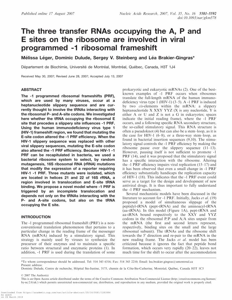
literature to account for -1 PRF. Initially, Jacks et al. (19)proposed a model of simultaneous slippage of thepeptidyl-tRNA (pept-tRNA) and the aminoacyl-tRNA(aa-tRNA). In this model (Figure 1A), pept-tRNA andaa-tRNA bound respectively to the XXY and YYZcodons in the ribosomal P/P and A/A sites unpair fromthe mRNA (the first and second letters represent,respectively, binding sites on the small and the largeribosomal subunit). The tRNAs and the ribosome shifttowards the 5’ direction and re-pair to the mRNA in thenew reading frame. The Jacks et al. model has beencriticized because it ignores the fact that peptide bondformation, which occurs very rapidly (20–22), leaves notmuch time for the shift to occur after the accommodation
Present address:Dominic Dulude, Centre de recherche, Hopital Ste-Justine, 3175, chemin de la Cote-Ste-Catherine, Montreal, Quebec, Canada H3T 1C5
*To whom correspondence should be addressed. Tel: 514 343 6316; Fax: 514 343 2210; Email: [email protected]
� 2007 The Author(s)
This is an Open Access article distributed under the terms of the Creative Commons Attribution Non-Commercial License (http://creativecommons.org/licenses/
by-nc/2.0/uk/) which permits unrestricted non-commercial use, distribution, and reproduction in any medium, provided the original work is properly cited.
Downloaded from https://academic.oup.com/nar/article-abstract/35/16/5581/1281884by gueston 28 March 2018
of the aa-tRNA in the A/A site. A refinement to the Jackset al. model had been provided by Plant et al. (23), whosuggested that a movement of 9 A of the anticodon loop ofthe aa-tRNA upon occupancy of the A/A site creates atension on the mRNA because of the resistance tounwinding of the stimulatory signal. This tension wouldbe relieved by the unpairing of the tRNAs, slippage of themRNA by one base in the 30 direction and re-pairing ofthe tRNAs in the new reading frame. However, theproposed 9 A displacement of the anticodon loop of theaa-tRNA is not supported by structure analysis and bylarge-scale molecular dynamics (24,25). A second modelwas proposed by Weiss et al. (26), where -1 PRF occursduring the translocational step of the elongation cycle.Translocation proceeds in a stepwise manner and requiresconformational changes within the ribosome (27–31). In a
simplified way, after peptide bond formation, the acceptorstem of the newly deacylated-tRNA (deac-tRNA) and ofpept-tRNA move, respectively, from the P to E site andfrom the A to P site of the large ribosomal subunit. Theresulting positions of both tRNAs are thus described asthe P/E and A/P sites, respectively. In the next step, theanticodon stem–loop of the tRNAs moves to the E and Psites on the small ribosomal subunit, dragging the mRNAby one codon (32,33). Weiss et al. (26) suggested that,when the tRNAs occupy these P/E and A/P sites, they canunpair from the mRNA, move in the 30 direction withthe ribosome and re-pair in the new reading frame. Theanticodon stem–loops of the tRNAs then move with themRNA to the E and P sites of the small ribosomal subunit(Figure 1B). The analysis of an electron cryo-microscopy(cryo-EM) ribosome-mRNA pseudoknot complex stalled
Figure 1. Models discussed in the literature to account for -1 PRF. The ribosomal subunits, the stimulatory signal (represented here by a stem–loop),the classic slippery sequence (X XXY YYZ) and the ribosomal sites (E, P and A sites) occupied by the corresponding tRNAs are indicated.The deac-tRNA, pept-tRNA and aa-tRNA are, respectively, shown in green, pink and blue, and coloured circles represent amino acids attached tothe 30 end of tRNAs. Elongation factors were omitted from the figure. (A) The two-tRNA simultaneous slippage model as proposed by Jacks et al.(19) and refined by Plant et al. (23): the -1 PRF occurs when the pept-tRNA and the aa-tRNA occupy, respectively, the P/P and the A/A sites, priorto peptide bond formation. (B) Model proposed by Weiss et al. (26), in which the -1 PRF occurs after peptide bond formation, when the deac-tRNAand the pept-tRNA occupy, respectively, the P/E and A/P sites. It should be noted that the relative position of the subunits is modified (ratchet-likerotation) compared to their position after peptide bond formation (27). (C) Model initially proposed by Farabaugh (38) and experimentallysupported by Leger et al. (35), in which the -1 PRF occurs before peptide bond formation and occupancy of the A/A site, when the incomingaa-tRNA occupies the A/T entry site. This model is updated so as to include the deac-tRNA in the E site, which is ejected from the ribosome whenthe aa-tRNA occupies the A/A site (60).
5582 Nucleic Acids Research, 2007, Vol. 35, No. 16
Downloaded from https://academic.oup.com/nar/article-abstract/35/16/5581/1281884by gueston 28 March 2018
in the process of -1 PRF supports the hypothesis that thisevent occurs during translocation (34), by showing thatthe elongation factor EF2 (eEF2), the eukaryotic homo-logue of EF-G, is bound to the stalled complex. However,it was observed that -1 PRF can be affected by mutationsin the small subunit rRNA that alter the accommodationof the aa-tRNA in the A/A site (35) as well as bymutations in the elongation factor 1A (eEF1A) (36,37),the eukaryotic homologue of EF-Tu, that contributes tothe accommodation process. These observations cannotbe explained by any of the two previous models, but theyare taken into account by a third model, which wasinitially proposed by Farabaugh (38) and further sup-ported by experimental data from our group (35). Thismodel proposes that the -1 PRF occurs when aa-tRNAand pept-tRNA are located, respectively, in the A/T entrysite and in the P/P site (Figure 1C). Under theseconditions, the -1 PRF has more time to take place,compared to the model of Jacks et al. Still, someobservations related to -1 PRF cannot be explained bythis model. In particular, swapping a fragment of eightnucleotides located immediately 5’ from the slipperysequence in the HIV-1 frameshift region with thecorresponding fragment in the human T-cell leukaemiavirus type 2 (HTLV-2) was found to decrease -1 PRFefficiency (39).
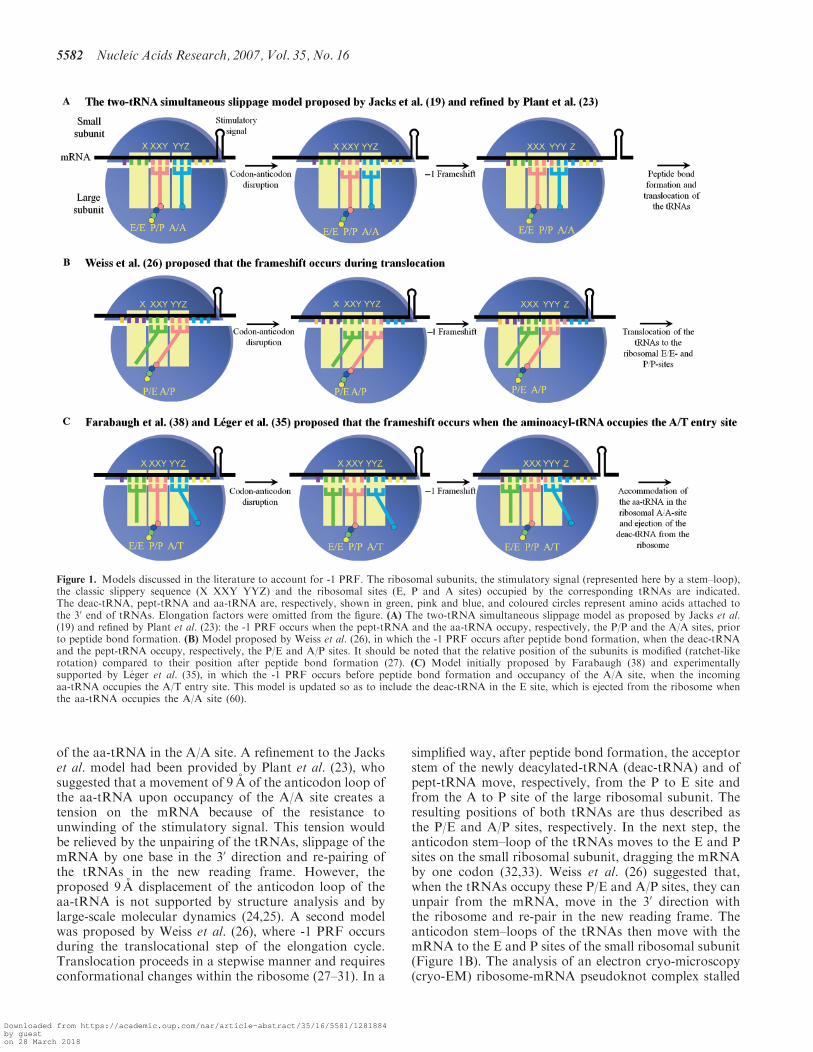
In this study, we reveal additional aspects that areimportant for -1 PRF, using the HIV-1 frameshift regionas a model. In HIV-1, the slippery sequence is U UUUUUA (40) and the stimulatory signal is a two-stem helix,where an internal three-nucleotide bulge separates thelower stem from the upper stem–loop (6–8) (Figure 2).This signal is separated from the slippery sequence by onenucleotide. We previously suggested that the lower stemfavours a specific interaction between the upper stem–loopand the ribosome, when the stimulatory signal firstencounters the ribosome. After this first contact, theribosome progresses along the mRNA and unwindsthe lower stem. Once the slippery sequence occupies thedecoding site, the upper stem–loop, which is at a distanceof eight nucleotides from this slippery sequence, acts as theeffective frameshift stimulatory signal. It allows the -1PRF to occur, involving the tRNAs interacting with the P-and A-site codons. With use of a dual-luciferase reportersystem in which the HIV-1 frameshift region was insertedbetween the coding sequences for the Renilla (Rluc) andfirefly luciferase (Fluc) such that the Fluc expressionrequires the -1 PRF of HIV-1, we demonstrated thatmutations in the E-site codon can alter the -1 PRFefficiency. The same observation was made for other viralslippery sequences. In addition, since we had recentlyshown that the -1 PRF of HIV-1 can be recapitulated inEscherichia coli and that prokaryotic ribosomes respondin the same manner as eukaryotic ribosomes to mutationsin the HIV-1 frameshift stimulatory signal (35), we used a-1 PRF system harboured in E. coli to undertake arandom search of mutations in 16S rRNA that affect -1PRF. We found three mutations of this kind, all located inthe platform region of the small ribosomal subunit. Thisregion is positioned close to the binding site of the E-sitetRNA (41) and is known to be involved in conformational
changes of the ribosome during translocation (27,28,31).Based on these findings, we propose a novel model inwhich -1 PRF is triggered by an incomplete translocationand requires the slippage of not only the tRNAsinteracting with the P- and A-site codons, but also ofthe tRNA occupying the E site.
MATERIALS AND METHODS
Plasmid constructions
All primers used to generate constructs were purchasedfrom Integrated DNA Technologies Inc. (Coralville, IA,USA). Plasmids used in frameshift assays in mammaliancultured cells are derivatives of pDual-HIV (–1) and(0) plasmids described in Dulude et al. (18). The pDual-HIV (–1) plasmid contains the HIV-1 group M subtypeB frameshift region inserted between the sequences codingfor Rluc and Fluc, such that the Fluc coding sequence is inthe –1 reading frame relative to the Rluc initiator codon.In pDual-HIV (0), Fluc is in frame with the Rluc initiatorcodon, by addition of an adenine 30 adjacent to theslippery sequence, which is inactivated by mutagenesis toC UUC CUC. The Rluc expression is used to normalizethe Fluc expression. The -1 PRF efficiency of each (–1)construct is obtained by dividing the Fluc/Rluc ratio bythe corresponding ratio of the (0) frame construct(Figure 2). The M1 to M7 mutants and C1 to C6constructs were generated by amplification of the mutatedDNA from pDual-HIV (–1), with use of standard PCRprocedures and a primer spanning the KpnI site, 50-GCAGGGGGTACCTGGAAAGGAAGGACACCAAATGAAAGATTGTTCGAGAGACAGGCNNNNNNNNNNGGGAAGATCTGG-30, (N corresponds to the mutated
Figure 2. HIV-1 frameshift region in eukaryotic and prokaryotic dual-luciferase systems. Plasmids contain the HIV-1 frameshift regioninserted between the coding sequence of the Rluc (white) and theFluc (grey) genes, as described in Dulude et al. (18). The HIV-1 slipperysequence is underlined. The HIV-1 two-stem helix frameshift stimula-tory signal (6–8) and the restriction sites (KpnI and Pfl23II) used in thisstudy are indicated. The insertion of the HIV-1 frameshift region issuch that the Fluc coding sequence is in the -1 reading frame relative tothe Rluc AUG initiator codon (-1 constructs). For the (0) frameconstruct, the slippery sequence is inactivated by mutagenesis toC UUC CUC and an A is inserted 30 to the slippery sequence, such thatthe Fluc coding sequence is in-frame with the Rluc AUG initiatorcodon. The Fluc gene is always expressed as a fusion protein with Rluc.
Nucleic Acids Research, 2007, Vol. 35, No. 16 5583
Downloaded from https://academic.oup.com/nar/article-abstract/35/16/5581/1281884by gueston 28 March 2018
nucleotides as shown in Figures 3A, 4A and 5B) and aprimer spanning the Pfl23II site, 50-GCCAACCGAACGGACATTTCG-30, for the forward and reversereactions, respectively. The mutated DNA was subclonedinto pDual-HIV (–1) previously digested by KpnI andPfl23II restriction enzymes.Mutations in 16S rRNA (�G666, iC739 and G604A)
were selected with a specialized bacterial ribosome system(42,43), using the p3RGFP-HIV (–1) plasmid, described indetails elsewhere. Briefly, p3RGFP-HIV (–1) contains theE. coli rrnB operon under the control of the induciblelacUV5 promoter, as well as two reporter genes coding forthe red (RFP) and green (GFP) fluorescent proteins. TheDsRed T4 plasmid coding for RFP was a generous giftfrom Dr B.S. Glick, from the University of Chicago (44),and the GFP coding sequence was obtained from thepGFPemd-N1 plasmid, a kind gift from Dr M. Bouvier,from the Universite de Montreal. RFP is expressed byconventional translation whereas the HIV-1 frameshiftregion is inserted in the beginning of the coding sequenceof GFP, so that its expression requires a –1 PRF. Theribosome-binding sites (RBS) of the reporters are changedto 50-AUCCC and the messenger-binding site (MBS) ofthe 16S rRNA is changed to 50-GGGAU, so that thereporters are exclusively translated by the ribosomes thatcontain the plasmid-encoded 16S rRNA (GFP/RFPsystem). Mutations iG666 and �C739 were introducedin the 16S rRNA by amplification of the mutated DNAfragments from p3RGFP-HIV (–1) with a two-step PCRapproach encompassing restriction sites BlnI and DraIII,using an overlap extension procedure (45). The resultantPCR fragment was subcloned into p3RGFP-HIV (–1)previously digested with the same enzymes.The specialized bacterial dual-luciferase system was
created as follows: the Rluc to Fluc coding sequenceencompassing the HIV-1 frameshift region in pDual-HIV(–1) and (0) constructs was amplified by PCR, using theprimer spanning the NsiI site, 50-CTAGAGCCACCATGCATACCAGCAAGG-30, and the primer spanningthe Pfl23II site, 50-GTTTCATAGCTTCTGCCAACCGAACG-30, for the forward and reverse reactions,respectively. The resultant PCR fragments were subclonedinto the pRNAluc2 plasmid digested with NsiI andPfl23II, as described in Belanger et al. (42), generatingpDual-HIV/P (0) and (–1) plasmids (where P stands forprokaryote plasmids). The pRNAluc2 plasmid containsthe E. coli rrnB operon under the control of the induciblelacUV5 promoter and a reporter gene coding for Fluc.As in the GFP/RFP system, the RBS of the dual-luciferasereporter and the MBS of the 16S rRNA were mutated sothat the dual-luciferase reporter is exclusively translatedby ribosomes that contain plasmid-encoded 16S rRNA.The 16S rRNA mutations in p3RGFP-HIV (–1) (�G666,iC739, G604A, iG666 and �C739) were cloned in pDual-HIV/P (0) and (–1), using two ApaI restriction sites.
Randommutagenesis of 16S rRNA
Random mutations were introduced into the 16S rRNAfragment using a high-copy plasmid (pUC18, Fermentas).A BamHI–SacI fragment encompassing 16S rRNA from
p3RGFP-HIV (–1) was cloned into pUC18 digested withthe same enzymes, generating pUC18/16S. E. coli XL1-Red mutator strain (Stratagene) was used to producerandom mutations into 16S rRNA. Escherichia coli XL1-Red competent cells were transformed with pUC18/16S asdirected by the manufacturer to obtain randomly mutatedplasmids (pUC18/R16S, where R stands for randomized16S rRNA) (46,47). The procedure was repeated seventimes, so that more than 90% of the clones analysedcontained a mutation in the 16S rRNA fragment.A second BlnI restriction site located in the beginning ofthe gene coding for 23S rRNA in p3RGFP-HIV (–1) wasmutated to an AflII restriction site, using a standardmutagenesis procedure, so as to conserve a unique BlnIrestriction site in the 16S rRNA gene. The 16S rRNArandom mutant library from pUC18/R16S was clonedinto p3RGFP-HIV (–1), using BlnI–DraIII restrictionsites, which generated p3RGFP-HIV/R16S. Each isolated16S rRNA mutation was re-introduced into p3RGFP-HIV (–1) to verify that the phenotype was conserved.
Frameshift assays in mammalian cultured cells
Frameshift assays in eukaryotes were monitored bytransient transfection of the dual-luciferase plasmids(pDual-HIV derivatives) into human embryonic kidney293T cells (HEK293T) maintained in Dulbecco’s modifiedEagle’s medium (DMEM) supplemented with 10% (v/v)FBS (Wisent), with 2� 105 cells/well seeded in 6-wellplates. Two mg of plasmids were diluted in 3% of theinitial culture volume in fresh serum-free medium andmixed with an equivalent volume containing 5 mg of PEI(polyethylenimine, Polysciences Inc.). The mixture wasincubated for 15min at room temperature prior to theaddition to cell culture. Cells were cultured for 48 h beforebeing washed twice with 2ml of PBS, and lysed with 450 mlof the Cell Passive Lysis Buffer 1X (Promega). Fluc versusRluc activities of each construct were measured for 10 s asrelative light units (RLU) with an EG&G Berthold LumatLB 9507 luminometer, using a non-commercial dual-luciferase enzyme assay system (48).
Bacterial frameshift assays
Dual-luciferase and GFP/RFP assays in E. coli wereconducted as follows: overnight cultures of E. coli Top 10strain (Invitrogen) were transformed with pDual-HIV/Por p3RGFP-HIV derivatives. Plasmids were grown in LBcontaining 100 mg/ml of ampicillin at 378C. The cultureswere diluted to an absorbance of 0.1 at 600 nm andincubated for 1 h at 378C. The plasmid-encoded rRNAexpression was induced with 1mM of isopropyl-b-D-thiogalactopyra noside (IPTG) for 3 h for dual-luciferaseassays, and 6 h for GFP/RFP assays at 378C. The dual-luciferase assays were carried as the luciferase assaysdescribed in Belanger et al. (42), except that the cells werelysed with 50 ml of the Cell Passive Lysis Buffer 1X andincubated for 15min at room temperature. Fluc and Rlucactivities were measured as described above. For GFP/RFP assays, 1ml of cultured cells was centrifuged. Thepellet was washed three times with 500 ml of PBS and re-suspended in 200 ml of PBS. Fluorescence was measured
5584 Nucleic Acids Research, 2007, Vol. 35, No. 16
Downloaded from https://academic.oup.com/nar/article-abstract/35/16/5581/1281884by gueston 28 March 2018
with a Fusion Universal Microplate Analyser (FusionTM
a-FP, Packard) at a 485 nm excitation wavelength(bandpass: 20 nm) for both GFP and RFP and at a535 nm (bandpass: 25 nm) and 580 nm (bandpass: 15 nm)emission wavelength for GFP and RFP, respectively.
RESULTS
Modulating role in -1 PRF of the three nucleotidesimmediately upstream of the HIV-1 slippery sequence
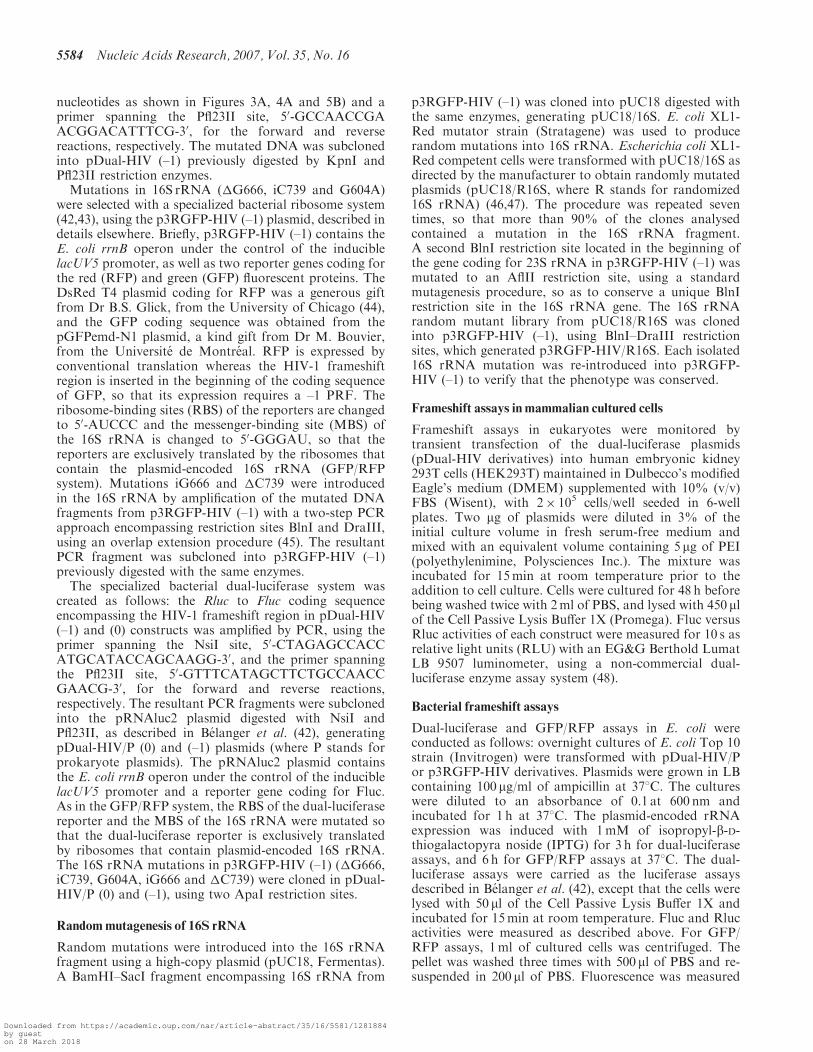
As mentioned earlier, mutations within the eight nucleo-tides immediately preceding the slippery sequence canaffect the efficiency of -1 PRF (39). We may expect that thecloser a nucleotide of this region is to the slipperyheptamer X XXY YYZ, the higher its influence on -1PRF. In this study, we focused on the three nucleotidespositioned immediately upstream of the slippery hepta-mer, which we name A, B and C. Together with theslippery heptamer, these nucleotides form the decamerA BCX XXY YYZ, which we call the extended slipperysequence. In the last model describing -1 PRF [Figure 1C,(35,38)], the XXY and YYZ codons of the classic slipperysequence (X XXY YYZ) are located in the P and A sites,respectively, when the -1 PRF occurs. The A BCnucleotides that are directly upstream of the slipperysequence are positioned such that the BCX and the ABCcodons are located, respectively, in the E site before andafter the shift. To investigate whether the identities ofnucleotides A BC can affect the -1 PRF, we studied sevenmutants of the frameshift region of HIV-1, in which theA BC nucleotide sequence (U AAU UUU UUA), wasreplaced by the corresponding sequence found in otherviruses. We used a dual-luciferase reporter system (18)in mammalian cultured cells. In this system, the HIV-1frameshift region is positioned between Rluc and Fluccoding sequences, such that Fluc expression requires the–1 PRF of HIV-1. In Figure 3A, one sees the extendedslippery sequences of the wild-type HIV-1 (WT; U AAUUUU UUA) and of seven mutants numbered from M1 toM7. The M1 mutant corresponds to a variant isolatedfrom a protease inhibitor-exposed patient, and M2 is anatural variant of HIV-1 (49). In M3 to M7 mutants,nucleotides A BC are taken from HIV type 2 (HIV-2; M3),the giardia virus (M4), the severe acute respiratorysyndrome coronavirus (SARS-CoV; M5), the humanT-cell lymphotropic virus type 1 (HTLV-1; M6) and theequine infectious anaemia virus (EIAV; M7). The absolutevalue of the WT -1 PRF efficiency was 8.0� 1.0%. InFigure 3B, a value of 100% is arbitrarily assigned to thisefficiency, and the efficiencies of all mutants are shownrelative to WT. Among all investigated mutants, M1 ischaracterized by the lowest level of -1 PRF efficiency,which is only 5% of the WT level. Such a drop of theefficiency can be explained by the fact that M1 is the onlyvariant in which the re-pairing of the pept-tRNA in thenew reading frame is not allowed and this variant is mostlikely very poorly infectious. With M2, M5, M6 and M7mutants, the -1 PRF efficiency was increased by about50%. In contrast, with M3, the -1 PRF efficiencydecreased slightly to 70% of the wild-type value, while
the frameshift level remained unchanged with M4.Although the effects are not dramatic, they are allsignificant and well-reproducible [with M1, M2, M6 andM7 mutants; P values are 0.0001 (n5 4) and with M5mutant, P-value is 0.001 (n=4), as determined byStudent’s t-test]. To verify whether the -1 PRF could bealso influenced by mutations upstream the A BC nucleo-tides, we mutated the GCU codon, which is immediatelyupstream the BCX codon (GCU was exchanged withAAU, UCU, GGU or CAU, so that the A positionremained unchanged). These mutations did not alter the -1PRF efficiency (data not shown). We can conclude thatmutations in positions A BC, immediately upstream of theclassic slippery sequence, can noticeably affect the -1 PRFefficiency of HIV-1 and, therefore, that the nucleotideslocated at positions A BC could be involved in the -1 PRFof HIV-1. This was not the case for the nucleotidesupstream of A BC.
-1 PRF efficiencies with other viral frameshift regionsmutated at positions A BC
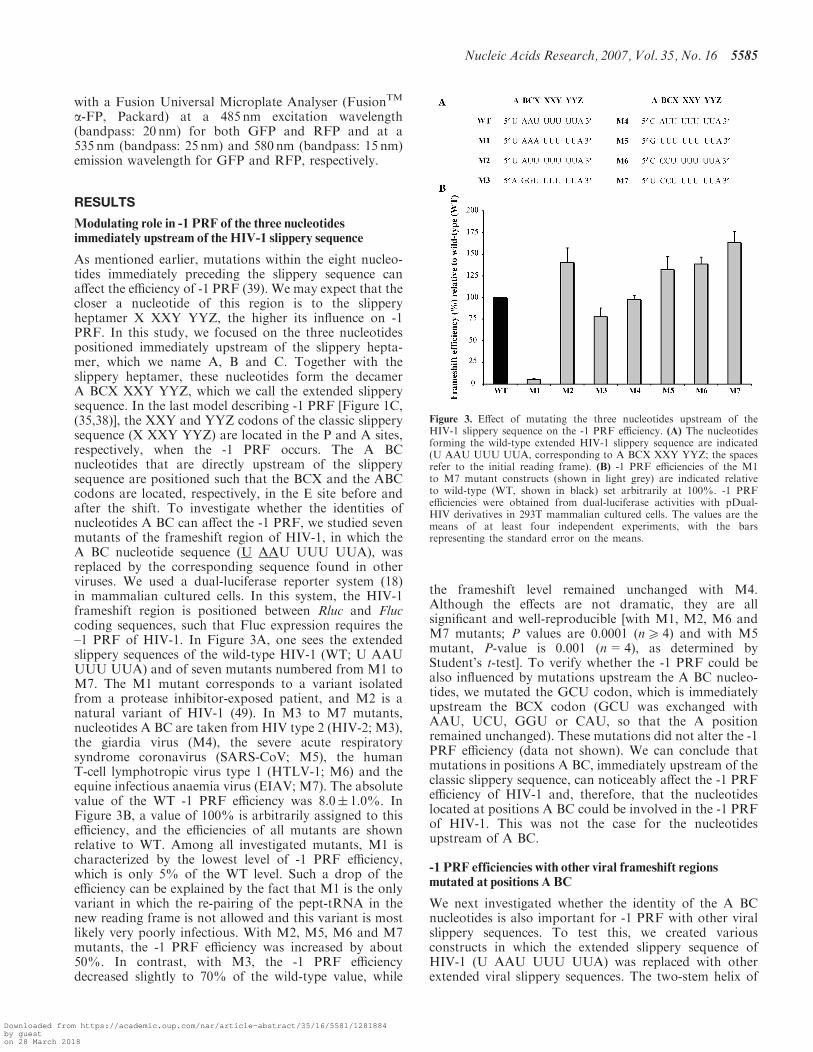
We next investigated whether the identity of the A BCnucleotides is also important for -1 PRF with other viralslippery sequences. To test this, we created variousconstructs in which the extended slippery sequence ofHIV-1 (U AAU UUU UUA) was replaced with otherextended viral slippery sequences. The two-stem helix of
Figure 3. Effect of mutating the three nucleotides upstream of theHIV-1 slippery sequence on the -1 PRF efficiency. (A) The nucleotidesforming the wild-type extended HIV-1 slippery sequence are indicated(U AAU UUU UUA, corresponding to A BCX XXY YYZ; the spacesrefer to the initial reading frame). (B) -1 PRF efficiencies of the M1to M7 mutant constructs (shown in light grey) are indicated relativeto wild-type (WT, shown in black) set arbitrarily at 100%. -1 PRFefficiencies were obtained from dual-luciferase activities with pDual-HIV derivatives in 293T mammalian cultured cells. The values are themeans of at least four independent experiments, with the barsrepresenting the standard error on the means.
Nucleic Acids Research, 2007, Vol. 35, No. 16 5585
Downloaded from https://academic.oup.com/nar/article-abstract/35/16/5581/1281884by gueston 28 March 2018
HIV-1 was used as a stimulatory signal with these differentslippery sequences, which were assayed in the eukaryoticdual-luciferase system described above and are shown inFigure 4A. The C1, C3 and C5 constructs contain theextended slippery sequences found, respectively, in giardiavirus (C AUC CCU UUA), EIAV (U CCA AAA AAC)and in SARS-CoV (G UUU UUA AAC). The -1 PRFefficiencies obtained with these three constructs varybetween 4% and 5%, about 40–60% of the HIV-1 -1PRF efficiency (Figure 4B). The nucleotides at positionsA BC in each of the C1, C3 and C5 constructs weremutated to UAA, the nucleotides found at positions A BCupstream of the classic slippery sequence of HIV-1,generating the C2, C4 and C6 chimeric constructs.This decreased -1 PRF efficiency by about 50%,compared to the C1, C3 and C5 constructs, whichconfirms the observations made with HIV-1 slippery
sequence concerning the importance of nucleotides A BCfor -1 PRF.
It was previously demonstrated that exchanging slip-pery sequences from different frameshift regions alters the-1 PRF efficiency (18,50). However, in these studies, onlythe heptanucleotide slippery sequence was exchanged.Figure 4B also compares the effect of exchanging eitherthe classic or the extended HIV-1 slippery sequence withother viral slippery sequences on -1 PRF efficiency. Whenthe classic slippery sequence of HIV-1 is replaced with theclassic slippery sequence of giardia virus, EIAV or thatof SARS-CoV, the -1 PRF efficiency drops to 20–30%when compared to HIV-1 -1 PRF efficiency. When theHIV-1 extended slippery sequence is replaced with theextended slippery sequence found in giardia virus, EIAVor in SARS-CoV, the -1 PRF efficiency also decreases,but much less than when the substitution involves onlythe classic heptamer, being 50–60% of HIV-1 -1 PRFefficiency. These results confirm that exchanging slipperysequences alters the -1 PRF efficiency, and, in addition,shows that the extent of the decrease depends uponwhether the extended slippery sequence or only the classicslippery sequence is exchanged. These results also confirmthe importance of positions A BC for -1 PRF.
Identification of 16S rRNAmutations that affect HIV-1 -1PRF efficiency
We also attempted to identify rRNA mutants that couldinterfere with the -1 PRF of HIV-1. So far, the rRNAmutations that were found to influence -1 PRF perturb theaccommodation process (35). Our aim was to selectmutations that influence -1 PRF by affecting the E-sitetRNA binding, since we had found that mutations in theE-site codon can alter -1 PRF. Our strategy consisted inusing the bacterial ribosome and introducing randommutations in 16S rRNA with the high-mutation-rateE. coli XL1-Red mutator strain (see Materials andMethods section for details). A pool of randomly mutatedplasmids was obtained, where 90% of the clones analysedcontained a mutation in the 16S rRNA. This 16S rRNArandom library was then cloned into a plasmid containingreporter genes coding for RFP and GFP, in which RFP isexpressed by conventional translation, whereas GFPexpression requires the -1 PRF of HIV-1. The RBS ofthe reporters and the MBS site of the 16S rRNA randomlibrary are mutated and remain complementary, so thatthe reporters are exclusively translated by ribosomes thatcontain plasmid-encoded 16S rRNA. A small screen wasindividually performed (about 2000 clones), using theGFP/RFP assay to select clones for which GFP expressionwas affected but for which conventional translation wasonly moderately decreased (<50%). Three mutants wereselected: mutant �G666 with a guanosine deleted from thestretch of guanosines between positions 666 to 671,mutant iC739 with a cytosine inserted into the stretch ofcytosines between positions 735 to 739, and mutantG604A with guanosine 604 replaced by an adenosine.Figure 5A shows the variations in HIV-1 -1 PRF efficiencyobtained with each selected mutant relative to wild-type16S rRNA, which was set arbitrarily at 100%, based on
Figure 4. -1 PRF efficiencies obtained with various slippery sequences.The HIV-1 slippery sequence is replaced with slippery sequences fromother viral frameshift regions. The stimulatory signal of HIV-1 is usedin all constructs. (A) The nucleotides forming the extended HIV-1slippery sequence (U AAU UUU UUA, corresponding to A BCXXXY YYZ; the spaces refer to the initial reading frame) are shown.The C1, C3 and C5 constructs contain the extended slippery sequencesfound, respectively, in the frameshift region of giardia virus (C AUCCCU UUA), EIAV (U CCA AAA AAC) and SARS-CoV (G UUUUUA AAC). Positions A BC in the C2, C4 and C6 chimeras aremutated to U AA, the nucleotides found at positions A BC in theextended slippery sequence of HIV-1. (B) -1 PRF efficiencies obtainedfrom dual-luciferase activities with pDual-HIV derivatives in 293Tmammalian cultured cells. The -1 PRF efficiencies with the C1 to C6constructs are indicated relative to HIV-1 wild-type frameshift region,which is set arbitrarily at 100%. Constructs containing a C CCU UUA,A AAA AAC and a U UUA AAC heptanucleotide sequence are shownin dark grey, light grey and in white, respectively. The values are themeans of at least four independent experiments, with the barsrepresenting the standard error on the means.
5586 Nucleic Acids Research, 2007, Vol. 35, No. 16
Downloaded from https://academic.oup.com/nar/article-abstract/35/16/5581/1281884by gueston 28 March 2018
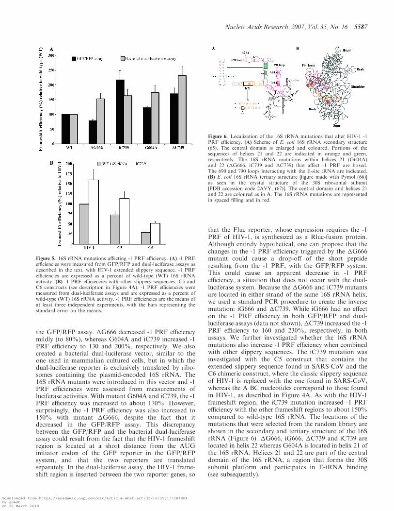
the GFP/RFP assay. �G666 decreased -1 PRF efficiencymildly (to 80%), whereas G604A and iC739 increased -1PRF efficiency to 130 and 200%, respectively. We alsocreated a bacterial dual-luciferase vector, similar to theone used in mammalian cultured cells, but in which thedual-luciferase reporter is exclusively translated by ribo-somes containing the plasmid-encoded 16S rRNA. The16S rRNA mutants were introduced in this vector and -1PRF efficiencies were assessed from measurements ofluciferase activities. With mutant G604A and iC739, the -1PRF efficiency was increased to about 170%. However,surprisingly, the -1 PRF efficiency was also increased to150% with mutant �G666, despite the fact that itdecreased in the GFP/RFP assay. This discrepancybetween the GFP/RFP and the bacterial dual-luciferaseassay could result from the fact that the HIV-1 frameshiftregion is located at a short distance from the AUGinitiator codon of the GFP reporter in the GFP/RFPsystem, and that the two reporters are translatedseparately. In the dual-luciferase assay, the HIV-1 frame-shift region is inserted between the two reporter genes, so
that the Fluc reporter, whose expression requires the -1PRF of HIV-1, is synthesized as a Rluc-fusion protein.Although entirely hypothetical, one can propose that thechanges in the -1 PRF efficiency triggered by the �G666mutant could cause a drop-off of the short peptideresulting from the -1 PRF, with the GFP/RFP system.This could cause an apparent decrease in -1 PRFefficiency, a situation that does not occur with the dual-luciferase system. Because the �G666 and iC739 mutantsare located in either strand of the same 16S rRNA helix,we used a standard PCR procedure to create the inversemutation: iG666 and �C739. While iG666 had no effecton the -1 PRF efficiency in both GFP/RFP and dual-luciferase assays (data not shown), �C739 increased the -1PRF efficiency to 160 and 230%, respectively, in bothassays. We further investigated whether the 16S rRNAmutations also increase -1 PRF efficiency when combinedwith other slippery sequences. The iC739 mutation wasinvestigated with the C5 construct that contains theextended slippery sequence found in SARS-CoV and theC6 chimeric construct, where the classic slippery sequenceof HIV-1 is replaced with the one found in SARS-CoV,whereas the A BC nucleotides correspond to those foundin HIV-1, as described in Figure 4A. As with the HIV-1frameshift region, the iC739 mutation increased -1 PRFefficiency with the other frameshift regions to about 150%compared to wild-type 16S rRNA. The locations of themutations that were selected from the random library areshown in the secondary and tertiary structure of the 16SrRNA (Figure 6). �G666, iG666, �C739 and iC739 arelocated in helix 22 whereas G604A is located in helix 21 ofthe 16S rRNA. Helices 21 and 22 are part of the centraldomain of the 16S rRNA, a region that forms the 30Ssubunit platform and participates in E-tRNA binding(see subsequently).
Figure 6. Localization of the 16S rRNA mutations that alter HIV-1 -1PRF efficiency. (A) Scheme of E. coli 16S rRNA secondary structure(65). The central domain is enlarged and coloured. Portions of thesequences of helices 21 and 22 are indicated in orange and green,respectively. The 16S rRNA mutations within helices 21 (G604A)and 22 (�G666, iC739 and �C739) that affect -1 PRF are boxed.The 690 and 790 loops interacting with the E-site tRNA are indicated.(B) E. coli 16S rRNA tertiary structure [figure made with Pymol (66)]as seen in the crystal structure of the 30S ribosomal subunit[PDB accession code 2AVY, (67)]. The central domain and helices 21and 22 are coloured as in A. The 16S rRNA mutations are representedin spaced filling and in red.
Figure 5. 16S rRNA mutations affecting -1 PRF efficiency. (A) -1 PRFefficiencies were measured from GFP/RFP and dual-luciferase assays asdescribed in the text, with HIV-1 extended slippery sequence. -1 PRFefficiencies are expressed as a percent of wild-type (WT) 16S rRNAactivity. (B) -1 PRF efficiencies with other slippery sequences: C5 andC6 constructs (see description in Figure 4A). -1 PRF efficiencies weremeasured from dual-luciferase assays and are expressed as a percent ofwild-type (WT) 16S rRNA activity. -1 PRF efficiencies are the means ofat least three independent experiments, with the bars representing thestandard error on the means.
Nucleic Acids Research, 2007, Vol. 35, No. 16 5587
Downloaded from https://academic.oup.com/nar/article-abstract/35/16/5581/1281884by gueston 28 March 2018
DISCUSSION
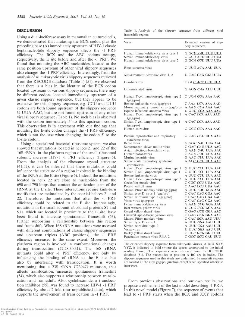
Using a dual-luciferase assay in mammalian cultured cells,we demonstrated that mutating the BCX codon plus thepreceding base (A) immediately upstream of HIV-1 classicheptanucleotide slippery sequence affects the -1 PRFefficiency. The BCX and the ABC codons occupy,respectively, the E site before and after the -1 PRF. Wefound that mutating the ABC nucleotides, located at thesame position upstream of other viral slippery sequencesalso changes the -1 PRF efficiency. Interestingly, from theanalysis of 41 eukaryotic virus slippery sequences retrievedfrom the RECODE database (Table 1) (51), we observedthat there is a bias in the identity of the BCX codonlocated upstream of various slippery sequences: there maybe different codons located immediately upstream of agiven classic slippery sequence, but they appear to beexclusive for this slippery sequence, e.g. CCU and UUUcodons are both found upstream of the slippery sequenceU UUA AAC, but are not found upstream of any otherviral slippery sequence (Table 1). No such bias is observedwith the codon immediately 5’ to this upstream codon.This observation is in agreement with our findings thatmutating the E-site codon changes the -1 PRF efficiency,which is not the case when changing the codon 5’ to theE-site codon.Using a specialized bacterial ribosome system, we also
showed that mutations located in helices 21 and 22 of the16S rRNA, in the platform region of the small ribosomalsubunit, increase HIV-1 -1 PRF efficiency (Figure 5).From the analysis of the ribosome crystal structure(41,52), it can be inferred that these mutations couldinfluence the structure of a region involved in the bindingof the tRNA at the E site (Figure 6). Indeed, the mutationslocated in helix 22 are close to nucleotides from the690 and 790 loops that contact the anticodon stem of thetRNA at the E site. These interactions require kink-turnmotifs that are maintained by the coaxial helices 21 and22. Therefore, the mutations that alter the -1 PRFefficiency could be related to the E site. Interestingly,mutations in the small subunit ribosomal proteins S7 andS11, which are located in proximity to the E site, havebeen found to increase spontaneous frameshift (53),further supporting a relationship between this regionand frameshift. When 16S rRNA mutations were assessedwith different combinations of classic slippery sequencesand upstream triplets (ABC positions), the -1 PRFefficiency increased to the same extent. Moreover, theplatform region is involved in conformational changesduring translocation (27,28,30,31). The 16S rRNAmutants could alter -1 PRF efficiency, not only byinfluencing the binding of tRNA at the E site, butalso by interfering with translocation. It is worthmentioning that a 23S rRNA C2394G mutation, thataffects translocation, increases spontaneous frameshift(54), which also supports a relationship between translo-cation and frameshift. Also, cycloheximide, a transloca-tion inhibitor (55), was found to increase HIV-1 -1 PRFefficiency by about 2-fold (our unpublished data), whichsupports the involvement of translocation in -1 PRF.
From previous observations and our own results, wepropose a refinement of the last model describing -1 PRF.In this novel model (Figure 7), the sequence of events thatlead to -1 PRF starts when the BCX and XXY codons
Table 1. Analysis of the slippery sequence from different viral
frameshift regions
Virus Extended version of slip-pery sequences
Human immunodeficiency virus type 1 G GCU AAU UUU UUA
Simian immunodeficiency virus G GCA AAU UUU UUA
Human immunodeficiency virus type 2 G GCA GGU UUU UUA
Rous sarcoma virus C UUG ACA AAU UUA
Saccharomyces cerevisiae virus L-A U CAG CAG GGU UUA
Giardia virus C GCC AUC CCU UUA
Gill-associated virus G AGG CAA AUU UUC
Human T-cell lymphotropic virus type 2(gag/pro)
C UGA GGA AAA AAC
Bovine leukaemia virus (gag/pro) C AAA UCA AAA AAC
Mouse mammary tumour virus (gag/pro) A AAU UCA AAA AAC
Equine infectious anaemia virus U GUU CCA AAA AAC
Human T-cell lymphotropic virus type 1(gag/pro)
A CAC CCA AAA AAC
Simian T-cell lymphotropic virus type 1(gag/pro)
A CAC CCA AAA AAC
Human astrovirus G GCC CCA AAA AAC
Porcine reproductive and respiratorysyndrome virus
G CAG UGU UUA AAC
Berne virus G GGU GAU UUA AAC
Subterranean clover mottle virus C GAG CAU UUA AAC
Avian infectious bronchitis virus G AAU UAU UUA AAC
Human coronavirus C AGU UAU UUA AAC
Murine hepatitis virus G AAC UUU UUA AAC
Severe acute respiratory syndromecoronavirus
A ACG UUU UUA AAC
Human T-cell lymphotropic virus type 1 G UUC CCU UUA AAC
Simian T-cell lymphotropic virus type 1 G UUC CCU UUA AAC
Bovine leukaemia virus A UUC CCU UUA AAC
Human T-cell lymphotropic virus type 2 A UUU CCU UUA AAC
Cocksfoot mottle virus C CGG CCU UUA AAC
Potato leafroll virus C AAG CCU UUA AAU
Mason–Pfizer monkey virus (gag/pro) A UUU CAG GGA AAC
Simian type D virus 1 (gag/pro) C CAU CAG GGA AAC
Simian retrovirus type 2 (gag/pro) C CAU CAG GGA AAC
Visna virus (gag/pro) C CAU CAG GGA AAC
Feline immunodeficiency virus G AAU UCG GGA AAC
Beet western yellow virus U CUG UCG GGA AAC
Cereal yellow dwarf virus C GAG UCG GGA AAC
Cucurbit aphid-borne yellows virus C GAG UCG GGA AAC
Mason–Pfizer monkey virus C CAU GGA AAU UUU
Simian type D virus 1 A UUU GGA AAU UUU
Simian retrovirus type 2 A UUU GGA AAU UUU
Visna virus U UUU GGA AAU UUU
Barley yellow dwarf virus C UCU GUG GGU UUU
Peaenation mosaic virus RNA 2 C GCG GCG GAU UUU
The extended slippery sequence from eukaryotic viruses, A BCX XXYYYZ, is indicated in bold (where the spaces correspond to the initialreading frame). The sequences were retrieved from the RECODEdatabase (51). The nucleotides at position A BC are in italics. Theslippery sequences used in this study are underlined. Frameshift regionsare located at the viral gag/pol junction except when specified otherwise(gag-pro).
5588 Nucleic Acids Research, 2007, Vol. 35, No. 16
Downloaded from https://academic.oup.com/nar/article-abstract/35/16/5581/1281884by gueston 28 March 2018
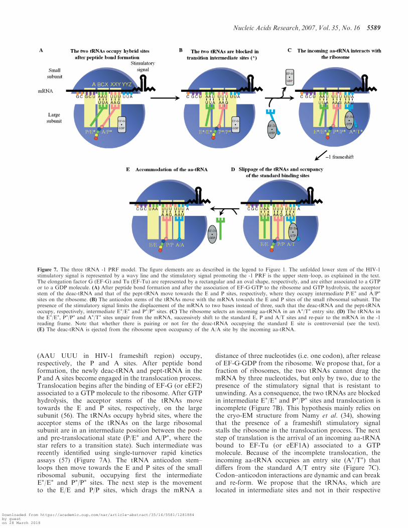
(AAU UUU in HIV-1 frameshift region) occupy,respectively, the P and A sites. After peptide bondformation, the newly deac-tRNA and pept-tRNA in theP and A sites become engaged in the translocation process.Translocation begins after the binding of EF-G (or eEF2)associated to a GTP molecule to the ribosome. After GTPhydrolysis, the acceptor stems of the tRNAs movetowards the E and P sites, respectively, on the largesubunit (56). The tRNAs occupy hybrid sites, where theacceptor stems of the tRNAs on the large ribosomalsubunit are in an intermediate position between the post-and pre-translocational state (P/E� and A/P�, where thestar refers to a transition state). Such intermediate wasrecently identified using single-turnover rapid kineticsassays (57) (Figure 7A). The tRNA anticodon stem–loops then move towards the E and P sites of the smallribosomal subunit, occupying first the intermediateE�/E� and P�/P� sites. The next step is the movementto the E/E and P/P sites, which drags the mRNA a
distance of three nucleotides (i.e. one codon), after releaseof EF-G�GDP from the ribosome. We propose that, for afraction of ribosomes, the two tRNAs cannot drag themRNA by three nucleotides, but only by two, due to thepresence of the stimulatory signal that is resistant tounwinding. As a consequence, the two tRNAs are blockedin intermediate E�/E� and P�/P� sites and translocation isincomplete (Figure 7B). This hypothesis mainly relies onthe cryo-EM structure from Namy et al. (34), showingthat the presence of a frameshift stimulatory signalstalls the ribosome in the translocation process. The nextstep of translation is the arrival of an incoming aa-tRNAbound to EF-Tu (or eEF1A) associated to a GTPmolecule. Because of the incomplete translocation, theincoming aa-tRNA occupies an entry site (A�/T�) thatdiffers from the standard A/T entry site (Figure 7C).Codon–anticodon interactions are dynamic and can breakand re-form. We propose that the tRNAs, which arelocated in intermediate sites and not in their respective
Figure 7. The three tRNA -1 PRF model. The figure elements are as described in the legend to Figure 1. The unfolded lower stem of the HIV-1stimulatory signal is represented by a wavy line and the stimulatory signal promoting the -1 PRF is the upper stem–loop, as explained in the text.The elongation factor G (EF-G) and Tu (EF-Tu) are represented by a rectangular and an oval shape, respectively, and are either associated to a GTPor to a GDP molecule. (A) After peptide bond formation and after the association of EF-G�GTP to the ribosome and GTP hydrolysis, the acceptorstem of the deac-tRNA and that of the pept-tRNA move towards the E and P sites, respectively, where they occupy intermediate P/E� and A/P�
sites on the ribosome. (B) The anticodon stems of the tRNAs move with the mRNA towards the E and P sites of the small ribosomal subunit. Thepresence of the stimulatory signal limits the displacement of the mRNA to two bases instead of three, such that the deac-tRNA and the pept-tRNAoccupy, respectively, intermediate E�/E� and P�/P� sites. (C) The ribosome selects an incoming aa-tRNA in an A�/T� entry site. (D) The tRNAs inthe E�/E�, P�/P� and A�/T� sites unpair from the mRNA, successively shift to the standard E, P and A/T sites and re-pair to the mRNA in the -1reading frame. Note that whether there is pairing or not for the deac-tRNA occupying the standard E site is controversial (see the text).(E) The deac-tRNA is ejected from the ribosome upon occupancy of the A/A site by the incoming aa-tRNA.
Nucleic Acids Research, 2007, Vol. 35, No. 16 5589
Downloaded from https://academic.oup.com/nar/article-abstract/35/16/5581/1281884by gueston 28 March 2018
standard high-affinity sites on the ribosome, are prone toshift to these standard sites, and, after the shift, re-pair tothe mRNA in the -1 reading frame (Figure 7D). Thedriving force for the change in the reading frame istherefore the tendency of the tRNAs to occupy theirstandard binding sites. The slippage of the tRNAs wouldstart with the tRNA located in the E�/E� site, followed bythe successive slippage of the tRNAs located in the P�/P�
and A�/T� sites, towards the E/E, P/P and A/T sites[see ref. (58) for the concept of successive tRNA slippage].The -1 PRF would be followed by the aa-tRNAaccommodation in the A/A site, which is coupled withthe E-site tRNA ejection from the ribosome (59,60)(Figure 7E) and conventional translation would resumewith unfolding of the frameshift stimulatory signal. In ourmodel, there are two steps where the ribosome pauses:when it is blocked during translocation and when the threetRNAs shift to their standard sites. As mentioned in the‘Introduction’ section, Weiss et al. (26) were the first tosuggest that -1 PRF was linked to translocation, but intheir model, -1 PRF occurs when the XXY and YYZcodons occupy the P and A sites (Figure 1B). We suggestthat a translocation anomaly leading to -1 PRF occurs atthe preceding elongation cycle, when the BCX and XXYcodons occupy the P and A sites, which takes into accountthe fact that the E-site codon participates in -1 PRF.In our model, three tRNAs participate in -1 PRF. The
event is triggered by the blockade of the ribosome at atransition state following an incomplete translocation dueto the presence of the stimulatory signal. How can weexplain that the identity of the BCX codon influences the -1 PRF? It is well known that mutating the classic X XXYYYZ motif prevents the slippage of the tRNAs located inthe P and A/T sites, since, after the shift, the tRNAscannot re-pair to the mRNA in the new reading frame.However, whether there is a codon–anticodon interactionat the E site is still a matter of debate. From the analysis ofcrystal structures, no codon–anticodon interaction hasbeen observed between the mRNA and the deac-tRNA inthe E site (52,61). In contrast, in vitro translationalexperiments support a codon–anticodon interaction atthe E site (60,62). From our slippery sequences analysis(Table 1), it can be observed that, after -1 PRF, only onestandard base-pairing is possible between the tRNA andthe ABC nucleotides, in most cases. If there is a codon–anticodon interaction at the E site, it could be suggestedthat base pairs other than Watson–Crick or G-U wobblepairs are tolerated at the E site. Under these relaxedconditions, a major consequence of mutating the BCXcodon plus the preceding base is not likely to alter the -1PRF efficiency by influencing the codon–anticodon inter-action of the E-site tRNA in the shifted frame, althoughminor effects cannot be excluded. We propose that themain consequence of making these changes is to modifythe identity of the E-site tRNA. Our hypothesis is thatthere is a relationship between the structural peculiaritiesof this tRNA and its capacity to shift from an inter-mediate to a classic site, since the movement of this tRNAprecedes and thus controls the movement of the twoother tRNAs. The group of Rousset (63) had proposedthat, in -1 PRF, an E-site tRNA carrying a pseudouridine
modification at position 39 was coupled to high -1 PRFefficiency. However, these results could not be reproduced(Rousset J.P. 2006, personal communication). Also, froma search using the tRNA Compilation 2000 database (64),our present results do not support any relationshipbetween the presence of a tRNA modification at position39 and the -1 PRF efficiency (data not shown).
In conclusion, we propose a novel model to describe -1PRF, in which three tRNAs are involved. A detailedunderstanding of -1 PRF mechanism should contribute tothe development of novel anti-frameshift agents that affectthe replication capacity of viruses, such as HIV-1.
ACKNOWLEDGEMENTS
This study was supported by a grant from the CanadianInstitutes of Health Research (CIHR) to L.B-G. S.V.S.acknowledges fellowships from the CIHR and from theFonds de la Recherche en Sante du Quebec (FRSQ). M.L.is a recipient of a scholarship from the FRSQ. We thankFrancois Belanger, Pascal Chartrand, Jim Omichinskiand the members of the Brakier-Gingras lab for criticalreading of the manuscript and stimulating discussions.We thank Ian Boudreau for his participation in this studyas a summer undergraduate student in Biochemistryfrom Universite de Montreal. Funding to pay the OpenAccess publication charges for this article was providedby the Canadian Institutes of Health Research.
Conflict of interest statement. None declared.
REFERENCES
1. Brierley,I. (1995) Ribosomal frameshifting viral RNAs. J. Gen.Virol., 76 (Pt 8), 1885–1892.
2. Namy,O., Rousset,J.P., Napthine,S. and Brierley,I. (2004)Reprogrammed genetic decoding in cellular gene expression. Mol.Cell, 13, 157–168.
3. Atkins,J.F., Baranov,P.V., Fayet,O., Herr,A.J., Howard,M.T.,Ivanov,I.P., Matsufuji,S., Miller,W.A., Moore,B. et al. (2001)Overriding standard decoding: implications of recoding for ribo-some function and enrichment of gene expression. Cold SpringHarb. Symp. Quant. Biol., 66, 217–232.
4. Brierley,I. and Pennell,S. (2001) Structure and function of thestimulatory RNAs involved in programmed eukaryotic -1ribosomal frameshifting. Cold Spring Harb. Symp. Quant. Biol., 66,233–248.
5. Brierley,I. and Dos Ramos,F.J. (2006) Programmed ribosomalframeshifting in HIV-1 and the SARS-CoV. Virus Res., 119, 29–42.
6. Dulude,D., Baril,M. and Brakier-Gingras,L. (2002)Characterization of the frameshift stimulatory signal controlling aprogrammed -1 ribosomal frameshift in the human immunodefi-ciency virus type 1. Nucleic Acids Res., 30, 5094–5102.
7. Gaudin,C., Mazauric,M.H., Traikia,M., Guittet,E., Yoshizawa,S.and Fourmy,D. (2005) Structure of the RNA signal essential fortranslational frameshifting in HIV-1. J. Mol. Biol., 349, 1024–1035.
8. Staple,D.W. and Butcher,S.E. (2005) Solution structure andthermodynamic investigation of the HIV-1 frameshift inducingelement. J. Mol. Biol., 349, 1011–1023.
9. Rettberg,C.C., Prere,M.F., Gesteland,R.F., Atkins,J.F. andFayet,O. (1999) A three-way junction and constituent stem-loops asthe stimulator for programmed -1 frameshifting in bacterialinsertion sequence IS911. J. Mol. Biol., 286, 1365–1378.
10. Baranov,P.V., Fayet,O., Hendrix,R.W. and Atkins,J.F. (2006)Recoding in bacteriophages and bacterial IS elements. TrendsGenet., 22, 174–181.
5590 Nucleic Acids Research, 2007, Vol. 35, No. 16
Downloaded from https://academic.oup.com/nar/article-abstract/35/16/5581/1281884by gueston 28 March 2018
11. Tu,C., Tzeng,T.H. and Bruenn,J.A. (1992) Ribosomal movementimpeded at a pseudoknot required for frameshifting. Proc. Natl.Acad. Sci., 89, 8636–8640.
12. Somogyi,P., Jenner,A.J., Brierley,I. and Inglis,S.C. (1993)Ribosomal pausing during translation of an RNA pseudoknot. Mol.Cell. Biol., 13, 6931–6940.
13. Lopinski,J.D., Dinman,J.D. and Bruenn,J.A. (2000) Kinetics ofribosomal pausing during programmed -1 translational frameshift-ing. Mol. Cell. Biol., 20, 1095–1103.
14. Kontos,H., Napthine,S. and Brierley,I. (2001) Ribosomal pausing ata frameshifter RNA pseudoknot is sensitive to reading phase butshows little correlation with frameshift efficiency. Mol. Cell. Biol.,21, 8657–8670.
15. Park,J. and Morrow,C.D. (1991) Overexpression of the gag-polprecursor from human immunodeficiency virus type 1 proviralgenomes results in efficient proteolytic processing in the absence ofvirion production. J. Virol., 65, 5111–5117.
16. Karacostas,V., Wolffe,E.J., Nagashima,K., Gonda,M.A. andMoss,B. (1993) Overexpression of the HIV-1 gag-pol polyproteinresults in intracellular activation of HIV-1 protease and inhibitionof assembly and budding of virus-like particles. Virology, 193,661–671.
17. Shehu-Xhilaga,M., Crowe,S.M. and Mak,J. (2001) Maintenance ofthe Gag/Gag-Pol ratio is important for human immunodeficiencyvirus type 1 RNA dimerization and viral infectivity. J. Virol., 75,1834–1841.
18. Dulude,D., Berchiche,Y.A., Gendron,K., Brakier-Gingras,L. andHeveker,N. (2006) Decreasing the frameshift efficiency translatesinto an equivalent reduction of the replication of the humanimmunodeficiency virus type 1. Virology, 345, 127–136.
19. Jacks,T., Madhani,H.D., Masiarz,F.R. and Varmus,H.E. (1988)Signals for ribosomal frameshifting in the Rous sarcoma virus gag-pol region. Cell, 55, 447–458.
20. Pape,T., Wintermeyer,W. and Rodnina,M.V. (1998) Completekinetic mechanism of elongation factor Tu-dependent binding ofaminoacyl-tRNA to the A site of the E. coli ribosome. EMBO J.,17, 7490–7497.
21. Rodnina,M.V. and Wintermeyer,W. (2001) Fidelity of aminoacyl-tRNA selection on the ribosome: kinetic and structural mechanisms.Annu. Rev. Biochem., 70, 415–435.
22. Gromadski,K.B. and Rodnina,M.V. (2004) Kinetic determinants ofhigh-fidelity tRNA discrimination on the ribosome. Mol. Cell, 13,191–200.
23. Plant,E.P., Jacobs,K.L., Harger,J.W., Meskauskas,A., Jacobs,J.L.,Baxter,J.L., Petrov,A.N. and Dinman,J.D. (2003) The 9-A solution:How mRNA pseudoknots promote efficient programmed -1ribosomal frameshifting. RNA, 9, 168–174.
24. Frank,J., Sengupta,J., Gao,H., Li,W., Valle,M., Zavialov,A. andEhrenberg,M. (2005) The role of tRNA as a molecular spring indecoding, accommodation, and peptidyl transfer. FEBS Lett., 579,959–962.
25. Sanbonmatsu,K.Y., Joseph,S. and Tung,C.S. (2005) Simulatingmovement of tRNA into the ribosome during decoding. Proc. NatlAcad. Sci., 102, 15854–15859.
26. Weiss,R.B., Dunn,D.M., Shuh,M., Atkins,J.F. and Gesteland,R.F.(1989) E. coli ribosomes re-phase on retroviral frameshiftsignals at rates ranging from 2 to 50 percent. New Biol.,1, 159–169.
27. Frank,J. and Agrawal,R.K. (2000) A ratchet-like inter-subunitreorganization of the ribosome during translocation. Nature, 406,318–322.
28. Valle,M., Zavialov,A., Sengupta,J., Rawat,U., Ehrenberg,M. andFrank,J. (2003) Locking and unlocking of ribosomal motions. Cell,114, 123–134.
29. Peske,F., Savelsbergh,A., Katunin,V.I., Rodnina,M.V. andWintermeyer,W. (2004) Conformational changes of the smallribosomal subunit during elongation factor G-dependent tRNA-mRNA translocation. J. Mol. Biol., 343, 1183–1194.
30. Spahn,C.M., Gomez-Lorenzo,M.G., Grassucci,R.A., Jorgensen,R.,Andersen,G.R., Beckmann,R., Penczek,P.A., Ballesta,J.P. andFrank,J. (2004) Domain movements of elongation factor eEF2 andthe eukaryotic 80S ribosome facilitate tRNA translocation. EMBOJ., 23, 1008–1019.
31. Wilson,K.S. and Nechifor,R. (2004) Interactions of translationalfactor EF-G with the bacterial ribosome before and after mRNAtranslocation. J. Mol. Biol., 337, 15–30.
32. Noller,H.F., Yusupov,M.M., Yusupova,G.Z., Baucom,A. andCate,J.H. (2002) Translocation of tRNA during protein synthesis.FEBS Lett., 514, 11–16.
33. Sharma,D., Southworth,D.R. and Green,R. (2004) EF-G-indepen-dent reactivity of a pre-translocation-state ribosome complex withthe aminoacyl tRNA substrate puromycin supports an intermediate(hybrid) state of tRNA binding. RNA, 10, 102–113.
34. Namy,O., Moran,S.J., Stuart,D.I., Gilbert,R.J. and Brierley,I.(2006) A mechanical explanation of RNA pseudoknotfunction in programmed ribosomal frameshifting. Nature,441, 244–247.
35. Leger,M., Sidani,S. and Brakier-Gingras,L. (2004) A reassessmentof the response of the bacterial ribosome to the frameshiftstimulatory signal of the human immunodeficiency virus type 1.RNA, 10, 1225–1235.
36. Dinman,J.D. and Kinzy,T.G. (1997) Translational misreading:mutations in translation elongation factor 1 alpha differentiallyaffect programmed ribosomal frameshifting and drug sensitivity.RNA, 3, 870–881.
37. Harger,J.W., Meskauskas,A. and Dinman,J.D. (2002) An‘‘integrated model’’ of programmed ribosomal frameshifting.Trends Biochem. Sci., 27, 448–454.
38. Farabaugh,P.J. (1997) Programmed Alternative Reading of theGenetic Code. Landes Bioscience, Austin, Texas and Springer,Heidelberg, Germany, pp. 69–102.
39. Kim,Y.G., Maas,S. and Rich,A. (2001) Comparative mutationalanalysis of cis-acting RNA signals for translational frameshifting inHIV-1 and HTLV-2. Nucleic Acids Res., 29, 1125–1131.
40. Jacks,T., Power,M.D., Masiarz,F.R., Luciw,P.A., Barr,P.J. andVarmus,H.E. (1988) Characterization of ribosomal frameshifting inHIV-1 gag-pol expression. Nature, 331, 280–283.
41. Yusupov,M.M., Yusupova,G.Z., Baucom,A., Lieberman,K.,Earnest,T.N., Cate,J.H. and Noller,H.F. (2001) Crystal structure ofthe ribosome at 5.5 A resolution. Science, 292, 883–896.
42. Belanger,F., Leger,M., Saraiya,A.A., Cunningham,P.R. andBrakier-Gingras,L. (2002) Functional studies of the 900 tetraloopcapping helix 27 of 16S ribosomal RNA. J. Mol. Biol., 320,979–989.
43. Brakier-Gingras,L., Belanger,F. and O’Connor,M. (2003)Translation mechanisms. In Lapointe,J. and Brakier-Gingras,L.(eds), Landes Bioscience, Georgetown, Texas andKluwer Academic/Plenum Publishers, New-York, New-York,pp. 247–263.
44. Bevis,B.J. and Glick,B.S. (2002) Rapidly maturing variants of theDiscosoma red fluorescent protein (DsRed). Nature biotechnology,20, 83–87.
45. Ho,S.N., Hunt,H.D., Horton,R.M., Pullen,J.K. and Pease,L.R.(1989) Site-directed mutagenesis by overlap extension using thepolymerase chain reaction. Gene, 77, 51–59.
46. Belanger,F., Theberge-Julien,G., Cunningham,P.R. andBrakier-Gingras,L. (2005) A functional relationship betweenhelix 1 and the 900 tetraloop of 16S ribosomal RNA within thebacterial ribosome. RNA, 11, 906–913.
47. Yassin,A., Fredrick,K. and Mankin,A.S. (2005) Deleterious muta-tions in small subunit ribosomal RNA identify functional sites andpotential targets for antibiotics. Proc. Natl Acad. Sci., 102,16620–16625.
48. Dyer,B.W., Ferrer,F.A., Klinedinst,D.K. and Rodriguez,R. (2000)A noncommercial dual luciferase enzyme assay system for reportergene analysis. Anal. Biochem., 282, 158–161.
49. Bally,F.M.R, Peters,S., Sudre,P. and Telenti,A. (2000)Polymorphism of HIV type 1 Gag p7/p1 and p1/p6 cleavage sites:clinical significance and implications for resistance to proteaseinhibitors. AIDS Res. Hum. Retrovir., 16, 1209–1213.
50. Biswas,P., Jiang,X., Pacchia,A.L., Dougherty,J.P. and Peltz,S.W.(2004) The human immunodeficiency virus type 1 ribosomalframeshifting site is an invariant sequence determinant and animportant target for antiviral therapy. J. Virol., 78, 2082–2087.
51. Baranov,P.V., Gurvich,O.L., Hammer,A.W., Gesteland,R.F. andAtkins,J.F. (2003) Recode 2003. Nucleic Acids Res., 31, 87–89,http://recode.genetics.utah.edu/.
Nucleic Acids Research, 2007, Vol. 35, No. 16 5591
Downloaded from https://academic.oup.com/nar/article-abstract/35/16/5581/1281884by gueston 28 March 2018
52. Korostelev,A., Trakhanov,S., Laurberg,M. and Noller,H.F. (2006)Crystal structure of a 70S ribosome-tRNA complex revealsfunctional interactions and rearrangements. Cell, 126, 1065–1077.
53. Robert,F. and Brakier-Gingras,L. (2003) A functional interactionbetween ribosomal proteins S7 and S11 within the bacterialribosome. J. Biol. Chem., 278, 44913–44920.
54. Sergiev,P.V., Lesnyak,D.V., Kiparisov,S.V., Burakovsky,D.E.,Leonov,A.A., Bogdanov,A.A., Brimacombe,R. and Dontsova,O.A.(2005) Function of the ribosomal E-site: a mutagenesis study.Nucleic Acids Res., 33, 6048–6056.
55. Vazquez,D. (1979) Inhibitors of protein biosynthesis. Mol. Biol.Biochem. Biophys., 30, 1–312.
56. Wilden,B., Savelsbergh,A., Rodnina,M.V. and Wintermeyer,W.(2006) Role and timing of GTP binding and hydrolysis duringEF-G-dependent tRNA translocation on the ribosome. Proc. NatlAcad. Sci., 103, 13670–13675.
57. Pan,D., Kirillov,S.V. and Cooperman,B.S. (2007) Kineticallycompetent intermediates in the translocation step of proteinsynthesis. Mol. Cell, 25, 519–529.
58. Baranov,P.V., Gesteland,R.F. and Atkins,J.F. (2004) P-sitetRNA is a crucial initiator of ribosomal frameshifting. RNA,10, 221–230.
59. Dinos,G., Kalpaxis,D.L., Wilson,D.N. and Nierhaus,K.H. (2005)Deacylated tRNA is released from the E site upon A siteoccupation but before GTP is hydrolyzed by EF-Tu.Nucleic Acids Res., 33, 5291–5296.
60. Nierhaus,K.H. (2006) Decoding errors and the involvement of theE-site. Biochimie, 88, 1013–1019.
61. Selmer,M., Dunham,C.M., Murphy,F.V. IV., Weixlbaumer,A.,Petry,S., Kelley,A.C., Weir,J.R. and Ramakrishnan,V. (2006)Structure of the 70S ribosome complexed with mRNA and tRNA.Science, 313, 1935–1942.
62. Wilson,D.N. and Nierhaus,K.H. (2006) The E-site story: theimportance of maintaining two tRNAs on the ribosome duringprotein synthesis. Cell. Mol. Life Sci., 63, 2725–2737.
63. Bekaert,M. and Rousset,J.P. (2005) An extended signal involved ineukaryotic -1 frameshifting operates through modification of theE site tRNA. Mol. Cell, 17, 61–68.
64. Sprinzl,M. and Vassilenko,K.S. (2005) Compilation of tRNAsequences and sequences of tRNA genes. Nucleic Acids Res., 33,D139–D140.
65. Cannone,J.J., Subramanian,S., Schnare,M.N., Collett,J.R.,D’Souza,L.M., Du,Y., Feng,B., Lin,N., Madabusi,L.V. et al. (2002)The comparative RNA web (CRW) site: an online database ofcomparative sequence and structure information for ribosomal,intron, and other RNAs. BioMed. Centr. Bioinformatics, 3, 15,http://www.rna.icmb.utexas.edu.
66. DeLano,W.L. (2002) The PyMOL Molecular Graphics System.DeLano Scientific, San Carlos, CA, USA, http://www.pymol.org.
67. Schuwirth,B.S., Borovinskaya,M.A., Hau,C.W., Zhang,W.,Vila-Sanjurjo,A., Holton,J.M. and Cate,J.H. (2005) Structures ofthe bacterial ribosome at 3.5 A resolution. Science, 310, 827–834.
5592 Nucleic Acids Research, 2007, Vol. 35, No. 16
Downloaded from https://academic.oup.com/nar/article-abstract/35/16/5581/1281884by gueston 28 March 2018