Embed Size (px)
Citation preview
The Thorax
Axial & Appendicular Skeleton
Mammary Glands
Surface Anatomy
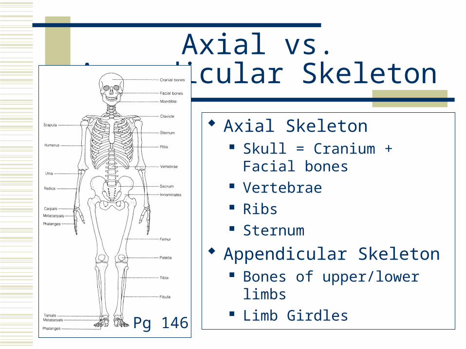
Axial vs. Appendicular Skeleton
Axial Skeleton Skull = Cranium + Facial bones Vertebrae Ribs Sternum
Appendicular Skeleton Bones of upper/lower limbs Limb Girdles
Pg 146
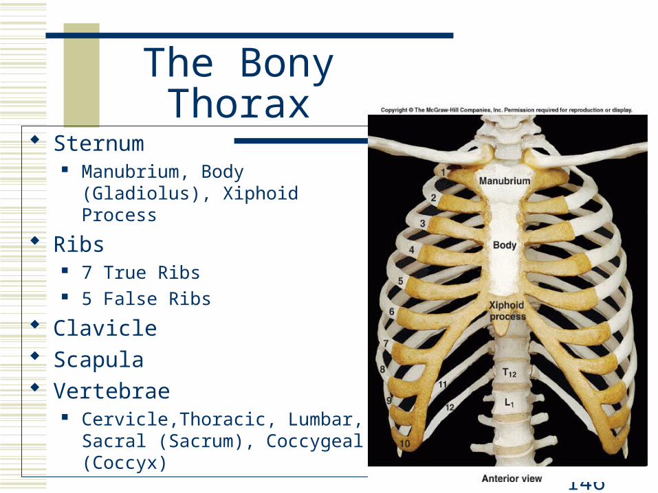
The Bony Thorax Sternum
Manubrium, Body (Gladiolus), Xiphoid Process
Ribs 7 True Ribs 5 False Ribs
Clavicle Scapula Vertebrae
Cervicle,Thoracic, Lumbar, Sacral (Sacrum), Coccygeal (Coccyx)
Pg 146
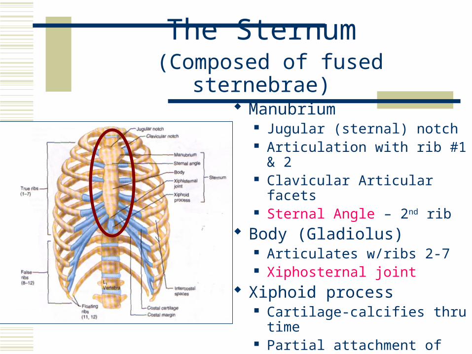
The Sternum (Composed of fused sternebrae)
Manubrium Jugular (sternal) notch Articulation with rib #1 & 2 Clavicular Articular facets Sternal Angle – 2nd rib
Body (Gladiolus) Articulates w/ribs 2-7 Xiphosternal joint
Xiphoid process Cartilage-calcifies thru time Partial attachment of many
muscles
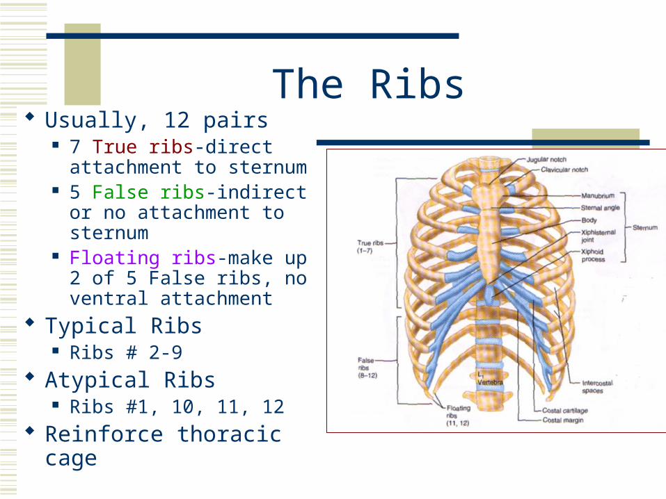
The Ribs Usually, 12 pairs
7 True ribs-direct attachment to sternum
5 False ribs-indirect or no attachment to sternum
Floating ribs-make up 2 of 5 False ribs, no ventral attachment
Typical Ribs Ribs # 2-9
Atypical Ribs Ribs #1, 10, 11, 12
Reinforce thoracic cage
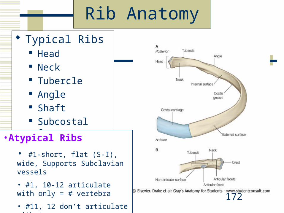
Rib Anatomy Typical Ribs
Head Neck Tubercle Angle Shaft Subcostal Groove
•Atypical Ribs
• #1-short, flat (S-I), wide, Supports Subclavian vessels
• #1, 10-12 articulate with only = # vertebra
• #11, 12 don’t articulate with transverse processes, or anteriorly at all
Pg 172
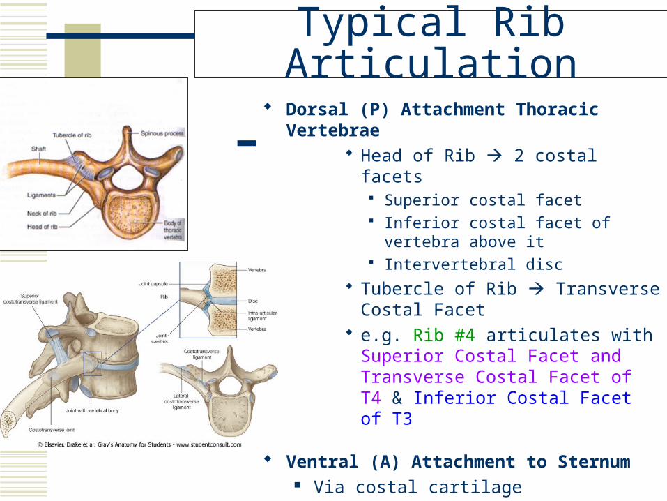
Typical Rib Articulation
Dorsal (P) Attachment Thoracic Vertebrae Head of Rib 2 costal facets
Superior costal facet Inferior costal facet of vertebra above it Intervertebral disc
Tubercle of Rib Transverse Costal Facet
e.g. Rib #4 articulates with Superior Costal Facet and Transverse Costal Facet of T4 & Inferior Costal Facet of T3
Ventral (A) Attachment to Sternum Via costal cartilage
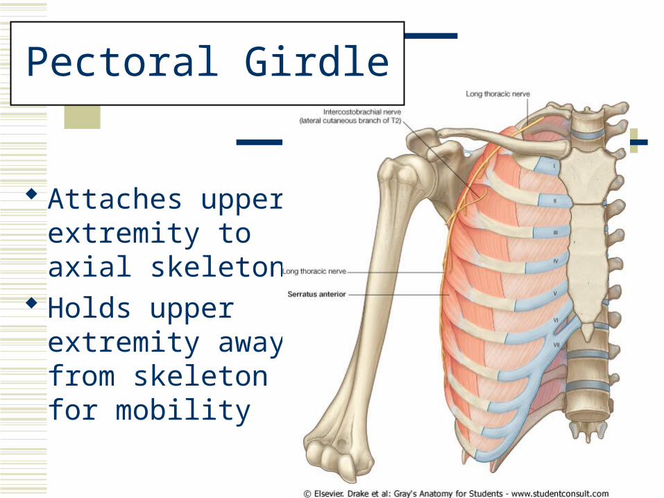
Attaches upper extremity to axial skeleton
Holds upper extremity away from skeleton for mobility
Pectoral Girdle
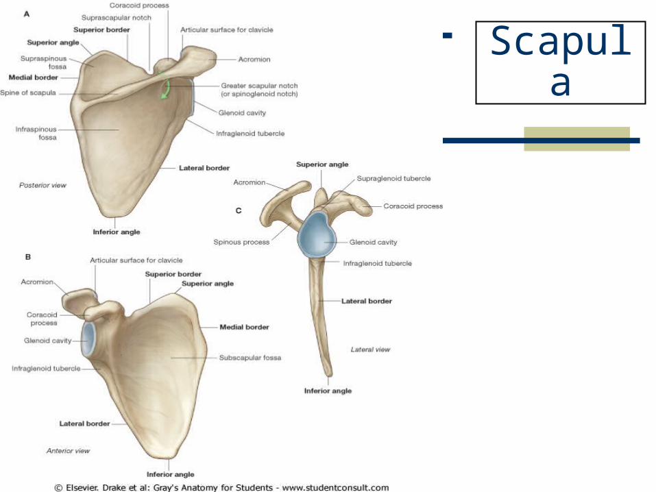
Scapula
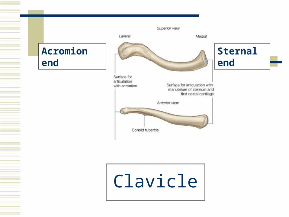
Acromion end Sternal end
Clavicle
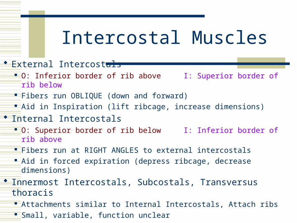
Intercostal Muscles External Intercostals
O: Inferior border of rib above I: Superior border of rib below Fibers run OBLIQUE (down and forward) Aid in Inspiration (lift ribcage, increase dimensions)
Internal Intercostals O: Superior border of rib below I: Inferior border of rib above Fibers run at RIGHT ANGLES to external intercostals Aid in forced expiration (depress ribcage, decrease dimensions)
Innermost Intercostals, Subcostals, Transversus thoracis Attachments similar to Internal Intercostals, Attach ribs Small, variable, function unclear
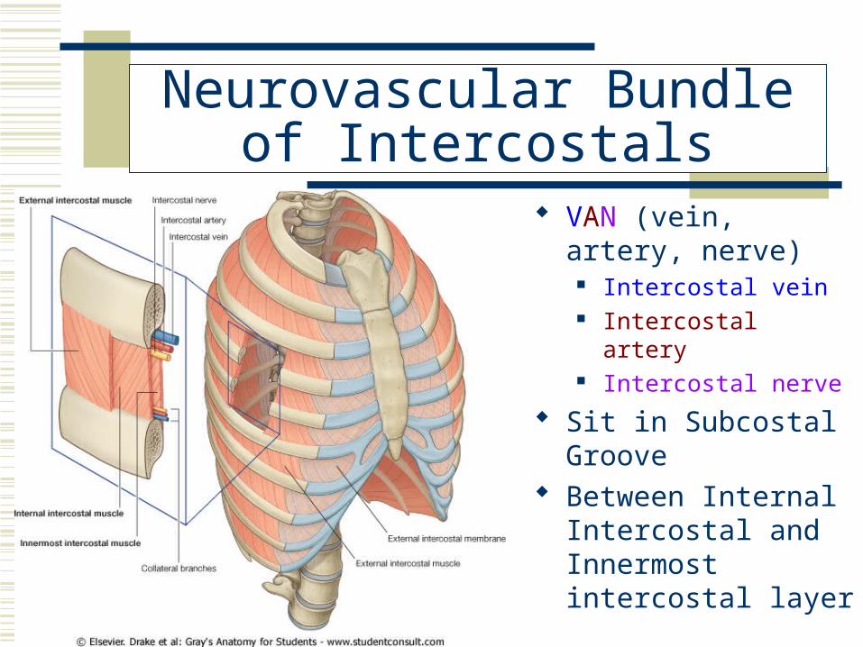
Neurovascular Bundle of Intercostals
VAN (vein, artery, nerve)
Intercostal vein Intercostal artery Intercostal nerve
Sit in Subcostal Groove Between Internal
Intercostal and Innermost intercostal layer
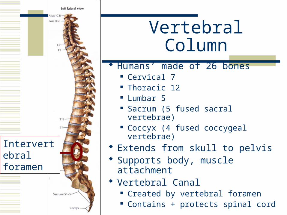
Vertebral Column
Humans’ made of 26 bones Cervical 7 Thoracic 12 Lumbar 5 Sacrum (5 fused sacral vertebrae) Coccyx (4 fused coccygeal vertebrae)
Extends from skull to pelvis Supports body, muscle attachment Vertebral Canal
Created by vertebral foramen Contains + protects spinal cord
Intervertebral foramen
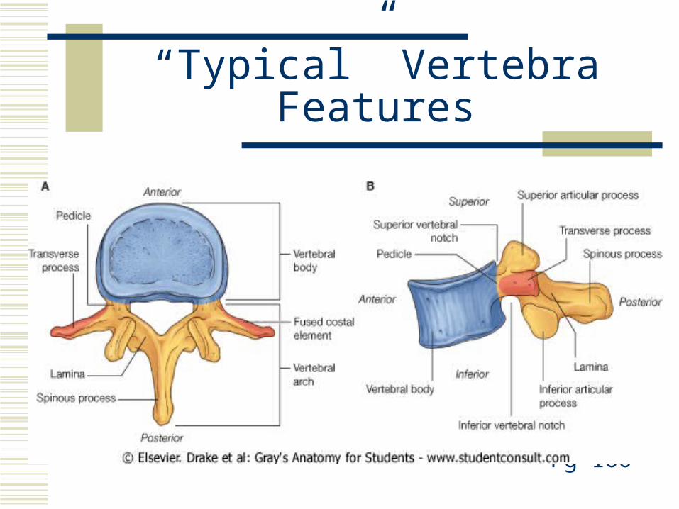
“Typical” Vertebra Features
Pg 166

Cervical Vertebrae (7) **Transverse Foramen ** Superior Articular Facets face
superoposteriorly Inferior Articular Facets face
inferoanteriorly Allows wide range of motion Spinous process fairly short, bifid
(except for C7) Vertebral Foramen is Triangular Body is wider laterally than in A-P
direction
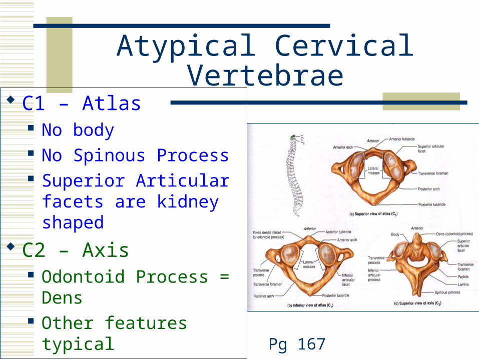
Atypical Cervical Vertebrae C1 – Atlas
No body No Spinous Process Superior Articular facets
are kidney shaped
C2 – Axis Odontoid Process = Dens Other features typical
Pg 167
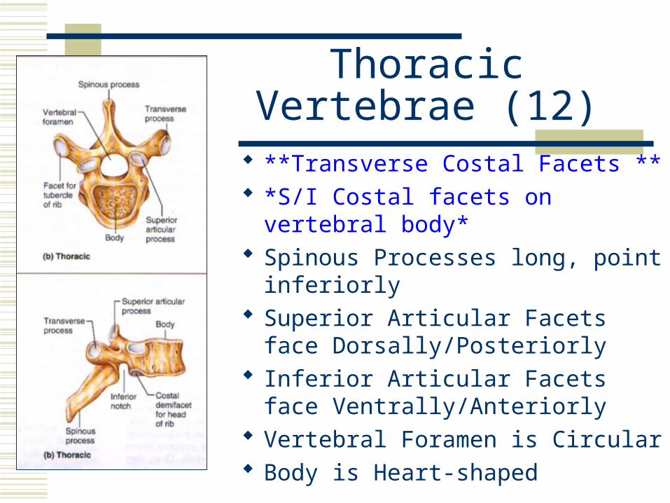
Thoracic Vertebrae (12)
**Transverse Costal Facets ** *S/I Costal facets on vertebral body* Spinous Processes long, point
inferiorly Superior Articular Facets face
Dorsally/Posteriorly Inferior Articular Facets face
Ventrally/Anteriorly Vertebral Foramen is Circular Body is Heart-shaped
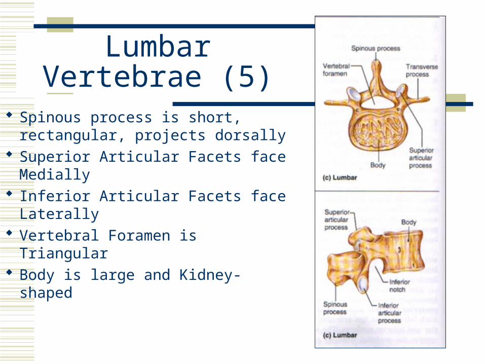
Lumbar Vertebrae (5)
Spinous process is short, rectangular, projects dorsally
Superior Articular Facets face Medially
Inferior Articular Facets face Laterally
Vertebral Foramen is Triangular Body is large and Kidney-shaped
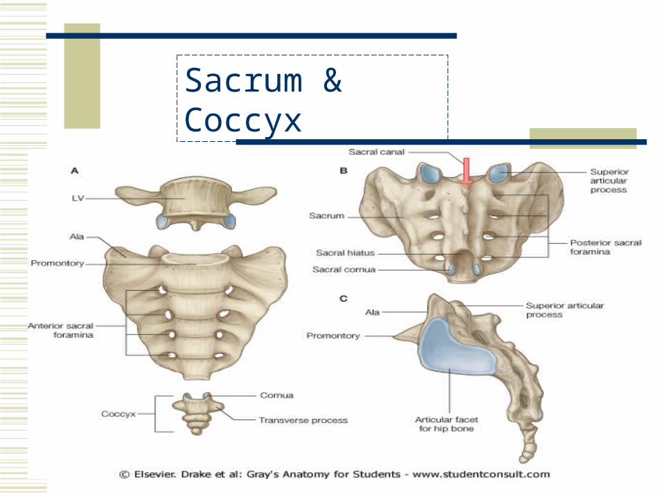
Sacrum & Coccyx
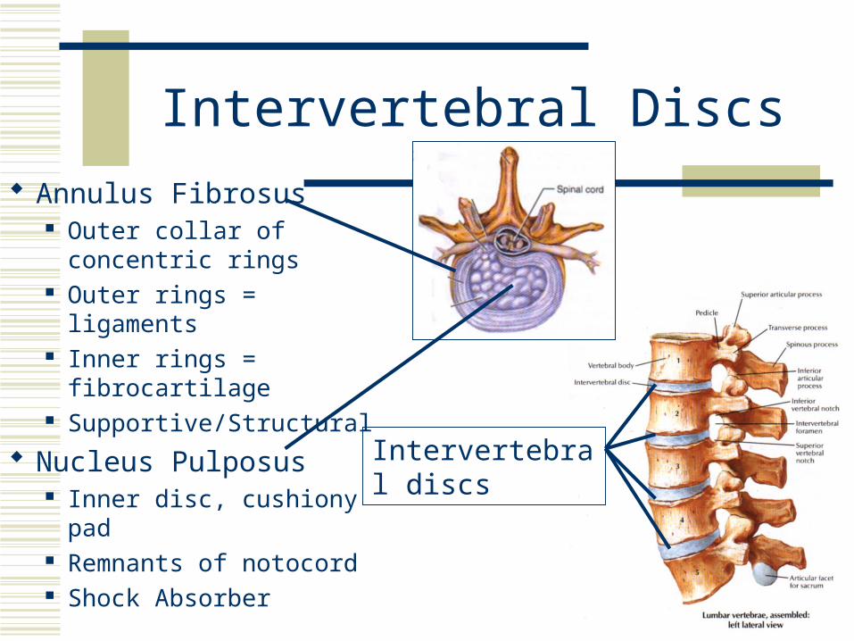
Intervertebral Discs
Annulus Fibrosus Outer collar of concentric
rings Outer rings = ligaments Inner rings = fibrocartilage Supportive/Structural
Nucleus Pulposus Inner disc, cushiony pad Remnants of notocord Shock Absorber
Intervertebral discs
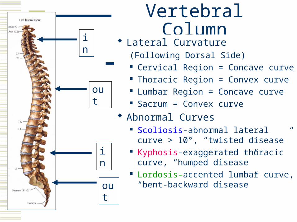
Vertebral Column Lateral Curvature
(Following Dorsal Side) Cervical Region = Concave curve Thoracic Region = Convex curve Lumbar Region = Concave curve Sacrum = Convex curve
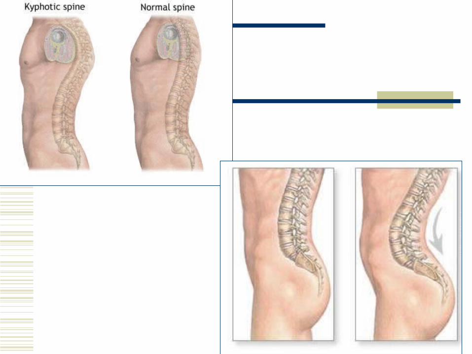
Abnormal Curves Scoliosis-abnormal lateral curve >
10°, “twisted disease” Kyphosis-exaggerated thoracic curve,
“humped disease” Lordosis-accented lumbar curve,
“bent-backward disease”
in
out
in
out
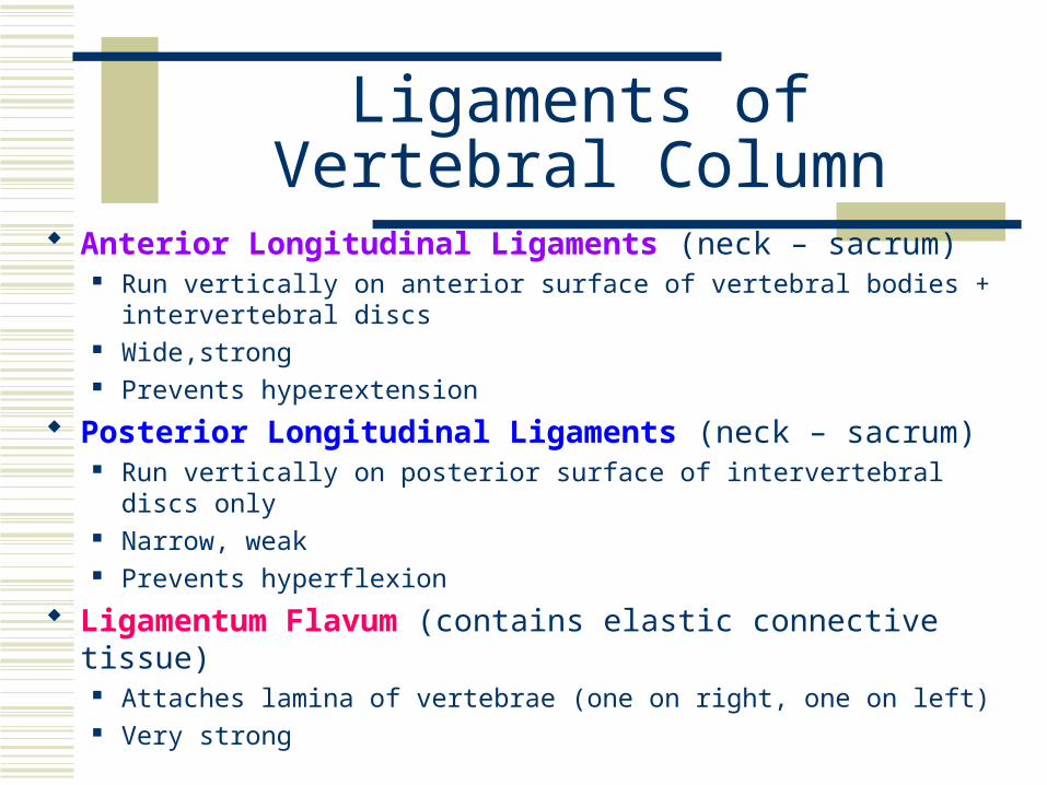
Ligaments of Vertebral Column
Anterior Longitudinal Ligaments (neck – sacrum) Run vertically on anterior surface of vertebral bodies + intervertebral
discs Wide,strong Prevents hyperextension
Posterior Longitudinal Ligaments (neck – sacrum) Run vertically on posterior surface of intervertebral discs only Narrow, weak Prevents hyperflexion
Ligamentum Flavum (contains elastic connective tissue) Attaches lamina of vertebrae (one on right, one on left) Very strong
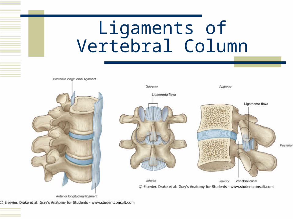
Ligaments of Vertebral Column
Ligamentum flavum
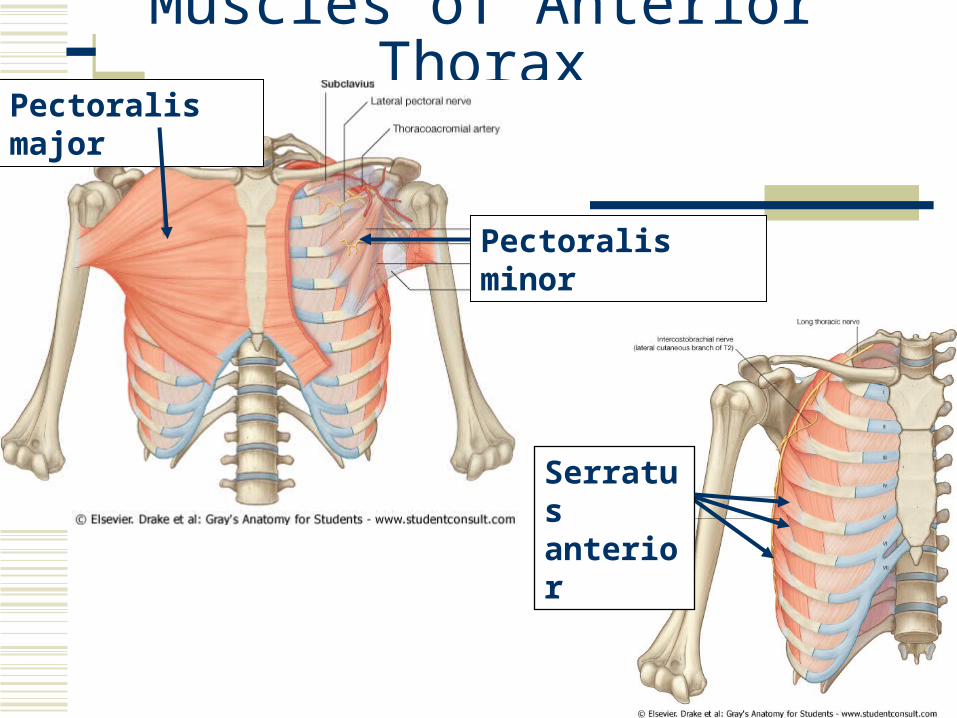
Muscles of Anterior ThoraxPectoralis major
Pectoralis minor
Serratus anterior
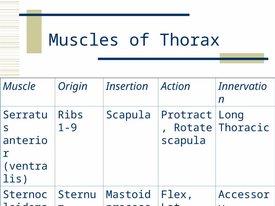
Muscle Origin Insertion Action Innervation
Serratus anterior (ventralis)
Ribs 1-9 Scapula Protract, Rotate scapula
Long Thoracic
Sternocleidomastoid
Sternum, clavicle
Mastoid process, S. nuchal line
Flex, Lat flex, Rotate head
Accessory
Muscles of Thorax
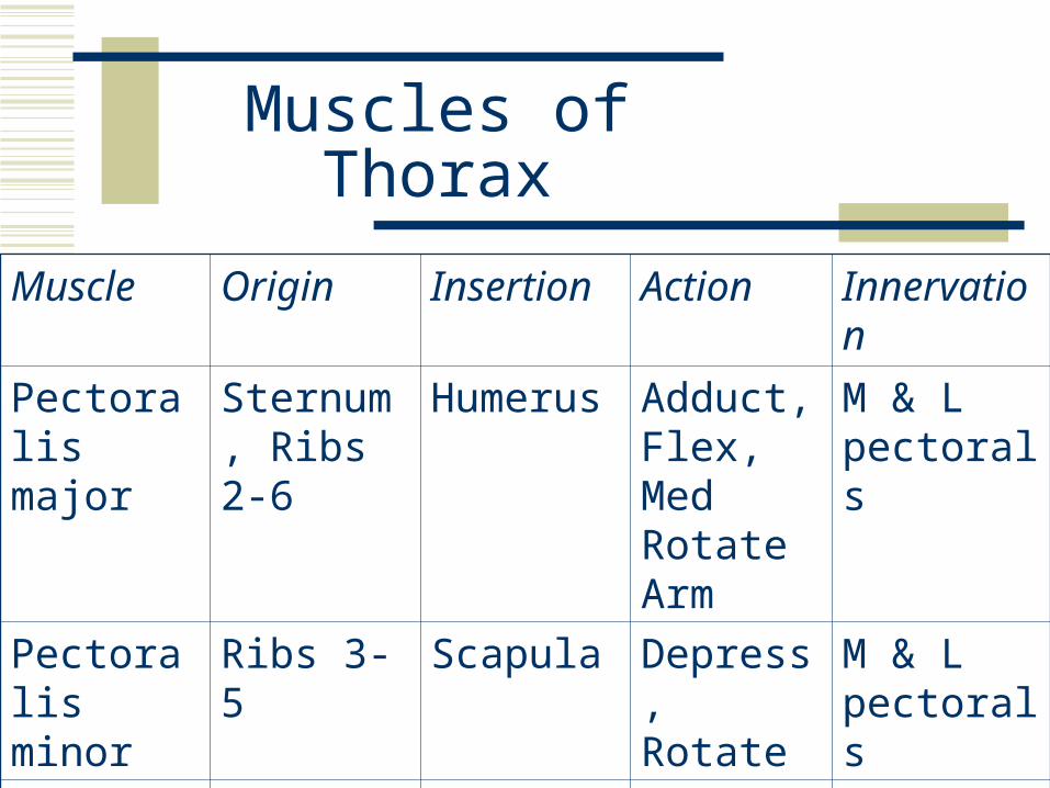
Muscles of Thorax
Muscle Origin Insertion Action Innervation
Pectoralis major
Sternum, Ribs 2-6
Humerus Adduct, Flex, Med Rotate Arm
M & L pectorals
Pectoralis minor
Ribs 3-5 Scapula Depress, Rotate
M & L pectorals
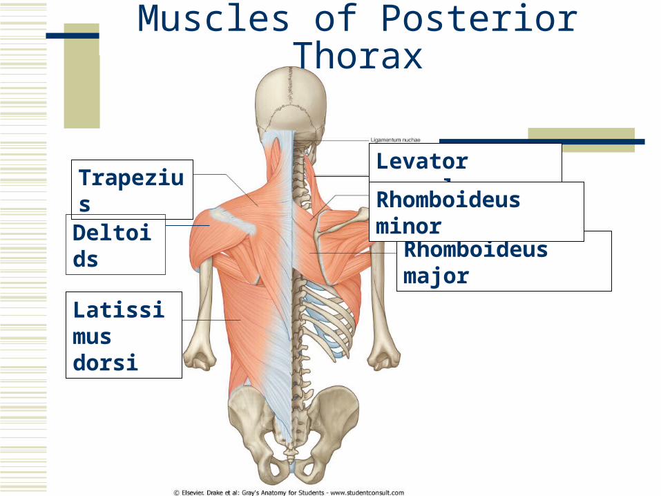
Muscles of Posterior Thorax
Trapezius
Latissimus dorsi
Deltoids
Levator scapulae
Rhomboideus major
Rhomboideus minor
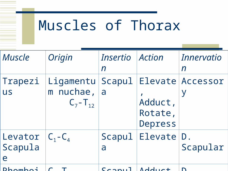
Muscle Origin Insertion Action Innervation
Trapezius Ligamentum nuchae, C7-T12
Scapula Elevate, Adduct, Rotate, Depress
Accessory
Levator Scapulae
C1-C4 Scapula Elevate D. Scapular
Rhomboids C7-T5 Scapula Adduct, Elevate, Rotate
D. Scapular
Muscles of Thorax
Muscles of Thorax
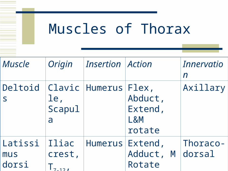
Muscle Origin Insertion Action Innervation
Deltoids Clavicle, Scapula
Humerus Flex, Abduct, Extend, L&M rotate
Axillary
Latissimus dorsi
Iliac crest,
T7-12, Lumbar fascia
Humerus Extend, Adduct, M Rotate Arm
Thoraco-dorsal

Location: (female breast) Superior border: 2nd rib Inferior border: 6th rib Medial border: Sternum Lateral border: Midaxillary line
Location: (male nipple) Fourth Intercostal Space, Midclavicular line
Underlying muscle Pectoralis major and minor Part of serratus anterior, external obliques
Lateral Thoracic Artery, branches of Internal Thoracic A., Post. Intercostals
Intercostal, Internal Thoracic, Axillary Veins Branches of Intercostal Nerve
The Breast
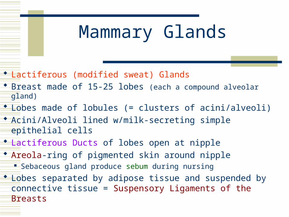
Mammary Glands
Lactiferous (modified sweat) Glands Breast made of 15-25 lobes (each a compound alveolar gland)
Lobes made of lobules (= clusters of acini/alveoli) Acini/Alveoli lined w/milk-secreting simple epithelial cells Lactiferous Ducts of lobes open at nipple Areola-ring of pigmented skin around nipple
Sebaceous gland produce sebum during nursing
Lobes separated by adipose tissue and suspended by connective tissue = Suspensory Ligaments of the Breasts
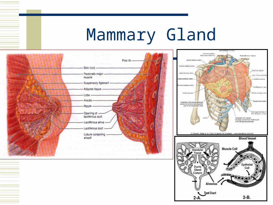
Mammary Gland
Surface Anatomy
Use the next 3 slides and follow the book to
palpate (feel) the features listed
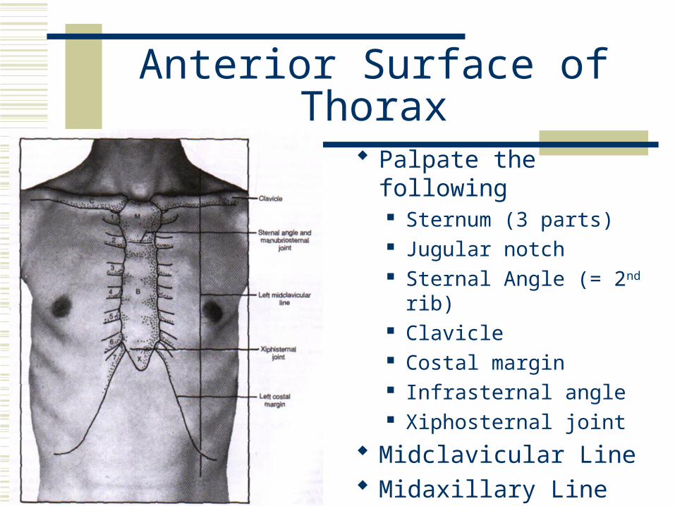
Anterior Surface of Thorax
Palpate the following Sternum (3 parts) Jugular notch Sternal Angle (= 2nd rib) Clavicle Costal margin Infrasternal angle Xiphosternal joint
Midclavicular Line Midaxillary Line
Posterior Surface of Thorax
Palpate the following Spinous Process of C7 Scapula (ribs 2-7)
Scapular spineAcromion Process Inferior Angle of Spine Inferior Border
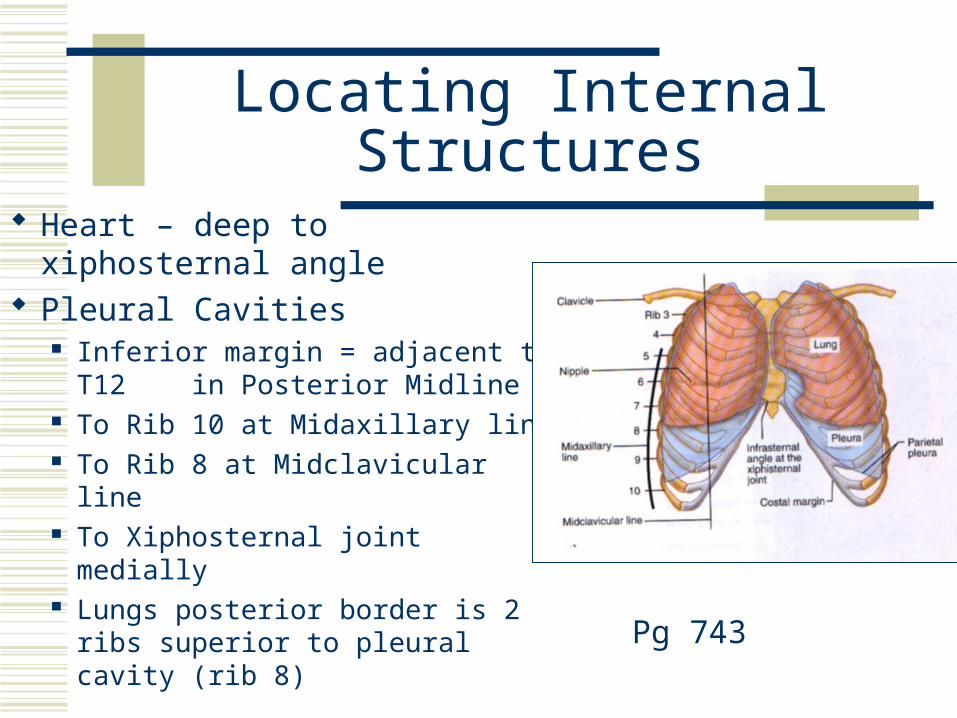
Locating Internal Structures
Heart – deep to xiphosternal angle
Pleural Cavities Inferior margin = adjacent to T12
in Posterior Midline To Rib 10 at Midaxillary line To Rib 8 at Midclavicular line To Xiphosternal joint medially Lungs posterior border is 2 ribs
superior to pleural cavity (rib 8) Pg 743