Embed Size (px)
Citation preview
THE STATUS OP THE GENUS ERYSIPELOTHRIX
George Curtis Langford, Jr.
Thesis submitted to the faculty of the Graduate of the University of Maryland in partial fulfillment of the requirements for the
degree of Doctor of Philosophy
1933
chool
fi

UMI Number: DP70451
All rights reserved
INFORMATION TO ALL USERS The quality of this reproduction is dependent upon the quality of the copy submitted.
In the unlikely event that the author did not send a complete manuscript and there are missing pages, these will be noted. Also, if material had to be removed,
a note will indicate the deletion.
UMIDissertation Publishing
UMI DP70451
Published by ProQuest LLC (2015). Copyright in the Dissertation held by the Author.
Microform Edition © ProQuest LLC.All rights reserved. This work is protected against
unauthorized copying under Title 17, United States Code
uestProQuest LLC.
789 East Eisenhower Parkway P.O. Box 1346
Ann Arbor, Ml 48106- 1346
i
TABIE OF CONTENTSpage
INTRODUCTION - - - - - - - - - - - - - - - - - - - - - - 1
HISTORICAL - - - - - - - - - - - - - - - - - - - - - - ifMATERIALS AND METHODS
MEDIA - - - - - - - - - - - - - - - - - - - - - 3 8
METHODS - - - - - - - - - - - - - - - - - - - - 43
CULTURES - - - - - - - - - - - - - - - - - - - - 47RESULTS
MORPHOLOGY AND CULTURAL CHARACTERISTICS - - - - 53
PHYSIOLOGICAL CHARACTERSEffect of Temperature on Growth - - - - - 62pH Range - - - - - - - - - - - - - - - - - 7 ifHydrogen Sulfide - - - - - - - - - - - - - 75
Catalase,, Nitrate Reduction,, Litmus Milk - 7 6
Fermentations - - - - - - - - - - - - - - 7 7
Tolerance to Sodium Chloride - - - - - - - 84Tolerance to Phenol - - - - - - - - - - - 85
Tolerance to Potassium Tellurite - - - - - 85
Selective-Differential Medium - --- - - - 85
DISCUSSION AND CONCLUSIONS - - - - - - - - - - - - - - 86
LITERATURE CITED ------ 95
ii
TABLES
Number
1 FINAL pH OBTAINED BY ACTION OF VARIOUS STRAINS OF ERYSIPELOTHRIX ON SUBSTRATES IN FERMENTATION MEDIUM NO. 1.
2 FINAL pH OBTAINED BY ACTION OF VARIOUS STRAINS OF ERYS IPELOTHRIX ON SUBSTRATES IN FERMENTATION MEDIUM NO. 2.
3 FINAL pH OBTAINED BY ACTION OF AMERICAN STRAINS OF ERYSIPELOTHRIX ON SUBSTRATES IN FERMENTATION MEDIUM NO. 3.
4 FINAL pH OBTAINED BY ACTION OF STRAINS OF ERYSIPELOTHRIX OBTAINED FROM NATIONAL COLLECTION OF TYPE CULTURES ON SUBSTRATES IN FERMENTATION MEDIUM NO. 3.
page
79
81
82
83
iii
FIGURES
Number page
1 Photomicrograph of ErysipelothrIx sp. shoving organism at 24 hours in Basal Broth Medium 55
2 Photomicrograph of Erysipelothrix sp. shovinglong chains that may appear in certain media 56
3 Photomicrograph of Erysipelothrix sp. shoving granules vhich may appear in certain media 59
4 Growth Curves for Strain No. 175 at 30, 3 5 ,and 37 C 63
5 Growth Curves for Strain No. 50 at 2 5 , 3 0 ,and 37 C 6[
6 Growth Curves for Strain No. 50 at 25 C on two different days 66
7 Growth Curves at 33 C with varied amounts ofStrain No. 55 inoculated into 15 ml medium 6 8
8 Growth Curve of Strain No. 175 at 25 C 6 9
9 Growth Curve of Strain No. 1 75 at 35 C 7010 Growth Curve of Strain No. 175 at 40 C 7111 Growth Curve of Strain No. 1 75 at 41 C 73

INTRODUCTION
The organism Erysipelothrix rhusiopathiae is the etiological agent of svine erysipelas, a disease vhich has long been the cause of a serious illness in the swine of Europe. The resulting economic loss served as a stimulus to the investigation of the disease, its cause, prevention, and cure. Woodbine (1950) states that: ‘’The mortality of svine in Germany (1898-1924) costs over 10 million marks and the disease, diagnosed in 17 of the United States, is suggested as a major cause of the 4.8 per cent of swine condemned in 1 9 3 1 -3 2 after post-mortem inspection for arthritis and other bone diseases.” The incidence in the United States vas rare in the latter years of the nineteenth and the early years of the twentieth century, but in the last thirty years, Its occurrence has increased at such a rate that Breed (1933) listed It as one of the three most important diseases of swine.
The clinical entity known as ’’erysipeloid” in man has also been known for many years, and although its incidence is not such that it is considered a disease of major importance, its occasional appearance in sporadic outbreaks among fish handlers, slaughterhouse employees,
2
veterinarians, butchers and other people coming in contact vith meat, fish and sea-food or their products, makes it of considerable importance from a diagnostic standpoint.The name E . erysipeloid!s has been given to organisms isolated from such cases.
Many early investigators noted that when rotten, putrid or decayed material such as horse meat or blood was injected Into mice they died in a few days of an overwhelming septicemia. The organism which caused this mouse septicemia is known as E. muriseptica.
There has been much confusion and controversy In the classification and nomenclature of these organisms. The ‘'splitters1' have cited differences in origin, morphology, and cultural characteristics as reasons for considering them as three separate species of the genus Erysipelothr!x while the “lumpers" disclaim these reasons, ascribe them to variability of the organism, and on the basis of practical identity from a biochemical and serological standpoint wish to consider them as all one species.
The present work vas undertaken with the object of clarifying the status of the members of the genus Erysipe- lothrlx. A large number of these strains representing all of the species listed vhich are available have been collected. These have been studied from a standpoint of their

clnssi fieat ion and idcnt I f:! cat i on . The indorsation thus
obtained has been compared with all previous reports in
order to determine whether there is .justification for
retaining three species of this genus.
4
HISTORICAL
Breed e_t al (1948) list Erysipelothrix as one of the three genera under the family Corynebacteriaceae Lehmann and Neumann. At various times since their discovery, these organisms have been placed in the genera: Bacillus,Pasteurella, Streptothrlx, Babesia, Oospora, Bacterium, Mycobacterium, Nocardia, Discomyces, Actinomyces, and now finally in Erys1pelothrix. The fact that these organisms could have been considered as members of such diverse groups may be attributed to: (1 ) the extreme variabilityof the organisms as observed under different conditions by the various investigators, (2 ) the changes made by the systematists In the definitions of bacterial genera during the last sixty years, and (3 ) the difficulty of classifying the organisms from the conflicting and sometimes very meager descriptions given by previous workers. In addition, there have been reported by several authors, a number of cases in which organisms possessing the characteristics generally ascribed to this group have been isolated from various sources, the earliest being mice, then swine, and then man. Recently they have also been isolated from a variety of animals including dogs, cats, foxes, rats, mink, deer, and
birds such as turkeys, pigeons, pheasants, parrots and sparrovs. The organisms have also been isolated from different species of fresh and salt water fish and crabs.
In the cases of the earlier isolated types from mice, svine, and man, the cultures from these sources were considered as various species of the same genus and were called Erysipelothrix muriseptica, E . porci, and E. ery- sipeloldes respectively. They were so divided because of the marked variability of the microbe in the different animals, although the early Investigators noted the similarity of the three types. Considerable difficulty was encountered by some workers in infecting swine experimentally with the organism. Mice and pigeons were, however, usually readily susceptible, while svine were much more resistant. In addition, the strains lost their virulence or were considerably weakened after many laboratory transfers on artificial media. These facts were not known by the early workers and lead to this division of the genus into three species. Although several papers have been written both for and against this division, there has never been sufficient evidence elucidated to allow a definite separation and identification of the three as they are now listed.
One of the earliest accounts of the disease in the
6
literature vhich may he attributed to organisms of the genus Erysipelothrlx is given by Baker (l87'3). He described cases of erythema serpens in which the patients shoved mild erythema vhich vas pink rather than red, no axillary lymphatic involvement, history of a slight injury, probably a scratch, no pus, and a marginal advancing redness. The disease vas of 2 to 6 weeks duration with eventual disappearance of the lesions. From this description we can be almost sure that these patients were infected with organisms of the genus Erysipelothrix. Baker, of course, had no knowledge of the etiology of the disease.
Koch (1881), in an article discussing studies vhich he had made of pathogenic organisms, mentioned the ’’Bacillus of mouse septicemia” vhich vas isolated from mice that had died of septicemia. This septicemia vas caused by injection of putrid blood or meat infusion broths. Very little information vas given about the organism except for a brief description of the appearance of its growth in nutrient gelatin:
"Colonien von Bacillen der Mausesepticamie in Nahrgelatine geimpft. Namentlich im o b e m Theil des Impfstriehs erscheint die eigenthumliche, verzveigte Form der kleinen Colonien.”
This was the first report of a member of the genusErysipelothrix found in the literature.
Pasteur in a letter to Dumas (1 8 8 2 ) stated that these organisms vere very similar to the agent of fowl cholera with the exception that they were smaller and differed in their physiological properties (no specific details were mentioned). He was able to cause the disease in pigs by inoculation with the culture and observed that rabbits and sheep were also affected by cultures of the organism. He mentioned the work of Klein, who claimed to have isolated the etiological agent of swine erysipelas, but who had described the organism as a bacillus with many spores. Although the disease entity known to the Germans as "rotlauf” or ’’schweinesrothlauf", to the French as ”rouget” or ”mal rouge des pores”, and to the English speaking people as "swine erysipelas” had been recognized for some time, this is one of the first reports concerning the bacteriology of the disease.
The next year (I8 8 3 ) Pasteur and Thuillier made use of a culture of the organisms to produce immunity in swine. This was one of the first experiments using the causative agent of a disease for this purpose.
Trevisan (l8 8h) gave the name Bacillus insidiosus to the bacillus of mouse septicemia. This is the first binomial term used for this organism. Ho description of the organism was given but it was listed under the group
8
of bacteria which fomed spores,Schroeter (1886) classified the mouse septicemia
bacillus as Bacillus murlnus. The organisms occurred frequently in pairs and sometimes in chains of four. No filaments were formed in the blood of the mouse, but they could be seen in cultures. Spores were often formed. Gelatin was never liquefied, but the organisms formed outgrowths like clouds in the medium. They were found in the blood and meat infusions which had putrefied. In the same year (1886) Flugge called this microbe Bacillus murisepticus.
Loeffler (1886) isolated from dead pigs an organism which killed a guinea pig and a mouse into which it was inoculated. He considered this organism to be different from that which caused mouse septicemia. Schutz (1886) reported on the inoculation of pigs with the organisms isolated from cases of swine "rotlauf" and noted that while some of the pigs showed symptoms, many remained apparently quite healthy and the organisms which had been injected could not be recovered from their blood, spleen, or joints at autopsy. He considered the agent isolated from cases of "rotlauf" to be identical to that causing "rouget du pores" and similar to that causing mouse septicemia. Experiments indicated the possibility of immunizing pigs against erysipelas by injections of cultures of
9
the causative agent.In his earlier studies of human erysipeloid, Rosen-
bach (1887) isolated an organism from a case of erythema migrans and placed it in the group Cladothr jbc. He vas able to produce typical symptoms of erysipeloid in himself by Inoculating his arm with this organism.
Trevisan (I8 8 9 ) in his description of the genera and species of bacteria, classified the causative agent of rotlauf as Bacillus thulllieri. In the same year however, (1 8 8 9 ), DeToni and Trevisan changed the genus name and called it Pasteurella thuillierl.
In 1 8 9 1 , Emmerich and Mastbaum reported the Immunization of rabbits by intravenous injection of a fully virulent culture of "rotlauf" bacilli. Antisera from these rabbits was used to protect mice and other rabbits from Injections of the fully virulent culture.
According to Brumpt (1 9 2 7 ) the name Oospora rosen- bachi Savageau and Radais 1 8 9 2 , was listed as a synonym for Actinomyces rosenbachi.
Moore (I8 9 2 ) Isolated an organism from the spleen of pigs with acute septicemic lesions which he called the bacillus of mouse septicemia. It vas a slender, non-motile gram-positive rod which showed occasional filaments in culture. The colonies were bluish gray by reflected light
10
and the growth In gelatin showed a f'test tube brush" appearance. It was pathogenic for mice and pigeons but rabbits and guinea pigs were immune. Long cultivation seemed to attenuate its pathogenic powers for mice. Pigs which were inoculated with cultures of the organism developed no symptoms.
Preisz (I8 9 2 ) described several morphological and cultural differences between the swine and mouse strains of the organism and on the basis of these differences considered them to be different organisms.
In an attempt to clarify the reason for the pathogenicity of the organism of "schweinerotlaufs". Petri and Massan (1 8 9 3 ) noted the presence of the characteristic "test tube brush" appearance of the growth in gelatin stabs, and by the use of lead acetate paper suspended over the culture medium demonstrated that the organisms produced hydrogen sulfide.
Kitt (1 8 9 3 ) called the causative agent of swine erysipelas Bacillus rhuslopathiae suis, described them as very small bacilli, 1 to 1 . 5 long and 1 /7 as wide as a red blood cell. He mentioned the "test tube brush" appearance in gelatin and the fact that the organisms killed mice and pigeons regularly and would sometimes kill rabbits.
Smith (1 8 9 5 ) considered the bacillus of mouse
11
septicemia to be the same as the bacillus of svine erysipelas. He described these bacilli as gram-positive rods vhich formed acid in glucose and lactose, and vere pathogenic for mice and pigeons. These organisms gave a "test tube brush" appearance in gelatin stabs, but this character did not appear vhen the medium vas alkaline.
Migula (1895) called the rotlauf organism Bacterium erysipelatos suutn and that of mouse septicemia Bacterium murisepticum. He considered them to be different species and said that the cells of B. erysipelatos suum vere alvays shorter and thinner than those of B. murisepticum.
Kruse (1 8 9 6 ) described the organism vhich caused erysipeloid in man as groving in very fine branched filaments vith formation of spores. He said that it grev in gelatin stabs vith the characteristic fuzzy lateral out- grovths from the line of inoculation and called it Strep- tothrix rosenbachli.
Lehmann and Neumann (1 8 9 6 ) said that the organism of erysipeloid most nearly resembled that of mouse septicemia, but named it Qospora erysipeloidis. They said that it grev better at 20 C than at 37 C.
An antiserum vas obtained from rabbits by LeClainche(1 8 9 7 ) vhich protected mice against virulent cultures ofthe organisms isolated from cases of rouget.
12
The trinomial Bacterium rhusiopathlae suis vas given to the organism by Chester (I8 9 7 ) in his classification of the bacteria, but in a later classification (1 9 0 1 ) he changed it to Mycobacteriam rhusiopathlae. He described this organism as a very small bacillus, slender, bent or curved vhich also formed filaments. It vas 0.2 to 0.6 ja by 1.8 p. In the depths of gelatin stabs, gray cloudy radiating outgrovths appeared vith softening of the gelatin- after some time. Indol vas not produced. The organism vas pathogenic for mice, vhite rats, and pigeons in 3 to 4 days. The mice died in a sitting position. He also listed Mycobacterium murisepticum as: "Probably identical viththe preceding."
Lachner-Sandoval (I8 9S) classified the agent of erysipeloid as Actinomyces eryslpeloides, vhile Migula (1900) thought that it should be called Bacterium rhusiopathlae. Migula considered B. rhus iopathlae to be different from the bacillus of mouse septicemia.
Prettner (1901) indicated that the immunity of svine to the rotlauf bacillus depended upon many things, among them: age, particular strain of pigs, and number of transfers of the organism in artificial media or in other animals. He claimed, for these reasons, that the results obtained by Preisz vere not sufficient to justify the latters conclusions.
13
He substantiated this view by injecting pigs of various ages with organisms directly from spleens of swine sick: with rotlauf. He also compared several strains of the bacillus of mouse septicemia with strains of freshly isolated rotlauf bacilli and concluded that they were Identical. He stated that the name Bacillus murisepticus was used incorrectly and should be removed from the literature.
The same year^ (1901), Lubowski Isolated from the feces of a 5 year old child with icteric intestinal catarrh, an organism with the following characteristics: Itwas a fine, small, gram-negative (!) rod. The broth culture became cloudy and the organism formed hydrogen sulfide. In gelatin stabs, it developed small knotty vegetations along the line of Inoculation and after several transfers It took on the characteristic ’’test tube brush11 appearance. It was very strongly pathogenic for mice. He found that they could be protected by antiserum against the swine erysipelas organism. After disappearance of the icterus no rotlauf bacilli could be recovered from the stool.
Gedoelst (1902) classified the organism of erysipeloid as Discomyces rosenbachi.
Gilchrist (1904) reported 329 cases of dermatitis, mostly in fish handlers, in which 323 were caused by crab bites or lesions produced by crabs. Ho organisms were
14
found in cultures or in histological sections of the lesions, but the erysipeloid condition was thought to be caused by the bacillus of svine erysipelas.
Caminiti (190?) in a discussion of the genus Strep- tothrlx, listed Streptothrix erysipeloides as one of the species in this grouo. No description vas given of the organism.
A very complete early study of erysipeloid and svine erysipelas vas done by Rosenbach (1909). He found that the agent of mouse septicemia behaved in its pathogenicity just as that of svine erysipelas and as erysipeloid of man. Pigeons were susceptible to the microbes as well as mice. Rabbits vere moderately susceptible and guinea pigs vere completely immune. Rosenbach gave an excellent description of the clinical picture of the disease in man, svine, and mice. He attached considerable importance to the extent of the growth of various strains in gelatin and also to morphological changes between the three strains. He only worked vith a few cultures vhich he divided into the following species; Erysipelothrix porci, E. erysipeloidis and E. muriseptica.
Rosenbach studied one culture only from erysipeloid, as pointed out by Rickmann (1909), and vas therefore liable to error. Rickmann studied Rosenbach1s culture from erysipeloid and over 1 0 0 others isolated from rotlauf cadavers.
15
He said that he could find no peal morphological difference in a large number of cultures from rotlauf, erysipeloid, and mouse septicemia and concluded that they were absolutely identical, since the immune and agglutination reactions were also about the same for all three organisms.
The organism was classified as Nocardia rosenbach! by Castellan! and Chalmers (1913) stnd vas described only very briefly as follows: "Mycelial threads, very slender, some terminate in club-like swellings. Can be cultivated on the usual laboratory media. Does not liquefy gelatin."
Crimi (191*0 gave an excellent description of the characters affecting the growth of the organism in gelatin stabs. He noted that the growth from all strains vas not necessarily always of the "brush" type in this medium. This characteristic depended, he found, upon the number of transfers in gelatin, the type of nutrient material added, and the pH of the medium. He found that cultures vhich did not at first give "brush" type growth, could usually be induced to do so after having been carried through several transfers at a neutral or slightly alkaline pH. This type of growth vas better in fresh meat infusion broth vith gelatin added than in solutions of Liebig Meat Extract vith gelatin added. The gelatin vas never liquefied regardless of the type of growth. The production of hydrogen sulfide could not be
16
demonstrated with lead acetate paper and the organisms did not reduce neutral red or methylene blue. In a broth containing potassium tellurite * a brownish color was formed in the medium and a black deposit settled out after several days Incubation. Catalase was never formed.
Cotoni (1919) studied 11 strains, of which 9 were isolated from swine and 2 were obtained from Jouan and Cesari. These non-motile, gram-positive rods grew as very small transparent colonies on agar. Some cultures split glucose, hydrogen sulfide was produced and they did not liquefy gelatin or coagulated serum. They were insoluble in bile and had no hemolytic action against guinea pig, rabbit, sheep, horse and cow blood cells. One culture which was sealed in a Martin’s broth tube at 37 C kept for 5 years. All strains were pathogenic for mice and pigeons, while rabbits survived large doses and guinea pigs were completely resistant. He called them "bacille du roget".
Tenbroek (1920) maintained that there was no essential difference between Erysipelothrix rhus iopathlae and Bacillus murisepticus. He showed that both would form the "test tube brush" type of growth in gelatin stabs, but It did not happen when the medium was acid. This was, incidentally, in direct contradiction to Smith’s (cit.) statement. Hydrogen sulfide was formed, blackening lead acetate
17
medium in 2.K hours. Acid was formed in glucose and lactosebut no acid was formed in sucrose, xylose, dulcitol, manni-tol, inulin, salicin, dextrin, starch, maltose, and glycerol.There was a distinct zone of hemolysis around the colonies.Indol was not formed. The organisms were quite pathogenicfor gray and white mice, pigeons and sparrows.
Winslow et al (1920) listed the genus ErysipelothrixRosenbach with the following description:
Rod shaped organisms with a tendency to the formation of long filaments which may show branching. The filaments may also thicken and show characteristic granules. No spores. Non- motile. Gram-positive. Do not produce acid. Microaerophilic. Usually parasitic. The type species is Erysipelothrix rhusiopathiae (Bacillus rhusiopathiae suls Kitt, 1093; Mycobacterium rhus iopathiae Chester, 1901; E rys i pe1othr ix porci Rosenbach, 1 9 0 9 ), the causal organism of swine erysipelas.
In addition, two species were listed under this genus by Holland (1920): (1) Erys ipelothrix erys ipelatos-suis and(2) Erysipelothrix rhus1opathiae Kitt.
Glucose, lactose, and fructose were reported by Creech (1921) to be fermented. Andrade*s indicator was used. There was no indication of acid in arabinose, dulcitol, galactose, inulin, maltose, mannitol, rafflnose, sucrose, salicin, and xylose. The organism was easily passed in pigeons.
Dumont and Cotoni (1921) isolated an organism
18
resembling the bacillus of "rouget du pore" from the cerebrospinal fluid of an Italian soldier who died of meningitis. They noted two types of growth in gelatin stabs: "beaded11
and "brush" forms. There was no liquefaction of the gelatin and the organisms were very viable over long periods of time.
Neveu-Lemaire (1921) classified the agent of rouget as Bacillus ruboris suis. He said that it was of very small dimensions (1 p long). It occurred as Isolated rods or in pairs In the blood and was a gram-positive, non-motlie rod. The organism was a facultative aerobe or anaerobe, but preferred anaerobic conditions. It did not liquefy gelatin.
Using both precipitin and agglutinin methods,Teichmann (1922) was not able to demonstrate any clear difference between individual strains of svine rotlauf.
Zibert (1922) studied 25 cultures Isolated from swine and mice, some freshly isolated and some passed through doves, mice, or on media for many passages and found that acid was produced in many carbohydrates. The strongest acid production was in lactose, then maltose, glucose and galactose.It was somewhat weaker in fructose, sucrose, arabinose, and rhamnose. Still weaker but definitely acid were mannitol and dulcitol. There vas absolutely no acid In glycerol. Organisms Isolated from swine and mice were morphologically
19
the same. He concluded there fore, that the kinds of rotlauf bacilli (from mice or svine) could not be differentiated by chemical or morphological tests. He said that chemical tests vere not reliable and morphological tests vere not constant.
As a result of microscopic studies made from sections of gelatin and agar cultures, Zosel (1 9 2 2 ) decided that Baclllus murisepticus acted just like the svine rotlauf bacillus in these media.
From a fatal disease of young pigs, Giltner (1922) isolated an organism vith the following properties: Leadacetate vas blackened in 24 hours and indol vas not formed. Acid but no gas vas formed in glucose, galactose, lactose, maltose, and fructose. There vas no acid formed in sucrose, salicin, mannitol, dulcitol, arabinose, or raffinose.
Bergey (1923) said that Pasteurella muriseptica vas gram-negative. The organism had a filiform arborescent growth in gelatin stabs. It did not form indol nor did it reduce nitrates. It vas microaerophilic and the optimum temperature vas 37 C.
The viability of rotlauf bacilli in various, soils vas reported by Hesse (1924) to vary considerably. The most important factor was the reaction of the soil. The organisms could be recovered after 90 days from alkaline soil vhich had been Inoculated and he believed that they could
20
probably live a year in such soil. In acid soils, theydied out in a day or so.
Cornell and Glover (1925) isolated organisms from joint-ill in lambs. These organisms gave a characteristic appearance in gelatin stabs. They produced acid but no gas in glucose, lactose, maltose, and fructose and slight acid in inulin. There was no change in sucrose, mannitol, dulcitol, salicin, arabinose, and glycerol. The organisms were recovered from the heart, spleen, kidney, articular cavities and subcutaneous abcesses of lambs which had died of erysipelas infection. 1 ml of the culture inoculated subcutaneously was pathogenic for lambs.
Klauder (1 9 2 6 ) indicated that the organism of erysipeloid in man and the organism of svine erysipelas werethe same. He stated that there was no report of the infection of man in this country up to the date of the article. However, in a publication appearing only a short time later, Klauder, Highter, and Harkins (1 9 2 6 ) gave a very good clinical description of the disease and stated that it was familiar to physicians along the New Jersey coast and that it also appeared from Maine to Florida.
Lehmann and Neumann (1927) classified the erysipeloid organism as Bacterium erys ipeloides, and retained the names Bacterium murisepticum and Bacterium erysipelatos suum
21
already given these organisms.Brumpt (192Y) classified the organism as Actinomyces
rosenbachl (Kruse I8 9O). He gave no information about the organisms except to note the characteristic appearance in gelatin and the appearance of the lesions.
Harkins (I927) demonstrated that the morphological, biological and serological characteristics of organisms isolated from erysipeloid of fish-handlers were alike. He considered the etiological agent to be the same as that of swine erysipelas.
Another report of viability was by Helm (1 9 2 8 ) who found that cultures of rotlauf bacilli in broth could remain viable and virulent for a year if the tubes were sealed to avoid the loss of moisture.
Spryszak and Szmanowski (I929) grew the organisms in broth with antiserum added and eventually obtained two types of colonies; a smooth regular colony and a rough type with the surface folded and granular with a dentate border. The normal smooth colonies contained short bacteria and the rough colonies had long filamentous chains of bacteria.
In an investigation of arthritis in lambs, Marsh (1 9 3 0 ) isolated organisms which he considered identical with those of swine erysipelas. He reported a green zone
22
of hemolysis around deep colonies. The organism formed acid in glucose, fructose, lactose, and gelatose. No Acid was formed in sucrose, mannitol, salicin, dextrin, maltose, and glycerol. The organisms killed mice easily and caused arthritis after a month or so in lambs.
Vuillemin (1931) classified the organism of rouget as Nocardia thuillierl.
Meyn (1931) in addition to describing the rough and smooth forms of colonies of rotlauf bacteria, also indicated that the smooth type formed an even turbidity in broth cultures and that the rough type formed a fine or heavy flocculant turbidity. He also said that the smooth colonies did not give the characteristic appearance in gelatin stabs, but that the rough colonies would do so. These two types could be changed back and forth by cultivation on artificial media or by animal passage.
Marsh (1931) reported further that organisms isolated from arthritis in lambs fermented glucose, fructose, lactose, gelatose, and arabinose. They failed to ferment sucrose, mannitol, salicin, dextrin, maltose, and glycerol.
Human infection was more frequently obtained by contact with fish and crustacea than by contact with swine flesh according to Klauder and Harkins (I9 3I).
Schoening, Creech and Grey (1932) described a
23
diagnostic test for svine erysipelas which depended upon the agglutination of cultures of the organisms by blood or serum from the suspected animal.
Creech (1933) tried the inoculation of young pigs with cultures of the bacillus of swine erysipelas and found that only 11 of 22 inoculated with the organism showed definite symptoms of the disease. Of these 11 only 5 died.
Typical erysipelothritic arthritis could be caused in lambs by exposing the stump of the umbilical cord, the docking wound, or the castration wound to soil which had been impregnated with cultures of E. rhusiopathiae, as was shown by Marsh (1933). In a later publication (Marsh, I933t>) he showed that Erysipelothrix strains from lambs and swine were serologically identical.
Schoening and Creech (1933) reported on an improvement of the agglutination diagnostic test by using a killed antigen rather than live broth cultures.
Sewage from Konigsberg was injected into mice by Hettche and Daneel (193*0 and organisms which resembled rotlauf bacilli were isolated from the mice.
The swine erysipelas organism was reported by Stiles and Davis (193*0 ferment glucose, lactose, fructose, and galactose. No acid formation occurred in dulcitol, sucrose, salicin, or mannitol. Pigeons and white mice were
2k
susceptible to the disease,, succumbing in 2 to 5 days .An outbreak of 210 cases of erysipeloid occurred
in the first 10 months of operation of a bone button factory (McGinnes and Spindle, 193*0- The organism was isolated from the workers by injecting and reaspirating sterile saline and then injecting this into mice. Cattleand hog bones were used in this factory.
Material such as scrapings from marine and fresh water fish, frogs, houseflies, and putrefied horseflesh were incubated in broth at 37 C and 0 . 2 ml of this broth was injected subcutaneously into mice by Kondo and Sugimura (1935). Most of the mice died in several days showing septicemic infection with rotlauf bacilli. Schoop (1 9 3o) also found erysipelothrix organisms on many different types of fish. His cultures would not grow in 2.7 per cent sodium chloride medium.
The organism of swine rotlauf was isolated by Mikschofsky (1 9 3 6 ) from the blood and heart valves of a dogwhich had died of endocarditis.
A pure culture of the bacillus of swine erysipelas was obtained from the autopsy of each of 20 turkeys which had died within a short time of each other in a single flock (Beaudette and Hudson, 1936). All strains produced characteristic growth in gelatin stabs and blackened lead
acetate agar along the path of Inoculation. Fermentations were studied in two series. In the first, acid was produced in arabinose, glucose, lactose, galactose, and fructose with the exception of one strain which failed to attack arabinose. (In all cases only slight acid was formed in arabinose). The authors stated that there was some suggestion of acid formation in xylose. The organisms did not attack adonitol, amygdalin, dextrin, dulcitol, erythri- tol, inositol, salicin, inulin, maltose, mannitol, melezi- tose, raffinose, rhamnose, sorbitol, soluble starch, sucrose, and trehalose. In the second series, galactose, fructose, and inositol were not Included. Glucose and lactose were regularly fermented as in the first series but no strains attacked arabinose. A single strain produced a very slight acid reaction In xylose. The substrates not attacked In the first series were likewise negative in the second series.
Deem and Williams (1936) studied 37 strains of E. rhuslopathlae, of which 26 came from Europe. With a medium at pH 7.2 using Andrade!s indicator they found that glucose, lactose, fructose, and galactose were fermented by all strains mannose was fermented by 2 0 of the 37 strains, arabinose and xylose were fermented by a few strains each and the following carbohydrates gave no acid: maltose, sucrose, raffinose,dextrin, inulin, starch, salicin, dulcitol, glycerol, and

2 6
mannitol.A liver digest medium with the addition of 1 per
cent Bacto-peptone and 0.5 per cent sodium chloride was suggested by Vawter (1937) as a good culture medium for ]£. rhusiopathiae. The medium was adjusted to pH 7.6 or 7*7 before sterilizing and 2 to 4 per cent of sterile horse serum was added after sterilization.
In further studies on the occurrence of the rotlauf organism in city sewage, Hettche (1937) indicated that he had been able to obtain these microbes from the sewage of Konigsberg and Munich a number of times. He also found that the organisms could exist for from 4 days to several weeks in sea water, aquarium water and sewage. The viability of the organisms was greater In aquarium water and sewage than in tap water.
Schoening, Gochenour and Grey (1938) made a study of the smooth and rough forms of E. rhusiopathiae. They found that maximum changes were not obtained until after 7 days on solid media. Typical smooth colonies were circular with a sharply defined regular outline, and shoved granular Inclusions which were also of rather regular outline. The entire colony was smooth, with a well raised, convex surface. The typical rough colony was quite irregular In outline and was also flat, especially as the margins were approached. It
27
was coarsely granular on the surface and presented in the center a coarsely granular dense area that was raised with a convex surface. The granules appeared to have exploded into irregularly pointed masses of varying sizes. Intermediate types were also noted. The pH seemed to affect the change to rough types, with media under pH 7*2 and over 7 . 8
tending to form rough colonies.Paterson (1938) reported that an organism isolated
from an infected horse gave acid but no gas in glucose, lactose, .maltose, fructose, soluble starch, dextrin, galactose, arabinose (slight), and xylose (slight). No acid was formed in sucrose, mannitol, dulcitol, inositol, glycerol, Inulin, salicin, raffinose, rhamnose, and litmus milk.
Klauder (1 9 3 8 ) reported on 100 cases of human erysipeloid of which most were workers In an abatoir. There were a few food and fish handlers. There was one death from suicide after 2 9 months of chronic infection with the organism.
Another incidence of the outbreak of erysipelas in turkey flocks was discussed by VanRoekel, Bullis, and Clarke (1 9 3 8 ). The organisms were readily isolated from the infected organs by heavy inoculation on chicken infusion agar plates. Ability to attack fermentable substances was studied in a cattle serum water medium. All 2k strains studied
28
formed acid in glucose, galactose, lactose, fructose, and mannose. Nine strains were slow in attacking mannose, Xylose shoved slight change with eight strains. No strains attacked arabinose, dextrin, dulcitol, inositol, inulin, maltose, mannitol, raffinose, rhamnose, salicin, sorbitol, sucrose, or xylose. A strain of knovn E. mur3 septica produced acid in glucose, fructose and sucrose and very late in galactose and lactose. It did not attack arabinose, dulcitol, inositol, inulin, maltose, mannitol, mannose, raffinose, rhamnose, salicin, sorbitol, or xylose. None darkened lead acetate agar stab cultures although grovth was noted along the inoculation. Grovth in gelatin stabs was characteristic in all strains. The organisms could be passed in chickens. These chickens shoved a titer against the organisms which was sometimes quite high.
Breed*s (1948) description of E. rhusiopathiae in Bergey*s Manual was taken largely from the work of Karlson(1938). A study of 52 cultures revealed little difference between them. When Isolated from tissue the organism was a short slender rod 1 to 2 jjl long. It formed round shiny colonies about 1 mm in diameter. When kept on agar, filaments 4 to 15 p. long could be found. These filamented types formed Irregular, opaque colonies 2 to 4 mm in diameter. There was no liquefaction of gelatin, but the growth
29
radiated in "test tube brush" fashion from the line of inoculation. Litmus milk could become slightly acid. A narrow zone of green hemolysis was formed on blood agar plates around deep colonies. Indol was not produced. Voges- Proskauer, methylene blue reductase and catalase tests were all negative. Aesculin was not hydrolyzed, and no gas was formed on any fermentable medium. Acid was formed in glucose, galactose, fructose, and lactose. There was a delayed reaction on mannose and cellobiose. No acid was formed in arabinose, xylose, rhamnose, maltose, melibiose, sucrose, trehalose, raffinose, melezitose, dextrin, starch, inulin, amygdalin, salicin, glycerol, erythritol, mannitol, sorbitol, dulcitol, or inositol. A reaction of pH 7 . 6 gave maximum growth at 35 C .
Barber ( 1939) studied six strains of Erysipelothrix and five strains of Listerella monocytogenes to determine whether they should be included in one genus. She mentioned the fact that staining was sometimes irregular, but no true granules were found in any strains studied. She gave a very detailed description of the cultural characteristics of the smooth, intermediate, and rough forms on agar. In broth, the smooth colonies showed a uniform turbidity with slight sediment which disintegrated upon shaking. The rough forms showed little turbidity, with
30
thread-like masses of deposit which were difficult to disintegrate. In gelatin stabs, four of the strains shoved "lamp brush” tyne of grovth while the other two gave a filiform grovth. No liquefaction occurred. A trace of hemolysin was produced on blood agar plates.Acid was formed in glucose and lactose by all strains.One strain formed acid in fructose and three strains formed acid in galactose. No acid was formed by arabinose, xylose, rhamnose, sucrose, maltose, trehalose, raffinose, starch, inulin, dextrin, glycogen, glycerol, mannitol, dulcitol, sorbitol, salicin, or inositol. A trace of hydrogen sulfide was formed by one strain and the others were negative. All strains were catalase and indol negative. Organisms were pathogenic to pigeons and mice. Rabbits died of the infection if large doses were given. A circulating monocytosis was caused in mice and rabbits by injection with strains of Erys ipelothrix.
Morrill (1939) reported that strains of organisms isolated from lesions of a veterinary student and from a horse cadaver produced acid from glucose, lactose, and sucrose, but not from maltose.
Organisms isolated from a fatal disease of ducks in Illinois by Graham, Levine, and Hester (1939) were reported to form no acid on glucose, maltose, lactose or sucrose.
31
Using phenol red broth base, Rosenvald and Dickenson(1 9 3 9 ) found that four strains of svine erysipelas organisms isolated from turkeys fermented glucose, lactose, galactose, and fructose. One strain produced a small amount of acid in xylose. There was no blackening of lead acetate agar.
Kohl (1940) compared the morphological, cultural, biochemical, serological and biological properties of B. murisepticum and B. erysipelatis suis t using 7 strains of mouse and 4 strains of svine bacilli. He stated that there was not the slightest morphological difference between the two types of bacteria, they were culturally the same, and were also alike in their reactions on 16 sugar and alcohol media, Biologically, the MLD for white mice was about the same for both tyoes. Strains of both were agglutinated by antiserum from either.
Watts (1940) studied 43 strains, of which 24 were English, 6 French, 4 German, 4 Japanese, 3 American, and 2 Dutch. All produced acid in 48 hours in glucose, maltose, lactose, fructose, starch, and dextrin. Some, after further incubation for 14 days, produced a slight but definite acid reaction in arabinose (o strains), dulcitol (10 strains), glycerol ( 8 strains), inulin (12 strains), mannitol 5(8 strains) sucrose (12 strains), xylose (10 strains), and inositol
32
(4 strains). Salicin was not fermented. The best grovth was obtained on 1 per cent glucose agar at pH 7.8.
An organism was obtained by Russell and Lamb (1940) from six different blood cultures of a lobster fisherman with endocarditis. It was a small, slender, straight or curved gram-positive rod which formed a "test tube brush" appearance in gelatin and produced acid in glucose, galactose, fructose, and lactose. No acid was produced in arabinose, xylose, mannose, sucrose, maltose, raffinose, starch, inulin, dextrin, dulcitol, glycerol, mannitol, or salicin. Nitrates were not reduced, and the organism was catalase negative.
Atkinson (1941) reported that 33 Australian strains of Erysipelothrix formed acid from glucose, lactose, and maltose, but not from sucrose and mannitol.
Strains of rhusiopathiae isolated from 16 outbreaks in turkeys fermented galactose, glucose, lactose, and fructose (Rosenwald and Dickenson, 1941). Xylose was occasionally fermented. Parenteral injection killed mice, pigeons and turkeys, but guinea pigs and chickens survived such injections.
Karlson and Merchant (1941) reviewed the literature and studied 6 0 different strains obtained from different laboratories in this country. Meat Infusion broth was used,
33
with Andrade*s indicator added for fermentation studies.They made note of the straight, curved, or comma shaped rods with frequent occurrence of paired cells. In gelatin stab cultures all strains developed the ’’test tube brush” type of growth In 48 hours. Some cultures showed a slight reaction in litmus milk after seven days incubation. A positive test for hydrogen sulfide was obtained in 24 hours in stab cultures on lead acetate agar. Serum was added to the broth for the first series of carbohydrate tests, and positive reactions were obtained on glucose, maltose, sucrose, arabinose, xylose, and dextrin in 24 hours. Serum was then omitted with the result that no reaction occurred in sucrose, maltose, arabinose, xylose, or dextrin. Only fpur showed acid formation in 48 hours: glucose, galactose,lactose, and fructose. In mannose a faint reaction was seen after two days. Delayed reactions occurred in 6 more mannose cultures during the 21 days of incubation. The reaction in cellobiose was positive in all but nine cultures in 12 days and in 2 1 days all cultures showed acid in this substrate. Cultures dried over the summer for 4 months remained viable for the most part. One set of cultures kept 8 months In the refrigerator were found to be viable. No acid formation was ever observed on arabinose, xylose, rhamnose, maltose, melibiose, sucrose, trehalose, raffinose, melezitose, dextrin,
34
starch, inulin, amygdalin, salicin, glycerol, erythritol, adonitol, mannitol, sorbitol, dulcitol, or inositol.
Van Es and McGrath (1942) gave a very complete review of the history of swine erysipelas. They discussed the etiology, epizootiology, clinical manifestations, pathologic anatomy, diagnosis, prophylaxis, and therapy of this disease at length.
Van Es (1942) described in a similar manner the infection in humans caused by this organism.
Hutner (1942) found that bile helped the growth of IS. rhusiopathiae when it was incorporated into the medium. Glucose, when added to the medium, speeded the growth of the organism. The addition of oleic acid and saponin also gave good growth.
Packer (1943) reported that E. rhus iopathiae grew well in a medium containing 1 :1 0 0 , 0 0 0 crystal violet and 1 :1 , 0 0 0
sodium azide in tryptose broth at pH 6.8. Most other organisms studied were inhibited by this medium.
A culture isolated at an autopsy of a human case of E. rhusiopathiae septicemia and investigated by Klauder, Kramer, and Nicholas (1943) produced acid in glucose, xylose, and lactose, but did not form hydrogen sulfide. Maltose, sucrose, or mannitol were not acidified. Sulfa drugs were not effect tive in controlling the disease.
35
Gledhill (19^5) vas able to separate 20 of 31 strains of Erysipelothrix into 4 serological groups by agglutina- tion-absorption methods. The remainder would have required several more groups for classification. The strains were all serologically, qualitatively homogenous with regard to their antigens of which there were many. He felt that the difference between the serological groups arose from a difference in the quantitative or spatial distribution of their antigens.
Van Es, Olney, and Blore (19^5) reported that penicillin was effective in treatment of pigeons infected with E . rhus iopathiae.
Drake and Hall (1 9 4 7 ) isolated a strain of E. rhusiopathiae from a rat collected in a rat survey. The organism formed acid in glucose and lactose, but not in sucrose and mannitol. The hydrogen sulfide test was weakly positive. The organism grew on a medium containing 1:1,000 sodium azide and 1:100,000 crystal violet. It would also grow on 1:2,000 potassium tellurite medium.
Stiles (19^7) reported that a serious chronic case of erysipeloid with skin eruptions of nine months duration was cleared up when treated with penicillin.
In cultures of E. rhusiopathiae isolated from fowl, Hudson (19^9) found that all strains fermented glucose and
lactose, but none maltose or sucrose.Brown e_t al (19^9) isolated the organism of svine
erysipelas from an outbreak of the disease in turkeys. It vas a small, slim, gram-positive, non-motile rod vhich grew very delicately on plain agar. A suspension of the grovth vas agglutinated by svine erysipelas antiserum. It did not form indol, but it did produce hydrogen sulfide. Glucose and lactose were fermented without gas production, but sucrose vas not attacked. When pigeons were injected intramuscularly with the culture, they died in 48 to 96 hours. Penicillin seemed to help prevent the death -of sick birds.
broth culture killed them in 9 to 11 days.A review by Woodbine (1950) described the forms of the
disease in man, animals, and birds. A cutaneous form of the Infection may appear in man, usually mild and localized to the hands, but occasionally spreading extensively. An Ery- sipelothrix septicemia may occur In man leading to a fatal endocarditis. The cutaneous form in pigs usually takes the form of ’’diamonds" of Infected skin. In addition to this milder form of the disease, an acute septicemic type with high mortality may occur or the disease may become chronic and localize In vegetations on the heart valves, or infect
(1 9 5 0 ) found that hamsters were sus ceptible to strain of E. rhus iopathiae. 1 ml of a
the joints to cause arthritis. Infection in lambs and sheep is usually of the arthritic type. Ery3 ipelothrix, like Listeria monocytogenes can cause monocytosis in rabits, but in the case of infection by ErysIpelothrix the condition is usually non-fatal. The septicemic form of the disease frequently occurs in birds. Woodbine was able, by the use of a peptone vater-horse serum medium,, to demonstrate production of acid but no gas from glucose, galactose, lactose, and fructose. Maltose gave an acid reaction when 5 per cent bovine serum was added to 1 per cent maltose peptone water.In addition, hydrogen sulfide was usually produced, the "test tube brush" appearance usually occurred In gelatin medium and mice were killed in 3 to 5 days. E. rhus iopathiae was agglutinated by antiserum prepared against any one strain, the organisms were sensitive to penicillin and streptomycin In vivo and these drugs appeared acceptable for treatment of the Infection in man.
38
MATERIALS AND METHODS
MEDIAIt vas found, as described In the literature, that
Erysipelothrix would grow on the ordinary media such as nutrient broth and agar, meat infusion broth and agar, and on blood agar plates. The development on these media, however, was slow and the growth was very scanty. The stock cultures were maintained on Cystine Trypticase Agar:
C.T.A. MEDIUM Cystine Trypticase Agar
Baltimore Biological Laboratories No. 174 Formula in grams per liter
Cystine 0 . 5 gmTrypticase 20.0 gmAgar 4 3.1 gmSodium chloride P.O gmSodium sulfite 0.5 gmPhenol red 0.017 gm2 9 * 5 gm of the powder is suspended in a liter
of water, boiled until solution Is complete and dispensed in suitable tubes. Sterilized at 118 C for 15 minutes.
This made an excellent conservation medium when dispensed In screw-capped vials or when the tubes were covered with plastic Celon^ caps after burning off the excess
■^Celon caps are obtained from the Celon Company, 2034 Pennsylvania Ave., Madison 4, Wisconsin.
39
cotton. These methods minimized loss of moisture but the screw-capped vials were unfortunately susceptible to contamination with molds. We did not experience any contamination during several months, when Celon caps were used. Stabs of Erysipelothrix strains into this medium, kept in the refrigerator at 4 C after initial incubation for 2 to 3 days at 33 C have been found to be viable for periods of at least 10 months.
The rapidity and amount of growth of a number of strains of the organism was observed on several commercially available dehydrated media. This vas for the two-fold purpose of observing the types of growth on different media employed frequently in diagnostic laboratories and at the same time finding a suitable medium to serve as a basal for further studies. The media employed were: Trypticase SoyBroth (BBL No. 162), Trypticase Soy Agar (BBL No. 168), Fluid Thioglycollate Medium (BBL No. 140), Cystine Trypticase Agar (BBL No. 1,4), Eos in Methylene Blue Agar (BBL No. 178), Desoxycholate Agar (BBL No. Ill), Eugonagar (BBL No. 265), and Salmonella Shigella Agar (BBL No. 270). Since none of the broth media tried supported luxuriant growth of the organism, Trypticase and Yeast Extract in varying concentrations and various combinations were used. A basal with the following composition was found to support rapid,
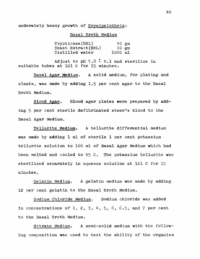
40
moderately heavy grovth of Erysipelothrix;Basal Broth Medium
Trypticase(BBL) 40 gmYeast Extract(BBL) 10 gmDistilled water 1000 mlAdjust to pH 7 . 8 * 0.1 and sterilize in
suitable tubes at 1 2 1 C for 15 minutes.Basal Agar Medium. A solid medium* for plating and
slants* was made by adding 1.5 per cent agar to the Basal Broth Medium.
Blood Agar. Blood agar plates were prepared by adding 5 per cent sterile defibrinated steerfs blood to the Basal Agar Medium.
Tellurite Medium. A tellurite differential medium was made by adding 1 ml of sterile 1 per cent potassium tellurite solution to 100 ml of Basal Agar Medium which had been melted and cooled to 45 C. The potassium tellurite was sterilized separately in aqueous solution at 121 C for 15 minutes.
Gelatin Medium. A gelatin medium was made by adding 12 per cent gelatin to the Basal Broth Medium.
Sodium Chloride Medium. Sodium chloride was added in concentrations of 1 , 2 * 3 * 4, 5* 6 * 6.5* and 7 per cent to the Basal Broth Medium.
Nitrate Medium. A semi-solid medium with the following composition was used to test the ability of the organism
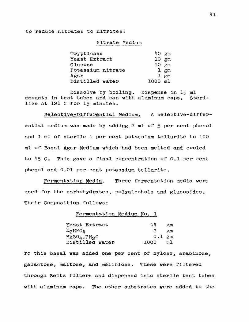
41
to reduce nitrates to nitrites:Nitrate Medium
Trypticase 40 gmYeast Extract 10 gmGlucose 10 gmPotassium nitrate 1 gmAgar 1 gmDistilled water 1000 mlDissolve by boiling. Dispense in 15 ml
amounts in test tubes and cap with aluminum caps. Sterilize at 121 C for 15 minutes.
Selective-Differential Medium. A selective-differential medium vas made by adding 2 ml of 5 per cent phenol and 1 ml of sterile 1 per cent potassium tellurite to 1 00
ml of Basal Agar Medium which had been melted and cooled to 45 C. This gave a final concentration of 0.1 per cent phenol and 0 . 0 1 per cent potassium tellurite.
Fermentation Media. Three fermentation media wereused for the carbohydrates, polyalcohols and glucosides.Their Composition follows:
Fermentation Medium No. 1Yeast Extract 44 gmK^HPOif 2 gmMgSO^.7H2 O 0 . 1 gmDistilled water 1000 ml
To this basal vas added one per cent of xylose, arabinose,galactose, maltose, and melibiose. These were filteredthrough Seitz filters and dispensed into sterile test tubeswith aluminum caps. The other substrates were added to the
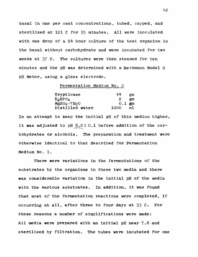
42
basal in one per cent concentrations, tubed, capped, and sterilized at 121 C for 15 minutes. All were inoculated with one drop of a 24 hour culture of the test organism in the basal without carbohydrate and were incubated for two weeks at 37 C. The cultures were then steamed for ten minutes and the pH was determined with a Beckmann Model G pH Meter, using a glass electrode.
In an attempt to keep the initial pH of this medium higher, it was adjusted to pH 8.010.1 before addition of the carbohydrates or alcohols. The preparation and treatment were otherwise identical to that described for Fermentation Medium No. 1.
There were variations in the fermentations of the substrates by the organisms in these two media and there was considerable variation in the initial pH of the media with the various substrates. In addition, it was found that most of the fermentation reactions were completed, if occurring at all, after three to four days at 33 C. For these reasons a number of simplifications were made:All media were prepared with an Initial pH near 7.8 and sterilized by filtration. The tubes were incubated for one
Fermentation Medium No. 2TrypticaseKgHPOi*MgSOi*-7H20 Distilled water
44 gm 2 gm 0 . 1 gm
1 0 0 0 ml
43
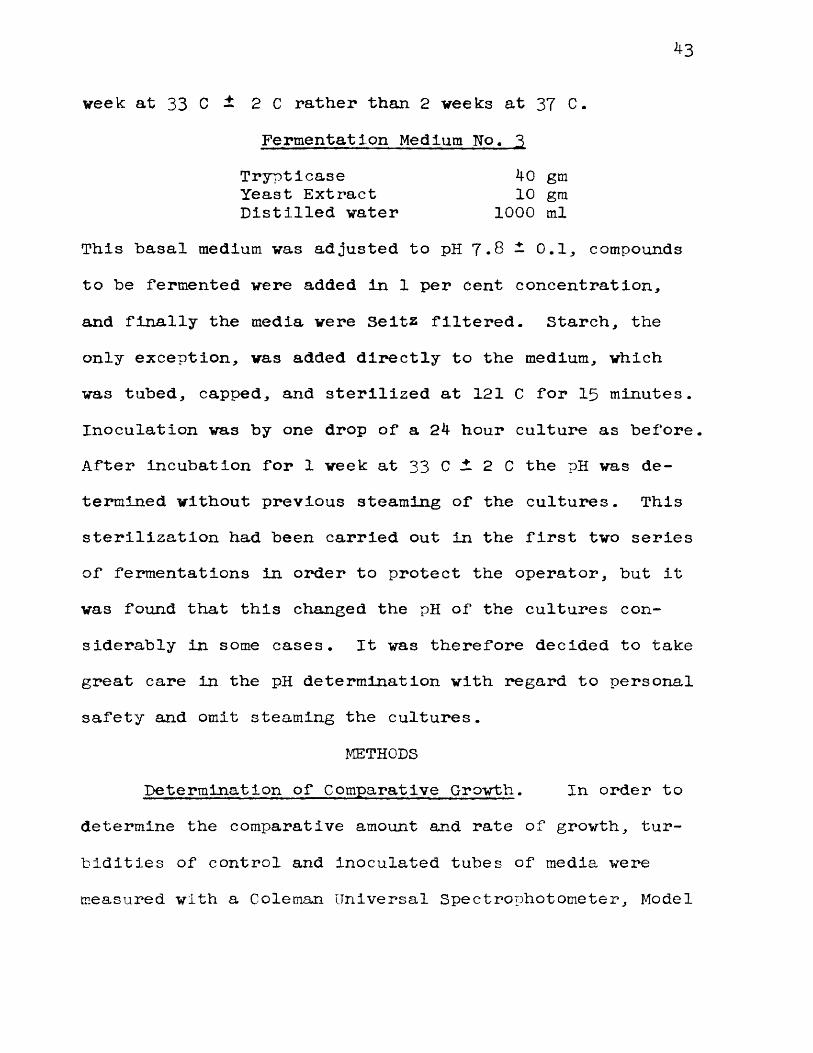
week at 33 C - 2 C rather than 2 weeks at 37 C.Fermentation Medium No, 3
Trypticase 40 gmYeast Extract 10 gmDistilled water 1000 ml
This basal medium was adjusted to pH 7.8 — 0.1, compounds to be fermented were added in 1 per cent concentration, and finally the media were Seitz filtered. Starch, the only exception, was added directly to the medium, which was tubed, capped, and sterilized at 121 C for 15 minutes. Inoculation was by one drop of a 24 hour culture as before. After incubation for 1 week at 33 C i 2 C the pH was determined without previous steaming of the cultures. This sterilization had been carried out in the first two series of fermentations in order to protect the operator, but it was found that this changed the pH of the cultures considerably in some cases. It was therefore decided to take great care in the pH determination with regard to personal safety and omit steaming the cultures.
METHODSDetermination of Comparative Growth. In order to
determine the comparative amount and rate of growth, turbidities of control and Inoculated tubes of media were measured with a Coleman Universal Spectrophotometer, Model
No. 11. Pyrex test tubes * 19 x 150 mm were selected and matched (while filled with Trypticase Soy Broth) so that variations of one per cent or less existed between readings on the galvanometer scale of the spectrophotometer. (This size tube vas selected because it could be inserted directly into the cuvette holders of the spectrophotometer. The necessity for transferring the medium to cuvettes is thus eliminated, and a series of readings can be made over an extended time without fear of contamination of the culture.) Aluminum caps were used on these culture tubes without cotton plugs. All readings were taken as "per cent transmittance" at a wave length of 5 3 5 mu using an uninoculated control set at 100 "per cent transmittance" and which had been incubated with the inoculated tubes. The readings in "per cent transmittance" were changed to Optical Density by the formula;
O.D. as 2 —log G where G is the galvanometer reading in "per cent transmittance". This method was used to determine the medium in which the organisms grew best, using maximum turbidity as the criterion of "best growth".
In addition, the comparative rate of grovth at different temperatures, and the maximum growth at these temperatures was also determined by this method. 15 ml
45
amounts of Basal Broth Medium in the selected tubes were inoculated with one ml of a 24 hour culture of the test organism. Initial turbidity was determined, and. hourly observations of turbidity were made in the spectrophotometer. The temperatures were maintained vithin±1.0 C by means of thermostatically controlled water baths of the type ordinarily used in serological work.
Colonial Appearance and Morphology. Colonial appearance was determined on Basal Agar Medium, Basal Agar Blood Medium, and Tellurite Medium.
Morphological studies were made on gram-stained smears of cultures from Basal Broth and Agar Media at various times after incubation at 33 C and 37 C.
Motility. Darkfield examinations were made of 18 to 24 hour cultures of the organism to determine motility. C.T.A. semi-solid medium was also inoculated and observed for evidence of motility.
Hemolysis. Both pour and streak plates were made using Basal Agar Blood Medium to observe the action of the organisms on blood. As an alternative to the defibrinated steer*s blood used in this medium, outdated, citrated human blood obtained from the blood bank vas found to be satisfactory .
Gelatin. Gelatin medium vas Inoculated by making a
46
single stab to the bottom of the tube. It vas incubated at 25 C and observed daily for 2 veeks for type of growth and liquefaction.
pH Range. Basal Broth Medium, adjusted to initial pH 5*9 to 9.2 was used to determine the pH range of the organism. Tubes were observed daily for 2 veeks for evidence of growth.
Nitrate Reduction. Nitrate medium vas inoculated and Incubated at 33 C for 14 days. Samples were removed aseptically each day and tested for nitrites. Tests were also made for nitrates since no nitrite vas demonstrable.A known positive control was run with each series of determinations. All tests made were those recommended in the "Manual of Methods for Pure Culture Study of Bacteria" unless otherwise indicated.
Hydrogen Sulfide and Indol. 48 hour cultures in Basal Broth Medium were used for hydrogen sulfide and indol tests. Strips of lead acetate paper were suspended over the Basal Broth Medium to test for the production of hydrogen sulfide. The Ehrlich-Bohme test was used to determine the formation of indol.
Catalase. Slants of Basal Agar Medium and tubes of Basal Broth Medium were inoculated with test and control organisms and incubated at 33 C. After 24 hours at
47
this temperature, 3 per cent hydrogen peroxide was poured over the slants and into the broth cultures. If bubbles of gas were evolved, catalase was considered present. Positive controls showed vigorous frothing when treated in this manner while the test organisms showed no sign of catalase activity.
Fermentation Studies. The methods used for the preparation, inoculation, incubation and reading of the fermentation tests are described under each of the fermentation media listed in the previous section.
Relationship to Oxygen. Shake tube cultures and plates of Basal Agar Medium incubated aerobically and anaerobically were used to determine the oxygen requirements of these organisms.
Inhibitors. Inhibitory substances such as phenol, crystal violet, and potassium tellurite were added to Basal Broth Medium and Basal Agar Medium in various concentrations to test the ability of the organism to grow in their presence 9
CULTURESIn order to avoid a one sided, and perhaps not typical
point of view, and to search for the greatest possible variety, cultures were collected from widely separated sources.
48
Stock Cultures, All strains were maintained on C.T.A. Medium in cotton plugged tubes covered with Celon caps. Transfers were made to Basal Broth Medium and 24 hour cultures in this medium were used to inoculate other media.
Preservation of Cultures. All strains were grown on Basal Agar Slants at 33 C for 48 hours, washed off with double strength skim milk and lyophilized.
Sources of Cultures. The culture number is followed by the name and locality of the donor,, strain number des ig- nated by the donor, animal and tissue from which isolated, and date of isolation when this information was available. All cultures received were designated as Erysipelothrix rhusiopathiae or Erysipelothrix spp. except No. 2 8 5 and No. 2 9 0 which were labeled Eryslpelothrix muriseptica.
LIST OF CULTURES
No. 30 - P.A. Hansen, Livestock Sanitary ServiceLaboratory, College Park, Md., Dead Hog, Spleen, January 10, 1949.
No. 3 5 - P.A. Hansen, L.S.S.L., College Park, Md.,Dead Shoat with pericarditis, Spleen, January 26, 1949.
No. 40 - P.A. Hansen, L.S.S.L., College Park, Md.,Dead Shoat with pericarditis, Liver,January 2 9 , 1949.
No. 45 - P.A. Hansen, L.S.S.L., College Park, Md.,Dead Hog, Spleen, September, 16, 1949-
50
55
6 0
65
70
75
80
35
90
95
100
105
110
120
125
1 30
k9
- F.H. Smiley* Army Medical School, Washington* D.C.* No. 8139* Isolated by H.W. Schoening, No. S-l.
- S.M. Morrison, Colorado A & M College* Fort Collins* Colorado* No. 8 9 * Hog* acute swine erysipelas* September* 19^9-
- S.M. Morrison* Colorado A & M College* Fort Collins* Colorado, No. 9 0 , History unknown.
- I.A. Merchant, Iova State College* Ames, Iowa* No. 3 7 * History unknown.
- I.A. Merchant* Iowa State College, Ames, Iowa, No. 3 8 * History unknown.
- G.C. Langford* University of Maryland, College Park, Maryland.
- G.C. Langford, University of Maryland, College Park, Maryland.
- G.C. Langford * University of Maryland, College Park* Maryland.
- G.C. Langford* University of Maryland, College Park* Maryland.
- New York State Veterinary College, Ithaca* New York, No. 5 2 9 .
- New York State Veterinary College* Ithaca, New York, No. 5^8.
- R .D . Shuman, USDA, Washington, D . C . * Pig * joint, (from Texas, isolated by Creech).
- R.D. Shuman* USDA* Washington* D.C., (isolated by Jensen In Copenhagen).
- R.D. Shuman, USDA, Washington, D.C., Hog* tonsil* (Animal Husbandry, Landrace, 1936).
- R.D. Shuman* USDA, Washington* D.C.* Hog, joint, (from North Carolina).
- R.D. Shuman* USDA* Washington* D.C.*History unknown.
50
Wo.
No.
No.
No.
No.
No.
No.
No.
No.
No.
No.
No.
No.
No.
No.
No.
140 - R.D. Shuman, USDA, Washington, D.C., Hog, Spleen, (from Austin, Minnesota).
145 - R.D. Shuman, USDA, Washington, D.C., Hog, cholera virus.
150 - R.D. Shuman, USDA, Washington, D.C., Hogwith skin placques, Spleen, April 2 6 , 1949.
155 “ R.D. Shuman, USDA, Washington, D.C., six passages of No. 145 in turkeys.
160 - R.D. Shuman, USDA, Washington, D.C., Hog,with skin lesions, Spleen, October, 1949.
1 6 5 - R.D. Shuman, USDA, Washington, D.C., 14hams ter pas s age s .
170 - R.D. Shuman, USDA, Washington, D.C., Pig, joint, (from Texas), mouse virulent.
175 - R.D. Shuman, USDA, Washington, D.C., Labstrain passed through pigeons three times.
180 - R.D. Shuman, USDA, Washington, D.C., Hog, with heart lesions, Spleen, (from Austin, Minnesota).
1 8 5 - R.D. Shuman, USDA, Washington, D.C., turkey, (from Montgomery, Alabama).
190 - R.D. Shuman, USDA, Washington, D.C., From 11 hamster passages.
195 - R.D. Shuman, USDA, Washington, D.C., Pig, with erys ipelas, Blood.
200 - R.D. Shuman, USDA, Washington, D.C., From hog cholera virus.
210 - H.M. DeVolt, L.S.S.L., College Park, Md., No. 1070, turkey, 1949.
215 - New York State Veterinary College, Ithaca, New York., No. 5 3 0 .
220 - H.M. DeVolt, L.S.S.L., College Park, Md., No. 1475, turkey, Fall, 1949.

51
No
No.
No.
No.
No.
No.
No.
No.
No.
No.
No.
No.
No.
225 “ H.M. DeVolt, L.S.S.L., College Park, Md.,No. 1490, turkey, Fall, 1950.
230 - J.V. Klauder, Philadelphia, Penna., Pig, with diamond back disease.
235 “ National Collection of Type Cultures,London, England, No. 1224,(W.G. Wragge, Pig, intestine, February 11, 1922).
240 - N.C.T.C., London, England, No. 2422, (from R.V. Solly, Pig, with ulcerative endocarditis, heart blood, September, 1 9 2 7 ).
245 - N.C.T.C., London, England, No. 8 1 6 3 , (from L.P. Garrod, pig with endocarditis, spleen, March, 1950).
2 5 0 - N.C.T.C., London, England, No. 7999* (from Staub, Pasteur Institute, July, 1949).
255 - N.C.T.C., London, England, No. 6 3 3 3 , (from A.W. Gledhill, duck, August, 1941).
260 - N.C.T.C., London, England, No. 6 3 3 2 , (fromA.W. Gledhill, lamb, joint-ill, November, 1939)
2 6 5 - N.C.T.C., London, England, No. 3406, (from T. Hare, pig with erysipelas, spleen,June, 1931).
270 - N.C.T.C., London, England, No. 2 8 2 5 , (fromBammerton and Lovell at London Zoo, AfricanJacana, intestine, 1928).
275 " N.C.T.C., London, England, No. I6 9 4 , (fromJ. Smith, pig, vegetations on mitral valve,September 4, 1 9 2 3 ).
280 - N.C.T.C., London, England, No. 3260, (fromM.R. Seddon, Veterinary Research Laboratory, N.S.W., lamb, polyarthritis, November, 1930).
2 8 5 - N.C.T.C., London, England, No. 8 0 7 , (from W.W.C. Topley, mice, 1921, (Erysipelothrlx murlseptica).

No. 290 - N.C.T.C., London, England, No. 4304, (from W.W.C. Topley, mice, 1921, (Erysipelothrlx muriseptica).
RESULTS
MORPHOLOGY AND CULTURAL CHARACTERISTICS
Morphology. ErysIpelothrix rhu3 iopathiae Is a gram- positive, non-sporeforming rod which varies greatly In size and shape. Depending upon the type of medium, its Initial pH, the incubation temperature, and the age of the culture, growth on nutrient agar and In nutrient broth has resulted in organisms which vary from short, stubby, almost coccoid rods to very long, thin, filamentous structures. Any of these forms may contain granules, but they are most common in older cultures. All of the different forms were frequently found in a single preparation from the culture.
Motility. All strains of Erysipelothrlx examined were found to be non-motile. Observations were made with the dark-field microscope on wet preparations of 18 hour cultures in Basal Broth Medium incubated at 33 C. Motility was never observed. The organisms grew well in semi- solid C. T. A. Medium. There was uniform growth from the point of entrance of the needle or loop into the medium all along the track of inoculation. No evidence of motility was seen in this medium.
Growth in Broth. A medium consisting of 4 per cent
5*
yeast extract in water caused some strains of the organism to grow in long tangled chains, especially when the pH of the medium was initially a little on the acid side of neutrality. Many authors have correlated the presence of these long tangled chains with a rough form of the organism which appears on solid media. This rough type of growth was favored by incubation at 33 C and was not encountered as frequently at 37 C.
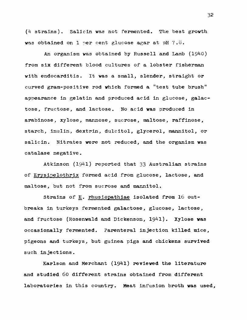
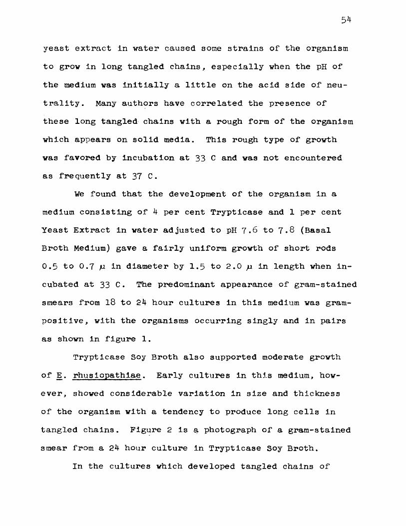
We found that the development of the organism in a medium consisting of 4 per cent Trypticase and 1 per cent Yeast Extract in water adjusted to pH 7 . 6 to 7.8 (Basal Broth Medium) gave a fairly uniform growth of short rods0 . 5 to 0 . 7 u in diameter by 1 . 5 to 2 . 0 /i in length when incubated at 33 C. The predominant appearance of gram-stained smears from 1 8 to 24 hour cultures in this medium was gram- positive, with the organisms occurring singly and in pairs as shown in figure 1 .
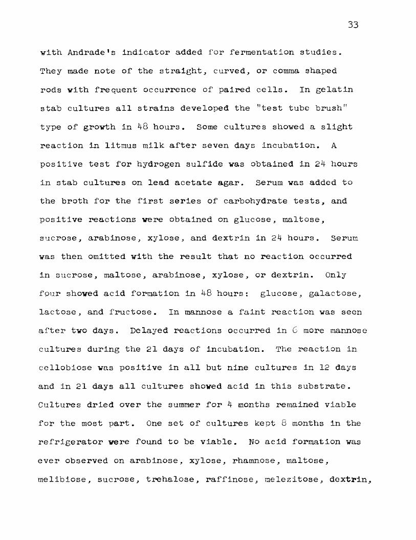
Trypticase Soy Broth also supported moderate growth of E. rhuslopathlae. Early cultures in this medium, however, showed considerable variation in size and thickness of the organism with a tendency to produce long cells in tangled chains. Figure 2 is a photograph of a gram-stained smear from a 24 hour culture in Trypticase Soy Broth.
In the cultures which developed tangled chains of
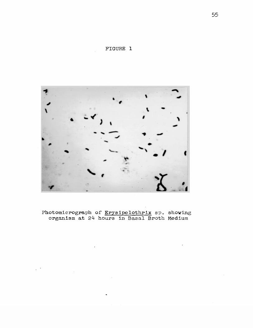
FIGURE 1
-f
*\
fc
Photomicrograph of Erysipelothrix sp, shovingorganism at 2k hours in Basal Broth Medium
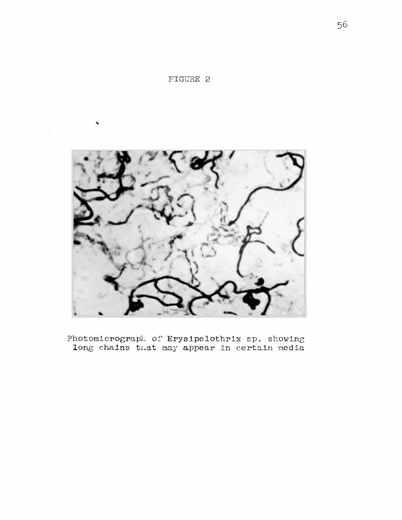
FIGURE 2
Photomicrograph of Erysipelothrix sp. shovinglong chains tuat may appear in certain media
very long cells, the macroscopic appearance of the tubes vas characteristic. In the very early stages of grovth, at 12 to 14 hours, a uniform turbidity appeared and vhen the tubes vere shaken, they took on a peculiar swirling, opalescent, cloudy appearance. In 24 to 48 hours there vas considerable flocculant deposit in the bottom of the tubes. After several days of incubation, this deposit became quite adherent to the bottom and could only be broken avay by vigorous agitation of the tube. When agitated, this adherent, thread-like material spiraled up and could be suspended in the medium only with difficulty. Stained smears of this material invariably shoved complex, twisted chains of gram-positive rods. These rods varied considerably in length from very short stubby ones to long thick filaments of enormous length which covered several oil immersion fields. Many of the rods appearing in the cultures were distinctly curved.
The "smooth’’ cultures, or the cultures which regularly formed short rods with only an occasional long filament developed the same typical opalescent turbidity as the "rough" tangled chain cultures. The only change which occurred in these cultures vhen incubated further was an increase in density, which reached a maximum usually after 48 hours incubation at 33 C. Considerable difference with
regard to ''smooth,r and "rough" types of growth was noted between the various strains on the same medium.
Gram Reaction. The reaction to Gram stain was also variable, depending again upon the medium employed. We found that, for the most part, early cultures of from 12
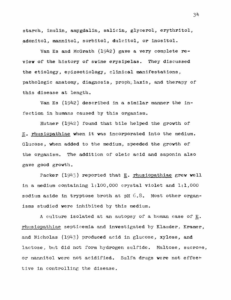
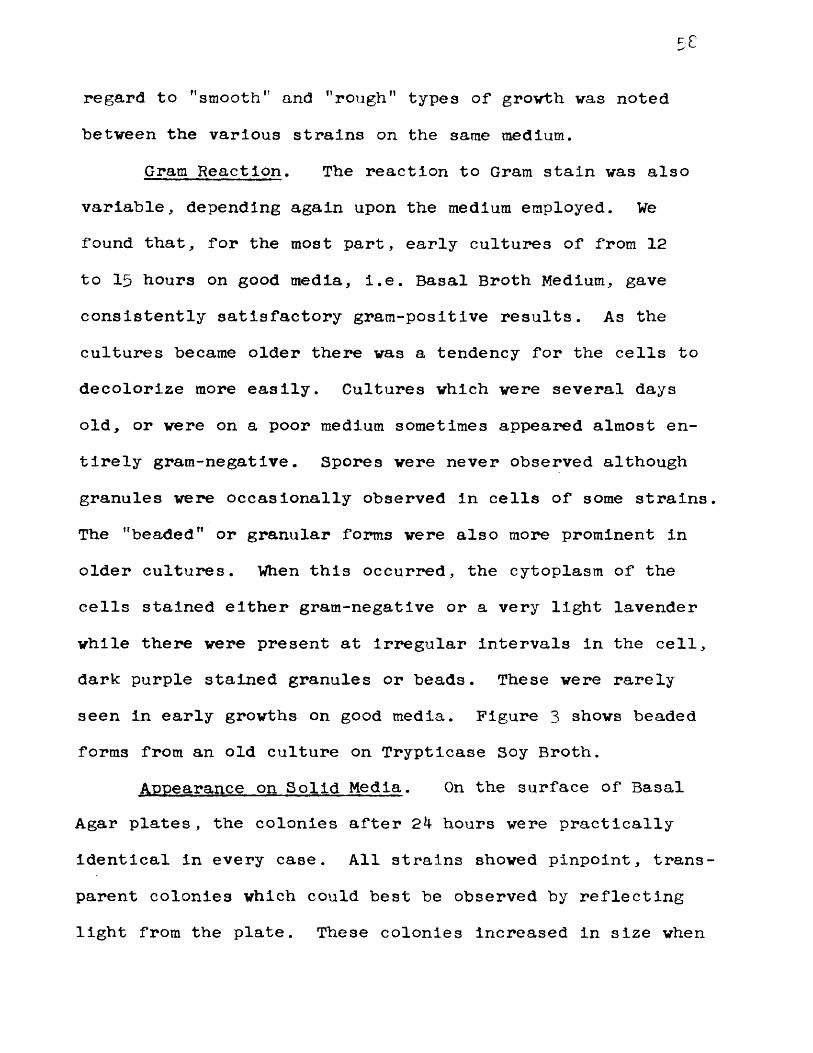
to 15 hours on good media, i.e. Basal Broth Medium, gave consistently satisfactory gram-positive results. As the cultures became older there was a tendency for the cells to decolorize more easily. Cultures which were several days old, or were on a poor medium sometimes appeared almost entirely gram-negative. Spores were never observed although granules were occasionally observed in cells of some strains The "beaded" or granular forms were also more prominent in older cultures. When this occurred, the cytoplasm of the cells stained either gram-negative or a very light lavender while there were present at irregular intervals in the cell, dark purple stained granules or beads. These were rarely seen in early growths on good media. Figure 3 shows beaded forms from an old culture on Trypticase Soy Broth.
Appearance on Solid Media. On the surface of Basal Agar plates, the colonies after 2k hours were practically identical in every case. All strains showed pinpoint, transparent colonies which could best be observed by reflecting light from the plate. These colonies increased in size when
59
FIGURE 3
Photomicrograph of Erys ipelothrlx sp. showinggranules which may appear in certain media
6o
well isolated,, reaching a maximum diameter of 1 to 1 . 5 mm after 48 to 7 2 hours incubation at 33 C. After this time, there was little change in size. The fully developed colonies were transparent by transmitted light and had a very light bluish sheen by reflected light. They were round and smooth with a glistening surface.
The size and appearance of the colonies were the same on Blood Agar Medium. After 24 hours incubation at either 33 C or 37 C there was usually no change in the medium.Some strains caused a faint greenish discoloration during this period, and all strains caused discoloration after 36
hours. Within the next 2 days this discoloration disappeared leaving a faint clear area around the colony. This clearing did not extend all the way through the medium to the bottom of the plate and was never very pronounced, but was clearly evident in every case if observed by looking directly down on the plate with bottom illumination.
Colonies of Erysipelothrix were pinpoint in size and greyish colored after 24 hours incubation at 33 C on Tellurite Medium. As they increased in size to a maximum diameter of 1 to 1 . 5 mm, they developed a shiny jet black color.
Since these organism may appear in clinical material for culture, it may be of diagnostic value to note that they are completely inhibited by Desoxycholate Medium , Eos in
Methylene Blue Agar and Bismuth Sulfite Medium. They develop very poorly on Salmonella Shigella Medium with isolated colonies seldom increasing beyond pinpoint size. Massive Inoculation on this medium gave rise to a scant white growth after several days incubation at 33 C. Growth was fairly good on Eugonagar and Trypticase Soy Agar.
Gelatin Stabs. Gelatin Medium supported growth of all Erysipelothrix strains studied but was never liquefied. There were two types of growth in the gelatin stabs: Moststrains grew in the ,Ttest tube brush” form which has been so frequently described in the literature. The organism developed first along the lines of inoculation, producing a filiform growth. Lateral, radiating, brush-like projections next appeared all around this filiform stab, and usually by 48 hours, the typical "test tube brush" appearance was evident. Several strains showed filiform growth but never developed lateral outgrowths to give the "test tube brush" even after repeated transfer in good media before inoculating the gelatin. These were strains 50* 95$ 215* 235* 280, 2 8 5 , and 290. Meyn (1931) indicated that transfers to agar from the "test tube brush" type gelatin tubes would give rise to rough colonies, while transfers from filiform type gelatin tubes would produce smooth colonies on agar plates. Using the Basal Agar Medium at 33 C we were unable to obtain
rough colonies from either type of growth in gelatin. Smears of the material from these gelatin tubes likewise showed little correlation between smooth and rough forms in so far as the morphology was concerned. Smears from gelatin tubes showing the ’’test tube brush” appearance demonstrated tangled chains of long rods in some cases and medium length rods, singly and in pairs in others.
PHYSIOLOGICAL CHARACTERS
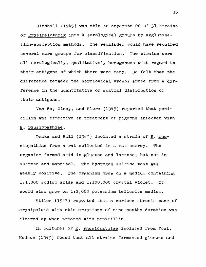
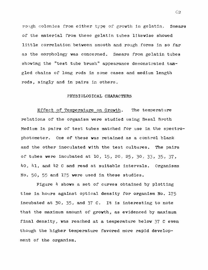
Effect of Temperature on Growth. The temperature relations of the organism were studied using Basal Broth Medium in pairs of test tubes matched for use in the spectrophotometer. One of these was retained as a control blank and the other inoculated with the test cultures. The pairs of tubes were incubated at 1 0 , 1 5 , 2 0 , 2 5 , 3 0 , 33 * 35 * 37 *40, 41, and 42 C and read at suitable intervals. Organisms No. 5 0 , 55 and 175 were used in these studies.
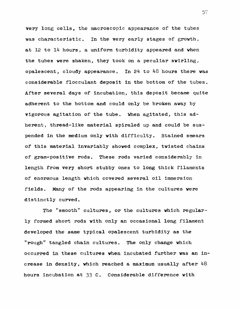
Figure 4 shows a set of curves obtained by plotting time in hours against optical density for organism No. 175 incubated at 3 0 , 35, and 37 C. It Is interesting to note that the maximum amount of growth, as evidenced by maximum final density, vas reached at a temperature below 37 C even though the higher temperature favored more rapid development of the organism.
63FIGURE 4
Growth Curves for Strain No. 175 at 30, 3 5 ,and 37 C
o
LAon
_ J osm
OJ
o o ocn cn cn ir\
o 1—1om oOJ
o o• . • •
jLLisNaa TyoiMo
HOURS
64
Figure 5 is a similar set of curves for Organism No.50 incubated at 26, 30, and 37 C. Again the tubes incubated at the lower temperature had a larger crop of cells than the ones incubated at 37 C. There was also considerable autolysis in the tube held at 37 C. The tubes incubated at 37 C however, developed faster as evidenced by earlier turbidity rise in comparison to the tubes incubated at lower temperatures. These findings agree with the accepted views as expressed by Hahn (1932): "The temperature which gives the largest crop is considerably lower than that giving the most rapid growth rate, but it is still far above the minimum temperature of growth. An increase above the optimum temperature causes a very rapid decline of the crop.” At 25 C there was a delay in the development of from 8 to 10 hours and the comparative rate of growth was somewhat slower than at 3 0 , 35, and 37 C. The final maximum crop was only slightly lower than the maximum cell crop for growth at 37
C. At 42 C there was little apparent development over a period of 48 hours as measured in the spectrophotometer. There was likewise no apparent growth at 10 C .
Figure 6 shows curves for organism No. 60 with two cultures incubated at 25 C on different days. Although attempts were made to keep the inoculum constant by using the same inoculating loop, small variations in amount of
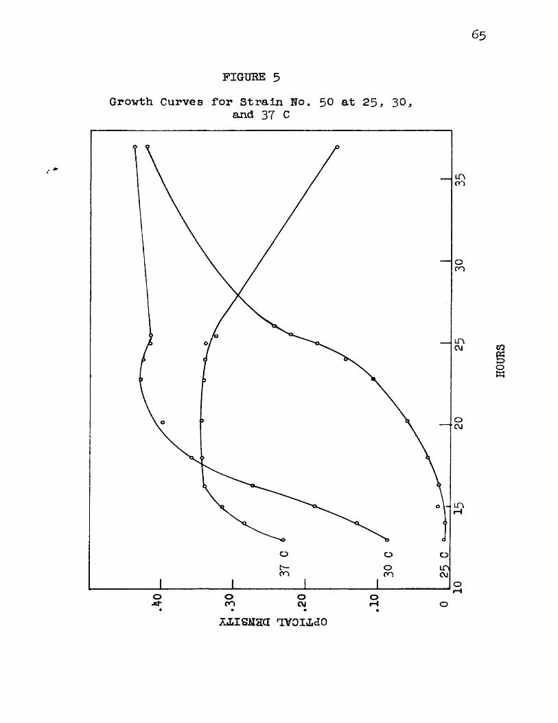
65
FIGURE 5Grovth Curves for Strain Ho. 5 0 at 25, 30.,
and 37 C
_inon
oon
inCM
O CM
O O OOon in
CMonopH
oxiiSHaa t v o i m o
HOURS
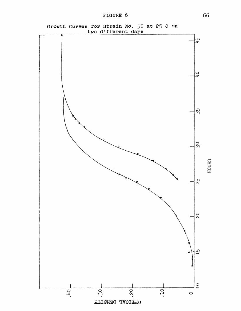
FIGURE 6 66Grovth Curves for Strain No. 5 0 at 25 C on
two different days
o-=j*
m
oC\J
or-iOOJ
omo.=3- oM isuari uvoiido
HOURS

67
inoculum inevitably occurred. This lead to some displacement of the curve but the form of the growth curve and the maximum cell crop remained essentially the same.
As shown in figure 7, the tubes which had a largerinoculum began to increase in density first. A large inoculum of 1 to 3 ml imparted a slight initial turbidity to the tube but enabled the comparative readings to be made in a much shorter time than was the case when an inoculum of a loop or a drop of culture was used.
At 25 C, the culture of organism No. 175 showed analmost constant increase in density for the first 11 hours and never reached an optical density greater than 0 .2 8 . Figure 8 shows the development of No. 175 the first 11hours of growth at 25 C and the maximum density obtained.One ml of inoculum was added to 15 ml of Basal Broth Medium.
At 35 Cj as seen in figure 9, the rate of development was fairly rapid with a good crop of cells obtained after 11 hours of incubation. The maximum density had apparently not been reached in this time and there was some autolysis of the cells after 25 hours incubation.
Figure 10 shows that the development of this organism was more rapid at 40 C and a larger cell crop was obtained than at 25 C.
At 41 C, the growth was similar to that at 25 C and
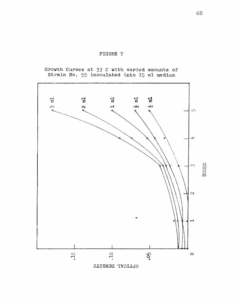
68
FIGURE 7
Growth Curves at 33 C with varied amounts of Strain No. 55 inoculated into 15 ml medium
rH1—IrHjCVJCVIon
-=t
on
OJ
rH
in o in o
ziiSMaa nvoiMo
HOUR
S
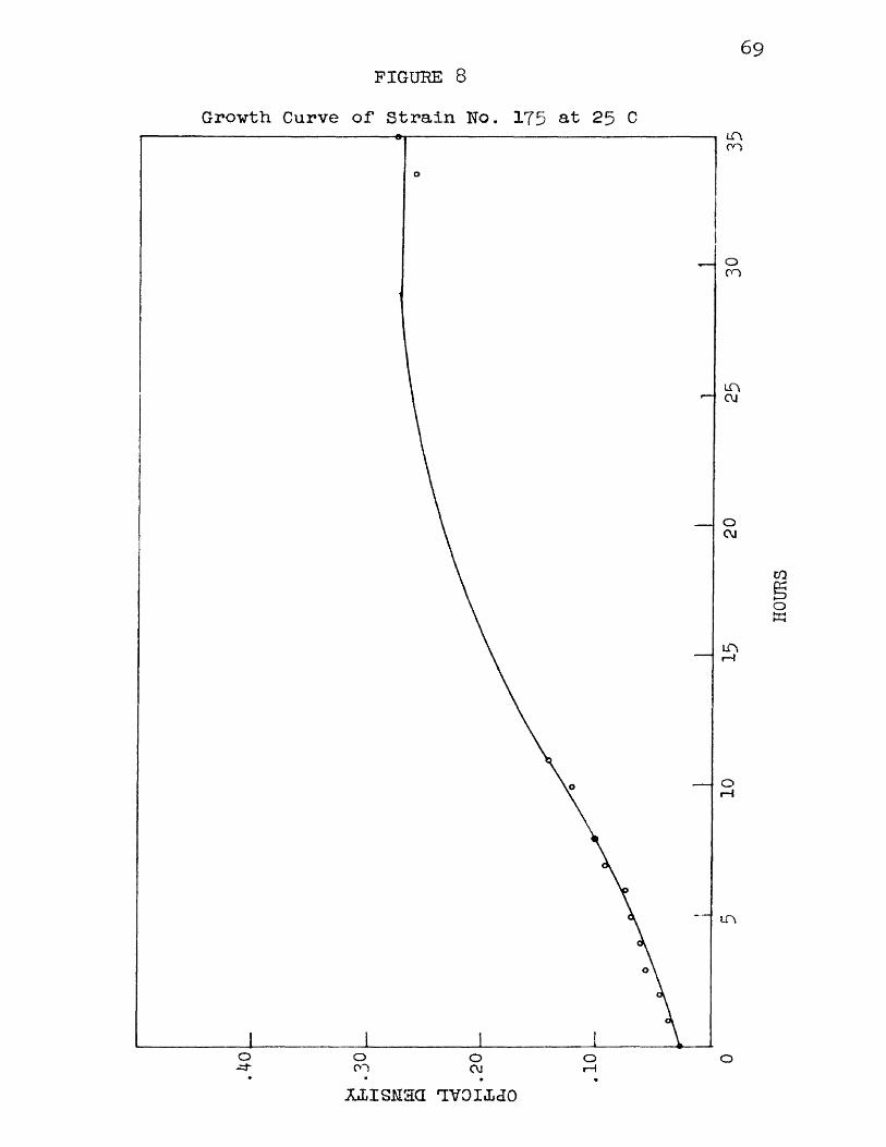
FIGURE 869
Growth Curve of Strain No. 175 at 25 CL T \cn
om
mOJ
oC\J
in
oOJ orH
O• ♦
JLiLISMan TOIidO
HOURS
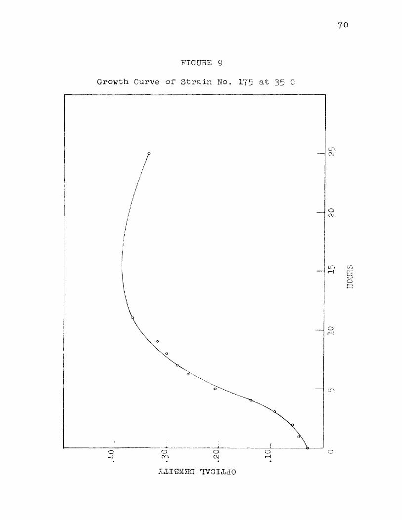
70
FIGURE 9Growth Curve of Strain No. 175 at 35 C
L T \
OrH
OC\J
oCO
xiis iuaa Ttvoiido
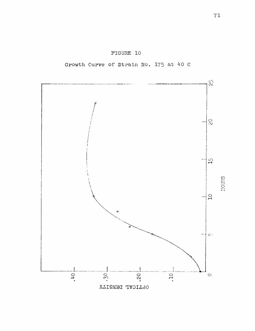
71
FIGURE 10Growth Curve of Strain No. 175 at 40 C
LT\CM
OCYJ
(—1
O o o o o
o
on cm
xiiSMaa uvoitLio
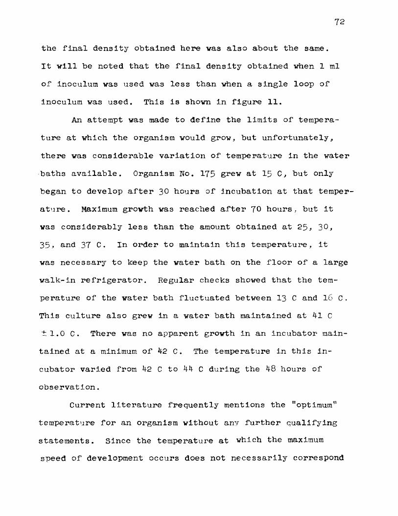
72
the final density obtained here vas also about the same.It will be noted that the final density obtained when 1 mlof inoculum was used was less than when a single loop ofinoculum was used. This is shown in figure 11.
An attempt was made to define the limits of temperature at which the organism would grow* but unfortunately, there was considerable variation of temperature in the water baths available. Organism No. 175 grew at 15 C, but onlybegan to develop after 30 hours of Incubation at that temperature. Maximum growth was reached after 70 hours, but It was considerably less than the amount obtained at 2 5 * 3 0 ,35., and 37 C. In order to maintain this temperature, it was necessary to keep the water bath on the floor of a large walk-in refrigerator. Regular checks showed that the temperature of the water bath fluctuated between 13 C and lb C . This culture also grew in a water bath maintained at 41 C ±1.0 C. There was no apparent growth In an incubator maintained at a minimum of 42 C. The temperature in this incubator varied from 42 C to 44 C during the 48 hours of observation.
Current literature frequently mentions the "optimum” temperature for an organism without any further qualifying statements. Since the temperature at which the maximum speed of development occurs does not necessarily correspond
OPTICAL DENSITY
! ^i
O It? 1^ OAT 'CM u-px/ti^S j c OAano t lV io J Q
£L I I MHXiOL
74
to the temperature at vhich maximum cell crops are obtained, statements in the literature could be clarified considerably by indicating the particular criterium by vhich ’’optimum” grovth was evaluated. For comparative purposes ve have found that a simple observation of the optical density of the cultures is sufficient to determine the maximum grovth or speed of development in different media. For more exact determinations of grovth rate and-numbers of organisms a more closely regulated vater bath vould be necessary and a method of measurement of grovth vhich vould indicate numbers of organisms vould be required.
pH Range. All pH studies .vere done at 33 C in Basal Broth Medium adjusted to the proper pH. Organisms No. 50 and No. 6 5 developed at pH 6.4 but only very slowly, shoving turbidity after 48 hours. Eight of eleven strains examined grev at pH 6 . 5 after 72 hours, and all strains shoved a moderate amount of turbidity in 24 hours at 33 C vhen the initial pH of the medium vas above 6 .7 . Rapid, heavy grovth occurred in media vith initial pH 7-3 to 9.0. 'When the organisms were grown in Basal Broth Medium vith no added fermentable carbohydrate and vith the initial reaction below about 7 . 8 to 8 .0 , the medium became slightly more.alkaline. Morphological observations were made on eleven strains vhich vere grown in media adjusted to pH 6.4 to pH 9,2 Their
75
morphologic changes were similar in the different pH ranges. Organisms used were No. 50 5 5 , 6 5 , 7 5 , 100. 150, 175, 190,205. and 225. In the range of pH 0.4 to 7 . 0 there was an increased tendency toward the formation of long tangled filaments. Prom pH 7-5 to about 8 .5 , the organisms grew regularly in short stubby rods. These occurred singly and in pairs which stained solidly gram-positive when examined after 24 hours. In the pH range above 8 .5 , there was a tendency to form small distorted cells which did not stain evenly and which were frequently gram-negative.
None of the strains examined produced Indol in Basal Broth Medium.
Hydrogen Sulfide. Inoculation of all strains of Eryslpelothrix available into lead acetate medium gave uniformly negative results for production of hydrogen sulfide. The growth was very light in this medium, which apparently proved inhibitory, and suspending strips of lead acetate paper above the Basal Broth Medium was found to be a far more satisfactory procedure. Most Erysipelothrlx strains blackened or caused a brownish discoloration of the paper after 24 hours incubation at 33 C. Several strains caused only a very slight discoloration of the lead acetate paper and two strains took as long as 7 2 hours to give a positive reaction, but all strains definitely produced hydrogen
76
sulfide. The paper over an uninoculated control remained white with no trace of discoloration after a week of incubation .
Catalase. None of the strains of Erysipelothrix examined produced catalase. Basal agar slant cultures and cultures in Basal Broth Medium were treated with 3 per cent hydrogen peroxide after 24 hours incubation at 33 C. No bubbles were ever observed, while a culture of Micrococcus pyogenes var aureus used as a positive control caused numerous bubbles to be given off by the hydrogen peroxide.
Nitrate Reduction. In the first series of examinations, the tubes of Nitrate Medium were examined each day for 14 days by aseptically removing a portion of the culture and testing for nitrites. Nitrite was never observed In the cultures of Erysipelothrix and tests for nitrate showed that it was still present. Thereafter, the tubes were incubated for 14 days and a single test performed at the end of that time for nitrite. No organism examined was able to form nitrite from nitrate. A control organism, Micrococcus pyogenes var aureus. run in parallel with each series of tests gave a good positive reaction.
Litmus Milk. Cultures Inoculated into Litmus Milk were incubated at 33 C and shoved no change after 1 week. Probably the addition of Trypticase and/or Yeast Extract
77
vould have allowed them to ferment the milk, but it was felt that little information of value would be gained in any case by employing such a medium.
Fermentations. The fermentation reactions were variable, depending upon the medium employed and the initial pH of the medium. Apparently, one week was sufficient time to allow for complete fermentation if it took place at all. Cultures which contained fermentable substrates were found to be self-sterilizing after 3 to 4 days incubation. Acid was regularly produced in all three fermentation media containing fructose, glucose, galactose, and lactose. In Fermentation Medium No. 1 and Fermentation Medium No. 2, all strains regularly fermented mannose also except No. 5 0 , which formed no acid in mannose in Fermentation Medium No.1. In Fermentation Medium No. 3, all strains fermented mannose except No. 6 5 , 75, 145 * 210, 270, and 290. In Fermentation Media Nos. 1 and 2, cellobiose was variable, but in Fermentation Medium No. 3 all strains fermented cellobiose except No. 235, 245, 250, 255, 260, 2 6 5 , 270, 275,280, 2 8 5 , and 290, received from the National Collection of Type Cultures. Xylose, arabinose, maltose, and melibiose were fermented by all strains In Fermentation Medium No. 1 except No. 105, which did not form acid in xylose. None of these substances was fermented in Fermentation Medium No. 2.
78
An occasional strain shoved slight acid production in glycerol, arabinose, sorbitol, inositol, trehalose, melibiose, raffinose, and salicin. No gas was ever produced by any strain.
Table 1 shows the reactions of the organisms on the substrates when used with basal medium. In these media, sterilization was by autoclave except for the ones containing xylose, arabinose, galactose, and melibiose. The cultures were steamed before the pH determinations were made. The problem which arose here was: "What shall we consider the criterion of fermentation of a substrate?" It Is evident from a glance at the table that all strains of Ery- sipelothrix are singularly inactive with respect to acid formation, and even when fermentation occurs, there Is usually very little change in the pH, i.e. from 0.1 to 0.8 pH units, although an occasional strain may change the pH as much as 1.6 units. In every case, the change in PH means the difference between the pH of the control after steaming and the pH of the medium in which the organism had grown. It was noted that when the organisms grew In Basal Broth Medium without a substrate or with a substrate which was not fermented, the final pH of the medium after Incubation and steaming was usually slightly more alkaline. This change in pH was small but constant. In addition to the four substrates, fructose, glucose, galactose, and lactose,

79
FINAL pH OBTAINED BY ACTION OF VARIOUS STRAINS OF EKYSIPELOTHRIX ON SUBSTRATES IN FERMENTATION MEDIUM NO. 1.
Control before 6.7 Inoculation 6.7Control after steaming 6.8
105110120125210220225
Summary ofFermentationCharacters
6.96.8
7.0
7.0
6.6.4
6.86.7
6.86.7
G.B6.6
6.8 6.5 6.9 6.4 6.8 6.8 6.86.8 6.5 6.8 6.5 6.7 6.8 6.86.8 6.6 6.8 6.4 6.7 6.8 6.86.8 6.6 6.9 6.5 6.9 6.9 6.86.8 6.7 6.8 6.4 6.8 6.8 6.86.8 6.7 6.8 6.4 6.8 6.8 6.86.8 6.6 6.8 6.5 6.8 6.8 6.86.8 6.7 6.8 6.4 6.8 6.8 6.86.8 6.7 6.8 6.5 6.9 6.8 6.86.8 6.7 6.8 6.5 6.9 6.8 6.86.8 6.6 6.8 6.5 6.8 ND* 6.86.9 6.6 6.8 6.4 6.9 6.8 6.86.8 6.8 6.9 6.5 6.8 6.8 6.86.7 6.6 6.8 6.4 6.8 6.8 6.76.8 6.6 6.8 6.4 6.8 6.8 6.86.8 6.4 6.9 6.4 6.8 6.8 6.86.8 6.5 6.8 6.5 6.8 6.9 6.86.8 6.6 6.8 6.4 6.8 6.8 6.86.8 6.7 6.8 6.5 6.8 ND 6.8
0 0-.4 .1-.2 0 0 0 0
6.56.3
6 .06.06.06 .05.85-86.06.0 6.0 5-9 5-9 5-9 6.0 5-8 5-8 5-8 5-9 5-9 5-9
6.4
6.3
5.9 6.0 5-95.9 5-9 6 .0 6 .0 6 .0 6.15.95.95.9 6.05.75.9 5-95.95.85.8
mannose galactose sucrose trehalose maltose cellobiose melibiose lactose melesitose
6.3 7.5 6.8 6.7 7-7 6.7 7.6 6.7 6.8
6.3 7.4 6.8 6.7 7.7 6.6 7.6 6.5 6.7
6.2 6.6 6.8 6.8 7.5 6.6 7.4 6.1 6.86.2 6.5 6.8 6.8 7.4 6.55 7.5 6.1 6.86.2 6.4 6.8 6.8 7.4 6.6 7.4 6,1 6.86.3 6.4 6.8 6.9 7.5 6.7 7.5 6.4 6.86.2 6.5 6.8 6.7 7.4 6.6 7.4 6.1 6.86.2 6.1 6.8 6.8 7.4 6.6 7.4 6.1 6.86.2c 6.3 6.8 6.8 7.5 6.6 7.4 6.2 6.86.25 6.4 6.8 6.8 7.5 6.6 7.4 6.2 6.86.2 6.3 6.9 6.9 ND 6.6 7.5 6.2 6.96.2 6.5 6.9 6.9 7.3 6.6 7.5 6.2 6.96.1 6.2 6.8 6.8 7.5 6.6 7.4 6.1 6.86.1 6.2 6.8 6.8 7.5 6.6 7.4 6.1 6.86.2 5.7 6.9 6.9 7.5 6.7 7.5 6.1 6.86.0 ND 6.8 6.7 7-4 6.5 7-4 6.1 6.76.2 6.5 6.8 6.8 7.4 6.6 7.4 6.1 6.86.2 6.6 6.8 6.8 7.5 6.6 7.4 6.1 6.86.2 6.2 ND 6.8 7.4 6.5 7.5 6.1 6.86.2 6.5 6.9 6.8 7-5 6.6 7.5 6.1 6.86.1 6.2 6.9 6.8 7.5 6.4 7.5 6.1 6.8
0-.3 .8-1.3 0 0 .2-.3 0-.2 .1- .2 .1-.4 0
+ + + i + + _
6.86.8
6.86.86.86.9 6.8 6.8 6.8 6.86.96.96.96.96.9
6.9 6.86.9
6.76.8
6.86.76.76.86.76.86.76.76.96.76.86.9 6.86.76.76.76.76.76.7
6.86.76.76.8 6.8 6.8 6.8 6.8 7.06.96.96.9 6.8 6.8 6.8
6.96.96.96.96.96.96.96.96.96.9 6.8 6.86.9 6.86.96.9
7.06.96.96.96.97.06.96.96.97.06.96.96.96.96.9 .6.9
6.96.9 6.86.9 6.86.9 6.8 6.86.96.97.07.07.0 6.86.96.96.9 6.86.9
• ND not determined
8o
which were fermented by every strain when tested In all three basal Fermentation Media, except No. 270, 280, and 290, there was a slight but noticeable lowering of the pH in xylose, arabinose, mannose, maltose, and melibiose. A few strains produced acid In cellobiose, but the majority of the strains failed to make the medium more acid than the uninoculated control.
Fermentation Medium No. 2 is similar to Fermentation Medium No. 1 with Trypticase substituted for Yeast Extract. Table 2 shows the action of the organisms on the substrates In this medium. Even though the Initial pH of the medium had been kept higher In order to allow more range on the acid side before acidity became inhibitory, it was noted that some substrates attacked in Fermentation Medium No. 1 were not attacked In Fermentation Medium No. 2. These were xylose, arabinose, maltose, and melibiose. The action on cellobiose was variable in this medium as it was in Fermentation Medium No. 1, but the strains were not constant In their action In the two media. More strains formed acid from cellobiose in Fermentation Medium No. 2 than In Fermentation Medium No. 1.
Tables 3 and show the reaction of tested organisms on substrates in Fermentation Medium No. 3. In this medium, all strains produced acid from cellobiose except two strains

TABIE 2FINAL pH OBTAINED BY ACTION OP VARIOUS STRAINS OP BKY3IPSL0THRIX ON
SUBSTRATES IN FERMENTATION MEDIUM NO. 2.
Control before inoculation Control after boiling
ORGANISM
105110120125210220225
Summary ofFermentationCharacters
cerol xylose arabinose rhamnose sorbitol mannitol inositol fructose glucose mannose galactose sucrose trehalose maltose cellobiose melibiose lactose oelexltose rafflnose dextrin starch inulin salicin No CHO
.0 6.9 6.8 7-5 8.0 8.0 8.0 7.5 7.4 7.4 6.9 8.0 8.0 7-0 7.7 6.9 7.7 8.0 8.0 7.8 8.0 8.0 8.0 8.0
.0 6.1 6.4 7-2 8.0 8.0 8.0 7-3 7.1 7.1 6.7 8.0 8.0 7-0 7.5 6.8 7.7 8.0 8.05 7.8 8.0 8.0 8.0 8.0
.1 6.4 6.6 7-4 8.1 8.1 8.1 6.5 6.2 6.9 6.1 8.1 8.1 7-3 7.3 7.1 6.2 8.1 8.1 8.1 8.0 8.1 8.1 8.1
.1 6.4 6.5 7.4 8.1 8.1 8.1 6.4 6.0 6.8 6.2 8.1 8.1 7-3 7.3 7.1 6.3 8.1 8.1 8.1 8.1 8.1 8.1 8.1
.2 6.4 6.5 7.5 8.1 8.1 8.1 6.3 6.1 7.0 6.2 8.1 8.1 7-3 7.3 7.1 6.3 8.1 v 8.1 8.1 8.0 8.1 8.1 8.0
.1 6.4 6.6 7.5 8.1 8.1 8.1 6.4 6.6 6.9 6.6 8.1 8.1 7.3 7.6 7.2 7.1 8.1 8.1 8.1 8.1 8.1 8.1 8.1
.1 6.2 6.6 7.5 8.1 8.1 8.1 6.2 6.0 6.7 6.1 8.1 8.1 7.3 7.5 7.2 6.1 8.1 8.1 8.1 8.1 8.2 8.1 8.1
.1 6.5 6.6 7.5 8.1 8.1 8.1 6.7 6.2 6.4 6.2 8.2 8.1 7.3 7.3 7.2 6.2 8.1 8.1 8.1 8.1 8.2 8.1 8.1
.2 6.4 6.5 7.5 8.1 8.1 8.1 6.3 6.3 7.0 6.4 8.1 8.1 7.3 7.4 7.1 6.6 8.1 8.2 8.1 8.1 8.1 8.1 8.1
.1 6.4 6.5 7.5 8.1 8.1 8.1 6.3 6.4 7.0 6.3 8.1 8.1 7.3 7.4 7.2 6.7 8.2 8.1 8.1 8.1 8.1 8.1 8.1
.1 6.6 6.6 7.5 8.1 8.1 8.1 6.5 6.3 6.9 6.3 8.1 8.2 7.3 7.7 7.2 6.5 8.1 8.1 8.1 8.1 8.1 8.1 8.1
.2 6.5 6.6 7.5 8.1 8.1 8.1 6.3 6.3 6.9 6.5 8.1 8.2 7.3 7.5 7.2 6.7 8.1 8.1 8.1 8.1 8.1 8.1 8.1
.1 6.6 6.6 7.5 8.1 8.1 8.1 6.5 6.2 6.8 6.2 8.1 8.1 7.3 7.4 7.1 6.5 8.1 8.1 8.1 8.1 8.1 8.1 8.1
.1 6.5 6.6 7.5 8.1 8.1 8.2 6.4 6.5 7.0 6.4 8.1 8.2 7.3 7.6 7.2 6.8 8.1 8.1 8.1 8.1 8.1 8.1 8.1
.2 6.5 6.6 7.5 8.1 8.2 8.1 6.3 6.0 6.8 6.1 8.2 8.2 7.3 7.6 7.2 6.2 8.i 8.1 8.1 8.1 8.2 8.1 8.1
.1 6.4 6.6 7.5 8.1 8.1 8.1 6.3 6.1 6.7 6.2 8.1 8.1 7.3 7.1 7.2 6.4 8.1 8.1 8.1 8.1 8.1 8.1 8.1
.1 6.5 6.6 7.5 8.1 8.1 8.1 6.3 6.0 7.0 6.1 8.1 8.1 7.3 7.6 7.2 6.3 8.1 8.1 8.1 8.1 8.1 8.1 8.1
.1 6.5 6.6 7.5 8.1 8.1 8.1 6.2 6.0 6.9 6.0 8.2 8.1 7.3 7.2 7.2 6.1 8.1 8.1 8.1 8.1 8.2 8.1 8.1
.1 6.4 6.6 7.5 8.1 8.2 8.1 6.6 ' 6.0 7.0 6.1 8.1 8.1 7.3 7.5 7.2 6.1 8.1 8.1 8.1 8.1 8.1 8.1 8.1
.2 6.5 6.6 7.5 8.1 8.2 8.1 6.2 6.1 7.0 6.2 8.1 8.2 7.4 7.3 7.2 6.1 8.1 8.1 8.1 8.1 8.1 8.1 8.1
.1 6.5 6.6 7.5 8.1 6.2 6.1 6.3 6.1 6.8 6.1 8.1 8.2 7.4 7.5 7.2 6.2 8.1 8.1 8.1 8.1 8.1 8.1 8.1
0 0 0 0 0 0 0 .6-1.1 .5-1-1 .1-.7 ■ 1-.7 0 0 0 0-.4 0 .6-1.6 0 0 0 0 0 0 0
_ _ _ - _ + + + + + + _ . _ -
TABUS 3FINAL pH OBTAINED BY ACTION OF AMERICAN STRAINS OF ERYSIPBLOTHRIX
ON SUBSTRATES IN FERMENTATION MEDIUM NO. 3.82
glycerol xylose arabinose rhamnose sorbitol mannitol inositol fructose glucose mannose galactose sucrose trehalose maltose cellobiose melibiose lactose melezitose raffinose starch inulin sallcin No CHOControl beforeInoculation 7.8 7.8 7.8Control afterincubation 7.9 7.8 7.8
ORGANISM
30 8.1 7.9 7.935 7.8 7.8 7.640 8.2 7.9 7.945 8.1 8.0 7-950 8.2 7.9 7.955 8.2 7.9 7.96o 8.2 7.9 7.965 8.2 7-9 7.970 8.2 7.9 7.975 8.2 7.9 7.985 8.2 7.9 8.090 8.1 7.9 8.095 8.1 7-9 7.9100 8.1 7-9 7.9105 8.2 7-9 8.0110 8.2 7-9 7.9120 8.2 7-9 7.9125 8.1 7.9 7.9130 8.1 8.0 7.9140 8.2 8.0 7.9145 8.1 8.0 7.9150 8.2 8.0 7.9155 8.2 8.0 7.9160 8.2 8.0 7.9165 8.1 8.0 7.9170 8.1 8.0 7.9175 8.1 8.0 7.9180 8.2 8.0 7.9185 8.2 8.0 7.9190 8.2 8.0 7.9195 8.2 8.0 7-9200 8.2 8.0 7.9210 8.2 7.9 7.9215 8.2 7.9 7.9220 8.2 8.0 7.9225 8.2 8.0 7.9230 8.1 8.0 7-9
pH of Controlminus final pH 0 0 0
Summary ofFermentationCharacters
7.8 7.9 8.0 7.8 8.0
8.0 8.0 8.0 7.9 8.1
8.2 8,2 8.2 8.1 6.88.0 7.9 8.1 7.8 6.78.3 8.2 8.2 8.1 6.98.1 8.2 8.2 8.1 7.08.2 8.2 8.2 8.2 7.08.2 8.1 8.2 8.2 6.58.1 8.2 8.2 8.2 6.78.2 8.2 8.3 8.2 7.08.2 8.2 8.3 8.2 7.28.1 8.2 8.3 8.2 6.78.2 8.2 8.2 8.2 6.68.1 8.2 8.3 8.2 7.68.2 8.2 8.2 8.1 7.48.2 8.2 8.2 8.1 6.68.1 8.2 8.2 8.1 6.68.3 8.2 8.2 8.1 6.88.2 8.1 8.2 8.1 6.78.1 8.1 8.2 8.1 6.78.2 8.2 8.3 8.2 6.78.1 8.2 8.2 8.2 6.68.1 8.2 8.3 8.1 6.88.2 8.2 8.2 8.2 6.68.2 8.2 8.2 8.2 6.78.2 8.2 8.2 8.2 6.78.2 8.2 8.2 8.1 6.68.2 8.2 8.2 8.2 6.78.2 8.2 8.3 8.2 6.58.2 8.2 8.2 8.2 6.88.2 8.2 8.2 8.2 6.88.1 8.2 8.2 8.2 6.68.2 8.2 8.2 8.2 6.68.1 8.2 8.2 8.2 6.68.1 8.2 8.2 8.2 7.28.2 8.2 8.2 8.2 6.58.2 8.2 8.2 8.1 6.78.2 8.2 8.2 8.1 7.08.2 8.2 8.2 8.2 6.6
0 0 0 0 .5-1.
_ _ _ +
7.8 7.7 7.8 7-9 8.0
7.7 7.6 7.8 7.9 8.1
6.6 7.1 6.9 8.2 8.26.4 6.9 6.6 7.9 8.06.6 7.4 7.1 8.1 8.26.6 7.4 6.9 8.2 8.37.0 7.4 7.2 8.2 8.26.7 7.1 6.8 8.2 8.26.5 7.0 6.8 8.2 8.26.7 7.6 6.9 8.2 8.26.7 7.5 6.8 8.2 8.26.9 7.7 7.0 8.2 8.26.6 7.4 6.8 8.2 8.26.7 7.3 6.8 8.1 8.26.7 7.4 6.8 8.2 8.26.6 7.1 6.7 8.2 8.16.6 7.2 6.6 8.2 8.16.5 7.4 6.8 8.2 8.26.7 7.5 6.9 8.2 8.26.7 7.4 6.9 8.1 8.36.5 7.2 6.8 8.1 8.2ND 7.1 6.7 8.1 8.36.7 7.6 6.9 8.1 8.26.5 7.4 6.8 8.1 8.26.6 7.0 6.8 8.2 8.26.6 7.0 6.8 8.2 8.26.5 6.8 6.7 8.2 8.26.5 7-0 6.7 8.3 8.26.4 7.1 6.7 8.2 8.26.6 7.0 6.9 8.2 8.26.3 6.9 7.0 8.1 8.26.3 7.0 6.8 8.2 8.26.5 6.9 6.8 8.2 8.26.4 6.9 6.9 8.2 8.26.7 7.6 6.8 8.3 8.26.6 7.3 6.8 8.1 8.26.5 7.2 6.8 8.2 8.36.7 7.3 7.0 8.2 8.2ND 7.0 6.9 8.2 8.2
.7-1.4 .l-.B .6-1.2 0 0
+ + - _
7.9 7.8 7.9 7-9 7.8
7-9 7.8 8.1 7-7 7.9
8.2 7.4 8.1 6.4 8.18.1 7.6 7.9 6.1 8.18.1 7.4 8.1 6.3 8.28.2 7.3 8 .2 7.0 8.28.2 7.6 8.2 6.6 8.28.2 7.5 8.2 6.4 8.28.2 7.2 8.1 6.3 8.28.2 7.4 8.2 6.6 8.38.2 7.3 8.1 6.7 8.38.2 7.3 8.1 6.3 6.38.2 7.3 8.2 6.2 8.28.2 7.7 8.2 6.6 8.38.2 7.5 8.2 6.6 8.28.0 7.4 8.0 5.9 8.28.0 7.4 8.1 6.0 8.28.1 7.3 8.2 6.3 8.28.1 7.5 8.2 6.2 8.28.2 7.2 6.2 6.2 6.28.2 7.3 8.2 6.2 8.28.2 7.2 8.2 6.1 8.28.2 7.2 8.2 6.3 8.28.2 7.3 8.2 6.2 8.28.2 7.2 8.1 6.2 8.28.2 7.3 8.2 6.2 8.28.2 7.1 8.2 6.3 8.28.2 7-5 8.2 6.2 8.28.2 7.3 8.2 6.2 8.28.2 7.3 8.2 6.4 8.28.2 7.4 8.2 6.1 HD8.2 7.5 ND 6.1 8.28.2 7.3 8.2 6.5 8.28.2 7.2 8.2 6.2 8.18.2 7.3 8.2 6.4 8.28.2 7-4 8.1 6.3 8.28.1 7.4 8.2 6.3 8.28.2 7.7 8.2 ND 8.28.2 7.3 8.2 6.2 ND
0 .1-.7 0 .7-1.8 0
- + _ + -
7.8 7.8 7.8 7.8 7.8
8.0 7.8 7.9 7.9 7.7
8.1 8.1 8.1 8.2 7.87.9 8.1 8.1 7.8 8.18.2 8.2 8.1 8.1 8.18.2 8.2 8.1 8.0 8.18.2 8.2 8.1 8.1 8.18.3 8.2 8.1 8.2 8.28.2 8.2 8.1 8.1 8.18.2 8.2 8.1 8.1 8.18.2 8.2 8.1 8.2 8.28.2 8.2 8.1 8.1 8.28.2 8.2 8.1 8.1 8.28.1 8.2 8.1 8.1 8.18.1 8.2 8.0 8.2 7.98.2 8.2 8.1 8.1 7.88.2 8.2 8.1 8.1 8.28.2 8.2 8.1 7.9 8.28.3 8.1 8.1 8.0 8.28.1 8.2 8.1 8.0 8.28.2 8.1 8.1 8.0 8.28.2 8.1 8.2 7.9 8.28.2 8.2 8.1 8.0 8.28.2 8.2 8.1 8.0 8.18.2 8.2 8.0 8.0 8.18.2 8.2 8.1 7.9 8.28.2 8.1 8.2 7.8 8.16.2 8.2 8.2 7.8 8.28.2 8.2 8.2 8.0 8,38.2 8.2 8.1 8.0 8.28.2 8.2 8.1 8.0 8.28.2 8.2 8.1 8.0 8.28.2 8.1 8.1 7.9 8.28.2 8.1 8.1 8.0 8.26.2 8.1 8.1 8.0 8.18.1 8.2 8.1 8.0 8.28.2 8.1 8.1 8.0 8.28.2 8.1 8.2 7.9 8.28.1 8.2 8.1 7.9 6.2
0 0 0 0 0

83
FINAL pH OBTAINED BY ACTION OF STRAINS OF BHY3IPEL0THRIN OBTAINED FROM NATIONAL COUECTION OF TYPE CULTURES ON SUBSTRATES IN
FERMENTATION MEDIUM NO. 3.
Control beforeInoculation Control after Incubation
7.77.8
7.7
7.5
7.6
7.57.77.6
7.77.7
7.77.8
7-77.7
7.8
7.77-77.5
7.67.6
7.6
7.57-77.8
7.8
7.77.77.7
7-7
7.5ORGANISM
235 8.1 7.8 7.8 8.0 8.0 8.1 8.1 7.2 7.1 7.6 7.4 8.1 8.0 8.0 7.6240 8.1 7.8 7.9 8.0 8.0 8.1 8.0 7.1 7.1 7.6 7.3 8.1 8.0 8.0 7.4245 8.0 7.8 7.6 8.0 8.0 8.0 8.0 6.9 6.7 7.3 7.0 8.0 8.0 8.0 7.5250 8.0 7.8 7.6 7.9 8.0 8.0 8.0 6.5 6.7 7.2 6.9 8.0 8.0 7.9 7.5255 8.0 7.8 7.7 7.9 8.0 8.0 8.0 6.8 6.8 7.4 7.1 8.0 8.0 8.0 7.5260 8.0 7.7 7.7 8.0 8.0 8.0 8.0 6.8 6.7 7.2 7.0 8.0 8.0 7.9 7-5265 8.0 7.8 7.9 7.8 8.0 8.0 8.0 6.9 7.0 7.5 7.1 8.0 7-9 7.9 7.5270 7-9 7.8 7.7 8.0 8.0 8.1 8.0 7.1 7.3 7.7 7.6 8.0 8.0 7-9 7.6275 7-9 7.8 7.7 8.0 8.0 8.0 8.0 7.0 6.9 7.6 7-4 8.0 8.0 8.0 7.6280 8.1 7.8 7.8 8.0 8.0 8.0 8.0 7.2 7.2 7.6 7.6 8.1 8.0 8.0 7.8285 8.1 7.8 7.7 8.0 8.0 8.1 8.0 7.5 7.2 7-5 7.4 8.1 8.0 8.0 7.6290 8.1 7.8 7.8 8.0 8.0 8.1 8.0 7-9 7.5 7-7 7.8 8.1 8.0 8.0 7.8pH of Control minus final pH 0 0 0 0 0 0 0 0-1.2 0-.8 0-.4 0-.6 0 0 0 0-.1Summary ofFermentationCharacters - - - - - - + + + * - - -
melibiose lactose melesltose raffinose dextrin starch lnulln salicin No CHO
7.7 7.7 7.6 7.7 7.7' 7.7, 7.6 7-7 7.7
7.7 7.5 7.8 7-7 7.5 7.7 7.7 7-7. 7.7
8.0 7.1 8.1 8.0 8.0 8.1 8.1 8.0 8.18.0 6.8 8.1 8.0 8.0 8.0 8.0 8.0 8.18.0 6.6 8.0 8.0 8.0 8.0 _ 8.0 8.0 8.0
8.0 6.3 8.0 8.0 8.0 8.0 8.0 7.9 8.08.0 6.6 8.0 8.0 8.0 8.0 8.0 7.9 8.0
8.0 6.4 8.1 8.0 8.0 8.0 8.0 7-9 8.0
7.9 6.7 7.9 8.0 8.0 8.0 7.9 7.9 8.0
8.0 7.3 8.0 8.0 8.0 8.0 8.0 8.0 8.0
8.0 7.0 8.1 8.0 8.0 8.0 8.0 7-9 8.0
8.0 7.4 8.1 8.1 8.0 8.1 8.0 8.0 8.0
8.0 7.2 8.1 8.0 8.0 8.0 8.0 8.0 8.0
8.0 7-7 8.1 8.1 8.0 8.0 8.1 8.0 8.1
0 0-1.2 0 0 0 0 0 0 0
_ + . _ _ - _ _ _
84
obtained from the National Collection of Type Cultures* Several organisms showed slight acid production from various substrates in addition to the usual ones fermented. There was some irregularity in the fermentation of fructose, galactose and lactose in the case of three strains obtained from NCTC, but glucose was fermented by all strains. No.280 did not ferment galactose, nor did No. 270 attack this substrate. No. 290, a strain labeled Erysipelothrix murl- septica, fermented only glucose of the entire series of substrates. No. 290, it should be noted, has the same final pH as the control, namely pH 7-5. We feel that this must be regarded as fermentation since: (1) The control series containing no substrate has become 0.3 to 0.4 pH units more alkaline than its uninoculated control and (2 ) in almost all cases where the substrate was not attacked there was a definite shift toward an alkaline pH.
Tolerance to Sodium Chloride. All strains examined would grow in Basal Broth Medium with up to 7 per cent sodium chloride added. The growth was progressively slower as more salt was added, but there was good growth by almost all strains after 24 hours in 4 per cent salt. After 48 hours, there was also good grovth in 5 per cent salt. Growth in 6 and 7 per cent salt was much slower, but there was unmistakable turbidity after three days by most strains
85
In media containing these concentrations of salt.Tolerance to Phenol. Phenol vas added to the tubes
of Basal Broth Medium in varying concentrations and the tubes were observed for growth after incubation at 33 C.It was found that a concentration of O . 3 3 per cent phenol would not allow growth of organisms No. 50 and No. 175* but that a concentration of 0 . 2 per cent would allow them to develop.
Tolerance to Potassium Tellurite. All strains grew on Basal Agar Medium containing 0.05 per cent potassium tellurite. They appeared as jet black colonies that reached a maximum diameter of 1 to 2 mm after 3 days Incubation at 33 C.
Selective-Differential Medium. An attempt was made to use a selective-differential medium composed of Basal Agar Medium with 0.05 per cent potassium tellurite and 0.2 per cent phenol added. This medium was too highly inhibitory to allow growth of Erysipelothrix organisms although, as already noted, Basal Media containing these concentrations of potassium tellurite or phenol separately would support good growth. By reducing the concentration of phenol to 0 . 1 per cent and that of potassium tellurite to0 . 0 1 per cent, growth was obtained by all pure culture strains of the organism at 33 C after 24 to 48 hours.
DISCUSSION AND CONCLUSIONS
With a collection of 50 cultures obtained from widely separated sources, Including two strains of Erysipelothrix murlseptica, the remarkable homogeniety of this group of organisms from a morphological, cultural and biochemical standpoint has been demonstrated. Previous observations with regard to extreme pleomorphism and variability under varying conditions have been fully confirmed and the relative biochemical inactivity of this group of organisms has been demonstrated on the usual substrates employed for genus and species differentiation. Strains of Erysipelothrix species from human sources vere not available for this study but it was felt that the descriptions available In the literature amply sustained the assumption that these organisms, when available, vould prove to act in the same fashion as those already studied.
The very early reports vhich contained the first published data on many species of bacteria vere frequently incomplete and were sometimes Inadequate or erroneous In their descriptions of organisms. This lack of pertinent Information and the compounding of errors through the
failure of later investigators to check the work of their
87
predecessors In many cases resulted In considerable confusion with regard to the clcssification and naming of the organisms. In addition, the Inability to agree on the exact limitations and definitions of the genera and species of bacteria has lead to the deplorable situation in which each investigator, when isolating an organism from a new source or an organism which showed different characteristics on the particular medium he used, would hasten to create a new species or In some cases even a new genus for this Isolate.
Organisms belonging to the genus Erysipelothrix have frequently been shifted about in the taxonomic scheme, since they are very pleomorphic and assume forms ranging from coccoid rods to long filamentous structures, with or without granules. Pasteur (1882) discussed the work of Klein who claimed to have isolated the etiological agent of swine erysipelas, and said that this organism formed spores.
The pathogen, Bacillus anthracis, was among the first organisms In which spores were studied extensively. Many of the subsequent early investigators then began to regard any morphologically differentiated material within the cell as spores. Wilson and Miles (19^6) have pointed out that much of the early literature is full of mistaken references to spores or "sporogenous granules” In various bacteria. Thus it does not seem unreasonable that the granules, which may
88
be very prominent in some strains of Erysipelothrix, vere labeled "spores" by Klein, Trevisan, Schroeter, and other investigators of this period. We think it important that the authors in each case indicated that the organism referred to vas the etiologic agent of "mouse septicemia".The "bacillus of mouse septicemia" had been isolated as early as 1881 by Koch. Pasteur (1882) vas able to isolate these same organisms and cause disease in other animals by injection of cultures of these bacilli. Klein (cit. by Pasteur ., 1882) had isolated the agent of svine erysipelas but called the granules spores. In his classification of microorganisms, Trevisan (1 8 8 5 ) applied the name Bacillus lnsldiosus to these organisms. He also included them under the sporeforming rods, but clearly indicated that the organism referred to vas the causative agent of mouse septicemia. The names Babesia erysipeloidis and Bacillus thuillieri later given by Trevisan (I8 8 9 ) to the agents of human erysipeloid and svine erysipelas respectively vere superfluous, since the three have been shovn to be identical.
Rosenbach (1 9 0 9 ) placed all three organisms in the same genus Erys ipelothrix, but maintained that they vere three separate distinct species. Here again, a characteristic of the organism, namely its lov disease producing powers in svine brought on confusion. Many strains will

89
infect only a very small percentage of the svine into vhich they are injected and passage on laboratory media may cause even this small amount of infectivity to be lost. This vas one of the reasons for considering the organisms from various sources to be different. The name Erysipelothrix appears to be the first valid generic term applied to these organisms.
In the very early work, Loeffler (1886), Preisz (1 8 9 2 ), Migula (1895)j> and Rosenbach (1909) maintained that the various strains of Erysipelothrix available from human cases, mouse septicemia, and svine erysipelas vere different organisms. Five vorkers up to the time of Rosenbachfs report in I9 0 9 and at least five since that time (see historical) steadfastly maintained that all of these organisms belonged to a single species. They produced morphological, cultural, biochemical and serological evidence to shov that these organisms, vhether Isolated from man. mouse, swine, or more recently a wide variety of animals, were all identical. Culturally, they shoved the same types of grovth on the different media employed and they were all relatively so inactive biochemically that this means could not be used to differentiate one from another. The fermentation characters vere too weak and Inconstant to be used for purposes of differentiation although all strains under favorable conditions were usually able to ferment fructose, galactose,
90
glucose, and lactose. Antiserum prepared against one strain of Erysipelothrix agglutinated all other strains.
In spite of these evidences of unity, this organism is still listed as three species in the 6th Edition of Bergey!s Manual: Erysipelothrix rhusiopathiae, E . muri-septica. and E . erysipeloldis. It Is impossible, however, from the data given, to distinguish between the three. The source for each species seems to be the only outstanding mark of difference. The notation is made that Rosenbach considered them different and that RIckmann considered them to be identical. It seems that, in order to minimize confusion, only one description should be given, namely that of the type species since no essential difference between the three has been shown. As the three species have been shown to be Identical, the other names suggested for them are superfluous. Furthermore, the name E. rhusiopathiae can not be retained.The genus name Erysipelothrix seems to be valid. but the first acceptable specific epithet for the organism is Tre- vlsan's insidiosns. Of course. It brings up the euestion, whether Trevisan*s name for the species is valid, since noactual description of the organism was included when this
//name was published./ As has already been pointed out, however, the name was given for the specific organism which caused the disease in mice already well known as "mouse
91
septicemia" and had apparently been isolated easily by various investigators at that time. Even though the authors mistook the granules for spores, there is no doubt as to the identity of the organism. The masculine form insidiosus must be altered to agree vith the feminine gender of Erysipelothrix . Hence, the resulting combination vill be Erysipelothrix insidiosa.
It will be noted that the historical section dwells not only on the recognition of Erysipelothrix as the causative agent of mouse septicemia, human erysipeloid, and swine erysipelas, but also on the description of the organism, its cultural appearance on various media and its activity against various substrates. The historical section has clearly pointed out the not inconsiderable difference of opinion on many of these matters by the various Investigators. In view of the conflicting opinions expressed by these authors, a description of the organism is desirable. Members of the genus Erysipelothrix have been found to be characterized by the following:
1. The causative agent of mouse septicemia, human erysipeloid and swine erysipelas has been shown to be a very pleomorphic non-motile rod. As such it assumed many forms in liquid and solid media, varying from a small coccobacillus to extremely long filamentous threads. The rods vere curved In many cases.
92
2. In young (18 to 24 hour) cultures on good media such as Basal Broth Medium, this organism vas consistently gram-positive. Older cultures, or cultures on inadequate media vere frequently easily decolorized and appeared gram- negative .
3. No spores vere formed by this organism, but granules vere present in old cultures of some strains.
4. Fully developed colonies of this organism on Basal Agar Medium vere from 1 to 2 mm in diameter after 48 hours, smooth and glistening vith regular, veil defined edges. They vere almost colorless, vere transparent by transmitted light and had a very light bluish sheen vhen vieved by reflected light.
5. The colonial appearance on Blood Agar Medium vas identical to that on Basal Agar Medium. In the first 24 hours of grovth there vas in some cases a slight greenish discoloration of the blood. By 48 hours this greenish discoloration vas replaced by a definite though slight clearing around the colonies.
6 . On Tellurite Medium the appearance of the colonies vas similar to that on Basal Agar Medium except for the jet black color of the colonies.
7. Gelatin stab cultures of Erysipelothrix species frequently shoved a characteristic "test tube brush” typeof grovth. This character vas not constant for all strains.
93
When it is present with the other characteristics already described the organism may he considered to be Erysipelothrix . Absence of this feature, however, can not exclude the organism from this genus.
8. In Basal Broth Medium actively growing cultures produced a uniform turbidity which usually reached a maximum by 24 to 48 hours at 33 C. After this time a slight sediment appeared and after 5 to 7 days, this sediment showed a thread-like aggregation and could only be dislodged from the bottom of the test tube with difficulty.
9. The organism grew over a range of 16 C to 41 C. Fastest growth was at about 37 C while maximum cell crops were obtained near 33 C .
10. Slightly alkaline media at pH 7.^ to 7-8 gavebest growth of the organism. Acidities lower than pH 6.4were usually inhibitory for most strains.
11. Although the action on various substrates wasvery weak, fructose, glucose, galactose, and lactose wereusually fermented to form acid but no gas. The usual media with indicators were unsatisfactory for determination of this acid. The use of the pH meter to determine the pH of inoculated tubes and controls and comparison of these was necessary to determine acid production in the basal media used.
94
12. Hydrogen sulfide was produced by all strains, but frequently the amount was so small that it was difficult to detect. Lead acetate paper suspended over tubes of Basal Broth Medium was satisfactory.
1 3 . Indol and catalase were not produced by this organism, nor vas nitrate reduced to nitrite.
14. This organism showed considerable tolerance to various inhibitory substances such as phenol, potassium tellurite and sodium chloride.
15. All three species of the genus Erysipelothrix listed are identical and should be included in the description of a single type species. The name of this species should be Erysipelothrix insldiosa (Trevisan) Langford to conform with the International Bacteriological Code of Nomenclature; in particular with Rule 26 concerning the priority of names.
LITERATURE CITED
Atkinson, N. 1941 A study of some Australian strains ofErysipelothrix. Australian J. Exptl. Biol. Med. Sci., 1%, 49-50.
Baker, W. M. 1073 Erythema Serpens. St. Bartholomew’s Hospital Reports, 2> 198-211.
Barber, M. 1939 A comparative study of Listerella and Erysipelothrix. J. Path. Bact., 48, 11-23.
Beaudette, F. R., and Hudson, C. B. 1936 An outbreak of acute svine erysipelas Infection In turkeys. J. Am. Vet. Med. Assoc., _88, 475-488.
Bergey, D. H. , Harrison, F. C., Breed, R. S., Hammer, B. W. , and Huntson, F. M. 1923 Bergeyfs Manual of Determinative Bacteriology, 1st Ed. 2 6 5 -2 6 6 , Williams & Wilkins, Baltimore.
Breed, F. 1933 Recognition of the most Important Infectious diseases of swine in the United States. J. Am. Vet. Med. Assoc., 83., 6 5 6 -6 6 5 .
Breed, R. S., Murray, E. G. D., and Hitchens, A. P. 1948 Bergey13 Manual of Determinative Bacteriology. 6th Ed. Williams & Wilkins, Baltimore.
Brown, R. G., Doll, E. R., Bruner, D. W., and KInkaid, A. S.1949 A svine erysipelas outbreak In turkeys. J. Am. Vet. Med. Assoc., 114, 4 3 8 .
Brumpt, E. 1927 Precis de Parasitology. 4th Ed., 1201, Masson et Cie., Paris.
Caminiti, R. I907 Ueber neue Streptothrixspecies und die Streptothriceen im Allgemeinen. Zent.Bakt.Parasitk., Abt. I, Orig., 4_4, 193-208.
Castellani, A., and Chalmers, A. J. 1913 Manual of Tropical Medicine. 2nd Ed., 8 1 5 , Bailli^re, Tindall and Cox, London.
9 6
Chester, P. D. 1897 A preliminary arrangement of thespecies of the genus Bacterium. Ann. Reports Delaware College Agr. Exp. Sta., 9 8 .
Chester, F. D. 1901 A Manual of Determinative Bacteriology.352-353, Mac Millan, Nev York.
Cornell, R. L., and Glover, R. E. 1925 Joint ill in lambs.Vet. Record, .4£, 8 3 3 -8 3 8 .
Cotoni, L. 1919 Etude sur le bacille du roget. Ann. inst. Pasteur, 22.* 634-644.
Creech, G. T. 1921 The bacillus of swine erysipelas Isolated from urticarial lesions of swine In the UnitedStates. J. Am. Vet. Med. Assoc., 59, 139-150.
Creech, G. T. 1933 Experimental studies in the transmissionof swine erysipelas. Vet. Med., 2j3, 403-406.
Crimi, P. 1914 Sulla biologia del bacillo del mal rossino. Staz. sper. mal. Inf. best. Napoli, 2 , IO7 -I5 9 .
Deem, A. W., and Williams, C. L. 1936 The fermentationreactions of Erysipelothrix rhusiopathiae. J. Bact.,32, 303-306.
De Toni, J. B., and Trevlsan, V. 1 8 8 9 In Saccardo, P. A., Sylloge Fungorum, 8 , 995.
Drake, C. H., and Hall, E. R. 19^7 The common rat as asource of Eryslpelothrix rhusiopathiae. Am. J. Pub. Health, 37, 846-848.
Dumont, J., and Cotoni, L. 1921 Bacille semblable aubacille du rouget du pore rencontre dans le llquidecephalo-rachidien d*un meningitique. Ann. inst.Pasteur, 22* 6 2 5 -6 3 3 .
Emmerich, R., and Mastbaum, 0. 1 8 9 1 Die Ursache derImmunlt&t, die Heilung von Infektionskrankheiten, speciell des Rothlaufs der Schweine und ein neues Schutzimpfungsverfahren gegen diese Krankheit. Arch. Hyg., 1 2 , 2 7 5 -3 2 8 .
Flflgge, C. 1886 Die Mikroorganismen 2 Affl., 243-251,F. C. W. Vogel, Leipzig.
97
Gedoelst, L. 1902 Les Champignons Parasites de L 1Homme et des Animaux Domestlques . 177 > Lierre, J. Van In et Cie., Bruxelles.
Gilchrist„ T. C. 1904 Erysipeloid with a record of 329 cases of vhich 3 2 3 were caused by crab bites or lesions produced by crabs. J. Cutaneous Diseases,22, 507-519.
Giltner, L. T. 1922 A fatal disease of young pigsapparently caused by the bacillus of swine erysipelas. J. Am. Vet. Med. Assoc., 61, 540-543.
Gledhill, A. W. 1945 The antigenic structure of Erysipelo- thrix. J. Path. Bact., 1 7 9 -1 8 9 .
Graham, R., Levine, N. D. , and Hester, H. R. 1939 Erysl- pelothrix rhusiopathlae associated with a fatal disease In ducks. J. Am. Vet. Med. Assoc., 95* 211-216.
Harkins, M. J. 1927 The bacillus of swine erysipelascausing a distinctive and severe form of erysipeloid among fish handlers. J. Bact., 13., 32.
Harrison, A. P., and Hansen, P. A. 195° Lactobacilli from turkeys. J. Bact. 6 0 , 543-555.
Helm, R. 1928 Die Lebensdauer der Rotlaufbazillen und ihreVIrulenzschwankungen unter verschiedenen Bedingungen. Berlin, tier&rztl. Wochschr., _44, 8 3 5 -6 3 8 .
Hesse, E. 1924 Einfluss der Reaktion, Insbesondere des Bodens, auf die Portentwicklung des Rotlauf - bzw. Murisepticus-Bacillus. Arch. wiss. u. prakrt.Tierheilk., 5 0 , 168-191.
Hettche, H. 0., and Daneel, R. 1934 Rotlaufferreger in st&dtischen Abw&ssem. Z. Hyg. Infektionskrankh.,1 1 6 , 41-43.
Hettche, H. 0. 1937 Zur Atiologie der Rotlaufinfektion.Arch. Hyg. u. Bakt., 119, 1 7 8-1 8 3 .
Hudson, C. B. 1949 Eryslpelothrlx rhusiopathlae infection in fowl. J. Am. Vet. Med. Assoc., 115, 36-39.
Hutner, S. H. 1942 Some growth requirements of Erysipelo- thrl* and Llsterella. J. Bact., 629-640
98
Karlson, A. G. 1938 The cultural characteristics ofErysipelothrix rhusiopathlae. J. Bact., 35, 205.
Karlson, A. G., and Merchant. I. A. 1941 The cultural and biochemic properties of Erysipelothrix rhusio- pathiae. Am. J. Vet. Research., £, 5-10.
Kitt, T. 1 8 9 3 Bacterienkunde und Pathologlsche .Mlkro- scopie flir Ttkler&rzte und Studierende der Thtermed ic In . 2nd Ed. , 284-209, M. Perles, Wien.
Klauder, J. V. 1 9 2 6a Erysipeloid and svine erysipelas in man. J. Am. Med. Assoc., 8 6 , 536-541.
Klauder, J. V., Righter, L.L., and Harkins, M. J. 1926b A distinctive and severe form of erysipeloid among fish handlers. Arch. Dermatol, and Syphilol., 14,6 6 2 -6 7 8 .
Klauder, J. V., and Harkins, M. J. 1931 Erysipeloid in the United States. J. Am. Med. Assoc., ^ 6 , 1205-1209.
Klauder, J. V. 1938 Erysipeloid as an occupational disease. J. Am. Med. Assoc., Ill, 1345-1348.
Klauder, J. V., Kramer, D. W., and Nicholas, L. 1943Erysipelothrix rhus iopathlae septicemia - Diagnosis and treatment. J. Am. Med. Assoc., 122, 938-943*
Koch, R. 1 8 7 8 Untersuchungen liber die Aetiologie desWundinfekt1onskrankhe11en. F. C. W. Vogel, Leipzig.
Koch, R. 1881 Zur Untersuchung von pathogenen Organismen.Mitt, a.d.k. Gesundheitsamte, JL, 1-48.
Kohl, K. 1940 Prilog poznavanju B. muriseptlcuma. Vet.Arh., 10, 500-515.
Kondo, S., and Sugimura, K. 1935 Experimental studies on swine erysipelas bacillus found in fish. J. Japan. Soc. Vet. Sci., JL4, III-I3 5 .
Kruse, W. 1 8 9 6 In Flligge, C., Die Mikroorganlsmen 3 Ed., 2_, 61, F. C. W. Vogel, Leipzig.
Lachner-Sandoval, V. 1 8 9 8 Ueber Strah^enpilze, Strauss- burg, 64.
99
Leclainche, M. E. 1 8 97 Sur la serotherapie du rouget du pore. Comp. rend. soc. biol., _4, 428-429. (10th Ser. )
Lehmann, K. B., and Neumann, R. 1 8 9 6 Atlas und Grundriss der Bakteriologie und Lehrbuch der speciellen bak- teriologischen Diagnostlk. 1st Aufl., 2 , 392, J. F. Lehmann7 M&nchen.
Lehmann, K. B., and Neumann, R. 1927 BakterloloRlsche Dlagnostik. 7 . Aufl. 2, 495-^99* J. F. I.ehmann, M&nchen.
Loeffler, F. 1886 Experimentelle Untersuchungen tiberSchweine-Rothlauf. Arb. a.d. Reichsgesundheltsamte,1 , 46-55.
Lubowski, R. 1901 Befund von Schweinerothlaufbazillenim Stuhle eines Ikterischen Kindes. Deut. Med. Wochschr., 2 7 , 1 1 6 .
Marsh, H. 1930 The bacillus of swine erysipelas associated with arthritis in lambs. J. Am. Vet. Med. Assoc., 7 7 . 1 0 7 -1 0 8 .
Marsh, H. 1931 The bacillus of swine erysipelas associated with arthritis in lambs. J. Am. Vet. Med. Assoc.,i§, 5 7 -6 3 .
Marsh, H. 1933a The serological identity of strains ofErysipelothrix rhus iopathlae of ovine and porcine origin. J. Am. Vet. Med. Assoc., §2, 5 4 8 -5 8 6 .
Marsh, H. 1933t> Experimental erysipelothritic arthritisin lambs. J. Am. Vet. Med. Assoc., 8*2, 753-766.
McGinnes, G. F., and Spindle, F. 1934 Erysipeloid condition among workers in a bone button factory due to the bacillus of swine erysipelas. Am. J. Pub. Health., 24* 32-35.
Meyn, A. 1931 Ueber Glatt- und Rauhformen bei Rotlauf- bakterien. Zent. Bakt. Parasitenk., 122, 507-510.
Migula, V. 1 8 9 5 In Engler and Prandtl, Die NflturlischenPflanzenfamillen. j., la, 24, Leipzig.
Migula, W. 1900 System der Bakterien. 2 , 431-433, G.Fischer, Jena.
179149

100
Mikschofsky, F. 1936 Rotlaufendokarditis und septikHmie beim Hund. Wien. tier&rztl. Monatsschr., 23 , 277.
Moore, V. A. 1892 Mouse septicemia bacilli in a pigs spleen with some observations on their pathogenic properties. J. Comp. Med. Vet. Arch., 333~34l.
Morrill, C. C. 1939 Erysipeloid; occurrence among veterinary students. J. Infectious Diseases, 6 >, 322-324.
Neveu-Lemaire, M. 1921 Prfecis de Parasitology Humaine,5th Ed., 24-25, F.-R. de Rudeval, Paris.
Packer, R. A. 1943 The use of sodium azide (NaN3 ) andcrystal violet in a selective medium for streptococci and Erysipelothrix rhusiopathiae. J. Bact., 46,343-349.
Pasteur, L. 1882 Sur le rouget, ou mal rouge des pores.Comp. rend. acad. sci.,. 1120-1121.
Pasteur, L. 1 8 8 3 La vaccination du rouget des pores al'aide du virus mortel att^nue de cette maladie.Comp. rend. acad. sci., <££, II63-H69.
Paterson, J. S. 1938 A case of infection of the horse vith Erysipelothrix rhusiopathiae(Svine erysipelas), Vet.J., 2±> 33-34.
Petri, R. J., and Massen, A. 1 8 93 Beitr&ge zur Biologie der Krankheitserregenden Bakterien insbesondere tiber die Bildung Schvefelvasserstoff durch dieselben unter- vornehmlicher Berhcksichtigung des Schveinerothlaufs. Arb. kaiserl. Gesund., J3, 3 1 8 -3 5 6 .
Preisz, H. 1 8 9 2 Adatok a sertesorbancy ismeretehez. (Bei- tr&ge zur Kenntniss des Schweinerothlaufs.) Zentr. Bakt. Parasitenk., Ref., JL1, 109-110.
Prettner, M. 1901 Ueber die Identit&t des bac. murisep- ticus und des bac. erysipelatis porci. Berlin, tier&rztl. Wochschr., (No volume) 6 6 9 -6 7 3 .
Rahn, 0. 1932 Physiology of Bacteria. 254, P. Blakiston 8c Co., Inc., Philadelphia.

101
Rickmann, V. 1909 Zur Frage der Identit&t der Erreger des Schveinerotlaufs, des Erysipeloids und der M&usesep- tik&mie. Z. Hyg. Infektionskrankh., 64, 362-364.
Rosenbach, F. J. 1 8 8 7 Ueber das Erysipeloid. Verhandl. deut. Ges. Chir., 16, Congress, Part 2, 75-79* Also published in Arch. klin. Chir., 346-350.
Rosenbach, F. J. 1909 Experimentelle, morphologische und klinische Studie fiber die Krankheitserregenden Mikro- organismen des Schveinrotlaufs, des Erysipeloids und der M&usesepsis. Z. Hyg. Infektionskrankh., 6 3 , 343-374.
Rosenvald, A. S., and Dickenson, E. M. 1939 A report ofsvine erysipelas in turkeys. Cornell Vet., 2_2, 6 1 -6 7 .
Rosenvald, A. S., and Dickenson, E. M. 1941 Svine erysipelas In turkeys. Am. J. Vet. Research, 2, 202-213.
Russell, V. 0., and Lamb, M. E. 1940 Erysipelothrix endocarditis: A complication of erysipeloid. J. Am. Med. Assoc., 114, 1045-1050.
Schoening, W. H. , Creech, G. T., and Grey, C. G. 1932 A laboratory tube test and a vhole blood rapid agglutination test for the diagnosis of svine erysipelas. North Am. Veterinarian, 13,, No. 12, 1 9 -2 5 .
Schoening, H. W., and Creech, G. T. 1933 An agglutination test for svine erysipelas. J. Am. Vet. Med. Assoc.,8 2 , 5 0 3 -5 0 8 .
Schoening, H. W., Gochenour, W. S., and Grey, C. G. 1938 Studies on Erysipelothrix rhus iopathlae vith special reference to smooth and rough type cultures. J. Am. Vet. Med. Assoc., c>2, 6 1 -6 7 .
Schoop, G. 1936 Rotlaufbakterien auf Seefischen. Deut. tier&rztl. Vochschr. , _44, 371-375*
Schroeter, J. 1886 In Cohn, Kryptogamen - Flora von Schlesien, First Half, 1 6 2 -1 6 3 , Breslau.
sfchuman, R. D., and Lee, A. M. 1950 The susceptibility of hamsters to Erysipelothrlx rhusiopathiae. J.Bact., 6 0 , 6 7 7 -6 7 8 .
102
Schdtz, J. W. 1886 U#ber den Rotl^lauf der Schveine und die Impfung desselben. Arb/ kaiserl. Gesundh., 1,56-57.
Smith, T. 1895-96 12th and 13th annual reports of the bureau of animal industry, U.S.D.A. , 166-174.
Spryszaz, A., and Szymanowski, Z. 1929 Sur la varia- bilite du bacille du roget du pore. Comp. rend, soc. biol., 1 0 0 , 1 1 5 1 -1 1 5 2 .
Stiles, G. W. , and Davis, C. L. 1934 Svine erysipelas and its economic importance. J. Am. Vet. Med.Assoc., 84, 8 9 5 -9 0 6 .
Stiles, G. W. 1947 Chronic erysipeloid(Swine erysipelas)in a man. The effect of treatment with penicillin.J. Am. Med. Assoc., 134, 953*955.
Teichmann, J. 1922 Zur Biologie des Schweinerotlauf-bazillus. Wien, tier&rztl. Monatsschr., ^33-443.
Tenbroeck, C. 1920 Studies on Bacillus murisepticus,or the rotlauf bacillus isolated from svine in the U.S. J. Exptl. Med., 3 2 , 331-343.
Trevisan, V. I8 8 5 Caratteri di alcuni nuovi generi diBatteriacee. Atti. della Acad. Fisio-Medico-Statis- tica, Milano, Ser. 4, 2l> 92-107.
Trevisan, V. 1 8 8 9 I Generi e le specie delle Batteriacee.13* L. Zanaboni e Gabuzzi, Milano.
Van Es, L., and McGrath, C. B. 1942a Swine Erysipelas. Univ. Neb. Agr. Exp. Sta. Research Bull. No. 128.
Van Es, L. 1942b Swine erysipelas infections in man.Univ. Neb. Agr. Exp. Sta. Research Bull. No. I3 0 .
Van Roekel, H., Bullis, K. L., and Clarke, M. K. 1938Erysipelas outbreaks in turkey flocks. J. Am. Vet. Med. Assoc., 22, 403-418.
Vawter, R. L. 1937 A culture medium for Erysipelothrix rhusiopathiae. J. Am. Vet. Med. Assoc., 90,£35-636.
103
Vuillemin, P. 1931 Les Champignons Parasites et lesMycoses de 1 1 Homme . Encyclopedle Mycologique , 2 ,125 > Lechevalier & fils, Paris,
Watts, P. S. 1940 Studies on Erysipelothrix rhusiopathiae.J. Path. Bact., £0, 355-369.
Wilson, G. S., and Miles, A. A. 1946 Topley and Wilson^Principles of Bacteriology and Immunity. 3rd Ed., JL, 24, Williams & Wilkins, Co., Baltimore.
Winslow, C. -E. A., Broadhurst, J., Buchanan, R. E.,Krumviede, C., Rogers, L. A., and Smith, G. H. 1920 The families and genera of the bacteria. J. Bact.,
191-229.Woodbine, M. 1950 Erysipelothrix rhusiopathiae. Bacterio
logy and Chemotherapy. Bact. Revs.,14, HdI-178.Zibert, S. 1922 Zur Biologie des SchveInerotlaufbazillus.
Wien, tier&rztl. Monatsschr., 396-408.Zosel, 0. 1922 Zur Biologie des SchweInerotlaufbazillus.
Wien, tier&rztl. Monatsschr., 2., 408-418.
ACKNOWLEDGEMENTS
The author acknowledges and expresses appreciation for the guidance, counsel3 and criticism of Dr. P. Arne Hansen3 Professor of Veterinary Bacteriology, University of Maryland, under whose direction this study was performed.
Dr. John E. Faber, Jr., Head of the Department of Bacteriology, University of Maryland made possible a graduate teaching Assistantship and also space and materials for this study. Dr. A. L. Brueckner, Director of the Livestock Sanitary Service Laboratory, University of Maryland also made space and equipment available. The author recognizes and appreciates the opportunity thus afforded for graduate study.
Sincere thanks are due my mother, Mrs. G. C.Langford for help in preparing the manuscript for duplication.

V I T A
Name: George Curtis Langford, Jr.Permanent address: 2116 Southview Avenue, Tampa 6, FloridaDegree to be conferred: Doctor of Philosophy, 1952Date of birth: August 26, 1921Place of birth: Marietta, GeorgiaSecondary education: Henry B. Plant High School, Tampa, Fla.Collegiate Institutions attended: Dates Degree
Tampa University 1941 - 1943University of Miami 1943 - 1944University of Florida 1947 - 1948 B.S. June, 1948University of Maryland 1948 - 1952 M.S. June, 1950
Positions held:Active duty, United States Naval Reserve (Hospital Corps) July, 1942 - April, 1946Medical Technologist, Tampa Municipal Hospital, Tampa, Florida, June, 1946 - September, 1946Technologist to V. C. Blake, M. D. (Internal Medicine) November, 1946 - February, 1947Technologist in Charge, Morrell Memorial Hospital, Lakeland, Florida, June 1947 - September, 1947Technologist in Charge, Mound Park Hospital, St. Petersburg, Florida, June, 1948 - September, 1948Graduate Research Fellow, University of Maryland, September, 1948 - June, 1949