Embed Size (px)
Citation preview

1
The genome of Erysipelothrix rhusiopathiae, the causative agent of swine erysipelas, 1
reveals new insights into the evolution of Firmicutes and its intracellular adaptations 2
3
4
Running title: Genome of E. rhusiopathiae 5
6
7
Yohsuke Ogawaa, Tadasuke Ookab, Fang Shia, Yoshitoshi Ogurac, Keisuke 8
Nakayamab, Tetsuya Hayashib,c, and Yoshihiro Shimojia,d,* 9
10
aNational Institute of Animal Health, 3-1-5 Kannondai, Tsukuba, Ibaraki 305-0856, 11
Japan,bDivision of Microbiology, Department of Infectious Diseases, Faculty of 12
Medicine, University of Miyazaki, 5200 Kiyotake, Miyazaki 899-1692, Japan, cDivision 13
of Bioenvironmental Science, Frontier Science Research Center, University of 14
Miyazaki, 5200 Kiyotake, Miyazaki 889-1692, Japan, dResearch Institute for 15
Biological Sciences, Tokyo University of Science, 2641 Yamazaki, Noda, Chiba 278-16
8510, Japan. 17
18
19
20
21
* Corresponding author. Yoshihiro Shimoji, 3-1-5 Kannondai, Tsukuba, Ibaraki 305-22
0856, Japan. Phone: +81-29-838-7790, Fax: +81-29-838-7790, 23
E-mail: [email protected]. 24
Copyright © 2011, American Society for Microbiology and/or the Listed Authors/Institutions. All Rights Reserved.J. Bacteriol. doi:10.1128/JB.01500-10 JB Accepts, published online ahead of print on 8 April 2011
on March 28, 2018 by guest
http://jb.asm.org/
Dow
nloaded from

2
Abstract 25
Erysipelothrix rhusiopathiae is a gram-positive bacterium that represents a 26
new class, Erysipelotrichia, in the phylum Firmicutes. The organism is a facultative 27
intracellular pathogen that causes swine erysipelas as well as a variety of diseases in 28
many animals. Here, we report the first complete genome sequence analysis of a 29
member of the class Erysipelotrichia. The E. rhusiopathiae genome (1,787,941bp) is 30
one of the smallest genomes in the phylum Firmicutes. Phylogenetic analyses based 31
on the 16S rRNA gene and 31 universal protein families suggest that E. 32
rhusiopathiae is phylogenetically close to Mollicutes, which comprises Mycoplasma 33
species. Genome analyses show that the overall features of the E. rhusiopathiae 34
genome are similar to those of other gram-positive bacteria; it possesses a complete 35
set of peptidoglycan biosynthesis genes, two-component regulatory systems, and 36
various cell wall-associated virulence factors, including a capsule and adhesins. 37
However, it lacks many orthologous genes for the biosynthesis of wall teichoic acids 38
(WTA) and lipoteichoic acids (LTA) and the dltABCD operon, which is responsible for 39
D-alanine incorporation into WTA and LTA, suggesting that the organism has an 40
atypical cell wall. In addition, like Mollicutes, its genome shows a complete loss of 41
fatty acid biosynthesis pathways and lacks the genes for the biosynthesis of many 42
amino acids, cofactors, and vitamins, indicating reductive genome evolution. The 43
genome encodes nine antioxidant factors and nine phospholipases, which facilitate 44
intracellular survival in phagocytes. Thus, the E. rhusiopathiae genome represents 45
evolutionary traits of both Firmicutes and Mollicutes and provides new insights into its 46
evolutionary adaptations for intracellular survival. 47
48
49
Introduction 50
The phylum Firmicutes consists of gram-positive bacteria with low 51
genomic G+C contents. It includes many important pathogens, including Bacillus, 52
Staphylococcus and Streptococcus species, and beneficial bacteria for humans, 53
including Lactobacillus and Lactococcus species. Firmicutes was previously 54
described as consisting of three classes of Bacilli, Clostridia and Mollicutes (14). 55
However, Mollicutes, which are represented by the genus Mycoplasma, has recently 56
been removed from the Firmicutes phylum and placed in a newly created phylum, 57
Tenericutes, because these species lack rigid cell walls and there is no alternative 58
on March 28, 2018 by guest
http://jb.asm.org/
Dow
nloaded from
3
marker except for 16S rRNA sequences to support retention of Mollicutes in 59
Firmicutes (23). On the other hand, the family Erysipelotrichaceae, which consists of 60
gram-positive walled bacteria that were previously classified as Mollicutes, was 61
retained in Firmicutes as a member of a newly generated class, Erysipelotrichia, 62
which comprises a single order and single family: the order Erysipelotrichales and the 63
family Erysipelotrichaceae. The family Erysipelotrichaceae is composed of eight 64
genera: Erysipelothrix, Allobaculum (a newly described genus), Bulleidia, 65
Catenibacterium, Coprobacillus, Holdemania, Solobacterium, and Turicibacter. The 66
latter six genera were transferred from other families to Erysipelotrichaceae (23). 67
E. rhusiopathiae, a gram-positive, non-spore-forming, rod-shaped 68
bacterium, is the standard species of the genus Erysipelothrix and comprises the 69
genus along with E. tonsillarum and E. inopinata (53). E. rhusiopathiae is ubiquitous 70
in nature and has been isolated from many species of wild and domestic mammals, 71
birds, reptiles, amphibians, and fishes (58). The organism can grow either aerobically 72
or anaerobically. As has been observed for Mesoplasma species in Mollicutes (41), it 73
does not grow at all or grows very slowly if the medium is not supplemented with 5-74
10% serum or 0.1% Tween 80 (polyoxyethylene sorbitan), even in a nutrient-rich 75
media such as brain-heart infusion (BHI) medium (11). 76
E. rhusiopathiae is generally regarded as an opportunistic animal 77
pathogen that causes a variety of diseases in many species of birds and mammals, 78
including humans. It is best known as a facultative intracellular pathogen that causes 79
swine erysipelas, which may occur as acute septicemia or chronic endocarditis and 80
polyarthritis (58). The organism has a capsule (52). Interestingly, it can survive inside 81
polymorphonuclear leukocytes and macrophages if it is phagocytosed (51). The 82
primary survival strategy in phagocytes appears to be escape from reactive oxidative 83
metabolites generated by phagocytic cells; however, the precise mechanisms for 84
intracellular survival of the organism are unknown (45, 51). 85
Here, we report the results of genome sequencing of E. rhusiopathiae strain 86
Fujisawa, the first complete genome sequence of a bacterium belonging to the class 87
Erysipelotrichia. Fujisawa is a highly virulent strain originally isolated from a diseased 88
pig and has been extensively used in studies of E. rhusiopathiae pathogenesis (32, 89
47, 49-52, 55). This genome analysis provides new insights into how this organism 90
has evolved and adapted as an intracellular pathogen. 91
92
on March 28, 2018 by guest
http://jb.asm.org/
Dow
nloaded from

4
Materials and Methods 93
DNA sequencing, annotation and data analyses. The E. rhusiopathiae strain 94
Fujisawa was grown at 37ºC in BHI (Becton, Dickinson and Company, Baltimore, 95
MD) supplemented with 0.1% Tween 80 (pH 8.0), and the genomic DNA was 96
prepared as described previously (50). The genome sequence was determined at 97
Dragon Genomics Center Co. Ltd. (Mie, Japan) by a combination of three sequence 98
technologies: the Genome Sequencer 20 (GS20; Roche), SOLiDTM (Sequencing by 99
Oligonucleotide Ligation and Detection) (Applied Biosystems) and Genome Analyzer 100
II (GAII; Illumina) systems. The genomic DNA was first sequenced with the GS20 to 101
generate a draft sequence. Then a total of 140 million SOLiD reads (25-bp sequence 102
for each read) produced from the 1.5-kb and 5.5-kb paired-end libraries were 103
mapped to the GS20 sequence to correct sequence errors. At this stage, the depth 104
coverage from high-quality reads was approximately 23.5-fold. The sequences 105
obtained by GS20 and SOLiD were further compared with data obtained by GAII, and 106
unmatched sequences and gap sequences in the contigs were corrected or closed 107
by PCR amplification followed by Sanger sequencing. 108
Finally, three gaps, all of which were derived from rRNA (rrn) operons, 109
remained to be closed. Because the organization of these rrn operons was too 110
complex to be closed by a simple procedure, we constructed a fosmid library. Using 111
the fosmid clones containing each rrn locus, we determined the copy number of rrn 112
operons and the gene composition (16S-23S-5S rRNA) of each locus by Southern 113
hybridization analysis, PCR, and Sanger sequencing with primer walking. This series 114
of analyses revealed that one locus contains a single rrn operon (16S-23S-5S rRNA) 115
and that the other two loci contain two and four operons, respectively, in a tandem 116
organization. Therefore, we were unable to determine the complete nucleotide 117
sequences of these six rrn operons. However, by careful inspection of the Sanger 118
sequencing data from these two rrn loci, we found no peak patterns suggestive of the 119
presence of sequence heterogeneity between copies. Therefore, we conclude that 120
the rrn sequences in each of the two loci are identical to those of the single rrn 121
operon. 122
Potential protein-coding sequences (CDSs) of greater than 150 bp were 123
identified using the gene prediction software MGA (MetaGeneAnnotator) (36). Short 124
CDSs of less than 150 bp in intergenic regions were identified by the IMCGE (in silico 125
Molecular Cloning Genomics Edition) software (37) with a BLASTP search 126
on March 28, 2018 by guest
http://jb.asm.org/
Dow
nloaded from

5
(http://blast.ncbi.nlm.nih.gov/Blast.cgi) as a guide. tRNA and rRNA genes were 127
identified by tRNAscan-SE 1.23 (22) and RNAmmer1.2 (20), respectively. Functional 128
annotation of each CDS was made according to the results from the BLASTP search 129
against the NCBI RefSeq database (release 34) (40). For the metabolic pathway 130
analysis, the KEGG (Kyoto Encyclopedia of Genes and Genomes) database 131
(http://www.genome.jp/kegg/) (18) was used. 132
133
Phylogenetic analysis. A phylogenetic tree based on 16S rRNA gene sequences 134
was constructed as described previously (56). A genomic phylogenetic tree was 135
constructed from alignments of the concatenated protein sequences of 31 universal 136
genes, which are all involved in translation and have been shown to be conserved in 137
all of the analyzed bacterial species (6). Briefly, multiple sequence alignments of 138
each COG were created using MUSCLE (8). The sequence alignments were 139
concatenated and poorly aligned and divergent regions were removed using Gblocks 140
(4). Based on the resulting final sequence alignments, a phylogenic tree was 141
constructed by Maximum likelihood method using the MEGA 5 program 142
(http://www.megasoftware.net/). 143
144
Genome comparison. Genome sequences of other Erysipelotrichia strains, 145
including E. rhusiopathiae ATCC 19414 strain, all of which are incomplete or 146
unfinished, were obtained from the National Center for Biotechnology Information 147
(NCBI data base) 148
(http://www.ncbi.nlm.nih.gov/genomes/MICROBES/microbial_taxtree.html) in 149
February 2011. E. rhusiopathiae strain-specific CDSs were identified by bidirectional 150
best hit analysis between the genomes of Fujisawa and ATCC 19414 strains with a 151
threshold of >90% amino acid identity and >60% aligned length coverage of a query 152
sequence. Conservation of the CDSs of Fujisawa in other Erysipelotrichia strains 153
(Solobacterium moorei, Coprobacillus sp., Holdemania filiformis, Catenibacterium 154
mitsuokai, Bulleidia extructa, Erysipelotrichaceae bacterium, Clostridium ramosum, 155
Clostridium spiroforme, Eubacterium biforme, and Eubacterium dolichum), for which 156
draft genome sequences were available, was examined by BLASTP analysis with an 157
E-value threshold of 10-10. 158
159
on March 28, 2018 by guest
http://jb.asm.org/
Dow
nloaded from

6
Data deposition. The genome sequence reported in this paper has been deposited 160
in the DDBJ/GenBank/EMBL (accession no. AP012027). 161
162
Results and Discussion 163
General genomic features. The general features of the genome of E. rhusiopathiae 164
strain Fujisawa are summarized in Table 1. The origin of replication (nucleotide 165
position 1) was assigned to a region showing a clear G+C skew transition and 166
containing the dnaA gene accompanied by several DnaA boxes. The genome 167
contains 1,704 CDSs. Of these, biological functions were assigned to 1,332 CDSs, 168
327 CDSs showed sequence similarities to proteins of unknown function, and the 169
remaining 45 had no significant database match. The genome contains only seven 170
recognizable pseudogenes (four caused by frameshift and three caused by point 171
mutations). 172
Like many other members of Firmicutes with lower genome G+C contents, the 173
majority (75.6%) of genes are located on the leading strand. The genome contains 174
seven rrn operons with the typical order of 16S, 23S, and 5S rRNA genes and 55 175
tRNA genes, including cognates for all amino acids (Fig.1). The seven rrn operons 176
are located at three loci and show a unique genetic organization: in the two loci 177
(nucleotide positions 78,525 to 89,100 and 1,117,642 to 1,139,038), two and four 178
operons, which probably have identical sequences (see the Materials and Methods), 179
are located in tandem, respectively. 180
We identified a 36.5-kb prophage (named PP_Erh_Fujisawa) encoding 49 181
genes (ERH_0581 through ERH_0629) but no CRISPR (clustered regularly 182
interspaced short palindromic repeats). We found 22 intact insertion sequence (IS) 183
elements and two truncated IS elements, none of which were inserted into CDSs of 184
known function. These IS elements were classified into six types; all types were 185
newly identified (named ISErh1 to ISErh6), with ISErh2 being the predominant type 186
(eight intact copies) (Table 2). Most of the IS elements belong to the IS30 and IS3 187
families, but one type (ISErh3) is unclassified. Notably, the two IS30 family members, 188
ISErh2 and ISErh4, are associated with atypically long (23-41 bp) direct repeats 189
(DRs). Within the IS30 family, such long DRs have so far been detected only in 190
IS1630 (19-26 bp) of Mycoplasma fermentans (3) and ISMbov6 (22-36 bp) of M. 191
bovis (24). The predicted transposases of both ISErh2 and ISErh4 show the highest 192
on March 28, 2018 by guest
http://jb.asm.org/
Dow
nloaded from

7
homology (32.4% and 32.0% amino acid sequence identity, respectively) to that of 193
IS1630 of M. fermentans. 194
195
Phylogenetic position of E. rhusiopathiae. We constructed a genomic 196
phylogenetic tree using the 31 universal genes (6) that are conserved among all 197
analyzed bacterial species and compared it with a tree that was constructed using 198
16S rRNA sequences. In this analysis, we included all Erysipelotrichia strains, draft 199
sequences of which were available, to better understand the phylogenetic position 200
not only of E. rhusiopathiae but also of the class Erysipelotrichia. In both trees (Fig. 201
2A and 2B), E. rhusiopathiae and other Erysipelotrichia strains, with the exception of 202
T. sanguinis, clustered together, forming a cluster distinct from other Firmicutes 203
species and placed at a position closest to Mollicutes. These results indicate a very 204
close phylogenetic relationship between Erysipelotrichia including E. rhusiopathiae 205
and Mollicutes. Furthermore, our results may raise a possibility that Erysipelotrichia 206
should be separated from Firmicutes and classified as a distinct phylum as 207
Mollicutes were moved to a separate phylum Tenericutes (23). In contrast, T. 208
sanguinis was placed in both trees at a very distant position from other 209
Erysipelotrichia strains and clustered together with other Firmicutes species, 210
suggesting that this species should be separated from Erysipelotrichia. 211
212
Reductive genome evolution and metabolic capabilities. Genome reduction has 213
also taken place in many bacteria living in nutrient-rich environments, including lactic 214
acid bacteria (25) and the members of Mollicutes (28). Among the sequenced 215
members of Firmicutes, including lactic acid bacteria, the genome of E. rhusiopathiae 216
is one of the smallest, indicating that reductive genome evolution occurred in this 217
organism (Table 3). 218
The loss of DNA repair systems, a genomic feature that is often observed in 219
bacteria showing genome reduction (30, 31), was evaluated and compared with DNA 220
repair system loss in other bacteria in Firmicutes and Mollicutes. The E. 221
rhusiopathiae genome contains 34 genes related to DNA repair functions (Table 3), 222
which is a greater number of DNA repair-related genes than is present in Mollicutes, 223
which varies from 11 to 33, and is similar to the number of genes in other Firmicutes 224
with reduced genome sizes (Table 3), although these numbers do not always 225
on March 28, 2018 by guest
http://jb.asm.org/
Dow
nloaded from

8
correlate with genome size. It may be noteworthy that, among the DNA repair-related 226
genes that are highly conserved in Firmicutes but are absent in Mollicutes, four (radA, 227
recD, rexA, and rexB homologs) are missing from E. rhusiopathiae. 228
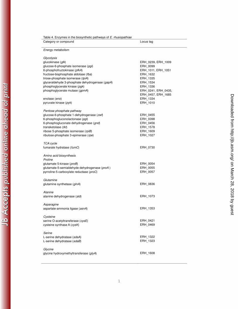
E. rhusiopathiae encodes a full set of enzymes for the glycolysis and 229
pentose phosphate pathways; some enzymes (glucokinase, 6-phosphofructokinase, 230
and phosphoglycerate mutase) are duplicated. However, as expected, the organism 231
lacks numerous genes for many other metabolic pathways (Fig. 3, Table 4). E. 232
rhusiopathiae lacks all of the genes for the tricarboxylic acid cycle, with the exception 233
of fumarate hydratase (Table 4). Importantly, the organism also lacks all of the genes 234
for the biosynthesis of unsaturated and saturated fatty acids. This genomic feature—235
the complete lack of genes for fatty acid biosynthesis—is observed in the genomes of 236
all Mollicutes species (with the exception of Acholeplasma laidlawii) but not in other 237
bacteria in the Firmicutes phylum, although several gene losses in fatty acid 238
biosynthetic pathways have been observed in some lactobacilli (25). 239
It appears that the mechanism by which Tween 80 enhances the growth of E. 240
rhusiopathiae does not involve oleic acid, which is the major ingredient of Tween 80, 241
because we were unable to demonstrate growth enhancement of the organism after 242
adding of various quantities of oleic acid. Its growth was completely inhibited by 243
concentrations of oleic acid greater than 0.001% (data not shown). This finding 244
suggests that Tween 80 may merely aid in membrane transport or another nutrient 245
utilization process. 246
E. rhusiopathiae also lacks many genes for amino acid biosynthesis; the 247
organism can synthesize only seven amino acids (alanine, asparagine, glutamine, 248
serine, cysteine, glycine and proline) through de novo pathways or using 249
intermediate molecules as derivatives. Moreover, all of the genes for the biosynthesis 250
of biotin, riboflavin, ubiquinone, and menaquinone and some of the genes involved in 251
the biosynthesis for pantothenate, thiamine, and folate are missing from the E. 252
rhusiopathiae genome (Table 4). 253
In agreement with its poor biosynthetic capacities, E. rhusiopathiae devotes 254
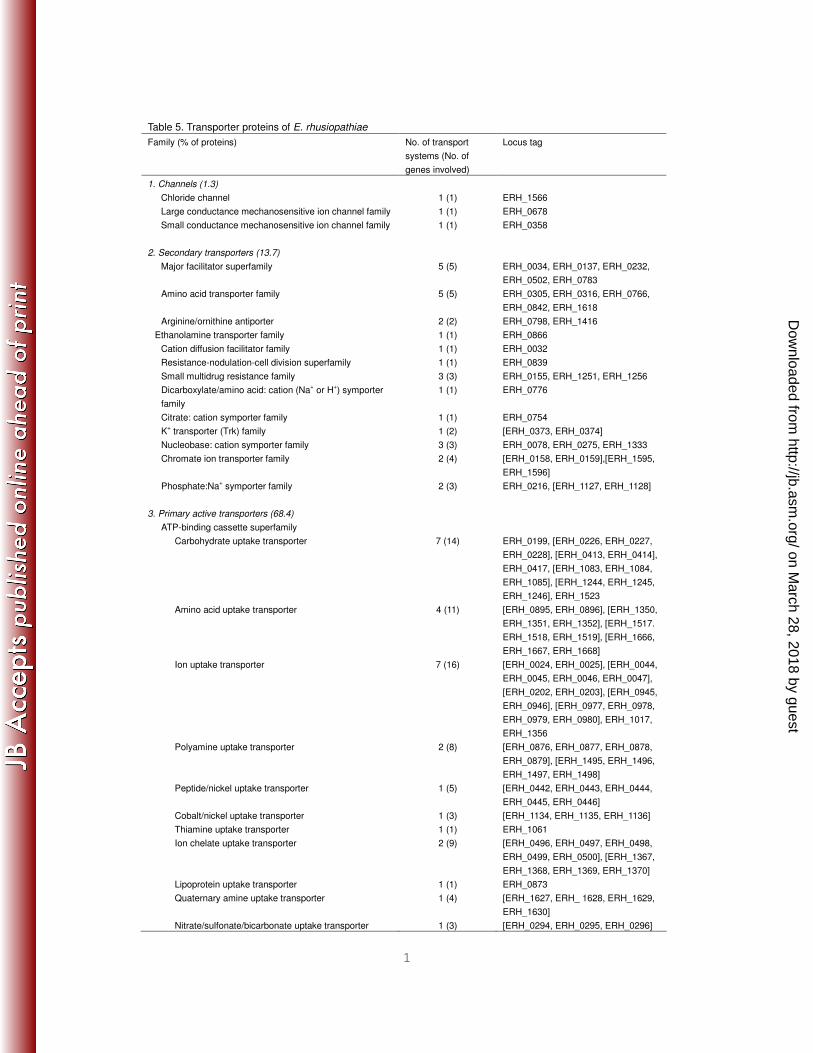
as much as 13.7% (minimally) of its genes to transport functions (Table 5), a level 255
similar to that observed for lactic acid bacteria (13-18%) (21). In particular, E. 256
rhusiopathiae contains a remarkably high percentage of primary active transporters 257
(68.4%). This property is also shared with Mycoplasma species (39). 258
on March 28, 2018 by guest
http://jb.asm.org/
Dow
nloaded from

9
259
Virulence-associated genes. 260
(i) Two-component signal transduction systems. Most bacteria employ two-261
component signal transduction systems to regulate the expression of many genes in 262
response to various changes in environmental conditions (54). In the E. rhusiopathiae 263
genome, we identified a total of 15 genes encoding response regulators, 14 of which 264
were adjacent to genes encoding cognate histidine kinases (Table 6). The numbers 265
of two-component signal transduction systems in the genomes of Streptococcus 266
pyogenes (12), Streptococcus pneumoniae (57), Listeria monocytogenes (15), 267
Enterococcus faecalis (16), and Bacillus subtilis (10) are 13, 14, 16, 17, and 35, 268
respectively. Compared to these bacteria, lactic acid bacteria contain fewer two-269
component systems (between five and nine) (9) and Mycoplasma species lack this 270
system entirely, with the exception of Mycoplasma penetrans (42). A loss of 271
regulatory systems is also an evolutionary pattern observed in many bacteria with 272
reduced genome sizes (31). Considering the presence of many two-component 273
systems in the E. rhusiopathiae genome compared to lactic acid bacteria and the 274
important roles of these systems in stress responses, such as oxidative or acid stress 275
responses (9), the two-component systems of E. rhusiopathiae may be very closely 276
associated with its virulence. 277
278
(ii) Cell wall synthesis. E. rhusiopathiae possesses a full set of the peptidoglycan 279
(PG) biosynthesis genes, which are relatively dispersed throughout the genome (Fig. 280
3). However, it is not clear whether all the genes involved in the complete 281
biosynthetic pathways of wall teichoic acids (WTA) and lipoteichoic acids (LTA) are 282
present. Furthermore, although the dlt operon encoding the enzymes that catalyze 283
the incorporation of D-alanine residues into WTA or LTA is always composed of 284
dltABCD genes in gram-positive bacteria (34), the gene order is not conserved, and 285
the dltD gene is missing in E. rhusiopathiae (Fig. 3), suggesting that the organism 286
contains atypical WTA and/or LTA. The dltABCD genes have been detected in all 287
Firmicutes bacteria with small genomes sequenced so far (19, 34), with the exception 288
of Oenococcus oeni, which is a lactic acid bacterium with the smallest genome of all 289
Firmicutes (Table 3). In the future, it will be worthwhile to intensively analyze the 290
structures and chemical compositions of WTA and LTA of E. rhusiopathiae. 291
on March 28, 2018 by guest
http://jb.asm.org/
Dow
nloaded from
10
292
(iii) Capsular polysaccharide synthesis. Immunological and microscopic 293
approaches were used to demonstrate that E. rhusiopathiae produces a capsule that 294
plays an important role in virulence (49, 51, 52); however, its chemical and biological 295
properties are uncharacterized. We previously showed that in the representative 296
acapsular mutant 33H6, which was generated by transposon mutagenesis with 297
Tn916 (52), the transposon was inserted into a gene (now corresponding to 298
ERH_0855) (48). The E. rhusiopathiae genome analysis revealed that this gene is 299
located in a cluster of genes encoding seven proteins (ERH_0855 to ERH_0861) that 300
appear to be involved in capsular polysaccharide biosynthesis (Table 7). Reverse 301
transcription (RT)-PCR analysis of the intergenic regions of the seven genes indicate 302
that these genes are transcribed as a polycistronic mRNA forming an operon (data 303
now shown). 304
305
(iv) Protein-secretion systems and surface-associated or extracellular 306
enzymes/proteins. Gram-positive bacteria produce a variety of extracellular or cell 307
surface-associated proteins, many of which play important roles in virulence (13). In 308
gram-positive bacteria, these proteins could be translocated across the membrane 309
via three pathways: the Sec (general secretory)-dependent pathway, the signal-310
recognition particle (SRP)-dependent pathway, and the twin-arginine translocation 311
(Tat) pathway (38). 312
E. rhusiopathiae encodes homologs for signal peptidase I, signal peptidase II 313
(lipoprotein signal peptidase), SRP, and a full set of Sec proteins. By contrast, as 314
seen in other members of Firmicutes with small genomes (Streptococcus species 315
and lactic acid bacteria) and in Mollicutes (7), E. rhusiopathiae does not contain the 316
Tat secretion system or any recognizable Tat substrates. 317
During the secretion process in gram-positive bacteria, many secreted proteins 318
are covalently attached to peptidoglycan through their carboxyl termini by 319
transpeptidases, called sortases, to be exposed as surface proteins (33). E. 320
rhusiopathiae contains a single sortase (ERH_0013) and 21 potential sortase 321
substrates that contain a sortase-recognition sequence (LPxTG) followed by a 322
membrane spanning hydrophobic domain and a positively charged tail (Table 8). 323
They include various hydrolyzing enzymes, such as proteases and peptidases, and 324
on March 28, 2018 by guest
http://jb.asm.org/
Dow
nloaded from

11
proteins that can potentially mediate host-bacteria interactions. Three of these 325
proteins possess KxW repeat modules as well, which are conserved in several 326
adhesins of gram-positive bacteria belonging to Firmicutes and have been shown to 327
play important roles in biofilm formation (50). 328
Of particular interest are three hyaluronidases (ERH_0150, ERH_0765, and 329
ERH_1210) and one neuraminidase (ERH_0299) that contain LPxTG motifs. Cell-330
surface associations of hyaluronidases and neuraminidase via LPxTG motif have 331
been shown only in S. pneumoniae (13). It is most likely that these four enzymes of E. 332
rhusiopathiae are also surface associated. Notably, the organism encodes an 333
additional neuraminidase (ERH_0761) that lacks the LPxTG motif and thus is 334
probably extracellulary secreted. 335
E. rhusiopathiae possesses another type of surface proteins with glycine-336
tryptophan (GW) dipeptide repeat modules, which attach surface proteins to the cell 337
wall through noncovalent interactions (59). In addition to the major surface protective 338
antigen SpaA.1 protein (ERH_0094), E. rhusiopathiae strain Fujisawa contains two 339
surface proteins (ERH_0407 and ERH_0768) with the GW dipeptide repeat modules. 340
Thus, like many other gram-positive pathogens (13), E. rhusiopathiae elaborates a 341
number of cell-surface components and extracellular products to interact with their 342
target cells and initiate infection. 343
344
(v) Putative virulence factors required for intracellular survival. E. rhusiopathiae 345
is vulnerable to oxidative stress, and the escape from reactive oxygen species (ROS) 346
is a major strategy for the intracellular survival of the organism (51). Genome 347
analysis revealed that E. rhusiopathiae possesses nine CDSs encoding enzymes 348
that potentially confer ROS resistance. They include a superoxide dismutase, two 349
thioredoxins, two thioredoxin reductases, a thiol peroxidase, and a glutaredoxin 350
(Table 8). Although E. rhusiopathiae lacks catalase, the organism may have evolved 351
these redundant defense mechanisms against oxidative stress within host cells. 352
Furthermore, the two alkyl-hydroperoxide reductases may be important in protecting 353
the organism against damage caused by nitric oxide (5). 354
In addition to these anti-ROS proteins, E. rhusiopathiae contains additional 355
enzymes that potentially help the organism survive within phagocytic cells (Table 8). 356
Phospholipases are considered virulence factors in many intracellular pathogens (43). 357
on March 28, 2018 by guest
http://jb.asm.org/
Dow
nloaded from

12
The E. rhusiopathiae genome contains at least nine CDSs encoding proteins with 358
sequence homologies to phospholipase family proteins, including phospholipase D 359
and patatin-like phospholipases. Patatin, a storage protein found in potatoes, also 360
has a phospholipase activity (29). E. rhusiopathiae possesses an extremely high 361
number of phospholipolytic enzymes compared to other intracellular bacteria (1). The 362
abundance of these enzymes of E. rhusiopathiae may be a reflection of the lack of 363
fatty acid biosynthesis pathways. Efficient acquisition of fatty acids from the host 364
membrane may be achieved by their combined actions. These phospholipases may 365
allow the organism to escape from phagosomes into the cytoplasm by disrupting the 366
phagosomal membrane (43). In fact, electron microscopic observation revealed that 367
the organism multiplies predominantly within the cytoplasm of macrophages at the 368
inoculation sites of mice (46), suggesting that the cytoplasm serves as a privileged in 369
vivo niche for E. rhusiopathiae, which allows the organism to circumvent host 370
immune responses. Antioxidant enzymes and phospholipases could also modulate 371
immunological cell-signaling pathways (2, 44). Thus, the organism may regulate host 372
cell functions by these enzymes for successful intracellular survival. 373
374
Genome comparison. By bidirectional best hit analysis with the draft sequence of E. 375
rhusiopathiae strain ATCC 19414, we found that 1594 (93.5%) CDSs of Fujisawa, 376
including the genes for capsular polysaccharide biosynthesis (ERH_0855 to 377
ERH_0861), are conserved in ATCC 19414. The orthologs exhibited a high level of 378
sequence similarity (99.3% amino acid sequence identity on average). The result of 379
dotplot analysis also revealed a high level of genomic synteny of the two genomes 380
(Figure S1). These data indicate that the genomic backbone is highly conserved 381
between the two strains. 382
In ATCC 19414, 71 out of the 1645 CDSs were strain-specific (Table S1). In 383
Fujisawa, 110 CDSs were absent in ATCC 19414 (Table S2). These Fujisawa-specific 384
CDSs include the 46 genes on PP_Erh_Fujisawa (ERH_0581 through ERH_0626) 385
and 23 genes that were also clustered in the genome (ERH_1273 through 386
ERH_1295) (Fig. 1 and Table S2). Because this gene cluster contains genes for an 387
integrase and a replication protein as well as several IS elements, it is most likely that 388
it represents an integrative element although direct repeat sequences were not found 389
at the boundaries. This integrative element, designated IE_Erh_Fujisawa, encodes 390
the genes for a restriction-modification system and a heavy metal transport system. 391
on March 28, 2018 by guest
http://jb.asm.org/
Dow
nloaded from

13
Moreover, Fujisawa appears to have a strain-specific pathway for polysaccharide 392
biosynthesis (ERH_1439 through ERH_1444). The G+C contents of most of the 393
strain-specific CDSs in the genomes of Fujisawa and ATCC 19414 differ significantly 394
from those of other parts of the genome (Tables S1 and S2), suggesting that they 395
have been acquired by each of the strains through horizontal gene transfer. 396
We further analyzed conservation of the Fujisawa CDSs in other 397
Erysipelotrichia strains. We excluded the T. sanguinis strain from this analysis 398
because it is very likely that this species is not a true member of Erysipelotrichia as 399
mentioned in the section for phylogenetic position of E. rhusiopathiae. This analysis 400
revealed that between 52 and 61% of the CDSs of Fujisawa are conserved in each of 401
these strains (Table S3) and a total of 625 CDSs (37%) are shared by all 402
Erysipelotrichia strains examined. Although this analysis is not complete because the 403
genome sequence information of all these Erysipelotrichia strains are draft 404
sequences with various levels of sequence quality, these 625 CDSs can be regarded 405
as roughly representing the core gene set of this bacterial group. It might be worth 406
mentioning that it appears that these Erysipelotrichia strains also lack the dltABCD 407
operon. 408
409
Conclusions. This study describes the reductive genome revolution of E. 410
rhusiopathiae and its unique strategy toward intracellular parasitism. The 411
phylogenetic study using the genome sequence information revealed that 412
Erysipelotrichia strains including E. rhusiopathiae form a cluster distinct from other 413
Firmicutes, and are phylogenetically closest to Mollicutes. The genomic features of E. 414
rhusiopathiae also represent evolutionary traits of both Firmicutes and Mollicutes. 415
Like Mollicutes, during genome reduction, E. rhusiopathiae lost the genes necessary 416
for fatty acid biosynthesis pathways and evolved redundant phospholipolytic 417
enzymes, which may be utilized for fatty acid acquisition in vivo. Furthermore, the 418
organism possesses many antioxidant factors, suggesting that E. rhusiopathiae 419
specifically adapted to the intracellular environments of phagocytic cells and showing 420
that its evolutionary adaptation is very unique in the phylum Firmicutes. Comparative 421
analysis with a draft genome sequence of another E. rhusiopathiae strain (ATCC 422
19414) revealed that these genomic features are highly conserved in this strain. 423
424
on March 28, 2018 by guest
http://jb.asm.org/
Dow
nloaded from

14
Acknowledgments 425
The authors are grateful to Vincent A. Fischetti for critically reading this 426
manuscript and to Ken Kurokawa for helpful suggestions on the phylogenetic data 427
analysis. 428
This work was supported by a Research and Development Projects for 429
Application in Promoting New Policy of Agriculture Forestry and Fisheries grant from 430
the Ministry of Agriculture, Forestry and Fisheries of Japan (to Y.S.) 431
432
433
References 434
1. Banerji, S., P. Aurass, and A. Flieger. 2008. The manifold phospholipases A of 435
Legionella pneumophila - identification, export, regulation, and their link to bacterial 436
virulence. Int. J. Med. Microbiol. 298:169-81. 437
2. Bogdan, C., M. Rollinghoff, and A. Diefenbach. 2000. Reactive oxygen and reactive 438
nitrogen intermediates in innate and specific immunity. Curr. Opin. Immunol. 12:64-76. 439
3. Calcutt, M. J., J. L. Lavrrar, and K. S. Wise. 1999. IS1630 of Mycoplasma fermentans, 440
a novel IS30-type insertion element that targets and duplicates inverted repeats of 441
variable length and sequence during insertion. J. Bacteriol. 181:7597-607. 442
4. Castresana, J. 2000. Selection of conserved blocks from multiple alignments for their 443
use in phylogenetic analysis. Mol Biol Evol. 17:540-52. 444
5. Chen, L., Q. W. Xie, and C. Nathan. 1998. Alkyl hydroperoxide reductase subunit C 445
(AhpC) protects bacterial and human cells against reactive nitrogen intermediates. Mol. 446
Cell 1:795-805. 447
6. Ciccarelli, F. D., T. Doerks, C. von Mering, C. J. Creevey, B. Snel, and P. Bork. 2006. 448
Toward automatic reconstruction of a highly resolved tree of life. Science 311:1283-7. 449
7. Dilks, K., R. W. Rose, E. Hartmann, and M. Pohlschroder. 2003. Prokaryotic 450
utilization of the twin-arginine translocation pathway: a genomic survey. J. Bacteriol. 451
185:1478-83. 452
8. Edgar, R. C. 2004. MUSCLE: multiple sequence alignment with high accuracy and high 453
throughput. Nucleic Acids Res. 32:1792-7. 454
9. El-Sharoud, W. M. 2005. Two-component signal transduction systems as key players in 455
stress responses of lactic acid bacteria. Sci. Prog. 88:203-28. 456
10. Fabret, C., V. A. Feher, and J. A. Hoch. 1999. Two-component signal transduction in 457
Bacillus subtilis: how one organism sees its world. J. Bacteriol. 181:1975-83. 458
on March 28, 2018 by guest
http://jb.asm.org/
Dow
nloaded from

15
11. Feist, H., K. D. Flossmann, and W. Erler. 1976. Investigations on the nutritional 459
requirements of erysipelas bacteria. Arch. Exp. Veterinarmed. 30:49-57. 460
12. Ferretti, J. J., W. M. McShan, D. Ajdic, D. J. Savic, G. Savic, K. Lyon, C. Primeaux, 461
S. Sezate, A. N. Suvorov, S. Kenton, H. S. Lai, S. P. Lin, Y. Qian, H. G. Jia, F. Z. 462
Najar, Q. Ren, H. Zhu, L. Song, J. White, X. Yuan, S. W. Clifton, B. A. Roe, and R. 463
McLaughlin. 2001. Complete genome sequence of an M1 strain of Streptococcus 464
pyogenes. Proc. Natl. Acad. Sci. USA 98:4658-63. 465
13. Fischetti, V. A. 2006. Surface proteins on gram-positive bacteria, p. 12-25. In V. A. 466
Fischetti, R. P. Novick, J. J. Ferretti, D. A. Portnoy, and J. I. Rood (ed.), Gram-positive 467
pathogens, 2nd ed. ASM Press, Washington, DC. 468
14. Garrity, G. M., T. G. Lilburn, J. R. Cole, S. H. Harrison, J. Euzeby, and B. J. Tindall. 469
March 6, 2007. Part 8, The Bacteria: Phylum Firmicutes: Class Mollicutes, p. 317-332. 470
In G. M. Garrity, T. G. Lilburn, J. R. Cole, S. H. Harrison, J. Euzeby, and B. J. Tindall 471
(ed.), The Taxonomic Outline of Bacteria and Archaea, Release 7.7. 472
http://www.taxonomicoutline.org/. 473
15. Glaser, P., L. Frangeul, C. Buchrieser, C. Rusniok, A. Amend, F. Baquero, P. Berche, 474
H. Bloecker, P. Brandt, T. Chakraborty, A. Charbit, F. Chetouani, E. Couve, A. de 475
Daruvar, P. Dehoux, E. Domann, G. Dominguez-Bernal, E. Duchaud, L. Durant, O. 476
Dussurget, K. D. Entian, H. Fsihi, F. Garcia-del Portillo, P. Garrido, L. Gautier, W. 477
Goebel, N. Gomez-Lopez, T. Hain, J. Hauf, D. Jackson, L. M. Jones, U. Kaerst, J. 478
Kreft, M. Kuhn, F. Kunst, G. Kurapkat, E. Madueno, A. Maitournam, J. M. Vicente, 479
E. Ng, H. Nedjari, G. Nordsiek, S. Novella, B. de Pablos, J. C. Perez-Diaz, R. Purcell, 480
B. Remmel, M. Rose, T. Schlueter, N. Simoes, A. Tierrez, J. A. Vazquez-Boland, H. 481
Voss, J. Wehland, and P. Cossart. 2001. Comparative genomics of Listeria species. 482
Science 294:849-52. 483
16. Hancock, L., and M. Perego. 2002. Two-component signal transduction in 484
Enterococcus faecalis. J. Bacteriol. 184:5819-25. 485
17. Hyyrylainen, H. L., A. Bolhuis, E. Darmon, L. Muukkonen, P. Koski, M. Vitikainen, 486
M. Sarvas, Z. Pragai, S. Bron, J. M. van Dijl, and V. P. Kontinen. 2001. A novel two-487
component regulatory system in Bacillus subtilis for the survival of severe secretion 488
stress. Mol. Microbiol. 41:1159-72. 489
18. Kanehisa, M. 1997. A database for post-genome analysis. Trends Genet. 13:375-6. 490
19. Kovacs, M., A. Halfmann, I. Fedtke, M. Heintz, A. Peschel, W. Vollmer, R. 491
Hakenbeck, and R. Bruckner. 2006. A functional dlt operon, encoding proteins 492
on March 28, 2018 by guest
http://jb.asm.org/
Dow
nloaded from

16
required for incorporation of d-alanine in teichoic acids in gram-positive bacteria, 493
confers resistance to cationic antimicrobial peptides in Streptococcus pneumoniae. J. 494
Bacteriol. 188:5797-805. 495
20. Lagesen, K., P. Hallin, E. A. Rodland, H. H. Staerfeldt, T. Rognes, and D. W. Ussery. 496
2007. RNAmmer: consistent and rapid annotation of ribosomal RNA genes. Nucleic 497
Acids Res. 35:3100-8. 498
21. Lorca, G. L., R. D. Barabote, V. Zlotopolski, C. Tran, B. Winnen, R. N. Hvorup, A. J. 499
Stonestrom, E. Nguyen, L. W. Huang, D. S. Kim, and M. H. Saier, Jr. 2007. Transport 500
capabilities of eleven gram-positive bacteria: comparative genomic analyses. Biochim. 501
Biophys. Acta 1768:1342-66. 502
22. Lowe, T. M., and S. R. Eddy. 1997. tRNAscan-SE: a program for improved detection of 503
transfer RNA genes in genomic sequence. Nucleic Acids Res. 25:955-64. 504
23. Ludwig, W., K. H. Schleifer, and W. B. Whitman. 2009. Revised road map to the 505
phylum Firmicutes, p. 1-13. In P. D. Vos, G. M. Garrity, D. Jones, N. R. Krieg, and W. 506
Ludwig (ed.), Bergey's Manual of Systematic Bacteriology, 2nd ed, vol. 3. Springer-507
Verlag, New York. 508
24. Lysnyansky, I., M. J. Calcutt, I. Ben-Barak, Y. Ron, S. Levisohn, B. A. Methe, and D. 509
Yogev. 2009. Molecular characterization of newly identified IS3, IS4 and IS30 insertion 510
sequence-like elements in Mycoplasma bovis and their possible roles in genome 511
plasticity. FEMS Microbiol. Lett. 294:172-82. 512
25. Makarova, K., A. Slesarev, Y. Wolf, A. Sorokin, B. Mirkin, E. Koonin, A. Pavlov, N. 513
Pavlova, V. Karamychev, N. Polouchine, V. Shakhova, I. Grigoriev, Y. Lou, D. 514
Rohksar, S. Lucas, K. Huang, D. M. Goodstein, T. Hawkins, V. Plengvidhya, D. 515
Welker, J. Hughes, Y. Goh, A. Benson, K. Baldwin, J. H. Lee, I. Diaz-Muniz, B. 516
Dosti, V. Smeianov, W. Wechter, R. Barabote, G. Lorca, E. Altermann, R. 517
Barrangou, B. Ganesan, Y. Xie, H. Rawsthorne, D. Tamir, C. Parker, F. Breidt, J. 518
Broadbent, R. Hutkins, D. O'Sullivan, J. Steele, G. Unlu, M. Saier, T. Klaenhammer, 519
P. Richardson, S. Kozyavkin, B. Weimer, and D. Mills. 2006. Comparative genomics 520
of the lactic acid bacteria. Proc. Natl. Acad. Sci. USA 103:15611-6. 521
26. Makino, K., H. Shinagawa, M. Amemura, and A. Nakata. 1986. Nucleotide sequence 522
of the phoB gene, the positive regulatory gene for the phosphate regulon of Escherichia 523
coli K-12. J. Mol. Biol. 190:37-44. 524
27. Makino, K., H. Shinagawa, M. Amemura, and A. Nakata. 1986. Nucleotide sequence 525
of the phoR gene, a regulatory gene for the phosphate regulon of Escherichia coli. J. Mol. 526
on March 28, 2018 by guest
http://jb.asm.org/
Dow
nloaded from

17
Biol. 192:549-56. 527
28. Merhej, V., M. Royer-Carenzi, P. Pontarotti, and D. Raoult. 2009. Massive 528
comparative genomic analysis reveals convergent evolution of specialized bacteria. Biol. 529
Direct 4:13. 530
29. Mignery, G. A., C. S. Pikaard, and W. D. Park. 1988. Molecular characterization of 531
the patatin multigene family of potato. Gene 62:27-44. 532
30. Moran, N. A. 2002. Microbial minimalism: genome reduction in bacterial pathogens. 533
Cell 108:583-6. 534
31. Moran, N. A., and J. J. Wernegreen. 2000. Lifestyle evolution in symbiotic bacteria: 535
insights from genomics. Trends Ecol. Evol. 15:321-326. 536
32. Nakato, H., K. Shinomiya, and H. Mikawa. 1986. Effect of C3 depletion on the 537
genesis of thrombocytopenia induced in rats by Erysipelothrix rhusiopathiae. Scand. J. 538
Haematol. 37:18-24. 539
33. Navarre, W. W., and O. Schneewind. 1999. Surface proteins of gram-positive bacteria 540
and mechanisms of their targeting to the cell wall envelope. Microbiol. Mol. Biol. Rev. 541
63:174-229. 542
34. Neuhaus, F. C., and J. Baddiley. 2003. A continuum of anionic charge: structures and 543
functions of D-alanyl-teichoic acids in gram-positive bacteria. Microbiol. Mol. Biol. Rev. 544
67:686-723. 545
35. Nikolskaya, A. N., and M. Y. Galperin. 2002. A novel type of conserved DNA-binding 546
domain in the transcriptional regulators of the AlgR/AgrA/LytR family. Nucleic Acids 547
Res. 30:2453-9. 548
36. Noguchi, H., T. Taniguchi, and T. Itoh. 2008. MetaGeneAnnotator: detecting species-549
specific patterns of ribosomal binding site for precise gene prediction in anonymous 550
prokaryotic and phage genomes. DNA Res. 15:387-96. 551
37. Ohyama, A., K. Kurokawa, K. Enai, H. Saitoh, S. Kanaya, M. Altaf-Ul-Amin, and 552
N.Ogasawara. 2006. Bioinformatics tool for genomic era; A step towards the in silico 553
experiments - focused on molecular cloning. J. Comp. Aid. Chem. 7:102-115. 554
38. Pallen, M. J., R. R. Chaudhuri, and I. R. Henderson. 2003. Genomic analysis of 555
secretion systems. Curr. Opin. Microbiol. 6:519-27. 556
39. Paulsen, I. T., L. Nguyen, M. K. Sliwinski, R. Rabus, and M. H. Saier, Jr. 2000. 557
Microbial genome analyses: comparative transport capabilities in eighteen prokaryotes. J. 558
Mol. Biol. 301:75-100. 559
40. Pruitt, K. D., T. Tatusova, and D. R. Maglott. 2007. NCBI reference sequences 560
on March 28, 2018 by guest
http://jb.asm.org/
Dow
nloaded from
18
(RefSeq): a curated non-redundant sequence database of genomes, transcripts and 561
proteins. Nucleic Acids Res. 35:D61-5. 562
41. Rose, D. L., J. G. Tully, J. M. Bove, and R. F. Whitcomb. 1993. A test for measuring 563
growth responses of mollicutes to serum and polyoxyethylene sorbitan. Int. J. Syst. 564
Bacteriol. 43:527-32. 565
42. Sasaki, Y., J. Ishikawa, A. Yamashita, K. Oshima, T. Kenri, K. Furuya, C. Yoshino, 566
A. Horino, T. Shiba, T. Sasaki, and M. Hattori. 2002. The complete genomic sequence 567
of Mycoplasma penetrans, an intracellular bacterial pathogen in humans. Nucleic Acids 568
Res. 30:5293-300. 569
43. Schmiel, D. H., and V. L. Miller. 1999. Bacterial phospholipases and pathogenesis. 570
Microbes Infect. 1:1103-12. 571
44. Schwarzer, N., R. Nost, J. Seybold, S. K. Parida, O. Fuhrmann, M. Krull, R. 572
Schmidt, R. Newton, S. Hippenstiel, E. Domann, T. Chakraborty, and N. Suttorp. 573
1998. Two distinct phospholipases C of Listeria monocytogenes induce ceramide 574
generation, nuclear factor-kappa B activation, and E-selectin expression in human 575
endothelial cells. J. Immunol. 161:3010-8. 576
45. Shimoji, Y. 2000. Pathogenicity of Erysipelothrix rhusiopathiae: virulence factors and 577
protective immunity. Microbes Infect. 2:965-72. 578
46. Shimoji, Y. 2004. Erysipelothrix rhusiopathiae, p. 111–116. In G. L. Gyles, J. F. Prescott, 579
J. G. Songer, and C. O. Thoen (ed.), Pathogenesis of bacterial infections in animals, 3rd 580
ed. Blackwell Publishing, Ames, Iowa. 581
47. Shimoji, Y., H. Asato, T. Sekizaki, Y. Mori, and Y. Yokomizo. 2002. Hyaluronidase is 582
not essential for the lethality of Erysipelothrix rhusiopathiae infection in mice. J. Vet. 583
Med. Sci. 64:173-6. 584
48. Shimoji, Y., Y. Mori, K. Hyakutake, T. Sekizaki, and Y. Yokomizo. 1998. Use of an 585
enrichment broth cultivation-PCR combination assay for rapid diagnosis of swine 586
erysipelas. J. Clin. Microbiol. 36:86-9. 587
49. Shimoji, Y., Y. Mori, T. Sekizaki, T. Shibahara, and Y. Yokomizo. 1998. Construction 588
and vaccine potential of acapsular mutants of Erysipelothrix rhusiopathiae: use of 589
excision of Tn916 to inactivate a target gene. Infect. Immun. 66:3250-4. 590
50. Shimoji, Y., Y. Ogawa, M. Osaki, H. Kabeya, S. Maruyama, T. Mikami, and T. 591
Sekizaki. 2003. Adhesive surface proteins of Erysipelothrix rhusiopathiae bind to 592
polystyrene, fibronectin, and type I and IV collagens. J. Bacteriol. 185:2739-48. 593
51. Shimoji, Y., Y. Yokomizo, and Y. Mori. 1996. Intracellular survival and replication of 594
on March 28, 2018 by guest
http://jb.asm.org/
Dow
nloaded from

19
Erysipelothrix rhusiopathiae within murine macrophages: failure of induction of the 595
oxidative burst of macrophages. Infect. Immun. 64:1789-93. 596
52. Shimoji, Y., Y. Yokomizo, T. Sekizaki, Y. Mori, and M. Kubo. 1994. Presence of a 597
capsule in Erysipelothrix rhusiopathiae and its relationship to virulence for mice. Infect. 598
Immun. 62:2806-10. 599
53. Stackebrandt, E. 2009. Genus I. Erysipelothrix, p. 1299-1306. In P. D. Vos, G. M. 600
Garrity, D. Jones, N. R. Krieg, and W. Ludwig (ed.), Bergey's Manual of Systematic 601
Bacteriology, 2nd ed, vol. 3. Springer-Verlag, New York. 602
54. Stock, J. B., A. J. Ninfa, and A. M. Stock. 1989. Protein phosphorylation and regulation 603
of adaptive responses in bacteria. Microbiol. Rev. 53:450-90. 604
55. Takahashi, T., N. Hirayama, T. Sawada, Y. Tamura, and M. Muramatsu. 1987. 605
Correlation between adherence of Erysipelothrix rhusiopathiae strains of serovar 1a to 606
tissue culture cells originated from porcine kidney and their pathogenicity in mice and 607
swine. Vet. Microbiol. 13:57-64. 608
56. Tamura, K., J. Dudley, M. Nei, and S. Kumar. 2007. MEGA4: Molecular 609
Evolutionary Genetics Analysis (MEGA) software version 4.0. Mol. Biol. Evol. 610
24:1596-9. 611
57. Throup, J. P., K. K. Koretke, A. P. Bryant, K. A. Ingraham, A. F. Chalker, Y. Ge, A. 612
Marra, N. G. Wallis, J. R. Brown, D. J. Holmes, M. Rosenberg, and M. K. Burnham. 613
2000. A genomic analysis of two-component signal transduction in Streptococcus 614
pneumoniae. Mol. Microbiol. 35:566-76. 615
58. Wood, R. L. 1999. Erysipelas, p. 419-430. In B. E. Straw, S. D'Allaire, W. L. Mengelng, 616
and D. J. Taylor (ed.), Diseases of swine, 8th ed. Iowa State University Press, Ames, 617
Iowa. 618
59. Yother, J., and J. M. White. 1994. Novel surface attachment mechanism of the 619
Streptococcus pneumoniae protein PspA. J. Bacteriol. 176:2976-85. 620
on March 28, 2018 by guest
http://jb.asm.org/
Dow
nloaded from
Figure 1. Circular representation of the genome of the E. rhusiopathiae strain
Fujisawa. Beginning with the outer region, the circle shows (i) nucleotide
positions in base pairs, (ii) predicted CDSs transcribed on the forward
(clockwise) and (iii) reverse (counterclockwise) DNA strands, (iv) positions of
Fujisawa strain-specific genes not present in the genome of ATCC 19414 strain, (v)
rRNA operon(s), (vi) tRNA genes, (vii) percent G+C content (red and blue,
respectively, represent regions with higher and lower G+C content compared to the
average value for the entire genome), and (viii) the G+C skew curve. The colors of
each CDS were assigned according to the COG functional grouping
(http://www.ncbi.nlm.nih.gov/COG/)
Figure 2. Phylogenetic position of E. rhusiopathiae. Phylogenetic trees based on
16S rRNA gene sequences (A) and based on concatenated protein sequence
alignments derived from 31 universal protein families (B) are shown. Species are
colored according to the current taxonomy: the phylum Firmicutes, blue;
Mollicutes, green; others, black. E. rhusiopathiae is indicated by boldface. The
scale bar represents the expected number of changes per sequence position. A
bootstrap test with 1000 replicates was used to estimate the confidence of the
branching patterns of the trees. Branches corresponding to partitions reproduced
in less than 50% bootstrap replicates are collapsed.
Figure 3. Overview of the basic metabolic pathways of E. rhusiopathiae. Pathways
or steps for which no enzymes were identified are indicated by red, as are the
compounds for which de novo synthetic pathways were not identified. The question
marks indicate that particular uncertainties exist. ABC, ATP-binding cassette
superfamily; Sec, secretion pathway; PTS, phosphotransferase system; GalNAc,
N-acetylgalactosamine; GlcNAc, N-acetylglucosamine; MscL, large conductance
mechanosensitive ion channel family; MscS, small conductance mechanosensitive
ion channel family; PP pathway, pentose phosphate pathway; PRPP,
phosphoribosyl-pyrophosphate; UMP, uridine 5'-monophosphate; AICAR,
5'-phosphoribosyl-4-carboxamide-5-aminoimidazole; IMP, inosine monophosphate;
PEP, phosphoenolpyruvate; ACP, acyl-carrier protein; TCA, tricarboxylic acid; THF,
tetrahydrofolate; DHF, dihydrofolate.
on March 28, 2018 by guest
http://jb.asm.org/
Dow
nloaded from

1
Table 1. General features of the genome of E. rhusiopathiae strain Fujisawa
Genome size (bp)
Overall G+C content (%)
G+C content of CDS (%)
Number of CDS
Number of pseudogenes
Number of rRNA operons (16S-23S-5S)
Number of tRNA genes
Prophage
Plasmid
1,787,941
36.6
36.7
1704
7
7
55
1
0
on March 28, 2018 by guest
http://jb.asm.org/
Dow
nloaded from

1
Table 2. Insertion sequences identified in E. rhusiopathiae strain Fujisawa
Name Number IS family No. of CDSs Size (bp) Length of DR (bp)
ISErh1 ERH_IS01 3 2 1280 3
ISErh1 ERH_IS02 3 2 1280 3
ISErh1 ERH_IS06 3 2 1280 3
ISErh1 ERH_IS14 3 2 1280 4
ISErh1 ERH_IS22 3 2 1280 3
ISErh2 ERH_IS03 30 1 1031 - a
ISErh2 ERH_IS08 30 1 1031 27
ISErh2 ERH_IS09 30 1 1031 29
ISErh2 ERH_IS15 30 1 1031 32
ISErh2 ERH_IS17 30 1 1031 23
ISErh2 ERH_IS18 30 1 1031 24
ISErh2 ERH_IS23 30 1 1031 30
ISErh2 ERH_IS24 30 1 1031 31
ISErh3 ERH_IS04 ? b
3 1351 6
ISErh3 ERH_IS07 c ?
b 3 1176 6
ISErh4 ERH_IS05 30 1 1032 30
ISErh4 ERH_IS10 30 1 1032 26
ISErh4 ERH_IS11 30 1 1032 37
ISErh4 ERH_IS16 30 1 1032 26
ISErh4 ERH_IS19 30 1 1032 41
ISErh5 ERH_IS12 3 2 1247 -
ISErh5 ERH_IS13 3 2 1247 -
ISErh5 ERH_IS20 c 3 2 1206 -
ISErh6 ERH_IS21 3 2 1240 - a -, not identified.
b ?, unclassified; both copies appear to contain internal deletions.
c Truncated IS.
on March 28, 2018 by guest
http://jb.asm.org/
Dow
nloaded from

Table 3. Numbers of orthologous genes related to DNA repaira
Name (Strain)
Genome size
(Mb)
GC contents
(%) phylum or class total adaA alkA alkB dinP
exoA
(xth) fpg ligA mfd mpg mutH mutL mutS mutT mutY nfo nth ogt pcrA phr polA priA radA radC recA recD recF recG recJ recN recO recQ recR recU rexA rexB ruvA ruvB ruvC tag1 umuC ung uvrA uvrB uvrC
Alkaliphilus oremlandii (OhILAs) 3.12 36.3 Firmicutes 43 1 2 0 1 1 0 1 1 1 0 1 2 1 1 1 1 2 2 0 1 1 1 1 1 1 1 1 1 1 1 1 1 0 2 1 1 1 1 1 0 1 1 1 1
Clostridium acetobutylicum (ATCC 824) 3.94 30.9 Firmicutes 40 0 1 0 1 1 0 2 1 1 0 1 2 0 1 1 1 1 2 0 1 1 1 1 1 2 1 1 3 1 1 1 1 0 2 1 1 1 0 0 0 0 1 1 1
Clostridium difficile (630) 4.29 29.1 Firmicutes 38 0 1 0 1 1 0 1 1 1 0 1 2 0 0 1 1 2 2 0 1 1 1 1 1 1 1 1 1 1 1 1 1 0 2 1 1 1 1 0 0 1 1 1 1
Clostridium novyi (NT) 2.55 28.9 Firmicutes 35 0 1 0 1 0 0 1 1 1 0 1 2 1 0 1 1 0 2 0 1 1 1 1 1 1 1 1 2 1 1 1 1 0 2 1 1 1 0 0 0 0 1 1 1
Clostridium thermocellum (ATCC 27405) 3.84 39.0 Firmicutes 36 0 1 0 1 1 0 1 1 0 0 1 2 0 0 0 1 1 2 0 1 1 1 2 1 2 1 1 2 1 1 0 1 0 2 1 1 1 1 0 0 0 1 1 1
Eubacterium eligens (ATCC 27750) 2.14 37.7 Firmicutes 39 0 1 0 0 0 0 1 1 0 1 1 2 1 0 1 1 0 3 0 1 1 1 1 2 2 1 1 1 1 1 1 1 1 2 1 1 1 0 0 1 1 1 1 1
Streptococcus mutans (UA159) 2.03 36.8 Firmicutes 37 0 0 0 1 1 1 1 1 1 0 1 2 1 1 0 1 1 1 0 1 1 1 1 1 1 1 1 1 1 1 0 1 1 2 1 1 1 0 1 0 1 1 1 1
Streptococcus pneumoniae (TIGR4) 2.16 39.7 Firmicutes 33 0 0 0 1 1 1 1 1 0 0 1 1 0 1 0 1 1 1 0 1 1 0 1 1 1 1 1 1 1 1 0 1 1 2 1 1 1 0 1 0 1 1 1 1
Streptococcus pyogenes (SF370) 1.85 38.5 Firmicutes 36 0 0 0 1 1 1 1 1 0 0 1 2 1 1 0 1 0 1 1 1 1 1 1 1 1 1 1 1 1 1 0 1 1 2 1 1 1 0 1 0 1 1 1 1
Anoxybacillus flavithermus (WK1) 2.85 41.8 Firmicutes 38 0 1 0 0 0 1 1 1 0 0 1 2 1 1 1 1 1 2 1 2 1 1 0 1 1 1 1 1 1 1 1 1 1 2 1 1 1 0 0 0 1 1 1 1
Bacillus subtilis (168) 4.22 43.5 Firmicutes 49 1 2 0 2 1 1 1 1 1 0 1 2 1 1 1 1 2 2 0 2 1 1 1 1 1 1 1 2 1 1 2 1 1 2 1 1 1 0 0 2 1 1 1 1
Geobacillus kaustophilus (HTA426) 3.54 52.1 Firmicutes 38 0 1 0 0 0 1 1 1 0 0 1 2 2 1 1 1 1 1 0 1 1 1 1 1 1 1 1 2 1 1 1 1 1 2 1 1 1 0 0 0 1 1 1 1
Listeria monocytogenes (EGD-e) 2.94 38.0 Firmicutes 49 2 0 0 1 1 1 1 1 1 0 1 2 1 1 1 1 3 1 1 2 1 1 1 1 1 1 1 1 1 1 2 1 1 2 1 1 1 0 1 1 2 1 1 2
Staphylococcus aureus (N315) 2.81 32.8 Firmicutes 41 0 0 0 1 0 1 1 1 1 0 1 2 0 1 1 1 1 1 1 2 1 1 1 1 1 1 1 1 1 1 2 1 1 2 1 1 1 0 1 1 1 1 1 1
Enterococcus faecalis (V583) 3.36 37.5 Firmicutes 47 1 0 0 1 1 1 1 1 1 0 1 2 0 1 1 1 1 1 1 1 1 1 1 1 1 1 1 1 1 1 2 1 2 2 1 1 1 0 1 5 1 1 1 1
Lactobacillus acidophilus (NCFM) 1.99 34.7 Firmicutes 37 0 0 0 1 2 1 1 1 1 0 1 2 0 0 0 1 1 1 0 1 1 1 1 1 1 1 1 1 1 1 1 1 1 2 1 1 1 0 1 0 1 1 1 1
Lactobacillus brevis (ATCC 367) 2.29 46.2 Firmicutes 41 0 0 0 1 1 1 1 1 1 0 1 2 2 1 1 1 1 1 0 1 1 1 1 1 1 1 1 1 1 1 2 1 1 2 1 1 1 0 1 0 1 1 1 1
Lactobacillus casei (ATCC 334) 2.90 46.6 Firmicutes 45 0 0 0 1 1 1 1 1 1 0 1 2 2 1 1 1 1 1 0 1 1 1 1 2 2 1 1 1 1 1 2 1 1 2 1 1 1 0 2 1 1 1 1 1
Lactobacillus delbrueckii subsp. bulgaricus (ATCC 11842) 1.86 49.7 Firmicutes 35 0 0 0 1 1 1 1 1 1 0 1 2 0 0 0 1 0 1 0 1 1 1 1 1 1 1 1 1 1 1 1 1 1 2 1 1 1 0 0 1 1 1 1 1
Lactobacillus fermentum (IFO 3956) 2.10 51.5 Firmicutes 36 0 0 0 0 1 1 1 1 1 0 1 2 1 0 0 0 1 1 0 1 1 1 1 1 1 1 1 1 1 1 1 1 1 2 1 1 1 0 1 1 1 1 1 1
Lactobacillus gasseri (ATCC 33323) 1.89 35.3 Firmicutes 39 0 0 0 1 2 1 1 1 1 0 1 2 0 0 0 1 2 1 0 1 1 1 1 1 1 1 1 1 1 1 1 1 1 2 1 1 1 0 1 1 1 1 1 1
Lactobacillus helveticus (DPC 4571) 2.08 37.1 Firmicutes 36 0 0 0 1 1 1 1 1 1 0 1 2 0 0 0 1 1 1 0 1 1 1 1 1 1 1 1 1 1 1 1 1 1 2 1 1 1 0 1 0 1 1 1 1
Lactobacillus johnsonii (NCC 533) 1.99 34.6 Firmicutes 39 0 0 0 1 2 1 1 1 1 0 1 2 1 0 0 1 2 1 0 1 1 1 1 1 1 1 1 1 1 1 1 1 1 2 1 1 1 0 1 0 1 1 1 1
Lactobacillus plantarum (WCFS1) 3.35 44.5 Firmicutes 42 0 0 0 1 1 1 1 1 1 0 1 2 2 1 1 1 1 1 0 1 1 1 1 1 1 1 1 1 1 1 2 1 1 2 1 1 1 0 1 1 1 1 1 1
Lactobacillus reuteri (JCM 1112) 2.04 38.9 Firmicutes 38 0 0 0 0 2 1 1 1 1 0 1 2 1 0 0 1 1 1 0 1 1 1 1 1 1 1 1 1 1 1 1 1 1 2 1 1 1 0 1 1 1 1 1 1
Lactobacillus rhamnosus (GG) 3.01 46.7 Firmicutes 45 0 0 0 1 1 1 1 1 1 0 1 2 2 1 1 1 1 1 0 1 1 1 1 2 2 1 1 1 1 1 2 1 1 2 1 1 1 0 2 1 1 1 1 1
Lactobacillus sakei (23K) 1.88 41.3 Firmicutes 40 0 0 0 1 1 1 1 1 1 0 1 2 0 1 1 1 1 1 0 1 1 1 1 1 1 1 1 1 1 1 2 1 1 2 1 1 1 0 1 1 1 1 1 1
Lactobacillus salivarius (UCC118) 1.83 32.9 Firmicutes 35 0 0 0 1 1 1 1 1 0 0 1 2 0 0 0 1 1 1 0 1 1 1 1 1 1 1 1 1 1 1 1 1 1 2 1 1 1 0 1 0 1 1 1 1
Leuconostoc mesenteroides (ATCC 8293) 2.04 37.7 Firmicutes 40 0 0 0 1 1 1 1 1 0 0 1 2 2 1 0 0 1 1 1 1 1 1 1 1 1 1 1 1 1 1 1 1 1 2 1 1 1 0 1 2 1 1 1 1
Lactococcus lactis subsp. cremoris (MG1363) 2.53 35.8 Firmicutes 40 1 0 0 1 1 1 1 1 0 0 1 2 4 1 0 1 1 1 0 1 1 1 1 1 1 1 1 1 1 1 1 1 1 2 1 1 1 0 0 0 1 1 1 1
Lactococcus lactis subsp. lactis (IL1403) 2.37 35.3 Firmicutes 41 1 0 0 1 1 1 1 1 0 0 1 2 3 1 0 1 1 1 0 1 1 1 1 1 1 1 1 1 1 1 1 1 1 2 1 1 1 0 1 1 1 1 1 1
Oenococcus oeni (PSU-1) 1.78 37.9 Firmicutes 33 0 0 0 0 1 1 1 1 0 0 0 1 3 0 0 0 1 1 0 1 1 1 0 1 1 1 1 1 1 1 0 1 1 2 1 1 1 0 1 1 1 1 1 1
Erysipelothrix rhusiopathiae (Fujisawa) 1.79 36.6 Firmicutes 34 0 0 0 0 1 1 1 1 0 0 1 2 1 1 1 1 1 1 0 1 1 0 1 1 0 1 1 1 1 1 1 1 1 0 0 1 1 1 1 1 1 1 1 1
Acholeplasma laidlawii (PG-8A) 1.50 31.9 Mollicutes 33 0 0 0 1 0 1 1 1 0 0 1 2 0 1 1 1 1 1 1 1 1 0 1 1 1 1 1 0 1 1 0 1 1 0 0 1 1 0 1 2 1 1 1 1
Candidatus Phytoplasma mali (AT) 0.60 21.4 Mollicutes 16 0 0 0 0 0 1 1 0 0 0 0 0 1 0 2 0 0 1 0 1 1 0 0 1 0 0 1 0 0 1 0 1 1 0 0 1 1 0 0 0 1 0 0 0
Candidatus Phytoplasma australiense 0.88 27.4 Mollicutes 11 0 0 0 0 0 1 1 0 0 0 0 0 1 0 1 0 0 1 0 1 1 0 0 0 0 0 0 0 0 0 0 0 0 0 0 0 0 0 0 0 1 1 1 1
Aster yellows witches'-broom phytoplasma (AYWB) 0.71 26.9 Mollicutes 11 0 0 0 0 0 1 1 0 0 0 0 0 1 0 1 0 0 1 0 1 1 0 0 0 0 0 0 0 0 0 0 0 0 0 0 0 0 0 0 0 1 1 1 1
Onion yellows phytoplasma (OY-M) 0.85 27.8 Mollicutes 11 0 0 0 0 0 1 1 0 0 0 0 0 1 0 1 0 0 1 0 1 1 0 0 0 0 0 0 0 0 0 0 0 0 0 0 0 0 0 0 0 1 1 1 1
Mesoplasma florum (L1; ATCC 33453) 0.79 27.0 Mollicutes 17 0 0 0 1 0 1 1 0 0 0 0 0 0 0 1 0 0 1 0 2 0 0 0 0 0 0 0 0 0 1 0 1 1 0 0 1 1 0 1 0 1 1 1 1
Mycoplasma agalactiae (PG2) 0.88 29.7 Mollicutes 16 0 0 0 1 0 1 1 0 0 0 0 0 0 0 1 0 0 1 0 1 0 0 0 0 0 0 0 0 0 1 0 1 2 0 0 1 1 0 0 0 1 1 1 1
Mycoplasma arthritidis (158L3-1) 0.82 30.7 Mollicutes 16 0 0 0 1 0 1 1 0 0 0 0 0 0 0 1 0 1 1 0 1 0 0 0 0 0 0 0 0 0 1 0 1 1 0 0 1 1 0 0 0 1 1 1 1
Mycoplasma conjunctivae (HRC/581T) 0.85 28.5 Mollicutes 16 0 0 0 1 0 1 1 0 0 0 0 0 0 0 1 0 0 1 1 1 0 0 0 0 0 0 0 0 0 1 0 1 1 0 0 1 1 0 0 0 1 1 1 1
Mycoplasma gallisepticum (R) 1.00 31.5 Mollicutes 15 0 0 0 1 0 1 1 0 0 0 0 0 0 0 1 0 0 1 0 1 0 0 0 0 0 0 0 0 0 1 0 1 1 0 0 1 1 0 0 0 1 1 1 1
Mycoplasma genitalium (G37) 0.58 31.7 Mollicutes 13 0 0 0 1 0 1 1 0 0 0 0 0 0 0 1 0 0 1 0 1 0 0 0 0 0 0 0 0 0 0 0 0 1 0 0 1 1 0 0 0 1 1 1 1
Mycoplasma hyopneumoniae (232) 0.89 28.6 Mollicutes 15 0 0 0 1 0 1 1 0 0 0 0 0 0 0 1 0 0 1 0 1 0 0 0 0 0 0 0 0 0 1 0 1 1 0 0 1 1 0 0 0 1 1 1 1
Mycoplasma mobile (163K) 0.78 25.0 Mollicutes 15 0 0 0 1 0 1 1 0 0 0 0 0 0 0 1 0 0 2 0 1 0 0 0 0 0 0 0 0 0 0 0 1 1 0 0 1 1 0 0 0 1 1 1 1
Mycoplasma capricolum (ATCC 27343) 1.01 23.8 Mollicutes 15 0 0 0 1 0 1 1 0 0 0 0 0 0 0 1 0 0 1 0 2 0 0 0 0 0 0 0 0 0 1 0 1 1 0 0 1 0 0 0 0 1 1 1 1
Mycoplasma mycoides (PG1) 1.21 24.0 Mollicutes 16 0 0 0 1 0 1 1 0 0 0 0 0 0 0 1 0 0 1 0 2 0 0 0 0 0 0 0 0 0 1 0 1 1 0 0 1 1 0 0 0 1 1 1 1
Mycoplasma penetrans (HF-2) 1.36 25.7 Mollicutes 17 0 0 0 1 0 1 1 0 0 0 0 0 0 0 1 0 1 1 0 1 0 0 0 0 0 0 0 0 0 1 0 1 1 0 0 1 1 0 1 0 1 1 1 1
Mycoplasma pneumoniae (M129) 0.82 40.0 Mollicutes 14 0 0 0 1 0 1 1 0 0 0 0 0 0 0 1 0 0 2 0 1 0 0 0 0 0 0 0 0 0 0 0 0 1 0 0 1 1 0 0 0 1 1 1 1
Mycoplasma pulmonis (UAB CTIP) 0.96 26.6 Mollicutes 18 0 0 0 1 0 1 2 0 0 0 0 0 0 0 1 0 1 2 0 1 0 0 0 0 0 0 0 0 0 1 0 1 1 0 0 1 1 0 0 0 1 1 1 1
Mycoplasma synoviae (53) 0.80 28.5 Mollicutes 15 0 0 0 1 0 1 1 0 0 0 0 0 0 0 1 0 0 1 0 1 0 0 0 0 0 0 0 0 0 1 0 1 1 0 0 1 1 0 0 0 1 1 1 1
Ureaplasma parvum (ATCC 700970) 0.75 25.5 Mollicutes 15 0 0 0 1 0 1 1 0 0 0 0 0 0 0 1 0 0 1 0 1 0 0 0 0 0 0 0 0 0 1 0 1 1 0 0 1 1 0 0 0 1 1 1 1
Ureaplasma urealyticum (ATCC 33699) 0.87 25.8 Mollicutes 14 0 0 0 1 0 1 1 0 0 0 0 0 0 0 1 0 0 1 0 1 0 0 0 0 0 0 0 0 0 1 0 1 0 0 0 1 1 0 0 0 1 1 1 1
Esherichia coli (K-12 MG1655) 4.64 50.8 Proteobacteria 44 1 1 1 1 1 1 1 1 0 1 1 1 1 1 1 1 2 2 1 1 1 1 1 1 1 1 1 1 1 1 1 1 0 1 1 1 1 1 1 1 1 1 1 1
Fusobacterium nucleatum (ATCC 25586) 2.17 27.2 Fusobacteria 30 0 0 0 1 1 0 1 1 0 0 1 2 0 0 0 1 1 1 0 1 1 1 1 1 0 1 1 2 1 1 1 1 0 0 0 1 1 1 0 0 1 1 1 1
a Orthologous genes were identified using the MBGD (Microbial Genome Database dor Comparative Analysis) database (http://mbgd.genome.ad.jp/).
on March 28, 2018 by guest
http://jb.asm.org/
Dow
nloaded from
1
Table 4. Enzymes in the biosynthetic pathways of E. rhusiopathiae
Category or compound
Locus tag
Energy metabolism
Glycolysis
glucokinase (glk)
glucose-6-phosphate isomerase (pgi)
6-phosphofructokinase (pfkA)
fructose-bisphosphate aldolase (fba)
triose-phosphate isomerase (tpiA)
glyceraldehyde 3-phosphate dehydrogenase (gapA)
phosphoglycerate kinase (pgk)
phosphoglycerate mutase (gpmA)
enolase (eno)
pyruvate kinase (pyk)
Pentose phosphate pathway
glucose-6-phosphate 1-dehydrogenase (zwf)
6-phosphogluconolactonase (pgl)
6-phosphogluconate dehydrogenase (gnd)
transketolase (tkt)
ribose 5-phosphate isomerase (rpiB)
ribulose-phosphate 3-epimerase (rpe)
TCA cycle
fumarate hydratase (fumC)
Amino acid biosynthesis
Proline
glutamate 5-kinase (proB)
glutamate-5-semialdehyde dehydrogenase (proA )
pyrroline-5-carboxylate reductase (proC)
Glutamine
glutamine synthetase (glnA)
Alanine
alanine dehydrogenase (ald)
Asparagine
aspartate-ammonia ligase (asnA)
Cysteine
serine O-acetyltransferase (cysE)
cysteine synthase A (cysK)
Serine
L-serine dehydratase (sdaA)
L-serine dehydratase (sdaB)
Glycine
glycine hydroxymethyltransferase (glyA)
ERH_0239, ERH_1009
ERH_0099
ERH_1011, ERH_1051
ERH_1632
ERH_1335
ERH_1534
ERH_1336
ERH_0241, ERH_0435,
ERH_0457, ERH_1685
ERH_1334
ERH_1010
ERH_0455
ERH_0398
ERH_0456
ERH_1578
ERH_1609
ERH_1027
ERH_0730
ERH_0054
ERH_0055
ERH_0057
ERH_0836
ERH_1073
ERH_1353
ERH_0421
ERH_0469
ERH_1322
ERH_1323
ERH_1608
on March 28, 2018 by guest
http://jb.asm.org/
Dow
nloaded from

2
Table 4. Continued.
Arginine (incomplete pathway)
arginine deiminase (arcA)
carbamate kinase (arcC)
ornithine carbamoyltransferase (argF)
Cofactors, vitamins, prosthetic groups and carriers
Pantothenate (incomplete pathway)
2-dehydropantoate 2-reductase (panE/apbA)
CoA
type III pantothenate kinase (coaX)
phosphopantothenate-cysteine ligase (coaB)
phosphopantothenoylcysteine decarboxylase (coaC)
pantetheine-phosphate adenylyltransferase (coaD)
dephospho-CoA kinase (coaE)
Folate (incomplete pathway)
folylpolyglutamate synthase (folC)
dihydrofolate reductase (folA)
Thiamine (incomplete pathway)
cysteine desulfurase
nucleoside-triphosphatase
thiamine biosynthesis protein (thiI)
ERH_0795
ERH_0797
ERH_0796
ERH_1317
ERH_0124, ERH_0188
ERH_0126
ERH_0125
ERH_1118
ERH_1014
ERH_1520
ERH_0263, ERH_0996
ERH_0498, ERH_0508
ERH_0545
ERH_0509
on March 28, 2018 by guest
http://jb.asm.org/
Dow
nloaded from
1
Table 5. Transporter proteins of E. rhusiopathiae Family (% of proteins) No. of transport
systems (No. of
genes involved)
Locus tag
1. Channels (1.3)
Chloride channel
Large conductance mechanosensitive ion channel family
Small conductance mechanosensitive ion channel family
2. Secondary transporters (13.7)
Major facilitator superfamily
Amino acid transporter family
Arginine/ornithine antiporter
Ethanolamine transporter family
Cation diffusion facilitator family
Resistance-nodulation-cell division superfamily
Small multidrug resistance family
Dicarboxylate/amino acid: cation (Na+ or H
+) symporter
family
Citrate: cation symporter family
K+ transporter (Trk) family
Nucleobase: cation symporter family
Chromate ion transporter family
Phosphate:Na+ symporter family
3. Primary active transporters (68.4)
ATP-binding cassette superfamily
Carbohydrate uptake transporter
Amino acid uptake transporter
Ion uptake transporter
Polyamine uptake transporter
Peptide/nickel uptake transporter
Cobalt/nickel uptake transporter
Thiamine uptake transporter
Ion chelate uptake transporter
Lipoprotein uptake transporter
Quaternary amine uptake transporter
Nitrate/sulfonate/bicarbonate uptake transporter
1 (1)
1 (1)
1 (1)
5 (5)
5 (5)
2 (2)
1 (1)
1 (1)
1 (1)
3 (3)
1 (1)
1 (1)
1 (2)
3 (3)
2 (4)
2 (3)
7 (14)
4 (11)
7 (16)
2 (8)
1 (5)
1 (3)
1 (1)
2 (9)
1 (1)
1 (4)
1 (3)
ERH_1566
ERH_0678
ERH_0358
ERH_0034, ERH_0137, ERH_0232,
ERH_0502, ERH_0783
ERH_0305, ERH_0316, ERH_0766,
ERH_0842, ERH_1618
ERH_0798, ERH_1416
ERH_0866
ERH_0032
ERH_0839
ERH_0155, ERH_1251, ERH_1256
ERH_0776
ERH_0754
[ERH_0373, ERH_0374]
ERH_0078, ERH_0275, ERH_1333
[ERH_0158, ERH_0159],[ERH_1595,
ERH_1596]
ERH_0216, [ERH_1127, ERH_1128]
ERH_0199, [ERH_0226, ERH_0227,
ERH_0228], [ERH_0413, ERH_0414],
ERH_0417, [ERH_1083, ERH_1084,
ERH_1085], [ERH_1244, ERH_1245,
ERH_1246], ERH_1523
[ERH_0895, ERH_0896], [ERH_1350,
ERH_1351, ERH_1352], [ERH_1517.
ERH_1518, ERH_1519], [ERH_1666,
ERH_1667, ERH_1668]
[ERH_0024, ERH_0025], [ERH_0044,
ERH_0045, ERH_0046, ERH_0047],
[ERH_0202, ERH_0203], [ERH_0945,
ERH_0946], [ERH_0977, ERH_0978,
ERH_0979, ERH_0980], ERH_1017,
ERH_1356
[ERH_0876, ERH_0877, ERH_0878,
ERH_0879], [ERH_1495, ERH_1496,
ERH_1497, ERH_1498]
[ERH_0442, ERH_0443, ERH_0444,
ERH_0445, ERH_0446]
[ERH_1134, ERH_1135, ERH_1136]
ERH_1061
[ERH_0496, ERH_0497, ERH_0498,
ERH_0499, ERH_0500], [ERH_1367,
ERH_1368, ERH_1369, ERH_1370]
ERH_0873
[ERH_1627, ERH_ 1628, ERH_1629,
ERH_1630]
[ERH_0294, ERH_0295, ERH_0296]
on March 28, 2018 by guest
http://jb.asm.org/
Dow
nloaded from

2
Drug exporter
Peptide exporter
F-type and V-type ATPase superfamily
P-type ATPase superfamily
Uncharacterized ABC-type transporters
4. Group translocations (16.2)
Phosphotranserase system (PTS)
5. Unclassified (0.4)
Metal ion transporter (MIT) family
18 (27)
14 (19)
2 (16)
7 (7)
8 (16)
23 (38)
1 (1)
[ERH_0095, ERH_0096], [ERH_0113,
ERH_0114], [ERH_0322, ERH_0323],
[ERH_0426, ERH_0427], [ERH_0449,
ERH_0450], [ERH_0689, ERH_0690],
ERH_0710, ERH_0714, [ERH_0792,
ERH_0793], ERH_0939, ERH_1112,
ERH_1372, [ERH_1376, ERH_1377],
ERH_1381, ERH_1387, [ERH_1411,
ERH_1412], ERH_1508, ERH_1683
ERH_0026, [ERH_0465, ERH_0466],
ERH_0704, ERH_0733, ERH_0874,
ERH_0986, ERH_1182, ERH_1184,
[ERH_1186, ERH_1187], ERH_1263,
[ERH_1360, ERH_1361], ERH_1383,
[ERH_1457, ERH_1458], [ERH_1491,
ERH_1492]
[ERH_0365, ERH_0366, ERH_0367,
ERH_0368, ERH_0369, ERH_0370,
ERH_0371, ERH_0372], [ERH_1042,
ERH_1043, ERH_1044, ERH_1045,
ERH_1046, ERH_1047, ERH_1048,
ERH_1049]
ERH_0074, ERH_1075, ERH_1200,
ERH_1283, ERH_1307, ERH_1338,
ERH_1401
[ERH_0409, ERH_0410, ERH_0411,
ERH_0412], ERH_0819, ERH_0846,
[ERH_1002, ERH_1003, ERH_1004],
ERH_1052, ERH_1125, ERH_1186,
[ERH_1477, ERH_1478, ERH_1479,
ERH_1480]
ERH_0005, ERH_0020, [ERH_0133,
ERH_0134, ERH_0135], ERH_0219,
ERH_0222, [ERH_0255, ERH_0256],
[ERH_0282, ERH_0283, ERH_0284],
ERH_0286, [ERH_0348, ERH_0349,
ERH_0350, ERH_0351], ERH_0680,
[ERH_0722, ERH_0723, ERH_0724],
ERH_0814, ERH_0849, ERH_0851,
ERH_1041, ERH_1120, ERH_1122,
ERH_1145, ERH_1208, [ERH_1219,
ERH_1220, ERH_1221], ERH_1327,
[ERH_1393, ERH_1394, ERH_1395,
ERH_1396], ERH_1399
ERH_1190
Total 132 (234)
on March 28, 2018 by guest
http://jb.asm.org/
Dow
nloaded from

1
Table 6. Two-component systems in E. rhusiopathiae
Kinase Response
regulator
Organization a
Predicted function Reference
ERH_0119
ERH_0212
ERH_0230
ERH_0270
ERH_0310
ERH_0313
ERH_0805
ERH_0872
ERH_0920
ERH_0981
ERH_1188
ERH_1193
ERH_1430
ERH_1493
ERH_0118
ERH_0211
ERH_0231
ERH_0269
ERH_0309
ERH_0312
ERH_0806
ERH_0871
ERH_0921
ERH_0982
ERH_1189
ERH_1194
ERH_1319
ERH_1429
ERH_1494
RH
RH
HR
RH
RH
RH
HR
RH
RH
RH
RH
RH
Orphan b
RH
RH
Similar to secretion stress response
Unknown
Unknown
Unknown
Unknown
Unknown
Similar to AgrA family
Unknown
Unknown
Similar to phosphatase assimilation
Unknown
Similar to LytTR family
Unknown
Unknown
(17)
(35)
(26, 27)
(35)
a Organization of each kinase-regulator pair on the E. rhusiopathiae chromosome (RH, 5’ response regulator-3’
histidine kinase; HR, 5’ histidine kinase-3’ response regulator) b “Orphan” designates a response regulator gene that is not directly associated with a histidine kinase gene
on March 28, 2018 by guest
http://jb.asm.org/
Dow
nloaded from

1
Table 7. BLAST results for E. rhusiopathiae capsular polysaccharide biosynthetic homologs
Locus tag Size
(aa) a
GenBank accession no., gene product (bacterial species), and hit length/length of
best hit homolog (% aa sequence identity)
ERH_0855
ERH_0856
ERH_0857
ERH_0858
ERH_0859
ERH_0860
ERH_0861
442
590
401
337
369
374
390
EEG30921, sugar transferase (Clostridium methylpentosum), 182/469 (38%)
ADE82409, O-antigen polymerase (Prevotella ruminicola), 90/344 (26%)
EFI08982, glycosyltransferase (Bacteroides sp. 3 1 19), 174/407 (42%)
EEI59831, UDP-glucose 4-epimerase (CapD) (Enterococcus faecium), 238/335
(71%)
EEV61046, NAD-dependent epimerase/dehydratase (Enterococcus faecium),
224/370 (60%)
EFF20431, UDP-N-acetylglucosamine 2-epimerase (Enterococcus faecium),
299/374 (79%)
EEK97598, glycosyltransferase (Bacillus cereus), 172/360 (47%)
a aa, amino acids.
on March 28, 2018 by guest
http://jb.asm.org/
Dow
nloaded from

1
Table 8. List of possible virulence factors of E. rhusiopathiae
Locus tag Gene Predicted function and/or description Motif/modules*
Surface proteins
ERH_0075
ERH_0094
ERH_0150
ERH_0161
ERH_0201
ERH_0221
ERH_0260
ERH_0278
ERH_0299
ERH_0407
ERH_0561
ERH_0668
ERH_0669
ERH_0728
ERH_0765
ERH_0768
ERH_0777
ERH_1139
ERH_1210
ERH_1258
ERH_1436
ERH_1454
ERH_1472
ERH_1687
Antioxidant proteins
ERH_0162
ERH_0175
ERH_0356
ERH_0375
ERH_1065
ERH_1311
ERH_1345
ERH_1500
ERH_1541
Phospholipase
ERH_0072
ERH_0083
ERH_0148
ERH_0333
ERH_0334
ERH_0347
ERH_0388
ERH_1214
ERH_1433
Hemolysins
ERH_0467
ERH_0649
Other extracellular proteins/
enzymes
ERH_0761
ERH_1034
ERH_1356
spaA.1
hylA
nanH.1
cbpA
rspA
rspB
hylB
cbpB
ushA
hylC
rspC
tpx
ahpC
nrdH
trxA.1
sodA
trxB.1
ahpD
trxA.2
trxB.2
pldB
cls
nanH.2
Collagen-binding protein
Unknown, protective antigen
Hyaluronidase
Peptidase M14
Pectin lyase fold-containing protein
Glycoside hydrolase, family 16
Proteinase
Unknown
Neuraminidase
Unknown, choline-binding protein
Glycosyl hydrolase, family 85
Biofilm formation, protective antigen
Biofilm formation
Internalin-like
Hyaluronidase
Unknown, choline-binding protein
Dipeptidase
5'-nucleotidase
Hyaluronidase
Unknown
Collagen-binding protein
Unknown
Internalin-like
Biofilm formation
Thiol peroxidase
Alkyl-hydroperoxide reductase
Glutaredoxin
Thioredoxin
Superoxide dismutase
Thioredoxin-disulfide reductase
Alkylhydroperoxide reductase
Thioredoxin
Thioredoxin-disulfide reductase
Patatin-like phospholipase
Phospholipase/Carboxylesterase family
Lysophospholipase
Cardiolipin synthetase
Patatin-like phospholipase
Phospholipase/Carboxylesterase
Phospholipase D
Lysophospholipase Lysophospholipase
Hemolysin-related protein
Hemolysin III
Neuraminidase
Fibronectin-binding protein
Adhesin
LPxTG
GW
LPxTG
LPxTG
LPxTG
LPxTG
LPxTG
LPxTG
LPxTG
GW
LPxTG
LPxTG/KxW
LPxTG/KxW
LPxTG
LPxTG
GW
LPxTG
LPxTG
LPxTG
LPxTG
LPxTG
LPxTG
LPxTG
LPxTG/KxW
on March 28, 2018 by guest
http://jb.asm.org/
Dow
nloaded from

2
ERH_1467 Biofilm formation
* LPxTG, sortase motif (31); GW, choline-binding module (58); KxW, module identified in extracellular matrix-
binding proteins of gram-positive bacteria (49).
on March 28, 2018 by guest
http://jb.asm.org/
Dow
nloaded from