Embed Size (px)
Citation preview
©2017 MFMER | slide-1
The “Standard” 3D Exam Kent H. Rehfeldt, MD, FASE Associate Professor of Anesthesiology Mayo Clinic Rochester, MN
No Disclosures
©2017 MFMER | slide-2
Standard 3D Exam
Is 3D “standard” ?
Are certain 3D images required / recommended intraoperatively ?
If standard, how and when should we obtain them ?
©2017 MFMER | slide-3
Goals / Objectives
• Examine guidelines for the use of 3D TEE in intraoperative / procedural settings
• Determine how / when 3D imaging should be employed during exam sequence
What should we do with 3D, and how should we do it?
©2017 MFMER | slide-4
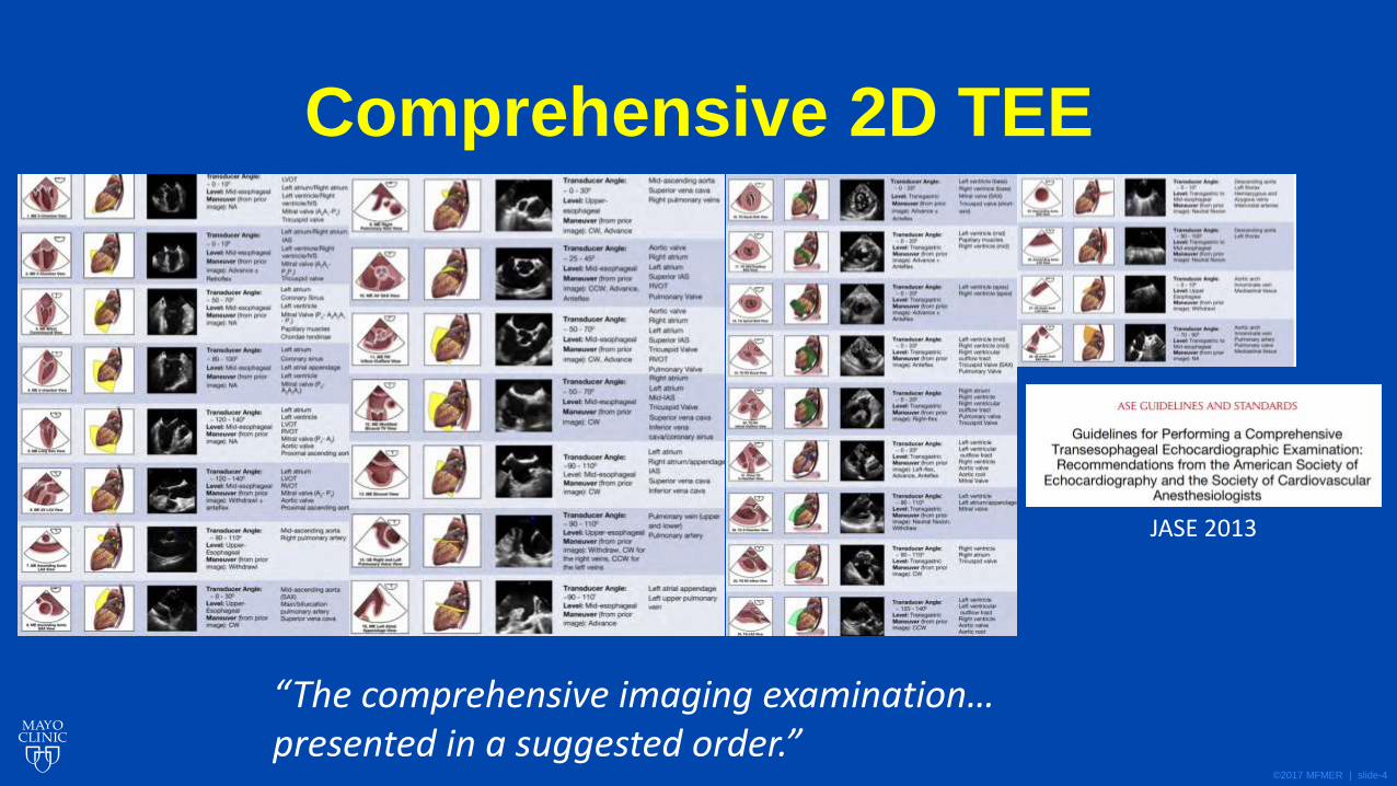
Comprehensive 2D TEE
JASE 2013
“The comprehensive imaging examination… presented in a suggested order.”
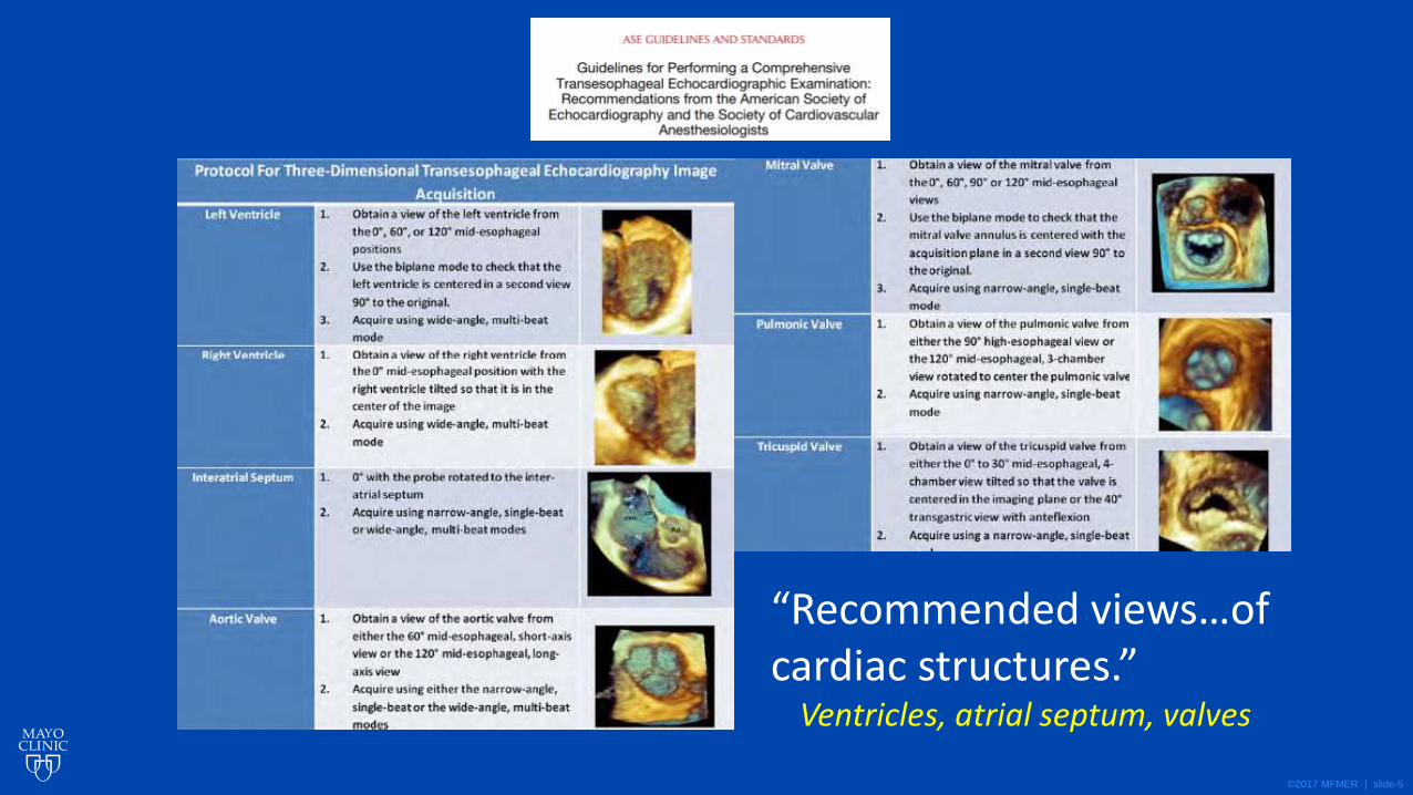
©2017 MFMER | slide-5
“Recommended views…of cardiac structures.”
Ventricles, atrial septum, valves
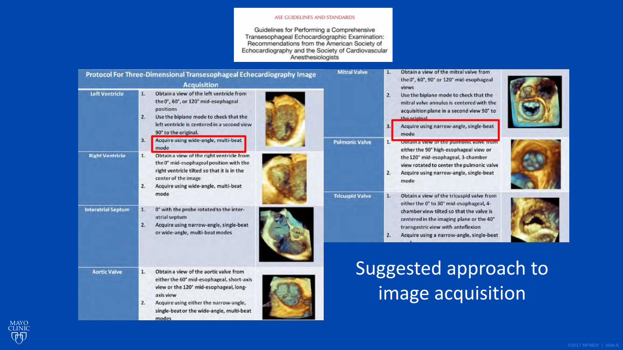
©2017 MFMER | slide-6
Suggested approach to image acquisition
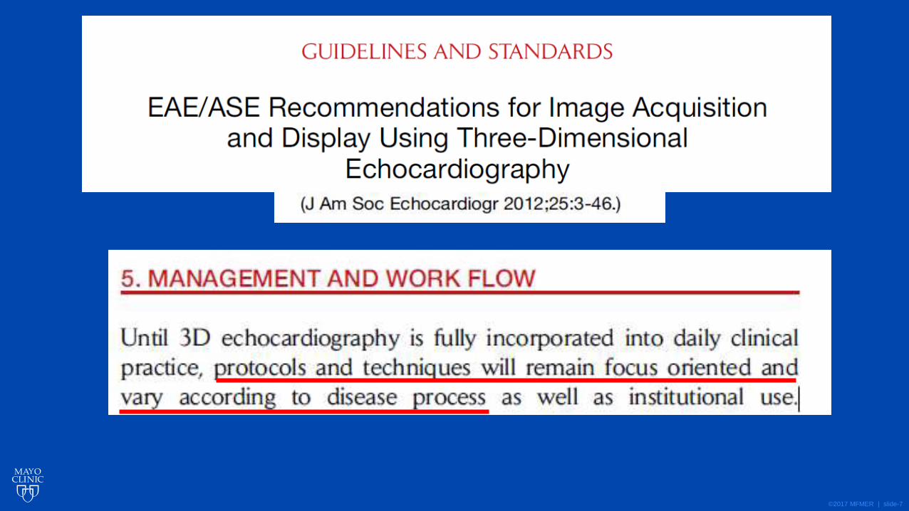
©2017 MFMER | slide-7
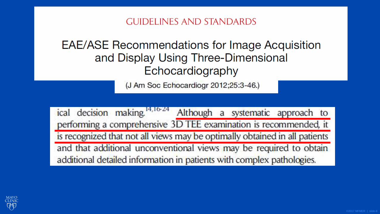
©2017 MFMER | slide-8
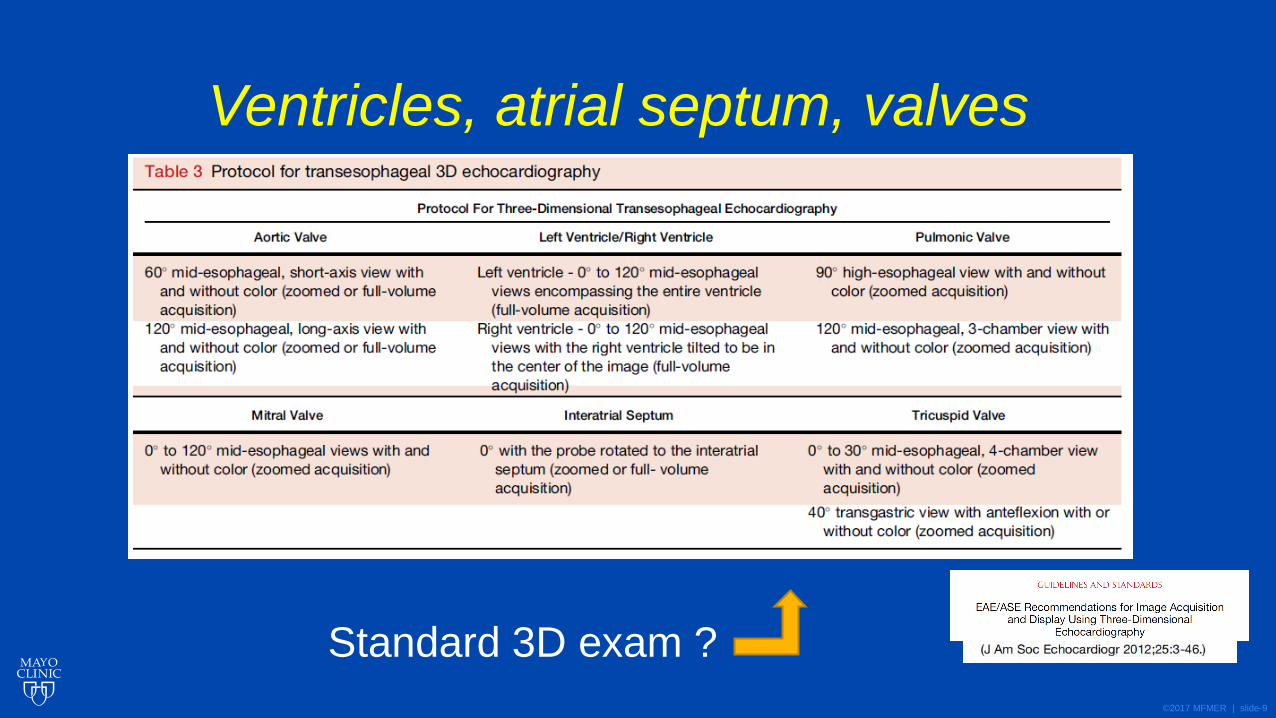
©2017 MFMER | slide-9
Standard 3D exam ?
Ventricles, atrial septum, valves
©2017 MFMER | slide-10
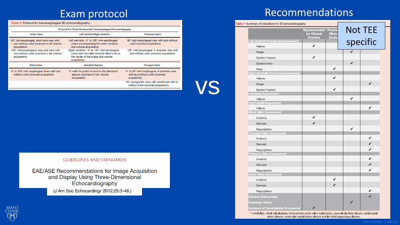
vs
Exam protocol Recommendations
Not TEE specific
©2017 MFMER | slide-11
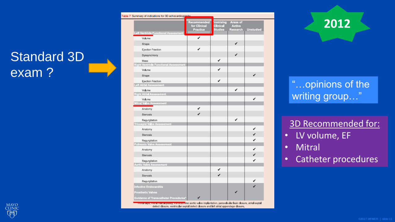
Standard 3D
exam ? “…opinions of the
writing group…”
3D Recommended for: • LV volume, EF • Mitral • Catheter procedures
2012
©2017 MFMER | slide-12
What is a “standard” 3D Exam?
©2017 MFMER | slide-13
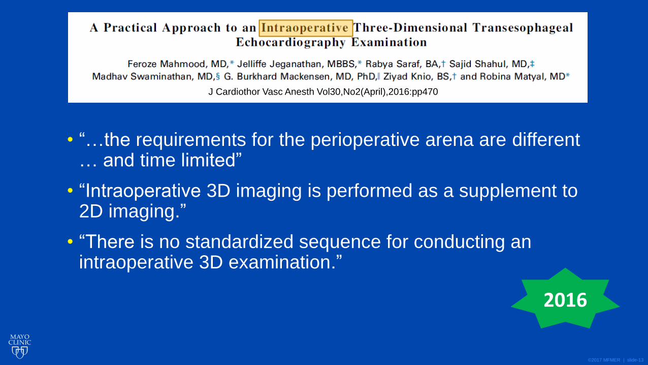
• “…the requirements for the perioperative arena are different … and time limited”
• “Intraoperative 3D imaging is performed as a supplement to 2D imaging.”
• “There is no standardized sequence for conducting an intraoperative 3D examination.”
J Cardiothor Vasc Anesth Vol30,No2(April),2016:pp470–
2016
©2017 MFMER | slide-14
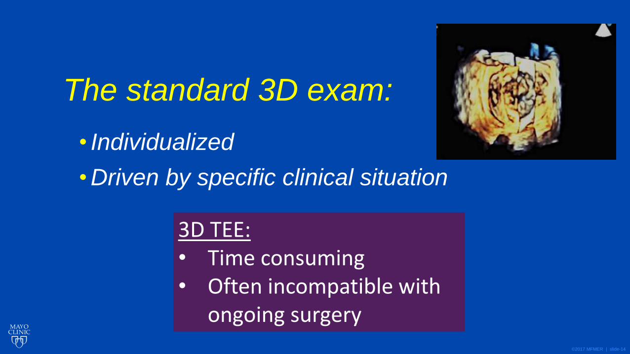
The standard 3D exam:
• Individualized
• Driven by specific clinical situation
3D TEE: • Time consuming • Often incompatible with
ongoing surgery
©2017 MFMER | slide-15
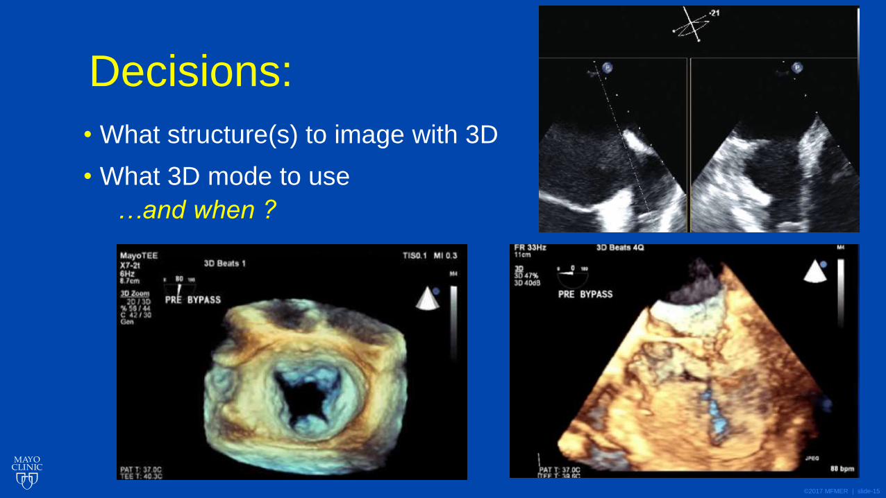
Decisions:
• What structure(s) to image with 3D
• What 3D mode to use
…and when ?
©2017 MFMER | slide-16
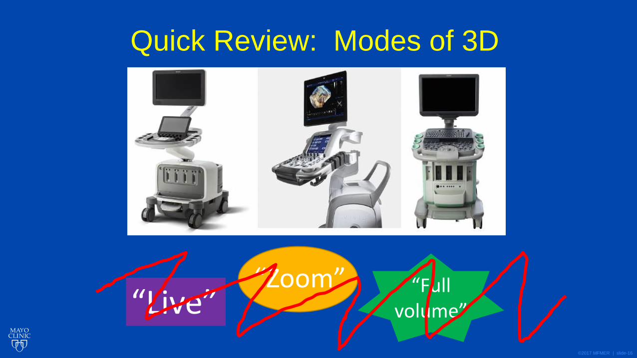
Quick Review: Modes of 3D
“Live” “Zoom” “Full
volume”
©2017 MFMER | slide-17
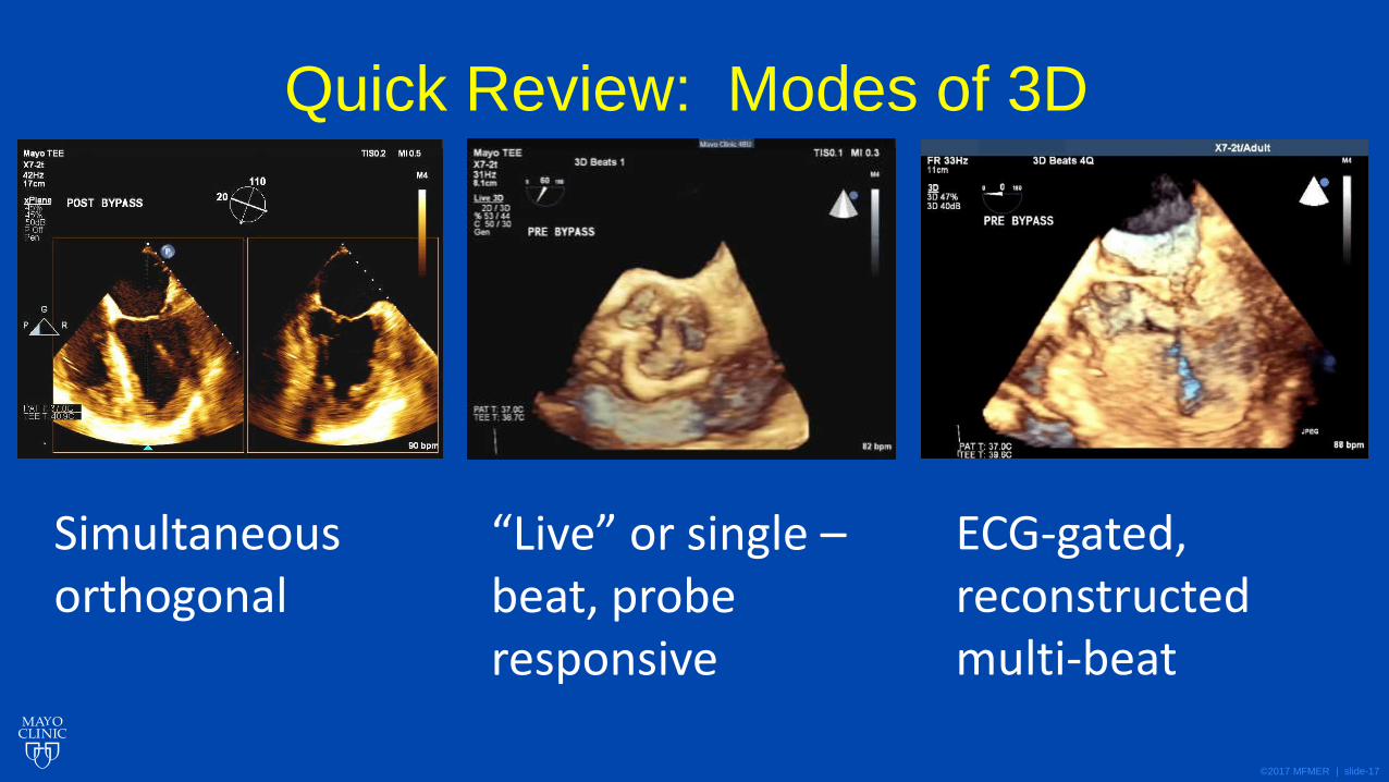
Quick Review: Modes of 3D
Simultaneous orthogonal
“Live” or single –beat, probe responsive
ECG-gated, reconstructed multi-beat
©2017 MFMER | slide-18
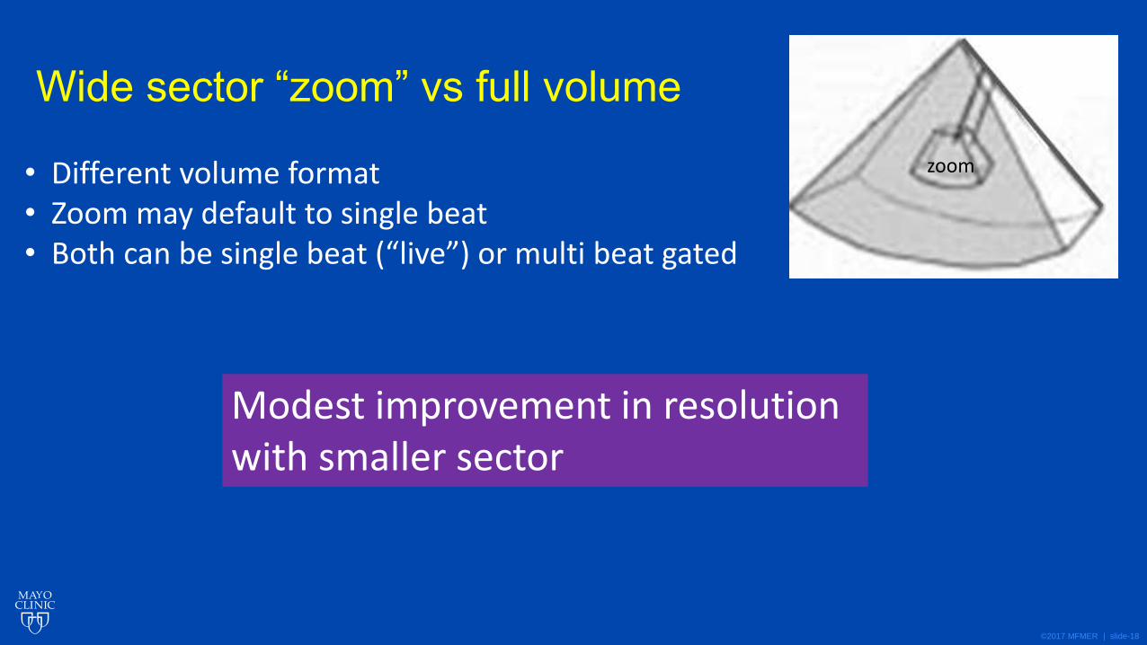
Wide sector “zoom” vs full volume
• Different volume format • Zoom may default to single beat • Both can be single beat (“live”) or multi beat gated
zoom
Modest improvement in resolution with smaller sector
©2017 MFMER | slide-19
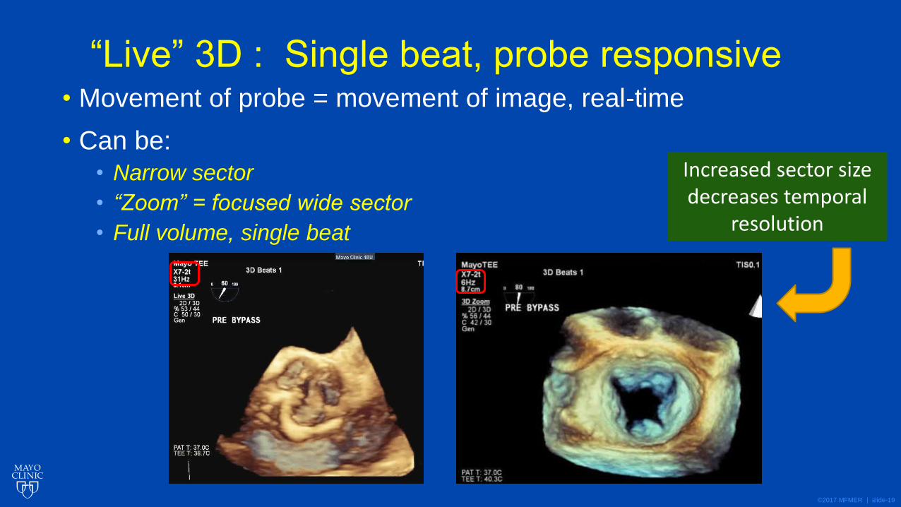
“Live” 3D : Single beat, probe responsive • Movement of probe = movement of image, real-time
• Can be:
• Narrow sector
• “Zoom” = focused wide sector
• Full volume, single beat
Increased sector size decreases temporal
resolution
©2017 MFMER | slide-20
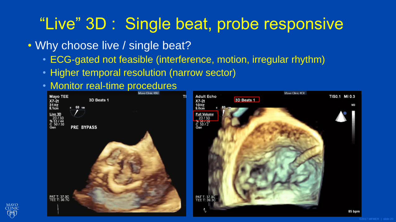
“Live” 3D : Single beat, probe responsive
• Why choose live / single beat?
• ECG-gated not feasible (interference, motion, irregular rhythm)
• Higher temporal resolution (narrow sector)
• Monitor real-time procedures
©2017 MFMER | slide-21
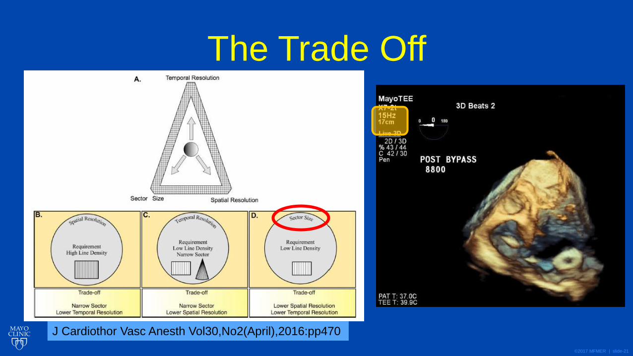
The Trade Off
J Cardiothor Vasc Anesth Vol30,No2(April),2016:pp470
©2017 MFMER | slide-22
Clinical Scenarios: When is 3D “standard”?
Debatable, but…
©2017 MFMER | slide-23
Catheter based procedures
©2017 MFMER | slide-24
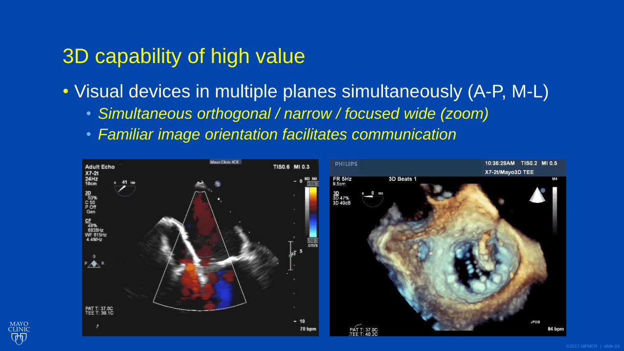
3D capability of high value
• Visual devices in multiple planes simultaneously (A-P, M-L)
• Simultaneous orthogonal / narrow / focused wide (zoom)
• Familiar image orientation facilitates communication
©2017 MFMER | slide-25
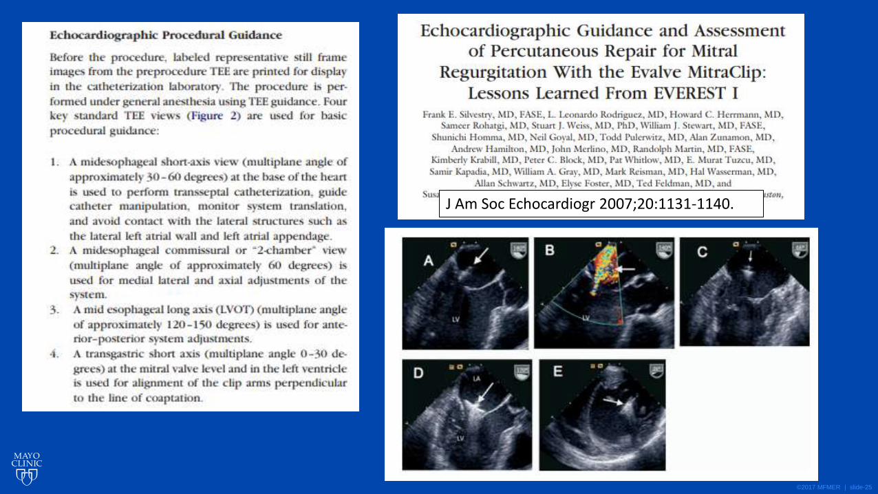
J Am Soc Echocardiogr 2007;20:1131-1140.
©2017 MFMER | slide-26
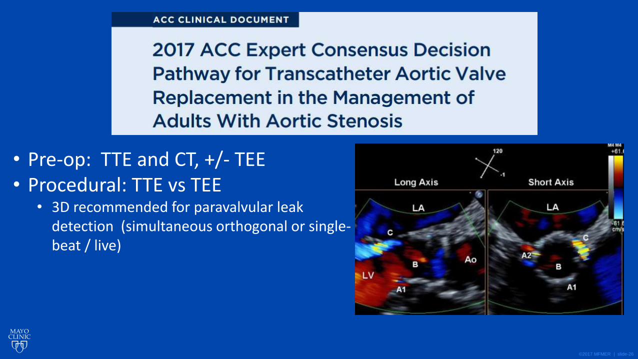
• Pre-op: TTE and CT, +/- TEE • Procedural: TTE vs TEE
• 3D recommended for paravalvular leak detection (simultaneous orthogonal or single-beat / live)
©2017 MFMER | slide-27
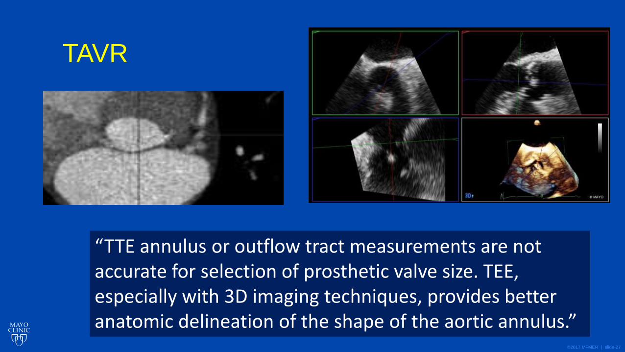
TAVR
“TTE annulus or outflow tract measurements are not accurate for selection of prosthetic valve size. TEE, especially with 3D imaging techniques, provides better anatomic delineation of the shape of the aortic annulus.”
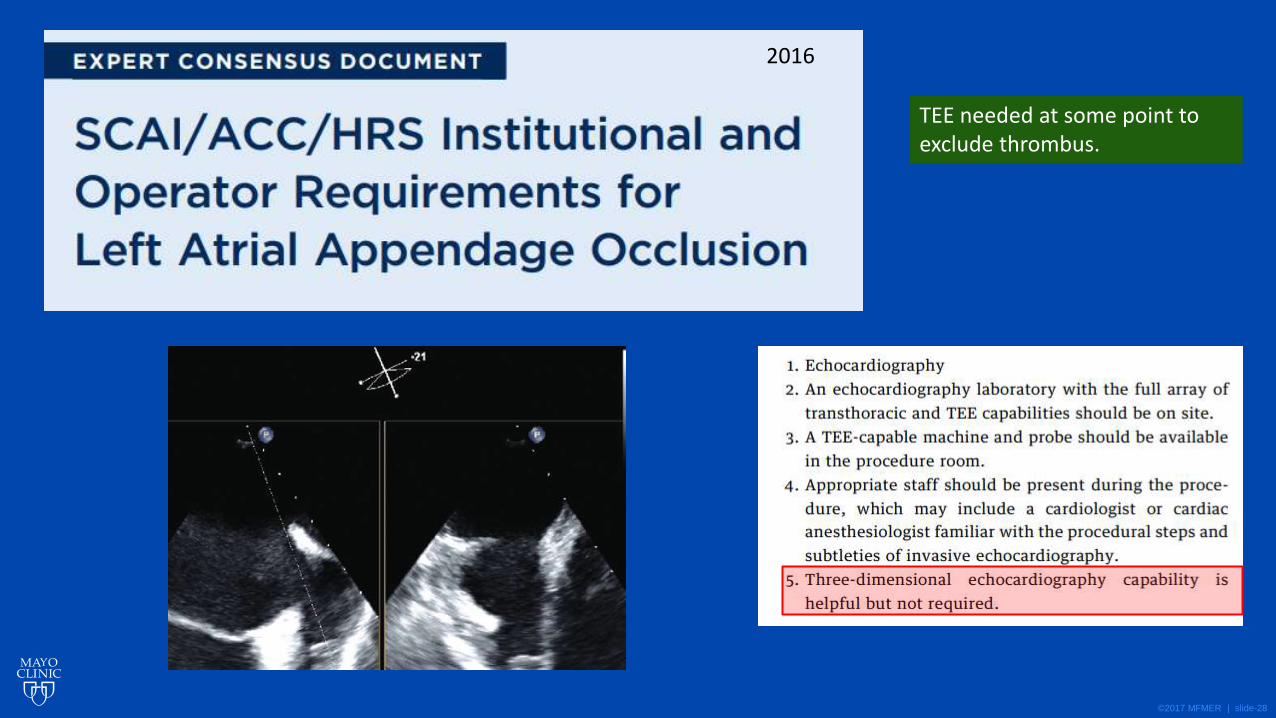
©2017 MFMER | slide-28
2016
TEE needed at some point to exclude thrombus.
©2017 MFMER | slide-29
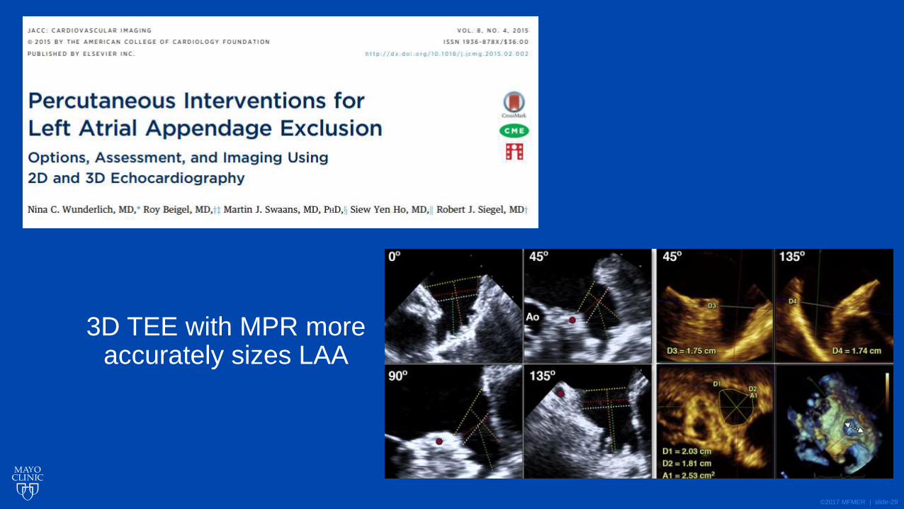
3D TEE with MPR more accurately sizes LAA
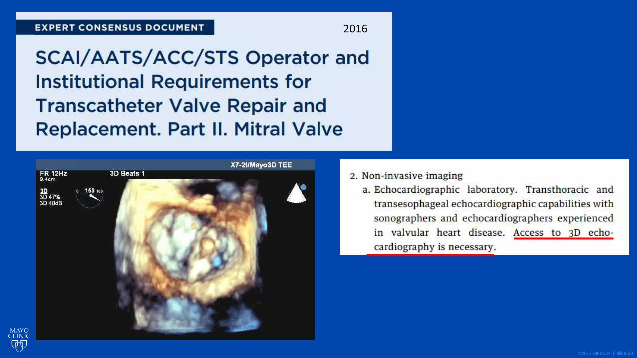
©2017 MFMER | slide-30
2016
©2017 MFMER | slide-31
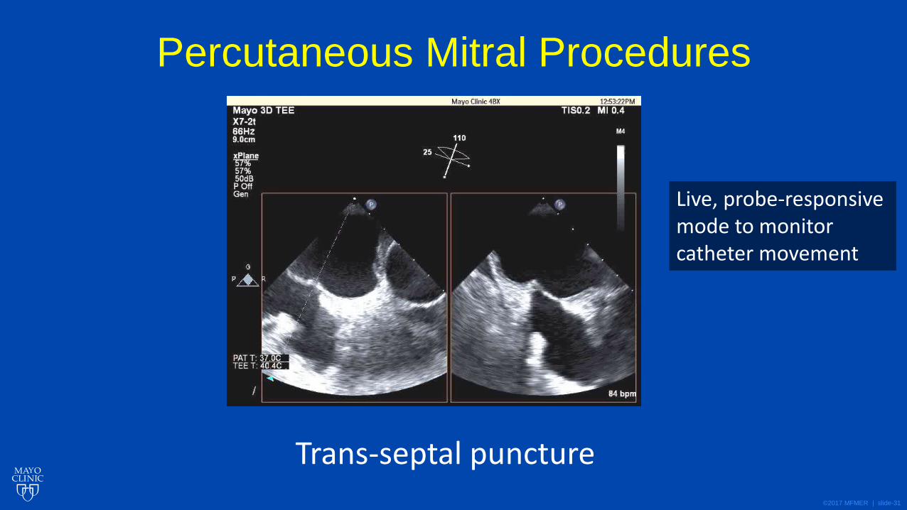
Percutaneous Mitral Procedures
Trans-septal puncture
Live, probe-responsive mode to monitor catheter movement
©2017 MFMER | slide-32
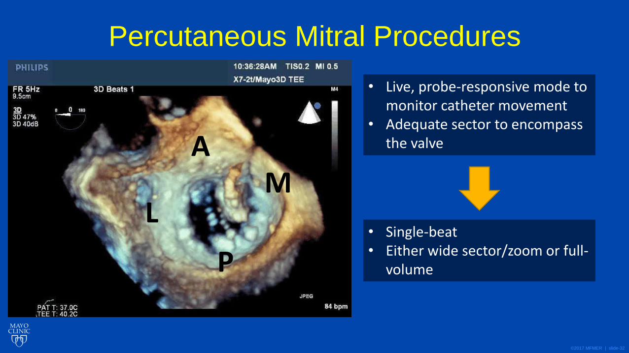
Percutaneous Mitral Procedures
• Live, probe-responsive mode to monitor catheter movement
• Adequate sector to encompass the valve
• Single-beat • Either wide sector/zoom or full-
volume
A
P
M L
©2017 MFMER | slide-33
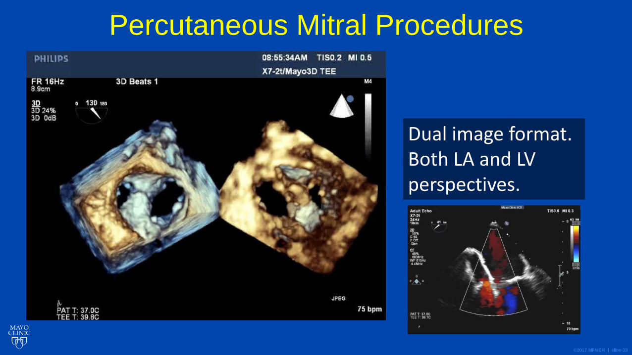
Percutaneous Mitral Procedures
Dual image format. Both LA and LV perspectives.
©2017 MFMER | slide-34
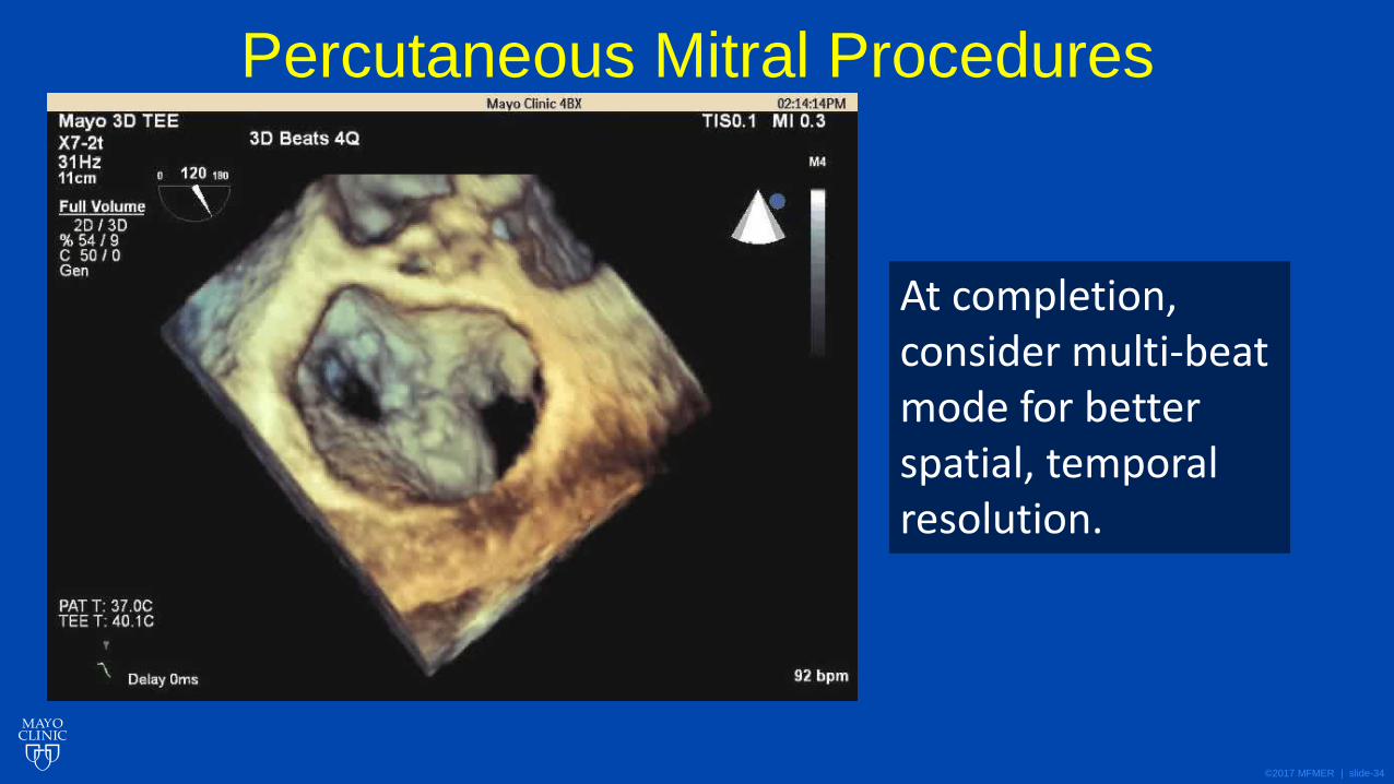
Percutaneous Mitral Procedures
At completion, consider multi-beat mode for better spatial, temporal resolution.
©2017 MFMER | slide-35
Valve Pathology, Repair, and Replacement
©2017 MFMER | slide-36
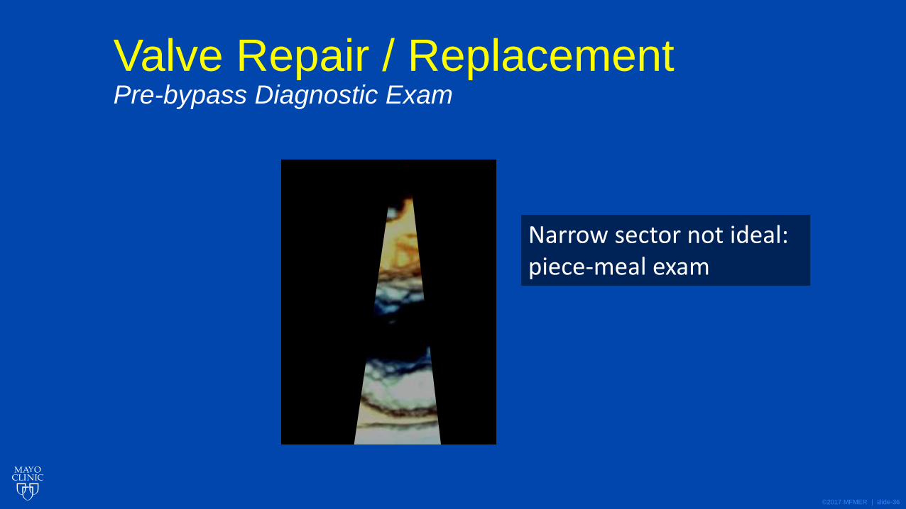
Valve Repair / Replacement Pre-bypass Diagnostic Exam
Narrow sector not ideal: piece-meal exam
©2017 MFMER | slide-37
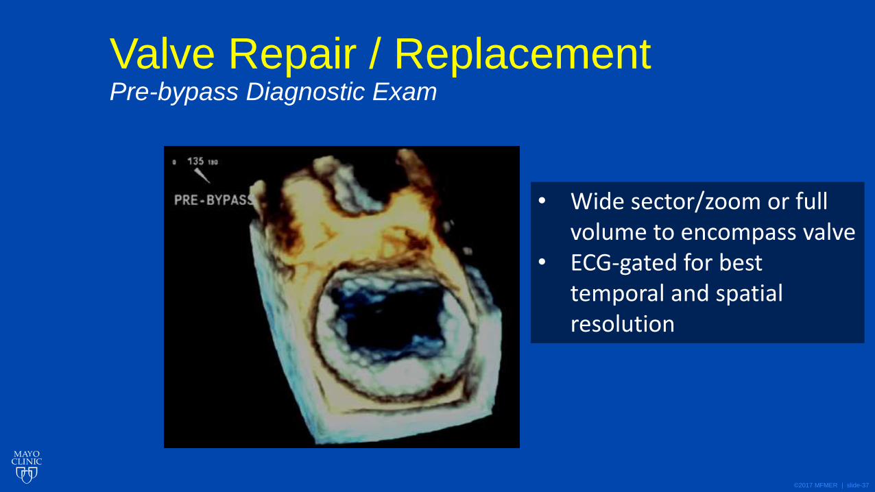
Valve Repair / Replacement Pre-bypass Diagnostic Exam
• Wide sector/zoom or full volume to encompass valve
• ECG-gated for best temporal and spatial resolution
©2017 MFMER | slide-38
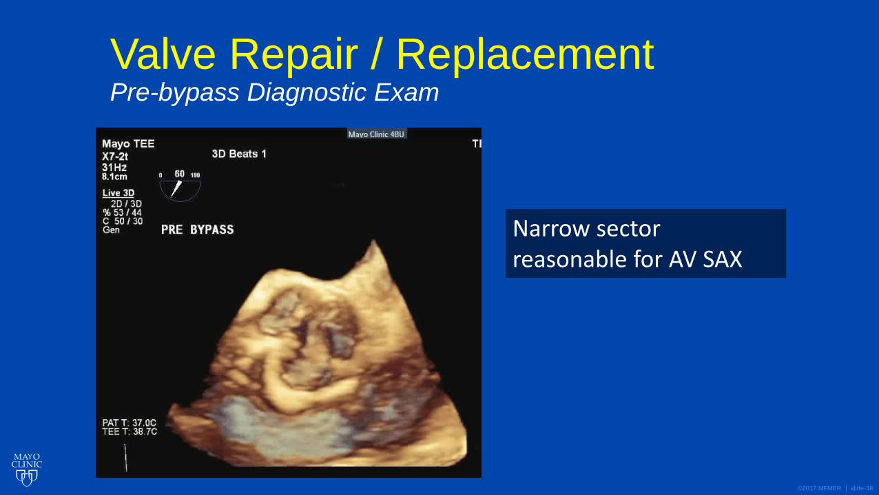
Valve Repair / Replacement Pre-bypass Diagnostic Exam
Narrow sector reasonable for AV SAX
©2017 MFMER | slide-39
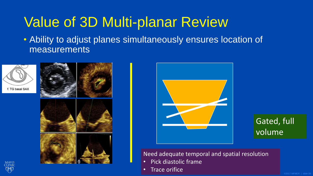
Value of 3D Multi-planar Review • Ability to adjust planes simultaneously ensures location of
measurements
Need adequate temporal and spatial resolution • Pick diastolic frame • Trace orifice
Gated, full volume
©2017 MFMER | slide-40
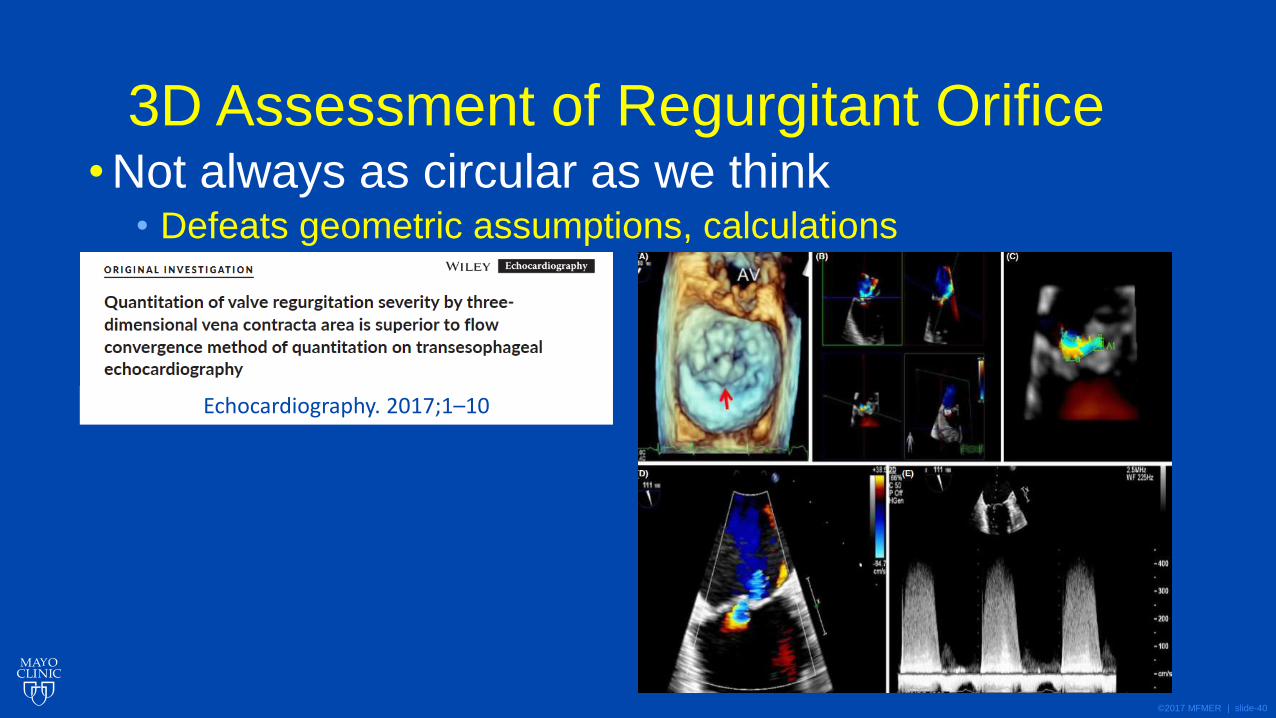
3D Assessment of Regurgitant Orifice • Not always as circular as we think
• Defeats geometric assumptions, calculations
Echocardiography. 2017;1–10
©2017 MFMER | slide-41
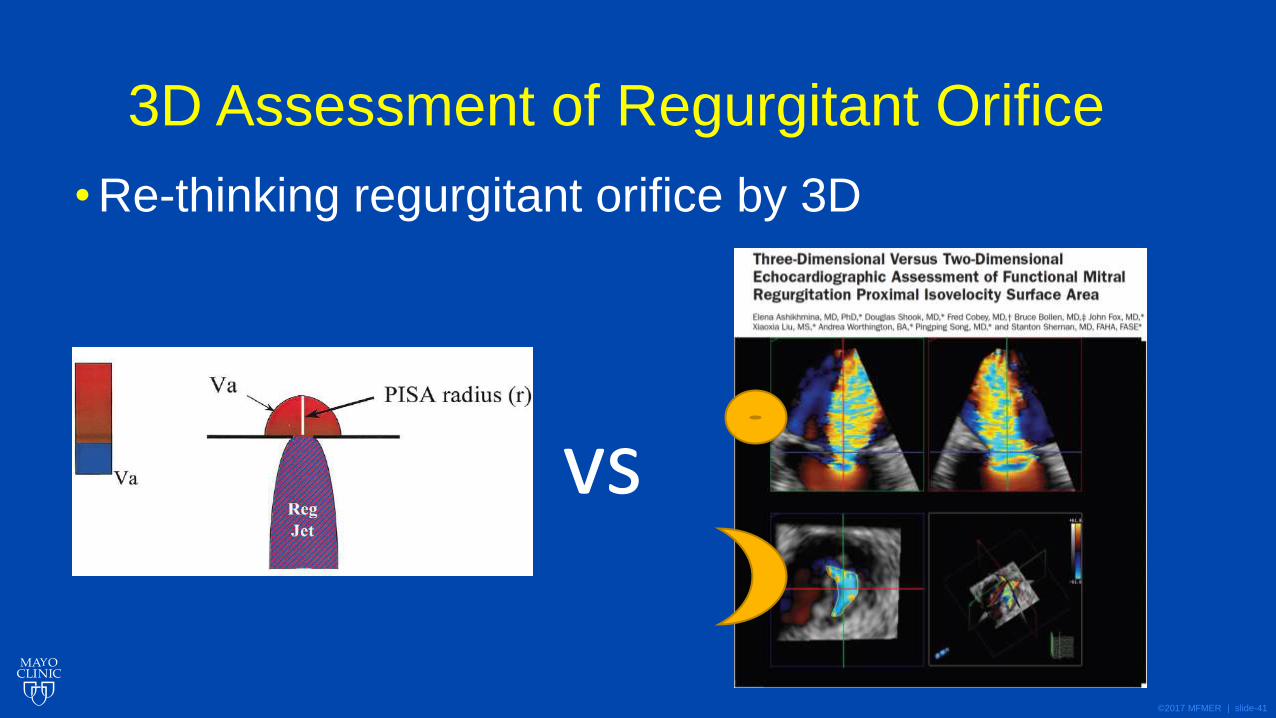
3D Assessment of Regurgitant Orifice
• Re-thinking regurgitant orifice by 3D
vs
©2017 MFMER | slide-42
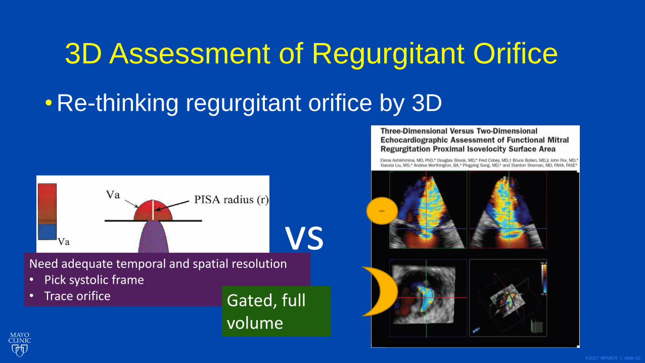
3D Assessment of Regurgitant Orifice
• Re-thinking regurgitant orifice by 3D
vs Need adequate temporal and spatial resolution • Pick systolic frame • Trace orifice Gated, full
volume
©2017 MFMER | slide-43
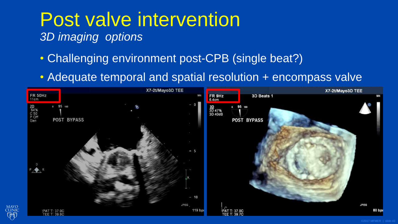
Post valve intervention 3D imaging options
• Challenging environment post-CPB (single beat?)
• Adequate temporal and spatial resolution + encompass valve
©2017 MFMER | slide-44
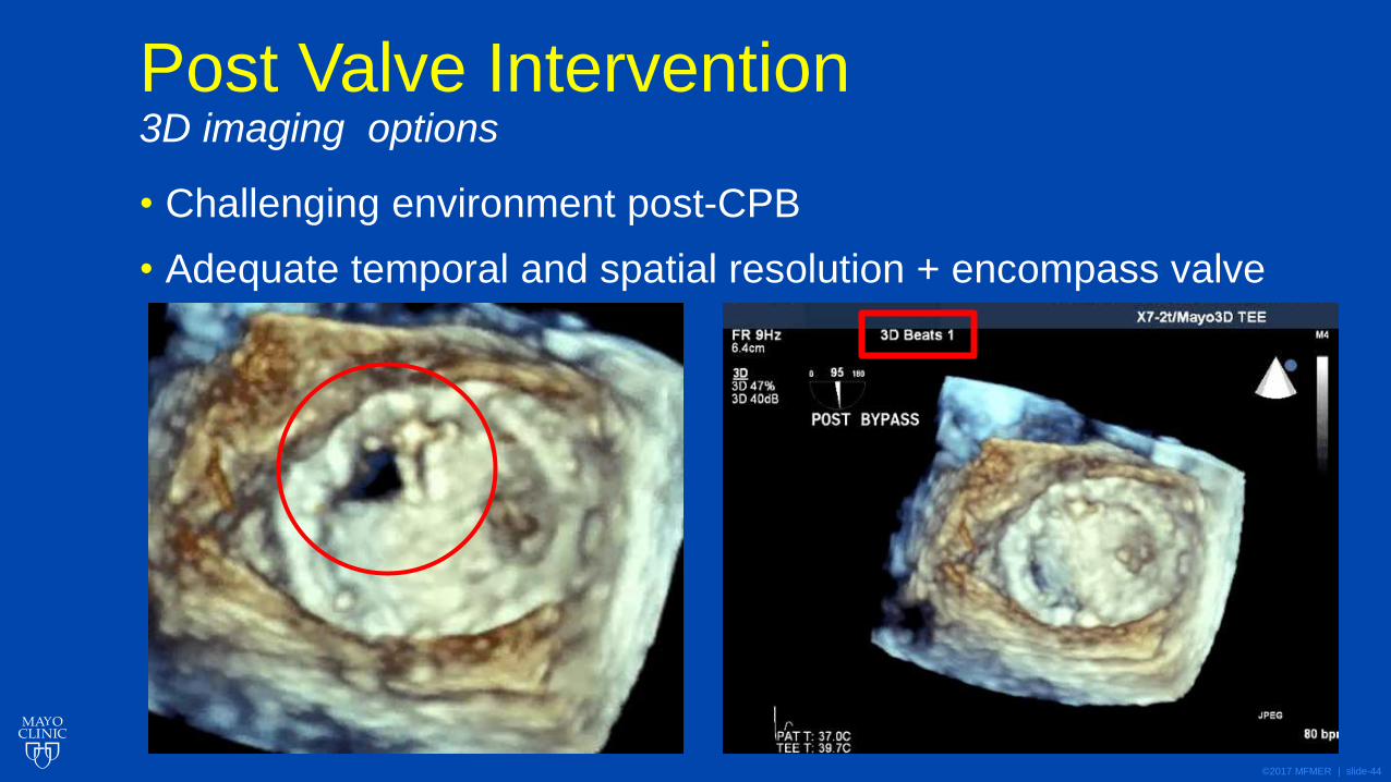
Post Valve Intervention 3D imaging options
• Challenging environment post-CPB
• Adequate temporal and spatial resolution + encompass valve
©2017 MFMER | slide-45
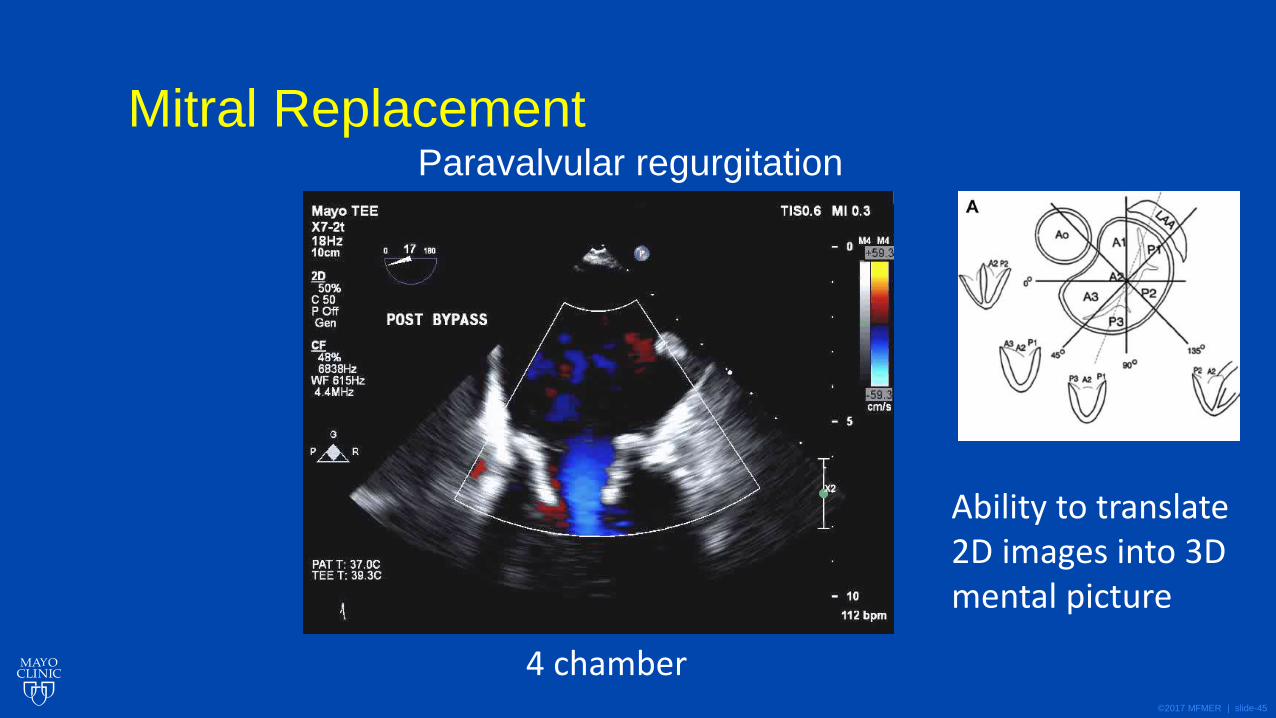
Mitral Replacement Paravalvular regurgitation
4 chamber
Ability to translate 2D images into 3D mental picture
©2017 MFMER | slide-46
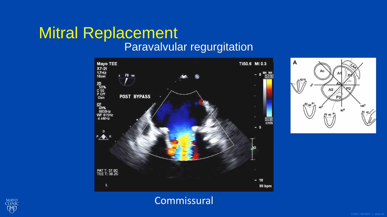
Mitral Replacement Paravalvular regurgitation
Commissural
©2017 MFMER | slide-47
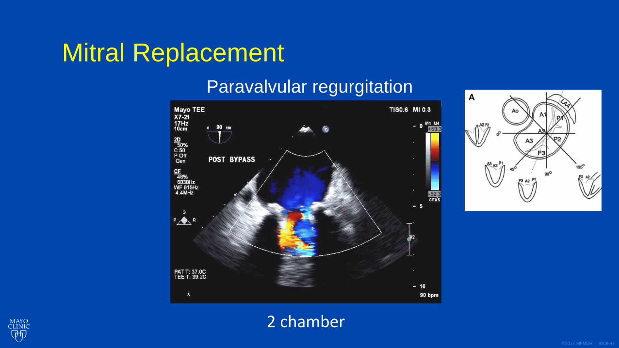
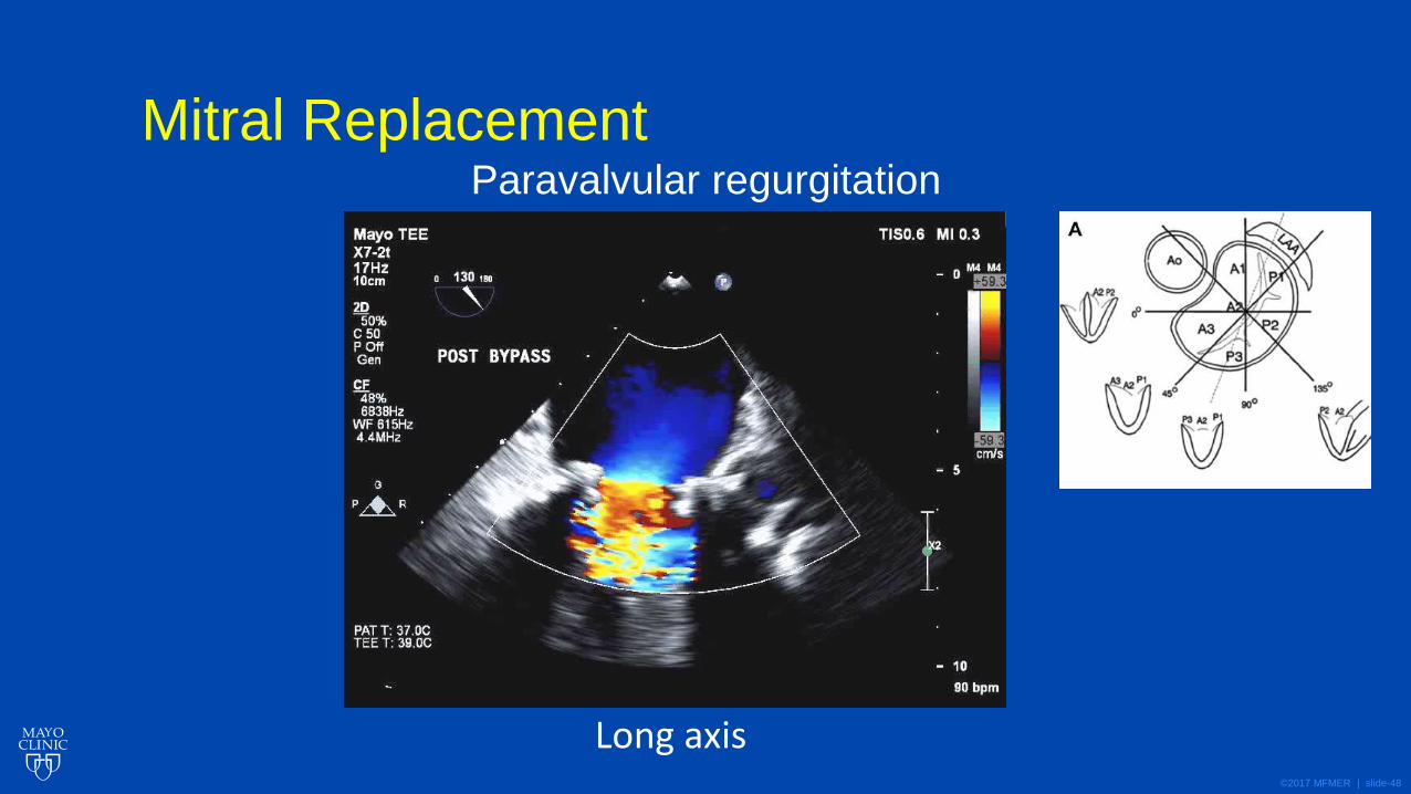
Mitral Replacement
Paravalvular regurgitation
2 chamber
©2017 MFMER | slide-48
Mitral Replacement Paravalvular regurgitation
Long axis

©2017 MFMER | slide-49
Mitral Replacement
Paravalvular regurgitation
Single beat mode immediately after bypass
• Spatial • Temporal • Encompass
valve
©2017 MFMER | slide-50
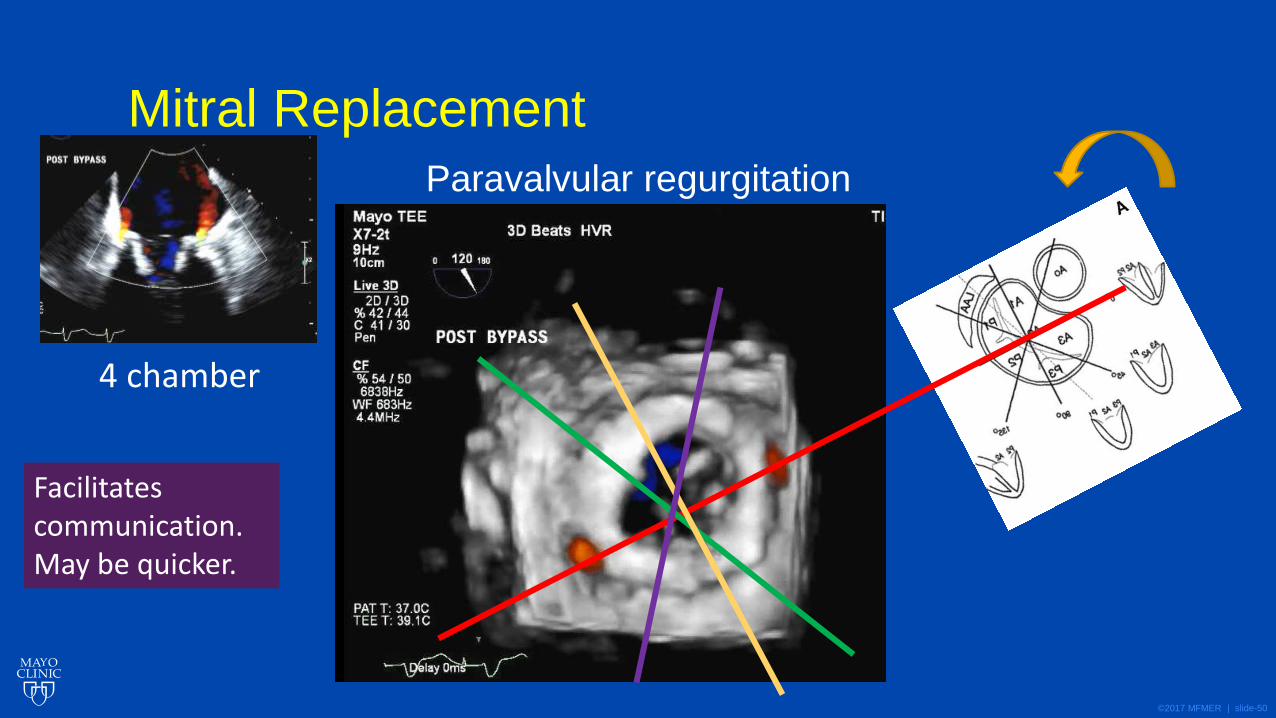
Mitral Replacement
Paravalvular regurgitation
4 chamber
Facilitates communication. May be quicker.
©2017 MFMER | slide-51
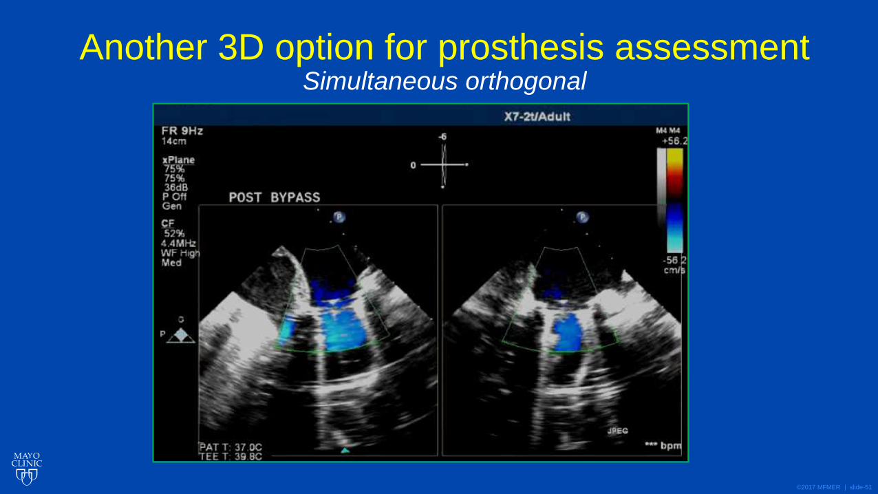
Another 3D option for prosthesis assessment Simultaneous orthogonal
©2017 MFMER | slide-52
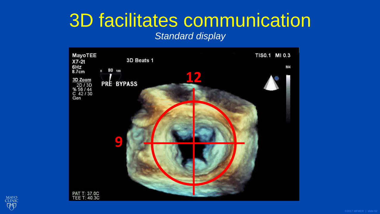
3D facilitates communication Standard display
12
9
©2017 MFMER | slide-53
Left Ventricular Function
©2017 MFMER | slide-54
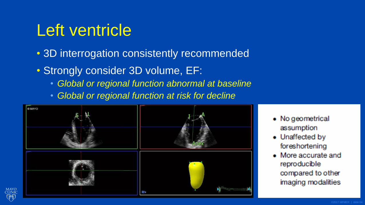
Left ventricle
• 3D interrogation consistently recommended
• Strongly consider 3D volume, EF:
• Global or regional function abnormal at baseline
• Global or regional function at risk for decline
©2017 MFMER | slide-55
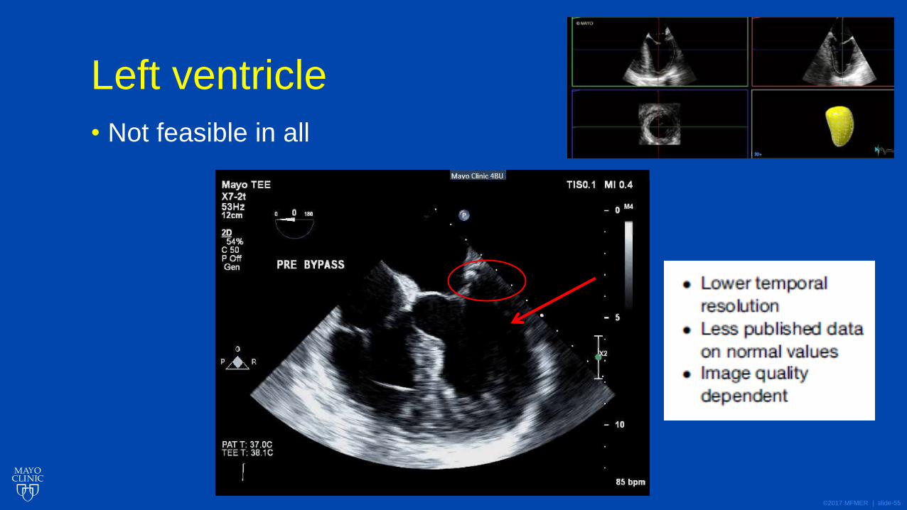
Left ventricle
• Not feasible in all
©2017 MFMER | slide-56
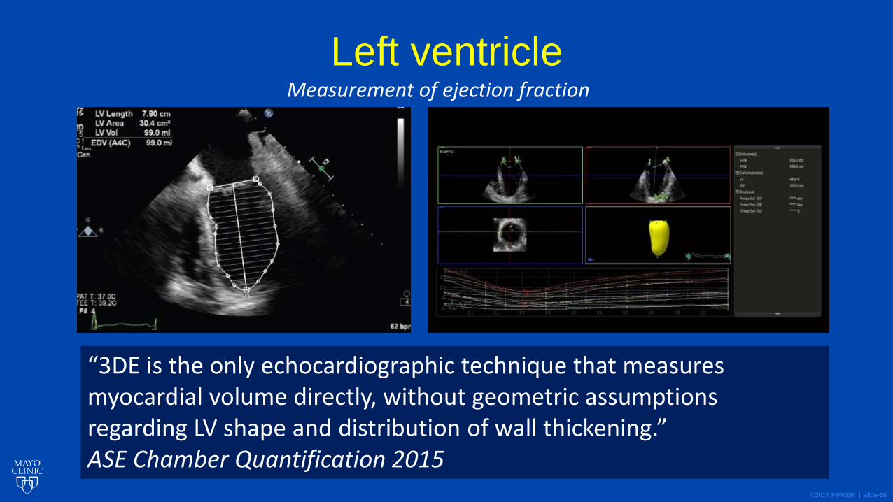
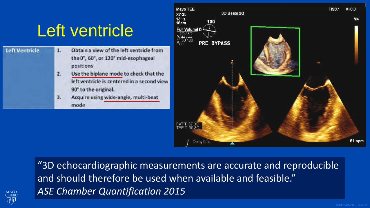
Left ventricle
“3DE is the only echocardiographic technique that measures myocardial volume directly, without geometric assumptions regarding LV shape and distribution of wall thickening.” ASE Chamber Quantification 2015
Measurement of ejection fraction
©2017 MFMER | slide-57
Left ventricle
“3D echocardiographic measurements are accurate and reproducible and should therefore be used when available and feasible.” ASE Chamber Quantification 2015
©2017 MFMER | slide-58
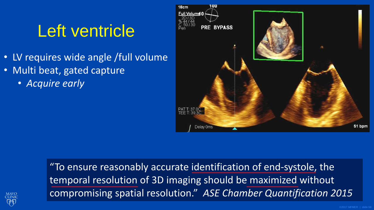
Left ventricle
“To ensure reasonably accurate identification of end-systole, the temporal resolution of 3D imaging should be maximized without compromising spatial resolution.” ASE Chamber Quantification 2015
• LV requires wide angle /full volume • Multi beat, gated capture
• Acquire early
©2017 MFMER | slide-59
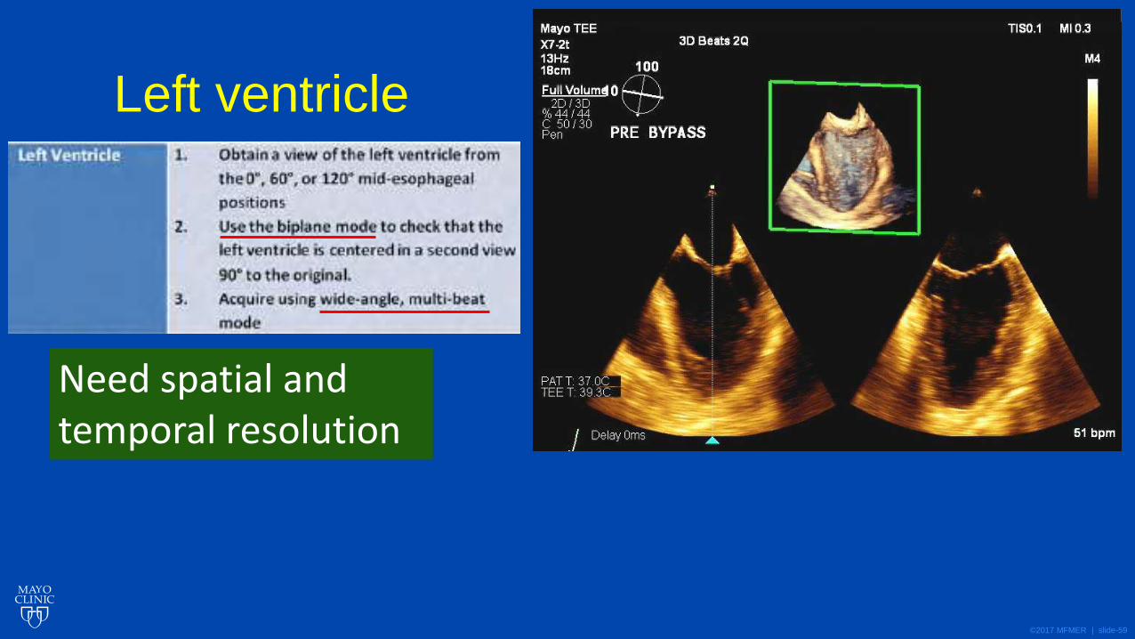
Left ventricle
Need spatial and temporal resolution
©2017 MFMER | slide-60
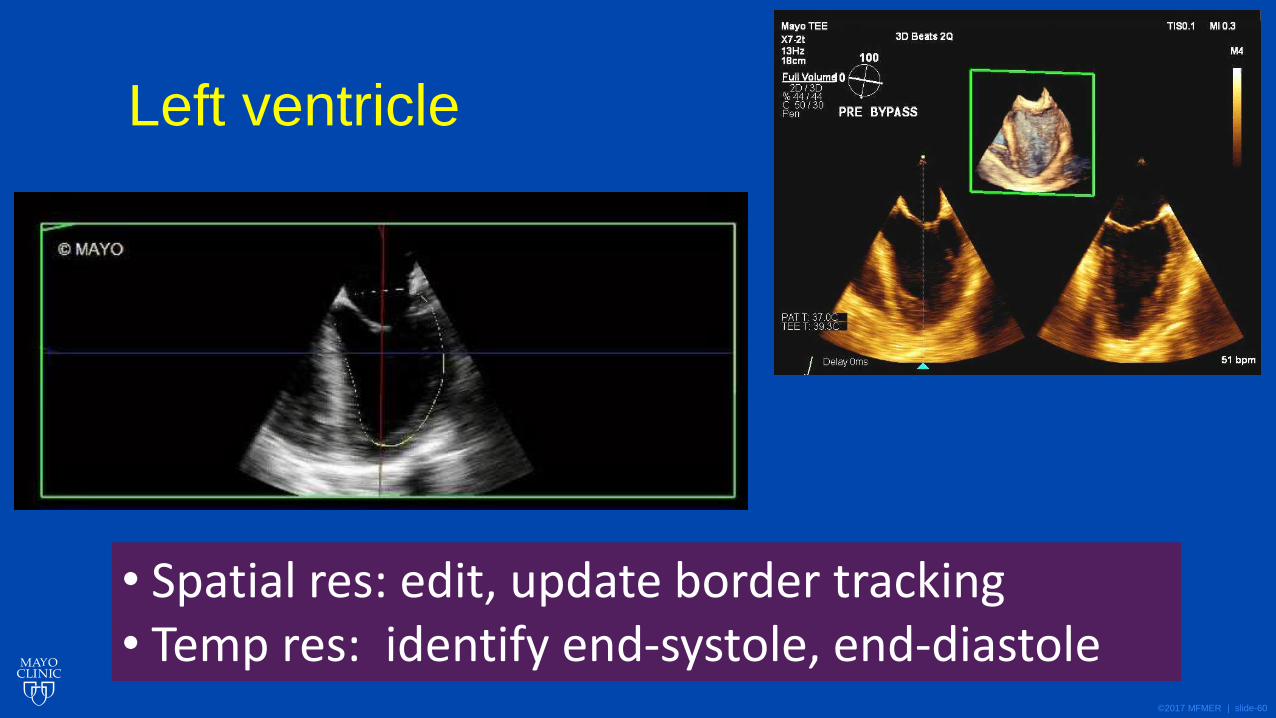
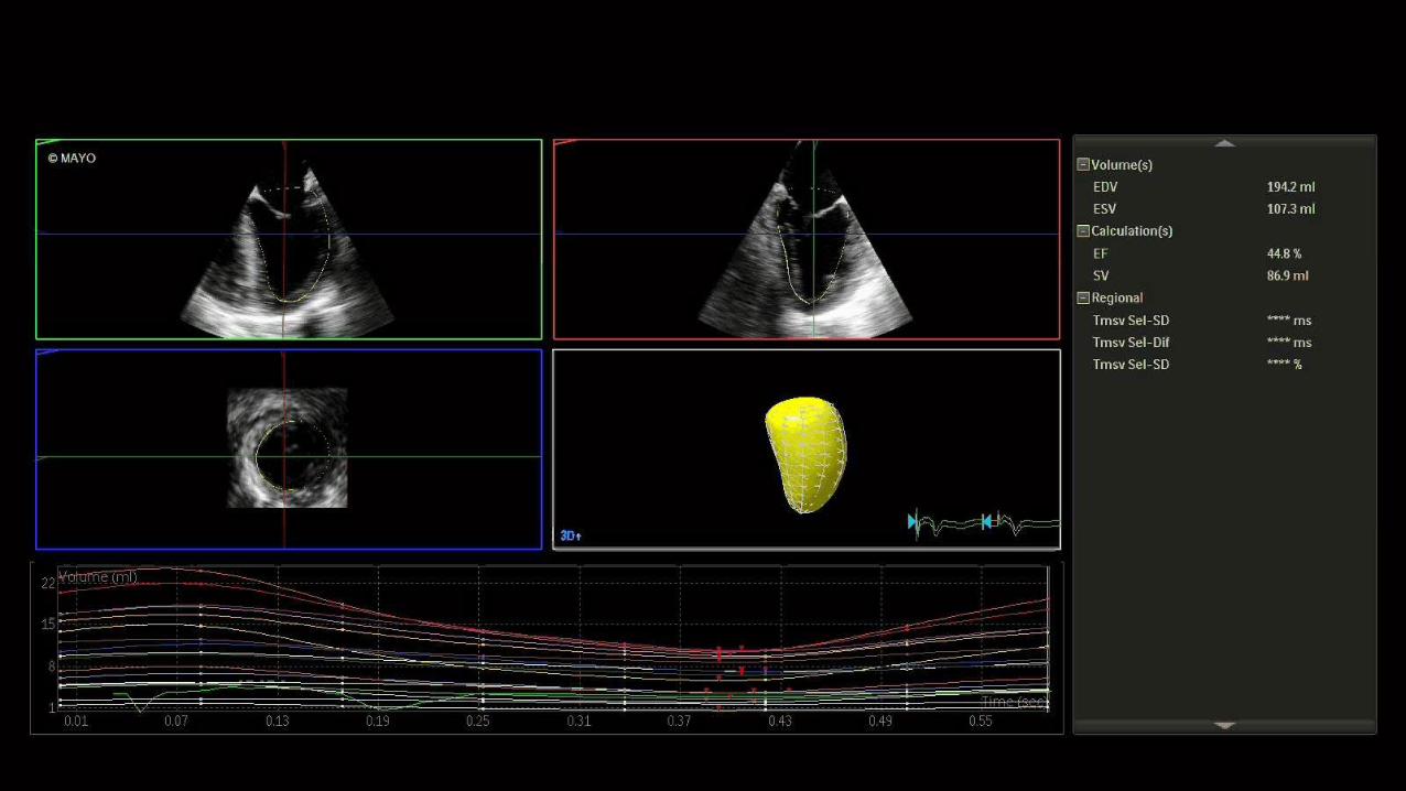
Left ventricle
• Spatial res: edit, update border tracking • Temp res: identify end-systole, end-diastole
©2017 MFMER | slide-61
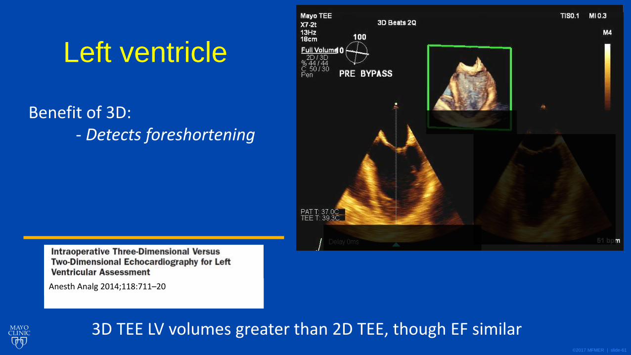
Left ventricle
Benefit of 3D: - Detects foreshortening
3D TEE LV volumes greater than 2D TEE, though EF similar
Anesth Analg 2014;118:711–20
©2017 MFMER | slide-62
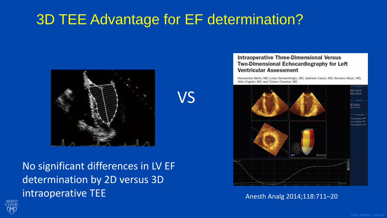
3D TEE Advantage for EF determination?
Anesth Analg 2014;118:711–20
VS
No significant differences in LV EF determination by 2D versus 3D intraoperative TEE
©2017 MFMER | slide-63
3D Assessment of LV EF
• Value in setting of wall motion abnormalities
©2017 MFMER | slide-64
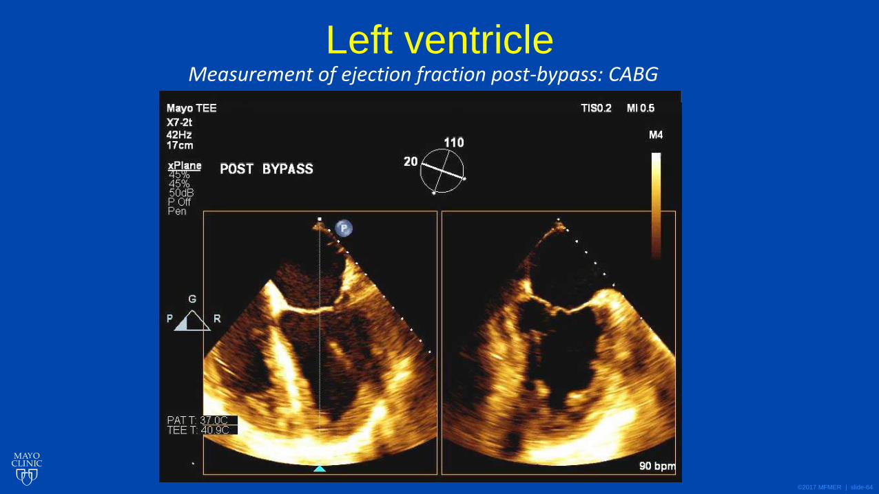
Left ventricle Measurement of ejection fraction post-bypass: CABG
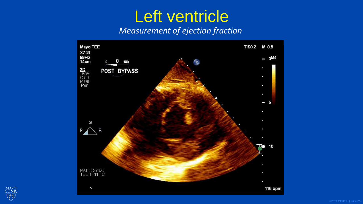
©2017 MFMER | slide-65
Left ventricle Measurement of ejection fraction
©2017 MFMER | slide-66
Left ventricle Measurement of ejection fraction
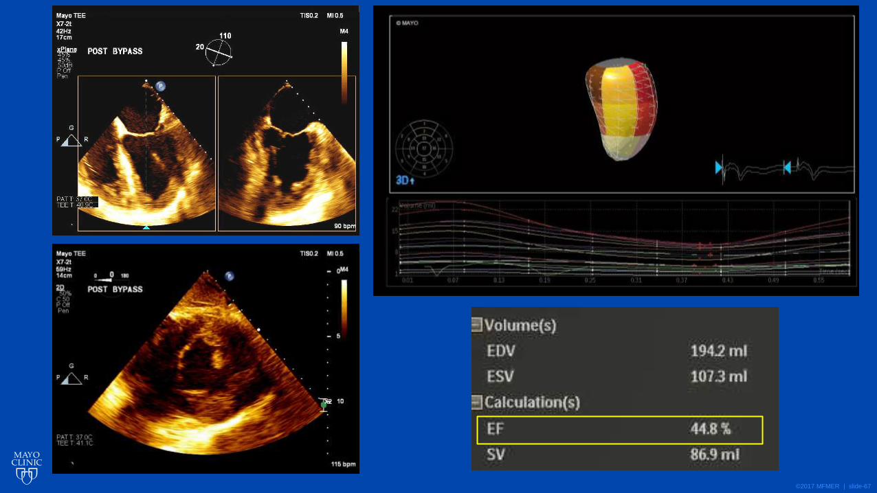
©2017 MFMER | slide-67
©2017 MFMER | slide-68
Right Ventricle
©2017 MFMER | slide-69
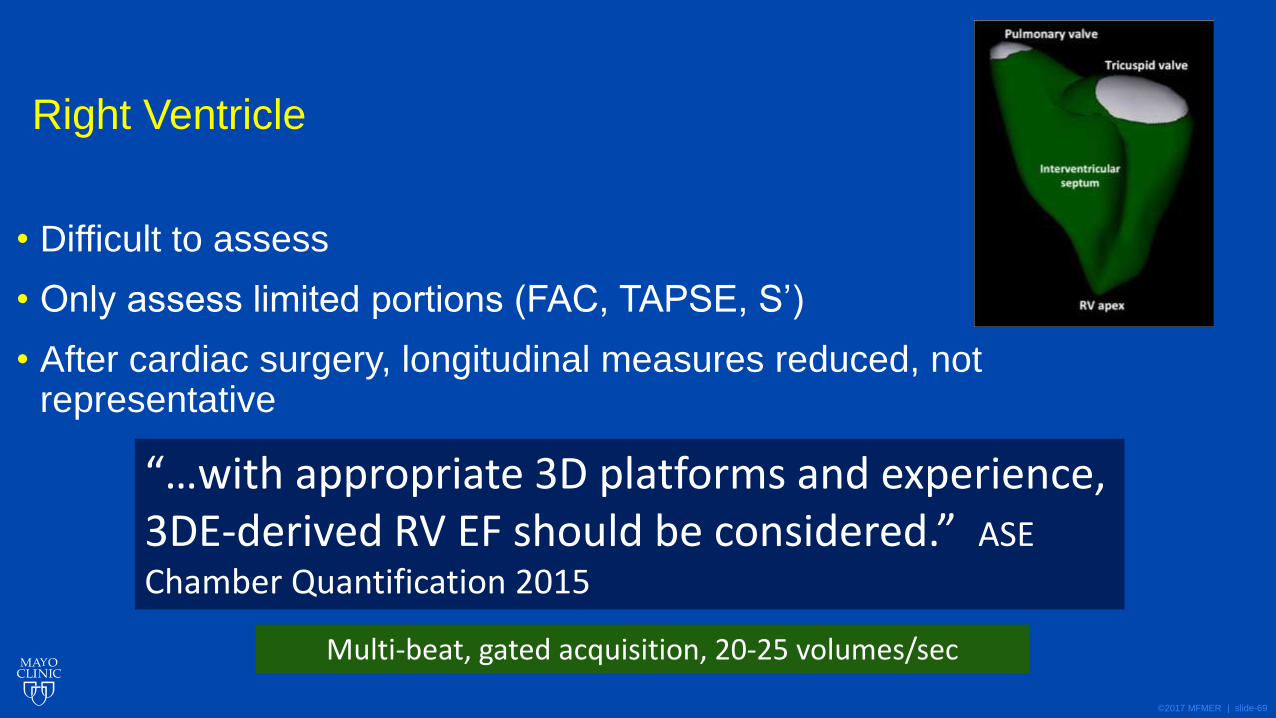
Right Ventricle
• Difficult to assess
• Only assess limited portions (FAC, TAPSE, S’)
• After cardiac surgery, longitudinal measures reduced, not representative
“…with appropriate 3D platforms and experience, 3DE-derived RV EF should be considered.” ASE
Chamber Quantification 2015
Multi-beat, gated acquisition, 20-25 volumes/sec
©2017 MFMER | slide-70
Congenital
©2017 MFMER | slide-71
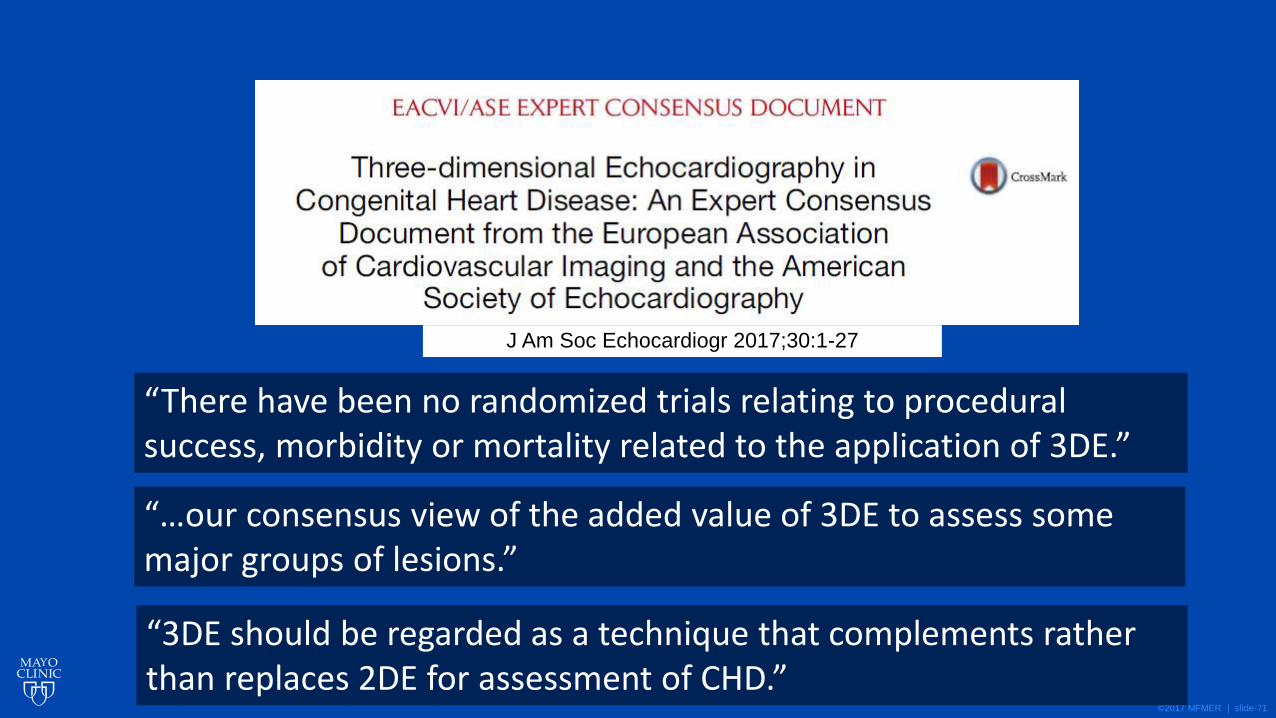
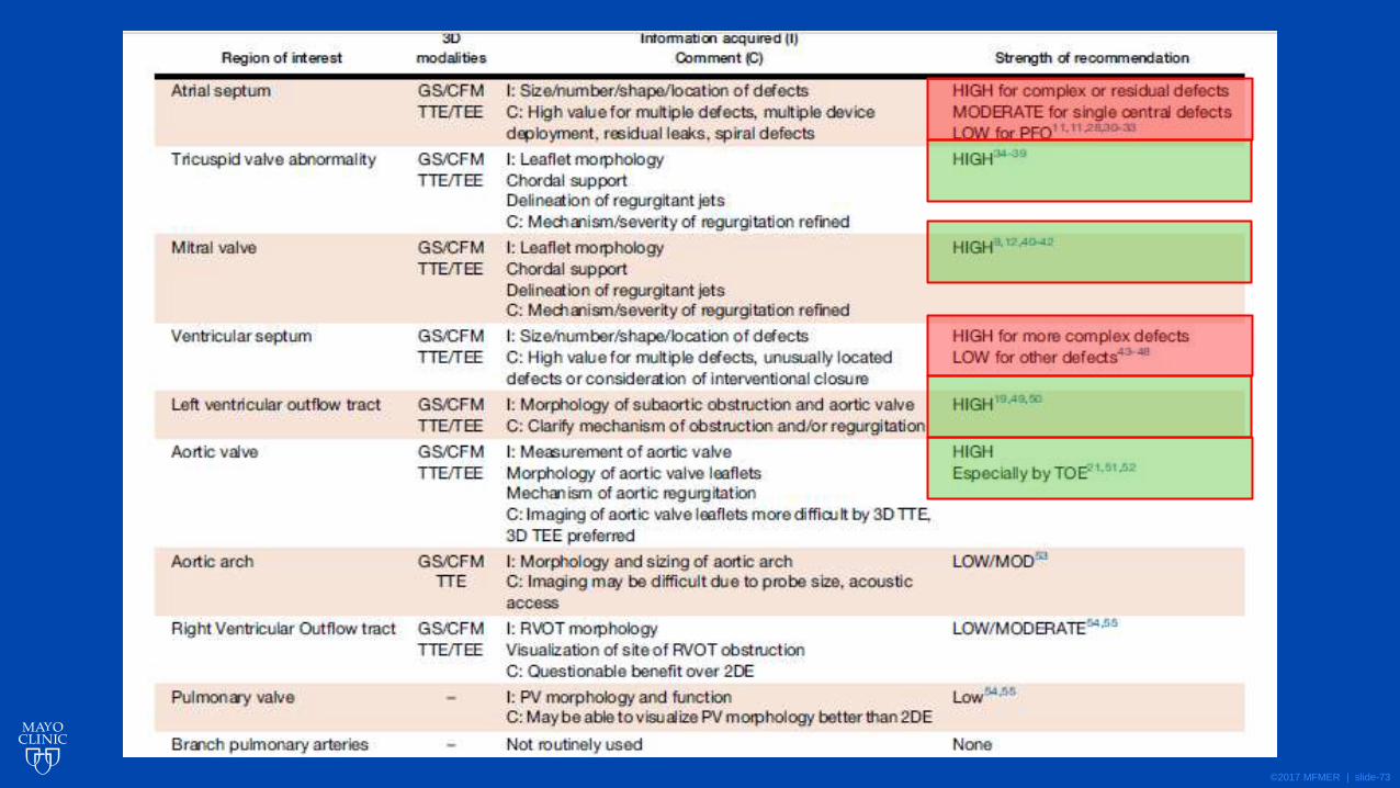
J Am Soc Echocardiogr 2017;30:1-27
“There have been no randomized trials relating to procedural success, morbidity or mortality related to the application of 3DE.”
“…our consensus view of the added value of 3DE to assess some major groups of lesions.”
“3DE should be regarded as a technique that complements rather than replaces 2DE for assessment of CHD.”
©2017 MFMER | slide-72
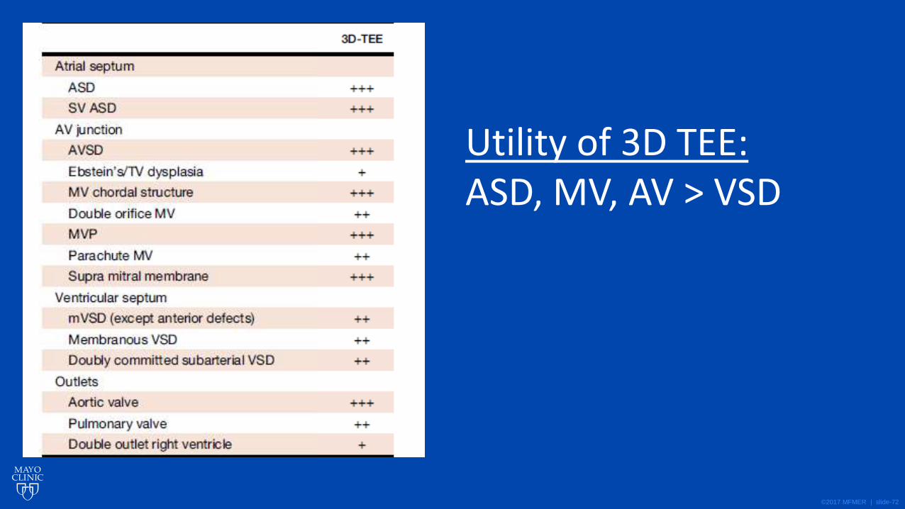
Utility of 3D TEE: ASD, MV, AV > VSD
©2017 MFMER | slide-73
©2017 MFMER | slide-74
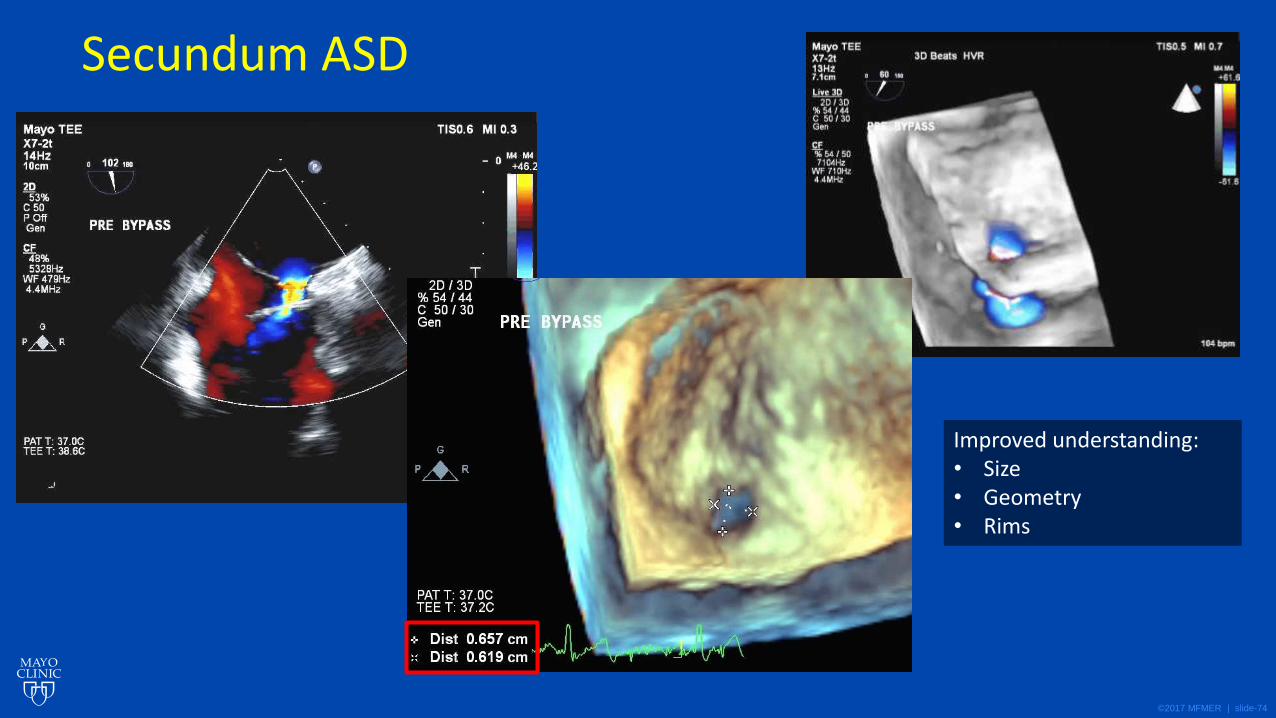
Secundum ASD
Improved understanding: • Size • Geometry • Rims
©2017 MFMER | slide-75
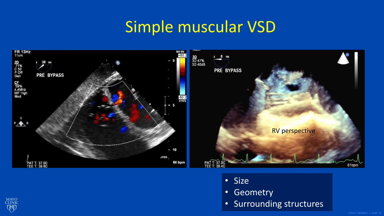
Simple muscular VSD
RV perspective
• Size • Geometry • Surrounding structures
©2017 MFMER | slide-76
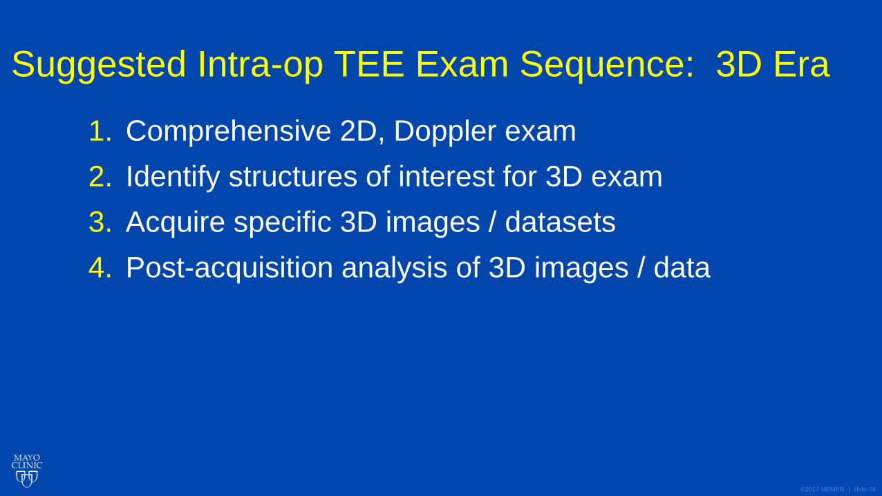
Suggested Intra-op TEE Exam Sequence: 3D Era
1. Comprehensive 2D, Doppler exam
2. Identify structures of interest for 3D exam
3. Acquire specific 3D images / datasets
4. Post-acquisition analysis of 3D images / data
©2017 MFMER | slide-77
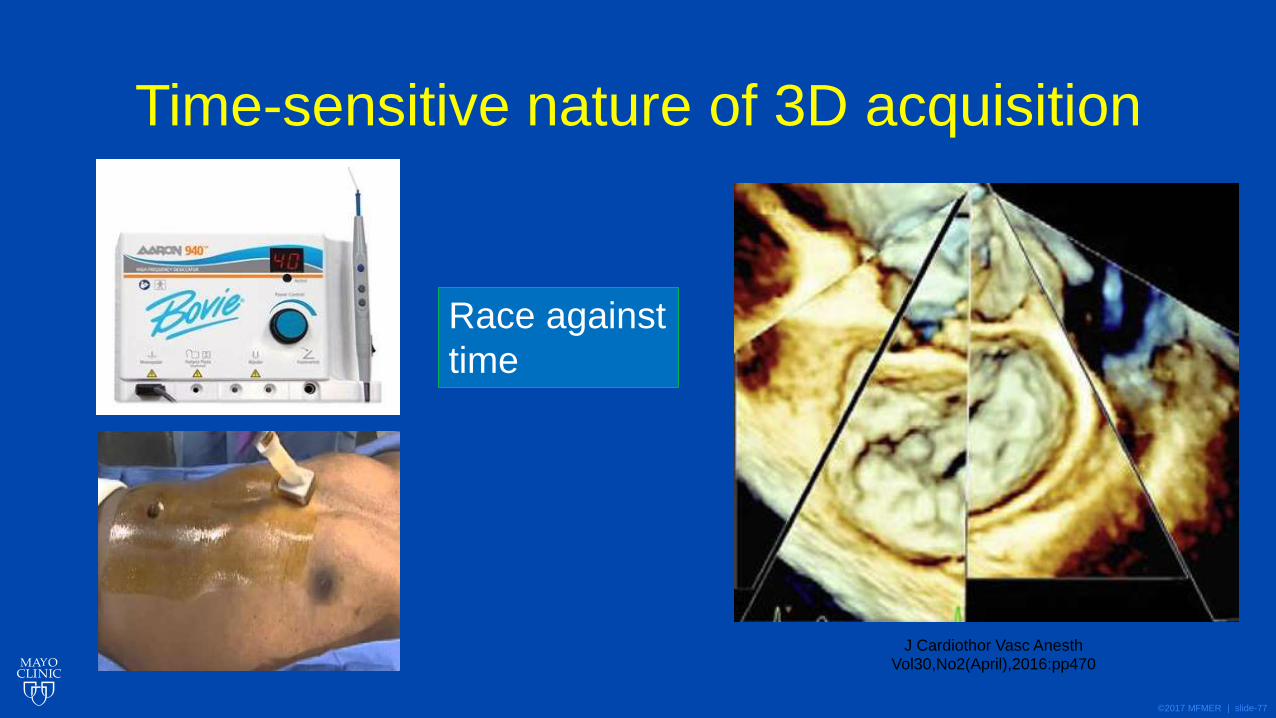
Time-sensitive nature of 3D acquisition
Race against
time
J Cardiothor Vasc Anesth Vol30,No2(April),2016:pp470
©2017 MFMER | slide-78
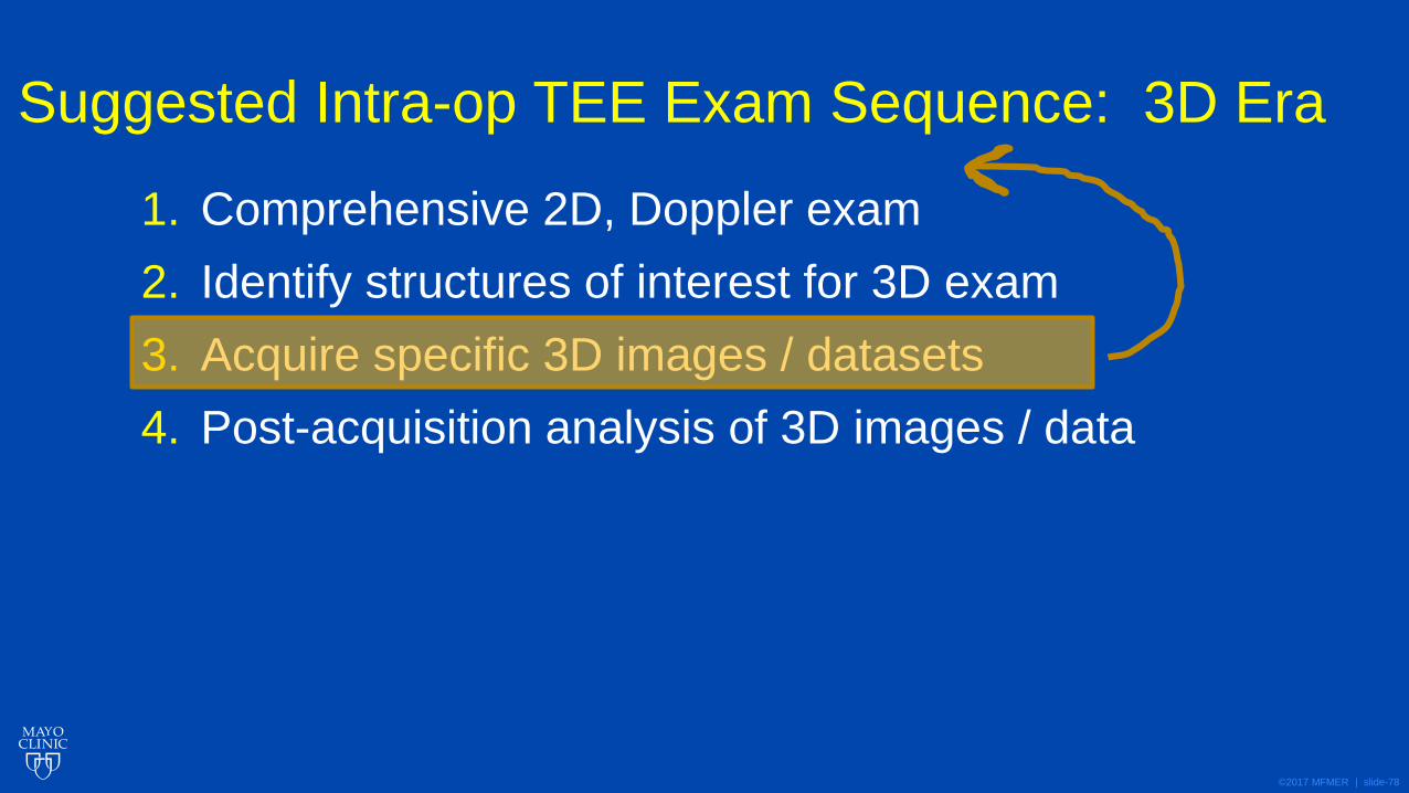
Suggested Intra-op TEE Exam Sequence: 3D Era
1. Comprehensive 2D, Doppler exam
2. Identify structures of interest for 3D exam
3. Acquire specific 3D images / datasets
4. Post-acquisition analysis of 3D images / data
©2017 MFMER | slide-79
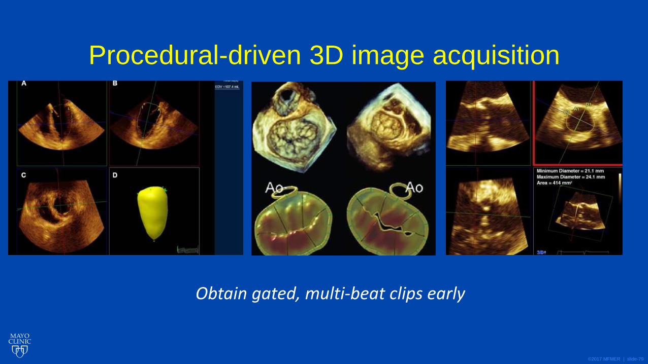
Procedural-driven 3D image acquisition
Obtain gated, multi-beat clips early

©2017 MFMER | slide-80
Take Home Points
©2017 MFMER | slide-81
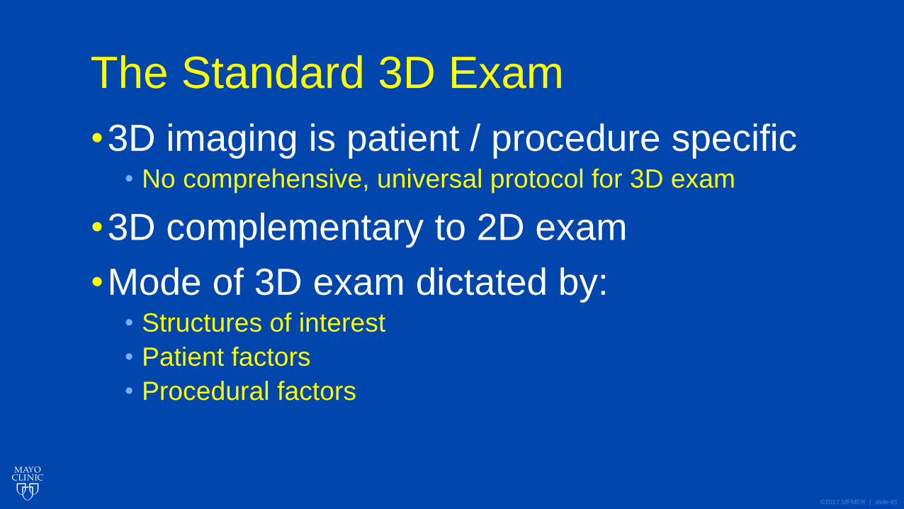
The Standard 3D Exam
•3D imaging is patient / procedure specific • No comprehensive, universal protocol for 3D exam
•3D complementary to 2D exam
•Mode of 3D exam dictated by: • Structures of interest
• Patient factors
• Procedural factors
©2017 MFMER | slide-82
The Standard 3D Exam
•Cases with high 3D yield • Catheter-based
• Valve repair / replacement
• Pre-operative diagnostic
• Post-intervention assessment
• Congenital
• Ventricular function