Embed Size (px)
Citation preview
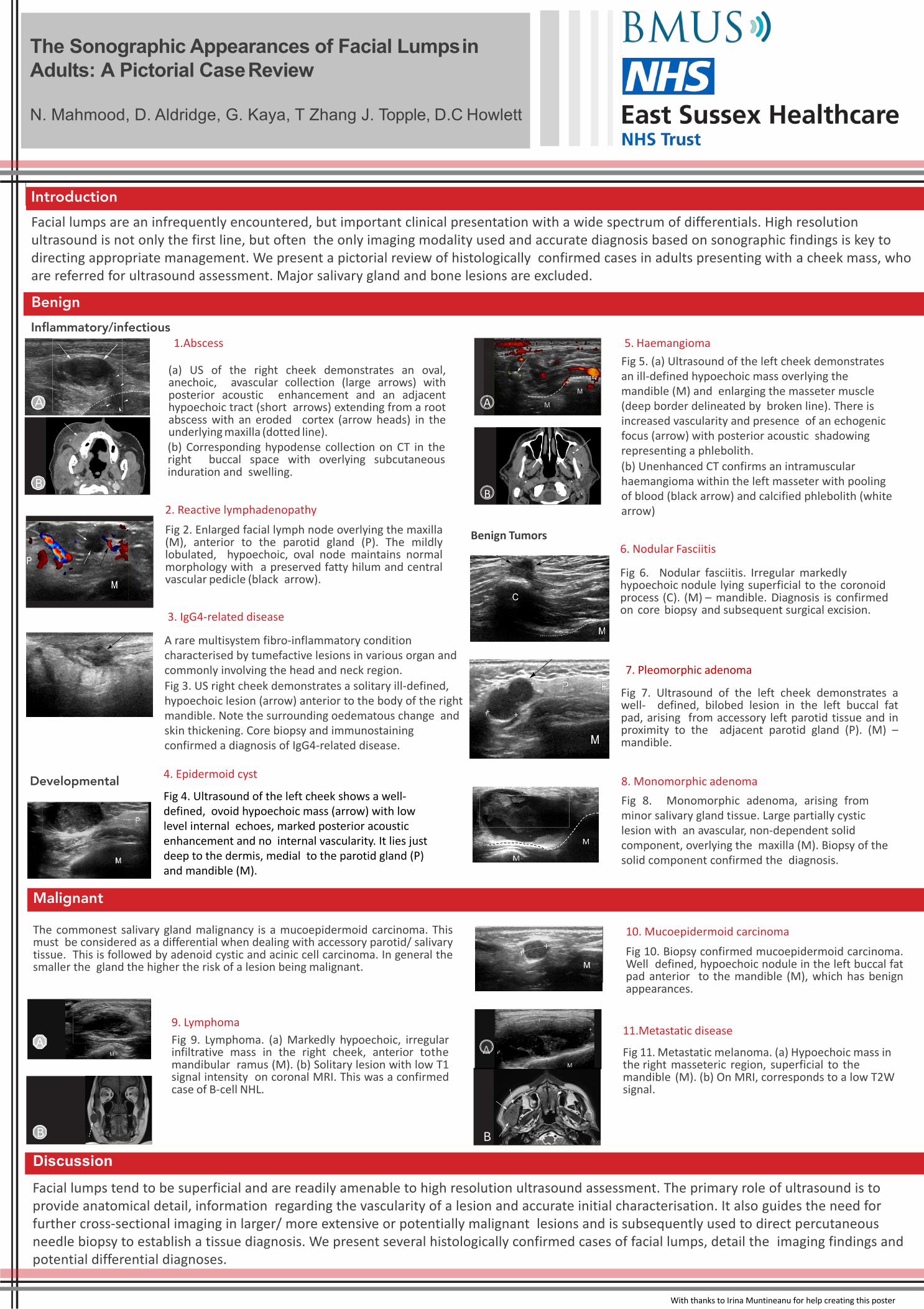
1.Abscess
(a) US of the right cheek demonstrates an oval,
anechoic, avascular collection (large arrows) with
posterior acoustic enhancement and an adjacent
hypoechoic tract (short arrows) extending from a root
abscess with an eroded cortex (arrow heads) in the
underlying maxilla (dotted line).
(b) Corresponding hypodense collection on CT in the
right buccal space with overlying subcutaneous
induration and swelling.
The Sonographic Appearances of Facial Lumpsin Adults: A Pictorial CaseReview
N. Mahmood, D. Aldridge, G. Kaya, T Zhang J. Topple, D.C Howlett
Introduction
Malignant
Benign
Discussion
8. Monomorphic adenoma
Fig 8. Monomorphic adenoma, arising from
minor salivary gland tissue. Large partially cystic
lesion with an avascular, non-dependent solid
component, overlying the maxilla (M). Biopsy of the
solid component confirmed the diagnosis.
10. Mucoepidermoid carcinoma
Fig 10. Biopsy confirmed mucoepidermoid carcinoma.
Well defined, hypoechoic nodule in the left buccal fat
pad anterior to the mandible (M), which has benign
appearances.
6. Nodular Fasciitis
Fig 6. Nodular fasciitis. Irregular markedly
hypoechoic nodule lying superficial to the coronoid
process (C). (M) – mandible. Diagnosis is confirmed
on core biopsy and subsequent surgical excision.
2. Reactive lymphadenopathy
Fig 2. Enlarged facial lymph node overlying the maxilla
(M), anterior to the parotid gland (P). The mildly
lobulated, hypoechoic, oval node maintains normal
morphology with a preserved fatty hilum and central
vascular pedicle (black arrow).
Fig 7. Ultrasound of the left cheek demonstrates a
well- defined, bilobed lesion in the left buccal fat
pad, arising from accessory left parotid tissue and in
proximity to the adjacent parotid gland (P). (M) –
mandible.
3. IgG4-related disease
Benign Tumors
5. Haemangioma
The commonest salivary gland malignancy is a mucoepidermoid carcinoma. This
must be considered as a differential when dealing with accessory parotid/ salivary
tissue. This is followed by adenoid cystic and acinic cell carcinoma. In general the
smaller the gland the higher the risk of a lesion being malignant.
11.Metastatic disease
Fig 11. Metastatic melanoma. (a) Hypoechoic mass in
the right masseteric region, superficial to the
mandible (M). (b) On MRI, corresponds to a low T2W
signal.
9. Lymphoma
Fig 9. Lymphoma. (a) Markedly hypoechoic, irregular
infiltrative mass in the right cheek, anterior tothe
mandibular ramus (M). (b) Solitary lesion with low T1
signal intensity on coronal MRI. This was a confirmed
case of B-cell NHL.
A
B
A
B
B
AA
B
Developmental
Inflammatory/infectious
7. Pleomorphic adenoma
A rare multisystem fibro-inflammatory condition
characterised by tumefactive lesions in various organ and
commonly involving the head and neck region.
Fig 3. US right cheek demonstrates a solitary ill-defined,
hypoechoic lesion (arrow) anterior to the body of the right
mandible. Note the surrounding oedematous change and
skin thickening. Core biopsy and immunostaining
confirmed a diagnosis of IgG4-related disease.
Facial lumps are an infrequently encountered, but important clinical presentation with a wide spectrum of differentials. High resolution
ultrasound is not only the first line, but often the only imaging modality used and accurate diagnosis based on sonographic findings is key to
directing appropriate management. We present a pictorial review of histologically confirmed cases in adults presenting with a cheek mass, who
are referred for ultrasound assessment. Major salivary gland and bone lesions are excluded.
Facial lumps tend to be superficial and are readily amenable to high resolution ultrasound assessment. The primary role of ultrasound is to
provide anatomical detail, information regarding the vascularity of a lesion and accurate initial characterisation. It also guides the need for
further cross-sectional imaging in larger/ more extensive or potentially malignant lesions and is subsequently used to direct percutaneous
needle biopsy to establish a tissue diagnosis. We present several histologically confirmed cases of facial lumps, detail the imaging findings and
potential differential diagnoses.
Fig 5. (a) Ultrasound of the left cheek demonstrates
an ill-defined hypoechoic mass overlying the
mandible (M) and enlarging the masseter muscle
(deep border delineated by broken line). There is
increased vascularity and presence of an echogenic
focus (arrow) with posterior acoustic shadowing
representing a phlebolith.
(b) Unenhanced CT confirms an intramuscular
haemangioma within the left masseter with pooling
of blood (black arrow) and calcified phlebolith (white
arrow)
4. Epidermoid cyst
Fig 4. Ultrasound of the left cheek shows a well-
defined, ovoid hypoechoic mass (arrow) with low
level internal echoes, marked posterior acoustic
enhancement and no internal vascularity. It lies just
deep to the dermis, medial to the parotid gland (P)
and mandible (M).
With thanks to Irina Muntineanu for help creating this poster