Embed Size (px)
Citation preview

The Skeletal System
1
Dr. Naim Kittana
Dr. Suhaib Hattab
Faculty of Medicine & Health SciencesAn-Najah National University

Declaration
• The content and the figures of this seminar were directly adopted from the text book “Human Anatomy and Physiology / Ninth edition/ Eliane N. Marieb2013”
2Dr. Naim Kittana, Dr. Suhaib Hattab

The Skeletal System
• Parts of the skeletal system:
1. Bones (skeleton)
2. Joints
3. Cartilages
4. Ligaments
• Divided into two divisions
1. Axial skeleton
2. Appendicular skeleton
3Dr. Naim Kittana, PhD

Functions of Bones
• Support of the body
• Protection of soft organs
• Movement due to attached skeletal muscles
• Storage of minerals and fats
• Blood cell formation
4Dr. Naim Kittana, PhD

Bones of the Human Body
• The skeleton has 206 bones
• Two basic types of bone tissue
• Compact bone
• Spongy bone
5Dr. Naim Kittana, PhD

Classification of Bones
6Dr. Naim Kittana, PhD

Structure of Flat Bones
7Dr. Naim Kittana, PhD

Structure of Long Bones
8Dr. Naim Kittana, PhD

Microscopic Anatomy of Compact Bone
• Five major cell types populate bone tissue: osteogenic cells, osteoblasts, osteocytes, bone lining cells, and osteoclasts
• Osteogenic cells (osteoprogenitor cells), are mitotically active stem cells found in the membranous periosteum and endosteum. When stimulated, these cells differentiate into osteoblasts or bone lining cells
• Osteoblasts are bone-forming cells that secrete the bone matrix.
• Osteocytes: The spidery osteocytes are mature bone cells that occupy spaces (lacunae) that conform to their shape. Osteocytesmonitor and maintain the bone matrix.
9Dr. Naim Kittana, PhD

Microscopic Anatomy of Compact Bone
• Bone lining cells: are flat cells found on bone surfaces where bone remodeling is not going on. Like osteocytes, they are thought to help maintain the matrix.
• Osteoclasts: are giant multinucleate cells located at sites of bone resorption
10Dr. Naim Kittana, PhD

Microscopic Anatomy of Compact Bone
• Osteon (Haversian System) The structural unit of compact bone.
• Each osteon is an elongated cylinder oriented parallel to the long axis of the bone.
• Functionally, osteons are tiny weight-bearing pillars.
11Dr. Naim Kittana, PhD

Microscopic Anatomy of Spongy Bone
• The trabeculae in spongy bone align precisely along lines of stress and help the bone resist stress.
12

Microscopic Anatomy of Compact Bone
13
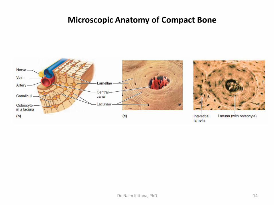
Microscopic Anatomy of Compact Bone
14Dr. Naim Kittana, PhD
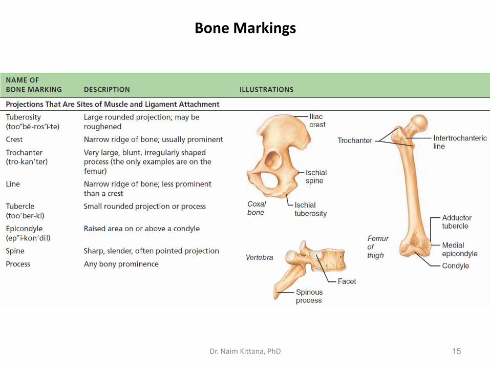
Bone Markings
15Dr. Naim Kittana, PhD

Bone Markings
16Dr. Naim Kittana, PhD

Capsule of hip-joint, Posterior aspect.
Upper surface of right tibia
17Dr. Naim Kittana, PhD

Long Bone Formation and Growth
18

The Axial Skeleton
• Forms the longitudinal part of the body
• Divided into three parts
1. Skull
2. Vertebral column
3. Bony thorax
19Dr. Naim Kittana, PhD

The
hu
man
ske
leto
n
20

The Skull
• Two sets of bones
1. Cranium
2. Facial bones
• Bones are joined by sutures
• Only the mandible is attached by a freely movable joint
21Dr. Naim Kittana, PhD

The
Sku
ll: A
nte
rio
r vi
ew
22

The Skull: Lateral View
23

The Skull: Superior View
24
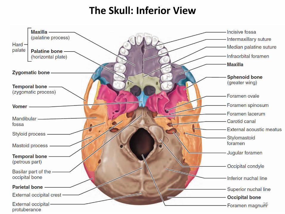
The Skull: Inferior View
25

Paranasal Sinuses
Hollow portions of bones surrounding the nasal cavity
26Dr. Naim Kittana, PhD

The Hyoid Bone
• The only bone that does not articulate with another bone
• Serves as a moveable base for the tongue
27Dr. Naim Kittana, PhD

The Vertebral Column
• Vertebrae separated by intervertebral discs
• The spine has a normal curvature
• Each vertebrae is given a name according to its location
28Dr. Naim Kittana, PhD

Structure of a Typical Vertebrae
29Dr. Naim Kittana, PhD

Regional Characteristics of Vertebrae
30

Regional Characteristics of Vertebrae
31Dr. Naim Kittana, PhD

Regional Characteristics of Vertebrae
32

The Bony Thorax
33

The Appendicular Skeleton
• Limbs (appendages)
• Pectoral girdle
• Pelvic girdle
34Dr. Naim Kittana, PhD

The Pectoral (Shoulder) Girdle
35

The humerus of the right arm
36

Bones of the Upper Limb
37

Bones of the Upper Limb
38

Bones of the Pelvic Girdle
39Dr. Naim Kittana, PhD

Bones of the Pelvic Girdle
40

Gender Differences of the Pelvis
41

Gender Differences of the Pelvis
42

Bones of the Lower Limbs
• The thigh has one bone: femur – thigh bone
43

Bones of the Lower Limbs
The leg has two bones:
• Tibia
• Fibula
The tibia and fibula of the right leg
44

Bones of the right foot
The foot:
1. Tarsus – ankle
2. Metatarsals – sole
3. Phalanges – toes
45Dr. Naim Kittana, PhD

Bones of the right foot
46

Arches of the Foot
Together, the arches of the foot form a half dome that distributes about half of a person’s standing and walking weight to the heel bones and half to the heads of the metatarsals. 47

Joints
• Articulations of bones
• Functions of joints:
• Hold bones together
• Allow for mobility
• Ways joints are classified:
• Functionally
• Structurally
48Dr. Naim Kittana, PhD

Functional Classification of Joints:
1. Immovable joints
2. Slightly moveable joints
3. Freely moveable joints
Structural Classification of Joints:
1. Fibrous joints: Generally immovable
2. Cartilaginous joints: Immovable or slightly moveable
3. Synovial joints: Freely moveable
Classifications of Joints
49Dr. Naim Kittana, PhD

Fibrous Joints
50

Cartilaginous Joints
51

Synovial Joints
52

The General Structure of Synovial Joint
53

Summary of joint classes
54Dr. Naim Kittana, PhD

Types of movements allowed by Synovial Joints
55

Types of movements allowed by Synovial Joints
• Gliding occurs when one flat, or nearly flat, bone surface glides or slips over another (back-and-forth and side-to-side)
• Angular movements increase or decrease the angle between two bones. These movements may occur in any plane of the body and include
flexion
extension
hyperextension
abduction
adduction
circumduction
56Dr. Naim Kittana, PhD

Types of movements allowed by Synovial Joints
• Flexion: bending movement, usually along the sagittal plane, that decreases the angle of the joint and brings the articulating bones closer together
• Extension: is the reverse of flexion and occurs at the same joints
• Hyperextension: Continuing such movements beyond the anatomical position is called
57Dr. Naim Kittana, PhD

Types of movements allowed by Synovial Joints
58

Types of movements allowed by Synovial Joints
• Abduction (“moving away”): is movement of a limb away from the midline or median plane of the body, along the frontal plane
• Adduction (“moving toward”): is the opposite of abduction, so it is the movement of a limb toward the body midline or, in the case of the digits, toward the midline of the hand or foot
• Circumduction: is moving a limb so that it describes a cone in space. The distal end of the limb moves in a circle, while the point of the cone (the shoulder or hip joint) is more or less stationary.
• Rotation: is the turning of a bone around its own long axis
59Dr. Naim Kittana, PhD
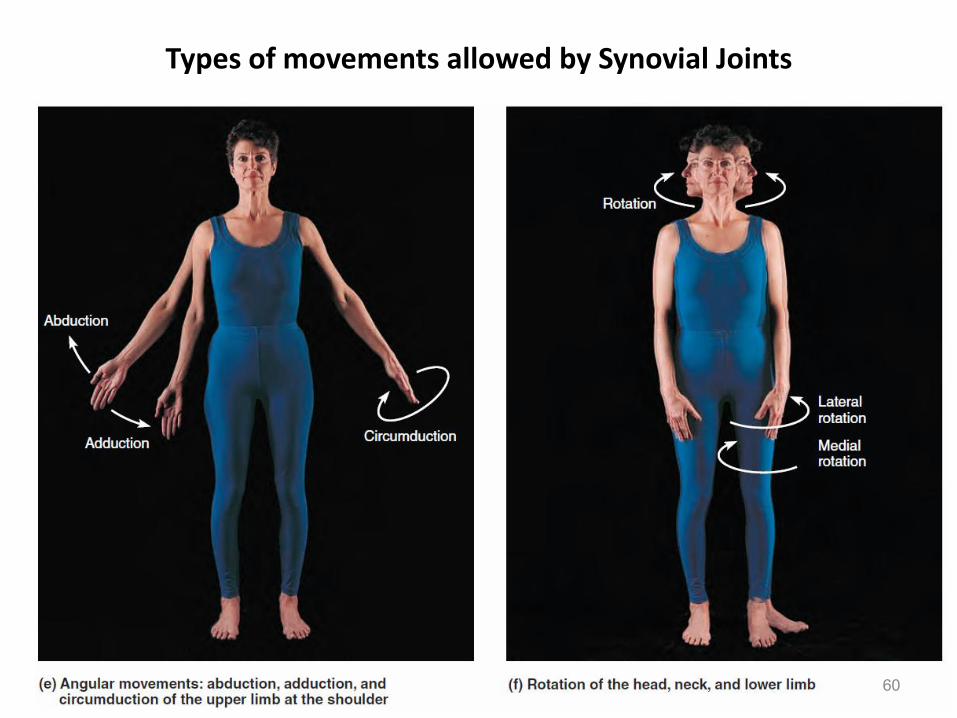
Types of movements allowed by Synovial Joints
60

Types of Synovial Joints Based on Shape
61

Synovial joints
1- Plane joints
Permit gliding or sliding movements in the plane of the articularsurfaces.
The opposed surfaces of the bones are flat or almost flat, with movement limited by their tight joint capsules.
Plane joints are numerous and are nearly always small.
An example is the acromioclavicular joint between the acromion of the scapula and the clavicle.
62Dr. Naim Kittana, PhD

Synovial joints
2- Hinge joints
Permit flexion and extension only
Movements occur in one plane (sagittal) around a single axis that runs transverse uniaxial joints
The joint capsule of these joints is thin and lax anteriorly and posteriorly where movement occurs
The bones are joined by strong, laterally placed collateral ligaments. The elbow joint is a hinge joint
63Dr. Naim Kittana, PhD

Synovial joints
3- Pivot joints
• Permit rotation around a central axis; thus they are uniaxial. In these joints, a rounded process of bone rotates within a sleeve or ring.
• The median atlantoaxial joint is a pivot joint in which the atlas (C1 vertebra) rotates around a finger-like process, the dens of the axis (C2 vertebra), during rotation of the head.
64Dr. Naim Kittana, PhD

Pivot joints
65Dr. Naim Kittana, PhD

Types of Synovial Joints Based on Shape
66

Synovial joints
4- Condyloid joints
• Permit flexion and extension as well as abduction and adduction; thus condyloid joints are also biaxial
• Movement in one plane (sagittal) is usually greater (freer) than in the other. Circumduction, more restricted than that of saddle joints, is also possible
• The metacarpophalangeal joints (knuckle joints) are condyloidjoints
67Dr. Naim Kittana, PhD

Types of Synovial Joints Based on Shape
68
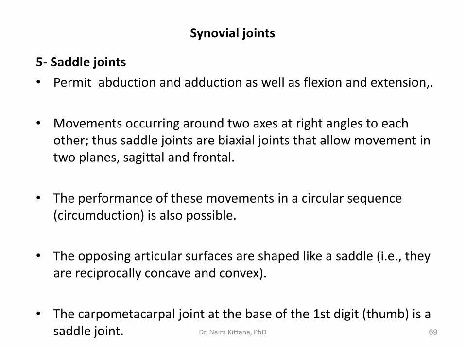
Synovial joints
5- Saddle joints
• Permit abduction and adduction as well as flexion and extension,.
• Movements occurring around two axes at right angles to each other; thus saddle joints are biaxial joints that allow movement in two planes, sagittal and frontal.
• The performance of these movements in a circular sequence (circumduction) is also possible.
• The opposing articular surfaces are shaped like a saddle (i.e., they are reciprocally concave and convex).
• The carpometacarpal joint at the base of the 1st digit (thumb) is a saddle joint. 69Dr. Naim Kittana, PhD
Synovial joints
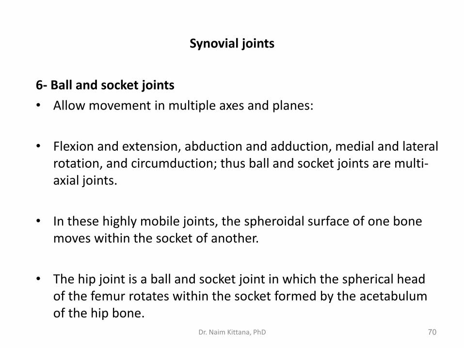
6- Ball and socket joints
• Allow movement in multiple axes and planes:
• Flexion and extension, abduction and adduction, medial and lateral rotation, and circumduction; thus ball and socket joints are multi-axial joints.
• In these highly mobile joints, the spheroidal surface of one bone moves within the socket of another.
• The hip joint is a ball and socket joint in which the spherical head of the femur rotates within the socket formed by the acetabulumof the hip bone.
70Dr. Naim Kittana, PhD