Embed Size (px)
Citation preview

The
SITS OPEN Artery by Thrombectomy in Acute Occlusive Stroke Study
Study Protocol Version 5.0 date 2016-11-24
An international, multicentre-controlled study of safety and efficacy of thrombectomy in acute occlusive stroke

SITS Open protocol version 5.0 2016-11-24 2
An open, prospective, blinded evaluation, international, multicentre, controlled study of safety and efficacy of thrombectomy and standard stroke care in clinical routine treatment of acute occlusive stroke compared to standard stroke care only Sponsored by
In collaboration with
3 SITS Open protocol version 5.0 2016-11-24
PROTOCOL SUMMARY
Study title The SITS open artery by thrombectomy in acute occlusive stroke study (SITS Open)
Primary Objective
To determine the benefit and safety of TBY in clinical routine practice by selected stent retrievers or other selected novel devices in addition to standard care in patients with major cerebral artery occlusion as compared to standard care only. Standard care may include IVT in accordance with current guidelines.
Secondary Objective
• To determine the benefit and safety of TBY by selected stent retrievers or other selected novel devices as additional therapy in proximal cerebral artery occlusion (Carotid T, M1, Basilar Artery) in patients receiving IVT according to current guidelines within 4.5 hours of ischaemic stroke onset as compared to stand-alone IVT.
• To determine whether TBY without prior IVT impacts the functional outcome of patients compared to standard stroke care including IVT, when indicated according to current guidelines.
• To determine the benefit and safety of TBY in clinical routine practice by selected stent retrievers or other selected novel devices in patients with major cerebral artery occlusion as compared to active arm of the pooled analyses of 5 randomized controlled trials, HERMES (1).
• To determine the study outcomes for patients in following subgroups: 1) In patients with M1/Car-T/BA occlusion, 2) M2/A1/P1 occlusion, 3) basilar artery occlusion, 4) in patients without prior treatment with IVT, 5) length of the occluding thrombus 8 mm, 6) moderately severe stroke at baseline (NIHSS 7-12) and for severe stroke (NIHSS 13- ).
Primary Hypothesis
Patients with ischaemic stroke caused by a major cerebral artery occlusion and offered thrombectomy will have improved functional outcomes (categorical shift towards lower, i.e. better, Rankin scores over the range of the scale) compared to those patients with comparable baseline characteristics and imaging who have been treated with standard stroke care only, including IVT when indicated.
H0: foTBY ≤ focontrol vs. HA: foTBY > focontrol
(≤ indicates worse or equal to, > indicates better than)
Where foTBY and focontrol represent the functional outcome undertaken on the full range (0-6, where 0 represents the best possible outcome and 6 the worst, i.e., death) of the modified Rankin Scale (mRS) using Cochran-Mantel-Haenszel shift test and proportional odds logistic regression subject to the validity of shift analysis model assumptions.

SITS Open protocol version 5.0 2016-11-24 4
Study Design Prospective, open, blinded evaluation (PROBE design), international, multicentre, controlled study of thrombectomy compared to standard treatment only, based on SITS clinical trial platform, of consecutively enrolled ischaemic stroke patients with confirmed occlusion of a major cerebral artery who will be considered eligible for thrombectomy in agreement with routine clinical criteria. Standard care may include IVT in accordance with current guidelines.
Inclusion Criteria
• Patients with acute stroke after exclusion of intracranial haemorrhage on CT/MRI scan.
• Confirmed diagnosis on CTA of persisting occlusion of the terminal Internal Carotid Artery (Car-T), proximal Middle Cerebral Artery (MCA, M1), proximal part of the insular segment of MCA (M2), proximal part of the anterior cerebral artery (A1), Basilar Artery (BA) or proximal part of the posterior cerebral artery (P1), consistent with the clinical symptoms. For inclusion in the study, CTA must not be performed later than 15 minutes after IVT start if given. For patients not treated with IVT, CTA should preferably be performed within 15 minutes of completion of the non-contrast CT but must be performed within 6 hours after stroke onset.
• Eligible patients for IVT are treated according to clinical guidelines (Attachment 1), and IVT, if given, initiated within 4.5 h.
• Initiation of thrombectomy is recommended within 6 hours after stroke onset but must be performed within to 8 hours if thrombectomy would still be of benefit for the patient as judged by the investigator.
• Baseline NIHSS Score at initiation of IVT is recommended between 7 and 25 for anterior circulation stroke and ≥7 without upper limit for posterior circulation stroke (baseline NIHSS score should be assessed by an NIHSS-certified physician), but patients may also be included beyond these scores if thrombectomy would still be of benefit for the patient as judged by the investigator.
• Age ≥18years.
• Anticipated life expectancy of at least 6 months.
• Patient or legal representative is competent to make a decision and has provided informed consent with regard to participation in the study, retrieval and storage of data and follow up procedures.
• Initiation of endovascular procedure (DSA/TBY, defined as start with groin puncture) within 2 hours from the start of IVT, or after CTA if IVT is not given (for TBY arm patients).

5 SITS Open protocol version 5.0 2016-11-24
Exclusion Criteria
• Known significant pre-stroke disability (mRS ≥2).
• Extended early ischemic changes for basilar artery occlusion, according to the judgment of treating physician based on routine clinical practice of the hospital; if technical possibility exists, early irreversible ischemic changes may be confirmed by pc-ASPECTS score < 8 on CTASI (2) or extensive DWI lesion on pre-treatment MRI.
• Known pregnancy.
• Participation in any other investigational drug or device study, currently or in the previous 30 days.
Clinical site locations
Approximately 45 study centres in Europe will participate.
Enrolment 600 patients in TBY arm and 300 patients in the control arm. Patients are enrolled after informed consent is obtained. Screening documentation will be performed before thrombectomy procedure (TBY group) or within one hour after IVT completion or within 6 hours if IVT is not given (control group).
Arms TBY arm consists of patients undergoing thrombectomy according to accepted critera in the judgement of investigator. Patients may be included even if they are not treated with intravenous thrombolysis because of contraindication or other reasons.
Control arm enrols patients at study centres that do not practice thrombectomy treatment. Control arm patients are treated with standard stroke care including IVT but do not receive TBY. As in the TBY arm the Control arm consists of patients fulfilling criteria for thrombectomy according to accepted critera in the judgement of investigator. Patients may be included even if they are not treated with intravenous thrombolysis because of contraindication or other reason.
Primary Endpoint
Categorical shift in mRS score at 3 months.
Secondary Endpoints
• Proportion of patients with functional independence (modified Rankin Scale, mRS, score 0-2) at 3 months after stroke onset
• Proportion of patients with excellent outcome (mRS score 0-1) at 3 months
• Recanalization of the occluded artery for TBY treated populations, defined as at least TICI 2b flow in the treated territory after procedure.
• Time from stroke onset to revascularisation to any TICI grade (defined as 2b or 3) for the TBY treated population.
• Recanalization (defined as AOL 2-3) of the occluded artery confirmed by 24h CTA/contrast-enhanced MRA.

SITS Open protocol version 5.0 2016-11-24 6
• Neurological improvement (difference in NIHSS from baseline to 12h, to 24h and to 7d post- IVT or discharge home/secondary care if earlier), and functional outcome at 3 months in relation to recanalization status and thrombus length (mm).
• Reduction in infarct size (TBY vs. control groups at 22-36 hours)
• Length of in-hospital stay (days to discharge from in-hospital ward to home/secondary care for survivors) in TBY groups vs. control groups
• Home Time: Number of days the patient stayed at home or at relative´s stay within the first 3 months after stroke onset, in TBY vs. Control groups.
• Recurrent stroke during within 3 month
• Proportion of patients with recanalization (defined as AOL 2-3) before thrombectomy.
Safety Endpoints
• Symptomatic intracerebral haemorrhage (SICH) according to SITS-MOST definition: local or remote parenchymal haemorrhage type 2 on the 22- to 36-hour post-treatment imaging scan, combined with a neurological deterioration of ≥4 points compared with baseline NIHSS or the lowest NIHSS value or death between baseline and 24 hours.
• Symptomatic intracranial haemorrhage (SICH) according to modified SITS-MOST definition; in addition to usual SITS-MOST criteria blood may be anywhere in the intracranial space (including in the intraventricular, intraparenchymal and/or subarachnoid space).
• Symptomatic intracranial haemorrhage (SICH) defined as an NIHSS decline of ≥4 points compared with baseline NIHSS or the lowest NIHSS value or death between baseline and 7 days, associated with any haemorrhage judged by core lab evaluation to be responsible for the decline. Blood may be anywhere in the intracranial space including in the intraventricular, intraparenchymal and/or subarachnoid space (modified ECASS III definition).
• All-cause mortality at 3 months.
• Neurological death within 7 days post treatment.
• Distal embolism/reocclusion demonstrated by follow-up CTA/MRA within 22-36 h post treatment or after CTA baseline
• Embolism into new territories (ENT)
• Any adverse event related to thrombectomy procedure such as ”symptomatic ischemic oedema” and “expansion of infarction or reinfarction”, including patients for whom the initiating angiography revealed recanalization by IVT only.

7 SITS Open protocol version 5.0 2016-11-24
STUDY FLOW CHART

SITS Open protocol version 5.0 2016-11-24 8
TABLE OF CONTENT
PROTOCOL SUMMARY 3
1. ABBREVATIONS 11
2. ADMINISTRATE INFORMATION 12 2.1. SPONSOR 12 2.2. CONSORTIUM COMMITTEE 12 2.3. STEERING COMMITTEE 12 2.4. COORDINATING INVESTIGATORS 13 2.5. CLINICAL STUDY CENTRES 13 2.6. CLINICAL STUDY MANAGEMENT 14 2.7. COORDINATION RESEARCH TEAM 14 2.8. DATA CAPTURE SYSTEM 14 2.9. STATISTICIANS 14 2.10. ADJUDICATION COMMITTEE OF mRS 15 2.11. NEUROIMAGING CORE LABORATORY 15 2.12. DATA AND SAFETY MONITORING COMMITTEE 15
3. BACKGROUND 16 3.1 THROMBOLYSIS 16 3.2 THROMBECTOMY 16 3.3 SUMMARY OF EARLIER MECHANICAL THROMBECTOMY CLINICAL TRIALS 16 3.4 PENUMBRA PIVOTAL STROKE TRIAL 17 3.5 NEWER MECHANICAL THROMBECTOMY DEVICES 17 3.6 BRIDGING CONCEPT 17 3.7 RECENT RANDOMISED CONTROLLED TRIALS 18 3.8 SUMMARY 18
4. OBJECTIVES 18 4.1 PRIMARY OBJECTIVES 18 4.2 SECONDARY OBJECTIVES 19
5. ENDPOINTS 19 5.1 PRIMARY ENDPOINT OF EFFICACY 19 5.2 SECONDARY ENDPOINTS OF EFFICACY 19 5.3 SAFETY ENDPOINTS 20
6. DESIGN 20 6.1 OUTLINE 20 6.2 MEASURES TO MINIMIZE BIAS 21 6.3 SELECTION OF STUDY CENTRES 21 6.4 ASSESSMENTS AND PROCEDURES 22
6.4.1 Clinical and Laboratory Procedures 22 6.4.2 Screening & Consent Procedures 22 6.4.3 Balance of recruitment and devices in the study 23 6.4.4 Medical Management 23 6.4.5 Thrombectomy (TBY arm only) 23 6.4.6 Arterial Access 24 6.4.7 Angiography 24

9 SITS Open protocol version 5.0 2016-11-24
6.4.8 Thrombectomy 24 6.4.9 In-Hospital Post Treatment Assessment 25 6.4.10 Rehabilitation measures 26 6.4.11 Outcome assessment at 3 Months (90 ± 14 days) 26
6.5 PATIENT RECRUITMENT AND WITHDRAWAL 28 6.5.1 Inclusion criteria 28 6.5.2 Exclusion criteria 28 6.5.3 Withdrawal and dropout 28 6.5.4 Screening log and SITS registry documentation 29
7. DEVICE 30 7.1 TRAINING 30 7.2 DESCRIPTION OF THE MEDICAL ENDOVASCULAR DEVICES 30
7.2.1 PreSET 30 7.2.2 Solitaire 32 7.2.3 Trevo Retriever 32
8. ASSESSEMENT OF EFFICACY AND SAFETY 34 8.1 INDEPENDENT CT/MR ADJUDICATION 34 8.2 DATA AND SAFETY MONITORING BOARD (DSMB) 34 8.3 REQUIREMENTS OF CLINICAL MEASUREMENTS 34 8.4 MODIFIED RANKIN SCALE AT 3 MONTHS 34 8.5 CLINICAL SAFETY ASSESSMENT 35
8.5.1 Clinical examination 35 8.5.2 Monitoring of vital signs 35 8.5.3 Laboratory tests 35 8.5.4 Imaging safety assessment 35 8.5.5 Classification of Intracerebral haemorrhage 36 8.5.6 Classification of Cerebral oedema 36 8.5.7 Definition of Symptomatic Intracerebral/Intracranial Haemorrhage (SICH) 36 8.5.8 Adverse Events and Reactions 37
8.6 CLINICAL EFFICACY ASSESSMENTS 37
9. IMAGING CORE LAB 37 9.1 ANGIOGRAPHY 37 9.2 BASELINE AND 24 H CT AND CTA EXAMINATIONS 37 9.3 OPTIONAL PRE-PROCEDURE CT PERFUSION OR MR DWI/PWI IMAGING 38
10. PROCEEDINGS FOR ADVERSE REACTIONS AND SERIOUS ADVERSE REACTIONS 38 10.1 DEFINITION and MANAGEMENT 38 10.2 ASSESSMENT OF ADVERSE REACTIONS OR SERIOUS ADVERSE REACTION 39 10.3 FOLLOW-UP OF ADVERSE REACTIONS and SERIOUS ADVERSE REACTIONS 40
11. STATISTICS AND DATA MANAGEMENT 40 11.1 DATA MANAGEMENT 40 11.2 STATISTICAL ANALYSIS PLAN 41
11.2.1 Determination of Sample Size 41 11.3 PREVENTION OF STATISTICAL BIAS 42 11.4 ANALYSIS OF PRIMARY ENDPOINT 44 11.5 ANALYSIS OF SECONDARY ENDPOINTS 44

SITS Open protocol version 5.0 2016-11-24 10
11.6 OTHER PLANNED ANALYSES 44 11.7 ADDITIONAL SUBGROUP ANALYSES 45
12. DIRECT ACCESS TO SOURCE DOCUMENTS 45
13. QUALITY CONTROL AND QUALITY ASSURANCE 46 13.1 SOURCE DATA 46
14. ETHICS 46 14.1 INDEPENDENT ETHICS COMITTEE 46 14.2 ETHICAL CONDUCT OF THE TRIAL 46 14.3 SUBJECT INFORMATION AND INFORMED CONSENT 47 14.4 RISK/BENEFIT ASSESSMENT 47
14.4.1 Potential risks of thrombectomy 47 14.4.2 Potential benefits of thrombectomy 48 14.4.3 Minimization of Risk of Thrombectomy 48 14.4.4 Potential Risks and Benefits of Neuro Imaging 48 14.4.5 Other Potential Risks 49
15. DATA HANDLING AND RECORD KEEPING 49 15.1 CASE REPORT FORMS AND RECORD KEEPING 49
16. FINANCING AND INSURANCE 50
17. PUBLICATION POLICY 50
18. SUPPLEMENTS 50 18.1 AMENDMENTS 50 18.2 PERSONELL INFORMATION 50
19. REFERENCE 51
20. SIGNED AGREEMENT OF THE TRIAL PROTOCOL 55
21. APPENDICES 56 21.1 STUDY LOGISTICS 56 21.2 SCHEDULE OF INVESTIGATIONAL EVENTS 57 21.3 NATIONAL INSTITUTE OF HEALTH STROKE SCALE (NIHSS) 58 21.4 MODIFIED RANKIN SCALE (MRS) 60 21.5 STRUCTURED INTERVIEW MODEL FOR MRS ASSESSMENT (also see instruction – attachment 3) 61 21.6 SITS GLOBAL OUTCOME SCALE 62 21.7 THROMBOLYSIS IN CEREBRAL INFARCTION (TICI) SCORE 63
21.7.1 TICI score for angiographical assessment 63 21.7.2 Modified TICI score for CTA assessment 63 21.7.3 Arterial Occlusive Lesion (AOL) Score 63
21.8 INSTRUCTIONS FOR IMAGING 64

11 SITS Open protocol version 5.0 2016-11-24
1. ABBREVATIONS
AE Adverse event AOL Arterial Oclusive Leision Revascularization Scale
AR Adverse Reaction
BP Blood Pressure BA Basilar artery
Car-T Terminal Carotid Artery CI Coordinating Investigator
CCI Co-Coordinating Investigator
CRF Case Record Form CT/CTA Computer Tomography /Computer Tomography Angiography
DSMB Data and Safety Monitoring Board DWI Diffusion weighted imaging
ECASS European Cooperative Acute Stroke Study
ECG Electrocardiogram eCRF Electronic Case Record Form
ESO European Stroke Organisation
FDA Food and Drug Administration
GCP Good Clinical Practice
HI Haemorrhagic Infarction IA Intraarterial
ICH Intracerebral Haemorrhage IEC Independent Ethics Committee
INR International Normalised Ratio ITT Intention to Treat
IV Intravenously
IVT Intravenous Thrombolysis LC Local Coordinator
M1 Proximal Middle Cerebral Artery MCA Middle Cerebral Artery
MFS Mission Fighting Stroke (Uppdrag Besegra Stroke)
MR Magnetic Resonance MRA Magnetic Resonance Angiography
MRI Magnetic Resonance Imaging
MRP Magnetic Resonance Perfusion
mRS Modified Rankin Scale
NC National Coordinator NIHSS National Institutes of Health Stroke Scale
OR Odds Ratio
PCA Posterior Cerebral Artery
pcASPECTS Posterior circulation Acute Stroke Prognosis Early CT score
PH Parenchymal Haemorrhage

SITS Open protocol version 5.0 2016-11-24 12
PI/c Principle Clinical Investigator
PI/i Principle Interventional Investigator PP Per Protocol
PWI Perfusion weighted imaging rt-PA Recombinant Tissue Plasminogen Activator
SAH Subarachnoid hemorrhage
SAE Serious Adverse Event SAR Serious Adverse Reaction
SC Steering Committee
SICH Symptomatic Intracerebral Haemorrhage
SITS Safe Implementation of Treatments in Stroke
SITS-MOST SITS Monitoring Study TBY Thrombectomy
TCD Transcranial Doppler study TICI Thrombolysis In Cerebral Infarction (score)
UBS Uppdrag Besegra Stroke (Mission Fighting Stroke)
2. ADMINISTRATE INFORMATION
2.1. SPONSOR Department of Clinical Neuroscience, Karolinska Institutet, representatives:
Prefekt: Jan Hillert, M.D., Ph.D. Department of Clinical Neuroscience, Karolinska Institutet
Nils Wahlgren, M.D., Ph.D. Department of Clinical Neuroscience, Karolinska Institutet
2.2. CONSORTIUM COMMITTEE Chair: Nils Wahlgren, M.D., Ph.D Department of Clinical Neuroscience, Karolinska Institutet
Kennedy Lees, M.D., FRCP University of Glasgow Glasgow, United Kingdom
Rüdiger von Kummer, Prof.Dr.med University Hospital of Dresden Dresden, Germany
2.3. STEERING COMMITTEE Chair Nils Wahlgren, M.D., Ph.D. Department of Clinical Neuroscience, Karolinska Institutet Stockholm, Sweden
13 SITS Open protocol version 5.0 2016-11-24
Co-chair Olav Jansen, M.D., Ph.D. University Hospital of Schleswig-Holstein Kiel, Germany
Niaz Ahmed, M.D., Ph.D. Karolinska University Hospital, Karolinska Institutet Stockholm, Sweden
Staffan Holmin, M.D., Ph.D. Karolinska University Hospital, Karolinska Institutet Stockholm, Sweden
Kennedy Lees, M.D., FRCP University of Glasgow Glasgow, UK
Salvatore Mangiafico, M.D., Ph.D. Careggi University Hospital Florence, Italy
Lawrence Wong, M.D., Ph.D. Chinese University of Hong Kong Hong Kong, China
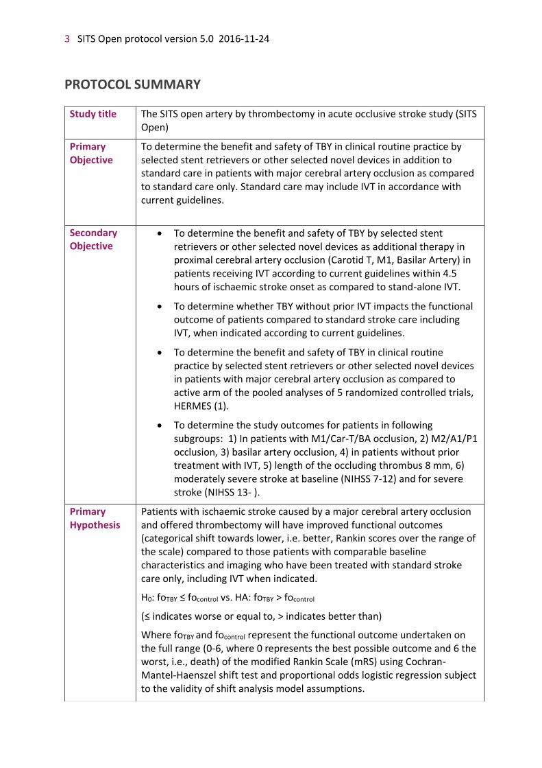
2.4. COORDINATING INVESTIGATORS Nils Wahlgren, M.D., Ph.D. Department of Clinical Neuroscience, Karolinska Institutet Stockholm, Sweden
Olav Jansen, M.D., Ph.D. University Hospital of Schleswig-Holstein Kiel, Germany
2.5. CLINICAL STUDY CENTRES Coordinating Study Centre: R2:03 Department of Neurology Karolinska University Hospital 171 76 Stockholm, Sweden
Primary study contact person: Professor Nils Wahlgren, [email protected] Telephone +46 (0)8-517 756 00
Primary neurointerventionist contact person: Professor Olav Jansen, [email protected] Telephone +49 (0) 431 597 4808

SITS Open protocol version 5.0 2016-11-24 14
2.6. CLINICAL STUDY MANAGEMENT Study Manager Kia Bengtsson R2:03 Department of Neurology Karolinska University Hospital 171 76 Stockholm, Sweden [email protected]
Study Centre Coordinator/Main centre contact Project Manager Karin Flood R2:03 Department of Neurology Karolinska University Hospital 171 76 Stockholm, Sweden
General inbox and Coordinator Hotline [email protected]; +46 (0) 76 945 96 70
2.7. COORDINATION RESEARCH TEAM
Niaz Ahmed, M.D., Ph.D. [email protected] Tatiana Kharitonova, M.D., Ph.D [email protected] Tiago Moreira, M.D., Ph.D. [email protected]
2.8. DATA CAPTURE SYSTEM SITS Thrombectomy Registry (SITS-TBY) and SITS International Stroke Thrombolysis Registry (SITS-ISTR)
2.9. STATISTICIANS Matteo Bottai, Sc.D. Biostat Core, Institute of Environmental Medicine Nobel’s väg 13 Karolinska Institutet 17177 Stockholm, Sweden
Sharon Kean Director Information Systems Robertson Centre for Biostatistics Level 11, Boyd Orr Building University of Glasgow, Glasgow, UK

15 SITS Open protocol version 5.0 2016-11-24
2.10. ADJUDICATION COMMITTEE OF mRS Chair Kennedy Lees, M.D., FRCP, Professor Co-chair Jesse Dawson, Clinical Reader Kate MacArthur, Consultant
Terence J Quinn, Clinical Senior Lecturer
All at University of Glasgow
Glasgow, United Kingdom
2.11. NEUROIMAGING CORE LABORATORY Chair Rüdiger von Kummer, Prof.Dr.med University Hospital of Dresden Department of Neuroradiology Dresden, Germany
Systematic Managment Archiving and reviewing Trial Imagies service (SMARTIS) University of Edinburgh (Neurosciences Imaging) Centre for Clinical Brain Sciences Att: Eleni Sakka The Chancellor's Building 49 Little France Crescent, Edinburgh EH16 4SB United Kingdom
2.12. DATA AND SAFETY MONITORING COMMITTEE
Chair: Gary Ford, M.D. FRCP. Magdalen Centre North The Magdalen Centre Robert Robinson Avenue Oxford Science Park OX4 4GA Markku Kaste, MD, PhD Department of Neurology Helsinki Central University Hospital Helsinki, Finland
David Liebeskind, MD UCLA Stroke Center 710 Westwood Plaza, 4-121 Reed Los Angeles, CA 90095, USA

SITS Open protocol version 5.0 2016-11-24 16
3. BACKGROUND 3.1 THROMBOLYSIS Intravenous thrombolysis (IVT) by recombinant tissue plasminogen activator (Alteplase) is an effective treatment within 4.5 hours after onset of neurological symptoms in patients with acute ischemic stroke (3–9). There is an increased risk of symptomatic, even fatal, intracranial haemorrhage, but this is offset by a reduction in the proportion of patients being dependent or dead (5,8,9). The proportion of patients who benefit from thrombolysis by at least one point on the modified Rankin Scale (mRS) has been estimated to 32% (10). IVT has been shown improve outcomes across a wide range of baseline neurological severities as these were expressed on the National Institutes of Health Stroke Scale (NIHSS). (11) Accumulating experience suggests that although intravenous thrombolysis improves outcomes also for severe ischaemic stroke, the improvements are frequently incomplete and may leave patients with a significant neurological and functional deficit. In the Safe Implementation of Treatments in Stroke-International Stroke Thrombolysis Registry (SITS-ISTR) based study of patients with dense Middle Cerebral Artery (MCA) sign on admission, representing the proximal MCA occlusion, has demonstrated that up to 45% of patients do not respond to IVT, and their outcomes are poor: mortality 30%, independence (mRS 0-2 on day 90) 19% (12). Moreover, evidence of recanalization of previously occluded vessel accompanied by early neurological improvement after IVT results in 75% of patients with mRS 0-2 at 3 months in this subgroup (13). Additional strategies for recanalization of occluded arteries would be needed to improve final outcome. Mechanical removal of the thromboembolic occlusion, thrombectomy, has a potential to fill this role and is currently in fast development(14–16).
3.2 THROMBECTOMY In recent years, development of diagnostic imaging methods has enabled rapid localisation of cerebral artery occlusions and their impact on cerebral perfusion and tissue integrity. New data suggest that thromboembolic occlusions 8 mm or longer may not be dissolved by intravenous treatment alone(17).
3.3 SUMMARY OF EARLIER MECHANICAL THROMBECTOMY CLINICAL TRIALS The MERCI trial(18) evaluated the use of the MERCI Retrieval System including the X6 and X5 Retrievers in patients with large vessel occlusions who were ineligible for IV t-PA and who were treated within 8 hours of symptom onset. The Multi MERCI trial(19) also used these devices and allowed for inclusion of the L5 Retriever, a next-generation device, and permitted the inclusion of patients who had persistent clot following failed IV thrombolysis. In both trials, procedural success was defined as restoration of at least TIMI Grade II or III flow in all treatable vessels (ICA, M1, M2, Vertebral, Basilar). According to the pooled analysis of results of both trials (20), successful revascularisation was achieved in 65% of cases, 90-day mortality was 38%, and good functional outcome (90-day mRS 0-2) achieved in 32%. During 2015 new trials have been presented supporting the use of stent retrievers for mechanical thrombectomy (International Journal of stroke, consensus statement Karolinska stroke update) (21) and pooled analyses of 5 randomised controlled trials, HERMES (1).

17 SITS Open protocol version 5.0 2016-11-24
3.4 PENUMBRA PIVOTAL STROKE TRIAL Penumbra System is a thrombectomy device designed to remove the thrombus from major intracranial vessels using the principle of clot aspiration. The system has two revascularisation options: first with thrombus debulking and aspiration, second, direct total thrombus aspiration. Inclusion criteria in the PENUMBRA pivotal stroke trial (22) were ischemic stroke with NIHSS score ≥ 8 and angiografically confirmed large vessel occlusion. Patients were treated within 8 hours from stroke onset; those treated within the first 3h were ineligible for IVT. Partial or complete recanalization was achieved in 82% of the treated vessels; 90-day mortality was 33%, 90-day mRS 0-2 reported in 25% of cases. (22)
3.5 NEWER MECHANICAL THROMBECTOMY DEVICES A number of other TBY devices have been reported in the treatment of arterial occlusion in ischemic stroke but none of them have been systematically studied in large clinical trials (23). In vitro study of a model system of cerebrovascular occlusion has demonstrated the superiority of a novel Solitaire revascularisation device (self-expanding fully retrievable stent) in terms of recanalization rate (100% in the experimental study) over the older devices (24).The success of recanalization in the clinical setting was achieved in 90% and the recanalization rate varied from 67% to 100% in a systematic review of 13 studies (25). When Solitaire device was applied as a part of stroke management protocol, including TBY as rescue, combined or stand-alone method under the thorough selection of eligible patients by neuroimaging criteria (MRI ASPECT score <5), the recanalization rate achieved 84%, functional independence (mRS 0-2) at 3 months 54%, mortality only 12%(26). In a recent single-centre study of another stent-like retriever, the TREVO device, demonstrated its safety and efficacy: in the largest reported to date consecutive series of stroke patients, about half of which were unsuccessfully treated by IVT, recanalization was achieved in 73% of those who were treated only with TREVO device, and in 87-93% of patients when additional devices or intra-arterial tissue-type plasminogen activator were required (27). Good functional outcome in this study was reported in 45% of cases, mortality was 28%. Recently, a study of the TREVO device demonstrated 55% functional independence (mRS score 0-2) in patients mostly with occlusions of the proximal MCA and terminal carotid artery and a median baseline stroke severity of 17 on the NIHSS(28). Trials with the Solitaire device also demonstrated a favourable outcome compared to the older MERCI device(29). Thus, it can be emphasized that the modern recanalization devices improve functional outcome of severe stroke patients compared to older devices or IVT only without an increase in stroke mortality in this subgroup.
3.6 BRIDGING CONCEPT The accepted explanation of comparatively modest clinical efficacy reported in TBY studies is that the advantage of higher rates of recanalization with TBY is weakened by longer time needed to achieve it compared to IVT (30,31). To avoid time delay and keep the good chance for final successful recanalization, a combination of IVT and endovascular treatment, the so-called bridging concept, was proposed and first tested in a randomised trial of combined IVT and local intra-arterial (IA) rt-PA therapy for stroke within 3 hours of onset of symptoms versus standard IVT alone (32). In a recent meta-analysis, pooled estimates associated with bridging therapy of any kind were 69.6% for recanalization rates, 48.9% for favourable
SITS Open protocol version 5.0 2016-11-24 18
outcome, 17.9% for mortality, and 8.6% for SICH (33), thus confirming the safety and efficacy of bridging approach in stroke patients.
The advantage of combined IVT+TBY approach was demonstrated by a prospective registry-based study of patients with confirmed arterial occlusion: successful recanalization was achieved in 87% of patients treated with combination of IVT and endovascular treatment (33). However, most of the studies of TBY after failed IVT came from small observational studies (or from subgroups of subjects in larger studies) (30,34,35), and have been criticized because they lacked concurrent control arms, and the primary endpoints were procedural (e.g. recanalization), as opposed to clinical (e.g. mRS outcomes at 3 months).
There are published data suggesting that IVT, even if failed, enhances the efficacy of subsequent mechanical revascularisation (30,36), though this observation was not tested in a controlled study.
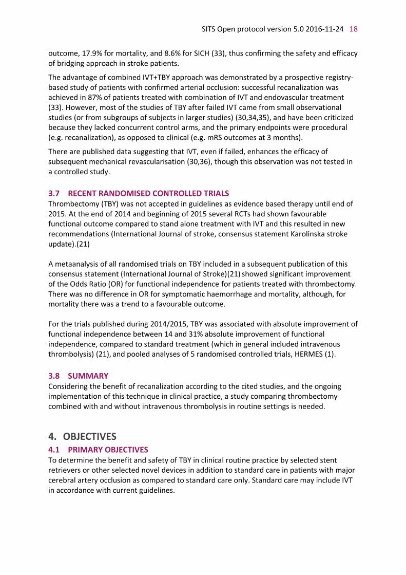
3.7 RECENT RANDOMISED CONTROLLED TRIALS Thrombectomy (TBY) was not accepted in guidelines as evidence based therapy until end of 2015. At the end of 2014 and beginning of 2015 several RCTs had shown favourable functional outcome compared to stand alone treatment with IVT and this resulted in new recommendations (International Journal of stroke, consensus statement Karolinska stroke update).(21)
A metaanalysis of all randomised trials on TBY included in a subsequent publication of this consensus statement (International Journal of Stroke)(21) showed significant improvement of the Odds Ratio (OR) for functional independence for patients treated with thrombectomy. There was no difference in OR for symptomatic haemorrhage and mortality, although, for mortality there was a trend to a favourable outcome.
For the trials published during 2014/2015, TBY was associated with absolute improvement of functional independence between 14 and 31% absolute improvement of functional independence, compared to standard treatment (which in general included intravenous thrombolysis) (21), and pooled analyses of 5 randomised controlled trials, HERMES (1).
3.8 SUMMARY Considering the benefit of recanalization according to the cited studies, and the ongoing implementation of this technique in clinical practice, a study comparing thrombectomy combined with and without intravenous thrombolysis in routine settings is needed.
4. OBJECTIVES 4.1 PRIMARY OBJECTIVES To determine the benefit and safety of TBY in clinical routine practice by selected stent retrievers or other selected novel devices in addition to standard care in patients with major cerebral artery occlusion as compared to standard care only. Standard care may include IVT in accordance with current guidelines.

19 SITS Open protocol version 5.0 2016-11-24
4.2 SECONDARY OBJECTIVES
5. ENDPOINTS 5.1 PRIMARY ENDPOINT OF EFFICACY Categorical shift in modified Rankin Scale (mRS) score at 3 months after stroke onset.
5.2 SECONDARY ENDPOINTS OF EFFICACY • Proportion of patients with functional independence (modified Rankin Scale, mRS,
score 0-2) at 3 months after stroke onset.
• Proportion of patients with excellent outcome (mRS score 0-1) at 3 months.
• Recanalization of the occluded artery for TBY treated populations, defined as at least TICI 2b flow in the treated territory after procedure.
• Time from stroke onset to revascularisation to any TICI grade (defined as 2b or 3) for the TBY treated population.
• Recanalization (defined as AOL 2-3) of the occluded artery confirmed by 24h CTA/contrast-enhanced MRA.
• Neurological improvement (difference in NIHSS from baseline to 12h, to 24h and to 7d post- IVT or discharge home/secondary care if earlier), and functional outcome at 3 months in relation to recanalization status and thrombus length (mm).
• Reduction in infarct size (TBY vs. control groups at 22-36 hours).
• Length of in-hospital stay (days to discharge from in-hospital ward to home/secondary care for survivors) in TBY groups vs. control groups
• To determine the benefit and safety of TBY by selected stent retrievers or other selected novel devices as additional therapy in proximal cerebral artery occlusion (Carotid T, M1, Basilar Artery) in patients receiving IVT according to current guidelines within 4.5 hours of ischaemic stroke onset as compared to stand-alone IVT.
• To determine whether TBY without prior IVT impacts the functional outcome of patients compared to standard stroke care including IVT, when indicated according to current guidelines.
• To determine the benefit and safety of TBY in clinical routine practice by selected stent retrievers or other selected novel devices in patients with major cerebral artery occlusion as compared to active arm of the pooled analyses of 5 randomized controlled trials, HERMES (1).
• To determine the study outcomes for patients in following subgroups: 1) In patients with M1/Car-T/BA occlusion, 2) M2/A1/P1 occlusion, 3) basilar artery occlusion, 4) in patients without prior treatment with IVT, 5) length of the occluding thrombus 8 mm, 6) moderately severe stroke at baseline (NIHSS 7-12) and for severe stroke (NIHSS 13- ).
SITS Open protocol version 5.0 2016-11-24 20
• Home Time: Number of days the patient stayed at home or at relative´s stay within the first 3 months after stroke onset, in TBY vs. Control groups.
• Recurrent stroke during within 3 month.
• Proportion of patients with recanalization (defined as AOL 2-3) before thrombectomy.
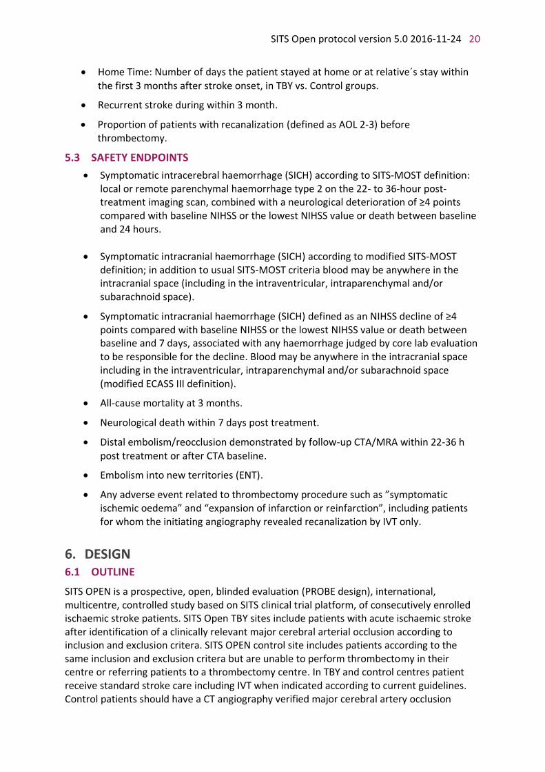
5.3 SAFETY ENDPOINTS
6. DESIGN 6.1 OUTLINE
SITS OPEN is a prospective, open, blinded evaluation (PROBE design), international, multicentre, controlled study based on SITS clinical trial platform, of consecutively enrolled ischaemic stroke patients. SITS Open TBY sites include patients with acute ischaemic stroke after identification of a clinically relevant major cerebral arterial occlusion according to inclusion and exclusion critera. SITS OPEN control site includes patients according to the same inclusion and exclusion critera but are unable to perform thrombectomy in their centre or referring patients to a thrombectomy centre. In TBY and control centres patient receive standard stroke care including IVT when indicated according to current guidelines. Control patients should have a CT angiography verified major cerebral artery occlusion
• Symptomatic intracerebral haemorrhage (SICH) according to SITS-MOST definition: local or remote parenchymal haemorrhage type 2 on the 22- to 36-hour post-treatment imaging scan, combined with a neurological deterioration of ≥4 points compared with baseline NIHSS or the lowest NIHSS value or death between baseline and 24 hours.
• Symptomatic intracranial haemorrhage (SICH) according to modified SITS-MOST definition; in addition to usual SITS-MOST criteria blood may be anywhere in the intracranial space (including in the intraventricular, intraparenchymal and/or subarachnoid space).
• Symptomatic intracranial haemorrhage (SICH) defined as an NIHSS decline of ≥4 points compared with baseline NIHSS or the lowest NIHSS value or death between baseline and 7 days, associated with any haemorrhage judged by core lab evaluation to be responsible for the decline. Blood may be anywhere in the intracranial space including in the intraventricular, intraparenchymal and/or subarachnoid space (modified ECASS III definition).
• All-cause mortality at 3 months.
• Neurological death within 7 days post treatment.
• Distal embolism/reocclusion demonstrated by follow-up CTA/MRA within 22-36 h post treatment or after CTA baseline.
• Embolism into new territories (ENT).
• Any adverse event related to thrombectomy procedure such as ”symptomatic ischemic oedema” and “expansion of infarction or reinfarction”, including patients for whom the initiating angiography revealed recanalization by IVT only.
21 SITS Open protocol version 5.0 2016-11-24
indicating thrombectomy. Major cerebral artery occlusion is defined if CT angiography confirmed occlusion which can be reached by the endovascular device approved for the study, including the proximal part of the Middle Cerebral Artery (MCA, M1), proximal part of the insular segment of MCA (M2), proximal part of the anterior cerebral artery (A1), terminal Carotid Artery (Car-T), or Basilar Artery (BA) and proximal part of the posterior cerebral artery (P1).
Both TBY and control sites will be selected from a list of highly qualified medical centres, with comparable experience in acute stroke care and results in terms of stroke outcomes. A TBY site is defined as a centre which performs TBY, a control site is defined as a centre offering only non-endovascular treatment for ischaemic stroke.
The patients with matching baseline characteristics and imaging are compared for the primary endpoint, which is categorical shift in modified Rankin Scale (mRS) score at day 90. The proportion of patients with functional independence (modified Rankin Scale, mRS, score 0-2), and excellent recovery (mRS score 0-1) at 3 months after stroke onset are secondary endpoints.
Key safety variables are proportion of patients with symptomatic intracranial haemorrhage (SICH according to SITS-MOST definition and modified SITS-MOST definition), and mortality at three months.
6.2 MEASURES TO MINIMIZE BIAS Measures to minimise bias include selection of comparable TBY and control centres (see below), and blinded and independent evaluation of functional outcome on the mRS at three months as well as of imaging studies by a core lab.
For the purpose of including potential centre effects on treatment outcomes in final matching, all TBY and control centres are encouraged to keep up their register of ischaemic stroke patients (outside the study cohort) in the SITS-ISTR during the study period.
Considerations of minimising bias in data collection and analysis are given in the statistical chapter (see Chapter 11).
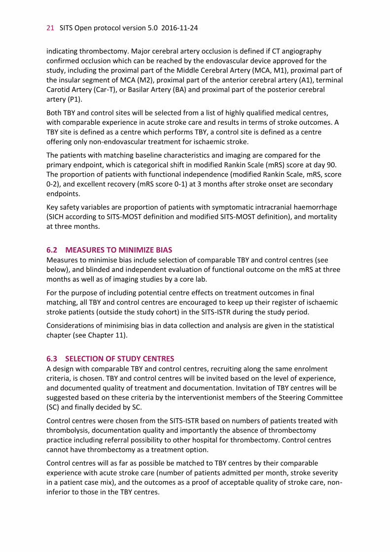
6.3 SELECTION OF STUDY CENTRES A design with comparable TBY and control centres, recruiting along the same enrolment criteria, is chosen. TBY and control centres will be invited based on the level of experience, and documented quality of treatment and documentation. Invitation of TBY centres will be suggested based on these criteria by the interventionist members of the Steering Committee (SC) and finally decided by SC.
Control centres were chosen from the SITS-ISTR based on numbers of patients treated with thrombolysis, documentation quality and importantly the absence of thrombectomy practice including referral possibility to other hospital for thrombectomy. Control centres cannot have thrombectomy as a treatment option.
Control centres will as far as possible be matched to TBY centres by their comparable experience with acute stroke care (number of patients admitted per month, stroke severity in a patient case mix), and the outcomes as a proof of acceptable quality of stroke care, non-inferior to those in the TBY centres.

SITS Open protocol version 5.0 2016-11-24 22
6.4 ASSESSMENTS AND PROCEDURES
6.4.1 Clinical and Laboratory Procedures All tests required by this protocol are considered within the range of standard clinical care pre- and post- intervention (some radiological procedures are considered study specific at some centres, see chapter 6.4.9). Appendix 2 (21.2) provides a detailed schedule of events.
6.4.2 Screening & Consent Procedures All eligible patients will be screened to determine their suitability for entry into the study. Patients who meet all the inclusion criteria and none of the exclusion criteria, including identification of a major cerebral artery occlusion on CTA, will be registered for preliminary enrolment in the study to a 24 h automatic telephone service.
Since TBY and control centres use their treatments in clinical practice, consent for the treatment is granted by clinical routine procedures, as will all routine treatments. The informed consent will be collected for participation in the study and for documentation of the data after the treatments have been initiated to avoid delays in clinical management. This should be done as soon as practically possible, and always within 20 h (7 days approved in applicable countries) after baseline.
In case a patient is unable to personally provide consent, e.g. because of aphasia, a routine according to local guidelines needs to be implemented at centres. e.g. the informed consent may be signed by a legal representative of the patient.
In case a patient is capable of providing informed consent, but unable to personally sign the consent form, a routine according to local guidelines has to be established, e.g. a witness, independent from the study, may certify the consent in writing.
If a patient has passed away, and no family member is available, the principle investigator may sign the consent form, provided that there are no indications that the patient would have resisted inclusion in the study. Alternatively, a similar measure as approved by the IRB would be applied according to local regulations. This procedure is of importance to avoid that patients with the worst outcomes of the procedure are systematically excluded from the study.
Every participating hospital will get access to a telephone number to notify that a patient meets the criteria for inclusion. This call must be done before initiation of thrombectomy (TBY centres) and within two hours of initiation of IVT (control centres) or after plain baseline CT (control patients not receiving IVT). Information will include a patient screening number; no patients’ ID will be documented in the call. The recorded information will be sent to the coordinating team and regarded as a part of the screening log. The centre will later be asked to provide a motivation if patients recorded on the screening log were not enrolled in the study. This strategy is chosen to avoid delays in the clinical management and to prevent criticism of bias due to selective enrolment of patients with positive outcome. The screening log thus vouches for a prospective enrolment even though informed consent might be received first after the procedure is completed.
After appropriate informed consent from the patient, or an acceptable representative, in agreement with local law, the patient will be registered in SITS Open. Upon admission, the patient will undergo a plain CT preceding the decision to initiate IVT, and a CTA will be

23 SITS Open protocol version 5.0 2016-11-24
performed to confirm the occlusion of the relevant artery. The CTA should not be done later than 15 minutes after the IVT initiation. For patients not treated with IVT, CTA be preferably performed within 15 minutes of completion of the non-contrast CT but must be performed within 6 hours after stroke onset.
At 22-36 hours after the IVT inititation or after plain CT scan (if IVT is not given), a follow up CT and CTA will be performed to identify any haemorrhage following the procedures, infarct extension and recanalization status. If additional irradiation is a concern, MRI and contrast-enhanced MRA may be done at follow-up instead of CT/CTA.
Stroke patients receiving IVT or TBY outside of the study should as completely as possible be registered in SITS-ISTR.
6.4.3 Balance of recruitment and devices in the study In order to avoid a skewed study in respect to centre capacity the number of subjects recruited by one site is limited to 40 per TBY hospital and 40 per control hospital. To reduce the risk of prevalence of one study device over the others, balanced selection of study sites according to their experience with devices is provided, and tools for steering the enrolment have been implemented.
6.4.4 Medical Management IVT is defined as the guidelines adapted from the European Stroke Organisation (ESO), SITS-MOST and American Stroke Association/American Heart Association (Attachment 1). Blood pressure (BP) should be tightly controlled during the first 24 h after the IVT initiation or after baseline plain CT scan, if IVT is not given, to less than 180/105. Intravenous labetalol is a treatment of choice if prompt BP reduction is required. Other drug therapy will be at the discretion of the investigator in accordance with IVT guidelines and national standards.
6.4.5 Thrombectomy (TBY arm only) The time of accessing the thrombus with a microcatheter, time from stroke onset of achieving recanalization TICI grades 2a, 2b, and 3 (if applicable) and the time of the final angiogram will be recorded. Only patients allocated to TBY will undergo invasive angiographic evaluation. Any of the TBY devices identified for the study must be used. The selection of the first study device (from those defined for the study) is at the discretion of the interventionist, if the interventionist decides to change to another device, it should preferably be any of the other study devices. Other (non-study) devices should be avoided as far as possible as rescue therapy after incomplete effect of study devices. It is in the interest of the study that the three study devices are reasonably equally distributed, although difference in practice may occur between centres.
If for any reason, the thrombectomy cannot be performed, or failed to open the vessel, the patient may be treated with additional intra-arterial thrombolytics, if clinically appropriate. The dose of intra-arterial rt-PA should be recorded in eCRF. These patients will be enrolled and included in intention-to-treat analysis.
If required, angioplasty and stenting of proximal artery stenosis is accepted in relation to the thrombectomy procedure. A guideline for a uniform policy of antiplatelet medications following stenting is available in Attachment 2.

SITS Open protocol version 5.0 2016-11-24 24
6.4.6 Arterial Access Arterial access should be obtained per standard practices at the treating institution.
6.4.7 Angiography Contrast is injected to determine the angiographic characteristics of the vessel/occlusion treated. This must be confirmed prior to treatment. The location of occlusion will be recorded. It is important that the CTA is done not later than 15 minutes after IVT start or within 15 min after baseline plain CT scan (must be performed within 6 hours after stroke onset) if IVT is not given for the patient to be included in the study.
An additional analysis is to estimate the size limit of a thrombus, which can be dissolved by IVT. The core lab records the dimensions of the thrombi based on NCCT imaging. Imaging should be done with a < 2,5 mm slice thickness at baseline and on follow-up (to allow measurement of the clot size by imaging core lab).
If thrombus is identified in an appropriate intracranial artery such as the M1 segment of the middle cerebral artery, or the terminal internal carotid, or basilar, artery, no further pre-treatment injections are required, and thrombectomy may be initiated.
For patients included in the study and subjected to thrombectomy, and found by angiography to have been recanalised before initiation of the endovascular procedure, informed consent should be required as planned and all follow up measures should be observed, including 3-month follow-up. These patients will be included in the intention-to-treat analysis.
Angiographic images of the occlusion being treated must allow clear visualisation of the target artery. Frontal and lateral projections will be saved if possible before thrombectomy and after the last thrombectomy attempt, preferably after the each thrombectomy attempt. The final run must include the whole head. The time point should be available on the angiograms. The same orientation should be used before and after treatment to allow a valid analysis of the data. (For detailed instructions for imaging see 21.8)
6.4.8 Thrombectomy a) Physicians should follow the relevant instructions for use for specific devices being
utilised in the study. For thrombi in the anterior circulation, it is mandatory to use either balloon guide catheter or a distal access catheter (DAC). Time records are made at the beginning of the procedure and on achieving recanalization TICI grades 2a, 2b, and 3 (if achieved), and with the final angiogram. The number of thrombectomy attempts should be limited to 6.
b) Procedural Heparin: A heparin flush solution may be administered in the guide catheter and in the access sheath at the discretion of the physician.
c) IA thrombolytics: IA thrombolytics (IAT) are permitted at the investigator’s discretion when required to achieve satisfactory recanalization. The need of IAT should be weighed against an increased risk of haemorrhage. For a patient treated with full dose IV rtPA (0.9 mg/kg), an additional dose of 0.2 mg/kg, corresponding to 14 mg in a 70 kg person, should not be exceeded.
d) Anti-vasospasm agents: Prudent use of anti-vasospasm agents is permitted. e) Proximal stenoses: In order to facilitate access to the intracranial occlusion, investigators
will sometimes need to traverse a proximal stenosis. Investigators may use their
25 SITS Open protocol version 5.0 2016-11-24
judgement to best address the proximal stenosis with (1) no treatment, (2) balloon angioplasty, or (3) stenting. The primary goal is to rapidly restore intracranial flow; so proximal stenoses sometimes may be best addressed at the end of the case. Care should be taken when stenting because patients must then receive antiaggregant treatment (e.g., clopidogrel) whose safety is not well established in the setting of acute stroke, in particular when undergoing IVT. Instructions for initiation of antiaggregant treatment in the study are attached (Attachment 2).
Termination of the intra-arterial treatment procedure will occur if:
a) Angiographic findings suggest vessel damage or extravasation of contrast.
b) Neurological deterioration or alteration in function is detected leading to the suspicion of an intracranial haemorrhage.
Final angiography will include ipsilateral anterio-posterior and lateral injections in the involved arterial system.
Aside from procedurally administered heparin, IV heparin is prohibited until after the 24-hour neuro-imaging has been performed to minimize the risk of intracranial haemorrhage. Blood pressure will be tightly controlled during the first 24 h to less than 180/105. Other drug therapy will be at the discretion of the investigator in accordance with IVT guidelines.
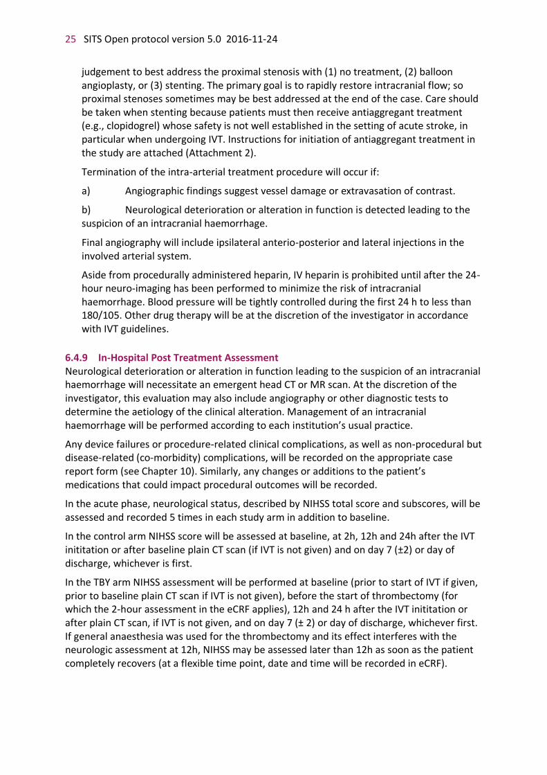
6.4.9 In-Hospital Post Treatment Assessment Neurological deterioration or alteration in function leading to the suspicion of an intracranial haemorrhage will necessitate an emergent head CT or MR scan. At the discretion of the investigator, this evaluation may also include angiography or other diagnostic tests to determine the aetiology of the clinical alteration. Management of an intracranial haemorrhage will be performed according to each institution’s usual practice.
Any device failures or procedure-related clinical complications, as well as non-procedural but disease-related (co-morbidity) complications, will be recorded on the appropriate case report form (see Chapter 10). Similarly, any changes or additions to the patient’s medications that could impact procedural outcomes will be recorded.
In the acute phase, neurological status, described by NIHSS total score and subscores, will be assessed and recorded 5 times in each study arm in addition to baseline.
In the control arm NIHSS score will be assessed at baseline, at 2h, 12h and 24h after the IVT inititation or after baseline plain CT scan (if IVT is not given) and on day 7 (±2) or day of discharge, whichever is first.
In the TBY arm NIHSS assessment will be performed at baseline (prior to start of IVT if given, prior to baseline plain CT scan if IVT is not given), before the start of thrombectomy (for which the 2-hour assessment in the eCRF applies), 12h and 24 h after the IVT inititation or after plain CT scan, if IVT is not given, and on day 7 (± 2) or day of discharge, whichever first. If general anaesthesia was used for the thrombectomy and its effect interferes with the neurologic assessment at 12h, NIHSS may be assessed later than 12h as soon as the patient completely recovers (at a flexible time point, date and time will be recorded in eCRF).

SITS Open protocol version 5.0 2016-11-24 26
IVT = Intravenous thrombolysis; TBY = Thrombectomy
*If general anaesthesia is used, this time point should be postponed until complete recovery from the anaesthesia **The day of discharge if the patient is discharged before day 5. Blood pressure (systolic and diastolic) should be measured and recorded at the same time with the NIHSS assessments.
22-36 hours after the IVT initiation (or after baseline plain CT scan, if IVT is not given) the NIHSS and CTA/contrast-enhanced MRA shall be obtained in both treatment arms to assess patency of the neurovasculature. Also at this time, a separate CT or MR will be obtained to assess haemorrhage, infarct and oedema. Any events related to the study arm, investigational device or procedure should be documented.
If CTA and MRA with contrast enhancement are not possible or contraindicated in individual patients, these follow-up studies in the absence of other solutions may be replaced by transcranial Doppler studies, though this is the least preferable option.
At 7 days (± 2) after the IVT initiation (or after baseline plain CT scan, if IVT is not given), or prior to hospital discharge, whichever is earlier, the patient’s neurological status will be assessed and recorded using the NIHSS, and mRS. Local investigators will enter their evaluation using the mRS (see 21.4-21.5 and Attachment 3). Any events related to the study arm, investigational device or procedure should be documented.
6.4.10 Rehabilitation measures Rehabilitation should be provided in accordance with the attached guidelines for management of acute ischemic stroke (based on ESO recommendations 39, see Attachment 4). It is not practically feasible or realistic to expect every subject in the study to receive identical care, but every subject at a given institution should have documented consistent care and rehabilitation options.
6.4.11 Outcome assessment at 3 Months (90 ± 14 days) Patient neurological and medical status will be assessed and recorded using the mRS (see 21.5). Follow-up mRS assessment is the last study procedure and is done at the last visit on day 90 ± 14 after baseline.
The investigator interviews the patient at the last study visit either in the hospital or, if patient is unable to visit the hospital, at the patient’s home. The interview is video recorded (see 21.4 and Attachment 3), and the investigator will be instructed to avoid any indication of the treatment and/or procedure that the patient has received. The result of assessment by the investigator will be entered into eCRF. Video records will be blinded for any personal

27 SITS Open protocol version 5.0 2016-11-24
information and treatment details and uploaded for further assessment by the members of the adjudication committee, who are unaware of the treatment the patient has received. Video files will be translated into English before the expert assessment.
The independent outcome assessment will be performed by the Outcomes Coordinating Centre at the Western Infirmary in Glasgow, UK. Upon upload of a mRS assessment, the relevant Outcomes Manager will be notified by an automated email. The manager or a deputy fluent in the relevant language will then review the assessment (via the web portal and within 3 working days after submission) and ensure that no indication as to treatment assignment is contained in the assessment; if there is such indication, the Outcomes Manager will remove this from the clip. The Outcomes Manager will also verify that the assessor is currently certified in mRS assessment. If the assessor is not trained, the assessment may need to be repeated by a trained observer. The Outcomes Manager will also perform an initial evaluation of adequacy of the clip. Major concerns with technical or clinical adequacy of the clip will thus be identified expeditiously. Assuming a valid assessment, the Outcomes Manager will then release this to 4 members of the endpoint assessment committee for review. If any editing has occurred to ensure blinded assessment, the original clip will be maintained and the nature of this editing recorded in the SITS Open trial website; however, no part of a clip that identifies the patient by name will be stored. The edited clip will be the one used by the endpoint assessment committee. The endpoint committee members chosen will be selected from a pool according to availability and language capability and will be notified by email. The aim is to review the assessment and assign a mRS score within 5 working days of receipt.
The chosen members will review the mRS assessment, confirm adequacy of quality for review and assign a mRS score which will be done independently to the local investigator’s score and to scores of colleagues, blinded to all other patient information. The assessment will again be viewed via the web portal and the score entered into the portal. An automated process will establish whether the various central readings agree with each other and with the local score and if so, the patient will be assigned to the common mRS category. If there is disagreement, the patient will be coded as “misclassified” and the video clip may be submitted for further review by the adjudication oversight committee at one of its scheduled meetings. Note that the website will indicate agreement or disagreement to the Outcomes Manager but will not reveal the original investigator’s score in cases of disagreement. Once the Outcomes Manager is notified of a disagreement, he or she will notify the committee by email and coordinate the review of clips at scheduled meetings.
After group review, the oversight adjudication committee will assign the patient to one of the following groups: technically inadequate assessment, unable to assign score; inadequate assessment, unable to assign score; or adequate assessment with mRS score assigned. Where committee classification is possible, the patient will be assigned to that Rankin category. The submitting centre may be asked for further information (e.g. to put a specific additional question to a patient) or to repeat the assessment if deemed necessary. Further questions can usually be answered by email within a few days. Most controversial scores are due to the weakness of the scoring definitions coupled with variation in clinical circumstances. A consensus score can generally be achieved.
At the final visit, the investigator will ask how many days of the first 90 days after stroke the patient spent at home or in relative’s care. Days spent at hospital, secondary care, etc., including repeated admissions, should be subtracted from the 90 days period.

SITS Open protocol version 5.0 2016-11-24 28
6.5 PATIENT RECRUITMENT AND WITHDRAWAL
6.5.1 Inclusion criteria
• Patients with acute stroke after exclusion of intracranial haemorrhage on CT/MRI scan.
• Confirmed diagnosis on CTA of persistent occlusion of the terminal Internal Carotid Artery (Car-T), proximal Middle Cerebral Artery (MCA, M1), proximal part of the insular segment of MCA (M2), proximal part of the anterior cerebral artery (A1), Basilar Artery (BA) or proximal part of the posterior cerebral artery (P1), consistent with the clinical symptoms. For inclusion in the study, CTA must not be performed later than 15 minutes after IVT start if given. For patients not treated with IVT, CTA be preferably performed within 15 minutes of completion of the non-contrast CT but must be performed within 6 hours after stroke onset.
• Eligible patient for IVT are treated according to clinical guidelines (Attachment 1), and IVT initiated within 4.5 h.
• Initiation of thrombectomy is recommended within 6 hours after stroke onset but may be extended to 8 hours if thrombectomy would still be of benefit for the patient as judged by the investigator.
• Baseline NIHSS score at initiation of IVT is recommended between 7 and 25 for anterior circulation stroke and ≥7 without upper limit for posterior circulation stroke (baseline NIHSS score should be assessed by an NIHSS-certified physician), but patients may also be included beyond these scores if thrombectomy would still be of benefit for the patient as judged by the investigator.
• Age ≥18years.
• Anticipated life expectancy of at least 6 months.
• Patient or legal representative is competent to make a decision and has provided informed consent with regard to participation in the study, retrieval and storage of data and follow up procedures.
• Initiation of endovascular procedure (DSA/TBY, defined as start with groin puncture) within 2 hours from the start of IVT, or after CTA if IVT is not given (for TBY arm patients).
6.5.2 Exclusion criteria
• Known significant pre-stroke disability (mRS ≥2).
• Extended early ischemic changes for basilar artery occlusion, according to the judgment of treating physician based on routine clinical practice of the hospital; if technical possibility exists, early irreversible ischemic changes may be confirmed by pc-ASPECTS score < 8 on CTASI (2) or extensive DWI lesion on pre-treatment MRI.
• Known pregnancy.
• Participation in any other investigational drug or device study, currently or in the previous 30 days.
6.5.3 Withdrawal and dropout Patients are enrolled in the study after initiation of intravenous thrombolysis treatment. The patient would be withdrawn from thrombectomy if recanalization has occurred during IVT,
29 SITS Open protocol version 5.0 2016-11-24
or if later found to be unsuitable for this treatment, even if patient is meeting all enrolment criteria, e.g., because of tortuous proximal arteries. These patients will be enrolled and included in the final statistical analysis. Rapid improvement after IVT does not make a patient unsuitable for thrombectomy, if angiography identifies persistent occlusion.
If patients are withdrawn from thrombectomy treatment (but not from the study), all other study procedures, including 3 months’ follow-up, will continue. A decision to withdraw a patient from treatment is made by the available study physician or, if necessary, by another doctor for the time being clinically responsible for the patient’s care. A patient can also be withdrawn from treatment on request from the investigator as indicated above, or by the patient. These latter patients are included in intention-to-treat analyses.
A patient will be dropped out from the study if requested by the patient. All study specific procedures and analyses will cease. Drop out does not exclude that patient remains in the SITS thrombectomy registry (SITS-TBY), unless this is expressed by the patient.
The study Coordinating Investigator must be informed if a patient requests to be withdrawn from further follow up or from the study as a whole, reasons (if provided by the patient, although this is not required) and confirm withdrawal. If a patient requires withdrawal from further follow up, patient may be asked if three months’ outcome data may be collected from a family member or a caregiver. If this accepted, a video interview is preferred.
6.5.4 Screening log and SITS registry documentation Patients will be entered into the SITS Open eCRF (which has SITS Registry as platform) if eligible and confirmed by the Investigator with regard to informed consent. These patient files will be labelled as SITS Open patients but the data will also be available for the registries. Data can be entered and changed as usual in the registries, and the investigators ought to supply imaging information and follow up mRS. Notably, core lab evaluations and independent mRS will not be available through the eCRF, since they are collected and evaluated entirely separated from the registry structure.
Patients not eligible for the study will be entered as usual to the applicable SITS Registry and no Informed consent is needed.
After follow up of all patients and declaration of clean file, the data from the registry will be downloaded to a separate file which cannot be changed except for entry of the independently generated mRS scores and the core lab evaluations of angiography and CT results. This study file will be subjected to analysis of the study statisticians.
Following the final study report and publication of the SITS Open study, the local Investigators are free to change the outcome data in the registries in accordance with the independent evaluations. Publications or reports at scientific meetings of results based on patient data from SITS Open patients are prohibited before publication of SITS Open results.
Patient data will be verified by the investigator. A “PHYSICIAN’S MEDICAL VERIFICATION FORM” will be completed by principle or sub investigator and signed off for each patient included in the study. The “PHYSICIAN’S MEDICAL VERIFICATION FORM” will be send to Sits coordination team. This verification will confirm that medical data of the patient and inclusion into SITS Open, including “enrolment ID” is in the hospital patient file.

SITS Open protocol version 5.0 2016-11-24 30
7. DEVICE
7.1 TRAINING
Minimal training requirements for acute stroke interventions:
• 3 or more years of prior training and experience in neuro-interventional recanalization therapy
• minimum of 100 endovascular stroke interventional procedures (i.a. thrombolysis, neuro-thrombectomy, intra- and extracranial stenting and percutaneous transluminal angioplasty), from this
• minimum of 30 intracranial procedures
• minimum of 30 extracranial procedures
Previously credentialed physicians who perform intra-arterial catheter-directed stroke procedures at their local institutions should have documented procedural and clinical outcomes that meet national standards and published evidence-based guidelines. The Steering Committee may decide to exclude a centre if not all training requirements are completed.
It is also recommended that the physician who performs mechanical thrombectomy should have successfully completed a training course for use of any specific device.
In individual cases the manufacturer may provide additional training with a specific study device.
7.2 DESCRIPTION OF THE MEDICAL ENDOVASCULAR DEVICES Three devices will be included in the trial, PreSET, Solitaire and TREVO. The investigator decides in individual cases which device to use, although the study encourages use of all three devices so that the use of them in the study will be proportional (estimated proportion of the use of the device is between 25% and 40%).
7.2.1 PreSET The pREset® (LT) Thrombectomy Stent (37) is designed for mechanical clot retrieval from intracranial arteries as acute ischemic stroke treatment.
The device is available in different designs:
“pREset 4-20” and “pREset 6-30” are used for large thrombus load in carotid “T” and proximal MCA occlusions. Whereas the “pREset LT 4-20” and “pREset LT 3-20” are used for the treatment of distal MCA occlusions.
For the treatment of rather small vessel diameters (minimum: 1.5 mm), the pREset LT versions were designed. Respectively, the pREset 4-20 and 6-30 are used for minimum vessel diameters of 2 mm and 3 mm.
The pREset (LT) device is indicated for the treatment of patients who are ineligible for Intravenous thrombolysis or for the treatment of patients who failed thrombolysis therapy and it is also indicated as a supplement treatment of initiated thrombolysis therapy.

31 SITS Open protocol version 5.0 2016-11-24
The unique proximal “ring” design ensures stable opening and reduced tapering when withdrawn. The helical slit maintains cell shape integrity and enables optimized radial force distribution.
pREset 4-20 has a CE mark approval since 2011, pREset 6-30 has a CE mark approval since 2012 and both pREset LT-sizes were CE mark approved in 2013. Overview regarding the product range and the respective sizes, including compatibility of microcatheters:
The pRESET (LT) consists of a self-expanding Nitinol structure, carries one x-ray visible marker on its proximal ‚ and two on its distal end and is firmly attached to the body of an insertion wire. The device is stored in a compressed condition in an introducer sheath (not displayed).
The pRESET (LT) is introduced into the target vessel through a suitable microcatheter and deployed inside the thrombus or distal to the thrombus.
SITS Open protocol version 5.0 2016-11-24 32
After complete deployment the slow withdrawal of the instrument occurs under continuous aspiration via the guiding catheter or aspiration catheter. (37)
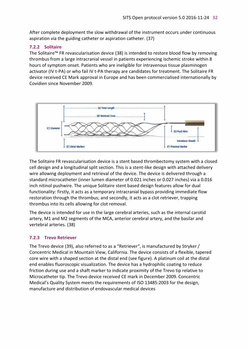
7.2.2 Solitaire The Solitaire™ FR revascularisation device (38) is intended to restore blood flow by removing thrombus from a large intracranial vessel in patients experiencing ischemic stroke within 8 hours of symptom onset. Patients who are ineligible for intravenous tissue plasminogen activator (IV t-PA) or who fail IV t-PA therapy are candidates for treatment. The Solitaire FR device received CE Mark approval in Europe and has been commercialised internationally by Covidien since November 2009.
The Solitaire FR revascularisation device is a stent based thrombectomy system with a closed cell design and a longitudinal split section. This is a stent-like design with attached delivery wire allowing deployment and retrieval of the device. The device is delivered through a standard microcatheter (inner lumen diameter of 0.021 inches or 0.027 inches) via a 0.016 inch nitinol pushwire. The unique Solitaire stent based design features allow for dual functionality: firstly, it acts as a temporary intracranial bypass providing immediate flow restoration through the thrombus; and secondly, it acts as a clot retriever, trapping thrombus into its cells allowing for clot removal.
The device is intended for use in the large cerebral arteries, such as the internal carotid artery, M1 and M2 segments of the MCA, anterior cerebral artery, and the basilar and vertebral arteries. (38)
7.2.3 Trevo Retriever
The Trevo device (39), also referred to as a “Retriever“, is manufactured by Stryker / Concentric Medical in Mountain View, California. The device consists of a flexible, tapered core wire with a shaped section at the distal end (see figure). A platinum coil at the distal end enables fluoroscopic visualization. The device has a hydrophilic coating to reduce friction during use and a shaft marker to indicate proximity of the Trevo tip relative to Microcatheter tip. The Trevo device received CE mark in December 2009. Concentric Medical’s Quality System meets the requirements of ISO 13485:2003 for the design, manufacture and distribution of endovascular medical devices

33 SITS Open protocol version 5.0 2016-11-24
Trevo device engineering drawing
The indication for the Trevo device is to restore blood flow in the neurovasculature by removing thrombus in patients experiencing ischemic stroke. Patients who are ineligible for treatment with intravenous tissue plasminogen activator (IV t-PA) or who fail IV t-PA therapy are candidates for treatment.
The Trevo device is delivered through a Microcatheter and the shaped section is unsheathed via pulling back on the Microcatheter, leaving the device directly in the thrombus and allowing it to immediately begin expanding and incorporating into the thrombus. This thereby anchors the device to the thrombus and facilitates retrieval of the thrombus into the guide catheter.
The Trevo device has been designed and tested to perform multiple retrieval attempts in a single patient. After each deployment of the device it should be thoroughly inspected before reloading. Refer to the Instructions for Use (IFU) for detailed instructions on how to reload the device, and the maximum number of retrieval attempts per device.
There are no specific contraindications for the use of the Trevo device apart from the inclusion and exclusion criteria of this study protocol. (39)
Item A B C D E F
Overall Length
Total Shaped Section Length
Active Shaped Section Length
Distal Platinum Coil Length
Shaft/Coil Diameter [in]
Unconstrained Shaped Section Diameter
Trevo™ 4 x 20 mm
180 cm 40 mm 20 mm 4.0 mm 0.018 max 4.0 mm

SITS Open protocol version 5.0 2016-11-24 34
8. ASSESSEMENT OF EFFICACY AND SAFETY
8.1 INDEPENDENT CT/MR ADJUDICATION
All baseline and 24 hour CT scans (target interval 22-36 h) will be read and scored by an independent core lab reviewer. The 24 hours’ scans will be evaluated for haemorrhage. All scans positive for 24-hour haemorrhage will be categorised whether they meet criteria for SICH according to the SITS-MOST, modified SITS-MOST and modified ECASS III definitions. In case of haemorrhage on 22—36 h CT scan, centres are advised to perform an additional CT the following day to differentiate penetration of contrast dye after Angiography/thrombectomy from true haemorrhage. This examination will be included in the material sent to core lab. Alternatively, newer so-called double-energy technique can be applied to perform this differentiation directly on the 22-36 h images. See 21.8 for instructions.
8.2 DATA AND SAFETY MONITORING BOARD (DSMB) An independent board consisting of stroke neurologists and interventional neuroradiologists who are not participating in the study and are not affiliated with the sponsor or have any conflicts of interest with the supportive companies, will be responsible for monitoring the data during the trial and making recommendations regarding safety and about extending the trial. The role of the DSMB will be to:
• Make recommendations to the Steering Committee regarding safety of the study.
• Make recommendations to the Steering Committee on extending the trial.
8.3 REQUIREMENTS OF CLINICAL MEASUREMENTS • NIHSS – Only doctors or other staff certified in the collection of NIHSS may perform
the assessments required in the study. Please note that certification is also requested when a first evaluation of NIHSS is done at a primary centre before initiating IVT and sending a patient to an active thrombectomy centre.
• Post-procedure TICI will be scored by the investigator or the sub-investigator. Additionally, all angiograms and baseline and post-procedure CTs and/or MRs will be independently evaluated by the core lab.
• Cause of death will be recorded for all patients that expire during the study.
8.4 MODIFIED RANKIN SCALE AT 3 MONTHS The local investigator or sub-investigators will perform a structured interview for determining the mRS score (see 21.4, 21.5 and Attachment 3) and document this in the eCRF. The interview will be recorded on video for additional independent evaluation. The investigator will be instructed to avoid any indication in the interview of the treatment and/or procedure that the patient has received. Video records will be blinded for any personal information and treatment details and uploaded for further evaluation. Video files will be translated into English before the external assessment.
Three adjudicators, not informed about the study details and the treatment alternatives, will independently assess the score. If adjudicators agree, this score will be saved in an

35 SITS Open protocol version 5.0 2016-11-24
independent database until all study data have been collected and the study database is locked. If adjudicators disagree the matter will be solved at a committee meeting.
8.5 CLINICAL SAFETY ASSESSMENT
8.5.1 Clinical examination Physical and neurological examination of the patient will be performed by a medical professional at baseline, initiation of treatment/procedure, 2h, 12h, 24h, 7 (± 2) days(after the IVT initiation or after plain CT scan at baseline if IVT is not given) or day of discharge home/secondary care, if earlier. The NIH-Stroke Scale (NIHSS) will be used for standardised evaluation of the neurological deficit. Certification in NIHSS use is a requirement for the rater. Post-procedure assessment in the active study arm should include evaluation of arterial puncture site for the signs of bleeding.
8.5.2 Monitoring of vital signs Vital signs taken at baseline are arterial blood pressure (systolic and diastolic), heart rate, respiratory rate, and body temperature. During and following the IVT blood pressure, heart rate and respiratory rate are monitored noninvasively every 15 minutes for the first 2h, then every 30 minutes for the following 24h. In the TBY arm, endovascular treatment is performed under the supervision of anaesthesiologist when required; method of anaesthesia, if applied, and intraoperational monitoring is chosen according to the local standard clinical practice. Type of anaesthesia and medications given should be recorded in the eCRF. Post-procedure monitoring in the TBY arm will be similar to that in the control arm.
8.5.3 Laboratory tests Laboratory Laboratory testing at baseline should comply with the standard guidelines for IVT patients; these include CBC, serum glucose, serum cholesterol, urea, creatinine, bilirubin and liver enzymes, electrolytes, coagulation tests (PT, APTT, INR). Blood glucose, cholesterol and APTT are recorded in the CRF.
8.5.4 Imaging safety assessment A local radiologist will perform primary evaluation of 22-36 h CT scan. All angiograms and baseline and post-procedure CTs and/or MRs will be provided to the core lab. CT and/or MR scans will be evaluated for any type haemorrhage (including SAH) and the degree of cerebral oedema, if present.
If detection of haemorrhage on the follow-up CT scan is ambiguous because of suspected extravasation of contrast media after mechanical thrombectomy, dual-energy imaging or another follow-up CT scan (preferably the next day after 24-hour assessment) is required.

SITS Open protocol version 5.0 2016-11-24 36
8.5.5 Classification of Intracerebral haemorrhage
8.5.6 Classification of Cerebral oedema
COED 1: Focal brain swelling up to one third of the hemisphere
COED 2: Focal brain swelling greater than one third of the hemisphere
COED 3: Brain swelling with midline shift
8.5.7 Definition of Symptomatic Intracerebral/Intracranial Haemorrhage (SICH)
SICH will be defined as follows:
SITS-MOST definition: local or remote parenchymal haemorrhage type 2 on the 22- to 36-hour post-treatment imaging scan, combined with a neurological deterioration of ≥4 points compared with baseline NIHSS or the lowest NIHSS value or death between baseline and 24 hours
Modified SITS-MOST definition: in addition to the SITS-MOST criteria listed above, extravasated blood may be anywhere in the intracranial space (including in the intraventricular, intraparenchymal and/or subarachnoid space).
ECASS III definition: any apparently extravascular blood in the brain or within the cranium that was associated with clinical deterioration, as defined by an increase of 4 points or more in the score on the NIHSS, or that led to death and that was identified by core lab evaluation as the predominant cause of the neurologic deterioration
Modified ECASS III definition: NIHSS decline of ≥4 points compared with baseline NIHSS or the lowest NIHSS value or death between baseline and 7 days associated with any
H1: Small petechiae along the margins of the infarct
H2: More confluent petechiae within the infarct area but without space-occupying effect
PH1: Blood clot(s) not exceeding 30% of the infarct area with some mild space-occupying effect
PH2: Blood clots exceeding 30% of the infarct area with significant space occupying effect
PHr1: Small or medium sized blood clots located remote from the actual infarct; a mild space occupying effect could be present
PHr2: Large confluent dense blood clots in an area remote from the actual infarct; significant space occupying effect may be present
SAH: Presence of haemorrhage in any compartment of the subarachnoid space

37 SITS Open protocol version 5.0 2016-11-24
haemorrhage judged by core lab evaluation to be responsible for the decline; blood may be anywhere in the intracranial space including in the intraventricular, intraparenchymal and/or subarachnoid space
8.5.8 Adverse Events and Reactions
Investigators and other study personnel will monitor each subject for possible adverse events and serious adverse events caused by the study procedure, i.e. adverse reactions and serious adverse reaction. Documenting and reporting of adverse reactions and serious adverse reactions is described at Chapter 10.
8.6 CLINICAL EFFICACY ASSESSMENTS
Functional outcome is evaluated on the modified Rankin Scale (mRS) on day 90 according to the Saver structured interview (please see 21.5) and the instructions in Attachment 3.
National Institutes of Health Stroke Scale (NIHSS) is used for evaluation of neurological symptoms (please see 21.3)
Post-procedure TICI score is used for TBY treated population; revascularisation of the occluded territory is defined as at least TICI 2b flow in the treated territory after procedure. (please see 21.7)
Time from stroke onset to revascularisation to TICI grades 2 and/or 3 in the TBY group.
CTA (recanalization at 24h, as measured by Modified TICI score and AOL (Arterial Occlusive Lesion) scores)
CT (reduction in infarct size at 24 h)
9. IMAGING CORE LAB All imaging data to be reviewed by Core lab should be sent in DICOM format and should have patient identifying elements removed and replaced with the patient’s study code. See Section 21.8 for instructions.
9.1 ANGIOGRAPHY
An independent core lab reviewer will review pre-procedure, during -procedure and final post-procedure angiograms. Procedure notes should have patient identifying elements removed and be sent with angiographic images.
9.2 BASELINE AND 24 H CT AND CTA EXAMINATIONS
Baseline CT examinations will be evaluated for early infarct signs (hypodensity), dense artery sign) and findings, which may contraindicate IVT. Appropriate indication for IVT will be confirmed or rejected.
Baseline CTA examinations will be evaluated for location of the qualifying artery occlusion and for any proximal artery stenosis
SITS Open protocol version 5.0 2016-11-24 38
24 (22-36 h) hour CT examinations will define any haemorrhage and score per the haemorrhage classification (8.5.5); in addition, an evaluation of infarct volume will be performed. If required and available, differentiation of blood and contrast using double-energy technique should be applied.
24 hour CTA examination will define the status of recanalization of the occluded artery and any reocclusion.
An additional CT examination (next day after 24 h follow up assessment) will be performed if necessary to differentiate between haemorrhage and contrast extravasation on 24 hour CT – as an alternative to double-energy technique at 24 h images.
In the case of renal insufficiency developing after CTA/thrombectomy procedure or in the case of impending renal failure or other clinical contraindications to repeated contrast administration, non-contrast-enhanced MRA or, as a least preference, TCD could be used instead of follow-up CTA if the patient is already included in the study. TCD should be performed by an accredited neurosonologist.
9.3 OPTIONAL PRE-PROCEDURE CT PERFUSION OR MR DWI/PWI IMAGING
Although this is not a required element, any centres that routinely perform pre-procedure CT Perfusion or MR DWI/MRP/PWI are strongly encouraged to enter these data in the data form. These imaging data should be submitted for core lab review if it has substantially influenced clinical decision (for instance, cancellation of TBY). In these cases, patient will be included in the study provided initial criteria are fulfilled.
CT Perfusion or MR DWI/MRP/PWI should be sent in DICOM format (without any post-processing of collected images) and should have patient identifying elements removed and replaced with the patient’s study code.
10. PROCEEDINGS FOR ADVERSE REACTIONS AND SERIOUS ADVERSE REACTIONS
10.1 DEFINITION and MANAGEMENT All study medications and devices are approved for the clinical use in any participating country.
An Adverse Reaction (AR) is defined as any untoward medical occurrence, including an exacerbation of a pre-existing condition in a subject who was treated by the study procedure/device and which is judged to have a causal relationship with the treatment under study (i.e., thrombectomy combined with IVT).
The study population consists of the subjects who are acutely and severely ill, and frequently have several comorbidities. Expected fluctuations or expected deterioration of the underlying disease and other pre-existing conditions should not be recorded as an AR, if not causally related to the study procedure.
AR are reported in the eCRF and to health authorities per the clinical practise and EU rules.
All Serious Adverse Reactions (SAR) should be reported into the eCRF within 24 hours. SARs and SAEs should also be reported to health authorities per clinical practice and EU rules.

39 SITS Open protocol version 5.0 2016-11-24
A SAR/SAE is defined as:
• Death
• Life-threatening events resulting in extended hospitalisation or death
• Events which result in permanent impairment of a body function or permanent damage to body structure
• Events which necessitate medical or surgical intervention by a health care professional (this includes medications) –
o to preclude permanent impairment of a body function or permanent damage to body structure; (or)
o to relieve unanticipated temporary impairment of a body function or unanticipated temporary damage to body structure.
In addition to events covered by the above definition of SAR, as a primary measure for assessing safety, all cases of symptomatic intracranial haemorrhage, any suspected vessel perforation, any suspected vessel dissection, embolization to a previously uninvolved territory, and arterial puncture site complication requiring surgical repair or blood transfusion should be considered SARs regardless of whether the patient experienced any clinical sequelae associated with that event.
The local investigator at each site will review all clinical ARs and will be responsible for relaying information regarding SAR into the CRF within 24 hours.
In the event of death this should be reported in the CRF on the page for Adverse Event and the page “Death”.
Unexpected ARs or Unexpected Adverse Device Effects are defined as those not listed in the section “Risk/Benefit Assessment”. Investigational sites must report any UAR/UADR within 24 hours by filling the CRF.
All suspected, unexpected, serious Adverse Reaction(SUSAR) are subject to expedited reporting. There is a checkbox in the CRF whether the SAR should be classified as a SUSAR. If the event qualifies as SUSAR this box should be marked.
It is important to report serious and / or unknown/unexpected ARs or those expected ARs who seem to be increasing in frequency, lack of efficacy with a medicinal product/device used in treating a life-threatening disease, associated with misuse, poisoning, overdose, misuse or use outside approved indication to health authorities per clinical practise.
In the event of unforeseen death, efforts will be made to secure a copy of any autopsy results and to obtain post-mortem specimens, for gross and histological evaluation.
10.2 ASSESSMENT OF ADVERSE REACTIONS OR SERIOUS ADVERSE REACTION Adverse Reactions will be documented if the occur during the acute phase, lasting up to and including 7 days or the day of discharge from the hospital if this occurs earlier, and continued to be monitored until the last visit when 3 months’ outcome is assessed. ARs will be documented in the appropriate part of the electronic case record form (eCRF). Key features to be reported:
• Reaction type:
• Serious: Definition above

SITS Open protocol version 5.0 2016-11-24 40
• Non-serious: Not meeting above definition
• Timing of reaction
• Kind of reaction
• Relation of reaction to thrombectomy or IVT
• Degree of intensity of non-serious reaction: – Mild: Sign(s) or symptom(s) is/are easily tolerated – Moderate: Discomfort which interferes with usual activity – Severe: Incapacitating or causing inability to work or to perform usual activities
or leading to death.
• If serious adverse reaction, categorisation: – Death, Life-threatening events resulting in extended hospitalisation or death,
events which result in permanent impairment of a body function or permanent damage to body structure, events which necessitate medical or surgical intervention by a health care professional (this includes medications).
The investigator assesses the relationship between the study intervention and the occurrence of AR or SAR, based on his/her medical judgement. The causality should be recorded in eCRF.
10.3 FOLLOW-UP OF ADVERSE REACTIONS and SERIOUS ADVERSE REACTIONS The investigator who has reported an AR or SAR is supposed to actively follow the subject's condition and provide further information to the coordination centre. The duration of follow-up is determined by the time needed for AR or SAR resolution or stabilisation of patient's condition, or until the event is otherwise explained, or until the subject is lost to follow-up. Follow-up of AR or SAR may require additional laboratory tests or investigations, or consultation with other health care professionals, which will be managed by the investigator.
Hypothetically, a pregnant woman unaware of her pregnancy may be inadvertently included into the study. The study protocol includes procedures associated with irradiation of the body, i.e. angiography and thrombectomy under X-ray control, and repeated CT scanning. In this case, the investigator must discuss all the potential risks with the family; the decision about the maintenance of pregnancy should be made after discussion with the patient and spouse and after considering the individual circumstances and national standards of care. The case must be followed up until the outcome of the pregnancy.
11. STATISTICS AND DATA MANAGEMENT
11.1 DATA MANAGEMENT All study data relating must be recorded in the eCRF (see User’s Guide). The eCRF should be completed promptly after each observation (results of in-hospital observations within 2 weeks after discharge, 3-months outcome data within 2 weeks after the last visit). The investigator is responsible for the verification of accuracy of data entries. In parallel, all the relevant information should be recorded in hospital patient files in accordance with local regulations. The eCRF may be compared with source documents to check for accuracy. The source documents provide evidence for the case and verify the data in eCRF.

41 SITS Open protocol version 5.0 2016-11-24
For any data transfer, measures will be undertaken to protect patient data handed over against disclosure to unauthorized third parties and patient confidentiality will be maintained at all times. Data validation and data queries will be handled by the study coordination or reresentative. After the study database closure, the database will be saved in Excel format for further statistical analysis.
The study will utilise an independent Adjudication Board for primary outcome evaluation, an Imaging Core Lab, and a Data Safety Monitoring Board (DSMB) to assist in the oversight and analysis of the study data.
Data from video recordings of investigators’ mRS at 3 months’ evaluations will be managed independently of the eCRF system and SITS database. These data will be blinded with regard to site and treatment arm and stored under the supervision of the adjudication committee until the final data lock of the SITS database. The mRS web portal will include a system that will make new videos available to the outcomes manager for quality checks and pre-review editing, transcription or translation, assign new videos and data to assessors, permit them to make notes and to complete an adjudication form online. Allocation to review committees for further assessment can be readily implemented. The web portal will be secure and end-users will access the system by entering a username and password. Assessors will be grouped by region. Access to the study data will be restricted to those who are part of the study team.
Data from the Imaging Core Lab evaluations will be stored in an independent database under the supervision of the Core Lab until the final data lock of the SITS database.
The local investigator is responsible for the storage of the original signed informed consent form. Once the investigator has obtained the patient’s informed consent, he/she will make an appropriate record (point the tick box) of this in the eCRF.
11.2 STATISTICAL ANALYSIS PLAN Statistical analysis of the primary and main secondary outcomes will be performed by the Biostatistical Core Facility at Karolinska Institutet in collaboration with the Robertson Centre for Biostatistics. Additional secondary analyses will be performed after decision by the Steering Committee.
11.2.1 Determination of Sample Size Sample size calculation is based on previously observed rates for known clinical outcomes for IVT treated and stent-retriever-assisted thrombectomy treated patients; data available from the latest SITS-ISTR database (December 2002 - December 2011) and recent reports of stroke caused by large vessel occlusion. For the calculation, the most recent data from SITS-ISTR database were preferred over published but old data, since these reflect any increase in the quality of stroke care in the last few years.

SITS Open protocol version 5.0 2016-11-24 42
Data source 90-day mRS 0-2 in IVT
group
90-day mRS 0-2 in TBY group
Patients in the SITS-ISTR with dense middle cerebral artery sign on admission CT scan
(Recruited December 2002 - October 2006, n=1905) 41
31%
Unpublished SITS-ISTR data based on dense artery sign and NIHSS score ≥7 at baseline
(Database accessed December 2011, n=6178)
31%
Unpublished SITS-ISTR data based on occlusion on CTA and NIHSS score ≥7 at baseline
(Database accessed December 2011, n=3324)
41%
Unpublished SITS-ISTR data based on NIHSS score ≥7 and occlusion on CTA or dense artery sign at baseline
(Database accessed December 2011, combined dataset, n=9343)
38%
Range observed in recent studies of stent retrievers (Solitaire, Trevo, Revive)
42-60%
TREVO study
(Thrombectomy REvascularization of large Vessel Occlusions in acute ischemic stroke, final results presented at the International Stroke Conference, New Orleans, 2012)
55%
(33 of 60 patients, 70% received rtPA
prior to TBY)
SWIFT study
(Comparison between Solitaire and MERCI, primary results presented at the International Stroke Conference, New Orleans, 2012)
58%*
(32 of 55 patients, with the use of
Solitaire FR device as non-rescue
therapy)
* 90-day good neurological outcome defined as mRS ≤2, or equal to the prestroke mRS if the prestroke mRS >2, or NIHSS score improvement ≥10
The sample size of 600 TBY and 300 control patients will achieve 98,9 % power to demonstrate 17% difference in binary outcome (proportion of 90-day mRS 0-2) with alpha level 0.01. The 17% (55% – 38%)difference is calculated as the difference in binary functional outcome in thrombectomy patients in TREVO study (55%, most of those treated with IVT before the procedure) versus the patients in the combined SITS-ISTR dataset of cases with baseline NIHSS score ≥7 and large vessel occlusion detected by dense artery sign or CTA/MRA (38%).
11.3 PREVENTION OF STATISTICAL BIAS This study aims to minimise statistical bias, which can potentially arise from 3 sources. First, outcome assessment may be influenced by knowledge of treatment assignment, coupled with prejudiced views of the benefits and risks of the alternative treatment options. This will be handled by arranging objective, blinded outcome assessment by an independent observer (in this case, video recordings of the assessments will be adjudicated by a committee). Second, other aspects of care apart from the treatment under study may vary between treatment groups as a result of the non-randomised treatment assignment. All aspects of care will be documented, and analysis will consider these as potential sources of

43 SITS Open protocol version 5.0 2016-11-24
variation. Also, selection of centres will take into account issues related to the quality of acute stroke care, ensuring the comparable experience and patient case mix in TBY and control centres. Third, selection bias potentially is a major issue. The second and third possible sources of bias will be handled through the following statistical approach.
With non-randomised assignment to groups, the starting premise is that the case mix will differ between treatment arms. It is likely to differ in three main ways: a) the selection criteria applied within centres may not completely overlap; b) the distribution of known prognostic variables will likely differ between centres that treat with IVT only versus those that offer endovascular treatment; and c) there may be unexpected differences in unrecorded/immeasurable factors that were not predicted as important prognostic variables but that still may influence the global outcome. Further, there is a risk that measurement of prognostic variables could differ by treatment group, through bias in baseline assessment.
The strategy is to control all of these factors rigorously. First, the most crucial baseline variables will be assessed objectively. NIHSS will be used as a semi-objective measure of stroke severity, which will be assessed by trained observers only. Central adjudication of imaging will take place, and the baseline readings will be completed before the existence or nature of follow-up scans is revealed to the adjudication panel.
Whilst it may appear desirable to match the two populations in a prospective manner, or even to review the degree of balance as the study progresses and to revise selection criteria prospectively, this would compromise enrolment and would render the study less generalizable. Alternatively, it may be proposed to delay final selection until the enrolment is complete and to define the target population on a blinded adjudication by an independent committee having access only to baseline data. In particular, imaging appearances will be graded according to the site and extent of vessel occlusion, and degree of collateralisation (as far as possible). The imaging data and the investigator-recorded baseline variables will be used for matching of patients from TBY and control groups; using an appropriate matching method. Patients from either group who could be adequately matched will be retained in the target population, whereas unmatched patients’ outcomes will be described as part of intent to treat sensitivity analysis only.
Following selection of the target population, analysis of the primary and secondary endpoints may proceed using the outcomes that have remained blinded to the adjudication panel until this stage. The primary analysis will be adjusted for each factor that was predicted in advance to act as an important prognostic variable (age, NIHSS, OTT and imaging grading) as well as for any other factor that appeared to influence the outcome. Evaluation of the influence of centre effect may be considered based on IVT and TBY registry data for non-study patients within participating centres, such as independence rate at 3 months.
Thus, the matching approach will be used to restrict case mix so that they are roughly comparable, the covariate adjustment on a-priori defined prognostic factors will be used to account for the known residual differences in case mix, and the inclusion of further factors which contributed to the variance of primary outcome will handle the “unexpected” variables. No adjustment procedure can be 100% efficient or reliable, and unanticipated barriers may arise. In this event, any necessary change to the analysis plan will be introduced in consultation with an external statistical group, and to protect against the possible bias, all group allocations to this stage will be handled as “A” and “B” without specification of actual

SITS Open protocol version 5.0 2016-11-24 44
treatment, so that the direction of any trends in favour of either treatment cannot be determined.
11.4 ANALYSIS OF PRIMARY ENDPOINT The primary hypothesis is: Patients with ischaemic stroke caused by major cerebral artery occlusion and thrombectomy as primary or additional treatment after intravenous rt-PA with alteplase will have improved functional outcomes (as reflected by categorical shift towards lower, i.e. better, Rankin scores over the range of the scale) compared to those patients with comparable baseline characteristics and imaging who have been treated with intravenous thrombolysis only.
Ho: foTBY < focontrol vs. HA: foTBY> focontrol
Where foTBY and focontrol represent the functional outcome undertaken on the full range (0-6) of the modified Rankin Scale (mRS) using Cochran-Mantel-Haenszel shift test and proportional odds logistic regression subject to the validity of shift analysis model assumptions in the TBY and control arm, respectively. A robust alternative to the proportional odds model is the Mann-Whitney measure, which will be used in the event that the proportional odds assumptions are not fully satisfied.
11.5 ANALYSIS OF SECONDARY ENDPOINTS Secondary endpoints will be presented using descriptive statistics. For categorical data, the number with the condition, the number evaluated, the percentage, and the exact 95% confidence intervals on the percentage will be provided. For continuous variables, the summary statistics (N, mean, median, standard deviation, interquartile range, minimum and maximum) will be reported. For each endpoint or measure, results will be summarized for all enrolled patients on the basis of intention-to-treat analysis. Proportion of patients who achieve functional independence (mRS < 2) at 90 days in the TBY and control groups, as well as the other binary endpoints (proportion of excellent recovery, mRS 0-1; proportion of dead; etc.) and safety outcomes, will be explored using appropriate statistical methods, including multiple logistic regression with adjustment for the prognostically important covariates, both predefined from the previous knowledge (like age, baseline NIHSS score) and those which appear significant after exploratory analysis. Continuous endpoints (onset-to-treatment time, length of in-hospital stay, home time) may be analysed with linear regression and quantile regression when the model assumptions are met. Wherever possible, centre effect will be incorporated to the multivariable models.
11.6 OTHER PLANNED ANALYSES The clinical temporal profile, as reflected by the curve of NIHSS assessments at baseline and after the treatment and/or procedure, will be analysed in both active and control arms in relation to vessel recanalization status described by CTA.
The correlation of blood pressure with the vessel recanalization status will be analysed separately in the active arm.
The subgroup of active arm patients who recanalize after IVT, i.e. demonstrate absence of occlusion on DSA, will be studied separately for baseline factors, including thrombus length and clinical profile to identify possible predictors/signs of higher IVT efficacy compared to other cases.
45 SITS Open protocol version 5.0 2016-11-24
11.7 ADDITIONAL SUBGROUP ANALYSES Subgroups analyses will be based on the initial matching process (11.3)
Categorical shift in mRS score at 3 months after stroke onset in patients with M1/Car-T/BA occlusion
Proportion of patients with functional independence (modified Rankin Scale, mRS, score 0-2) at 3 months after stroke onset in patients with M1/Car-T/BA occlusion
Categorical shift in mRS score at 3 months after stroke onset in patients with M2/A1/P1 occlusion
Proportion of patients with functional independence (modified Rankin Scale, mRS, score 0-2) at 3 months after stroke onset in patients with M2/A1/P1 occlusion
Proportion of patients with excellent recovery (mRS score 0-1) at 3 months in patients with M2/A1/P1 occlusion
Categorical shift in mRS score at 3 months after stroke onset in patients with basilar artery occlusion
Proportion of patients with functional independence (modified Rankin Scale, mRS, score 0-2) at 3 months after stroke onset in patients with basilar artery occlusion
Proportion of patients with excellent outcome (mRS score 0-1) at 3 months in patients with basilar artery occlusion
Categorical shift in mRS score at 3 months after stroke onset in patients with large cerebral artery occlusion without prior treatment with IVT
Proportion of patients with functional independence (modified Rankin Scale, mRS, score 0-2) at 3 months after stroke onset in patients with large cerebral artery occlusion without prior treatment with IVT
Proportion of patients with excellent recovery (mRS score 0-1) at 3 months in patients with major cerebral artery occlusion without prior treatment with IVT
Categorical shift in mRS score at 3 months after stroke onset in patients with a length of the occluding thrombus 8 mm
Proportion of patients with functional independence (modified Rankin Scale, mRS, score 0-2) at 3 months after stroke onset in patients with a length of the occluding thrombus 8 mm
Study outcomes for moderately severe stroke at baseline (NIHSS 7-12) and for severe stroke (NIHSS 13- )(see section statistical analysis).
12. DIRECT ACCESS TO SOURCE DOCUMENTS
The study coordination or repesentative must be given direct access to source documents at centre if requested. Direct access includes permission to examine, analyse, verify and reproduce any record(s) and report(s) that are important to the evaluation of the trial. In order to that, a written consent from patient should be fulfilled.

SITS Open protocol version 5.0 2016-11-24 46
13. QUALITY CONTROL AND QUALITY ASSURANCE
13.1 SOURCE DATA Inclusion in the study and the study inclusion number must be documented in the hospital patient file (HPF).
Source data verification will be performed by a “PHYSICIAN’S MEDICAL VERIFICATION FORM”. The form will be completed by principle or sub investigator and signed off for each patient included in the study. The completed “PHYSICIAN’S MEDICAL VERIFICATION FORM” will be send to Sits coordination team. This verification will confirm that medical data of the patient and inclusion into SITS Open, including “enrollment ID” is in the hospital patient file.
Blinded, independent core laboratories perform adjudications and assessments of images and mRs videos.
SITS National Coordinators or representatives (NC:s, optionally the SITS ordinary NC:s) may assist in source data monitoring of hospital patient files and visit the centres after the trial termination.
If needed, PIs (Local clinical coordinators) may be asked to send source data to SITS NCs for monitoring. Anonymous hospital patient file extracts are scanned and email to the NC who compare the eCRF with the anonymous hospital patient file for basic information according to instructions (hospital patient file in local language, thus the SITS NC is best suited to monitor). The anonymous hospital patient file will be indentified with the unique SITS Open Enrolement number.
When the study has been terminated, a representative for the study coordination (or, in some cases, NC) may visit the centres for monitoring that the documentation is complete and stored appropriately.
14. ETHICS 14.1 INDEPENDENT ETHICS COMITTEE The clinical study plan and informed consent will be submitted to the appropriate Ethics Committee by the coordinating centre. A written ethical approval will be obtained before the recruitment of patients’ starts. The investigator will be responsible for reporting any Serious Adverse Events according to clinical practise.
14.2 ETHICAL CONDUCT OF THE TRIAL The study will be conducted in agreement with the study protocol, GCP principles and the Helsinki Declaration (2008) (40).
The study procedures implemented in the present study are within the range of routine clinical practice, with sufficient amount of previous experience in stroke patients. Hence, no ethical approval is needed for accepted treatments and procedures. Ethical approval for the participation in the registry and documentation of the data should be obtained in each participating country in accordance with national legislation.
The study protocol includes procedures associated with irradiation of the body, i.e. angiography and thrombectomy under X-ray control, and repeated CT scanning, but there is no high degree of suspicion of severe adverse reactions.

47 SITS Open protocol version 5.0 2016-11-24
14.3 SUBJECT INFORMATION AND INFORMED CONSENT Written information and oral explanations will be provided, and the patient asked whether she or he accepts to join the study. To avoid time delay in the acute stroke setting, the informed consent form may be signed after the urgent treatments/procedures are done, given the fact that clinical strategy is not changed by the study protocol, and the informed consent is needed for the registration of data only.
Since TBY and control centres use their treatments in clinical practice, consent for the treatment is granted by clinical routine procedures, as will all routine treatments. The informed consent will be collected for participation in the study and for documentation of the data after the treatments have been initiated to avoid delays in clinical management. This should be done as soon as practically possible, and always within the acute hospital stay.
For the basic informed consent form in English, please see Attachment 5.
14.4 RISK/BENEFIT ASSESSMENT All study procedures and investigations are considered within the range of routine clinical practice. Nevertheless, any of the treatments, including intravenous thrombolysis, thrombectomy of large intracranial vessels does, and imaging studies, may carry risk to the patient. Since study treatments, procedures and devices are approved for clinical use, their implementation is unlikely to result in unexpected events.
14.4.1 Potential risks of thrombectomy The potential risks associated with the current mechanical procedures used to remove blood clots from the brain include:
• An air bubble (air embolus) introduced into the blood vessels
• Bleeding or bruising in the access site, or where the puncture is made
• Infection at the access site, or sepsis
• Embolization of a fragment, or of the entire thrombus, to a previously uninvolved territory
• Vessel spasm
• New clot formation (thrombosis)
• A blood vessel tear or puncture
• Distal thrombus – embolization of pieces of the original thrombus “downstream” in the same vascular territory as the original thrombus
• Blood vessel becomes acutely occluded (re-thrombosis)
• Ischemia (reduced blood flow) in the brain
• Intra-cerebral haemorrhage (bleeding into the brain)
• False aneurysm formation
• Neurological deficits, including a new stroke
• Headache/pain during the retrieval procedure
• Death

SITS Open protocol version 5.0 2016-11-24 48
14.4.2 Potential benefits of thrombectomy Stent-retrievers were designed to restore blood flow to the occluded vessel(s) and it has been shown in numerous studies that revascularisation increases the chance of a good clinical outcome in revascularised patients. Based on pre-clinical testing and earlier clinical studies, it is believed that the study devices will perform at least as well as the prior generations of Merci Retrievers ® which have been cleared for commercial use in Europe since 2002 and North America since 2004. (41)
In a recent study of TREVO stent-retriever (final results reported at the International Stroke Conference, New Orleans, 2012) the rate of device-related serious adverse events was 5%, compared to that of 3.5-10% in various subgroups of patients in the pooled analysis of MERCI and multi-MERCI trials. (41)
14.4.3 Minimization of Risk of Thrombectomy Study management procedures designed to minimize risks to patients:
• Only trained and experienced physicians will be allowed to use the study devices within the study.
• Only CE-marked devices will be used in this study.
• Patients will be closely monitored during and after the procedure through discharge, and will be followed for up to 90 days post procedure to assess clinical outcomes.
• Study personnel at each site will receive training on proper informed consent procedures, required study data points, NIHSS and mRS assessment tools, data collection forms and/or use of eCRF, and proper reporting of adverse events and adverse device effects.
• Reported adverse reactions/unanticipated serious reactions device effects/device malfunctions will be reviewed and analysed on an on-going basis throughout the study, and appropriate medical measures will be taken by the treating physician(s) to resolve the adverse reactions.
• The data is remotely evaluated by reviewing EDC forms for inconsistencies and/or missing information.
14.4.4 Potential Risks and Benefits of Neuro Imaging Angiography, CT and MR are standard neuro-imaging procedures that aid physicians in diagnosing and treating large vessel occlusions in patients experiencing an ischemic stroke. The risks and benefits are well studied and understood within the medical community, and these procedures are used routinely in this same patient population at the same time points to determine eligibility for endovascular procedures, and/or to assess neurological decline and/or the presence of haemorrhage post treatment/procedure.
CT/MR scans of the brain obtained at baseline and at 22-36 hours’ post treatment/procedure/baseline plain CT scan (if IVT was not given) are considered standard medical care. The radiation dose that is received is the same dose that would be received from the clinical care to assess and treat the underlying medical condition.
CT angiography (CTA) is widely used in the work-up of stroke patients with large vessel occlusion to access the occlusion site, guide the treatment decisions and estimate the individual prognosis. It is increasingly included into the standards of acute stroke imaging in tertiary as well as in primary care stroke centres. The risks associated with performing a follow-up CTA in control patients are the ionizing radiation exposure and potential adverse

49 SITS Open protocol version 5.0 2016-11-24
effects of administration of contrast dye. However, it is accepted by medical community that benefit of an accurate visualisation of cerebral vessels in stroke patients will generally outweigh the small radiation risks of a single study. National and international radiology technical standards are developed to use the lowest radiation dose possible while producing the best images for evaluation, provided adjustment of the scan parameters to one’s body type and weight and thorough limitation of the scanning area to the area of interest to avoid unnecessary radiation to other body parts.
Procedure-related complications of CTA may include:
• Allergic reaction to contrast material
• Kidney failure in patients who already have borderline kidney function of severe diabetes (advanced kidney disease is a contraindication to CTA).
• Leakage of contrast dye from the peripheral vein and other problems related to the peripheral venous access
Nevertheless, in a clinical study of stroke patients who received IVT and were performed CT angiography even without knowledge of baseline creatinine level serious renal injury was never observed. (42)
If CTA and MRA with contrast enhancement are not available or contraindicated, these follow-up studies may be replaced by a transcranial Doppler study, though this is the least preferable option.
14.4.5 Other Potential Risks
• Possible discomfort from vital sign monitoring and related equipment during, and immediately following, the procedure.
• Possible discomfort from frequent neurological examinations performed before, during, and after the procedure.
• Pain or bruising at the site of blood draw.
• There also may be risks and discomforts which are yet unknown.
15. DATA HANDLING AND RECORD KEEPING
15.1 CASE REPORT FORMS AND RECORD KEEPING An electronic case report form (eCRF) will be obtained and completed for each included patient. Investigators will ensure completion and review of the eCRF. Investigators have personal responsibility for the accuracy and authenticity of all clinical and laboratory data that are entered into the eCRF. User’s Guide provides list of variables included into the eCRF with the explanations.
The eCRF will be integrated into the SITS (Safe Implementation of Treatments in Stroke) framework with secure Internet connection and with high security level password protection. CRFs will be archived electronically.
Inclusion in the study medical data of the patient will be documented in hospital patient file according to clinical practice.

SITS Open protocol version 5.0 2016-11-24 50
16. FINANCING AND INSURANCE
Separate financial protocol will be set up.
Financing and insurance will be in accordance with the regulations of individual countries and are addressed in separate documents. Since all the study procedures are the part of approved care, no insurance responsibility from the study organisation is required.
17. PUBLICATION POLICY
The results of the trial will be published in the name of all investigators in an international peer-reviewed scientific journal not later than 12 months after the study has ended.
Research data collected during the study are jointly owned by SITS International and Karolinska Institutet. Karolinska Institutet and SITS International Coordinating Centre have the responsibility for storage and maintenance of the data. Industrial partners will be allowed to share data on their specific devices for internal purposes after publication of the main results.
SITS Open Steering Committee has the primary role in planning and approval of publications. All manuscripts based on SITS Open data will be presented to the Steering Committee members for discussion and approval before submission to any scientific journal.
18. SUPPLEMENTS
18.1 AMENDMENTS The CI decides about amendment of the study protocol and is responsible to request approval from the Ethics Committee.
18.2 PERSONELL INFORMATION The Principal Investigator is accountable for the conduct of the trial at the site. If any tasks are delegated, the Principal Investigator should maintain a list of appropriately qualified persons to whom he/she has delegated specified significant trial-related duties

51 SITS Open protocol version 5.0 2016-11-24
19. REFERENCE 1. Goyal M, Menon BK, van Zwam WH, Dippel DWJ, Mitchell PJ, Demchuk AM, et al.
Endovascular thrombectomy after large-vessel ischaemic stroke: a meta-analysis of individual patient data from five randomised trials. Lancet. 2016;387(10029):1723–31.
2. Puetz V, Khomenko A, Hill MD, Dzialowski I, Michel P, Weimar C, et al. Extent of hypoattenuation on CT angiography source images in basilar artery occlusion: prognostic value in the Basilar Artery International Cooperation Study. Stroke [Internet]. 2011 Dec [cited 2016 Nov 3];42(12):3454–9. Available from: http://www.ncbi.nlm.nih.gov/pubmed/21960577
3. ESO Writing Committee. Guidelines for Management of Ischaemic Stroke and Transient Ischaemic Attack 2008. Cerebrovasc Dis [Internet]. 2008 [cited 2016 Nov 3];25(5):457–507. Available from: http://www.karger.com/doi/10.1159/000131083
4. Hacke W, Donnan G, Fieschi C, Kaste M, von Kummer R, Broderick JP, et al. Association of outcome with early stroke treatment: pooled analysis of ATLANTIS, ECASS, and NINDS rt-PA stroke trials. Lancet (London, England) [Internet]. 2004 Mar 6 [cited 2016 Nov 16];363(9411):768–74. Available from: http://www.ncbi.nlm.nih.gov/pubmed/15016487
5. Wahlgren N, Ahmed N, Dávalos A, Ford GA, Grond M, Hacke W, et al. Thrombolysis with alteplase for acute ischaemic stroke in the Safe Implementation of Thrombolysis in Stroke-Monitoring Study (SITS-MOST): an observational study. Lancet [Internet]. 2007 Jan [cited 2016 Nov 3];369(9558):275–82. Available from: http://linkinghub.elsevier.com/retrieve/pii/S0140673607601494
6. Wahlgren N, Ahmed N, Dávalos A, Hacke W, Millán M, Muir K, et al. Thrombolysis with alteplase 3–4·5 h after acute ischaemic stroke (SITS-ISTR): an observational study. Lancet. 2008;372(9646):1303–9.
7. Hacke W, Kaste M, Bluhmki E, Brozman M, Dávalos A, Guidetti D, et al. Thrombolysis with Alteplase 3 to 4.5 Hours after Acute Ischemic Stroke. N Engl J Med [Internet]. 2008 Sep 25 [cited 2016 Nov 3];359(13):1317–29. Available from: http://www.nejm.org/doi/abs/10.1056/NEJMoa0804656
8. Lees KR, Bluhmki E, von Kummer R, Brott TG, Toni D, Grotta JC, et al. Time to treatment with intravenous alteplase and outcome in stroke: an updated pooled analysis of ECASS, ATLANTIS, NINDS, and EPITHET trials. Lancet. 2010;375(9727):1695–703.
9. Wardlaw JM, Murray V, Berge E, del Zoppo G, Sandercock P, Lindley RL, et al. Recombinant tissue plasminogen activator for acute ischaemic stroke: an updated systematic review and meta-analysis. Lancet. 2012;379(9834):2364–72.
10. Saver JL, SD W, R von K, RJ C, FA M, GH G, et al. Number Needed to Treat Estimates Incorporating Effects Over the Entire Range of Clinical Outcomes. Arch Neurol [Internet]. 2004 Jul 1 [cited 2016 Nov 3];61(7):452–4. Available from: http://archneur.jamanetwork.com/article.aspx?doi=10.1001/archneur.61.7.1066
11. Mishra NK, Lyden P, Grotta JC, Lees KR. Thrombolysis Is Associated With Consistent Functional Improvement Across Baseline Stroke Severity. Stroke. 2010;41(11).
12. Kharitonova T, Thorén M, Ahmed N, Wardlaw JM, von Kummer R, Thomassen L, et al. Disappearing hyperdense middle cerebral artery sign in ischaemic stroke patients treated with intravenous thrombolysis: clinical course and prognostic significance. J

SITS Open protocol version 5.0 2016-11-24 52
Neurol Neurosurg Psychiatry [Internet]. 2009 Mar [cited 2016 Nov 3];80(3):273–8. Available from: http://www.ncbi.nlm.nih.gov/pubmed/18931005
13. Kharitonova T. Large cerebral artery occlusion and recanalization in stroke patients treated with intravenous thrombolysis: clinical and radiological markers and their clinical relevance. Karolinska Institute; 2011.
14. Barreto AD, Alexandrov A V. Adjunctive and Alternative Approaches to Current Reperfusion Therapy. Stroke. 2012;43(2).
15. Gralla J, Brekenfeld C, Mordasini P, Schroth G. Mechanical Thrombolysis and Stenting in Acute Ischemic Stroke. Stroke [Internet]. 2012 Jan 1 [cited 2016 Nov 3];43(1):280–5. Available from: http://stroke.ahajournals.org/cgi/doi/10.1161/STROKEAHA.111.626903
16. Bösel J, Hacke W, Bendszus M, Rohde S. Treatment of Acute Ischemic Stroke With Clot Retrieval Devices. Curr Treat Options Cardiovasc Med [Internet]. 2012 Jun 6 [cited 2016 Nov 3];14(3):260–72. Available from: http://link.springer.com/10.1007/s11936-012-0172-y
17. Riedel CH, Zimmermann P, Jensen-Kondering U, Stingele R, Deuschl G, Jansen O. The Importance of Size: Successful Recanalization by Intravenous Thrombolysis in Acute Anterior Stroke Depends on Thrombus Length. Stroke [Internet]. 2011 Jun 1 [cited 2016 Nov 3];42(6):1775–7. Available from: http://stroke.ahajournals.org/cgi/doi/10.1161/STROKEAHA.110.609693
18. Smith WS, Sung G, Starkman S, Saver JL, Kidwell CS, Gobin YP, et al. Safety and Efficacy of Mechanical Embolectomy in Acute Ischemic Stroke: Results of the MERCI Trial. Stroke [Internet]. 2005 Jul 1 [cited 2016 Nov 3];36(7):1432–8. Available from: http://stroke.ahajournals.org/cgi/doi/10.1161/01.STR.0000171066.25248.1d
19. Smith WS. Safety of mechanical thrombectomy and intravenous tissue plasminogen activator in acute ischemic stroke. Results of the multi Mechanical Embolus Removal in Cerebral Ischemia (MERCI) trial, part I. AJNR Am J Neuroradiol [Internet]. [cited 2016 Nov 3];27(6):1177–82. Available from: http://www.ncbi.nlm.nih.gov/pubmed/16775259
20. Levy EI, Siddiqui AH, Crumlish A, Snyder K V., Hauck EF, Fiorella DJ, et al. First Food and Drug Administration-Approved Prospective Trial of Primary Intracranial Stenting for Acute Stroke: Stroke [Internet]. 2009 Nov 1 [cited 2016 Nov 3];40(11):3552–6. Available from: http://stroke.ahajournals.org/lookup/doi/10.1161/STROKEAHA.109.561274
21. Wahlgren N, Moreira T, Michel P, Steiner T, Jansen O, Cognard C, et al. Mechanical thrombectomy in acute ischemic stroke: Consensus statement by ESO-Karolinska Stroke Update 2014/2015, supported by ESO, ESMINT, ESNR and EAN. Int J Stroke [Internet]. 2016 Jan [cited 2016 Nov 24];11(1):134–47. Available from: http://www.ncbi.nlm.nih.gov/pubmed/26763029
22. Penumbra Pivotal Stroke Trial Investigators. The Penumbra Pivotal Stroke Trial: Safety and Effectiveness of a New Generation of Mechanical Devices for Clot Removal in Intracranial Large Vessel Occlusive Disease. Stroke [Internet]. 2009 Aug 1 [cited 2016 Nov 3];40(8):2761–8. Available from: http://stroke.ahajournals.org/cgi/doi/10.1161/STROKEAHA.108.544957
23. Ellis JA, Youngerman BE, Higashida RT, Altschul D, Meyers PM. Endovascular treatment strategies for acute ischemic stroke. Int J Stroke [Internet]. 2011 Dec [cited 2016 Nov 3];6(6):511–22. Available from: http://doi.wiley.com/10.1111/j.1747-

53 SITS Open protocol version 5.0 2016-11-24
4949.2011.00670.x 24. Chueh JY, Wakhloo AK, Gounis MJ. Effectiveness of Mechanical Endovascular
Thrombectomy in a Model System of Cerebrovascular Occlusion. Am J Neuroradiol [Internet]. 2012 Nov 1 [cited 2016 Nov 3];33(10):1998–2003. Available from: http://www.ajnr.org/cgi/doi/10.3174/ajnr.A3103
25. Koh JS, Lee SJ, Ryu C-W, Kim HS. Safety and Efficacy of Mechanical Thrombectomy with Solitaire Stent Retrieval for Acute Ischemic Stroke: A Systematic Review. Neurointervention [Internet]. 2012 [cited 2016 Nov 3];7(1):1. Available from: http://synapse.koreamed.org/DOIx.php?id=10.5469/neuroint.2012.7.1.1
26. Costalat V, Machi P, Lobotesis K, Maldonado I, Vendrell JF, Riquelme C, et al. Rescue, Combined, and Stand-Alone Thrombectomy in the Management of Large Vessel Occlusion Stroke Using the Solitaire Device: A Prospective 50-Patient Single-Center Study: Timing, Safety, and Efficacy. Stroke [Internet]. 2011 Jul 1 [cited 2016 Nov 3];42(7):1929–35. Available from: http://stroke.ahajournals.org/cgi/doi/10.1161/STROKEAHA.110.608976
27. San Roman L, Obach V, Blasco J, Macho J, Lopez A, Urra X, et al. Single-Center Experience of Cerebral Artery Thrombectomy Using the TREVO Device in 60 Patients With Acute Ischemic Stroke. Stroke [Internet]. 2012 Jun 1 [cited 2016 Nov 3];43(6):1657–9. Available from: http://stroke.ahajournals.org/cgi/doi/10.1161/STROKEAHA.111.640011
28. Wahlgren N, Macho M, Killer M, Liebeskind DS, Jansen O. Final results from the TREVO study (Thrombectomy REvascularization of large Vessel Occlusions in acute ischemic stroke). 2012.
29. Saver J, Jahan R, Levy E, Jovin T, Baxter B, Nigueira R, et al. Primary results of the SOLITAIRE FR With the Intention For Thrombectomy (SWIFT) multicenter randomized clinical trial.
30. Nogueira RG, Smith WS, Sung G, Duckwiler G, Walker G, Roberts R, et al. Effect of Time to Reperfusion on Clinical Outcome of Anterior Circulation Strokes Treated With Thrombectomy: Pooled Analysis of the MERCI and Multi MERCI Trials. Stroke [Internet]. 2011 Nov 1 [cited 2016 Nov 3];42(11):3144–9. Available from: http://stroke.ahajournals.org/cgi/doi/10.1161/STROKEAHA.111.624163
31. Rha J-H, Saver JL. The Impact of Recanalization on Ischemic Stroke Outcome: A Meta-Analysis. Stroke [Internet]. 2007 Mar 1 [cited 2016 Nov 3];38(3):967–73. Available from: http://stroke.ahajournals.org/cgi/doi/10.1161/01.STR.0000258112.14918.24
32. Lewandowski CA, Frankel M, Tomsick TA, Broderick J, Frey J, Clark W, et al. Combined intravenous and intra-arterial r-TPA versus intra-arterial therapy of acute ischemic stroke: Emergency Management of Stroke (EMS) Bridging Trial. Stroke [Internet]. 1999 Dec [cited 2016 Nov 3];30(12):2598–605. Available from: http://www.ncbi.nlm.nih.gov/pubmed/10582984
33. Mazighi M, Meseguer E, Labreuche J, Amarenco P. Bridging Therapy in Acute Ischemic Stroke: A Systematic Review and Meta-Analysis. Stroke [Internet]. 2012 May 1 [cited 2016 Nov 3];43(5):1302–8. Available from: http://stroke.ahajournals.org/cgi/doi/10.1161/STROKEAHA.111.635029
34. Mazighi M, Serfaty J-M, Labreuche J, Laissy J-P, Meseguer E, Lavallée PC, et al. Comparison of intravenous alteplase with a combined intravenous–endovascular approach in patients with stroke and confirmed arterial occlusion (RECANALISE study): a prospective cohort study. Lancet Neurol [Internet]. 2009 Sep [cited 2016 Nov

SITS Open protocol version 5.0 2016-11-24 54
3];8(9):802–9. Available from: http://linkinghub.elsevier.com/retrieve/pii/S1474442209701826
35. Shi ZS, Loh Y, Walker G, Duckwiler GR. Endovascular Thrombectomy for Acute Ischemic Stroke in Failed Intravenous Tissue Plasminogen Activator Versus Non-Intravenous Tissue Plasminogen Activator Patients: Revascularization and Outcomes Stratified by the Site of Arterial Occlusions. Stroke [Internet]. 2010 Jun 1 [cited 2016 Nov 3];41(6):1185–92. Available from: http://stroke.ahajournals.org/cgi/doi/10.1161/STROKEAHA.109.568451
36. Pfefferkorn T, Holtmannspötter M, Patzig M, Brückmann H, Ottomeyer C, Opherk C, et al. Preceding intravenous thrombolysis facilitates endovascular mechanical recanalization in large intracranial artery occlusion. Int J Stroke [Internet]. 2012 Jan [cited 2016 Nov 3];7(1):14–8. Available from: http://doi.wiley.com/10.1111/j.1747-4949.2011.00639.x
37. pREset - Phenox [Internet]. Available from: http://www.phenox.net/products/preset.html
38. Solitaire Revascularization Device | Medtronic [Internet]. Available from: http://www.medtronic.com/us-en/healthcare-professionals/products/neurological/revascularization-stroke/solitaire.html
39. Stryker Neurovascular | Trevo XP ProVue Retriever 4x20mm [Internet]. Available from: https://www.strykerneurovascular.com/products/ais/trevo-xp-provue-retriever-4x20mm
40. WMA Declaration of Helsinki - Ethical Principles for Medical Research Involving Human Subjects [Internet]. 2013 [cited 2016 Nov 3]. Available from: http://www.wma.net/en/30publications/10policies/b3/
41. Fields JD, Lutsep HL, Smith WS. Higher Degrees of Recanalization after Mechanical Thrombectomy for Acute Stroke Are Associated with Improved Outcome and Decreased Mortality: Pooled Analysis of the MERCI and Multi MERCI Trials. Am J Neuroradiol [Internet]. 2011 Dec 1 [cited 2016 Nov 3];32(11):2170–4. Available from: http://www.ajnr.org/cgi/doi/10.3174/ajnr.A2709
42. Aulicky P, Mikulik R, Goldemund D, Reif M, Dufek M, Kubelka T. Safety of performing CT angiography in stroke patients treated with intravenous thrombolysis. J Neurol Neurosurg Psychiatry [Internet]. 2010 Jul 1 [cited 2016 Nov 3];81(7):783–7. Available from: http://jnnp.bmj.com/cgi/doi/10.1136/jnnp.2009.184002
43. Mishra NK, Ahmed N, Andersen G, Egido JA, Lindsberg PJ, Ringleb PA, et al. Thrombolysis in very elderly people: controlled comparison of SITS International Stroke Thrombolysis Registry and Virtual International Stroke Trials Archive. BMJ [Internet]. 2010 Nov 24 [cited 2016 Nov 3];341(nov23 1):c6046–c6046. Available from: http://www.bmj.com/cgi/doi/10.1136/bmj.c6046
44. Ford GA, Ahmed N, Azevedo E, Grond M, Larrue V, Lindsberg PJ, et al. Intravenous Alteplase for Stroke in Those Older Than 80 Years Old. Stroke [Internet]. 2010 Nov 1 [cited 2016 Nov 3];41(11):2568–74. Available from: http://stroke.ahajournals.org/cgi/doi/10.1161/STROKEAHA.110.581884
45. Sandercock P, Wardlaw JM, Lindley RI, Dennis M, Cohen G, Murray G, et al. The benefits and harms of intravenous thrombolysis with recombinant tissue plasminogen activator within 6 h of acute ischaemic stroke (the third international stroke trial [IST-3]): a randomised controlled trial. Lancet (London, England) [Internet]. 2012 Jun 23 [cited 2016 Dec 1];379(9834):2352–63.

55 SITS Open protocol version 5.0 2016-11-24
20. SIGNED AGREEMENT OF THE TRIAL PROTOCOL
Coordinating investigator
Date
Signature
Co-coordinating investigator
Date
Signature
Study statistician
Date
Signature
Chair DSMB
Date
Signature
Chair Adjudication Committee
Date
Signature
Chair Imaging Core Centre
Date
Signature
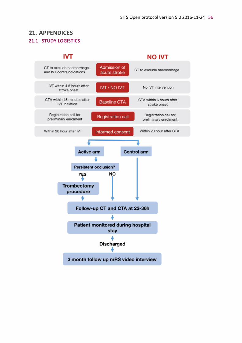
SITS Open protocol version 5.0 2016-11-24 56
21. APPENDICES 21.1 STUDY LOGISTICS

57 SITS Open protocol version 5.0 2016-11-24
CHEDULE OF INVESTIGATIONAL EVENTS
Trial Period Baseline 1 Check point 1
2 Check point 2
24h post IVT start
7 2 days or discharge
Final follow-
up 90 14 days
Visit No. 1 2 3 4 5 6
Day of visit 0 0 0 1 7 89 (75-104)
Screening log X3
Informed consent X Inclusion/exclusion criteria
X
Pregnancy excluded4 X
Time of stroke onset X
Time of rtPA start X
Demographic data X
Medical history X4
Intake of medications at stroke onset
X
CBC, including platelet count
X
Biochemistry panel, including blood glucose and cholesterol
X
Coagulation tests X
NIHSS X X X X X Blood pressure X X X X X
CT X X5,6 X7
MRI6 X X5 X7
CT OR MRA X X7
TCD X X X X
IVT X TBY8 X
Intake of medications at discharge
X
ADR/SAE Reporting X X X X X X
Final stroke diagnosis X
Global Outcome X Modified Rankin Scale, mRS
X9 X X
1 Check point 1 Control /Active arm: 90-150 min post-IVT start or plain CT(baseline) 2 Check point 2 Control arm: 10-14h post-IVT start/ or plain CT(baseline) Active arm: 10-14h post-IVT start or after the full recovery from anaesthesia/ or plain CT(baseline) 3 The telephone call to the coordinating centre must be done before initiation of thrombectomy and/or within two hours of initiation of IVT /plain CT(baseline) (if IVT is not given) 4 by interview
5 in case of suspected ICH, at the discretion of the investigator; accepted interval 2-24h
6 optional 7 accepted interval 22-36h
8 TBY arm only
9 mRS before stroke

SITS Open protocol version 5.0 2016-11-24 58
21.2 NATIONAL INSTITUTE OF HEALTH STROKE SCALE (NIHSS)
National Institute of Health Stroke Scale (NIHSS) Date and time DD-MM-YYYY HH:MM (24h)
1.a. Level of Consciousness 0: Alert 1: Not alert, but rousable with minimal stimulation 2: Not alert, requires repeated stimulation to attend 3: Coma
1.b. LOC questions (Ask patient the month and her/his age) 0: Answers both correctly 1: Answers one correctly 2: Both incorrect
1.c. LOC commands (Ask patient to open/close eyes & form/release fist) 0: Obeys both correctly 1: Obeys one correctly 2: Both incorrect
2. Best gaze (only horizontal eye movement) 0: Normal 1: Partial gaze palsy 2: Total gaze paresis or Forced deviation
3. Visual Field testing 0: No visual field loss 1: Partial hemianopia 2: Complete hemianopia 3: Bilateral hemianopia (blind including cortical blindness) 4. Facial Paresis (Ask patient to show teeth/ raise eyebrows & close eyes tightly) 0: Normal symmetrical movement 1: Minor paralysis (flattened nasolabial fold, asymmetry on smiling) 2: Partial paralysis (total or near total paralysis of lower face) 3: Complete paralysis of one or both sides (absence of facial movement in the upper and lower face)
5. Motor Function – Arm 0: Normal (extends arms 900 (or 450) for 10 seconds without drift) 1: Drift 2: Some effort against gravity 3: No effort against gravity 4: No movement 9: Untestable (Joint fused or limb amputated) (do not add score
Right
Left
5. Motor Function – Arm 0: Normal (extends arms 900 (or 450) for 10 seconds without drift) 1: Drift 2: Some effort against gravity 3: No effort against gravity 4: No movement 9: Untestable (Joint fused or limb amputated) (do not add score)
Right
Left
7. Limb Ataxia 0: No ataxia 1: Present in one limb 2: Present in two limbs
8. Sensory (Use pinprick to test arms, legs, trunk and face- compare side to side) 0: Normal

59 SITS Open protocol version 5.0 2016-11-24
9. Best Language (Ask patient to describe picture, name items, read sentences) 0: No aphasia 1: Mild to moderate aphasia 2: Severe aphasia 3: Mute
10. Dysarthria (Ask patient to read several words) 0: Normal articulation 1: Mild to moderate slurring of words 2: Near unintelligible or unable to speak 9: Intubated or other physical barrier (do not add score)
11. Extinction and inattention (Formerly Neglect) (Use visual or sensory double stimulation) 0: Normal 1: Inattention or extinction to bilateral simultaneous stimulation in one of the sensory modalities 2: Severe hemi-inattention or hemi-inattention to more than one modality
Total Score

SITS Open protocol version 5.0 2016-11-24 60
21.3 MODIFIED RANKIN SCALE (MRS)
MODIFIED RANKIN SCALE (MRS) Date/time DD-MM-YYYY HH:MM (24h)
0 No symptoms at all
1 No significant disabling symptoms
2 Slight disability but does not require substantial help from other person, can walk
3 Moderate disability, requires substantial help from other person, can walk
4 Moderately severe disability, requires substantial help from other person, unable to walk
5 Severe disability, bedbound
6 Dead

61 SITS Open protocol version 5.0 2016-11-24
21.4 STRUCTURED INTERVIEW MODEL FOR MRS ASSESSMENT (also see instruction – attachment 3)
STRUCTURED INTERVIEW MODEL FOR MRS ASSESSMENT 6 DEATH 5 BEDRIDDEN
5.1 Is the person bedridden? Yes (5) / No
4 ASSISTANCE TO WALK
4.1 Is another person’s assistance essential for walking? Yes (4) / No 3 ASSISTANCE TO LOOK AFTER OWN AFFAIRS
3.1 Is assistance ABSOLUTELY essential for preparing a simple meal? Yes (3) / No
3.2 Is assistance ABSOLUTELY essential for basic household chores? Yes (3) / No
3.3 Is assistance ABSOLUTELY essential for looking after household expenses? Yes (3) / No
3.4 Is assistance ABSOLUTELY essential for local travel? Yes (3) / No
3.5 Is assistance ABSOLUTELY essential for local shopping? Yes (3) / No
2 USUAL DUTIES AND ACTIVITIES
2.1 Work Has the new stroke substantially reduced (compared to prestroke status) the person’s ability to work (or, for a student, study)?
Yes (2) / No
2.2 Family responsibilities Has the new stroke substantially reduced (compared to prestroke status) the person’s ability to look after family at home?
Yes (2) / No
2.3 Social & leisure activities Has the new stroke reduced (compared to prestroke status) the person’s regular free-time activities by more than one half as often?
Yes (2) / No
2.4 Other physical/medical condition Are the patient’s work, family, and/or social/leisure activities substantially reduced by a physical/medical condition other than the stroke that led to trial enrollment?
Yes (2) / No
1 SPONTANEOUSLY REPORTED SYMPTOMS
1.1 Does the patient have any symptoms resulting from the new stroke? Yes (1) / No
1.2 SYMPTOM CHECKLIST
1.2.1 Does the person have difficulty reading or writing as a result of the new stroke?
Yes (1) / No
1.2.2 Does the person have difficulty speaking or finding the right word as a result of the new stroke?
Yes (1) / No
1.2.3 Does the person have problems with balance or coordination as a result of the new stroke?
Yes (1) / No
1.2.4 Does the person have visual problems as a result of stroke? Yes (1) / No
1.2.5 Does the person have numbness (face, arms, legs, hands, feet) as a result of the new stroke?
Yes (1) / No
1.2.6 Does the person have weakness or loss of movement (face, arms, legs, hands, feet) as a result of the new stroke?
Yes (1) / No
1.2.7 Does the person have difficulty with swallowing as a result of the new stroke?
Yes (1) / No
1.2.8 Does the person have any other symptoms related to the new stroke? Yes (1) / No
RANKIN GRADE=

SITS Open protocol version 5.0 2016-11-24 62
21.5 SITS GLOBAL OUTCOME SCALE
GLOBAL OUTCOME
Condition
Much better
Better
Unchanged
Worse
Much worse
Dead
The scale reflects the global impression of the patient’s performance, according to the judgement of the treating clinician. A global impression would include not only an estimation of change in neurological functions but also of the patient’s general medical and psychological condition. An approximate reference for change in neurological functions depends on baseline severity; for a baseline severity of 12 points on NIHSS ‘unchanged’ may correspond to absence of change or change of NIHSS score between -1 and +4 from baseline; “much better”, equivalent of dramatic/major neurological improvement by 8-10 points reduction from baseline or total recovery; “much worse” an increase in NIHSS score by 8-10 points from baseline, or death.
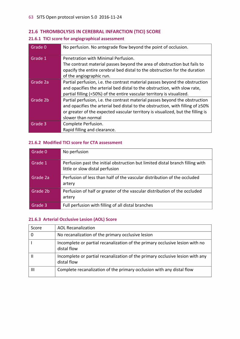
63 SITS Open protocol version 5.0 2016-11-24
21.6 THROMBOLYSIS IN CEREBRAL INFARCTION (TICI) SCORE
21.6.1 TICI score for angiographical assessment
Grade 0 No perfusion. No antegrade flow beyond the point of occlusion.
Grade 1 Penetration with Minimal Perfusion. The contrast material passes beyond the area of obstruction but fails to opacify the entire cerebral bed distal to the obstruction for the duration of the angiographic run.
Grade 2a Partial perfusion, i.e. the contrast material passes beyond the obstruction and opacifies the arterial bed distal to the obstruction, with slow rate, partial filling (<50%) of the entire vascular territory is visualized.
Grade 2b Partial perfusion, i.e. the contrast material passes beyond the obstruction and opacifies the arterial bed distal to the obstruction, with filling of ≥50% or greater of the expected vascular territory is visualized, but the filling is slower than normal
Grade 3
Complete Perfusion. Rapid filling and clearance.
21.6.2 Modified TICI score for CTA assessment
21.6.3 Arterial Occlusive Lesion (AOL) Score
Score AOL Recanalization
0 No recanalization of the primary occlusive lesion
I Incomplete or partial recanalization of the primary occlusive lesion with no distal flow
II Incomplete or partial recanalization of the primary occlusive lesion with any distal flow
III Complete recanalization of the primary occlusion with any distal flow
Grade 0 No perfusion
Grade 1 Perfusion past the initial obstruction but limited distal branch filling with little or slow distal perfusion
Grade 2a Perfusion of less than half of the vascular distribution of the occluded artery
Grade 2b Perfusion of half or greater of the vascular distribution of the occluded artery
Grade 3 Full perfusion with filling of all distal branches
SITS Open protocol version 5.0 2016-11-24 64
21.7 INSTRUCTIONS FOR IMAGING Imaging should be done with a < 2,5 mm slice thickness at baseline and on follow-up (in order to allow measurement of the clot size by imaging core lab). Angiographic images of the occlusion being treated must allow clear visualisation of the target artery. Same orientation should be used before and after treatment to allow a valid analysis of the data.
Baseline plain CT and CT angiography, 24-hour plain CT/MR scans and CT/MR angiographies, as well as thrombectomy imaging records from active centres, should be stored at CD/DVD and sent for independent core lab evaluation. In case of any ICH or suspected ICH on 24-hour assessment, imaging should be repeated on the next day, and included on CD/DVD for further assessment by core lab with regard to differentiation blood/contrast. All images should be saved in DICOM format. Importantly, only the raw data should be saved, i.e. the original imaging file before any processing. The name of the patient on images should be replaced by the patient study code. Study date and time on the image should be kept. If the software allows anonymity only, without any option to replace the patient and hospital information, images should be saved anonymously, but the study code and date/time of the imaging saved as the file name.
Evaluation of occlusion, qualifying for inclusion in the study, should be based on the first CT angiography performed after stroke onset, even if performed at another hospital before transport to the TBY centre (with or without initiated IVT), unless the quality of the images are insufficient for evaluation according to the radiologist at the TBY centre. The first CT and CT angiography (of sufficient quality) must be included in the material sent to core lab. Clinical study coordinator (a neurologist) will be asked to provide the raw imaging data for the core lab assessment, and will be assigned responsible for getting it from another hospital, if necessary.
For control centres, 2 CD/DVD are required: with baseline CT/MR scan and CT/MR angiography, and with 24h CT/MR scan and CT/MR angiography. For active centres, 3 CD/DVDs should be burned: the two mentioned above, plus the separate disk with thrombectomy records. In case of repeated follow-up imaging in suspicion of ICH, the second follow-up record may be saved together with 24-hour imaging at the same disk.
Centres are also encouraged to provide for core lab evaluation any additional imaging which may be of importance for understanding of clinical decisions.
Imaging records and corresponding transmittal sheet should be sent to the University of Edinburgh (Neurosciences Imaging) within 3 working days. In case of missing images, information, or poor quality the centre will be alerted within the next few days of CD receipt. Detailed instructions for handling of images is found in Imaging Procedures Manual.
Corresponding address:
University of Edinburgh (Neurosciences Imaging) Centre for Clinical Brain Sciences Att: Eleni Sakka The Chancellor's Building 49 Little France Crescent, Edinburgh EH16 4SB United Kingdom

65 SITS Open protocol version 5.0 2016-11-24
Attachment 1. Guidelines for intravenous thrombolysis
Patient undergoing thrombolytic therapy should be transferred to an intensive care or semi-intensive care/monitoring unit with the possibility to monitor vital signs and neurological status as soon as the radiological and (for active study centres only) any subsequent endovascular procedure is completed. Invasive procedures (e.g. gastric tube, urinary catheter, arterial line) should be avoided. Thrombolytic therapy consists of intravenously administered alteplase (0.9 mg/kg body weight, maximum dose 90mg) given within 4.5 hours after stroke onset by a 10% of total dose in bolus and 90% by a 1-hour infusion. For active centres bridging to thrombectomy, there is no need to wait for the infusion to be completed or to discontinue the infusion before artery puncture, but this decision is on the investigator’s discretion. The final dose of alteplase must always be documented in eCRF. Reasons for discontinuation of rtPA infusion are severe headache, acute hypertension, nausea/vomiting, hypersensitivity /anaphylactic reaction (e.g. angio-edema, bronchospasm, rash, urticaria, hypotension, shock or any other symptom associated with allergic reactions). If a systolic blood pressure is above 180 mm Hg or if a diastolic blood pressure is above105 mm Hg, intravenous antihypertensive medications should be started. Indications
• Clinical diagnosis of ischaemic stroke
• Age ≥ 18 years
• Time from stroke onset ≤ 4.5 hours
• Absence of haemorrhage on admission CT scan Contraindications
• Hypersensitivity to rtPA or to any of the medication compounds.
• Severe stroke as assessed clinically (e.g. NIHSS>25 for anterior circulation stroke) and/or extensive early ischaemic changes on admission CT scan (defined as more than one third of the MCA territory or one half of other territories)
• Other thrombolytic treatment within the previous 72h
• Known platelet count below 100,000/mm3
• Clinically significant hypoglycaemia
• Uncontrollable hypertension (blood pressure exceeding: systolic 185 mm Hg and diastolic - 110 mm Hg on at least 2 measurements made with 10-minute interval) or aggressive management (by the discretion of the investigator, usually implying intravenous pharmacotherapy) necessary to reduce BP to these limits
• Current use of oral anticoagulants and a prolonged prothrombin time (INR > 1.7) or intake of direct oral anticoagulants with APTT above normal limits or within the latest 4 hours
• Use of glycoprotein IIb-IIIa inhibitors within the past 72 hours
• Clinical suspicion of subarachnoid haemorrhage (even if CT-scan is normal)
• Use of heparin in previous 48 hours, if aPTT is exceeding the upper limit of normal for laboratory
• Recent history of central nervous system damage (intracranial haemorrhage neoplasm, ruptured aneurysm, intracranial or spinal surgery)
• Active bleeding or acute trauma on examination, or recent severe or dangerous bleeding
• Known haemorrhagic diathesis/ bleeding disorder
SITS Open protocol version 5.0 2016-11-24 66
• Recent (less than 10 days) traumatic external heart massage, obstetrical delivery, recent puncture of a non-compressible blood-vessel (e.g. subclavian or jugular vein puncture)
• Prior stroke in previous 3 months
• Major surgery within the preceding 14 days, which poses a risk in the opinion of the investigator
• Documented ulcerative gastrointestinal disease during the last 3 months, oesophageal varices, arterial aneurysm, arterial/venous malformations
• Seizure at onset of stroke only if there is a clinical suspicion of postictal residual neurological impairments which mimics stroke
• Any condition that may be associated with increased bleeding risk or could impose hazards to the patient if intravenous thrombolysis is initiated (e.g., severe microangiopathy such as haemolytic uraemic syndrome or thrombotic thrombocytopenic purpura), at the discretion of the treating physician
Comments
Upper age limit was not set in the present study guidelines, since the recent research (43–45) has demonstrated the beneficial effect of IVT even in very elderly patients. Consideration of patient’s advanced age should be made on individual basis with regard to the general state of health and concomitant diseases.
The population of the present study is not a general stroke population, but a subgroup with severe stroke, as indicated by large cerebral vessel occlusion at baseline. For this reason, rapidly improving symptoms should not preclude vascular imaging.
In the list of contraindications, “recent” without the clarification of time intervals implies time period which poses a risk by the opinion of the investigator.
In case of unclear or relative contraindications to IVT, risk-benefit ratio should be estimated individually, and decision made on per patient basis.
67 SITS Open protocol version 5.0 2016-11-24
Attachment 2. Antiplatelet therapy following non-retrievable stent use
Guideline Peri-procedural:
• 500 mg of aspirin are given IV after the stent placement
• Abciximab IV (1/2 of standard bolus dose, i.e. 0,125 mg/kg, or other relevant dosing according to the local clinical routine) – at the discretion of the treating physician
24h after stenting:
• Aspirin 300 mg p.o. (loading dose)
• Clopidogrel 300 mg p.o. (loading dose)
• 48h after stenting and further on:
• Aspirin, 75 mg daily, for at least 6 months (or life-long)
• Clopidogrel, 75 mg daily, for at least 3 months
SITS Open protocol version 5.0 2016-11-24 68
Attachment 3. Instructions for mRS Assessment
Training
Successful certification in mRS training will be a pre-requisite of participation in the SITS Open trial. Where a site has more than one study coordinator, all coordinators should hold a valid training certificate. Only investigators holding a valid training certificate should perform outcomes assessment. If an investigator is replaced at a site, the training centre should be notified within 7 days to organise training of the replacement. Once initiated a site must maintain a minimum of two trained individuals so that a certified rater is always available for follow up assessments.
The training will be coordinated by the Outcomes Coordinating Centre at the Western Infirmary in Glasgow, UK. All participating centres will be invited to register with the training campus web site and complete mRS training. At these meetings researchers will receive training and explanation of how to perform mRS assessment in the SITS Open trial and be given the opportunity for discussion with the one of the Glasgow team. Investigators will submit their scores on-line and results are relayed to the Outcomes Manager at the Western Infirmary. All results will be collated into a database and will be checked by our Outcomes Manager. A hard copy of a certificate of completion can be printed by successful observers and kept at the local site.
Not all investigators and sub-investigators at a site need be qualified as raters, but only qualified raters may undertake outcome assessments. Thus, all raters must be trained in mRS assessment using a validated web-based training programme before beginning participation in the SITS Open trial. A link to the training web-sites can be found on the SITS Open trial website. Each participant must register their own training account. The link for mRS training will be provided by the Outcomes Coordinating Centre. Raters will be unable to upload any assessments if training has not been completed. Raters will also be shown how to operate the video camera and given a practical demonstration on video upload procedures.
Video recording
The mRS assessment will be recorded using a digital video camera. Cameras will be purchased centrally and distributed by the Coordinating Centre. An easily portable tripod will be used to mount the video camera.
Via web-based training materials, observers will be shown how to operate the video camera and given a practical demonstration on video upload procedures and use of the Rankin Outcome Adjudication web portal. Each site must submit a test video assessment during the pilot phase of the trial to ensure familiarity with technical issues and confirm correct operation of procedures. This will be reviewed by a member of the endpoint assessment committee.
The mRS assessment should be recorded with the head and shoulders of the patient in camera shot, unless the participant clearly has the most severe level of disability (for example a mRS score of 5) or where coma or intubation render assessment impractical. In this scenario we advise that a proxy be used to provide information and that they should be interviewed on video alongside or in place of the patient. If the patient has any cognitive
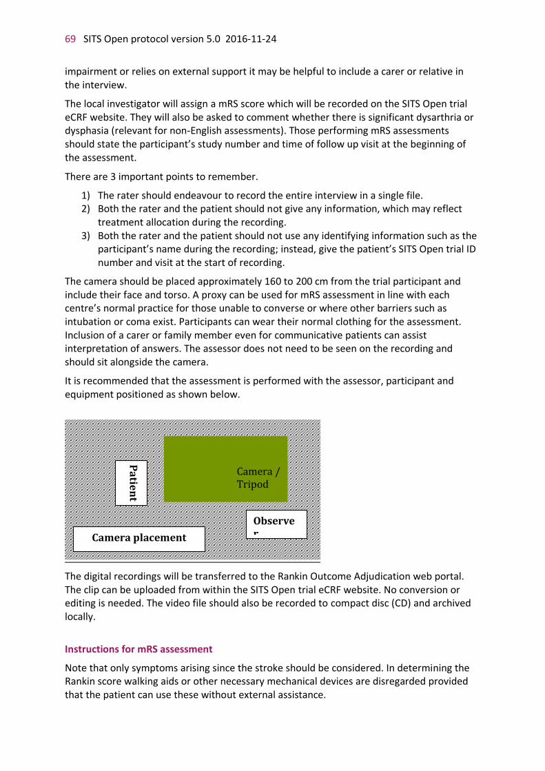
69 SITS Open protocol version 5.0 2016-11-24
impairment or relies on external support it may be helpful to include a carer or relative in the interview.
The local investigator will assign a mRS score which will be recorded on the SITS Open trial eCRF website. They will also be asked to comment whether there is significant dysarthria or dysphasia (relevant for non-English assessments). Those performing mRS assessments should state the participant’s study number and time of follow up visit at the beginning of the assessment.
There are 3 important points to remember.
1) The rater should endeavour to record the entire interview in a single file. 2) Both the rater and the patient should not give any information, which may reflect
treatment allocation during the recording. 3) Both the rater and the patient should not use any identifying information such as the
participant’s name during the recording; instead, give the patient’s SITS Open trial ID number and visit at the start of recording.
The camera should be placed approximately 160 to 200 cm from the trial participant and include their face and torso. A proxy can be used for mRS assessment in line with each centre’s normal practice for those unable to converse or where other barriers such as intubation or coma exist. Participants can wear their normal clothing for the assessment. Inclusion of a carer or family member even for communicative patients can assist interpretation of answers. The assessor does not need to be seen on the recording and should sit alongside the camera.
It is recommended that the assessment is performed with the assessor, participant and equipment positioned as shown below.
The digital recordings will be transferred to the Rankin Outcome Adjudication web portal. The clip can be uploaded from within the SITS Open trial eCRF website. No conversion or editing is needed. The video file should also be recorded to compact disc (CD) and archived locally.
Instructions for mRS assessment
Note that only symptoms arising since the stroke should be considered. In determining the Rankin score walking aids or other necessary mechanical devices are disregarded provided that the patient can use these without external assistance.
Camera / Tripod
Observer
Pa
tien
t
Camera placement

SITS Open protocol version 5.0 2016-11-24 70
The score of 0 is awarded to patients who have no residual symptoms after their stroke, not even minor symptoms.
If patients have any symptoms resulting from the stroke, whether physical or mental, then they should be scored at least 1 on the Rankin scale. For example, if they have any new difficulty in speech, reading or writing, in physical movement, sensation, vision or swallowing, or any change in their mood that does not limit their activities, they still should score 1. Patients in this category can continue to take part in all their previous work, social and leisure activities. For this purpose, “usual” is regarded as any activity that they used to undertake for a monthly basis or more frequently.
If there is any activity that they used to undertake that they can no longer do since the stroke, whether because of a physical limitation or because they have chosen to give up the activity as a result of the stroke, then they should be scored 2 on the Rankin. In this category the patient has slight disability and is unable to carry out all his previous activities, but he is still able to look after all of his own affairs without any external assistance. For example, a patient would be scored in this category if he used to play golf and is no longer able to do so, or if he used to have a job whereas he now no longer works. The patient should still be able to look after himself without any daily help. In other words, he will be able to dress, move around, eat, go to the toilet, prepare simple meals, undertake shopping and make short journeys by himself. He will not require any supervision from other people and could safely be left at home for periods of a week or more without any concern. An inability to drive purely because of legal impediment, ie where the participant is otherwise physically able, would not warrant a score of 2.
Rankin category 3 is for patients who have moderate disability. These patients require some external help for daily activities but are able to walk without assistance. They may use a stick or a frame for walking but the assistance of another person is not required for this. They will be able to manage daily activities such as dressing, toileting, feeding etc, but will need help for more complex tasks such as shopping, cooking or cleaning or will need to be visited more often than weekly for some other purpose. The external help may simply be advisory, for example supervision for their financial affairs.
Patients with moderately severe disability who are unable to walk without assistance and are unable to attend to their own bodily needs by themselves are given a score of 4. These patients are not independently mobile and will need help with daily tasks such as dressing, toileting or eating. They will need to be visited at least daily or will need to live in close proximity to a carer. To discriminate patients in category 4 from those in the most severe category, consider whether the patient can regularly be left along for moderate periods of a few hours during the day.
Patients who cannot be left alone even for a few hours should be given the score of 5. Patients in category 5 have severe disability and are usually bedridden, incontinent and require constant nursing care and attention. Someone else will always need to be available during the day and at time during the night, although this will not necessarily be a trained nurse.
Thus, in summary, to distinguish between patients in category 0 or 1 consider whether the patient has any remaining symptoms. To distinguish between categories 1 and 2 consider whether the patient can undertake all of his previous activities. If the patient is independent of others in activities of daily living, then he should be scored 2 rather than 3. To distinguish
71 SITS Open protocol version 5.0 2016-11-24
between category 3 and category 4 the crucial question is whether the patient can walk without the assistance of other people. Finally, a patient who can be left by himself for a few hours during the day would be given a score of 4 rather than 5.
It is important to note that patients do not always fall neatly into one category and some judgement is usually required when scoring them. When in doubt between 2 categories, always stick to the key discriminators of the scale. Thus, if the patient has remaining symptoms he scores at least 1. If the patient is unable to undertake previous activities he scores at least 2. If he is dependent upon others in activities of daily living he must score at least 3. If the patient is unable to walk without assistance, he must score at least 4 and if the patient is bedridden and requires constant nursing care he will score 5. Finally, if there is still some doubt between two alternatives on the scale, and both options appear equally valid, then the worse option should be chosen.
As an example we have included some key discriminating questions that should be considered when using the modified Rankin scale. These are shown in more detail below (the official definitions of each category are shown below in bold and the italicized text provides guidance that may reduce inter-observer variability, without requiring a structured interview).
0. No symptoms at all
The patient should be unaware of any new limitation of symptom caused by the stroke, however minor.
1. No significant disability despite symptoms; able to carry out all usual duties and activities
The patient has some symptoms as a result of the stroke, whether physical or cognitive – for example affecting speech, reading or writing; or physical movement; or sensation; or vision; or swallowing; or mood – but can continue to take part in all previous work, social and leisure activities. The crucial question to distinguish grade 1 from grade 2 (below) may be, ‘is there anything that you can no longer do that you used to do until you had the stroke? As a guide, an activity that was undertaken more frequently than monthly could be regarded as a ‘usual activity’.
2. Slight disability; unable to carry out all previous activities but able to look after own affairs without assistance
The patient will be unable to undertake some activity that was possible before the stroke (e.g. driving a car, dancing, reading or working) but is still able to look after him/herself without help from others on a day-to-day basis. Thus, the patient can manage dressing, moving around, feeding, toileting, preparing simple meals, shopping, and travelling locally without needing assistance from anyone else. Supervision is not necessary. This grade assumes that the patient could be left alone at home for periods of a week or more without concern.
3. Moderate disability; requiring some help, but able to walk without assistance
At this grade the patient is independently mobile (using a walking aid of frame if necessary) and can manage dressing, toileting, feeding, etc. but needs help from someone else for more complex tasks. For example, someone else may need to undertake shopping, cooking or cleaning and will need to visit the patient more often that weekly to ensure that these

SITS Open protocol version 5.0 2016-11-24 72
activities are completed. The assistance can be advisory rather than physical: for example, a patient who needs supervision or encouragement to cope with financial affairs would be in this grade.
4. Moderately severe disability; unable to walk without assistance and unable to attend to own bodily needs without assistance
The patient requires someone else to help with some daily tasks, whether walking, dressing, toileting or eating. This patient will be visited at least once and usually twice or more times daily, or must live in proximity to a carer. To distinguish grade 4 from grade 5 (below), consider whether the patient can regularly be left alone for moderate periods during the day.
5. Severe disability: bedridden, incontinent, and requiring constant nursing care and attention
Someone else will always need to be available during the day and at times during the night, though not necessarily a trained nurse.
6. Dead

73 SITS Open protocol version 5.0 2016-11-24
Attachment 4. Rehabilitation guidelines
Rehabilitation aims to enable people with disabilities to reach and maintain optimal physical, intellectual, psychological and/or social function.
None of the non-modifiable factors, such as stroke severity, gender, stroke aetiology, age, and topography of lesion should influence decisions on rehabilitation.
Rehabilitation for the stroke patients is recommended to be started in a stroke unit by a coordinated multidisciplinary team with stroke expertise. The rehabilitation program should start as early as possible, preferably in the first 24h of stroke onset if clinical condition of the patient allows it. Start of the rehabilitation program within 20–30 days is a due. Patients that are undergoing TBY rehabilitation are recommended to get physiotherapy and occupational therapy as much as possible, the recommended minimum is 1 h of active practice per day at least 5 days per week.
For medically stable patients with mild or moderate impairment early discharge from stroke unit to a rehabilitation unit is a preference.
Rehabilitation program may include the following items in accordance with the individual clinical symptoms and deficits:
• Physiotherapy
• Occupational therapy
• Speech and language disorders assessment and appropriate treatment
• Cognitive deficits assessment and appropriate treatment
• Monitoring for post-stroke depression for all patients; antidepressants and psychological interventions if indicated
• Medications for post-stroke emotionalism and mood disorders
• Tricyclic or anticonvulsant medications for post-stroke neuropathic pain, if indicated
• Botulinum toxin for post-stroke spasticity, if indicated
• Involvement of other therapists depending on patient-specific goals (dieticians, orthoptists, social workers, etc.)
For the severely disabled patients who are unable to participate actively, passive movements to prevent contractures or pressure sores have been recommended.
SITS Open protocol version 5.0 2016-11-24 74
Attachment 5. Informed consent
Information for a clinical trial participant – TBY ARM
Name of the trial: THE SITS OPEN ARTERY BY THROMBECTOMY IN ACUTE OCCLUSIVE STROKE STUDY (SITS Open)
Chair of the Steering Committee: professor Nils Wahlgren, MD, PhD, FESO
This is an example which needs modifications by country, e.g. with regard to contact details
Treatment of stroke by mechanically removing blood clots in cerebral arteries. You (or a close relative) is kindly asked to participate in this study, because you were diagnosed a stroke caused by a blood clot, which blocks one or more of the blood vessels supplying the brain. We kindly ask you to participate in this study, which is designed to measure the benefit of a procedure of clot removal from the blood vessel in the brain.
The procedure may be done as a first or second treatment to your stroke. You may receive a first step of stroke treatment, an injection of a medication that dissolves blood clots into your vein. However, your neurologist and attending neuroradiologist jointly evaluated your neurological conditions and current radiological studies and agreed that you will probably get better with an additional (or as first treatment) procedure that restores blood flow to the brain, i.e. mechanically removing the clot. This procedure is used in many stroke centres, and all the devices are approved by medical regulatory authorities, but there is still not enough data collected to make the exact measurements of the degree of improvement.
In some situations, the evaluation will show that the procedure is not necessary, however it is of great importance that you participate in the study anyway and perform the follow up procedures.
The main aim of our study is to measure the degree of recovery from stroke by an appropriate test, and compare the results in those who did with those who did not get the additional treatment. Also, we’ll record and study some details of your disease and treatment, such as devices used for the procedure, extent of restoration of the blood flow, improvement of body functions after the treatment, etc.
It is your free choice if you decide to participate in the study or not. If you decide to participate, from now information about your treatment and your recovery will be collected. Participation in the study does not affect the choice of treatment and the level of care that you get from the medical staff of your hospital. Participation in the study is not associated with any expenses.
You will get the best-known care whether you participate in the study or not. As the study patient, you can always cancel your participation in the study without giving any reason. If you choose to cancel your participation in the trial, further treatment will not be affected, and you will get the best possible care. Background and purpose For a patient who has a stroke, the first goal is to remove the blood clot that is the cause of the symptoms. The conventional treatment for this condition is infusion of the medication, which dissolves blood clots (thrombolytic). However, there are patients who for various

75 SITS Open protocol version 5.0 2016-11-24
reasons cannot be treated with thrombolytic infusion or where this treatment does not give the sufficient effect. A treatment is then to remove the clot mechanically via going directly into the blood vessel with a special device. Today there are various mechanical devices with different advantages and disadvantages. None of the devices are suitable for all types of clots. The purpose of this study is to collect the data from the clinical routine to measure the benefit that patient gets from additional procedure of mechanical removal of the blood clot that blocks blood flow to the brain, thus restoring blood supply of the brain. The treatment is standard practice in this hospital and all permissible instruments are approved. If thromectomy is not performed, alternative treatment will be performed according to clinical routine. Study Progress The duration of the study is 3 months from the onset of the stroke. During the first days of in-hospital stay your neurological symptoms will be checked and recorded every day by your physician in charge, according to clinical routine. You will not be asked to undergo any additional physical examinations in relation to the study. The samples taken before treatment are routinely collected from all patients who are expected to undergo some form of intervention treatment. No blood samples will be taken just for the study purposes.
Results of imaging studies will be saved with an anonymous study code and sent to an independent expert lab for review.
Angiography, i.e. imaging of blood vessels of the brain, is performed to guide the treatment, and then repeated after 24 hours. 24-hour repeated imaging of the brain vessels is the only study-related procedure, which is not a clinical routine, though it is widely used in hospitals in case of need to clarify the condition of a given vessel or the diagnosis. The repeated imaging is currently not routine at all hospitals but if you choose to participate in the study, it becomes mandatory. In special cases of contraindications this imaging may be replaced by an ultrasound investigation, though ultrasound is much less informative.
In addition to general clinical routine, after 3 months (±2 weeks) your investigator will arrange a meeting with you to ask several questions about your daily living, which describe the degree of your recovery from stroke, and record your answers by video for the experts who will give their estimation. No special investigations/tests/samples are needed for this follow-up evaluation. The interview is called a modified Rankin Scale assessment. It is usually performed by a nurse or doctor and takes between 5 and 10 minutes. At the end of the interview we can assign a grade on the Rankin stroke scale, which describes the extent of the patient’s recovery. All the records will be anonymous and confidential. The recording will be made in a private room in the stroke clinic or in a private room on the ward if you are still in hospital. If you cannot attend the hospital, we could visit you to perform the assessment. The recording will last between 5 and 10 minutes. You will not be identified by name on the recording but your face and voice will remain recognisable. The recording will be sent for viewing by trained stroke assessors who will decide whether they agree or disagree with the recovery grade that your own clinician has assigned to you.
SITS Open protocol version 5.0 2016-11-24 76
Alternative treatments Your participation in this study does not affect the choice of treatment. Nowadays various devices are used for removing clots in any of the main brain blood vessels. None of these devices are fast, efficient and reliable in all conditions and with all kinds of clots. The physician is free to choose among the most common devices which he / she believes is most appropriate.
Risks and benefits Since you get the same treatment regardless of whether you are in the study or not, there are no direct benefits to you personally to participate in the study. Possibly, your help to choose the optimal strategy for the other patients with a severe stroke. Also you will get detailed explanations about your stroke and the results of the brain vessels evaluation related to your stroke; and you will get an expert second opinion about your brain imaging results and about the degree of recovery that was assigned to your case by internationally renowned specialists. The second X-ray angiography is currently not routine at all the hospitals, though, of importance, it is a standard medical investigation that is used in many situations for many years for establishing diagnosis or guiding the treatment strategy. The potential disadvantages of that are possible discomfort from contrast dye administration to your vein (if you have a catheter in the vein, which is very likely for an in-hospital patient, no additional injections are needed), possible allergic reaction to contrast dye (if the first imaging which you underwent on admission was well tolerated, there is no reason to expect any problems) and also leads to an additional radiation dose. The advantage is that a more precise diagnosis and further treatment may be better adapted to your needs.
All the medical devices used in the study are the approved medical instruments, which have been properly tested and released on the market. There is a small risk that an unknown adverse effect, which has never been observed before, may appear, but participation in the study does not possess any additional risk of that, since no new treatments are implemented.
If you are pregnant or are planning to conceive in the nearest future, additional irradiation is generally not recommended. Discuss your individual situation with your physician before you make a decision about the participation on the study.
Privacy All personal data collected for study purposes are managed in accordance with the applicable laws and regulations. All data is encoded in such a way that no data can be linked to an individual study subject: your name will never appear in any of the study materials. Your face and voice will appear once at the video record of outcome assessment, but anonymously. The study data collected will be entered into a database that is currently used for the registration of stroke patients treated with clot-dissolving medication. Record-holder for this database, SITS, is Professor Nils Wahlgren, Department of Neurology, Karolinska University Hospital, Stockholm, Sweden. Study results will be presented in groups and not on the individual basis.
77 SITS Open protocol version 5.0 2016-11-24
Access to the data is strictly limited to the authorized personnel (i.e. your investigator, members of the research team, and independent experts who will evaluate images and video records). Of importance, all the data will be stored in the encoded form, where all the personal information is masked by a study code when subjected to statistical analysis.
The video recordings will be uploaded to a secure computer in the University of Glasgow, UK. Only named study investigators will have access to the videos and we anticipate they will be stored for a maximum of three years and subsequently destroyed.
Personal data collecting board of Karolinska University Hospital can be reached by phone + 46 (0) 8 517 700 00.
You are entitled to free of charge once per year upon written request to get information about what is recorded about you. The study is approved by the regional ethical review board in Stockholm.
Compensation and Insurance Standard Patient Injury insurance covers any damage caused by the treatment. You will not receive any form of compensation if you participate in the study. No special insurance is needed when the treatment is standard practice at your hospital. If you get any damage from the treatment, compensation should be provided according to the regulations of your medical insurance company. Please ask your investigator if clarify any questions that may arise after you have read the information above.
For any questions regarding the study, please contact:
Research nurse First name, Surname Phone: Mobile: Responsible study doctor First name, Surname Phone: Mobile:

SITS Open protocol version 5.0 2016-11-24 78
INFORMED CONSENT I____________________________________ hereby confirm that I have consulted and agree to participate in the SITS Open study. I have been informed about the aims and scope of the study. I confirm that I have read and understood the information given to patients, and I have been also given oral explanations about the study. The information given was complete and clear. I have had the opportunity to ask questions and the questions have been satisfactorily answered. I have received a copy of this leaflet. I understand that my participation is voluntary. I am able to cancel my participation in the study at any time without giving any reason and without risk that this will affect my medical care or my legal rights. I agree that my medical records will be audited by authorized persons from the organisation SITS International, its affiliates, and the medical controlling authorities or ethical review boards in case of my participation in the study. I agree that my personal information is collected, analysed and processed in accordance with the applicable Privacy Acts. I agree that my study doctor calls and collects all relevant information and/or documentation regarding my medical care/survey from my family doctor or from another clinic if I have been treated elsewhere. Date and time of giving informed consent Time (HH:MM) : Date (YYYY-MM-DD) 20 - - _________________________________________ _____________________ Signature of the patient or the legal representative Date _____________________________________________ _______________________ Printed name of the patient or the legal representative Legal representative's relationship to the patient
I provided the research participant with detailed explanation of what he or she can expect from the participation in the study. I certify here that at the time of signing this form the study participant understands the purpose, procedures, potential benefits and possible risks. I also certify that he or she: - knows the language in which it the explanations were given - is able to read and understand this form or, otherwise can hear and understand the information given above - Do not have any difficulties in understanding the meaning of participation in the study. _________________________________________ ____________________ The treating physician's signature Date _____________________________________________ Treating physician's printed name
79 SITS Open protocol version 5.0 2016-11-24
Information for a clinical trial participant – CONTROL ARM Name of the trial: THE SITS OPEN ARTERY BY THROMBECTOMY IN ACUTE OCCLUSIVE STROKE STUDY (SITS OPEN)
Chair of the Steering Committee: professor Nils Wahlgren, MD, PhD, FESO
This is an example which needs modifications by country, e.g. with regard to contact details
Treatment of stroke by mechanically removing blood clots in cerebral arteries.
You (or a close relative) is kindly asked to participate in this study, because you were diagnosed a stroke caused by a blood clot, which blocks one or more of the blood vessels supplying the brain.
The first step of treatment in this condition is to inject a medication that dissolves blood clots into your vein. In some hospitals in certain cases the treatment is extended with an additional procedure of mechanically removing the clot. However, this additional treatment cannot be offered at all hospitals, including this hospital.
We kindly ask you to participate in this study which is designed to measure the benefit of the different strategies (with and without an additional procedure of clot removal from the blood vessel in the brain) that are implemented as a standard practice in different stroke centres, because there is still not enough data collected to make the exact measurements of the effect of this procedure. The main aim of our study is to measure the degree of recovery from stroke by an appropriate test, and compare the results in those who did with those who did not get the additional treatment. Also, we’ll record and study some details of your disease and treatment, such as the extent of restoration of the blood flow, improvement of body functions after the treatment, etc.
In some situations, the evaluation will show that the treatment with intravenous injection of medication to dissolve your clot is not necessary, however it is of great importance that you participate in the study anyway and perform the follow up procedures.
It is your free choice if you decide to participate in the study or not. If you decide to participate, from now information about your treatment and your recovery will be collected. Participation in the study does not affect the choice of treatment and the level of care that you get from the medical staff of your hospital. Participation in the study is not associated with any expenses.
You will get the best known care whether you participate in the study or not. As the study patient, you can always cancel your participation in the study without giving any reason. If you choose to cancel your participation in the trial, further treatment will not be affected, and you will get the best possible care.
Background and purpose For a patient who has a stroke, the first goal is to remove the blood clot that is the cause of the symptoms. The conventional approved treatment for this condition is infusion of the medication which dissolves blood clots (thrombolytic). However, there are patients who for various reasons cannot be treated with thrombolytic infusion or where this treatment does not give the sufficient effect. In this case, in some stroke centres a trained physician tries to remove the clot mechanically via going directly into the blood vessel with a special device.

SITS Open protocol version 5.0 2016-11-24 80
None of the devices are suitable for all cases of stroke and/or all types of clots. Decision is made of individual basis, and the benefit of the additional treatment is still unmeasured. The purpose of this study is to collect the data from the clinical routine to measure the benefit that patient gets from additional procedure of mechanical removal of the blood clot that blocks blood flow to the brain, thus restoring blood supply of the brain. This treatment is NOT a standard practice in this hospital. Study Progress The duration of the study is 3 months from the onset of the stroke.
During the first days of in-hospital stay your neurological symptoms will be checked and recorded every day by your physician in charge, according to clinical routine. You will not be asked to undergo any additional physical examinations in relation to the study. The samples taken before treatment are routinely collected from all patients who are expected to undergo some form of intervention treatment. No blood samples will be taken just for the study purposes.
Results of imaging studies will be saved with an anonymous study code and sent to an independent expert lab for review.
Angiography, i.e. imaging of blood vessels of the brain, is performed to guide the treatment, and then repeated after 24 hours. 24-hour repeated imaging of the brain vessels is the only study-related procedure, which is not a clinical routine, though it is widely used in hospitals in case of need to clarify the condition of a given vessel or the diagnosis. The repeated imaging is currently not routine at all hospitals but if you choose to participate in the study, it becomes mandatory. In special cases of contraindications this imaging may be replaced by an ultrasound investigation, though ultrasound is much less informative.
In addition to general clinical routine, after 3 months (±2 weeks) your investigator will arrange a meeting with you to ask several questions about your daily living, which describe the degree of your recovery from stroke, and record your answers by video for the experts who will give their estimation. No special investigations/tests/samples are needed for this follow-up evaluation. The interview is called a modified Rankin Scale assessment. It is usually performed by a nurse or doctor and takes between 5 and 10 minutes. At the end of the interview we can assign a grade on the Rankin stroke scale, which describes the extent of the patient’s recovery. All the records will be anonymous and confidential. The recording will be made in a private room in the stroke clinic or in a private room on the ward if you are still in hospital. If you cannot attend the hospital, we could visit you to perform the assessment. The recording will last between 5 and 10 minutes. You will not be identified by name on the recording but your face and voice will remain recognisable. The recording will be sent for viewing by trained stroke assessors who will decide whether they agree or disagree with the recovery grade that your own clinician has assigned to you.
Alternative treatments Your participation in this study does not affect the choice of treatment. You have received thrombolytic infusion and additional treatment options are not available.

81 SITS Open protocol version 5.0 2016-11-24
Risks and benefits Since you get the same treatment regardless of whether you are in the study or not, there are no direct benefits to you personally to participate in the study. Possibly, your help to choose the optimal strategy for the other patients with a severe stroke. Also you will get detailed explanations about your stroke and the results of the brain vessels evaluation related to your stroke; and you will get an expert second opinion about your brain imaging results and about the degree of recovery that was assigned to your case by internationally renowned specialists.
The second X-ray angiography is currently not routine at all the hospitals, though, of importance, it is a standard medical investigation that is used in many situations for many years for establishing diagnosis or guiding the treatment strategy. The potential disadvantages of that are possible discomfort from contrast dye administration to your vein (if you have a catheter in the vein, which is very likely for an in-hospital patient, no additional injections are needed), possible allergic reaction to contrast dye (if the first imaging which you underwent on admission was well tolerated, there is no reason to expect any problems) and also leads to an additional radiation dose. The advantage is that a more precise diagnosis and further treatment may be better adapted to your needs.
Participation in the study does not possess any additional risk of any unknown adverse effects (i.e. the adverse effect which has never been observed before), since no new treatments are implemented.
If you are pregnant or are planning to conceive in the nearest future, additional irradiation is generally not recommended. Discuss your individual situation with your physician before you make a decision about the participation on the study.
Privacy All personal data collected for study purposes are managed in accordance with the applicable laws and regulations. All data is encoded in such a way that no data can be linked to an individual study subject: your name will never appear in any of the study materials. Your face and voice will appear once at the video record of outcome assessment, but anonymously. The study data collected will be entered into a database that is currently used for the registration of stroke patients treated with clot-dissolving medication. Record-holder for this database, SITS, is Professor Nils Wahlgren, Department of Neurology, Karolinska University Hospital, Stockholm, Sweden. Study results will be presented in groups and not on the individual basis.
Access to the data is strictly limited to the authorized personnel (i.e. your investigator, members of the research team, and independent experts who will evaluate images and video records). Of importance, all the data will be stored in the encoded form, where all the personal information is masked by a study code.
The video recordings will be uploaded to a secure computer in the University of Glasgow, UK. Only named study investigators will have access to the videos and we anticipate they will be stored for a maximum of three years and subsequently destroyed.
Personal data collecting board of Karolinska University Hospital can be reached by phone + 46 (0) 8 517 700 00.
SITS Open protocol version 5.0 2016-11-24 82
You are entitled to free of charge once per year upon written request to get information about what is recorded about you. The study is approved by the regional ethical review board in Stockholm.
Compensation and Insurance Standard Patient Injury insurance covers any damage caused by the treatment. You will not receive any form of compensation if you participate in the study. No special insurance is needed when the treatment is standard practice at your hospital. If you get any damage from the treatment, compensation should be provided according to the regulations of your medical insurance company.
Please ask your investigator if clarify any questions that may arise after you have read the information above.
For any questions regarding the study, please contact: Research nurse First name, Surname Phone: Mobile: Responsible study doctor First name, Surname Phone: Mobile:

83 SITS Open protocol version 5.0 2016-11-24
INFORMED CONSENT I________________________________ hereby confirm that I have consulted and agree to participate in the SITS OPEN study. I have been informed about the aims and scope of the study. I confirm that I have read and understood the information given to patients, and I have been also given oral explanations about the study. The information given was complete and clear. I have had the opportunity to ask questions and the questions have been satisfactorily answered. I have received a copy of this leaflet. I understand that my participation is voluntary. I am able to cancel my participation in the study at any time without giving any reason and without risk that this will affect my medical care or my legal rights. I agree that my medical records will be audited by authorized persons from the organisation SITS International, its affiliates, and the medical controlling authorities or ethical review boards in case of my participation in the study. I agree that my personal information is collected, analysed and processed in accordance with the applicable Privacy Acts. I agree that my study doctor calls and collects all relevant information and/or documentation regarding my medical care/survey from my family doctor or from another clinic if I have been treated elsewhere. Date and time of giving informed consent Time (HH:MM) : Date (YYYY-MM-DD) 20 - - ____________________________________________ ___________________ Signature of the patient or the legal representative Date ________________________________________________ _____________________ Printed name of the patient or the legal representative Legal representative's relationship to the patient
I provided the research participant with detailed explanation of what he or she can expect from the participation in the study. I certify here that at the time of signing this form the study participant understands the purpose, procedures, potential benefits and possible risks. I also certify that he or she: - knows the language in which it the explanations were given - is able to read and understand this form or, otherwise can hear and understand the information given above - Do not have any difficulties in understanding the meaning of participation in the study. ________________________________________ ________________________ The treating physician's signature Date ________________________________________ Treating physician's printed name