Embed Size (px)
Citation preview
The Significance of Sex (Continued)Author(s): Julius NelsonSource: The American Naturalist, Vol. 21, No. 2 (Feb., 1887), pp. 138-162Published by: The University of Chicago Press for The American Society of NaturalistsStable URL: http://www.jstor.org/stable/2451511 .
Accessed: 20/05/2014 13:47
Your use of the JSTOR archive indicates your acceptance of the Terms & Conditions of Use, available at .http://www.jstor.org/page/info/about/policies/terms.jsp
.JSTOR is a not-for-profit service that helps scholars, researchers, and students discover, use, and build upon a wide range ofcontent in a trusted digital archive. We use information technology and tools to increase productivity and facilitate new formsof scholarship. For more information about JSTOR, please contact [email protected].
.
The University of Chicago Press and The American Society of Naturalists are collaborating with JSTOR todigitize, preserve and extend access to The American Naturalist.
http://www.jstor.org
This content downloaded from 195.78.108.118 on Tue, 20 May 2014 13:47:43 PMAll use subject to JSTOR Terms and Conditions

I38 The Signiftcaice of Sex. [Feb.
travelled round Greenland from the north, while the West Green- landers came down southward along the shores of Baffin's Bay, meeting the others at the southern point of Greenland, and there forming a mixed race. The author considers that the differences described favor this hypothesis, but thinks it too early to draw a general conclusion from the facts at hand. He adds that the mixed race in all probability also contains Scandinavian elements, though not the slightest trace of Scandinavian culture is to be discovered.
In a foot-note at the beginning of the article Dr. Rink states that the direct inspiration of the paper was the fact that he had the opportunity of studying the rich ethnological collection from East Greenland in company with Captain Holm, and also per- sonally received information about the western Eskimos from the brothers Krause and A. Jakobson, and about those of the middle region from Dr. F. Boas. He also courteously acknowledges the information received from other sources, especially from those in America who are engaged in studying similar subjects.
U. S. NATIONAL MUSEUM.
THE SIGNIFICANCE OF SEX.
BY JULIUS NELSON.
(Continued from page 42.)
EXPLANATION OF PLATES VI.-VIII.
Figs. 94 to 124, h, illustrate cell-division (94-104 are Protozoan), and Figs. 124, j,-133 illustrate fertilization (i.e., the union of male and female pronuclei).
PLATE VI. FIG. 94, a-b. Opalina ranarum-Kent, Plate 26. See also Nussbaum, A. m. A.,
xxvi., and Zeller, Z. w. Z., xxix.-This "unicellular" animal is multinucleate, and the nuclei multiply by karyokinesis (see Figs. 104, I05) independently of cell-divis- ion. The latter takes place successively as in a, until small cells like b result, con- taining few nuclei. These become encysted and the nuclei fuse to become one. Then the mononucleate animal escapes and increases in size, while the nuclei be- come more numerous again. Their number may rise to hundreds.
FIG. 95, a-d. Oxytricha scutellur-Gruber, Z. w. Z., xl.-As this infusorian grows the number of nuclei increases by direct division until we have a form like c, then the nuclei fuse (d) to become one, and then once more divide. As this proceeds the cell-body is constricted between the groups of nuclei as shown in a and b.
FIG. 96, a-b. Polycricus schwar;ii-Biitschli, A. m. A., ix.-This infusorian
This content downloaded from 195.78.108.118 on Tue, 20 May 2014 13:47:43 PMAll use subject to JSTOR Terms and Conditions

PLATE VI.
, 0 O 4 0 0 % 0
00 d0 A.r.DZ~
0 0
62 ~c
00h~t}J~f; /?&a a- 0 CD~~~~~
/02,~~~~~~~
100
Kc6T
This content downloaded from 195.78.108.118 on Tue, 20 May 2014 13:47:43 PMAll use subject to JSTOR Terms and Conditions

i887] The Significance of Sex. I39
usually has a row of four nuclei as in a, but when division takes place the nuclei divide so as to furnish the daughters with the normal number (b).
FIG. 97, a-d. Slylonychia histrio-Nussbaum, A. m. A., xxvi.-Here we have two sorts of nuclei, a small spindle-shaped " paranucleus," which in division presents the spindle-fibres and microsomata of karyokinesis, and a large nucleus whose " nuclein" substance is more irregularly distributed. In a resting state (a) the para- nucleus is homogeneous and nearly spherical, the nucleus has small bodies in it that resemble the paranucleus. When cell-division takes place (b, c) both sorts of nuclei divide so that the daughter-cells are multinucleate, but when these return to the "1 resting" condition the nuclei fuse once more, as seen in d. Here the nuclein bodies of the nucleus are drawn out into filaments.
FIG. 97 ,X, a-b. Represents division of Paramauciurn,-from article " Protozoa" in Encyclopoed. Brit., by Lankester. Here the paranucleus divides into two groups of four each, but the nucleus divides up much finer and strongly suggests beaded fila- ments. BUtschli in A. m. At, ix., figures the nucleus as broken up more irregularly.
FIG. 98, a-g. Polyztoma uvella (a-c, f-g) and Dallingeria `drysdali (d and e) -Dallinger, Jour. R. Micr. Soc., April, 1886.-After conjugation (see Fig. I31) the fertilized nucleus of the monad is dissolved throughout the protoplasm in ultra mi- croscopic particles (gemmules), and when the cyst bursts these are projected out, and soon grow so as to be visible to a power of fifteen thousand diameters, until finally they attain the size and shape seen in a, then granules appear in their substance, and at the same time a clear zone of protoplasm (b) is secreted about this body, which henceforth is the nucleus (c). When division is to take place the granules arrange themselves in regular lines as in d, and a peculiar and simple karyokinesis follows (c-f ), with return of granules to normal distribution in g, a daughter-nucleus.
FIG. 99, a-c. Nucleus of Chromulina woroniana-Fisch, Z. w. Z., xlii.-The wall of the nucleus is thick and contains nuclein, but there is also a nucleolus which segments up into fine granules, while simple constriction of the nucleus ensues (a-c), and when the daughter-nuclei are established, these granules fuse and return by in- verse kinesis to the normal state.
FIG. 100, a-d. Nucleus of Cyathomonas truncata-Fisch, 1. c.-This is thin- walled, and most all the chromatin is in the nucleoli. In a four of these bodies are seen. In b we see rows of granules raying out from the nucleolus, and the nucleus and nucleolus behave in division much as if the former were a cell and the latter its nucleus; finally, after division (c-d), the rays disappear, and we get a simple nucleus with a nucleolus.
FIG. io1, a-e. Nucleus of Cordosiga bo/ry/is-Fisch, 1. c.-We have first a clear vesicle containing a nucleolus, the latter gradually dissolves into granules (a, b), and the granules fuse to filaments (d), which arrange themselves parallel to one another like a spindle, and then the fibre-bundle constricts, followed by the nucleus (e). The original state is assumed by the daughter-nuclei passing through an inverse series of changes: thus from e we get successively d, c, b, a, etc.
FIG. 102, a-b. Onychodactylus acrobates-Entz, Mitt. Neap., v.; c, Stylonychic mytilus-Btitschii (from Kent, Plate 1.).-Division of nucleus and paranucleus during cell-multiplication. The nucleus and paranucleus remain closely applied to each other; the latter leads in division. The segments of the former remain united by a bridge (c), the centre one only being severed by cell-division as in b.
FIG. 103, a-q .-a-c. Nucleus of Spfirochona gemmipara-Hertwig, Jenaische Zeit- schrift, xi. d-q is a nucleus of Actinos~pha-riuv eichornii-Hertwig, Z. z., xviii.; see Gruber, Z. w. Z., xxxviii.-a-c show the amceboid powers of nuclear substance;
This content downloaded from 195.78.108.118 on Tue, 20 May 2014 13:47:43 PMAll use subject to JSTOR Terms and Conditions

I40 The Significance of Sex. [Feb.
in a the phenomena are restricted to the nucleus; in b to the hyaline body which holds the nucleolus, and at last, in c, the nucleolus is sending out ray-like pseudo. podia, which become the chromatin fibrils. A spindle is finally formed with a hy- aline end-plate at each end. d-h show different states of a nucleus in the " resting" condition. In d we have a nucleolus and paranucleolus; these segment and become related, as in e and f. In g the nucleolus is much segmented. h shows us the nu- cleus preparing for division; the special protoplasm sheet lining the nucleus is amce- boid, as is also the nucleolus. The former, at last, gathers as two polar caps (j), while the latter dissolves to granules (ki); at first the granules are in the centre of the nucleus, then they pervade its whole substance, and finally a peripheral clear zone is established (I). The substance of this zone then moves to the poles, forming a " polar plate," while the granules aggregate in vertical lines, and fuse more and more towards the equator to form an equatorial plate of microsomata (1). Then segmentation begins again, and the daughter-microsomata move apart towards the poles (m). On their way they form a continuous plate or zone of very minute granules (n), but sometimes the groups may be shown to be still distinct, as at o, which also shows the polar stars (" asters") raying out from the protoplasmic cap. The microsomata are received and absorbed by the polar plate in a rosette-like figure (p). The polar plate invaginates like a gastrula, while the spindle constricts; the polar masses of proto- plasm flow down on the sides (q), and are at last themselves constricted to serve as an envelope for the daughter-nuclei; the spindle-fibres are absorbed into the cavity of the gastrula-like " calotte," and a stage-like h results. The substance of these fibrils is probably that which is separated from the nucleolus to form the paranu- cleolus.
FIG. 104, a-c. A nucleus of Opalina ranarum-Nussbaum, A. m. A., xxvi.-The nucleus divides by first forming four " microsomata nucleoli," seen in polar view in a. These microsomata divide and move along fibres to the poles, as in b, and simple Constriction, as in c, and reversion to uninuclear condition follows.
105, a-j. A nucleus of O.palina ranarum. According to Pfitzner (M. J., xi.), a shows an irregular reticulum or "knhuel" with a couple of nucleoli and irregular masses of chromatin at the surface. In b the chromatin has become aggregated in superficial microsomata. These are forms of resting nuclei. The initial condition from which division proceeds is seen in c. We have an abundant knduel and a few nucleoli; then in d the knituel (skein) filament segments; next, in e, the segments are concentrated to the centre. The nucleoli may or may not be absorbed. Now there ray out fibres from an " amphiaster" towards the centre from two opposite points, and the segments of nuclein arrange themselves into an equatorial plate (Af) and, splitting each into two, send the regular number of V-shaped loops along the fibres of the spindle to the poles (g, h). Constriction follows (U), and the segments once more fuse into a " skein-filament" or a " reticulum." This gives us as compli- cated a form of karyokinesis as we find in the cells of metazoa.
FIG. io6, a-d. Nucleus of embryonic cells of Scorjpion-Blochmann, M. J., x.-To. show " direct" division A nucleolus is built up from a to b; the nucleolus then divides, next the nucleus does so, and at last the cell constricts according to the Remakian scheme.
FIG. I07. Nucleus of Vorticella in division-Carnoy, p. 217.-Shows a simple con- striction without modification of the net-work.
FIG. io8. Epithelium nucleus from gut of Astacus-Frenzel, Mitt. Neap., v. -No karyokinesis during division, though we have a reticulum and nucleoli.
FIG. 109. Male ovum of Pelobatesfuscus-Carnoy, p. 40.-Cell and nucleus suffer
This content downloaded from 195.78.108.118 on Tue, 20 May 2014 13:47:43 PMAll use subject to JSTOR Terms and Conditions
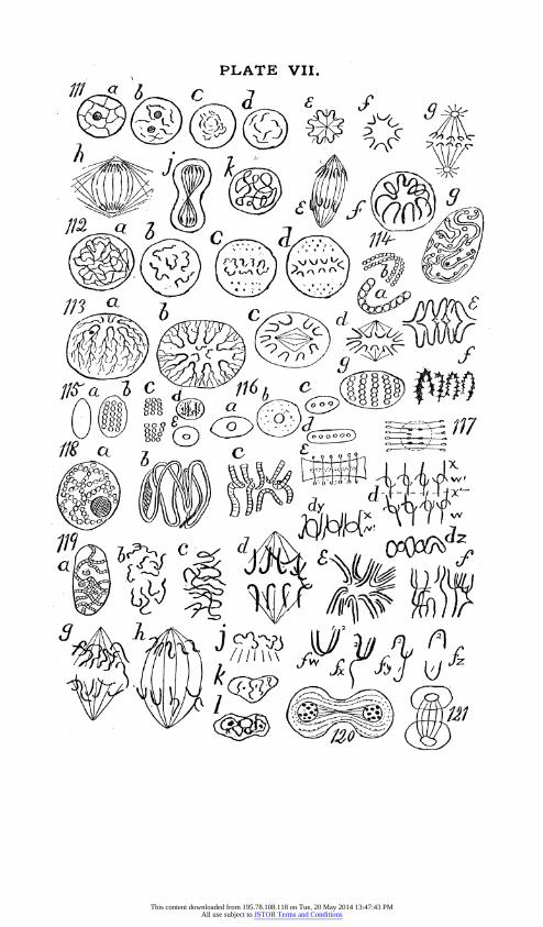
PLATE VII.
7/1P C d f o9 119 6 W an qS~~~ ~~ ~~ CA
~~ __ a ~ d
///i \\~~~~~/V
This content downloaded from 195.78.108.118 on Tue, 20 May 2014 13:47:43 PMAll use subject to JSTOR Terms and Conditions
i 887] The Significance of Sex. 141
simple constriction; the segments of the nucleolus are thus separated without a spindle,-a mode of division known as " stenosis."
FIG. I IO. Nucleus of muscle-fibre of larva of Hydrophzilus (Carnoy, p. 240) enter- ing into division. The segments of the nuclein filament are seen lying among the fibres of the spindle, which latter have been transformed from the karyoplasmic net- work.
PLATE VII.
FIG. I I I, a-k. Nucleus from cells of endoderm of Coelenterates except (k) which is ectodermal-Pfitzner, A. m. A., xxii.-a gives the " skein" reticulum; b shows the skein-filament segmented, while the nucleolus has divided. In c we get the segments in centre; in d the nucleoli have dissolved; in e the polar asters have formed a spindle, and the segments have formed a " rosette" in its equator; f shows the " ro- sette" broken up into loops by segmentation of outer limbs of the rosette. This is the " monaster" stage, and when the loops have split and duplicated themselves we get in g the " dyaster." h shows the spindle with the loops near the poles and with astral rays streaming out into the cytoplasm; j shows the constriction of spindle and of cell; in k we have a true skein-filament that does not form a reticulum.
The formation of the cell " plate," where (as in plants) there is no cell constriction, may be seen in Figs. 124, h and s, and modern text-books on botany.
FIG. 112, a-g. Karyokinesis from epithelium of Salamander-Flemming, A. m. A., xviii.-c, d are from testes as seen in living state. Here we see bodies at the poles nearly corresponding in number to the segments of the filament. When the dyaster is formed they are about twice as numerous, and strongly suggest that they are a species of paranucleolus. cf. 97)2, 103, etc. In the daughter-nuclei the series a, b, is inversely followed, as in e, f, g. In g the filament is cut across by the knife in many of its windings, thus giving us pseudo-nucleoli.
FIG. II3, a-g. Epitheliumn of Salamander according to Rabl, M. J., x.-a is a schematic side view, and b a polar view of a resting nucleus. According to Rabl the segments of the filament do not fuse or in any way anastomose in the resting nucleus, but simply branch out finer and finer. Then in kinesis the branches are withdrawn, and short thickish loops are formed. The spindle is first seen in its entirety at one pole, which, as seen in a and b, is different from the other pole, and ,then the spindle turns at an angle of 9o0 and forms the usual amphiaster (c, d). When the loops split, the halves are carried apart at their bend first, and the shorter arms of the U are first separated as seen in e. Arriving at the poles the loops branch out once more, as inf, to form the figure a. In g we see a daughter-nucleus from testicular epithelium of Proteus, where the branching does not take place, but the loops are formed of a row of microsomata (beaded filament).
FIG. 114, a-b, shows how such a beaded filament splits by each microsoma divid- ing in the general plane of the loop. Pfitzner, M. J., vii.
FIG. I 15, a-e. Nucleus from growing point of Tradescantia virginica-Strasburger, "Zellbildung u. Zelltheilung," Jena, I88o.-The nearly homogeneous protoplasm of the nucleus (a) becomes granular; the granules fuse and arrange themselves in rows of microsomata (b), and these rows are cut across (c) in the equator and pushed to- wards the. poles while undergoing various changes of segmentation back to granules again, but a central nucleolus remains undissolved, or rather is built up during the process of reconstruction of the daughter-nucleus (d, e).
FIG. I I 6, a-e. Nucleus of Spirogyra majuscula-Strasburger, 1. c.-In a we see the lenticular nucleus with its nucleolus; b is a face view showing it becoming granular. In c the nuclein has segmented (or aggregated?) into more and-more numerous masses
This content downloaded from 195.78.108.118 on Tue, 20 May 2014 13:47:43 PMAll use subject to JSTOR Terms and Conditions

142 The Szgnzficance of Sex. [Feb.
or microsomata which soon reach the granular state again (c, d, e). Meanwhile the nucleus flattens more and becomes biconcave; the granular protoplasm gathers in the concavities and sends across and through the nucleus the spindle-fibres (e); the marked boundary of the nucleus is dissolved, and the equatorial plate of granules splits and moves towards and into the polar masses, while the intervening portions of the spindle-fibres (" connecting fibrils") spread out and help build the cell-plate.
FIG. I17 is from Spirogyra ni/ida, where the nucleus is more spherical and the spindle-fibres are at first aggregated towards the centre in union with the central gran- ular. mass of chromatin, and they become more spread out as the nucleus loses in outline and the chromatin is divided into its daughter portions. We see also that the latter is confined to the central fibres of the spindle.
FiG. I i8, a-dz. Nuclei from protoplasmic layer next wall of embryo-sac of Galan- thus nivalis-Strasburger, A. m. A., xxiii.-a, first step towards karyokinesis; the microsomata form a continuous beaded filament, produced by the shortening of a finely-wound " skein" or " tangle," the meshes of whose reticulum give a finely gran- ular appearance to the protoplasm. A large nucleolus exists besides. This is dis- solving and adding itself to the filament in b, where the boundaries between the microsomata are not indicated in the diagram. c is the " segmented stage," in which we have on both sides of the equator segments whose equatorial ends are bending around like hooks (only a few segments are shown). Next, the hooks split longitu- dinally, and the halves of each hook seek opposite poles; to do this there must be a stage where one-half of the loops of the southern hemisphere cross the equator and meet corresponding hooks from the northern on their way to the southern hemisphere. (See d, where w and w' are the corresponding halves of the southern hooks, and x and x' of the northern hooks. w' and x' are represented as having just crossed.) While this "metakinesis" is progressing the hooks become more U-shaped, and taking the northern daughter-nucleus, as in dy, we can see how its skein filament is reconstituted in dz, by union of neighboring limbs of the sets of loops. (Meta- kinesis refers to the changes that take place after the splitting of the loops or micro- somnata to form the chromatin figures which are to occupy the daughter-nuclei. The preceding changes constitute the " prophase," the succeeding, the " anaphase.")
FIG. I i9, a-1. From wall of embryo-sac of Fritillria irnperialis-Strasburger, A. m. A., xxiii.-a-d represent the prophase, e-f, the metaphase, g-1, the anaphase. Here the first step of the metaphase is, as in the last case, one of mutual transfer be- tween the opposite sides of the equator. (Seef.) Following any one loop in the northern hemisphere, splitting into two halves (i, 2, Fig. fw), the part 2 is des- tined for the northern daughter-nucleus, and i for the southern. Separation between the two halves proceeds from one end to the other, so that the part I becomes pulled out straight, one limb of the part 2 is held back, while the other is dragged towards its own pole (fx), both halves are therefore acted on so as to be pulled into line par- allel with a meridian, but the end, as it approaches the pole, bends around hook- shaped to form a new loop, hence the part 2 passes through an " S" stage (fy), and the part I a hook stage, and they finally reach the U stage in fz, when the stages dy and dz ensue, as in the preceding figure. On comparing Figs. f-i, it will be seen that practically this mathematical regularity is never attained, but only distantly approximated. The number of loops in Strasburger's figures is far greater than in the diagram, but they may be deciphered and understood by means of the diagrams (Fig. f-fz). The main source of departure is found in the fact that most of the loops are not perfect U's in the anaphase, but are oftener hooks or even straight filaments lying in the meridians. In the last casefw andfx would be omitted.
This content downloaded from 195.78.108.118 on Tue, 20 May 2014 13:47:43 PMAll use subject to JSTOR Terms and Conditions
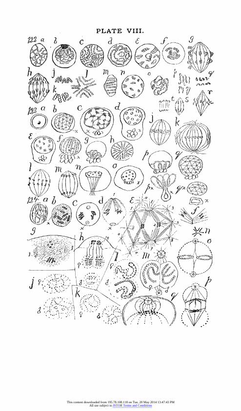
PLATE VIII.
C c 6 0 cX
t2K A), a X/
aI
k~~> ,'
This content downloaded from 195.78.108.118 on Tue, 20 May 2014 13:47:43 PMAll use subject to JSTOR Terms and Conditions
I 887] The Significance of Sex.
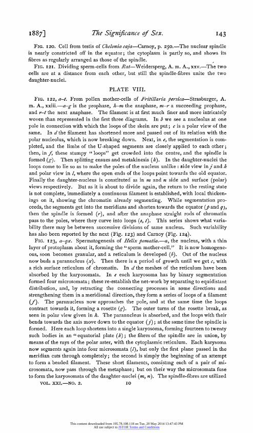
FIG. 120. Cell from testis of Chelonia caja-Carnoy, p. 250-The nuclear spindle is nearly constricted off in the equator; the cytoplasm is partly so, and shows its fibres as regularly arranged as those of the spindle.
FIG. 121. Dividing sperm-cells from Rat-Weidersperg, A. m. A., xxv.-The two cells are at a distance from each other, but still the spindle-fibres unite the two daughter-nuclei.
PLATE VIII.
FIG. 122, a-t. From pollen mother-cells of Frifillaria persica-Strasburger, A. m. A., xxiii.-a-g is the prophase, h-rm the anaphase, m-r a succeeding prophase, and r-t the next anaphase. The filament is at first much finer and more intricately woven than represented in the first three diagrams. In b we see a nucleolus at one pole in connection with which the loops of the skein are put; c is a polar view of the same. In d the filament has shortened more and passed out of its relation with the polar nucleolus, which is now breaking down. Next, in e, the segmentation is com- pleted, and the limbs of the U-shaped segments are closely applied to each other; then, in f, these stumpy " loops" get crowded into the centre, and the spindle is formed (g). Then splitting ensues and metakinesis (it). In the daughter-nuclei the loops come to lie so as to make the poles of the nucleus unlike: side view in j and k and polar view in 1, where the open ends of the loops point towards the old equator. Finally the daughter-nucleus is constituted as in m and n side and surface (polar) views respectively. But as it is about to divide again, the return to the resting state is not complete, immediately a continuous filament is established, with local thicken- ings on it, showing the chromatin already segmenting. While segmentation pro- ceeds, the segments get into the meridians and shorten towards the equator (p and q), then the spindle is formed (r), and after the anaphase straight rods of chromatin pass to the poles, where they curve into loops (s, I). This series shows what varia- bility there may be between successive divisions of same nucleus. Such variability has also been reported by the next (Fig. 123) and Carnoy (Fig. 124).
FIG. 123, a-qx. Spermatogenesis of Helix pomna/ia.-a, the nucleus, with a thin layer of protoplasm about it, forming the " sperm mother-cell." It is now homogene- ous, soon becomes granular, and a reticulum is developed (b). Out of the nucleus now buds a paranucleus (x). Then there is a period of growth until we get c, with a rich surface reticulum of chromatin. In d the meshes of the reticulum have been absorbed by the karyosomata. In e each karyosoma has by binary segmentation formed four microsomata; these re-establish the net-work by separating to equidistant distribution, and, by retracting the connecting processes in some directions and strengthening them in a meridional direction, they form a series of loops of a filament (f). The paranucleus now approaches the pole, and at the same time the loops contract towards it, forming a rosette (g). The outer turns of the rosette break, as seen in polar view given in h. The paranucleus is absorbed, and the loops with their bends towards the axis move down to the equator (j); at the same time the spindle is formed. Here each loop shortens into a single karyosoma, forming fourteen to twenty such bodies in an " equatorial plate (k); the fibres of the spindle are in union, by means of the rays of the polar aster, with the cytoplasmic reticulum. Each karysoma now segments again into four microsomata (1), but only the first plane passed in the meridian cuts through completely; the second is simply the beginning of an attempt to form a beaded filament. These short filaments, consisting each of a pair of mi- crosomata, now pass through the metaphase; but on their way the microsomata fuse to form the karyosomata of the daughter-nuclei (m, n). The spindle-fibres are utilized
VOL. XXI.-NO. 2. 10
This content downloaded from 195.78.108.118 on Tue, 20 May 2014 13:47:43 PMAll use subject to JSTOR Terms and Conditions

144 The Significance of Sex. [Feb.
in forming the paranucleus (see A. m. A., xxvi.), the karyosomata segment to form the rosette fi and px, where p is the figure in the first division and fix that of the last. The reticulum is again established in q and qx; the steps following the final division are shown in Fig. 8i, a, b, c, etc.
FIG. 124, a-s. " Cytodieresis" of the egg of Ascaris megalocepihala (a-g), from Car- noy-La Cellule, vol. ii., May, i886. h-s from Van Beneden,A. B., iv.-a is the nu- cleus of a young egg having a beaded filament forming a " skein." There is besides a nucleolus which Carnoy calls a " plasmatic-nucleolus" (x). Soon the filament seg- ments (b) into eight karyosomata, and then the reticulum of the nucleus can be seen remaining. The egg grows, and when mature, and containing the spermatozoon, the preparations for forming the polar globules are made. The poles of the egg become marked each by a plasmatic-nucleolus; and the eight karyosomata now take an equa- torial position in two groups (c), which gives to the germinal vesicle the appearance of having two germinal spots when viewed with moderate powers. The next stage shows the reticulum and wall of the germinal vesicle dissolved (which solution ap- pears with all nuclei at this stage). Two groups of fibres now ray down from one pole towards the karyosomata (d). The other half of the spindle is soon completed in a similar manner. Then from the poles, where is a granular mass called a " pla- teau," there ray out into the protoplasm, fibres to form primary asters (i). The plateaux may split and so form several secondary plateaux, one of which is shown at I., Fig. e, for each pole, but its aster is still primary, because a part of its rays enter into the nuclear spindle. There may be several of these formed by repeated split- ting of the " plateau." Secondary asters (2) are formed when the streamers flow from the karyosomata. Tertiary asters (3) are those connected with primary asters, but not a part of nuclear spindle; while quatenary asters (4) are small asters scattered through the yelk, but they may be connected with any other aster by one or two fila- ments. These asters are simply transformation of the ordinary reticulum, and no fixed law governs their production, and the utmost variety of combination may be found. The system of asters is much more complex in the formation of the second polar globule than in the first. Our description of Fig. e is from the second " cary- ocinetique figure." The asters finally fade out until only one plateau with its bilat- eral spindle is left; this often closes up on itself (f), so that the polar plate looks like an equatorial plate; the karyosomata are thus carried around into a plane at right angles to their old position, and tend to approach each other. But this mode of dis- appearance of the asters is only one of several. The last trace of the spindles and asters disappears (g), the plateaux are reduced to granular spots, the reticulum of the plasma is restored, but not as yet the membrane of the nucleus. Now the reticulum produces a simple spindle anew (the " spindle of separation") between the two groups of kary- osomata, and the most peripheral one is cut off by an equatorial cell-plate, much as in plants (A). The segment cut off, which sometimes contains part of the yelk, is the polar globule. Then the four karyosomata of the yelk separate into groups of two each, and the process is elaborately repeated, so that only two karyosomata are left in the yelk to form the female pronucleus. The karyosomata in the polar globules may divide by karyokinesis. After the last polar globule has been extruded (A) the two karyosomata of the female pronucleus segment up into microsomata, and a similar figure is seen with the nuclein of the male pronucleus. j shows the two in a stage where a beaded filament is being established by concentration and shortening; k is a further stage; I shows the filament splitting in each pronucleus, but each first segments transversely into two; the pronuclei are closely applied to each other in a bilateral way, as seen by the position of the polar asters, which now appear (m). The loops
This content downloaded from 195.78.108.118 on Tue, 20 May 2014 13:47:43 PMAll use subject to JSTOR Terms and Conditions

I8871 The Significance of Sex. 145
finally arrange themselves in the equator, so as to show in polar view as in n; two of these loops were furnished by the male and two by the female. A side-view of this stage is shown in o, where the filaments have split in the middle, but not yet at the ends and the centre. Inp the V-shaped loops have diverged towards the poles quite a ways; the central apices, however, move faster towards the poles than the outer limbs. g shows the microsomata somewhat irregularly segregated at the base of a figure formed by the polar aster and spindle of conducting fibre, and at the apex of the spindle of connecting fibrils, which is now constructing the cell-plate as in s. This seems to show that the connecting fibres and the conducting fibres belong to distinct systems, which is more clearly shown in r, where the karyosomata are placed at the cross-points of the meshes formed by the interlacing of the two systems. In the con- struction of the daughter-nucleus the microsomata pass by segmentation into a knauel like that seen inj and k, and only when the equatorial plate is again formed for sub- sequent division do we get the four loops once more established, as is seen respect- ively at the left and right of s. Van Beneden, however, ignores the evidence of his own figures, and states that the four loops remain distinct throughout. (See text for further discussion.)
(C) KARYOKINESIS.
TWO extreme types of cell-division are known; in one, the nucleus simply constricts into two halves that move apart,
followed by a similar constriction of the cell-body, so that each of the daughter-cells is provided with its own nucleus; in the other type the nucleus undergoes changes by which it becomes invisible to the microscope, unless the cell be treated with proper reagents, and as the partition which divides the cell-body appears, there is gradually built up a nucleus in each of the daughter-cells. The former type is known as, direct division, the latter as indirect division. The term karyokinesis (nuclear motion) is usually re- stricted to the latter kind of division, but we are learning that there are many forms of indirect division that gradually unite the two extremes, so that we can no longer make the above dis- tinction. The term karyokinesis admits readily of a broad sig- nification, and we shall use the word as including all sorts of nuclear transformations.
Our knowledge of cell-structure and of the nucleus has won- derfully increased since i833, when Robert Brown discovered the nucleus while studying the generative organs of orchids, and Von Mo/l (1835) first saw it divide. To-day we are making as rapid progress in this direction as ever, and there is no field of biological research which offers so great inducements to the in- vestigator, or so valuable results as this.
In all our progress there has been but one tendency, and that is to show us that the cell, and especially the nucleus, is a com-
This content downloaded from 195.78.108.118 on Tue, 20 May 2014 13:47:43 PMAll use subject to JSTOR Terms and Conditions
146 The Significance of Sex. [Feb.
plex and highly-organized structure. We can no longer use the term protoplasm in its old sense of one definite substance whose remarkable properties are due to the great chemical complexity of its molecule.
i. Historical.-In the works of Schieiden and Schwann (1838- I840), which established the cell-doctrine, the cell was described as originating by the activity of the cytoblast (the nucleus), which was itself due to a condensation of granules in the cell-substance of the mother-cell. The endogenous origin of cells and cell-nuclei was, however, gradually overthrown, and in i855 Remak estab- lished the generalization that all cells are due to the division of pre-existing cells in such manner that the nucleolus first divides, then the nucleus, and lastly the cell-body. This schema could rest only on the facts of direct division and a superficial observation of indirect division. As soon as the latter was carefully studied by Hoffrneister, in i867, he found that the nucleus disappears and two centres of attraction arise in the cell, in connection with which the daughter-nuclei were built up. These facts had been observed in animal-cells already in i858, by Munk, so that the view that a cell must return to a cytode condition to divide and so, in a manner, be rejuvenated, and produce new nuclei endoge- nously, was fairly established. This school was strengthened by receiving the support of all who had observed the maturation of the ovum (except Warneck, i850), for here it was seen that the germinal vesicle disappears before segmentation, and that the nuclei of the segmentation products arise as new structures, and, moreover, Valette St. George had, in i866, shown that the ovum is a cell, the germinal vesicle a nucleus, and the germinal dot a nucleolus.
The complex nature of cell-structure was surmised by Briicke as early as i86i, although the microscope had then only re- vealed granules, and that these were at times arranged in a radiate manner with reference to the nuclei.
In i865, Fromnmann, through extended research, described the reticulate nature of protoplasm and generalized that this was the typical structure of protoplasm, but his views remained for many years unnoticed.
In i873, Heitznann advocated the view that whenever a pro- toplasmic mass was fully developed it became vacuolated, and so showed a reticulated structure in which the nodal points ap-
This content downloaded from 195.78.108.118 on Tue, 20 May 2014 13:47:43 PMAll use subject to JSTOR Terms and Conditions
-i887] The Szgnzicance of Sex. I47
peared as granules. The nucleus is only a large nodal point in the centre, and as this developed it repeated the process, and finally the nucleolus in a mature cell takes on the reticulate structure. He laid the basis for Nigeli's theory of heredity by advancing the notion that the reticuli of all the cells in the body are continuous, and so anticipated modern studies of protoplasmic continuity.
This year is memorable as marking the beginning of studies on karyokinesis. The stimulus came from a paper by Schneider,' in which the different phases were pretty well described, though their connection and sequence were unknown. Even the spindle and cell plate were figured. Bfitschzli and Fol confirmed these results, the former mainly as to the nuclear rosette and its sepa- ration into two halves to constitute the daughter-nuclei, while the latter got the asters and spindle best; hence the former agreed with Schneider that there was no reconstitution of the nucleus, while the latter inclined to side with the orthodox school.
Auerbach now appeared with his "Organologische Studien" (i874). He starts with Heitzmann's views as to the organization of protoplasm, but considers the nucleus to be a sap-cavity into which molecules of protoplasm wander and grow to become nucleoli. These multiply by division, so that old cells have many nucleoli. The cells of highly-organized tissues, he says, have more nucleoli than cells lower in the scale of organization. The nucleoli are young cells, and they are simply separated into two groups in direct division; but in indirect division, which he dis- tinguishes as palingenetic, these are dissolved into a molecular state in the nuclear sap, and then absorbed with the sap by the cell-plasma. This process is termed karyolysis, the spindle with its polar asters is the karyolytic figure and the simple expression of the streaming out of the nuclear substance. Later, near each star, the sap and molecules return to form a daughter-nucleus. This seemed a pretty fair explanation, and Flemming at this time was much influenced by it.
Bfitschli, however (i875), opposed the theory, though he modi- fied his former view of the simple persistence and division of the nucleus to the view that the nucleus is reconstructed into a spindle, at whose equator the fibres become thickened to form the nu- clear plate, which plate by splitting passed its halves to the poles of the spindle to be re-formed into nuclei. In the same
A list of the papers referred to will be given at the close of the article.
This content downloaded from 195.78.108.118 on Tue, 20 May 2014 13:47:43 PMAll use subject to JSTOR Terms and Conditions

I48 The Significance of Sex. [Feb.
year the first edition of Strasburger's work on cell-division ap- peared. This treated in the main of the plant-cell, where the spindle thickenings after separating leave between themselves connecting fibrils that are more prominent than in the animal- cell. These he called nuclearfibrils, and at their equator a second set of thickenings appear that go to construct the dividing wall between the new cells, hence he named it the cell-plate. The second edition of this work appeared in 1876, and the third in i88o. In the last he changes the name he gave the connecting fibres to cell-fibres, because he supposed that they were formed from the cytoplasm penetrating into the nuclear matter at the time of the deconstitution of the latter.
Van Beneden, on the other hand, agreed with Biitschli that the spindle comes from the old nucleus. He distinguishes between the nuclear sap and the nuclear essence. The connecting fibrils are of the same essence as the nuclear disk, and are due to the draw- ing out of the elements as they segment into the daughter-disks.
In this year, also, Hertwig showed that the egg-nucleus does not disappear during cleavage, but passes through a metamorpho- sis similar to the cell-divisions described by Biltschli. At the poles of the spindle and in the centre of each aster he finds a polar corpuscle. Fol had seen corpuscles in the stars, but had confounded them with the daughter-nuclei. The following year, 1876, Fol corrects this error. Balbiani found the nuclear plate to be composed of rod-like elements that were composed oafgranules, but these views were unnoticed, so that Pfitzner received the honor of their discovery five years later.
At this time the elements which compose the nuclear plate were not distinguished from the spindle-fibres, due to the fact that reagents which made the one visible left the other obscure, hence there was a good deal of contradiction in the results, which was unreal.
In this year Biitschli's chief work appears. He supposes the infusorian nucleus to represent the original type of nucleus. He thinks the cytoplasm stimulates the nucleus to division, though it may not itself necessarily follow the example. The rays of the stars are not the expression of attractive forces of the nucleus, but are rather due to a chemical influence. He found that the asters when they first appear near the nucleus, are not necessa- rily at opposite poles. R. Hertwig had divided the nuclear mat-
This content downloaded from 195.78.108.118 on Tue, 20 May 2014 13:47:43 PMAll use subject to JSTOR Terms and Conditions

I 887] The Significance of Sex. I49
ter, like Van Beneden, into sap and nuclear substance, Strasburger now proposes the following scheme. There are in the nucleus three formed substances, one of which is active. By the excita- tion of the cytoplasm the active substance gathers at the poles of the nucleus, leaving the spindle-fibres stretching between; the latter are cytoplasmic in origin, and the polar substance acts on them just as it acts on the fibres of the cytoplasm which form the stars, hence the general disposition of the polarized mole- cules in rows radiating away from the polar area. The third substance is first repelled from the poles to form the nuclear equatorial plate; but in some way there is a change of polarity by which it is subsequently attracted to the poles, and so the plate splits in two (it might also do this through internal repul- sion, but, as we shall show farther on, there is no necessity for a physical explanation). This is perhaps the best of the few theo- ries which have been advanced to account for the phenomena.'
On the question of the solution of the nucleus, Fol now took a middle ground, holding with Strasburger that the cytoplasm entering formed the spindle, but the nuclear matter simply be- came continuous with the cytoplasm through the dissolution of the nuclear membrane.
In i878, Schleicker advanced the view that the protoplasm was composed not of parts that had a fixed relation to one another, but of units that were independently mobile, and so all the struc- tures were amceboid in form, hence there could be no definite phases during cell-division, wherefore he proposed the term karyokinesis to designate the phenomena. On the other hand, Flemming had, by careful staining, worked out the series of forms through which the nuclear matter passes during karyokinesis. He did not get the spindle well because, as he showed the next year, this was composed of non-stainable matter. The resting nucleus consists of a vesicle enclosing a reticulum and one or more nucleoli. This reticulum is changed to an exceedingly long and intricately wound filament, at the same time the nu- cleoli dissolve in the sap and the filament absorbs the material. This is the phase of the close kniiuel. (Fig. II 2, a.) The filament now shortens and grows thicker, passing through the open
TWe give the theory in its most developed form, third edition of Strasburger's work, i88o, where he slightly modified it from its original statement in the second edition.
This content downloaded from 195.78.108.118 on Tue, 20 May 2014 13:47:43 PMAll use subject to JSTOR Terms and Conditions

I50 The Szgnwficance of Sex. [Feb.
knauel stage, until it shows as a rosette (Fig. III, e), with loops turned peripherally and towards the centre. Then the outer limbs of the loops break, leaving a lot of V-shaped filaments having their apices towards a common centre. (Fig. 1o5,f.) This is the " mother-star." Meanwhile the central point of attraction splits and moves to the poles, where asters now appear. This is accompanied by alternate expansion and contraction of the nuclear star (diastole and systole), and finally results in its flat- tening into an "equatorial plate" (Fig. I 13, d); then each loop splits lengthwise, though it may have done so while still in the mother-star (Fig. I i9, e), and thus formed the "fine-rayed star." The halves of each loop become separated and grouped in a " dyaster," or two daughter-stars, passing through the phase which shows as a splitting of the equatorial plate. Then the apices of the loops (travelling along the spindle-fibres) are drawn towards the poles (Fig. I I2, e), drawing the limbs after them, and so reach the pole. Here they form into a figure like the old mother-star (Figs. II 2, ?, I05, A), and return, by uniting ends through the rosette (Fig. I I8,f) into the knaiuel form, and finally become like the mother-nucleus.
The next year Flemming divided cell-division into direct and indirect. He limited the former to motile cells, and accepted Schleicher's term as applying to the indirect kind. He thinks the nucleoli are an accidental thing in a nucleus, and according as nuclear substance stains or not he calls it chromnatin and achro- matin. In the same year, i879, Fol proposed his electrolytic theory of cell-division.- He believed the nuclear reticulum was directly transformed into the spindle, and the nuclear plate was due to an equatorial thickening of its fibres.
Strasburger's studies gave him different results from Flemming. In plants the phases are not so marked, but may give a spindle figure of chromatic granules arranged in rows like the staves of .a cask; and the daughter-nuclei arise through the simple break- ing of these across the middle. (Figs. I 15, a-d.)
In i88i, Retzius had confirmed the phases of Flemming, but showed that the rosette must be given up, as segmentation of the kniauel may take place while in the loose or open kniauel stage. (Fig. I I2, b.) He says the chromatic substance is contained in a hyaline matrix, as Pfitzner has shown, and most of it is absorbed by a particular node or nodes, and these are the nucleoli.
This content downloaded from 195.78.108.118 on Tue, 20 May 2014 13:47:43 PMAll use subject to JSTOR Terms and Conditions

I 887] The Szgnificance of Sex. 15 I
Pfitzner, this year, called attention to the fact that the nuclear loops are composed of granules like a row of beads, and that the loop splits by the segmentation of each granule. He thought these granules to be the protoplasmic molecules, but later (i883) said they were independent and individual units in the nuclear structure. (See Fig. I 14, a, b.)
In i882, Strasburger proposed the terms cytoplasm, microso- inata, nucleoplasm, etc., which we adopted in Section a of this paper. He studied more carefully the method of rearrangement of the loops in the equatorial plate during its division, and finds that it is more complex than Flemming made it, for the old bend straightens out, and one end of the loop gets drawn towards the pole, and then bends like a hook, and this new bend travels along the filament to its middle point, thus making the two limbs equal, while at the same time the loop is drawn polewards. Strasburger had not yet discovered the splitting of the loops, so that he had as yet only an imperfect notion of how the rearrangement took place. (See Figs. i i8 and I 19 for the actual facts.)
In this year appeared Flemming's systematic work on the cell, and in it he accepts the criticisms of Retzius and Strasburger, so far as they relate to the rosette phase and the " rearrangement." lie doubts if there is a reticulum in cells, or at least in nuclei. The appearance may be due to the optical effect of a closely- wound filament or mitomn, the sap is the paramitom, and karyo- kinesis should be termed mitokinesis, or mitosis. Besides the mitom there are chief and accessory nucleoli. He gives up the idea that the chromatin may dissolve in the cell-sap. He gives us the term spirem for the kniuel phase. Rearrangement in the equatorial plate is termed metakinesis. He uses the term aster for the star-form of the mother-nucleus, and dyaster for that of the daughter-nucleus.
In i883, Pfitzner makes three sorts of chromatic substance in the nucleus. The substance of the spindle, hitherto called achro- matic, he terms parachromatin, while the sap only is true achro- matin. The nucleolus has prochromatin, while the mitom has the true chromatin. In the resting nucleus there maybe, besides Strasburger's membrane, which belongs to the cytoplasm, a true nuclear membrane of parachromatin.
Roux, in this year, proposes a theory of karyokinesis, based on the idea that there is a mixture of qualities in the chromatin,
This content downloaded from 195.78.108.118 on Tue, 20 May 2014 13:47:43 PMAll use subject to JSTOR Terms and Conditions
152 The Szgnificance of Sex. [Feb.
etc., and that these have to be distributed in a definite way be- tween the daughter-cells at each division. Hence the complicated machinery.
In i884, Rabl seeks to show that there never is a simple fila- ment in a cell, but that the loops pre-exist even in the resting state, and that in this latter state the chromatin flows out along definite paths into finer and finer branches, which never anasto- mose, but may swell up at points, where a special lot of chro- matin gathers, and there form nucleoli. The cell is heteropolar, always having the apices of the loops directed towards the principal pole. (See Fig. I I3, a-f.)
Heuser agrees with Rabl in finding the mitom segmental in the resting phase.
In this year Strasburger discovers the splitting phase in certain plant-tissues. (Figs. I i.8, I i9.)
Carnoy now comes to the front with important contributions. He finds, what has not been noticed by previous observers in its true light, that there exists a true reticulum in the nucleus, like the reticulum in the cytoplasm (Fig. 4), but that the mitom in its convolutions hides this, and gives us the aspect of a coarser reticulum (Fig. 3), which is the one referred to by previous in- vestigators. The mitom may be itself reticulated (Fig. 13), or its segments be short and rod-like in some cells (Figs. 123, I24), but besides these there are the nucleoli. All the nucleoli are not composed of true chromatin. There are one or two that are composed of plastin (Figs. 44, I24, b, c), like the true reticulum, and with it and the membrane become transformed to the achro- matic or spindle part of the figure. The phases of Flemming are realized only in a limited number of cases, and there appears to be the utmost variety in the karyokinetic figures; we may get forms where the nuclear reticulum does not become transformed, although the mitom may segment. (Figs. io9, I io, slenotic di- vision.) Again, the achromatic part of the figure may in one and the same cell differ under different circumstances from great complexity to simplicity of structure.'
Platner has also made important contributions (see Fig. I23,
a-qx); but these will be considered in another connection.2
I See also, Lee, " Carnoy's Cell Researches." Q. J. M. S., April, i886. 2 The latest paper by Platner (see Bibliography) has just come to my notice.
Platner thinks the spindle fibres are one continuous filament in which is a proto-
This content downloaded from 195.78.108.118 on Tue, 20 May 2014 13:47:43 PMAll use subject to JSTOR Terms and Conditions

I 887] - The Significance of Sex. 153
Thus we get a notion of the cell as a most varied organism. Cells may be as varied among themselves as the higher organ- isms. But we have hardly begun to get an idea of the variety of karyokinetic phenomena; what is to appear by future study we can only vaguely imagine. The phenomena of cell-division can- not be purely physical phenomena. They are living phenomena, and show they are subject to the laws of heredity and evolu- tion of adaptation and variation. If this be so, we can under- stand karyokinesis only through comparative studies, just as we study the laws of variation and evolution, of homology and affin- ity with higher organisms. It becomes important, therefore, to understand karyokinetic phenomena among the Protozoa. Re- cent studies in this line have shown that here we may get nearly as complex figures of karyokinesis as in tissue-cells; but from this we get all grades down to the simplest direct division. We learn that nuclei may be alike in distantly-related forms, while closely-allied forms may have very diverse nuclei.
2. Protozoa.-These organisms present, as we should expect, a great variety of nuclear forms and karyokinesis. The differ- entiation has been in so many different directions, with the ac- quirement of secondary characters whose physiological signifi- cance we can hardly guess, that it is perhaps impossible to connect the forms. Frey thinks the vesicular form of nucleus is the primitive one, but most writers agree with R. Hertwig in de- riving this form from a solid form through vacuolation of the latter, which leaves the chromatin either all in a cortical zone, or else in one or more nucleoli; and this process may be repeated in a nucleolus when this becomes large and important. Gruber suggests that in a primitive state the chromatin was present throughout the cell in a granular form, as in Triclrosphmrium, Pleurophrys, Trachelocercus, Chcenia, and others, and that solid nuclei arose by fusion of these granules, or by some of them re- maining in close union following their multiplication. But it must be observed that this granular condition could easily arise by the segmentation of larger bodies and so be secondary in
plasmic streaming in one direction. Each meridian bears a karyosoma that splits, because the fibre splits; thus, always in a meridional plane. The parachromatic mitom is so wound that the current is towards opposite poles in neighboring fibres after the splitting, hence the daughter karyosomata are swept to their proper poles. This theory appears to be as weak as its predecessors.
This content downloaded from 195.78.108.118 on Tue, 20 May 2014 13:47:43 PMAll use subject to JSTOR Terms and Conditions

154 The Significance of Sex. [Feb.
character. In some forms we get either many small nuclei or else granules, besides one or more chief nuclei, and, according to Altmann, this is true of all tissue-cells, so that we have nuclear bodies first differentiated in two directions, the granules serving some nutritive or other function, while the nuclei retained the office of being the primary reproductive bodies.
The next differentiation arising would be the differentiation of the nuclei into two kinds, which in some Ciliates have acquired considerable independence and act quite differently during di- vision. We know them as nuclei and nucleoli, or as nuclei and paranuclei, respectively. Better terms are Huxley's endoplast and endoplastule, as not implying homologies which are probably false. In massive nuclei the chromatin exists in a fine net-work, which gives the appearance of granules in the resting phase and of fibrilla during division. In Gastrostyla the endoplastules divide by a true spindle and a nuclear plate of karyosomata. Nearly all the nuclei of Opalina are of this sort.
The substance of the nucleus may differentiate into two sorts that gather in two portions of the nucleus, either by polar dif- ferentiation or by centripetal differentiation. One substance is hyaline, the other granular; examples are Leptodiscus, Spiroc'hbna, and Noctiluca. In Spirochona it is the endoplast which has this structure, and it divides by a complex kinesis. A nucleolus appears in the clear part, which becomes transformed into fibrils while the hyaline portion gathers as two polar plates that sepa- rate, and so, as it were, tear the nucleus in two. The three endoplastules present divide by simple constriction at the same time; and here also there are polar plates of a substance different from the equatorial portion.
In a different direction we get the vesicular differentiation, and this is most common in the lower Protozoa, and is that from which the metazoan cell-nucleus is derived. In Rhizopods we may not only get many nuclei (about two hundred in Pelomyxa), but each nucleus may present a great variety of phenomena. There may be a central nucleolus, and this nucleolus may be composed of many microsomata, or the microsomata may sepa- rate as so many nucleoli, or again may fall into granules. To- gether with all these forms there may be a cortical shell of microsomata, for examples, see Figs. 14, a, b, 20, 22. The multinuclearity of many of the Protozoa is due to the fact that
This content downloaded from 195.78.108.118 on Tue, 20 May 2014 13:47:43 PMAll use subject to JSTOR Terms and Conditions
i887] The Signifcance of Sex. I55
cell-division is purely facultative, and has not been inseparably associated with nuclear division. When it takes place, as in Opalina, any number of nuclei may be separated away in the daughter-cell. In many cases the nuclear divisions affect only the chromatin, while the hyaloplasm and its nucleolemma remain as funiculi uniting the segments. (See Figs. 42, 31, 29, 28, 4I, etc.) The microsomata of a nucleolus or of a karyosoma behave in the same way; as in Figs. i9, d, 43, I14, 115, 123,g, 124, 0.
In Radiolaria we get a multitude of small " massive" nuclei that divide by ameeboid constriction (Fig. 25), and in the central capsule vesicular nuclei, whose different metamorphoses are shown in Figs. i6, I7, i8, and i9.
In division of vesicular nuclei we get, as the simplest form, the Remakian scheme. (Fig. ioo.) Next we may get the granular contents arranged in fibrils that are bisected by the constrict- ing nucleus (Fig. ioi), or they may remain in the granular state. (Fig. 99.) The most complex case is given by Actinosphcerium eickornii. (Fig. 103, a-q.) Here the nucleolus separates into two bodies, one containing chromatin and one the parachromatin; then each body segments into fine granules; these granules get arranged in fibrils; the chromatin-granules fuse to karyosomata lying in an equatorial plate in the spindle formed by the paeachro- matin; the karyosomata divide into daughter-karyosomata that pass towards the poles; on its way each daughter-karyosoina segments to microsomata; the microsomata segment to granules, which, however, form separate karyosoma-like masses until they are absorbed by the polar plates. The latter are due to the fact that the external protoplasm had gathered at two opposite poles of the nucleus and had attracted the parachromatic cortical layer of the nucleus. Why the protoplasm should gather at the poles and so induce nuclear division is unknown. Possibly substances in the nucleus have first passed to the poles and attracted the cytoplasm. These substances may be the segments of the para- nucleolus, for it is possible to derive the spindle-fibres from a dif- ferentiation of some of the chromatin-granules into hyaloplasm.
It is rare to find the nucleolus (or the granules that represent it when it is segmented) in the same condition during nuclear di- vision as during the resting phase. There is a cycle of changes, so that one condition has to be assumed for division, and then the chromatin returns through inverse stages to its resting state.
This content downloaded from 195.78.108.118 on Tue, 20 May 2014 13:47:43 PMAll use subject to JSTOR Terms and Conditions
156 The Significance of Sex. [Feb.
The phases of this cycle may be few or they may be many, and besides, the phase in which the nucleus rests may be in one case in one point of this cycle, in another it is a different phase of this cycle. But in all cases the cycle is made up of stages of fusion or of segmentation between the two extremes of one single nucleolus and of numerous granules. (The law is unchanged even if we sup- pose that the granules are the nodes of a reticulum.) Usually nuclear division follows that point in the cycle where the chro- matin is most condensed. Thus, even in the Remakian schema, this law is followed, as see Figs. 99, ioo, IOI. Cases like Figs. lOT, 103, 104, and karyokinesis in Metazoa are related to the Rema- kian schema by a compounding of the latter, for each karyosoma fol- lows the Rernakian schema. We thus see that direct division is to in- direct division as a unicellular or unisegmental animal is to one that is multicellular or multisegmented. If we study the cell-divi6ion of mzltinuclear Pro/ozoa we find thfat the same laws hold. Opalina is an exception, and such forms as Polykrikos (Fig. 96), that have only nuclear division during cell-division, are connecting links to the far larger class of cases, where, as in Oxytricha (Fig. 95), Paramoccium (Fig. 97'2), Stentor, etc., there is fusion of the nuclei (or of nuclear segments in moniliform endoplasts) before division and multiplication of these bodies preceding, during, or following division. In Paramoccium the resting phase concurs with the mononucleate phase. But even in the exceptions to this law there is fusion, when these forms encyst to produce spores (Fig. 97) by the successive or by simultaneous division of the fused nucleus. In such cases, as in conjugation of low forms, the phenomena may be facultative, the number of nuclei resulting from the fusion being one which varies from one to the original number, just as the resulting spores may vary in number.
What is the meaning of this ? It is evident that we have here to do with coinjugative phenomena. These self-same nuclei that with one form of cell-division fuse, may, in case of buds, be set free, as microgonidia to fertilize other gonidia. It may be that the granules and nuclei are in different parts of the cell subjected to different conditions, and thus come to vary slightly. If, now, the cell divided, the daughter-cells would differ, and ultimately new species be formed. But, as we find that if conjugation phe- nomena be left out in one place in the different modes of repro- duction it is apt to occur in other points of the cycle, and if
This content downloaded from 195.78.108.118 on Tue, 20 May 2014 13:47:43 PMAll use subject to JSTOR Terms and Conditions
1887] The Significance of' Sex. I57
prevented, as in male and female parthenogenesis, the resulting organisms are weak, we must conclude that the organism derives some benefit from the mixture of chromatins that are slightly dif- ferent. In the case assumed above, where cell-division is not ac- companied by fusion of the nuclei, we may presume that conju- gation of gametes supplies this lack,' though in Opalina the only form of fertilization as yet discovered is that of nuclear fusion during encystment. But Opalina is a parasite, and parasites, we know, get along remarkably well without fertilization.
This explanation of karyokinesis seems quite plausible, so that we may formulate the law that every cell-division is preceded by fertilization phenomena: is accompanied by close inbreeding. Other explanations have been suggested. Strasburger regards karyo- kinesis as resulting in like cells, therefore the different chroma- tins present must be carefully divided, so that each daughter-cell shall receive its proper ingredients. Roux and Weismann go further and call attention to the fact that dividing cells are not alike, so that we need a particular distribution of the gemmules for each division, so that, to follow the last author, in the first egg-segmentation, the histogenic plasm is separated from the generative plasm. Thus the soma is descended from one blasto- mere and the generative cells from another: In a similar way the endoderm-cells have a common ancestor, and the ectoderm another, and so on. This explanation seems to me difficult of application to the Protozoa, so that the laWv enunciated above appears to be unaffected.
3. General.-It would extend the length of this paper unduly, as well as be of no interest except to the specialist, to discuss the various views that have been advanced in interpretation of the special stages of karyokinesis. Nevertheless, some of the more prominent features should be noticed. We have already seen that cell-division need not be necessarily associated with nuclear division, whether this be direct or indirect, so in the higher tis- sues there occur cells that present such conditions. These are the internodal cells of Chara, many fungi where the nuclei are granules, and the generative tissues of both plants and animals; also in cartilage-cells and marrow-cells. In the generative tissues of plants cell-division does not take place until many endosperm
I The formation of varieties and species must take place in spite of this tendency of differentiated chromatins to fuse.
This content downloaded from 195.78.108.118 on Tue, 20 May 2014 13:47:43 PMAll use subject to JSTOR Terms and Conditions
158 The Significance of Sex. [Feb.
nuclei are present, and to this form of division the term free-cell formation has been applied, while the term in the sense in which Schleiden used it has been pretty nearly abandoned. Still, we saw that in the Protozoa nuclei could arise by the fusion of gran- ules, and return by fragmentation to the granular state, and it may be a question whether similar phenomena may not be found in tissue-cells. The cases of the endogenous origin of nuclei are reported in eggs that have so much yelk that it is hard to follow the nuclear changes.
Does the cytoplasm or the nucleus stimulate division ? Hertwig holds that the nucleus is the automatic centre which controls the individuality of the cell, but it must be confessed that the earliest changes are in the cytoplasm. Protoplasm gathers at two points and forms stars, between which the nucleus becomes stretched .out and transformed. Carnoy (Fig. 124), and lately (i886) Hert- wig, have found independent stars arising in the cytoplasm, and if more than two of these get connection with the nucleus, there are as many poles and spindles or resulting daughter-nuclei as there are asters. The rays about these asters are simply a transforma- tion of the reticulum. What their function is we can only guess with the numerous guesses made by predecessors. They may be nutritive, may be paths for travelling gemmules, may have a nervous function, or finally only serve motor functions. The spindle-fibres are a similar transformation of the nuclear reticu- lum (i.e., the parachromatin reticulum); whether the nuclear membrane in dissolving adds to their material, or gathers at the poles of the spindle as in Actinosphaerium, may be doubtful. In the latter case it would continue its original function of medi- ating between the intra- and the extra-nuclear reticulum. But Strasburger and others find that the astral- and spindle-fibres are continuous, and thinks the latter come by a penetration of the former through the poles of the nucleus. But the mass of evi- dence is against him, and besides, the spindle-fibres are composed of parachromatin (chemically, plastin) and react differently from the extra-nuclear fibres.
Then there are the polar corpuscles in the centres of the stars, and forming the apices of the spindle. Their origin is obscure. Possibly the plastin nucleolus of Carnoy may, by its division and migration, have initiated the division of the nucleus, and is rep- resented in these corpuscles. It is certain that these corpuscles
This content downloaded from 195.78.108.118 on Tue, 20 May 2014 13:47:43 PMAll use subject to JSTOR Terms and Conditions

I 887] The Szgnificance of Sex. I59
may divide, and so split the spindle (Fig. I24), and often the two stars first arise close together, and move later to opposite poles. We must also notice Rabl's discovery of a spindle intact in the nucleus, which rotates into its position (see Fig. II3, c) through an angle of go'.
In what condition is the chromatin in the resting nucleus ? It may be present as a fine or as a coarse reticulum, closely inter- penetrating the parachromatic reticulum, and perhaps fused with it. In the germinal vesicle it is present as a nucleus, which be- comes transformed into the reticulum before division. The next change this reticulum suffers is its transformation into a mitom,- i.e., a filament,-which, while it is very long and closely balled up, may not be distinguished from a reticulum. Some nuclei rest in this phase, or in subsequent phases of its shortening and conse- quent thickening. When thick it has been found to be com- posed of granules that fuse to form microsomata, so that finally the mitom is one microsoma thick. The next phase is one in which the mitom segments into loops or filamentous karyosomata. Some cells rest in this phase. (See Fig. 124.) When the segmen- tation occurs early, while the reticulum is being transformed into the mitom, we get a condition of things represented in Figs. I 13 and 45. The karyosomata are apt to be short or corpuscular in generative cells. (See Figs. I22 and 123.)
Now we have two ways in which the karyosomata are sepa- rated into two groups to form the daughter-nuclei. In one they are separated without accompanying division, as in Fig. 124. In the other they divide in such a way that each half of the karyo- soma is destined to pass into different daughter-nuclei. In this case we get two forms. In one form the karyosomata, if they are short, become arranged into a nuclear plate in the equator of the spindle, and by division and separation of the halves we get two daughter-plates that pass to the poles, and become the daughter-nuclei, but if they are long, they lie along each spindle- fibre and are bisected in the equator as in Figs. ioI and I I5. In the other case the daughter-segments are produced by a lonigi- tudinal splitting of the loop, as shown in Figs. 114, I i8, and I i9, in which splitting usually occurs early, while the spirem figure still persists, and in this case no true equatorial plate may form, but be only the expression of separating loops passing each other on the way to their respective poles. Between all these different
VOL. XXI.-NO. 2. I I
This content downloaded from 195.78.108.118 on Tue, 20 May 2014 13:47:43 PMAll use subject to JSTOR Terms and Conditions
i6o The Significance of Sex. [Feb.
methods there are connecting links. Carnoy calls such a case as is seen in Fig. io9 stenotic division, while similar separation with more complex spindle and asters, as in Fig. I24, he terms cyto- dieresis.
Finally, the karyosomata reach the poles and pass through stages of fusion of the larger bodies and segmentation of the smaller, so that the nucleus appears homogeneous because of its very fine reticulum. Then from this point the changes continue along the upward path to the resting phase, wherever that may be. While undergoing this fusion, the hyaloplasm in which the chromatin-granules are imbedded become much increased, so that if the chromatin is sparse we get vesicular unions, like Fig. i25. This hyaloplasm is parachromatin, and it undoubt- edly enters partly into the formation of the spindle-fibres; in Fig. 126 appears to be the only source of these.
Concerning the nucleoli that may be present, besides the re- ticulum or mitom and the plasmatic nucleolus, there exist the most diverse views. In the first place, we must call attention to the fact that very diverse structures have received this name by different writers. The nodes of the reticulum, the karyosomata, the groups these may form when unresolved by the lens, all have received this name. The true nucleolus seems to disappear during division, and to be gradually built up by fusion of gran- ules at its close. It has been supposed that it dissolved in the nuclear sap, and was absorbed by the mitom, or that it was di- rectly connected with the mitom, and so incorporated into it. Thus Pfitzner called its substance prochromatin, as being a store from which the mitom replenished itself, but has lately changed the name to pseudochromatin, and other authors think it is of accidental value. Those who think with Strasburger, Fraisse, Kassel, and Brass that the chromatin is food-substance and the hyaloplasm the real idioplasm, find no difficulty with this body. But it must be remembered that it also has hyaloplasm, so the difficulty is unsolved. What is the meaning of those polar cor- puscles (not to be confounded with the polar corpuscles consid- ered above) seen in Fig. I 12, c, o, d, which multiply as the loops multiply, and whose number is approximately two or three times that of the loops ? It almost seems as if they were related to the karyosomata, as the endoplastules of Protozoa are to the endo- plasts. They may have come by the segmentation of the nucle-
This content downloaded from 195.78.108.118 on Tue, 20 May 2014 13:47:43 PMAll use subject to JSTOR Terms and Conditions
1887] The Significance of Sex. i6i
olus, and in that case this body is a paranucleolus over against the mitom. But these bodies may have come from the plasmatic nucleolus of Carnoy, and so we are still in doubt.
Another unsolved problem is concerning the connecting fibres that remain between the retreating karyosomata. In plants they help to build the cell-plate, and the nucleus gets reconstructed without their being absorbed. In animal's they seem to be tab- sorbed, for, if left outside, they form the paranucleus of Platner (see Fig. I23), which is later absorbed by the nucleus to form the spindle. They are thus made of substances similar to those which enter into the parachromatic reticulum, and, when not absorbed by the nucleus, they join the other fibres of the cyto- plasmic reticulum, from which they can no longer be distin- guished. Thus the chromatin of the nucleus must make more hyaloplasm, from which a new parachromatic reticulum can arise.
There is some evidence of the existence of microsomata that are not chromatin; in the cytoplasmic reticulum these are not so active in their fusions and segmentations as the nuclear microso- mata, but still they do this, for the spindle-fibres and rays, when extra-nuclear, have been observed to segment and fuse. This can easily be understood by combining with Heitzmann's schema the idea of units in the cell. Strasburger and Pfitzner recognize the microsoma as such a unit, and we have shown that there are numerous units of differing complexity and degrees. When two organisms differ in the number of units that enter into their structure, such difference is one of degree in the ordinary sense, but when two organisms differ by belonging to higher or lower stadia of organization, such difference constitutes a discrete degree. Such degrees separate the Protozoa from Metozoa, a man from the social organism, a cell from the microsoma, a microsoma from a gemmule. Though here further study is needed to discover the number of stadia visible to the microscope, Nigeli has ad- mirably discussed the stadia that lie between the chemical mole- cule and the micellke, and Altmann has suggested that the bacterial organisms are of the same grade of organization as the microsoma. Each node in a reticulum may be conceived as a unit. We have already seen how, when chromatin segments, it may leave a funic- ulus of hyaloplasm (with or without a wall). This connecting piece of hyaloplasm may break by being drawn towards the
This content downloaded from 195.78.108.118 on Tue, 20 May 2014 13:47:43 PMAll use subject to JSTOR Terms and Conditions
i62 The Significance of Sex. [Feb.
centres on either side of it, or it may, like a pseudopodium, reach out and obtain a fresh connection with a neighbor. This is ad- mirably illustrated in Fig. I23. In this way it is that a reticulum can be transformed into a mitomn or a fibre. The observations of Rabl and of Retzius on the formation of the mitom become intelligible. By the attraction of the hyaloplasm along definite paths, and their separation along others, we may also see how a nucleus arises in a cell, as in Fig. 2. By mutual attraction the microsomata fuse. Why they segment may be explained by assuming that certain gemmules or societies of gemmules differ- entiate from the others and serve as governing centres, about which the rest flock. Individuality arises in this way everywhere. The cause of union between two units of like order, which con- stitutes sexual union, is not so apparent. We find this occurring only where a slight difference has arisen, so that, Lankester says, " they may mutually gain each other's experience." At bottom all the phenomena of the cell-life may be referred to attrac- tions, and through its action the reticulum becomes the organ of movement.
There is considerable evidence that successive cell-divisions differ in their karyokinetic phenomena. We know that the num- ber of karyosomata in the segmenting-egg are fewer than in the tissue-cells, and that they are shorter. There must be a change somewhere. But in gametogenesis two successive generations may differ, as can be seen by Figs. I23 and I24. We may start in gametogenesis with direct division, pass on to generations pro- duced by budding or by stenosis, and finally reach the complex phenomena of the segmenting embryo. We know only a little about this. Our knowledge compares with what we should know, as the knowledge of zoologists, before embryology, com- pares with their present knowledge. When we reflect that we must observe cells in all periods of their life and all the genera- tions of cells as they differentiate, and we must do this for all the different animals, and the results must be corroborated by different observers, morphologists need not quarrel for lack of room nor sit idle for lack of work. We can also understand why k-aryokinesis is such a mysterious phenomenon. Cell-division Vi ust be understood ontogenetically and phylogenetically.
(To be concluded.)
This content downloaded from 195.78.108.118 on Tue, 20 May 2014 13:47:43 PMAll use subject to JSTOR Terms and Conditions