Embed Size (px)
Citation preview
Br Heart J 1987;58:358-68
The aetiology and course of isolated severe aorticregurgitation: a clinical, pathological, andechocardiographic studyTIMOTHY E GUINEY,* M J DAVIES, D J PARKER, GRAHAM J LEECH,AUBREY LEATHAM
From the South West Thames Regional Cardiothoracic Unit and British Heart Foundation CardiovascularPathology Unit, St George's Hospital and Medical School, London
SUMMARY Seventy two consecutive patients with severe isolated aortic regurgitation were evalu-ated by preoperative echocardiographic and angiographic assessment of the aortic root. Biopsyspecimens of the aortic wall were taken at operation. Two major groups of patients were found:those with cusp derangement but normal aortic roots and those with normal cusps but dilatedaortic roots. Ofthe 42 cases ofabnormal cusps, 20 were rheumatic, 15 were infective, and six werebicuspid. One patient had a tear in an otherwise normal cusp. Of the 30 cases of abnormal roots
but normal cusps, six had inflammatory changes (syphilis, Reiter's disease, giant cell aortitis) and24 had root dilatation caused by non-inflammatory destruction of elastic laminae. Echo-cardiographic measurement of the aorta at the level of the top of the commissures predicted thefindings at pathology. In 37 of 39 patients with cusp disease the measurement was < 37 mm. In27 of 33 patients with root disease the measurement was > 37 mm. This difference was statisti-cally significant. There was no difference in the sizes of the prosthesis used in each group,
suggesting that it was the diameter of the junction of the aorta with the sinuses rather thanthe junction of the sinuses with the ventricle that was important in aortic regurgitation.
Clinical progression in patients with non-inflammatory aortic root disease is slower than inpatients with infective disease but faster than in those with rheumatic cusp disease.
Isolated aortic regurgitation has many known causesyet in a high proportion of patients who requirevalve replacement the exact aetiological diagnosis isuncertain. Because of this some clinical reports ofpatients with isolated aortic regurgitation have notdistinguished between aetiological groups that mayhave different rates of progression.' 2 In studieswhere aetiology was considered, it was unknown in22-25% of the patients.34 We have undertaken aprospective study in which the clinical course, pre-operative echocardiograms of the aortic valve, andaortograms were correlated with studies of the cusps
Requests for reprints to Professor M J Davies, St George's Hospi-tal Medical School, Cranmer Terrace, London SW17 ORE.
*Present address: Cardiac Unit, Massachusetts General Hospital, Boston,MA, USA.
Accepted for publication 14 May 1987
removed at operation and with biopsy specimens ofthe aortic root.
Patients and methods
We studied 72 (15 women aged 19-72 and 57 menaged 22-73) consecutive patients who had aorticvalve replacement for isolated aortic regurgitation.Forty six patients were of British origin and 26 wereforeign, mainly from the Middle East. All hadsymptoms: nine were in New York Heart Associ-ation class I, 18 in class II, 26 in class III, 15 in classIV, and four patients were operated on asemergencies because of rapidly worsening symp-toms at rest. Dyspnoea was present in 94% of thepatients, angina in 35%, and syncope or nearsyncope in 7%. Eighteen patients or 25% of the totalhad evidence of active or treated infective endo-carditis.
358
on Decem
ber 14, 2021 by guest. Protected by copyright.
http://heart.bmj.com
/B
r Heart J: first published as 10.1136/hrt.58.4.358 on 1 O
ctober 1987. Dow
nloaded from

The aetiology and course of isolated severe aortic regurgitation
In assessing the progress of symptoms in differentsubsets of this cohort of patients the concept of a
"break-point" was used.4 This was the intervalbetween the first appearance of symptoms and thetime at which symptoms intensified to greater thanNew York Heart Association class II or requiredboth digitalis and diuretics.
Preoperative electrocardiograms showed leftventricular hypertrophy in 85% of the study group
and probable left ventricular hypertrophy in another110%. The Romhilt-Estes criteria were used, andthe mean (SD) score was 7 4 (2 4).5M mode echocardiography was performed on all
patients before operation.6 The transducer was
placed over the fourth left intercostal space; angu-
lation of the transducer medially and cephalad per-
mitted visualisation of the aortic root. The widestdimension that still kept the free edge of the cusps inview was recorded. Ultrasound frequency was 2-25MHz. Aortic dimensions were measured by callipersinitially, and these were subsequently verified bydigitising the tracings with the Numonics ComputerSystem.Ninety two per cent (66/72) of the patients under-
went cardiac catheterisation including aortography.Of those who did not have a catheter study, threewere under 40 years of age making additional coro-
nary artery disease unlikely. In the other threepatients urgent surgery was indicated and catheterevaluation was thought inappropriate. In no patientwas there another cardiac diagnosis sufficient initself to warrant surgical intervention.There was no pressure gradient between the left
ventricle and the aorta on pull back tracings in 49patients. Minor peak-to-peak gradients of five to 25mmHg were noted in 14 patients. Three patientswith peak-to-peak gradients of 26-35 mmHg were
included because on clinical grounds the diagnosiswas severe aortic regurgitation rather than aorticstenosis.At operation, patients were given Hancock por-
cine heterograft valves (45), Starr Edwards (Model1126U) ball and cage prostheses (12), or fresh nutri-ent homografts (15).
Biopsy specimens were taken 1 cm above the com-missures from the edge of the aortotomy site andaveraged 10 x 4mm in size. Haematoxylin andeosin and Weigert-van Gieson's elastic tissue stainswere used. The cusps were photographed beforehistology. The cusp area was measured by digitisingthe photographs. Echocardiographic and patholo-gical examination were independent of any knowl-edge of the clinical findings.The size of the lower border of the aortic annulus
in each patient was assessed by the type and size ofthe prosthesis implanted. Patients in whom a homo-
graft valve was inserted were excluded. Normal aor-tic valves were obtained from 35 necropsies onpatients dying suddenly from non-cardiac causesand whose isolated left ventricular mass lay withinthe normal range for a subject with their total bodymass. The aortic valve was fixed in the closed posi-tion by perfusion of the cross clamped aorta withformal saline at a hydrostatic pressure equivalent tolOOmmHg.
Statistical analysis7 included one-way analysis ofvariance to compare groups of patients.
Results
NORMAL VALVESIn normal aortic valves the total (mean (SD)) cusparea was 838 (139) mm2 and bore a linear relation tothe cross sectional area of the aortic root at commis-sural level (r = 0 69).
REGURGITANT VALVESThere were two groups of abnormal valves: thosewith a normal aortic root in whom abnormal cuspswere responsible for the aortic regurgitation andthose with an abnormal aortic root and basically nor-mal cusps altered only by the secondary changesresulting from regurgitation.Cusp disease was responsible for aortic regur-
gitation in 42 patients (58&3%) (table). In twenty(2777%) the leaflets were shrunken, thickened, andretracted with reduction in the distance from base tothe free edge (fig 1). This is typical of chronic rheu-matic disease. The mean (SD) total cusp area was575 (96) mm2, significantly less than normal valvecusps (p < 0001). Microscopical examinationconfirmed that the fibrosis had obliterated all normalcusp structure.
Fifteen patients with infective endocarditis hadwell defined perforations in the central area of thecusp associated in three instances with cusp
Table Aetiology of aortic regurgitation in 72 cases
Aetiology No
Cusp disease:Rheumatic 20Infective 15Bicuspid 6Tom cusp 1
Total 42 (58 3%)
Root disease:Non-inflammatory 24Inflammatory 6
Syphilis (3)Reiter's disease (1)Ankylosing spondylitis (1)Giant cell aortitis (1)
Total 30 (41-7%)
359
on Decem
ber 14, 2021 by guest. Protected by copyright.
http://heart.bmj.com
/B
r Heart J: first published as 10.1136/hrt.58.4.358 on 1 O
ctober 1987. Dow
nloaded from

360
Fig 1 Comparison of excised aortic cusps in rheumaticaortic incompetence (a) and aortic root disease (b). Thecusps of rheumatic disease are smaller and thickened byfibrosis extendingfrom the base tofree edge. The cusps in rootdisease are larger and of normal thickness with only minimalfibrous thickening of the free edge.
aneurysms. Nine of the infections were on bicuspidvalves and six on tricuspid valves.
Six patients had non-infected bicuspid valves inwhich the leaflets were either strikingly unequal insize (fig 2) or had one leaflet with a deep notch in thefree edge. One patient had a bicuspid valve withsuperimposed rheumatic heart disease and onepatient had a spontaneous tear of a normal cusp.
Disease of the aortic root was present in 30patients (table). Six of these were found to haveinflammatory infiltration, usually in focal areas,associated with destruction of the aortic media. Withthe exception ofone patient whose biopsy specimensincluded large numbers of giant cells, the chronicinflammatory response was non-specific. Clinicalinformation allowed the diagnosis of syphilis inthree of these patients, ankylosing spondylitis inone, and Reiter's disease in another.
In contrast, the remaining 24 of these 30 patientshad non-inflammatory aortic root disease. Five ofthese 24 patients had coexistent bicuspid valves.The histological picture was that of patchydisruption and destruction of smooth muscle andelastic tissue in the aortic media. The degree ofmedial destruction varied, but all showed depositionof connective tissue mucins in areas of elastic fibredestruction (figs 3a-c). There was no inflammatoryinfiltrate within the media. The mean (SD) totalcusp area in the patients with a tricuspid aortic valvewas 942 (95) mm2. The total aortic cusp area washigher than in normal hearts (mean 942 vs 838 mm2p < 0 025). The cusps in these patients were thin,translucent, and pliable, and appeared normal in
Guiney, Davies, Parker, Leech, Leatham
every way (fig 1). The only structural abnormalitywas an area of thickening caused by linear fibrosis onthe free edge of the cusp. Histological examinationdid not show destruction or myxoid degeneration ofthe fibrous centre of the cusps. At the free edge,there was a knobbed fibrous thickening (fig 4).
ECHOCARDIOGRAPHYAll patients demonstrated left ventricular volumeoverload and diastolic fluttering of the anteriormitral leaflet or the septal endocardium and in somethere was premature closure of the mitral valve.The mean (SD) diameter of the aortic root at the
level of the free edge of the aortic cusps in thosepatients subsequently found to have abnormal cuspswas 32-1 (5-1)mm (fig-5). In patients with histo-pathological evidence ofxoot disease the mean (SD)diameter was significantly larger (p < 0 001) (42-3(9 0)mm) (fig 6). Despite the difference in aorticroot size, the diameter of the lower aortic ring orannulus as judged from the size of the prostheticvalves implanted was not different in the two groups(25 8mm vs 26 3 mm).The pathological findings in individual patients
were predicted by the preoperative echocardio-graphic assessment of the valve. Thirty nine patientshad root diameters at the commissural level of< 37 mm. Of these, 37 (95%) were found at surgeryto have normal aortic roots with the aortic regur-gitation caused by cusp disease. Of those 33 patientswith aortic root diameters >37mm, 28 (85%) werelater found to have disease of the aortic wall account-ing for the regurgitation. This difference is highlysignificant (p < 0-001).
Fig 2 Bicuspid aortic valve in which one cusp is larger andof a different shape than the other.
on Decem
ber 14, 2021 by guest. Protected by copyright.
http://heart.bmj.com
/B
r Heart J: first published as 10.1136/hrt.58.4.358 on 1 O
ctober 1987. Dow
nloaded from

The aetiology and course of isolated severe aortic regurgitation
Fig 3 Increasing degrees of aortic medial destruction causedby non-inflammatory disease. In the normal aorta (a) themedia contain regularly arranged elastic laminae. In (b)there arefocal areas in which the laminae have vanishedleaving spaces containing fine collagen. In (c) the media arealmost entirely replaced by fibrous tissue with only smallislands of residual elastic laminae. By this stage the media
.t;-'are much reduced in width. A, adventitia; In, intima.
r '
49
361
on Decem
ber 14, 2021 by guest. Protected by copyright.
http://heart.bmj.com
/B
r Heart J: first published as 10.1136/hrt.58.4.358 on 1 O
ctober 1987. Dow
nloaded from
patients, even though in no case would mitral valvesurgery have been contemplated. In patients withabnormal aortic roots the presence of a double echofrom the free edge of the cusps (fig 6) was matchedby finding fibrous thickening of the cusp edge in theoperation specimen.
Eccentricity of the aortic valve closure line wasseen in seven cases all of which had bicuspid valves.Thirteen patients without eccentric closure linesalso had bicuspid valves, however.
ANGIOGRAPHYAngiographic examination confirmed the clinical
.....s impression of severe aortic regurgitation. Thedegree of regurgitation was estimated by con-
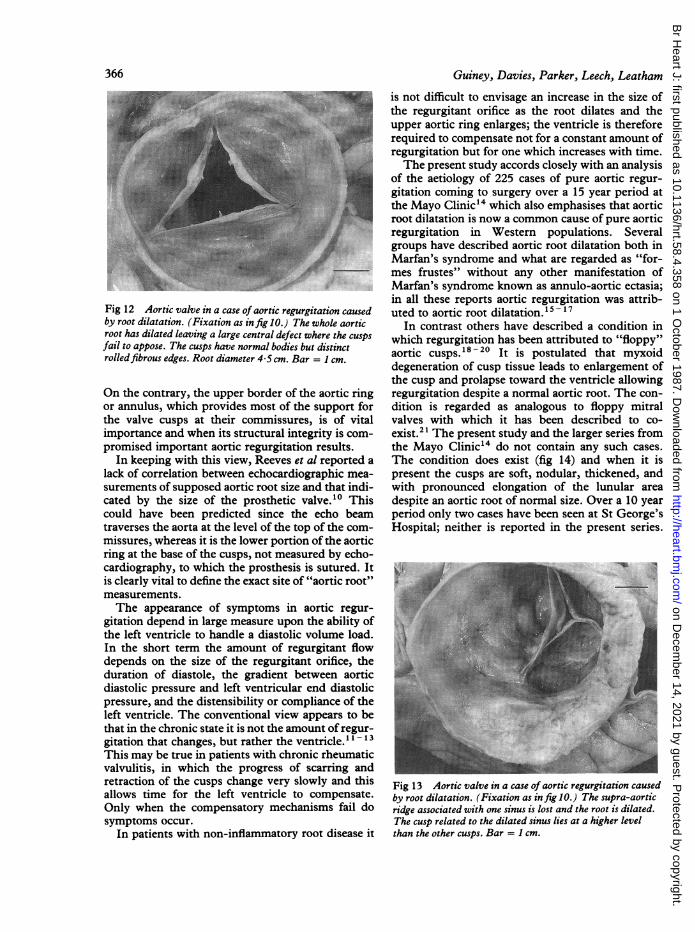
..._ 1 I |ventional criteria: width of the regurgitant jet,degree of opacification of the left ventricle, andrapidity of clearance of the dye from the leftventricle.8The aortic root where cusp disease was
responsible for the regurgitation was normal in sizeand marked by a definite waist just above the sinusesof Valsalva (fig 7). In cases in which aortic rootabnormality was advanced, there was completeeffacement of the aortic waist (fig 8).
PROGRESSION OF AORTIC REGURGITATIONAssessment of rate of progression of symptomsaccording to aetiological diagnosis showed that thecourse of disease differed in the three largest groups(fig 9). The mean (SD) course from the onset ofsymptoms to the break point was long in those with
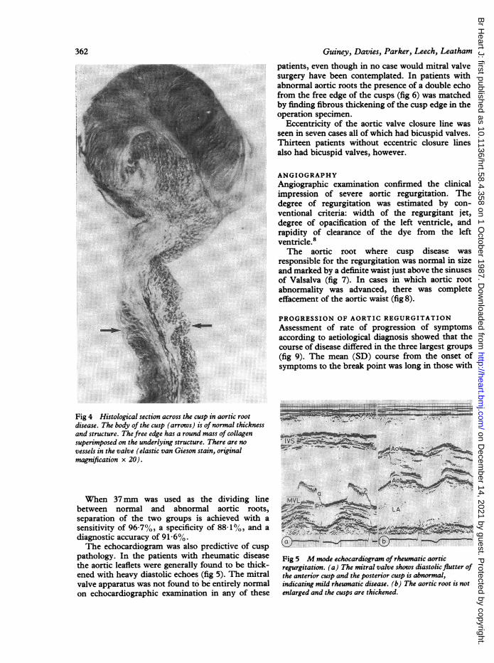
Fig 4 Histological section across the cusp in aortic rootdisease. The body of the cusp (arrows) is of normal thicknessand structure. The free edge has a round mass of collagensuperimposed on the underlying structure. There are no ;vessels in the valve (elastic van Gieson stain, originalmagnification x 20).
When 37mm was used as the dividing line VYLbetween normal and abnormal aortic roots,separation of the two groups is achieved with a ,"sensitivity of 96-7%, a specificity of 88100, and adiagnostic accuracy of 91 6%. `
The echocardiogram was also predictive of cusp Ipathology. In the patients with rheumatic disease Fig 5 M mode echocardiogram of rheumatic aorticthe aortic leaflets were generally found to be thick- regurgitation. (a) The mitral valve shows diastolic flutter ofened with heavy diastolic echoes (fig 5). The mitral the anterior cusp and the posterior cusp is abnormal,valve apparatus was not found to be entirely normal indicating mild rheumatic disease. (b) The aortic root is noton echocardiographic examination in any of these enlarged and the cusps are thickened.
Guiney, Davies, Parker, Leech, Leatham362
on Decem
ber 14, 2021 by guest. Protected by copyright.
http://heart.bmj.com
/B
r Heart J: first published as 10.1136/hrt.58.4.358 on 1 O
ctober 1987. Dow
nloaded from

The aetiology and course of isolated severe aortic regurgitation
r:1t¢fQtttttIl11StliMIIiitit1lttMlttt01tftltl~~~~~~~~~~~~~~~~~~~~~~~~~......... ..* .A ... ......... ..
.: *.: .. 's . ! aoJ :_
.pI-
1I -% S ., 0
Fig 6 Mmode echocardiogram of aortic regurgitation caused by a dilated aortic root. The diameter of the aorta atcommissural level is 4-3 cm. Thefree edges of the cusps produce a double echo (arrow).
rheumatic disease (82-6 (68) months) and short inthose with infective endocarditis, either controlledor uncontrolled (10 1 (19) months. In those withnon-inflammatory aortic root disease there was anintermediate course (20 6 (24 3) months). Thesedifferences are highly significant (p < 0 001).
Discussion
An understanding of the pathophysiological mech-anisms of aortic regurgitation requires a review ofthe normal anatomy and function of the valve. Forsuch a purpose normal aortic valves were fixed in theclosed position under a hydrostatic pressure equiv-alent to 100 mm Hg. This provided for distension ofthe sinuses of Valsalva to a degree approximatingtheir position in life (fig 10). When thus examined,the aortic root was seen to consist of a sleeve of about1-5 cm in length bounded by ring-like structuresboth above and below with a distended area, thesinuses, in between. The lower ring, which lies
below the cusps (fig 11) and forms the attachment ofthe aorta to the left ventricle, is made up in part bythe myocardium of the upper interventricular sep-tum and in part by the base of the anterior leaflet ofthe mitral valve. Its fibrous support structure can bedissected out as part of the so-called cardiac skeletonand is familiarly, but perhaps misleadingly, knownas the aortic annulus. The upper border of the aorticsleeve forms another definite circular ring at thelevel of the attachment of the aortic commissuresand forms a major part of the structural support forthe cusps. This has been termed the supra-aorticridge or sinotubular junction. The cusps are con-tained within the aortic sleeve and meet at the com-missures which are attached to the supra-aorticridge. Just above the sleeve with its enclosed cusps,the more familiar smooth muscle layers and elasticlaminae of the wall of the tubular portion of theascending aorta merge with the more collagenousconnective tissue of the sleeve. The tissue of the aor-tic sleeve bulges outward in life more than the elastic
363
on Decem
ber 14, 2021 by guest. Protected by copyright.
http://heart.bmj.com
/B
r Heart J: first published as 10.1136/hrt.58.4.358 on 1 O
ctober 1987. Dow
nloaded from
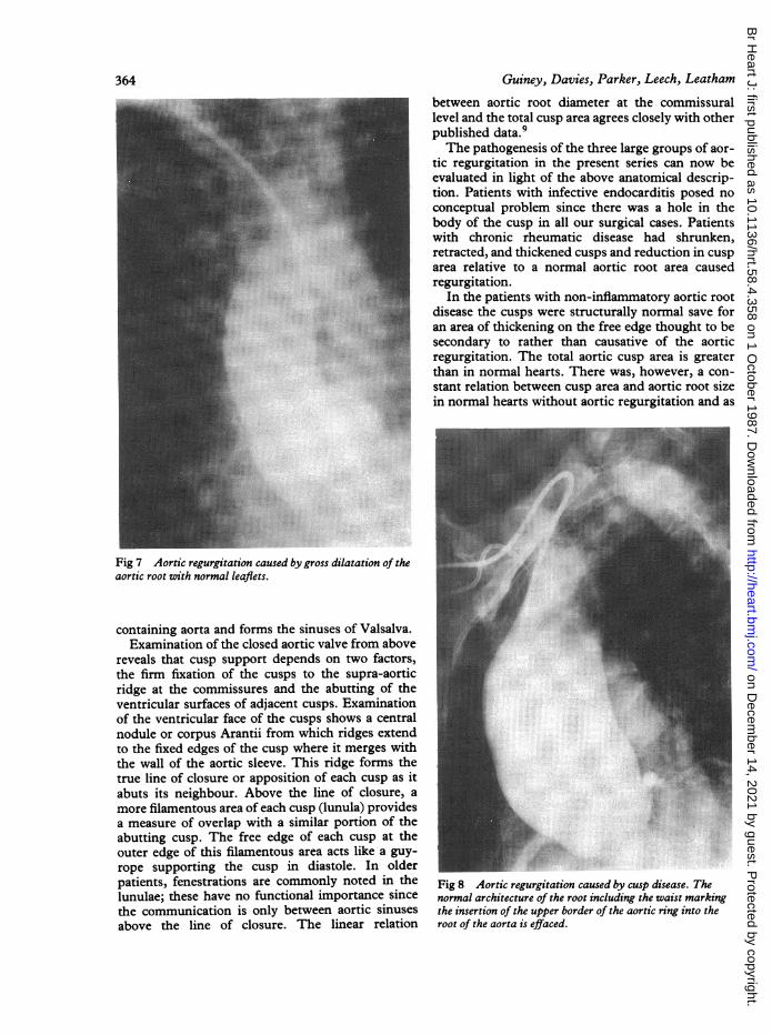
between aortic root diameter at the commissurallevel and the total cusp area agrees closely with otherpublished data.'The pathogenesis of the three large groups of aor-
tic regurgitation in the present series can now beevaluated in light of the above anatomical descrip-tion. Patients with infective endocarditis posed noconceptual problem since there was a hole in thebody of the cusp in all our surgical cases. Patientswith chronic rheumatic disease had shrunken,retracted, and thickened cusps and reduction in cusparea relative to a normal aortic root area causedregurgitation.
In the patients with non-inflammatory aortic rootdisease the cusps were structurally normal save foran area of thickening on the free edge thought to besecondary to rather than causative of the aorticregurgitation. The total aortic cusp area is greaterthan in normal hearts. There was, however, a con-
E f stant relation between cusp area and aortic root sizein normal hearts without aortic regurgitation and as
...~~~~~~~~~~~~~~~~~~~~~~<..?
Ms~~~~~~~~~~~~~~~~~~~~~~~~~~~~~~~~~~~~~~~~~~~~~~~~~~~~~~~~.. .z..
Fig 7 Aortic regurgitation caused by gross dilatation of theaortic root with normal leaflets
containing aorta and forms the sinuses of Valsalva.Examination of the closed aortic valve from above
reveals that cusp support depends on two factors,the firm fixation of the cusps to the supra-aorticridge at the commissures and the abutting of theventricular surfaces of adjacent cusps. Examinationof the ventricular face of the cusps shows a centralnodule or corpus Arantii from which ridges extendto the fixed edges of the cusp where it merges withthe wall of the aortic sleeve. This ridge forms thetrue line of closure or apposition of each cusp as itabuts its neighbour. Above the line of closure, amore filamentous area of each cusp (lunula) providesa measure of overlap with a similar portion of theabutting cusp. The free edge of each cusp at theouter edge of this filamentous area acts like a guy-rope supporting the cusp in diastole. In olderpatients, fenestrations are commonly noted in the Fig 8 Aortic regurgitation caused by cusp disease Thelunulae; these have no functional importance since normal architecture of the root including the waist markingthe communication is only between aortic sinuses the insertion of the upper border of the aortic ring into theabove the line of closure. The linear relation root of the aorta is effaced.
Guiney, Davies, Parker, Leech, Leatham364
on Decem
ber 14, 2021 by guest. Protected by copyright.
http://heart.bmj.com
/B
r Heart J: first published as 10.1136/hrt.58.4.358 on 1 O
ctober 1987. Dow
nloaded from
The aetiology and course of isolated severe aortic regurgitation
82 6 (68) 77 (5 8) months
A... ... ... .. B E.j C Rheumatic disease
20 6 (21.3) 6 75 (5 7) months
A g m B [ilC Aortic root disase
10-1 (19) 415 (9 5) months
A B f C Infective endocarditis
A: Onset of symptoms B: Breakpoint at > class II or both digoxin and diuretics requiredC: Operation
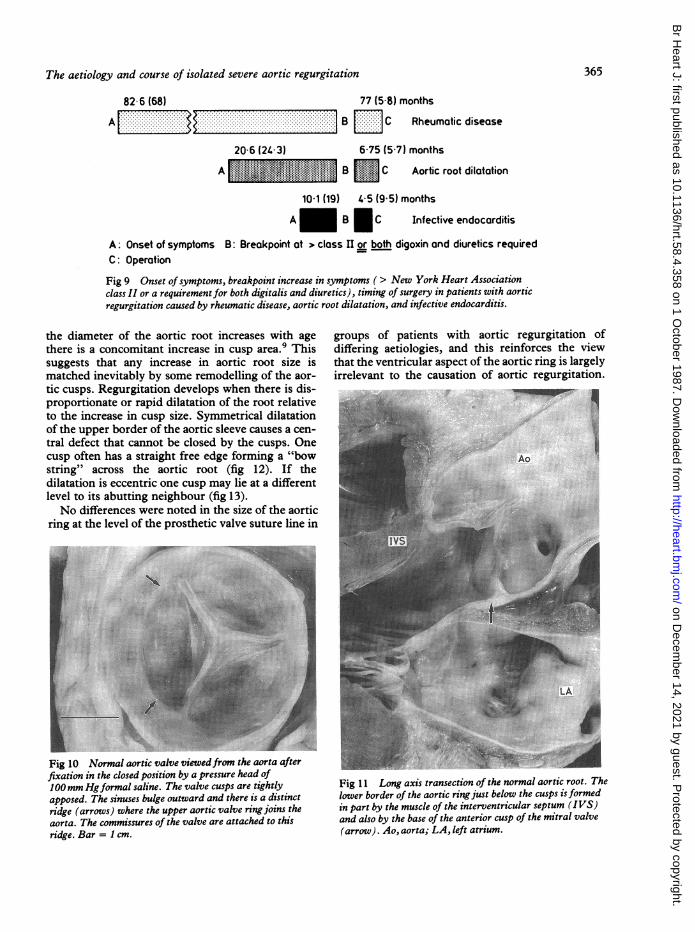
Fig 9 Onset ofsymptoms, breakpoint increase in symptoms ( > New York Heart Associationclass II or a requirement for both digitalis and diuretics), timing of surgery in patients with aorticregurgitation caused by rheumatic disease, aortic root dilatation, and infective endocarditis.
the diameter of the aortic root increases with age groups of patients with aortic reguthere is a concomitant increase in cusp area.9 This differing aetiologies, and this reinforcsuggests that any increase in aortic root size is that the ventricular aspect of the aortic rimatched inevitably by some remodelling of the aor- irrelevant to the causation of aortic retic cusps. Regurgitation develops when there is dis-proportionate or rapid dilatation of the root relativeto the increase in cusp size. Symmetrical dilatationof the upper border of the aortic sleeve causes a cen-tral defect that cannot be closed by the cusps. Onecusp often has a straight free edge forming a "bow Aostring" across the aortic root (fig 12). If thedilatation is eccentric one cusp may lie at a different .... ..
level to its abutting neighbour (fig 13).No differences were noted in the size of the aortic
ring at the level of the prosthetic valve suture line in
irgitation of-es the viewing is largelyegurgitation.
se:.
Fig 10 Normal aortic valve viewedfrom the aorta afterfixation in the closed position by a pressure head of100mm Hgformal saline. The valve cusps are tightlyapposed. The sinuses bulge outward and there is a distinctridge (arrows) where the upper aortic valve ring joins theaorta. The commissures of the valve are attached to thisridge. Bar = I cm.
Fig 11 Long axis transection of the normal aortic root. Thelower border of the aortic ringjust below the cusps isformedin part by the muscle of the interventricular septum (IVS)and also by the base of the anterior cusp of the mitral valve(arrow). Ao, aorta; LA, left atrium.
365
on Decem
ber 14, 2021 by guest. Protected by copyright.
http://heart.bmj.com
/B
r Heart J: first published as 10.1136/hrt.58.4.358 on 1 O
ctober 1987. Dow
nloaded from
366
Fig 12 Aortic valve in a case of aortic regurgitation causedby root dilatation. (Fixation as in fig 10.) The whole aorticroot has dilated leaving a large central defect where the cuspsfail to appose. The cusps have normal bodies but distinctrolledfibrous edges. Root diameter 4-5 cm. Bar = I cm.
On the contrary, the upper border of the aortic ringor annulus, which provides most of the support forthe valve cusps at their commissures, is of vitalimportance and when its structural integrity is com-promised important aortic regurgitation results.
In keeping with this view, Reeves et al reported alack of correlation between echocardiographic mea-surements of supposed aortic root size and that indi-cated by the size of the prosthetic valve.10 Thiscould have been predicted since the echo beamtraverses the aorta at the level of the top of the com-missures, whereas it is the lower portion of the aorticring at the base of the cusps, not measured by echo-cardiography, to which the prosthesis is sutured. Itis clearly vital to define the exact site of "aortic root"measurements.The appearance of symptoms in aortic regur-
gitation depend in large measure upon the ability ofthe left ventricle to handle a diastolic volume load.In the short term the amount of regurgitant flowdepends on the size of the regurgitant orifice, theduration of diastole, the gradient between aorticdiastolic pressure and left ventricular end diastolicpressure, and the distensibility or compliance of theleft ventricle. The conventional view appears to bethat in the chronic state it is not the amount of regur-gitation that changes, but rather the ventricle."1113This may be true in patients with chronic rheumaticvalvulitis, in which the progress of scarring andretraction of the cusps change very slowly and thisallows time for the left ventricle to compensate.Only when the compensatory mechanisms fail dosymptoms occur.
In patients with non-inflammatory root disease it
Guiney, Davies, Parker, Leech, Leatham
is not difficult to envisage an increase in the size ofthe regurgitant orifice as the root dilates and theupper aortic ring enlarges; the ventricle is thereforerequired to compensate not for a constant amount ofregurgitation but for one which increases with time.The present study accords closely with an analysis
of the aetiology of 225 cases of pure aortic regur-gitation coming to surgery over a 15 year period atthe Mayo Clinic"4 which also emphasises that aorticroot dilatation is now a common cause of pure aorticregurgitation in Western populations. Severalgroups have described aortic root dilatation both inMarfan's syndrome and what are regarded as "for-mes frustes" without any other manifestation ofMarfan's syndrome known as annulo-aortic ectasia;in all these reports aortic regurgitation was attrib-uted to aortic root dilatation. 15- 17
In contrast others have described a condition inwhich regurgitation has been attributed to "floppy"aortic cusps.18-20 It is postulated that myxoiddegeneration of cusp tissue leads to enlargement ofthe cusp and prolapse toward the ventricle allowingregurgitation despite a normal aortic root. The con-dition is regarded as analogous to floppy mitralvalves with which it has been described to co-exist.21 The present study and the larger series fromthe Mayo Clinic14 do not contain any such cases.The condition does exist (fig 14) and when it ispresent the cusps are soft, nodular, thickened, andwith pronounced elongation of the lunular areadespite an aortic root of normal size. Over a 10 yearperiod only two cases have been seen at St George'sHospital; neither is reported in the present series.
Fig 13 Aortic valve in a case of aortic regurgitation causedby root dilatation. (Fixation as in fig 1O.) The supra-aorticridge associated with one sinus is lost and the root is dilated.The cusp related to the dilated sinus lies at a higher levelthan the other cusps. Bar = 1 cm.
on Decem
ber 14, 2021 by guest. Protected by copyright.
http://heart.bmj.com
/B
r Heart J: first published as 10.1136/hrt.58.4.358 on 1 O
ctober 1987. Dow
nloaded from

The aetiology and course of isolated severe aortic regurgitation 367
Fig 14 Valve cusps from a case of aortic regurgitationcaused by "floppy" aortic valve cusps. The cusps are nodularand thickened without an appreciable increase in surfacearea. Unlike the cusps in rheumatic disease these felt soft andpliable; the lunulae of the cusps are elongated. Thedimensions of the aortic root were normal.
Histologically the cusp fibrosa is disrupted and con-tains an excess of acid connective tissue mucins.One study of 55 cases of aortic regurgitation
ascribed 13 cases to floppy cusps but it may besignificant that the size of the aortic root was notmeasured.20 A study from Italy found 12 (7T3%)floppy aortic valves in 177 cases of pure aortic regur-
* 22gitation. There may either be a true geographicalvariation or different morphological definitions ofwhat is called a "floppy" aortic valve. Before a sepa-rate primary abnormality of the cusps is diagnosedthe aortic root which supplies structural support forthe cusps must be seen to be normal in size and mor-phology. Patients with mitral valve prolapse withoutMarfan's syndrome do not usually have aortic rootdilatation.23There may be a fundamental difference between
patients with isolated aortic regurgitation caused byaortic root dilatation and those with mitral regur-gitation caused by floppy valves. The former prob-lem is marked by disruption or dissolution of elastictissue in the aortic root and the valve leaflets are nor-mal. The primary defect in mitral valve prolapse isone of collagen rather than of elastic fibres.2425
Roberts et al have reported bicuspid valves andisolated aortic regurgitation in which the anatomicalabnormalities and inequality in cusp size appearsuch that an additional abnormality of the aortic wallis not required to explain the regurgitation.26 Thesepatients are similar to six of our patients with bicus-pid valves and normal histology of the aortic root.Echocardiographic study has demonstrated prolapseof the larger cusp in bicuspid aortic valves21 andwhile minor prolapse is common in patients withbicuspid valves severe prolapse is directly related toregurgitation.27 The five other patients in our serieswho had bicuspid valves without other gross abnor-mality of the cusps had additional non-inflammatoryaortic root disease. The number of patients in ourseries with both aortic root dilatation and bicuspidvalves is so small that no firm conclusion should bedrawn on whether this is simply a chance associationor whether there is a genetic link between bicuspidaortic valves and aortic medial degeneration. Theincreased risk of dissecting aneurysms in theascending aorta21 29 associated with bicuspid valves,however, does support the concept of concomitantaortic medial disease.The important clinical message from our
investigation is that isolated primary dilatation ofthe upper aortic ring is a common cause of aorticregurgitation and is of particular importancebecause it is likely to progress much more rapidlythan rheumatic aortic regurgitation. With thedecline in rheumatic disease in Western populationsisolated aortic root dilatation will become relativelymore common.
This work was supported by the St George'sHospital Cardiac Research Fund, British HeartFoundation, and the Jensen Medical ResearchTrust.
References
1 Rapaport E. Natural history of aortic and mitral valvedisease. Am J Cardiol 1975;35:221-7.
2 Goldschlager N, Pfeifer J, Cohn K, Popper R,Selzer A. The natural history of aortic regurgitation:a clinical and hemodynamic study. Am J Med 1973;54:557-88.
3 Rotman M, Morris JJ Jr, Behar VS, Peter RH, Kong
on Decem
ber 14, 2021 by guest. Protected by copyright.
http://heart.bmj.com
/B
r Heart J: first published as 10.1136/hrt.58.4.358 on 1 O
ctober 1987. Dow
nloaded from

368 Guiney, Davies, Parker, Leech, LeathamY. Aortic valvular disease: comparison of types andtheir medical and surgical management. Am J Med1971;51:241-57.
4 Samuels DA, Curfman GD, Friedlich AL,Buckley MJ, Austen WG. Valve replacement foraortic regurgitation: long-term follow-up withfactors influencing the results. Circulation 1979;60:647-54.
5 Romhilt DW, Estes EH. A point score system for theECG diagnosis of left ventricular hypertrophy. AmHeart J 1968;75:752-8.
6 Feigenbaum H. Echocardiography. 3rd ed. Philadel-phia: Lea and Febiger, 1981:60-76.
7 Dixon WJ, Brown MB, Engelman L, et al. BMDPstatistical software. Berkeley: University of CaliforniaPress, 1981.
8 Dinsmore RE, Miller SW. Angiography in acquiredvalvular heart disease. Semin Roentgenol 1979;-14:153-66.
9 Silver MA, Roberts WC. Detailed anatomy of the nor-mally functioning aortic valve in hearts ofnormal andincreased weight. Am J Cardiol 1985;55:454-61.
10 Reeves WC, Ettinger V, Thomson K, et al. Limitationsin the echocardiographic assessment of aortic rootdimensions in the presence of aortic valve disease.Radiology 1979;132:411-3.
11 Henry WL, Bonow RO, Borer JS, et al. Observationson the optimum time for operative intervention foraortic regurgitation. I. Evaluation of the results ofaortic valve replacement in symptomatic patients.Circulation 1980;61:471-83.
12 Henry WL, Bonow RO, Rosing DR, Epstein SE.Observations on the optimum time for operativeintervention for aortic regurgitation. II. Serial echo-cardiographic evaluation of asymptomatic patients.Circulation 1980;61:484-92.
13 Bonow RO, Borer JS, Rosing DR, et al. Preoperativeexercise capacity in symptomatic patients with aorticregurgitation as a predictor of postoperative leftventricular function and long-term prognosis.Circulation 1980;62:1280-90.
14 Olson LT, Subramanian R, Edwards WD. Surgicalpathology of pure aortic insufficiency: a study of 225cases. Mayo Clin Proc 1984;59:835-41.
15 McKusick VA, Logue RB, Bahnson HT. Associationof aortic valvular disease and cystic medial necrosis ofthe ascending aorta. Report of 4 instances. Circu-lation 1957;16:188-94.
16 Weaver WF, Edwards JE, Brandenburg RO. Idio-pathic dilatation of the aorta with aortic valvularinsufficiency: a possible forme fruste of Marfan'ssyndrome. Mayo Clin Proc 1959;34:518-22.
17 Cooley DA. Annuloaortic ectasia. Ann Thorac Surg1979;28:303-4.
18 Rippe JM, Angoff G, Sloss LV, Wynne J, Alpert JS.Multiple floppy valves: an echocardiographic syn-drome. Am J Med 1979;66:817-24.
19 McKay R, Yacoub MH. Clinical and pathologicalfindings in patients with "floppy" valves treated sur-gically. Circulation 1973;48 (suppl III):1 163-73.
20 Allen WM, Matloff JM, Fishbein MC. Myxoiddegeneration of the aortic valve and isolated aorticregurgitation. Am J Cardiol 1985;55:439-44.
21 Shapiro LM, Thwaites B, Westgate C, Donaldson R.Prevalence and clinical significance of aortic valveprolapse. Br Heart J 1985;54:179-83.
22 Bellitti R, Caruso A, Festa M, et al. Prolapse of the"floppy" aortic valve as a cause of aortic regur-gitation. A clinico-morphological study. Int J Car-diol 1985;9:399-410.
23 Brown OR, Demots SH, Kloster FE, Roberts A,Menashe JD, Beals RK. Aortic root dilatation andmitral valve prolapse in Marfan's syndrome: anechocardiographic study. Circulation 1975;52:651-7.
24 King BD, Clark MA, Babs N, Kilman JW, Wooley CF."Myxomatous" mitral valves: collagen dissolution asthe primary defect. Circulation 1982;66:288-96.
25 Gravanis MB, Campbell WG. The syndrome of pro-lapse of the mitral valve. Arch Pathol Lab Med1982;106:369-74.
26 Roberts WC, Morrow AG, McIntosh CL, Jones M,Epstein SE. Congenitally bicuspid valve causingsevere pure aortic regurgitation without super-imposed infective endocarditis. Am J Cardiol1981;47:206-9.
27 Stewart WJ, King ME, Gillam LD, Guyer DE,Weyman AE. Prevalence of aortic valve prolapsewith bicuspid aortic valve and its relation to aorticregurgitation: a cross sectional echocardiographicstudy. Am J Cardiol 1985;54:1277-82.
28 Edwards WD, Leaf DS, Edwards JE. Dissecting aorticaneurysm associated with congenital bicuspid aorticvalve. Circulation 1978;57:1022-5.
29 Larson EW, Edwards JE. Risk factors for aorticdissection-a necropsy study of 161 cases. Am J Car-diol 1984;53:849-55.
on Decem
ber 14, 2021 by guest. Protected by copyright.
http://heart.bmj.com
/B
r Heart J: first published as 10.1136/hrt.58.4.358 on 1 O
ctober 1987. Dow
nloaded from