Embed Size (px)
Citation preview

The Sella and Parasellar Region
Anatomic and Pathologic Considerations:
A Practical Approach
Philip Chapman, MD
Assistant Professor
University of Alabama, Birmingham

The Sella and Parasellar Region
Outline
• Imaging Techniques
• Normal Anatomy
• Differential Diagnosis
– Sella
– Suprasellar
– Infundibulum

Recommended Imaging Techniques
MRI Imaging
• Multiplanar: • Sagittal and Coronal
• Small FOV 16-18 cm
• 3mm
• T1W, T2W
• Post T1W + FS
• Dynamic enhanced for pituitary lesions

Sella: Normal Anatomy
Pituitary Gland
• Anterior Lobe (75%)
• Pars Intermedia
• Posterior Lobe (25%)
• Infundibulum

Pituitary: Normal Anatomy
Anterior Lobe Lateral • PRL (10-30%) • GH (50%) Midline • ACTH (10-30%) • TSH (5%) • FSH/LH (10%)
• Location of adenomas parallels the distribution

Pars intermedia

Sella: Normal Anatomy
Pituitary Bright Spot The secretary granules carrying vasopression and oxytocin appear as the “bright spot” of the posterior lobe of the pituitary gland on T1-weighted unenhanced (MRI).

Pituitary: Normal Anatomy
Posterior Lobe
• Infundibulum
• Pituicytes (glial)
• Axons
• Vasopressin (ADH)
• Oxytocin
Ectopic Posterior Pituitary

Parasellar Region: Normal Anatomy
Parasellar Structures
• Cavernous Sinus
• Cranial Nerves
• III, IV, V1, V2, VI
• Cavernous ICA
• Optic Chiasm
• Hypothalamus
• Sphenoid Sinus

Parasellar Region: Normal Anatomy

Parasellar Region: Normal Anatomy
Bony Structures
• Planum sphenoidale
• Tuberculum sellae
• Sella turcica
• Dorsum sellae
***CT is complementary for evaluating central skull base lesions: • Effects on skull base • Calcifications

Sella and Parasellar Pathology
Differential Diagnoses
• Sellar
• Suprasellar
• Infundibular

Sellar Pathology
Non-neoplastic Lesions • Hyperplasia/Hypertrophy (physiologic, end organ failure) • Cysts (RCC, pars intermedia cyst) • Empty Sella Primary Neoplasms • Pituitary adenoma (Most common) • Craniopharyngioma (Only 5% purely intrasellar) • Meningioma (Purely intrasellar rare) • Abscess (Rare) • Pituitary carcinoma (Extremely rare) Metastasis (1%)

Pituitary Gland Size
Maximum normal height
• 6 mm infants and children
• 8 mm males, postmenopausal females
• 10 mm young women of childbearing age
• 12 mm late pregnancy, postpartum females
• “Elster’s Rule”

Pituitary Hyperplasia/Hypertrophy
• Must know age, gender!!
• Physiologic ↑
• 10-15 mm
• Convex upwards
• Strong, uniform
enhancement
• Can be indistinguishable
from:
• Macroadenoma
• Lymphocytic hypophitis
• Metastasis, lymphoma
Postpartum lactating ♀
21y menstruating ♀
14 mm
11 mm

Pituitary Hyperplasia/Hypertrophy
Postpartum lactating ♀
21y menstruating ♀
14 mm
11 mm
*** Dynamic imaging may
help distinguish physiologic
hyperplasia from
macroadenoma
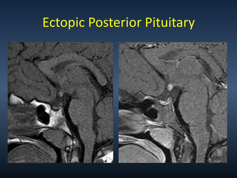
Pituitary Gland
Hyperplasia/Hypertrophy
Pathologic
hypertrophy
• End-organ failure
- Hypothyroidism
- Ovarian failure
• Neuroendocrine
tumors Pituitary hypertrophy secondary to untreated hypothyroidism

“EMPTY” SELLA
Considered normal variant. Loose association with Pseudotumor Cerebri

Pituitary Neoplasms
Adenoma type • Prolactinoma 30%-40%
• Null cell 25%
• GH 20%
• ACTH 10%
• FSH/LH 10%
• PRL-GH 5%
• Mixed, TSH 1-5%
• Incidental pituitary
lesions are common
• Adenomas comprise the majority (80%) of pituitary lesions. • A large percentage of these (approximately 75%) are functioning and result in endocrine abnormalities.
Adenoma
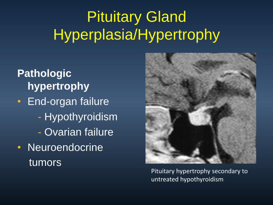
Sella: Pathology
Pituitary Microadenoma
• 10 mm or less
• 10-20% of autopsies
• Micro >>> Macro
• Convex margin
• Stalk deviation
• Sella floor thin

Recommended Imaging Techniques
Dynamic Imaging
• Microadenomas
• 4-5 slices
• T1 FSE, Turbo SE
• Image continuously
after contrast for app 2 minutes
• Increases sensitivity for small adenomas

Sella: Pathology
There is up to a 20% false-negative rate in the detection of microadenomas.
Dynamic Imaging
• In essence, the normal pituitary gland enhances at a faster rate than the microadenoma, so that early during contrast injection (90 seconds), the adenoma appears as hypointense against the backdrop of enhancing pituitary tissue.
• This difference is lost as microadenoma gradually accumulates contrast (after app 2 minutes)

Sella: Pathology
Microadenoma
Dynamic Imaging
• Increases sensitivity
• (10-30% seen only on
dynamic MR)
There is up to a 20% false-negative rate in the detection of microadenomas.

Pituitary Microadenoma

Clinical
• Intrasellar 40%
• Suprasellar extent 60%
• 3mm – 3cm
• Most incidental
• Symptomatic
Pituitary dysfunction
Visual change, HA
Sella: Rathke Cleft Cyst
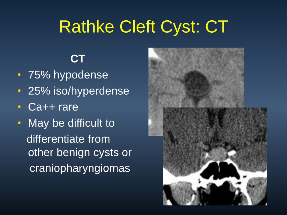
CT
• 75% hypodense
• 25% iso/hyperdense
• Ca++ rare
• May be difficult to
differentiate from
other benign cysts or
craniopharyngiomas
Rathke Cleft Cyst: CT

Imaging Features
• Signal varies - cyst
content
• 50-60% T1
hyperintense
• 30-40% follow CSF
• 75% intracystic
nodule
• +/- rim enhancement
Rathke Cleft Cyst: MR

Rathke Cleft Cyst

Rathke Cleft Cyst

Suprasellar Masses: The “Big Five”
• 75% of all sellar/parasellar masses
1. Pituitary macroadenoma (35%-50%)
2. Meningioma
3. Aneurysm
4. Craniopharyngioma
5. Astrocytoma (hypothalamic-chiasmatic)
10% each

Adult Lesions
• Pituitary
Macroadenoma
• Meningioma
• Aneurysm
Pediatric Lesions
• Craniopharyngioma
• Chiasmatic /
Hypothalamic
Glioma
• Hypothalamic Hamartoma
Suprasellar Differential Diagnosis
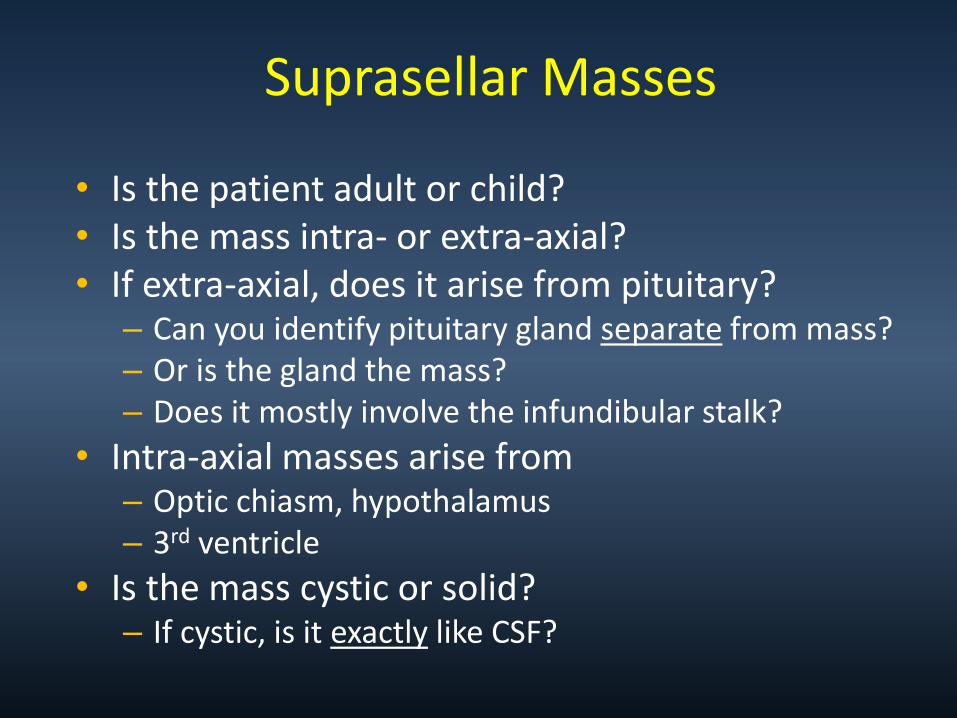
Suprasellar Masses
• Is the patient adult or child? • Is the mass intra- or extra-axial? • If extra-axial, does it arise from pituitary?
– Can you identify pituitary gland separate from mass? – Or is the gland the mass? – Does it mostly involve the infundibular stalk?
• Intra-axial masses arise from – Optic chiasm, hypothalamus – 3rd ventricle
• Is the mass cystic or solid? – If cystic, is it exactly like CSF?
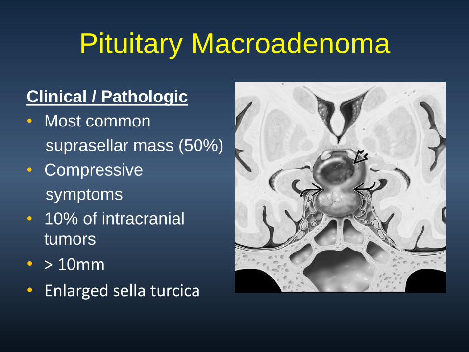
Clinical / Pathologic
• Most common
suprasellar mass (50%)
• Compressive
symptoms
• 10% of intracranial
tumors
• > 10mm
• Enlarged sella turcica
Pituitary Macroadenoma

Suprasellar: Pathology

Prolactinoma
• 30%-40% of adenomas
• Female >> Males
• Galactorrhea
• Amenorrhea
• Serum PRL > 150ng/mL
• If > 1000ng/mL invasion
Pituitary Adenoma

Imaging Features
• Isointense GM: T1, T2WI
• “Mass is the pituitary”
• May have hemorrhage,
cystic components
• Figure-eight, snowman
• Strong but heterogeneous
enhancement
Pituitary Macroadenoma: MR

Pituitary Macroadenoma: MR

Giant Pituitary Macroadenoma
Prolactinoma
4 cm

Giant Pituitary Macroadenoma

Cavernous Sinus Invasion
• Adenomas that involve the lateral margins of the adenohypophysis may grow laterally beyond the sellar margin and invade the adjacent cavernous sinus.
• 5- 10% of all pituitary adenomas involve the cavernous sinus and are considered to be invasive
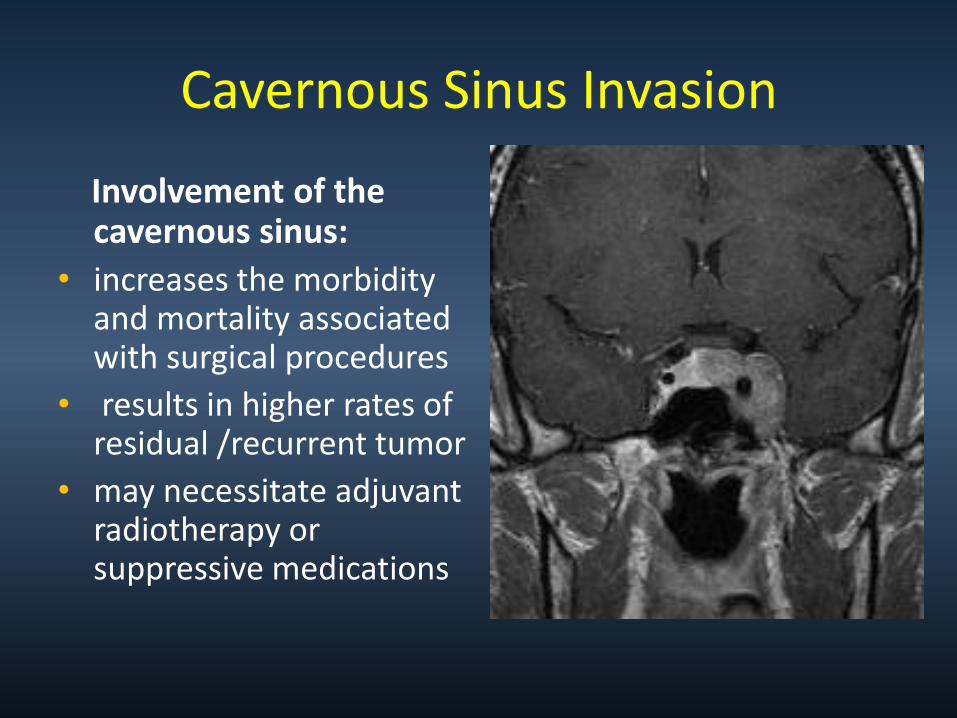
Cavernous Sinus Invasion
Involvement of the cavernous sinus:
• increases the morbidity and mortality associated with surgical procedures
• results in higher rates of residual /recurrent tumor
• may necessitate adjuvant radiotherapy or suppressive medications

Pituitary-Cavernous Interface
Songtao, Qi, et al. Membranous Layers of the Pituitary Gland, Operative Neurosurgery, March 2009

Cavernous Sinus Invasion
Knosp-Steiner Grading
0 1 2
3 4
Cavernous Sinus Invasion
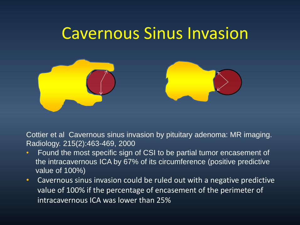
Cottier et al Cavernous sinus invasion by pituitary adenoma: MR imaging.
Radiology. 215(2):463-469, 2000 • Found the most specific sign of CSI to be partial tumor encasement of
the intracavernous ICA by 67% of its circumference (positive predictive
value of 100%)
• Cavernous sinus invasion could be ruled out with a negative predictive value of 100% if the percentage of encasement of the perimeter of intracavernous ICA was lower than 25%

Extrinsic narrowing of the carotid artery is rarely associated with pituitary adenomas and is more suggestive of meningiomas

Invasive Pituitary Macroadenoma

37 yo male with Prolactinoma

Growth hormone secreting Prolactin secreting
Infrasellar Extension
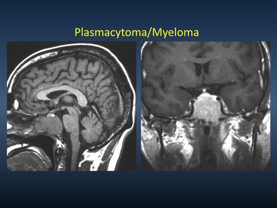
Plasmacytoma/Myeloma

Multiple Myeloma Of Clivus
69 yo with h/o mm 65 yo male with mm

Prolactinoma Chordoma

Pituitary Apoplexy
Clinical Syndrome
• Acute onset
• Visual changes
• Headache
• Vomiting
• Meningismus
• Rapid enlargement of Macroadenoma secondary to hemorrhagic infarction
• Rare, life threatening
T1 Shortening in subacute hemorrhage
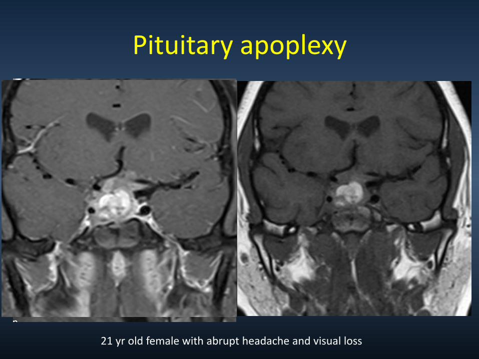
Pituitary apoplexy
21 yr old female with abrupt headache and visual loss
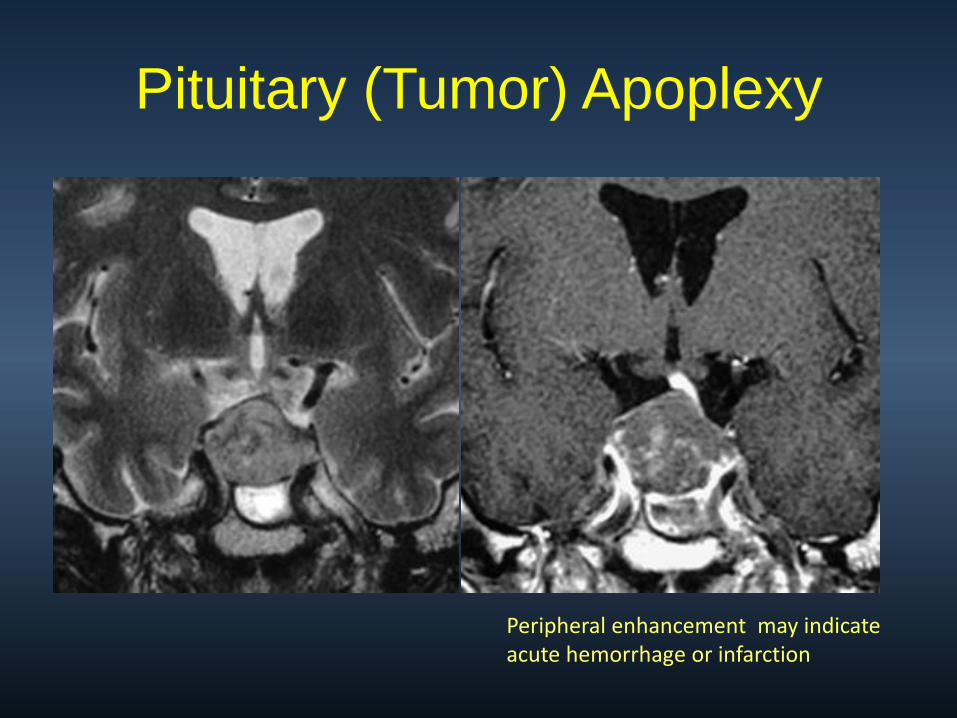
Pituitary (Tumor) Apoplexy
Peripheral enhancement may indicate acute hemorrhage or infarction

Macroadenoma with hemorrhage?


![Atypical Intracranial Epidermoid Cysts: Rare Anomalies with … · 2017-10-31 · the parasellar region [1]. Conversely, atypical epidermoid cysts are rare, with intra-axial epidermoid](https://img.pdfslide.us/doc/110x75/5f7ff1f90cbb51524d18b285/atypical-intracranial-epidermoid-cysts-rare-anomalies-with-2017-10-31-the-parasellar.jpg)