Embed Size (px)
Citation preview

The Roles of YbeY, RfaH, and Hfq in Gene Regulation and Virulence of Yersinia enterocolitica O:3
RESEARCH PROGRAMS UNIT, IMMUNOBIOLOGYDEPARTMENT OF BACTERIOLOGY AND IMMUNOLOGYFACULTY OF MEDICINEDOCTORAL PROGRAMME IN BIOMEDICINEUNIVERSITY OF HELSINKI
KATARZYNA LESKINEN
dissertationes scholae doctoralis ad sanitatem investigandam universitatis helsinkiensis 49/2016
49/2016
Helsinki 2016 ISSN 2342-3161 ISBN 978-951-51-2341-1
KA
TA
RZ
YN
A L
ES
KIN
EN
Th
e Roles of Y
beY, R
faH, an
d H
fq in
Gen
e Regu
lation an
d V
irulen
ce of Yersin
ia en
terocolitica O
:3
Recent Publications in this Series
28/2016 Sandra SöderholmPhosphoproteomic Characterization of Viral Infection29/2016 Mariann LasseniusBacterial Endotoxins in Type 1 Diabetes30/2016 Mette IlanderT and NK Cell Mediated Immunity in Chronic Myeloid Leukaemia31/2016 Ninja KarikoskiThe Prevalence and Histopathology of Endocrinopathic Laminitis in Horses32/2016 Michael BacklundRegulation of Angiotensin II Type 1 Receptor by Its Messenger RNA-Binding Proteins33/2016 Stanislav RozovCircadian and Histaminergic Regulation of the Sleep-Wakefulness Cycle34/2016 Bárbara Herranz BlancoMulti-Approach Design and Fabrication of Hybrid Composites for Drug Delivery and Cancer Therapy35/2016 Siri TähtinenCombining Oncolytic Immunotherapies to Break Tumor Resistance36/2016 Katri SääksjärviDiet, Lifestyle Factors, Metabolic Health and Risk of Parkinson’s Disease – A Prospective Cohort Study37/2016 Alexandre BorrelDevelopment of Computational Methods to Predict Protein Pocket Druggability and Profile Ligands using Structural Data38/2016 Gonçalo BarretoInnate Immunity in Osteoarthritis: The Role of Toll-Like Receptors and Cartilage Derived Mediators in the Disease Progression39/2016 Alexey YukinAnimal Models of Early Brain Disorders: Behavioural and Electrophysiological Approaches40/2016 Satu ValoWestern Diet and Genetic Predisposition as Risk Factors of Colon Cancer41/2016 Riccardo SiligatoHormonal Regulation of Primary and Secondary Growth in the Root of Arabidopsis thaliana42/2016 Janne TynellVirus-Host Interactions of Emerging Respiratory Pathogens43/2016 Katri KorpelaIntestinal Microbiota Development in Childhood: Implications for Health and Disease44/2016 Mikko MuonaIdentification of New Genetic Syndromes with Epilepsy by Whole-Exome Sequencing45/2016 Ana Cathia Dias MagalhãesOn the Mechanisms of Neural Development in the Ventral Telencephalon46/2016 Petri ArvilommiTreatment, Adherence and Disability in Bipolar Disorder47/2016 Annika ThomsonFinnish Pretrial Male Firesetters: Mortality, Suicidality, Psychopathy and Morbidity of Schizophrenia48/2016 Sami SvanbäckToward Accurate High-Throughput Physicochemical Profiling using Image-Based Single-Particle Analysis

1
The roles of YbeY, RfaH, and Hfq in gene regulation and virulence of Yersinia enterocolitica O:3
Katarzyna Leskinen
Immunobiology Research Program Department of Bacteriology and Immunology
Faculty of Medicine University of Helsinki
Finland
ACADEMIC DISSERTATION
To be presented, with permission of the Faculty of Medicine of the University of Helsinki,
for public examination in the Lecture Hall 1 in the Haartman Institute, Haartmaninkatu 3, Helsinki,
on August 22nd 2016 at 12.00
Helsinki 2016
2
Supervised by Professor Mikael Skurnik, PhD Immunobiology Research Program Department of Bacteriology and Immunology Faculty of Medicine University of Helsinki Helsinki, Finland
Reviewed by Professor Mikael Rhen, PhD Department of Microbiology, Tumor and Cell Biology (MTC) Karolinska Institutet Stockholm, Sweden
And
Arto Pulliainen, PhD
Medical Microbiology and Immunology Medical Biochemistry and Genetics Institute of Biomedicine University of Turku Turku, Finland
Opponent Professor Virginia Miller, PhD Departments of Microbiology and Immunology Genetics University of North Carolina School of Medicine Chapel Hill, USA
Illustrations by the author ISBN 978-952-10-9854-3 (paperback) ISBN 978-952-10-9855-0 (PDF) http://ethesis.helsinki.fi Printed at Hansaprint Oy Finland 2016

3
“Keep me incomplete”
-Liam Cormier
4
TABLE OF CONTENTS
LIST OF ORIGINAL PUBLICATIONS ...................................................................................................................... 7
ABBREVIATIONS ................................................................................................................................................. 8
ABSTRACT ........................................................................................................................................................ 10
1. INTRODUCTION ....................................................................................................................................... 12
2. REVIEW OF THE LITERATURE ................................................................................................................... 13
2.1. Gene regulation in bacteria ............................................................................................................. 13
2.1.1. Regulation of transcription ............................................................................................................ 14
2.1.1.1. Promoters ............................................................................................................................... 14
2.1.1.2. Sigma factors and anti-sigma factors ..................................................................................... 15
2.1.1.3. Operons and regulons ............................................................................................................ 16
2.1.1.4. Transcriptional regulators ...................................................................................................... 17
2.1.1.5. RfaH antiterminator ............................................................................................................... 19
2.1.1.6. The LysR-family transcriptional regulators ............................................................................. 21
2.1.2. Post-transcriptional regulation ...................................................................................................... 22
2.1.2.1. Translation initiation efficiency .............................................................................................. 22
2.1.2.2. Stability of mRNA .................................................................................................................... 23
2.1.2.3. Non-coding RNAs .................................................................................................................... 23
2.1.2.4. Usage of rare codons .............................................................................................................. 26
2.1.2.5. RNA-binding proteins ............................................................................................................. 26
2.1.2.6. RNA chaperone Hfq as a global post-transcriptional regulator ............................................. 28
2.1.2.7. Ribonuclease YbeY .................................................................................................................. 30
2.1.3. Post-translational regulation ......................................................................................................... 31
2.2. The genus Yersinia ........................................................................................................................... 32
2.2.1. Yersinia enterocolitica ................................................................................................................... 32
2.2.1.1. General characteristics ........................................................................................................... 34
2.2.1.2. Virulence factors ..................................................................................................................... 35
2.2.1.2.1. Plasmid encoded virulence factors .................................................................................. 35
2.2.1.2.2. Chromosomally encoded virulence factors ...................................................................... 38
2.2.1.2.3. Virulence-related features ............................................................................................... 40
2.3. Coordination of gene regulation in Yersiniae .................................................................................. 42
3. AIMS OF THE STUDY ................................................................................................................................ 46

5
4. MATERIALS AND METHODS ..................................................................................................................... 47
4.1. Materials .......................................................................................................................................... 47
4.1.1. Bacterial strains and bacteriophages ............................................................................................ 47
4.1.2. Plasmids ......................................................................................................................................... 48
4.1.3. Antibodies and antisera ................................................................................................................. 49
4.1.4. Bioinformatics resources used ...................................................................................................... 50
4.2. Methods .......................................................................................................................................... 50
4.2.1. Bacterial cultivation ....................................................................................................................... 50
4.2.2. Growth curves. .............................................................................................................................. 51
4.2.3. SDS-PAGE. ...................................................................................................................................... 51
4.2.4. Preparation of LPS samples. .......................................................................................................... 51
4.2.5. Detection of O-antigen in culture supernatants. .......................................................................... 51
4.2.6. DOC-PAGE. ..................................................................................................................................... 51
4.2.7. Immunoblotting ............................................................................................................................. 52
4.2.8. Total RNA extraction...................................................................................................................... 52
4.2.9. RNA-sequencing. ........................................................................................................................... 52
4.2.10. Quantitative RT-PCR. ................................................................................................................... 53
4.2.11. Quantitative proteomics. ............................................................................................................ 53
4.2.12. Resistance assays ......................................................................................................................... 53
4.2.13. Urease test................................................................................................................................... 54
4.2.14. Sugar utilization. .......................................................................................................................... 54
4.2.15. Motility and biofilm assays. ......................................................................................................... 54
4.2.16. Luminescence assay. ................................................................................................................... 55
4.2.17. Bacteriophage sensitivity. ........................................................................................................... 55
4.2.18. Electron microscopy. ................................................................................................................... 55
4.2.19. Serum killing assay. ...................................................................................................................... 55
4.2.20. Cell culture infection assay. ......................................................................................................... 56
4.2.21. Mouse experiments. .................................................................................................................... 56
5. RESULTS ................................................................................................................................................... 58
5.1. YbeY, Hfq and RfaH are involved in the gene expression and protein synthesis ............................ 58
5.1.1. Transcriptomics ............................................................................................................................. 58
5.1.2. Proteomics ..................................................................................................................................... 59
5.2. The role of ybeY, hfq, rfaH and rovM in the physiology of Y. enterocolitica O:3 ............................ 60
5.2.1. Bacterial growth ............................................................................................................................ 60

6
5.2.2. Cell and colony morphology .......................................................................................................... 61
5.2.3. Susceptibility to environmental stresses ....................................................................................... 61
5.2.4. Motility and biofilm formation ...................................................................................................... 62
5.2.5. Other physiological features affected by the mutations ............................................................... 62
5.3. The effect of ybeY, hfq, and rfaH mutations on the virulence and virulence-associated traits of Y. enterocolitica O:3 ........................................................................................................................................ 63
5.3.1. Production of Yop effector proteins .............................................................................................. 63
5.3.2. LPS synthesis .................................................................................................................................. 64
5.3.3. Production of other virulence factors ........................................................................................... 64
5.3.4. Resistance to normal human serum .............................................................................................. 65
5.3.5. Virulence in cell and animal models .............................................................................................. 66
6. DISCUSSION ............................................................................................................................................. 68
6.1. The impact of studied mutations on the gene expression .............................................................. 68
6.2. YbeY ................................................................................................................................................. 69
6.3. RfaH ................................................................................................................................................. 71
6.4. Hfq ................................................................................................................................................... 72
6.5. RovM ................................................................................................................................................ 75
7. CONCLUSIONS AND FUTURE CONSIDERATIONS ..................................................................................... 77
8. ACKNOWLEDGEMENTS ............................................................................................................................ 78
9. REFERENCES ............................................................................................................................................. 80

7
LIST OF ORIGINAL PUBLICATIONS
This thesis is based on the following publications, which are referred to in the text
by their Roman numerals (I-III).
I. Leskinen K, Varjosalo M, Skurnik M. 2015. Absence of YbeY RNase compromises the
growth and enhances the virulence plasmid gene expression of Yersinia
enterocolitica O:3. Microbiology 161:285-99.
II. Leskinen K, Varjosalo M, Li Z, Li CM, Skurnik M. 2015. The expression of the Yersinia
enterocolitica O:3 lipopolysaccharide O-antigen and outer core gene clusters is RfaH-
dependent. Microbiology 161: 1282-1294.
III. Leskinen K, Varjosalo M, Fernández-Carrasco H, Bengoechea JA, Skurnik M. Several
Hfq-dependent alterations in physiology of Yersinia enterocolitica O:3 are mediated
by derepression of the transcriptional regulator RovM. Submitted.
8
ABBREVIATIONS
Ail Attachment invasion locus
AHL N-Acyl Homoserine Lactone
asRNA antisense RNA
BHI Brain Heart Infusion
bp base pair
CFU Colony Forming Unit
Clm Cloramphenicol
ECA Enterobacterial Common Antigen.
DMEM Dulbecco's Modified Eagle's Medium
DOC 2,5-Dimethoxy-4-chloroamphetamine
DOC-PAGE 2,5-Dimethoxy-4-chloroamphetamine Polyacrylamide Gel
Electrophoresis
FC Fold Change
GMP Guanosine monophosphate
GTP Guanosine-5'-triphosphate
HIS Heat Inactivated Serum
IC Inner Core
Inv Invasin
kbp kilo base pair
Km Kanamycin
LB Lysogeny Broth
LC-MS/MS Liquid Chromatography-tandem Mass Spectrometry
LPS Lipopolysaccharide
ManpNAcA D-mannopyranuronic Acid
mRNA messenger RNA
MS Mass Spectrometry
ncRNA non-coding RNA
NHS Normal Human Serum
O-Ag O-Antigen
OC Outer Core
PAPI Poly(A) Polymerase I

9
PBS Phosphate-Buffered Saline
PTM Post-Translational Modifications
PTS Phosphotransferase system
pYV Yersinia virulence plasmid
RBP RNA-binding protein
RBS Ribosome-binding site
RNA-seq RNA sequencing
RT Room Temperature
SD Shine-Dalgarno
SDS Sodium Dodecyl Sulfate
SDS-PAGE Sodium Dodecyl Sulfate Polyacrylamide Gel Electrophoresis
Spec Spectinomycin
sRNA small RNA
Strep Streptomycin
T3SS Type III Secretion System
TCEP Tris(2-carboxyethyl)phosphine
TCS Two-Component regulatory System
TIR Translation Initiation Region
UTR Untranslated Region
YadA Yersinia adhesin A
Yops Yersinia outer membrane proteins
10
ABSTRACT
Understanding the molecular mechanisms of bacterial virulence has broad implications. In
addition to just academic interest many practical applications can be foreseen emerging from
virulence research: identification of novel antimicrobial drug targets, potential vaccines, and
diagnostics of infectious diseases. Different virulence factors are responsible for the initiation
of the disease and others for the disease symptoms. Consequently, elimination of a single
virulence factor can severely attenuate or even completely abrogate virulence. Due to the
increasing antibiotic resistance world-wide there is an urgent need for new antimicrobial
agents. The virulence factors and their eukaryotic interaction partners are recognized as
potential targets for vaccine and antibacterial drug development and therefore highly
prioritized research topics internationally.
Genus Yersinia consists of 17 species of which Y. pestis, Y. pseudotuberculosis and Y.
enterocolitica are human pathogens. Y. pestis causes bubonic plague while Y. pseudotuberculosis
and Y. enterocolitica cause mostly food-borne yersiniosis, usually a diarrheal disease
sometimes followed by post-infectious reactive arthritis. The pathogenic potential of these
bacteria resides on many essential virulence factors some of which are encoded by genes
located on a 70 kb virulence plasmid of Yersinia (pYV) and others by chromosomal loci.
Yersiniosis is considered to be the third most common cause of gastroenteritis in Europe. In
Finland both Y. pseudotuberculosis and Y. enterocolitica cause hundreds of human infections
annually.
The aim of the study is to characterize the intricate regulatory networks of Yersinia especially
those that control the expression of the virulence factors. To achieve that goal three regulators
were initially selected. The first gene studied, ybeY, was selected based on the literature due to
the fact its protein product is believed to affect the sRNA regulation similar to Hfq. YbeY was
recently recognized as an endoribonuclease playing an important role in the process of
ribosome biosynthesis. The absence of ybeY gene in Y. enterocolitica serotype O:3 resulted in
misprocessing of 16S rRNA and in severe decrease of growth rate with complete growth arrest
at elevated temperatures. Interestingly, the lack of YbeY disturbed severely the regulation of
the Yersinia virulence plasmid genes and affected the expression of regulatory small RNA
species. Furthermore, the ybeY mutant displayed impairment of many virulence-related
features, and decreased infectivity in the cell infection model.
11
The gene rfaH was selected as RfaH is implicated in regulation of different virulence factors in
pathogenic bacteria, where it is required for the expression of lipopolysaccharide (LPS),
capsule, hemolysin, exotoxin, hemin uptake receptor, and F pilus. This study revealed that RfaH
of Y. enterocolitica O:3 acts as a highly specific regulator that enhances the transcription of the
operons involved in biosynthesis of LPS O-antigen and outer core but does not affect the
expression of enterobacterial common antigen. Furthermore, the transcriptome of the rfaH
strain showed high similarity with the transcriptome of the O-antigen negative mutant, what
indicated that the some changes seen in the rfaH strain were actually due to indirect responses
to the loss of O-antigen. Moreover, the lack of RfaH resulted in attenuated stress response and
lower resistance to compounds such as sodium dodecyl sulfate and polymyxin B. Conversely,
the rfaH strain displayed higher resistance to complement-mediated killing by normal human
serum.
Due to an established role of non-coding RNAs in the gene regulation of bacteria, the small RNA
chaperone gene hfq was chosen for further study. Previous studies recognized the role of Hfq
in bacterial virulence. However, the effects of Hfq-deficiency differ between the bacterial
species. In Y. enterocolitica O:3 loss of Hfq caused impairment in growth, elongation of the
bacterial cells, and decreased the resistance of bacteria to heat, acid and oxidative stresses, as
well as attenuation in mouse infection experiments. Moreover, this study revealed that several
alterations typical for the hfq-negative phenotype were due to derepression of the
transcriptional factor RovM. The inactivation of the rovM gene of the hfq mutant reversed the
motility and biofilm formation defects, mannitol utilization changes, and partially
complemented the growth defect of the hfq mutant.
In conclusion, all the studied proteins affected the gene regulation of Y. enterocolitica O:3 in
different manner causing changes in gene and protein expression. The conducted experiments
demonstrated that all the studied mutations compromised the bacterial virulence. The studied
mutants showed significant decrease in resistance to different environmental conditions that
are normally encountered during the course of infection. Furthermore, the loss of studied
proteins resulted in such effects as growth defect, impairment of motility and biofilm formation,
changes in carbohydrates metabolism, and alterations in production of different virulence
factors that also contributes to vitality and ability to establish infection in host organism.

12
1. INTRODUCTION
Yersiniosis is currently the third most common food-borne gastroenteritis in Europe after
Salmonella and Campylobacter infections, with the Yersinia enterocolitica subsp. palearctica
serobiotype O:3/4 being most frequently isolated from humans and slaughter pigs. In Finland,
the number of Yersinia incidence is among the highest in European Union, with approximately
500-700 cases per year (ca. 10 cases per 100 000 population). Moreover, as a psychrotrophic
microorganism, Y. enterocolitica is able to proliferate at temperatures as low as 0⁰C, which
makes it a substantial concern for the public health.
Understanding the molecular mechanisms of bacterial gene regulation can bring many practical
applications: identification of novel antimicrobial drug targets, development of novel vaccines,
and improvements in diagnostics of infectious diseases. Due to the fact that different virulence
factors are needed during different stages of infection, elimination of a single factor can severely
attenuate the virulence. Therefore these factors are recognized as potential targets for vaccine
and antibacterial drug development. In this respect Yersinia makes an exceptionally good model
because it possesses tens of recognized virulence factors, there are good animal models for the
disease, and the genomic sequences of several Yersinia species are known. In the face of rapidly
emerging resistance it is vital that there is no diminish in the search for new antimicrobial
agents, particularly of new lines (e.g. the inhibitors of bacterial virulence).

13
2. REVIEW OF THE LITERATURE
2.1. Gene regulation in bacteria
A large reservoir of genetic information increases the versatility of a bacterium by allowing it
to adapt to variety of environmental conditions. The sequenced bacterial genomes contain from
700 up to 9 000 genes, although only approximately 600 – 800 are needed at a certain time
point (Dale & Park, 2010). Furthermore, both gene expression and protein synthesis is an
energy-consuming process. Therefore, in order to respond adequately to the external stimuli
and conserve the energy, the gene expression undergoes tight regulation.
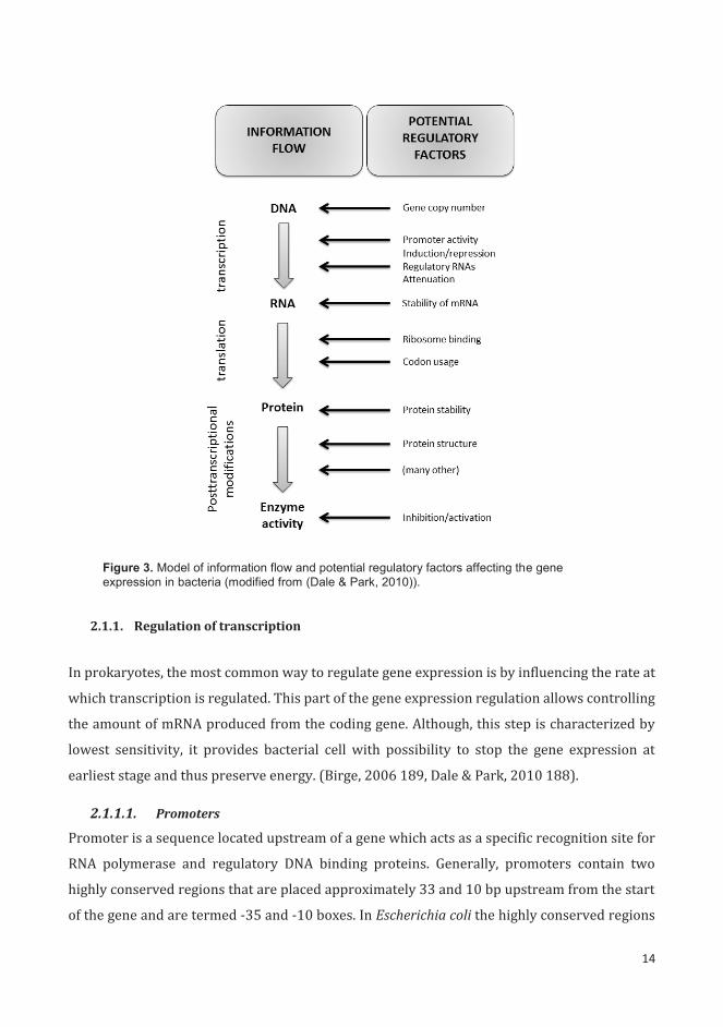
Regulation of gene expression in bacterial cell takes place at different levels (Fig. 3). The most
general control occurs at the level of transcription. The expression is further controlled at the
level of translation and subsequently undergoes the postranslational control. Following the
scheme presented in the Figure 3 a number of potential regulatory factors can be mentioned.
First of all, higher number of copies of the gene can increase the expression. In general, if the
genome harbors more copies of a certain gene, there are more sites available for the
transcription process to take place. However, most of the genes on the bacterial chromosome
exist only in one copy, excluding genes coding for such molecules as rRNA. Therefore, this type
of regulation is restricted to only several genes. Secondly, the promoter activity determines the
efficiency with which the gene is transcribed by affecting the level of initiation of transcription
by RNA polymerase. Promoter activity is considered to be the most important cue in control of
expression of individual genes in bacteria. Next step of regulation focuses on the stability of
mRNA molecules, which serve as the templates for translation. Most of the bacterial mRNAs are
short-lived and are typically degraded with half-lives below 2 minutes, whereas other forms of
RNA (rRNA or tRNA) are considerably more stable. All the above mentioned steps focus on
providing and maintaining the proper amount of mRNA available in bacterial cell. The process
of translation is controlled by the efficiency of initiation (ribosome binding) and the rate of
translation (codon usage). Furthermore, the abundance of protein reflects both the rate of
production and its stability. The last step of regulation involves different posttranscriptional
effects that may include events like protein folding, covalent modifications, as well as activation
and inhibition by other proteins. (Birge, 2006 189, Dale & Park, 2010 188).
14
Figure 3. Model of information flow and potential regulatory factors affecting the gene expression in bacteria (modified from (Dale & Park, 2010)).
2.1.1. Regulation of transcription
In prokaryotes, the most common way to regulate gene expression is by influencing the rate at
which transcription is regulated. This part of the gene expression regulation allows controlling
the amount of mRNA produced from the coding gene. Although, this step is characterized by
lowest sensitivity, it provides bacterial cell with possibility to stop the gene expression at
earliest stage and thus preserve energy. (Birge, 2006 189, Dale & Park, 2010 188).
2.1.1.1. Promoters
Promoter is a sequence located upstream of a gene which acts as a specific recognition site for
RNA polymerase and regulatory DNA binding proteins. Generally, promoters contain two
highly conserved regions that are placed approximately 33 and 10 bp upstream from the start
of the gene and are termed -35 and -10 boxes. In Escherichia coli the highly conserved regions

15
TTGACA and TATAAT constitute the consensus sequences for -35 and – 10 boxes, respectively.
The sequence separating these highly conserved regions is variable and for major of promoters
it is 16 to 18 bp long. The variations of the promoter sequence reflect both its strength and its
capacity to bind different classes of RNA polymerases. Promoters that have sequence close to
the ideal consensus are considered to be strong and they can direct the initiation of the
transcription of the gene every 2 seconds. Changes in the conserved boxes or in the separating
sequence lead to decrease in the promoter activity. Despite the diversity of the promoters, the
TATA motif of the -10 box is common for all classes of promoters, including eukaryotic and
archeal promoters. Therefore promoter constitutes a fixed level of control that determines the
potential level of expression of a certain gene. (Travers, 1987, Dale & Park, 2010).
2.1.1.2. Sigma factors and anti-sigma factors
RNA polymerase that conducts transcription is composed of four subunits (α2ββ’) and
additional dissociable element called -factor. The -factor allows recognition of the conserved
-10 and -35 boxes in the promoter region determining the specificity of the enzyme. Since
regulons of -factors can be comprised of hundreds of genes, this mechanism is frequently used
to respond to such stimuli as environmental stresses, nutritional downshifts, and variations in
pH and osmolarity (Kazmierczak et al., 2005, Dale & Park, 2010). The most common -factor,
70, is responsible for transcription of housekeeping genes required for the essential cellular
functions. The presence of alternative sigma factors allows bacterium to redirect the
transcription into set of a smaller number of genes linked to a specific function. In E. coli seven
different -factors have been identified (Table 3) (reviewed in (Landini et al., 2014)).
The general stress response alternative sigma factor RpoS ( 38) is responsible for
transcription of genes contributing to bacterial survival under unfavorable environmental
conditions. Its expression is activated during starvation, oxidative damage, reduced pH, as well
as during stationary phase of growth. The rpoS gene is not essential for growth of E. coli and its
deletion does not affect the growth rate in neither rich nor minimal medium. However, strains
lacking RpoS display high sensitivity to a variety of environmental stresses. Approximately 140
genes are induced directly by the increase in 38 levels, regardless of growth conditions and
environmental cues. Moreover, up to 500 genes together can be affected directly or indirectly
by the activity of RpoS, indicating interplay with additional regulators (reviewed in (Landini et
al., 2014)). Many studies have shown that in some species like S. enterica serovar Typhimurium
RpoS is needed for full virulence. The RpoS mutant of this species displayed alterations in
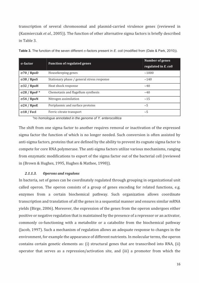
16
transcription of several chromosomal and plasmid-carried virulence genes (reviewed in
(Kazmierczak et al., 2005)). The function of other alternative sigma factors is briefly described
in Table 3.
Table 3. The function of the seven different -factors present in E. coli (modified from (Dale & Park, 2010)).
-factor Function of regulated genes Number of genes
regulated in E. coli
70 / RpoD Housekeeping genes 1000
38 / RpoS Stationary phase / general stress response 140
32 / RpoH Heat shock response 40
28 / RpoF * Chemotaxis and flagellum synthesis 40
54 / RpoN Nitrogen assimilation 15
24 / RpoE Periplasmic and surface proteins 5
18 / FecI Ferric citrate transport 5
*no homologue annotated in the genome of Y. enterocolitica
The shift from one sigma factor to another requires removal or inactivation of the expressed
sigma factor the function of which is no longer needed. Such conversion is often assisted by
anti-sigma factors, proteins that are defined by the ability to prevent its cognate sigma factor to
compete for core RNA polymerase. The anti-sigma factors utilize various mechanisms, ranging
from enzymatic modifications to export of the sigma factor out of the bacterial cell (reviewed
in (Brown & Hughes, 1995, Hughes & Mathee, 1998)).
2.1.1.3. Operons and regulons
In bacteria, set of genes can be coordinately regulated through grouping in organizational unit
called operon. The operon consists of a group of genes encoding for related functions, e.g.
enzymes from a certain biochemical pathway. Such organization allows coordinate
transcription and translation of all the genes in a sequential manner and ensures similar mRNA
yields (Birge, 2006). Moreover, the expression of the genes from the operon undergoes either
positive or negative regulation that is maintained by the presence of a repressor or an activator,
commonly co-functioning with a metabolite or a catabolite from the biochemical pathway
(Jacob, 1997). Such a mechanism of regulation allows an adequate response to changes in the
environment, for example the appearance of different nutrients. In molecular terms, the operon
contains certain genetic elements as: (i) structural genes that are transcribed into RNA, (ii)
operator that serves as a repression/activation site, and (iii) a promoter from which the
17
transcription starts. Furthermore, there is a regulator gene that codes for a regulatory molecule
which interacts with the operator (Birge, 2006). A regulon constitutes another form of
organized gene regulation in bacteria, where a set of genes is regulated by the same regulatory
gene product (Snyder & Champness, 2007).
2.1.1.4. Transcriptional regulators
Transcription process starts downstream from the promoter region, where the polymerase
begins the elongation of the RNA. Termination site designates the location at which the
polymerase is being released from the DNA and thus the elongation of the RNA molecule stops.
That occurs in some distance from the translation termination codon, leaving the 3’
untranslated region in between (Snyder & Champness, 2007). The dissociation of polymerase
can be caused by two types of mechanisms: Rho-dependent (factor-mediated) or intrinsic (Rho-
independent). Rho-independent termination occurs in the absence of auxiliary factors at
locations where the RNA forms a stable hairpin structure, whereas Rho-mediated termination
results from the action of Rho protein, which binds to specific sequences present in the RNA
(Boudvillain et al., 2013). Both the beginning and the termination of the transcription process
can be influenced by the activity of transcriptional factors.
The initiation of transcription can be positively or negatively influenced by the recruitment of
a specific activator or repressor, respectively. A repressor binds to an operator changing the
conformation of the upstream region and prevents the polymerase from binding and/or
advancing on the DNA template. An activator typically binds to an upstream activator sequence
located upstream of the promoter and promotes the initiation of transcription. Transcription
factors can either work solely as activators or repressors, or as both (dual regulators)
depending on the target promoter (Snyder & Champness, 2007). A computational analysis of E.
coli genome estimated presence of 314 transcriptional factors, out of which 35% were
activators, 43% repressors, and 22% dual regulators (Perez-Rueda & Collado-Vides, 2000).
Distinct group of transcriptional regulators comprises global transcriptional regulators, which
have the ability to regulate large numbers of genes belonging to different functional classes.
The action of these factors can be complex, as they not only directly affect the expression of
certain genes, but also indirectly regulate various cellular pathways by controlling different
regulators. In E. coli it has been estimated, that seven global transcriptional regulators (CRP,
FNR, IHF, Fis, ArcA, NarL and Lrp) control 50% of all regulated genes (Martinez-Antonio &
Collado-Vides, 2003).

18
The rate of transcription can be also altered by changes in the topology of the bacterial
chromosome. H-NS is a nucleid-associated protein that affects the DNA topology at specific loci
and therefore modulates gene transcription by selective supercoiling of the promoter regions.
The regulatory effects of H-NS are linked to metabolic and environmental conditions. The
primary direct effect of H-NS is repression. (Fang & Rimsky, 2008).
Additionally, the process of transcription can be influenced at the stage of termination. Several
proteins act to prevent transcriptional termination by utilizing two different mechanisms. The
non-processive transcription antitermination factors bind a specific RNA sequence preventing
the RNA from forming a transcription termination structure and allowing the polymerase to
continue the elongation beyond this sequence. The processive elongation factors modify the
polymerase so that it becomes resistant to the termination signal and it reads through the
termination site (Rutberg, 1997).
Two-component regulatory systems (TCRs) and phosphorelay systems constitute a mechanism
of sensing and responding to external stimuli by bacterial cells. Generally, this system
comprises of two elements: (i) a histidine protein kinase, an integral membrane protein, and
(ii) a cytoplasmic response regulator. Upon occurrence of a certain external stimulus, the
histidine protein kinase undergoes a conformational change, autophosphorylates and
subsequently transfers the phosphate group to the response regulator. The phosphorylation
activates the regulator and enables it to bind to target DNA sequences and thus to regulate the
expression of controlled genes. (Dale & Park, 2010).
Additionally, cyclic di-GMP signaling is implicated in regulation of wide range of bacterial
features, including adhesion to surfaces, biofilm formation, aggregation and the virulence. The
concentration of cyclic di-GMP inside of the cell results from the balance between the synthesis
and degradation. The GGDEF protein domain catalyzes the synthesis of cyclic di-GMP from two
GMP molecules, while EAL and HD-GYP domains catalyze the hydrolysis back to GMP. Cyclic di-
GMP functions through binding to different receptors or effectors with the pilZ domain, as well
as different transcription factors and riboswitches. Therefore, the regulation exerted by the
cyclic di-GMP can occur not only at the level of transcription, but also at the post-transcription
or post-translation level. (Ryan, 2013).
An exceptional situation, where bacterial cell requires nimble and adequate response to
changes in external environment occurs during the infection. The expression of virulence

19
factors is tightly and coordinately regulated during different stages of infection. Precise control
of virulence gene expression is ensured by the virulence-related transcriptional factors, which
can sense host signals such as changes in temperature, osmolarity, pH, iron levels, nutrient
availability, antimicrobial agents and oxygen levels. Disruption of these virulence factors leads
to reduced virulence or complete attenuation of the pathogen (Cotter & Miller, 1998, Zhou &
Yang, 2006).
2.1.1.5. RfaH antiterminator
RfaH is a bacterial transcriptional antiterminator that enables polymerase to overcome the
intrinsic termination signals and prevent the polarization of long operons. Genetic polarity is
caused by a failure in transcription of mRNA of particular part of the operon or reduced
translation of a certain region (Birge, 2006). In case of long operons genetic polarity leads to
reduced production of enzymes encoded by genes distal to the promoter. Loss of RfaH increases
transcription polarity of limited long operons without affecting the transcription initiation from
the operon promoters. In E. coli and Salmonella RfaH controls the transcription of a specialized
group of operons that direct the synthesis, assembly and export of the lipopolysaccharide core,
exopolysaccharide, F conjugation pilus and hemolysin toxin (Bailey et al., 1997).
For its activity RfaH requires a non-coding 8 bp motif 5’-GGCGGTAG-3’ termed ops (operon
polarity suppressor). In order to function, the ops element must be located downstream of an
active promoter in the correct orientation. The importance of the ops element was further
proven by the discovery, that the deletion of the ops sequence increased transcriptional polarity
within the operon in a similar way as rfaH null mutation. Moreover, insertion of this 8 bp motif
downstream of a non-native promoter resulted in increase of distal gene transcription (Nieto
et al., 1996). Further research showed that RfaH-dependent transcription elongation occurs
upon recruitment of RfaH into a transcription complex and that this recruitment is specifically
directed by the ops element (Bailey et al., 2000). Moreover, the examination of genomic
sequences revealed that ops element is conserved among gamma-proteobacteria, being present
and functional in such species as Shigella flexneri, Vibrio cholerae, Klebsiella pneumoniae and
Pseudomonas aeruginosa (Nieto et al., 1996, Rahn et al., 1999, Carter et al., 2004, Carter et al.,
2007).
The RfaH protein consists of two domains connected by a flexible linker. The N-terminal domain
is structurally similar to NusG and mediates the RNA polymerase binding and anti-pausing
functions. The C-terminal domain is a short α-helical hairpin. In a free RfaH molecule the two

20
domains interact and are tightly associated. The contact with the ops element triggers the
conformational change that separates the domains and allows the RfaH to bind to the
polymerase. Moreover, the C- terminal domains refolds into a β-barrel. The association of the
two domains in the free state restricts the RfaH actions to ops-containing operons and thus
helps avoiding interference with NusG (Belogurov et al., 2007). After binding to the ops-
element RfaH delays the transcription, but after the escape from the ops element, it enhances
the elongation by suppressing pausing and Rho-dependent termination (Artsimovitch &
Landick, 2002). Moreover, the RfaH protein shows high level of conservation between the
species. Orthologues of RfaH were also proven to complement an E. coli rfaH deletion
suggesting high level of functional homology (Carter et al., 2004).
RfaH was first described as a component of LPS synthesis machinery in Salmonella and believed
to function as an enzyme (Wilkinson & Stocker, 1968). Later it was shown that RfaH acts as a
positive regulator of the expression of a gene cluster involved in the lipopolysaccharide
biosynthesis pathway ( (Lindberg & Hellerqvist, 1980) and displays homology with E. coli sfrB,
a gene required for the expression of F-factor functions (Sanderson & Stocker, 1981). Further
studies revealed that RfaH is also needed for the synthesis and secretion of haemolysin (Bailey
et al., 1992) and the expression of the type II capsule K5 antigen in E. coli (Stevens et al., 1994).
In addition, RfaH enhances the expression of kps operons necessary for the synthesis of group
2 polysialic acid capsules (Navasa et al., 2014) and hemin receptor molecule ChuA (Nagy et al.,
2001). Recent research demonstrated that RfaH selectively controls fimB expression at the
post-transcriptional level by suppression of small RNA MicA inhibition (Moores et al., 2014).
Due to the fact that RfaH promotes the expression of components that are required for bacterial
virulence, loss of RfaH usually leads to decrease in pathogenicity (Nagy et al., 2002, Nagy et al.,
2006). The decrease in virulence was observed for example for Salmonella enterica serovar
Typhimurium, uropathogenic and avian pathogenic E. coli. The absence of RfaH in E. coli results
in downregulation of several virulence factors (LPS, K15 capsule, alpha-hemolysin and hemin
receptor ChuA) and subsequently to reduced urovirulence in the mouse model (Nagy et al.,
2002). The rfaH deletion mutant of Salmonella showed decreased intracellular net growth in
epithelial and macrophage cells. Similarly, the mutant was deficient in production of outer
membrane structures. In this case it was shown that the absence of rfaH results not only in
changes that are a result of polarization of long operons with ops sequence, but also leads to
indirect changes caused by rough-phenotype (Nagy et al., 2006).

21
2.1.1.6. The LysR-family transcriptional regulators
The LysR-type transcriptional regulators are widely distributed among the prokaryotes. They
are involved in the regulation of metabolic functions like sugar catabolism, amino-acid
synthesis, aromatic compound degradation, antibiotic resistance and virulence. Typically they
consist of two domains: N-terminal DNA binding domain with a helix-turn-helix motif and a C-
terminal regulatory domain that binds an effector. Classically, these regulators oligomerize to
form tetramers that bind to DNA to activate or repress transcription upon binding to one or
more effectors. The two domains are connected by a linker helix that together with N- terminal
domain plays also a role in oligomerization. (Schell, 1993).
In E. coli the LysR homologue A (LrhA) functions as a global transcriptional regulator of genes
related to motility, chemotaxis and flagella synthesis. In other bacteria, the LrhA homologs are
known under diverse names and functions. The PecT of Erwinia chrysanthemi and HexA of
Erwinia carotovora are 75-79% identical to LrhA, and were implicated to regulate several
virulence determinants (Surgey et al., 1996, Mukherjee et al., 2000). The Yersinia
pseudotuberculosis LrhA homolog RovM (ca. 70% identical to LrhA) represses the invasin
regulator RovA (Heroven & Dersch, 2006). The RovM of Y. enterocolitica O:3 is 88% identical to
RovM of Y. pseudotuberculosis and ca. 70 % identical to LrhA of E. coli.
The structure analysis of RovM from Y. pseudotuberculosis revealed that it most likely adopts a
tetrameric arrangement with two distant DNA-binding domains. Such a conformation would
cause the target DNA to bend around the regulator. Additionaly, it was shown that RovM
possesses a cavity that could bind small inducer molecules (Quade et al., 2011).
Y. pseudotuberculosis RovM was shown to recognize a 50 bp region upstream of promoters that
contains two palindromic sequences. Moreover, hyper-reactive bases were detected in the
RovM-binding sequence suggesting that RovM bends its binding site upon interaction (Heroven
& Dersch, 2006). LrhA is known to interact directly with the promoter of the flagellar flhDC
genes, and thereby affects indirectly the genes that are under the control of the FlhDC master
regulon (Lehnen et al., 2002). In E. coli, it was shown that LrhA protein binds directly to the
promoter region upstream of the lrhA gene and thus the expression of lrhA is subject to positive
autoregulation (Lehnen et al., 2002). Although, unlike the other orthologues, RovM does not
bind directly to its own promoter, it is positively autoregulated through an unknown
mechanism (Heroven & Dersch, 2006). In addition, the expression of lrhA in E. coli is repressed
by the RcsCDB phosphorelay system (a cell-envelope stress-sensing pathway) and induced by

22
mutations in the FtsK DNA motor protein (Peterson et al., 2006) and in Pantoea stewartii by the
regulatory protein EsaR (Ramachandran & Stevens, 2013, Ramachandran et al., 2014). In Y.
pseudotuberculosis the expression of rovM is medium-dependent and mediated by CsrC
(Heroven et al., 2008). It was also shown that the expression of lrhA in Salmonella is growth
phase-dependent (Mouslim & Hughes, 2014).
2.1.2. Post-transcriptional regulation
Expression of a gene can also be regulated at later stages, when the transcription process has
already happened and mRNA was produced. In both prokaryotes and eukaryotes, the
messenger RNA can be translated into a protein with different efficiency depending on its
sequence, structure and presence of different factors. Such type of control of expression is
called post-transcriptional or translational regulation. Generally, the post-transcriptional
control of expression involves interactions of different molecules with the mRNA transcripts
that affect the process of translation or may be based on different accessibility or stability of
the mRNA transcript.
2.1.2.1. Translation initiation efficiency
Commonly, bacterial mRNA contains a translation initiation region (TIR) composed of the
initiation codon and the Shine-Dalgarno sequence (SD). The SD site is located 4 to 15 bp
upstream of the initiation codon and contains a sequence of nucleotides with variable
complementarity to the 3’ end of the 16S rRNA. Such complementarity allows the 30S ribosomal
to bind to the mRNA upstream of the initiation codon. The level of complementarity is one of
the factors contributing to the efficiency of translation (Jacob et al., 1987). Moreover, TIRs can
also harbor a short U- or A/U-rich sequence that binds the ribosomal protein S1. The presence
of this sequence upstream of the initiation codon enhances the efficiency of translation
initiation (Boni et al., 1991). Another important factor that determines the efficiency of
translation is the structure of the mRNA transcript. Occasionally, the conformation of the mRNA
can inhibit the translation through hampering the access of the 30S ribosomal subunit to the
ribosomal binding site (de Smit & van Duin, 1990). Moreover, it has been recently suggested
that the synonymous mutations within the first 40 nt of the transcript can significantly affect
the abundance of the protein through alterations in the mRNA folding (Kudla et al., 2009).

23
2.1.2.2. Stability of mRNA
The stability of mRNA transcript comprises another level of post-transcriptional regulation.
Generally, the concentration of mRNA within the bacterial cell is a result of balance between
the synthesis and the degradation of the mRNA molecules. Therefore, not only the transcription,
but also the degradation of mRNA directly affects the synthesis of protein by decreasing the
concentration of mRNA available for translation. In many bacterial species, mRNA degradation
is modulated in response to changes in the environment and to stress conditions (Redon et al.,
2005, Shalem et al., 2008). This mechanism allows quick disposal of unnecessary mRNA and
thus prevents the cell from producing the proteins that are no longer required under changed
conditions.
In E. coli mRNA degradation is generally initiated by endoribonucleolytic cleavages induced by
single-stranded RNA-specific endoribonucleases (e.g., RNase E and RNase G) or double-
stranded RNA-specific endoribonucleases (e.g., RNase III). This initial nucleolytic step
generates primary decay intermediates which are further degraded by a combination of endo-
and exonucleases (PNPase, RNase II, RNase R). This stage yields short nucleotides which are
later converted to mononucleotides by oligoribonuclease. In addition to major ribonucleases,
bacteria possess a number of ancillary mRNA-modifying enzymes that assist the mRNA
degradation process (reviewed in (Kaberdin et al., 2011)).
Gene expression can be also influenced by the secondary structure of the RNA molecule. The
RNA helicases are proteins that are able to change the secondary structure of RNA molecules
by unwinding the RNA or DNA-RNA duplexes and by performing local strand separation. The
most well-known group of the RNA helicases are DEAD-box proteins that induce a local strand
separation. Such separation can be further used for either protein or regulatory RNA binding
to one of the strands. The activity of different bacterial DEAD-box proteins has been implicated
in ribosome biogenesis, RNA decay, and translation initiation. (Reviewed in (Khemici & Linder,
2016)).
2.1.2.3. Non-coding RNAs
Non-coding RNA (ncRNA) molecules are widely spread among all kingdoms of life and recently
have become recognized as a novel class of gene expression regulators. These molecules
encompass a large and diverse group of RNA species that do not encode for proteins and thus
do not undergo the translation process. Instead, they present regulatory functions. Regulatory
ncRNAs can be divided into several classes: (i) cis-encoded base-pairing RNAs (antisense RNAs,

24
asRNAs), (ii) trans-encoded base-pairing RNAs, (iii) riboswitches, (iv) ribozymes, (v) RNAs
modulating protein activity, and (vi) CRISPRs (clustered regulatory interspaced short
palindromic repeats) (Liang et al., 2011). The asRNAs are encoded on the DNA strand opposite
to the coding mRNA and they typically share over 75 nt complementarity with their target
mRNA. The trans-encoded RNAs are typically encoded in the intragenic regions away from their
target mRNA and they share only limited complementarity with the mRNA species. Moreover,
unlike cis-encoded RNAs, trans-acting RNAs can regulate more than one mRNA molecule
(Michaux et al., 2014). Small RNA molecules (sRNAs) are also known to interact directly with
proteins and alter their activity by sequestration (e.g. 6S RNA in E. coli) (Wassarman, 2007).
The ribozymes are catalytic RNAs which typically catalyze cleavage or ligation of another RNA
particle through a phosphodiester cleavage reaction (Serganov & Patel, 2007). Riboswitches
are metabolite-sensing RNA structures that response to such environmental cues as cations or
temperature shifts. Upon a change in the environment they change their conformation leading
to activation or inhibition of gene expression (Serganov & Patel, 2007). The last group, CRISPRs,
are highly variable regions of 24-47 bp, separated by a series of 2-249 repeat-spacer units. The
whole CRISPR region, preceded by a 550 bp leader sequence, determines resistance to
bacteriophages and foreign plasmids (Michaux et al., 2014).
Trans-encoded sRNAs range in size from 50 to 500 nt in length and present various secondary
structures. Base-pairing with the target mRNA is usually imperfect and based on 7-10 nt seed
sequence. Moreover, for proper functionality trans-encoded sRNAs often require the presence
of Hfq chaperone that simultaneously bind to both the sRNA and mRNA and facilitate the
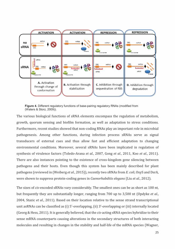
interaction (Gottesman & Storz, 2011). The sRNAs regulate the gene expression by base-pairing
with the target mRNAs, which leads to changes in mRNA translation or stability. sRNAs can be
both activators and repressors of gene expression depending on the location of pairing with the
target mRNA. They can act positively by changing the mRNA conformation and preventing
formation of an inhibitory structure that sequesters the ribosome-binding site (Fig. 4A). sRNAs
can act negatively by base pairing with 5’ untranslated region (UTR) and inferring with the
ribosome binding (Fig. 4C) or by targeting the sRNA-mRNA duplex for degradation by
ribonucleases (Fig. 4D) (Waters & Storz, 2009).
25
Figure 4. Different regulatory functions of base-pairing regulatory RNAs (modified from (Waters & Storz, 2009)).
The various biological functions of sRNA elements encompass the regulation of metabolism,
growth, quorum sensing and biofilm formation, as well as adaptation to stress conditions.
Furthermore, recent studies showed that non-coding RNAs play an important role in microbial
pathogenesis. Among other functions, during infection process sRNAs serve as signal
transducers of external cues and thus allow fast and efficient adaptation to changing
environmental conditions. Moreover, several sRNAs have been implicated in regulation of
synthesis of virulence factors (Toledo-Arana et al., 2007, Gong et al., 2011, Koo et al., 2011).
There are also instances pointing to the existence of cross-kingdom gene silencing between
pathogens and their hosts. Even though this system has been mainly described for plant
pathogens (reviewed in (Weiberg et al., 2015)), recently two sRNAs from E. coli, OxyS and DsrA,
were shown to suppress protein-coding genes in Caenorhabditis elegans (Liu et al., 2012).
The sizes of cis-encoded sRNAs vary considerably. The smallest ones can be as short as 100 nt,
but frequently they are substantially longer, ranging from 700 up to 3,500 nt (Opdyke et al.,
2004, Stazic et al., 2011). Based on their location relative to the sense strand transcriptional
unit asRNAs can be classified as (i) 5’-overlapping, (ii) 3’-overlapping or (iii) internally located
(Georg & Hess, 2011). It is generally believed, that the cis-acting sRNA species hybridize to their
sense mRNA counterparts causing alterations in the secondary structures of both interacting
molecules and resulting in changes in the stability and half-life of the mRNA species (Wagner,

26
1994). Eventually, asRNAs can influence the abundance of mRNA through regulation of
transcription, degradation of sense transcripts or their stabilization. Moreover, antisense
species can also regulate the translation process through binding to the SD sequence of their
target mRNA (reviewed in (Georg & Hess, 2011) and (Sesto et al., 2013)). Recently, the
transcriptome analysis revealed that in Mycoplasma pneumonie, Sinorizobium meliloti, Vibrio
cholerae and Staphylococcus aureus antisense transcription rates reach approximately 13%,
11%, 4.7% and 1.3%, respectively (Guell et al., 2009, Liu et al., 2009, Beaume et al., 2010,
Schluter et al., 2010). In addition, a recent RNA sequencing-based study conducted on the
transcriptome of E. coli identified about 1,000 different asRNA species (Dornenburg et al.,
2010).
Several recent studies led to identification of various non-coding sRNAs among Yersinia-
species. A deep RNA-sequencing approach resulted in identification of 150 sRNAs in Y.
pseudotuberculosis and 31 in Y. pestis (Koo et al., 2011, Beauregard et al., 2013). Another
approach based on cDNA-cloning allowed verification of 43 novel sRNA from Y. pestis (Qu et al.,
2012). Recently, several studies focused on their expression under different conditions and
their role in bacterial virulence (Koo et al., 2011, Yan et al., 2013). Moreover, it was shown, that
some sRNAs, although conserved in both Yersinia display different function, suggesting
evolutionary changes in sRNA regulation network between these two species (Koo et al., 2011).
2.1.2.4. Usage of rare codons
The efficiency of translation is strongly influenced by the codon bias. The same amino acid can
be coded by different triplets of nucleotides, and therefore different tRNAs have to be drawn
during the translation. Due to the fact that different tRNA species show different abundance,
the synonymous mutation can significantly affect the efficiency of translation (Parmley & Hurst,
2007, Tuller et al., 2010).
2.1.2.5. RNA-binding proteins
Bacterial post-transcriptional regulators typically influence RNA degradation, translation
initiation efficiency or transcript elongation. The RNA-binding proteins (RBPs) can use
different mechanisms to exert their regulatory functions: (i) change in the mRNA susceptibility
to RNases, (ii) modulation of mRNA RBS accessibility, or (iii) acting as a chaperone to facilitate
the interaction between mRNA and other factors (Van Assche et al., 2015).

27
Apart from RNases that are directly involved in mRNA decay (described in 2.2.2.2.), bacteria
also possess a number of mRNA modifying enzymes that can facilitate mRNA degradation. For
example in E. coli, the pyrophosphate removal at the 5’ end by pyrophosphate hydrolase (RppH)
and addition of a single-stranded poly(A) extension to the 3’ end of mRNA facilitated by poly(A)
polymerase I (PAPI) both enhance the mRNA degradation. Additionally, the degradation of
highly structured RNA molecules can be assisted by the RhlB, which unwinds the RNA
structures in an ATP-dependent manner. (Kaberdin et al., 2011, Van Assche et al., 2015).
Regulatory RBPs can alter the efficiency of translation initiation directly by competing with
ribosomes for the RBS or indirectly by altering the secondary structure of the mRNA near the
ribosome interaction region (Van Assche et al., 2015). One example of RBP that is conserved
among many bacterial species is CsrA, a central component of the global carbon storage
regulatory system. CsrA binds to GGA-motifs in the 5’ UTR near the Shine-Dalgarno region and
represses translation by competing with the 30S ribosomal subunit (Baker et al., 2007).
Another group of RBPs can affect RNA stability or translation initiation efficiency by assisting
in the interactions with other molecules. These proteins typically bind simultaneously the
mRNA target and its co-effector molecule that can be a sRNA or another protein. A well-known
example of such function is Hfq, RNA-chaperone implicated in global post-transcriptional
regulation (Geng et al., 2009, Schiano et al., 2010, Kakoschke et al., 2014). The selected
examples of RBPs are presented in Table 4.
Table 4. Selected examples of RNA-binding proteins involved in post-transcriptional gene expression
regulation in Yersinia species.
Regulator Target Function References CsrA GGA-motifs in
the 5’ UTR global carbon storage regulation; in Y. enterocolitica CsrA activates expression of genes encoding the master motility regulator flhDC, enhances resistance to osmolytes and allows growth at 4°C and 42°C
(LeGrand et al., 2015)
Hfq AU-rich regions sRNA chaperone that stabilizes the sRNA-mRNA interactions; highly pleiotropic; affects growth, metabolism and virulence
(Geng et al., 2009, Schiano et al., 2010, Kakoschke et al., 2014)
SmpB Small stable RNA A
in Y. pseudotuberculosis affects pathogenesis, resistance to environmental stresses, and motility; enables proliferation in macrophages, affects Yop-mediated cytotoxicity
(Okan et al., 2006)
YopD 5’ UTR of T3SS genes
represses expression of T3SS genes, shows highest affinity to effector Yops; prevents ribosome binding and accelerates degradation.
(Chen & Anderson, 2011)

28
2.1.2.6. RNA chaperone Hfq as a global post-transcriptional regulator
Hfq, an RNA chaperone required for maintaining the stability and function of many sRNAs, has
been recognized as a central component of global post-transcriptional regulation network
(reviewed in (Vogel & Luisi, 2011)). Hfq was first identified as a host bacterium factor required
for the replication of bacteriophage Qβ RNA (Franze de Fernandez et al., 1968). Subsequent
research revealed that it is widely distributed in the bacterial kingdom, present in many
different pathogenic species. Hfq is a bacterial homolog of the eukaryotic and archeal Sm/LSm
proteins, with a characteristic ring-like multimeric quaternary architecture supporting
interactions with other macromolecules. In eukaryotes, many different functions were
implicated for the Sm/LSm proteins, including role in mRNA splicing, RNA decapping and RNA
stabilization (reviewed in (Wilusz & Wilusz, 2005)).
The Hfq of E. coli is a 102 amino acid residue (11.2 kDa) highly abundant protein with an
estimated 50 000 to 60 000 copies per cell, of which 80 – 90% are found in association with
ribosomes (Brennan & Link, 2007). Hfq has a 25Å thick toroidal structure with an outer
diameter of around 70 Å and a 8-12 Å wide central pore. The protein is characterized by an N-
terminal α helix followed by β strands displaying the topology β5α1β1β2β3β4. Like other
proteins from Sm family Hfq contains Sm1 and Sm2 motives, two highly conserved regions. The
Sm1 motif encompasses the first three β strands, whereas the Sm2 motif is located in fourth
and fifth β strand. The hexamer structure is formed through the interactions between the
residues of β4 and β5 of pairing subunits. The α helix is located on the top of the β sheet and
constitutes the distal side of the protein (Sauter et al., 2003).
In E. coli, AU-rich sequences of sRNAs typically bind to the proximal surface of the Hfq protein
and A-rich sequences of mRNAs bind to the distal surface (Mikulecky et al., 2004). The structure
of S. aureus Hfq showed that the Sm1 and Sm2 motifs play an important role in RNA binding.
The sRNA molecule expands and fills the central pore on the proximal side binding to Hfq
through AU-rich regions in a circular manner. The Hfq structure possesses six AU nucleotide
binding pockets, yet as many of the sRNAs contain stretches shorter than six U or A, it is unlikely
that all the pockets are filled simultaneously (Schumacher et al., 2002). The motif required for
binding of A-rich sequences of mRNA is located on the distal side of the Hfq protein, opposite
to the AU-binding side (Mikulecky et al., 2004).
There are different mechanisms through which Hfq exhibits its regulatory functions. First, Hfq
can suppress protein synthesis by allowing the sRNA to bind to the 5’ region of the mRNA

29
sequestering the translation initiation site. It can also display opposite function by promoting
sRNA binding to the 5’ region of mRNA in order to disrupt a secondary structure that initially
inhibited the translation. By binding to sRNAs Hfq can also protect them from the ribonuclease
cleavage or promote the degradation. The mechanism of action depends on the RNA molecules
(reviewed in (Vogel & Luisi, 2011)). Recent studies using co-immunoprecipitation and
subsequent detection of sRNAs and mRNAs led to identification of a large number of Hfq targets
(Zhang et al., 2003, Sittka et al., 2008, Chao et al., 2012, Bilusic et al., 2014).
The regulation of Hfq expression is growth phase dependent. The study showed that the level
of Hfq protein is higher during the log phase and decreases when bacteria enter the stationary
phase (Kajitani et al., 1994). It is also known, that in E. coli CsrA can bind to hfq mRNA and
inhibit its synthesis by blocking the ribosome binding (Baker et al., 2007).
In most of the studied bacterial species the absence of Hfq results in pleiotropic phenotypic
alterations that compromise the fitness and the responses to external cues. Due to its
pleiotropic nature, many different defects were observed among Hfq-deficient strains:
impaired growth, inability to cope with different types of environmental stresses, higher
susceptibility to antimicrobial agents, defects in quorum sensing and host invasion. However,
the effects of Hfq-deficiency seemed to be always unique for each bacterial species. Moreover,
the virulence of many pathogenic species was attenuated upon depletion of Hfq. The highest
levels of attenuation were observed for Gram-negative pathogens like Brucella abortus,
Salmonella spp, Vibrio cholerae, uropathogenic E. coli, Neisseria meningitis and Y. pestis
(reviewed in (Chao & Vogel, 2010)). Considering the high levels of attenuation observed in
many pathogens, it is believed that the Hfq-deficient strains may serve as live attenuated
vaccines (Geng et al., 2009, Chao & Vogel, 2010, Schiano et al., 2010, Hayashi-Nishino et al.,
2012).
Previous studies showed that Hfq has a profound influence on the fitness of Y. enterocolitica O:8
including the metabolism of carbohydrates, nitrogen, iron, fatty acids and ATP synthesis.
Moreover, the depletion of Hfq led to slower bacterial growth, decreased resistance to stress
and impaired synthesis of urease and yersiniabactin. In addition, the role of Hfq in biofilm
formation was implicated for that species (Kakoschke et al., 2014). The hfq mutant of Y.
pseudotuberculosis presented hypermotility and increased production of a biosurfactant-like
substance. Furthermore, it showed decreased survival in macrophages, affected biofilm
formation, impaired production of T3SS effector proteins and high attenuation rate in mouse

30
model infection (Schiano et al., 2010, Bellows et al., 2012). Also in Y. pestis Hfq was implicated
in the persistence inside of macrophages and resistance to stress. Similarly, the loss of hfq led
to attenuation (Geng et al., 2009).
2.1.2.7. Ribonuclease YbeY
YbeY is a 17-kDa highly conserved protein from the UPF0054 family that was discovered during
the global transcriptional analysis as a product of a heat-shock gene. At first, due to the
sequence similarity to metal-dependent hydrolases, YbeY was believed to possess a hydrolytic
function (Oganesyan et al., 2003, Zhan et al., 2005). Although the structure homology analysis
showed similarity to eukaryotic extracellular proteinases such as collagenase and gelatinase, in
vitro studies failed to detect any hydrolase activity (Rasouly et al., 2009). Only recently it was
discovered that YbeY is a ribonuclease that plays a critical role in rRNA maturation, as well as
in late-stage 70S ribosome quality control (Jacob et al., 2013).
The structural study revealed that the overall protein structure of YbeY consists of six α helices
and four β strands in a βααβαββααα fold. It harbors a conserved domain, characteristic for the
UPF0054 family. Moreover it contains a metal ion, most probably a Ni2+ ion that is coordinated
by the residues of His114, His118 and His124 (Zhan et al., 2005). YbeY shows high level of
conservation between the bacterial species, as ybeY genes from four distantly related pathogens
can fully complement ybeY mutant strain of E. coli (Vercruysse et al., 2014).
In E. coli the YbeY functions as a single strand-specific endoribonuclease that in its purified form
effectively degrades total rRNA and mRNA, yet is unable to degrade double-stranded RNA.
Moreover, the ribonuclease activity is manifested at 37°C and at 45°C, though it significantly
decreases at 65°C, in line with previous findings about YbeY playing a role in heat-shock
response. It was also proposed that YbeY cleaves the 17S rRNA precursor generating a 3’
phosphate terminus. Furthermore, under standard growth conditions as well as under stress
YbeY together with RNase R removes defective 70S ribosomes from the cellular pool allowing
effective translation. It was proposed that YbeY acts as a sensor of defective ribosomes by
recognizing defects in 30S subunits, which subsequently initiates degradation of complete 70S
ribosomes by introducing endonucleolytic cuts in the rRNA. Such damage in rRNA leads to its
misprocessing and misfolding and further destruction by other ribonucleases (Jacob et al.,
2013). Moreover, YbeY plays a role in transcriptional antitermination of rRNA synthesis, that is
also critical for ribosome biogenesis (Grinwald & Ron, 2013). Recent studies show that in S.

31
meliloti, E. coli, as well as in V. cholerae YbeY has an impact on small non-coding RNAs (sRNAs)
(Pandey et al., 2011, Pandey et al., 2014, Vercruysse et al., 2014).
YbeY belongs to the 206 genes postulated to comprise the minimal bacterial genome set (Gil et
al., 2004), but is essential only in some bacteria like Vibrio cholerae, Haemophilus influenzae and
Bacillus subtilis (Akerley et al., 2002, Kobayashi et al., 2003) (Vercruysse et al., 2014). In other
species like E. coli and Shinorhizobium meliloti its loss causes increase in the sensitivity to
environmental stresses and in addition in S. meliloti its loss abrogates intracellular infection
necessary for the symbiosis (Davies & Walker, 2008, Rasouly et al., 2009). In E. coli the ybeY
deletion mutant presented severe translational defects caused by very low level of functional
polysomes and accumulation of free ribosomes and ribosomal subunits. Translational defects
were mostly manifested at elevated temperatures and resulted in growth failure (Rasouly et al.,
2009). In V. cholerae loss of YbeY resulted in complete loss of mouse colonization and biofilm
formation, reduced cholera toxin production, as well as alterations in expression of virulence-
related sRNAs (Vercruysse et al., 2014). Additionally, it was implicated that in E. coli YbeY plays
a role in apoptosis-like death (Erental et al., 2014).
An YbeY endoribonuclease homolog was also observed in eukaryotes. The ybeY null mutants of
Arabidopsis thaliana are seeding lethal, suggesting an important role of this protein in plant
growth. The ybeY mutant displayed slow growth, impaired photosynthesis, defective
chloroplast development and alterations in rRNA maturation (Liu et al., 2015).
2.1.3. Post-translational regulation
The last step of gene expression regulation is based on post-translational modifications (PTM)
that alter the structure or the function of a synthetized protein. PTMs can alter the activity of a
protein by having an impact on protein complex formation, enzyme catalysis or interactions
with other biomolecules. The possible modifications in prokaryotes include phosphorylation,
acetylation, methylation, carboxylation, glycosylation, lipidation, adenylation, ribosylation,
nitrosylation, oxidation, pupylation and deamination (reviewed in (Cain et al., 2014)).
It was previously shown that PTMs contribute significantly to bacterial adaptability and cell
cycle control. Protein phosphorylation, the attachment of phosphate onto the functional groups
of amino acid side chains, is probably the most extensively studied type of modification.
Different bacterial kinases are involved in signal trafficking in regulatory networks (reviewed
in (Cain et al., 2014)). PTMs have also a profound impact on bacterial physiology and virulence.

32
Campylobacter jejuni, impaired in protein glycosylation, showed decreased ability to adhere
and invade eukaryotic cells and lost ability to colonize intestinal tracts of mice (Szymanski et
al., 2002). Although single PTM can already change the function of a protein, in some cases a
protein can undergo several modifications at competing sites. Interplay between acylation and
phosphorylation was previously observed in bacteria (Liarzi et al., 2010).
2.2. The genus Yersinia
Yersiniae are Gram-negative, rod-shaped, facultative anaerobic, non-spore-forming bacteria,
about 2 μm long and 0.6 μm in diameter. They are named after Alexandre Yersin, a
bacteriologist who discovered Yersinia pestis in 1894. Yersinia species are widely distributed in
nature and adapted, depending on the species, to survive in the outside environment and/or
within a specific animal host.
The genus Yersinia belongs to the family Enterobacteriaceae, within the class
Gammaproteobacteria of the phylum Proteobacteria (Garrity, 2005). Currently, this genus
consists of 17 species of which three, Y. pestis, Y. pseudotuberculosis and Y. enterocolitica, are
infect humans, and one, Y. ruckeri, fish (Ewing et al., 1978). Y. pestis is a causative agent of
plaque, whereas Y. pseudotuberculosis and Y. enterocolitica are enteropathogens that usually
cause self-limited gastroenteritis, yersiniosis, typically restricted to the intestinal tract and
lymph nodes. Y. ruckeri causes enteric red mouth disease, which affects mainly salmonid fish
(Ewing et al., 1978). Other species belonging to this genus are considered to be environmental
bacteria appearing mainly in fresh water and terrestrial ecosystems and include Y. aldovae, Y.
alecksiciae, Y. bercovieri, Y. entomophaga, Y. fredriksenii, Y. intermedia, Y. kristensenii, Y.
massiliensis, Y. mollaretti, Y. nurmii, Y. pekkanenii, Y. rohdei, and Y. similis (Garrity, 2005, Sprague
& Neubauer, 2005, Merhej et al., 2008, Sprague et al., 2008, Hurst et al., 2011, Murros-
Kontiainen et al., 2011, Murros-Kontiainen et al., 2011)
2.2.1. Yersinia enterocolitica
Currently Y. enterocolitica is the most common pathogenic Yersinia infecting humans. Based on
DNA-DNA hybridization and differences in 16S rRNA sequences, Y. enterocolitica was divided
into two subspecies: enterocolitica and palearctica (Neubauer et al., 2000). A further division of
the species is based on the biochemical heterogeneity and it distributes the strains into six

33
biotypes: 1A, 1B, 2, 3, 4 and 5 (Wauters et al., 1987). Generally, the biotype 1B is considered to
be pathogenic, biotypes 2-5 are characterized by low pathogenicity, and biotype 1A includes
non-pathogenic strains (Bottone, 1997). Furthermore, Yersinia species are additionally divided
into different serotypes based on the variability of O-antigen structure. Y. enterocolitica subsp.
enterocolitica comprises the biotype 1B strains formerly termed North American and is
represented by the serotype O:8 ATCC 9610 strain, while Y. enterocolitica subsp. palearctica
comprises the former European strain from biotypes 1A and 2-5 with the serotype O:3 strain
Y11 being the postulated representative (Neubauer et al., 2000).
Typically Y. enterocolitica causes self-limiting gastroenteritis with non-specific diarrhea, fever,
abdominal pain, and sometimes nausea and vomiting. Normally symptoms of acute illness
appear after 1-11 days of incubation and last for about 5-14 days (Cover & Aber, 1989). The
most common post-infectious sequelae include reactive arthritis and erythema nodusum, more
rarely erythema multiforme, uveitis, and conjunctivitis (Saari et al., 1980, Bottone, 1997).
Moreover, Y. enterocolitica can lead to such extraintestinal complications as liver and spleen
abscesses, pneumonia, meningitis, empyema and endocarditis (Zheng et al., 2008). In
immunocompromised patients yersiniosis can result in septicemia (Zheng et al., 2008).
Additionally, asymptomatic carriage of Y. enterocolitica is has been reported (Van Ossel &
Wauters, 1990).
Most of the yersiniosis cases are sporadic, however, rare outbreaks have also been reported,
with sources tracked to contaminated water, dairy and porcine products (Thompson & Gravel,
1986, Marjai et al., 1987, Ackers et al., 2000). The incidence of yersiniosis is hard to estimate as
it is rarely reported, and usually only the genus name is recorded. Nevertheless, vast majority
(around 91%) of the reported cases are due to Y. enterocolitica, with the 4/O:3 being the most
common bio/serotype worldwide (Long et al., 2010, EFSA, 2014). Currently, Y. enterocolitica is
the third most commonly reported cause of enteric zoonosis in European Union, after
Salmonella and Campylobacter, with 6 471 confirmed cases of yersiniosis reported in 2013
(EFSA, 2014). Finland is among the countries with the highest incidence rate in EU, with the
number of reported cases between 500 and 700 per year. Even though a decreasing trend in
the number of cases was observed, the incidence rate of yersiniosis is still higher than
salmonellosis. Moreover, also in Finland the bio/serotype 4/O:3 is the most predominant one.
(EFSA, 2014).
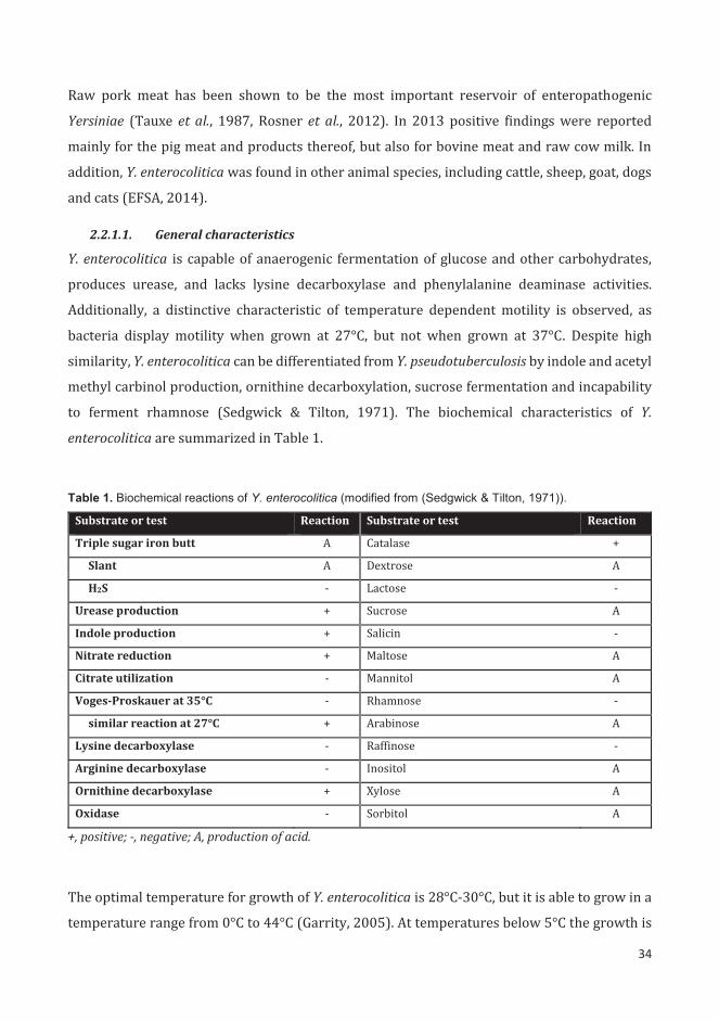
34
Raw pork meat has been shown to be the most important reservoir of enteropathogenic
Yersiniae (Tauxe et al., 1987, Rosner et al., 2012). In 2013 positive findings were reported
mainly for the pig meat and products thereof, but also for bovine meat and raw cow milk. In
addition, Y. enterocolitica was found in other animal species, including cattle, sheep, goat, dogs
and cats (EFSA, 2014).
2.2.1.1. General characteristics
Y. enterocolitica is capable of anaerogenic fermentation of glucose and other carbohydrates,
produces urease, and lacks lysine decarboxylase and phenylalanine deaminase activities.
Additionally, a distinctive characteristic of temperature dependent motility is observed, as
bacteria display motility when grown at 27°C, but not when grown at 37°C. Despite high
similarity, Y. enterocolitica can be differentiated from Y. pseudotuberculosis by indole and acetyl
methyl carbinol production, ornithine decarboxylation, sucrose fermentation and incapability
to ferment rhamnose (Sedgwick & Tilton, 1971). The biochemical characteristics of Y.
enterocolitica are summarized in Table 1.
Table 1. Biochemical reactions of Y. enterocolitica (modified from (Sedgwick & Tilton, 1971)).
Substrate or test Reaction Substrate or test Reaction
Triple sugar iron butt A Catalase +
Slant A Dextrose A
H2S - Lactose -
Urease production + Sucrose A
Indole production + Salicin -
Nitrate reduction + Maltose A
Citrate utilization - Mannitol A
Voges-Proskauer at 35°C - Rhamnose -
similar reaction at 27°C + Arabinose A
Lysine decarboxylase - Raffinose -
Arginine decarboxylase - Inositol A
Ornithine decarboxylase + Xylose A
Oxidase - Sorbitol A
+, positive; -, negative; A, production of acid.
The optimal temperature for growth of Y. enterocolitica is 28°C-30°C, but it is able to grow in a
temperature range from 0°C to 44°C (Garrity, 2005). At temperatures below 5°C the growth is
35
significantly slowed down and the bacterium undergoes cold adaptation that includes
alterations in the compositions of lipids and proteins in the cell membrane (Goverde et al.,
1994). Moreover, Y. enterocolitica can survive in frozen food product for extended period of
time as well as withstand repeated freezing and thawing (Asadishad et al., 2013). Y.
enterocolitica can grow in pH range between 4.0 and 10.0, with the optimum around 7.2 - 7.4,
and tolerate sodium chloride concentrations up to 5% (Garrity, 2005). It is able to grow under
both aerobic and anaerobic conditions, though the presence of carbon dioxide affects the
growth rate by prolonging the lag phase of the organism (Pin et al., 2000).
2.2.1.2. Virulence factors
2.2.1.2.1. Plasmid encoded virulence factors
Virulence factors of Y. enterocolitica are encoded both on the bacterial chromosome and on the
70-kb virulence plasmid (pYV). The latter one encodes, among the others, the proteins of the
type III secretion system (T3SS), effector proteins (Yops) and adhesion protein YadA (Table 2).
The presence of pYV is strictly necessary for virulence, yet not sufficient (Gemski et al., 1980,
Heesemann & Laufs, 1983, Heesemann et al., 1984). The injectosome of T3SS of Y. enterocolitica
consists of 29 Ysc proteins (schematic model with the major proteins presented in Fig. 1). Its
main function is to translocate the effector proteins from the bacterial cells into the eukaryotic
host cytosol without extensive leakage. Ten of these Ysc proteins (YscD, -J, -L, -N, -Q, _R, -S, -T,
U, and –V) are conserved, typical for all T3SSs and constitute the proximal part attached to the
membrane (Cornelis, 2002). The injectosome is anchored by the basal body in the
peptidoglycan and membranes with a ring-shaped structure with external diameter of about
200 Å and a central pore of about 50 Å. Basal body constitutes a base for a needle that is 600-
800 Å in length and 60-70 Å in width (Koster et al., 1997, Hoiczyk & Blobel, 2001). The needle
tip consists of a hypothetical structure called the pore complex, which connects the needle with
the target host cell and forms a pore within its membrane (Dewoody et al., 2013). Based on the
size of the molecules that can be transported through the pore complex, its size is estimated to
be about 16 – 23 Å (Neyt & Cornelis, 1999). T3SS was generally believed to translocate the
effector proteins from the bacterial cytosol to the target-cell cytoplasm in one step process, but
recent research showed that the translocation of Yops can also occur through a different
mechanism that involves an intermediate extracellular step (Akopyan et al., 2011).

36
Tabl
e 2.
Ove
rvie
w o
f the
maj
or v
irule
nce
fact
ors
of Y
. ent
eroc
oliti
ca.
Fact
or
Func
tion
R
efer
ence
pY
V en
code
d Ys
c (2
9 pr
otei
ns)
com
pone
nts o
f the
T3S
S in
ject
osom
e
revi
ewed
in (C
orne
lis, 2
002,
Dew
oody
et a
l., 2
013)
LcrV
in
tera
cts w
ith Y
opB
and
YopD
, req
uire
d fo
r tra
nslo
catio
n of
the
effe
ctor
pro
tein
s, re
gula
tory
role
in Y
op
synt
hesi
s (N
illes
et a
l., 1
998,
Sar
ker e
t al.,
199
8)
YopN
re
gula
tory
func
tion
invo
lved
in ca
lciu
m re
spon
se (p
art o
f cal
cium
plu
g co
mpl
ex)
(For
sber
g et
al.,
199
1)
YopB
Yo
pD
toge
ther
form
a p
ore
com
plex
with
in th
e ho
st m
embr
ane,
resp
onsi
ble
for t
rans
loca
tion
of o
ther
effe
ctor
pr
otei
ns fr
om b
acte
rium
to th
e cy
toso
l of t
he h
ost c
ell
(Mon
tagn
er e
t al.,
201
1)
YopE
cy
toto
xic,
antip
hago
cyto
sis a
ctiv
ity, i
nact
ivat
es R
ho fa
mily
of G
TPas
es a
nd d
isru
pts a
ctin
mic
rofil
amen
t of
the
targ
et c
ell
(Ros
qvis
t et a
l., 1
991,
Mej
ia e
t al.,
200
8)
YopH
ph
osph
otyr
osin
e ph
osph
atas
e, d
epho
spho
ryla
tes h
ost p
rote
ins (
p130
Cas ,
paxi
llin,
FAK
), ex
erts
a n
egat
ive
role
on
the
cyto
skel
eton
dyn
amic
s thu
s con
trib
utes
to re
sist
ance
to p
hago
cyto
sis b
y m
acro
phag
es
(Bla
ck &
Blis
ka, 1
997,
Per
sson
et a
l., 1
997,
Bla
ck e
t al.,
19
98)
YopT
de
poly
mer
izes
act
in b
y m
odify
ing
a GT
Pase
that
regu
late
s the
form
atio
n of
stre
ss fi
bers
(Rho
A),
cont
ribu
tes t
o re
sist
ance
to p
hago
cyto
sis b
y m
acro
phag
es
(Zum
bihl
et a
l., 1
999)
YopO
se
rine
-thr
eoni
ne k
inas
e, d
isru
pts a
ctin
cyt
oske
leto
n, in
tera
cts w
ith R
hoA
and
Rac-
1, c
ontr
ibut
es to
re
sist
ance
to p
hago
cyto
sis b
y m
acro
phag
es
(Bar
z et
al.,
200
0, D
ukuz
umur
emyi
et a
l., 2
000)
YopP
Yo
pJ
anti-
infla
mm
ator
y ac
tivity
, ind
uce
apop
tosi
s in
phag
ocyt
ic ce
lls, i
nter
fere
with
MAP
K si
gnal
ing
path
way
s, in
hibi
t act
ivat
ion
of N
F-κB
by
prev
entin
g th
e ph
osph
oryl
atio
n of
IκB,
redu
ce th
e re
leas
e of
TN
F-α
and
IL-8
, re
duce
the
pres
enta
tion
of IC
AM-a
and
E-s
elec
tin o
n th
e su
rfac
e of
end
othe
lial c
ells
(Mill
s et a
l., 1
997,
Bol
and
& C
orne
lis, 1
998,
Sch
esse
r et
al.,
199
8, O
rth
et a
l., 2
000,
Den
ecke
r et a
l., 2
002)
YopM
st
rong
ly a
cidi
c, le
ucin
e-ri
ch p
rote
in, i
s del
iver
ed to
the
targ
et ce
ll an
d tr
affic
s to
the
nucl
eus b
y a
vesi
cle-
asso
ciat
ed p
athw
ay, d
ocum
ente
d co
ntri
butio
n to
pat
hoge
nesi
s in
Y. p
estis
, its
act
ion
in th
e nu
cleu
s stil
l re
mai
ns u
nkno
wn
(Leu
ng e
t al.,
199
0, B
olan
d et
al.,
1996
, Skr
zype
k et
al.,
19
98)
YopQ
re
gula
tory
func
tion,
con
trol
s tra
nslo
catio
n fid
elity
(D
ewoo
dy e
t al.,
201
3)
YadA
ad
hesi
on to
epi
thel
ial c
ells
and
inhi
bitio
n of
rece
ptor
– m
edia
ted
phag
ocyt
osis
, bin
ds c
olla
gen,
fibr
onec
tin
and
lam
inin
, med
iate
s inv
asio
n in
to ti
ssue
cul
ture
cel
ls, b
inds
to n
eutr
ophi
ls, c
ruci
al fo
r ser
um re
sist
ance
by
bin
ding
fact
or H
and
C4B
p (E
l Tah
ir &
Sku
rnik
, 200
1, B
iedz
ka-S
arek
et a
l., 2
005)
Chro
mos
ome
enco
ded
Inv
Inva
sin;
bin
ds to
β1-
inte
grin
s and
pro
mot
es tr
ansl
ocat
ion
of b
acte
ria
acro
ss th
e in
test
inal
epi
thel
ium
, re
quir
ed fo
r col
oniz
atio
n of
the
lym
ph n
odes
(M
iller
& F
alko
w, 1
988,
Isbe
rg &
Leo
ng, 1
990,
Pep
e &
M
iller
, 199
3)
Ail
Atta
chm
ent i
nvas
ion
locu
s; m
edia
tes a
ttac
hmen
t and
inva
sion
into
euk
aryo
tic c
ells
, con
trib
utes
to se
rum
re
sist
ance
(M
iller
& F
alko
w, 1
988,
Bie
dzka
-Sar
ek e
t al.,
200
5)
LPS
Lipo
poly
sacc
hari
de; r
equi
red
for e
ffect
ive
colo
niza
tion
of h
ost t
issu
es in
ear
ly st
age
of in
fect
ion,
allo
ws
inva
sion
of d
eepe
r tis
sues
, ren
ders
resi
stan
ce to
cat
ioni
c an
timic
robi
al p
eptid
es, c
ontr
ibut
es in
dire
ctly
to
seru
m re
sist
ance
(al-H
endy
et a
l., 1
992,
Zha
ng e
t al.,
199
7, S
kurn
ik e
t al
., 199
9, B
iedz
ka-S
arek
et a
l., 2
005,
Rei
nes e
t al.,
20
12)
Yst
heat
-sta
ble
ente
roto
xin;
ass
ocia
ted
with
flui
d se
cret
ion
and
diar
rhea
l sym
ptom
s (D
elor
& C
orne
lis, 1
992)
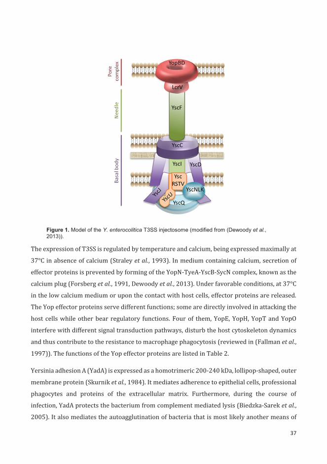
37
Figure 1. Model of the Y. enterocolitica T3SS injectosome (modified from (Dewoody et al., 2013)).
The expression of T3SS is regulated by temperature and calcium, being expressed maximally at
37°C in absence of calcium (Straley et al., 1993). In medium containing calcium, secretion of
effector proteins is prevented by forming of the YopN-TyeA-YscB-SycN complex, known as the
calcium plug (Forsberg et al., 1991, Dewoody et al., 2013). Under favorable conditions, at 37°C
in the low calcium medium or upon the contact with host cells, effector proteins are released.
The Yop effector proteins serve different functions; some are directly involved in attacking the
host cells while other bear regulatory functions. Four of them, YopE, YopH, YopT and YopO
interfere with different signal transduction pathways, disturb the host cytoskeleton dynamics
and thus contribute to the resistance to macrophage phagocytosis (reviewed in (Fallman et al.,
1997)). The functions of the Yop effector proteins are listed in Table 2.
Yersinia adhesion A (YadA) is expressed as a homotrimeric 200-240 kDa, lollipop-shaped, outer
membrane protein (Skurnik et al., 1984). It mediates adherence to epithelial cells, professional
phagocytes and proteins of the extracellular matrix. Furthermore, during the course of
infection, YadA protects the bacterium from complement mediated lysis (Biedzka-Sarek et al.,
2005). It also mediates the autoagglutination of bacteria that is most likely another means of
38
protection against host defenses (Skurnik et al., 1984). It was shown, that YadA is an essential
virulence factor for Y. enterocolitica (reviewed in (El Tahir & Skurnik, 2001)).
2.2.1.2.2. Chromosomally encoded virulence factors
Even though pYV is necessary for virulence of Y. enterocolitica, the chromosomally encoded
virulence factors are needed for full infectivity (Revell & Miller, 2001) (Table 2). One important
aspect of virulence is the ability of these bacteria to invade intestinal epithelial cells, which is
mediated by invasin (Inv) and Ail (attachment and invasion locus) (Miller & Falkow, 1988).
Invasin is a 92 kDa outer membrane protein, the expression of which is usually highest at
ambient temperature, prior to initiation of infection in the eukaryotic host. It serves as a
primary invasion factor and is necessary for efficient penetration of the intestinal barrier. In a
mouse infection model, Inv negative strains are unable to colonize Peyer’s patches early after
infection and showed 3-4 day delay in colonization when compared with the wild type (Pepe &
Miller, 1993). In vivo, invasin promotes the penetration of Y. enterocolitica into M cells by
adhesion to β1-integrins located on the surface of epithelial cells (Isberg & Leong, 1990). In Y.
enterocolitica serotype O:3 the synthesis of invasin is highly activated and nearly constitutive,
with no temperature dependence due to acquisition of IS1667 insertion sequence in the invA
regulatory region (Uliczka et al., 2011). Ail is a 17 kDa integral membrane protein with four
surface-exposed loops. In Y. enterocolitica it mediates the attachment and invasion into the host
cells, as well as serum resistance (Miller et al., 2001, Biedzka-Sarek et al., 2005). Previously, the
presence of attachment and invasion locus in the genome has been associated exclusively with
pathogenic Y. enterocolitica strains (Miller et al., 1989), yet recent study detected Ail in some
biotype 1A strains (Sihvonen et al., 2011).
One important virulence factor encoded chromosomally is lipopolysaccharide (LPS), a major
component of Gram-negative bacterial outer membrane. It consists of three main structural
components: the most external O-specific polysaccharide (O-antigen, O-Ag), the core region
(inner and outer cores) and the lipid A, which anchors the whole structure into the membrane
(Fig. 2) (Muszynski et al., 2013). The O-Ag of the O:3 serotype is a homopolymer composed of
6-deoxy-L-altrose repeating units, linked together by 1,2 linkages (Hoffman et al., 1980, Skurnik
et al., 1999). The inner core (IC) is composed of seven sugar residues, while the outer core (OC)
includes six residues (Radziejewska-Lebrecht et al., 1994, Skurnik et al., 1995). Untypically for
Gram-negative bacteria, in Y. enterocolitica O:3, there are two distinctive types of LPS molecules
present on the bacterial surface. One form constitutes of molecules where the O-Ag is linked to

39
the IC, whereas in the other form is the OC attached to the IC (Pinta et al., 2012, Muszynski et
al., 2013). The enterobacterial common antigen (ECA) is uniquely present in
Enterobacteriaceae and constitutes of a polysaccharide build of 3)-α-D-Fucp4NAc-(1 4)-β-
D-ManpNAcA-(1 4)-α-D-GlcpNAc-(1 repeats. The ECA polymer is either covalently
anchored to the outer membrane (via its own L-glycerophospholipid or via the core of LPS) or
is found in cyclic form in the periplasmic space (Lugowski et al., 1983, Kuhn et al., 1988,
Kajimura et al., 2005). The role of ECA is not fully known, it is assumed it has a protective
function against environmental stresses, confers resistance to low pH, bile salts, host defense
mechanisms, and is needed for motility (Danese et al., 1998, Barua et al., 2002, Ramos-Morales
et al., 2003, Castelli et al., 2008).
Figure 2. A schematic structure of two Y. enterocolitica O:3 LPS forms (modified from (Skurnik et al., 1995, Pinta et al., 2012, Muszynski et al., 2013)).
40
In Yersinia enterocolitica serotype O:3 the OC and O-ag biosynthetic genes are organized into
two distinct gene clusters. The OC gene cluster consists of nine genes (wzx, wbcKLMNOPQ and
gne) and is located between the hemH and gsk genes (Skurnik et al., 1995), whereas the O-
antigen gene cluster consists of two operons and eight genes (wbbSTU, wzm, wzt, wbbVWX)
(Zhang et al., 1993, Skurnik & Bengoechea, 2003). The expression of the O-ag is regulated at
transcriptional level and dependent on the temperature of growth. The optimal expression
occurs at 25°C, while at 37°C the number of repeating units per LPS molecule is much lower.
However, temperature seems to affect the transcription only in the stationary phase, whereas
in the exponential phase the O-ag is expressed constitutively (Lahtinen et al., 2003). The
detailed mechanism of this regulation is still unknown.
LPS confers to the virulence of Gram-negative bacteria and their resistance to antimicrobial
compounds (Al-Hendy et al., 1992, Biedzka-Sarek et al., 2005, Reines et al., 2012). It has been
shown previously that the O-antigen and OC are essential for full virulence of Y. enterocolitica
O:3 and O:8 and loss of either of them leads to severe attenuation in murine models (Al-Hendy,
1992, Zhang et al., 1997, Skurnik et al., 1999) . The O-ag is required for effective colonization of
host tissues during the first hours of infection, while the OC allows colonization of the deeper
organs and prolonged persistence of the bacteria in the Peyer’s patches (Al-Hendy, 1992, Zhang
et al., 1997, Skurnik et al., 1999). Moreover, it was shown that LPS contributes to serum
resistance indirectly, by blocking the other factors present on the surface of bacterial cell
(Biedzka-Sarek et al., 2005).
In addition, another gene located in the chromosome showed important involvement in the
virulence of Y. enterocolitica. The heat-stable enterotoxin Yst appears in three different
variants: YstA, YstB and YstC. It was shown in young rabbit model, that Yst plays an important
role in fluid secretion and development of diarrhea (Delor & Cornelis, 1992).
2.2.1.2.3. Virulence-related features
In addition to typical virulence factors, Y. enterocolitica possesses other metabolic features that
contribute to virulence. One of these features is production of urease, an enzyme that catalyzes
the hydrolysis of urea to ammonia and carbamic acid, which subsequently spontaneously
hydrolyzes to carbonic acid and ammonia. During the growth in external environment this
enzyme enables bacteria to utilize urea as a source of nitrogen. However, as the hydrolysis of
urea causes local increase in pH, urease is necessary for Y. enterocolitica to survive the passage
through the acidic stomach of the host (De Koning-Ward & Robins-Browne, 1995). Unlike in

41
some other enteropathogens, acid tolerance of Y. enterocolitica depends only on production of
urease. The regulation of urease expression in Y. enterocolitica is regulated by the growth
phase, with maximal activity during the stationary phase, and temperature, with the activity
being higher at 28°C than at 37°C. However, urease activity is not regulated by nitrogen
limitation (de Koning-Ward & Robins-Browne, 1997).
Iron is an essential nutrient for almost all bacterial species. While this chemical element is easily
available in natural environments and most of culture media, the concentration of free iron
within the host tissues is remarkably low. Usually in mammalian tissues iron is bound to carrier
proteins like transferrin and lactoferrin that further restricts its availability for bacteria. Many
pathogens acquire iron from the host by secreting high-affinity iron-binding siderophores.
Production of siderophores, mainly yersiniobactin, was reported for many strains of pathogenic
Yersinia, including Y. enterocolitica biotype 1B. Therefore, without available iron, the low-
pathogenic strains of Y. enterocolitica can cause only moderate intestinal syndromes, while
patients with iron overload a may develop a systematic yersiniosis (Chambers & Sokol, 1994,
Carniel, 2001). Moreover, patients treated with Desferal (commercially available form of iron-
chelating deferoxamine) have been reported to be more prone to develop septicemia caused by
Y. enterocolitica (Boelaert et al., 1987). The virulence-enhancing effect of Desferal depends on
the production of outer membrane protein FoxA that acts as a receptor for deferoxamine
(Baumler & Hantke, 1992). Morover, Desferal was also shown to have a potential
immunosuppressive effect on the host (Autenrieth et al., 1994, Autenrieth et al., 1995).
Another feature with implicated role in virulence is motility. It was shown, that flagellum-
dependent motility in Y. enterocolitica ensures migration of the bacteria to host cells. In HEp-2
cell culture model non-motile strain displayed impairment in invasion. However, the same
strain showed no difference in invasion when bacteria were brought into contact with host cells
by centrifugation (Young et al., 2000). Moreover, it was previously implicated, that some
virulence genes can be regulated as part of the flagellar regulon (Young et al., 1999). In Y.
enterocolitica ylpA, a gene required for survival in Peyer’s patches and stimulation of the acute
inflammatory response, is a part of flagellar regulon (Schmiel et al., 1998). Therefore the
alterations in motility may have both direct and indirect effect on the virulence of this
enteropathogen. Due to high similarity of flagellar regulon it is likely that many members of
Enterobacteriaceae share the same type of regulation, which follows a hierarchical cascade with
three major gene classes I, II, and III. The flhDC operon (class I) is at the top of the hierarchy
42
and it regulates the expression of class II genes. Class II consists of structural and accessory
proteins, as well as sigma factor, which allows the transcription of the class III genes (proteins
involved in maturation of flagellar system) (Young, 2004). Moreover, it has been demonstrated
that motility is regulated by AHL-dependent quorum sensing in Y. enterocolitica (Atkinson et
al., 2006).
2.3. Coordination of gene regulation in Yersiniae
Pathogenic Yersinia species are well adapted for survival in various external environments and
persistence in a variety of host organisms. During the invasion process, bacteria must adjust
rapidly to several extremely different environments, such as gastric acidity, altered osmolarity,
changing nutrient availability, and competition with the microbiota colonizing the host
organism. In order to establish infection several virulence factors must be employed. That
process involves precise regulation of expression that ensures that these virulence factors and
adequate metabolic pathways are produced at the correct spatiotemporal conditions. The
regulation is achieved at transcriptional, post-transcriptional and post-translational levels
through the employment of various transcription factors, nucleoid-associated proteins, ncRNAs
and ribonucleases (Heroven & Dersch, 2014, Erhardt & Dersch, 2015).
Although, the majority of the mechanisms and factors involved in gene regulation in Yersiniae
are highly conserved, yet the network composition and architecture may vary between the
species (Erhardt & Dersch, 2015). During the first stages of host colonization bacteria have to
survive the acidic environment of the stomach. For this the bacteria use urease, an enzyme that
counteracts low pH by neutralizing it with ammonia generated from urea (Young et al., 1996).
The regulators as TCS OmpR/EnvZ and Hfq chaperone have been implicated in the regulation
of urease biosynthesis in Yersinia species (Hu et al., 2009, Kakoschke et al., 2014). The
gastrointestinal tract is a rich source of food, yet the availability of nutrients depends on the
host and there is a constant competition with the resident microbiota for resources. This phase
of infection depends to a large extend on the ability to sense the available nutrients and to
response adequately by changing the expression of enzymes of the metabolic pathways. This
stage involves such regulators as the PhoP/PhoQ TCS, the global carbon storage regulator
(Csr), cAMP receptor protein Crp, the UvrY/BarA TCS, and the sRNA regulatory network in
cooperation with the Hfq chaperone (reviewed in details in (Heroven & Dersch, 2014)).

43
Subsequently, bacteria attach to and invade the M cells overlaying the Peyer’s patches in the
ileum (Grutzkau et al., 1990). The key virulence factors of this stage are the outer membrane
proteins invasin, Ail, and YadA, that are regulated by the MarR-type regulator RovA, H-NS, and
YmoA. Moreover, the ability of bacterium to internalize is also enhanced by the flagella-
mediated motility, which is controlled by the master operon flhDC (reviewed in (Erhardt &
Dersch, 2015)). From the Peyer’s patches bacteria disseminate to the mesenteric lymph nodes,
liver and spleen (Cornelis & Wolf-Watz, 1997). This phase of infection requires the expression
of ysc genes encoding the T3SS and the Yop effector proteins. These genes are encoded on the
pYV and their transcription is activated by the LcrF(VirF) regulator (Bolin et al., 1988). The
ability of Yersinia spp. to avoid the host defense mechanisms depend also on the expression of
LPS, YadA and Ail, due to their involvement in the resistance against the host serum
complement system (Biedzka-Sarek et al., 2005).
The sensing of the environment occurs through different cues, yet the changes in temperature
is the most important signal received by the pathogenic Yersiniae. Temperature-dependent
regulation of the gene expression happens through several different regulators. The expression
of T3SS and associated Yop effector proteins is allowed only at body temperatures and
controlled by LcrF/VirF, YmoA and levels of supercoiling of the pYV (Lambert de Rouvroit et
al., 1992, Hoe & Goguen, 1993, Rohde et al., 1999, Bohme et al., 2012). The temperature-
dependent regulation of Inv expression is governed by the RovA regulator, an intristic
temperature-sensing protein (Revell & Miller, 2000, Herbst et al., 2009, Quade et al., 2012). The
loss of motility at 37°C occurs through 28/FliA without the involvement of the flhDC master
operon (Kapatral et al., 2004). Several virulence genes of Yersinia are expressed in response to
changes in the environmental concentration of Ca2+ in a process referred to as low calcium
response (Goguen et al., 1984, Straley et al., 1993). The changes in temperature and osmolarity
are sensed by the TCS OmpR/EnvZ that controls the expression of the outer membrane porins
(Dorrell et al., 1998, Brzostek et al., 2012). Another transcriptional regulator, IscR, responds to
oxidative stress, oxygen limitation and iron availability and controls the expression of T3SS
through the LcrF(VirF) (Miller et al., 2014). In Yersinia the response to different carbon sources
is mediated by BarA/UvrY, Crp and Csr systems (Poncet et al., 2009, LeGrand et al., 2015). The
low pH and low levels of magnesium in the environment are the trigger factors affecting the
expression of the PhoP/PhoQ TCS (Groisman, 2001). In addition, the CpxAR system is
responsible for responses to the extra-cytoplasmatic stress in Yersinia (Liu et al., 2012). The
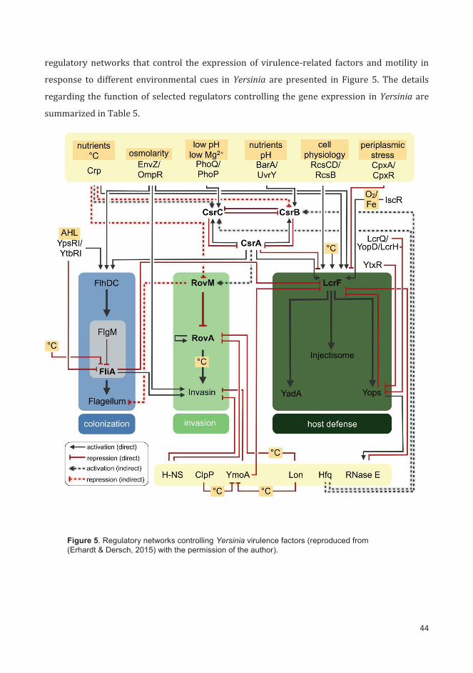
44
regulatory networks that control the expression of virulence-related factors and motility in
response to different environmental cues in Yersinia are presented in Figure 5. The details
regarding the function of selected regulators controlling the gene expression in Yersinia are
summarized in Table 5.
Figure 5. Regulatory networks controlling Yersinia virulence factors (reproduced from (Erhardt & Dersch, 2015) with the permission of the author).
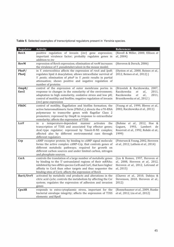
45
Table 5. Selected examples of transcriptional regulators present in Yersinia species.
Regulator Activity References RovA positive regulation of invasin (inv) gene expression;
important virulence factor; probably regulates genes in addition to inv
(Revell & Miller, 2000, Ellison et al., 2004)
RovM repression of RovA expression; elimination of rovM increases the virulence of Y. pseudotuberculosis in the mouse model;
(Heroven & Dersch, 2006)
PhoP/ PhoQ
in Y. enterocolitica affects the expression of rovA and lpxR; regulates lipid A deacylation; allows intracellular survival of Y. pestis; elimination of phoP in Y. pestis results in partial attenuation; shows positive and negative regulation of number of proteins
(Oyston et al., 2000, Reines et al., 2012, Reines et al., 2012) }
OmpR/ EnvZ
control of the expression of outer membrane porins in response to changes in the osmolarity of the environment; adaptation to high osmolarity, oxidative stress and low pH; control of motility and biofilm; negative regulation of invasin (inv) gene expression
(Brzostek & Raczkowska, 2007, Raczkowska et al., 2011, Raczkowska et al., 2011, Brzostkowska et al., 2012 )
FlhDC control of motility, flagellation and biofilm formation; the active heteromultimeric form (FlhD4C2) directs the 70-RNA polymerase to transcribe genes with flagellar Class 2 promoters; repressed by OmpR in response to extracellular osmolarity; affects the expression of T3SS
(Young et al., 1999, Bleves et al., 2002, Raczkowska et al., 2011)
LcrF in a temperature-dependent manner activates the transcription of T3SS and associated Yop effector genes; AraC-type regulator; repressed by YmoA-H-NS complex; affected also by different environmental cues through different regulators
(Bohme et al., 2012, Hoe & Goguen, 1993, Lambert de Rouvroit et al., 1992, Rohde et al., 1999)
Crp cAMP receptor protein; by binding to cAMP signal molecule forms the active complex cAMP-Crp, that controls genes of different metabolic pathways; required for growth on different carbon sources and under limited carbon, nitrogen and phosphate sources.
(Petersen & Young, 2002, Heroven et al., 2012, Lathem et al., 2014)
CsrA controls the translation of a large number of metabolic genes by binding to the 5’-untranslated regions of their mRNAs; inhibited by two sRNA species CsrB and CsrC, that have higher affinity to CsrA than other targets and thus sequester the binding sites of CsrA; affects the expression of RovA
(Liu & Romeo, 1997, Heroven et al., 2008, Heroven et al., 2012, Heroven et al., 2012, LeGrand et al., 2015)
BarA/UvrY activated by metabolic end products and alterations in the citric acid cycle; controls the metabolism by affecting the Csr system; regulates the expression of adhesion and invasion genes
(Chavez et al., 2010, Dahiya & Stevenson, 2010, Heroven et al., 2012)
CpxAR responds to extra-cytoplasmic stress, important for the bacterial envelope integrity; affects the expression of T3SS elements and RpoE
(Ronnebaumer et al., 2009, Hunke et al., 2012, Liu et al., 2012)

46
3. AIMS OF THE STUDY
During the course of infection bacterial cell passes through different niches. Sensing properly
environmental cues allows it to respond accordingly, survive and establish infection. Therefore
for pathogens, the gene regulation also means adaptation to the host and coordination of
expression of virulence factors, either directly or indirectly by affecting global regulators.
The details of bacterial gene expression regulation are not fully understood. This thesis work
was undertaken to increase the knowledge of the regulatory networks and involvement of
different regulators in bacterial physiology and virulence. In this study, I aimed at using two
high-throughput methods, RNA-sequencing (RNA-seq) and quantitative proteomics (LC-
MS/MS), to identify mRNAs and proteins of Yersinia enterocolitica O:3 dependent on selected
regulatory proteins, and to elucidate their role in the physiology and virulence of the pathogen.
The specific objectives of this thesis work were:
I. To identify the genes regulated directly or indirectly by Hfq, YbeY and RfaH in Y.
enterocolitica.
II. To investigate the role of Hfq, YbeY, RfaH and RovM in the physiology of Y.
enterocolitica
III. To investigate the role of Hfq, YbeY, RfaH and RovM in the virulence and virulence-
associated phenotypes of Y. enterocolitica.

47
4. MATERIALS AND METHODS
4.1. Materials
4.1.1. Bacterial strains and bacteriophages
All bacterial strains and bacteriophages used in this thesis are listed in Table 6. The detailed
descriptions of the strains, including the growth conditions, are available in the original
publications I, II and a manuscript (III).
Table 6. Bacterial strains and bacteriophages used in this study.
Bacterial strain Description Reference Used in Y. enterocolitica O:3 strains YeO3-wt 6471/76, serotype O:3, patient isolate, wild type (Skurnik,
1984) I, II, III
YeO3- ybeY YeO3-wt, ybeY:: pKNG101-ybeY; StrepR this study I YeO3-
ybeY/pMMB207- ybeY
YeO3- ybeY strain transformed with pMMB207-ybeY; StrepR, ClmR
this study I
YeO3-c Virulence plasmid cured derivative of YeO3-wt (Skurnik, 1984)
II
YeO3-c-R1 Spontaneous rough derivative of YeO3-c (al-Hendy et al., 1992)
II
YeO3- rfaH YeO3-wt, rfaH::pSW25T-rfaH; SpecR this study II YeO3-c- rfaH YeO3-c, rfaH::pSW23T-rfaH; ClmR this study II YeO3- rfaH/pTM100-rfaH
YeO3- rfaH strain transformed with pTM100-rfaH; SpecR, ClmR
this study II
YeO3-c-rfaH/pLux232oT-POC
YeO3-c- rfaH strain carrying OC gene cluster promoter reporter vector pLux232oT-POC, ClmR, KmR
this study II
YeO3-c-rfaH/pLux232oT-
PECA
YeO3-c- rfaH strain carrying ECA gene cluster promoter reporter vector pLux232oT-PECA, ClmR, KmR
this study II
YeO3-c-rfaH/pLux232oT-
POP1
YeO3-c- rfaH strain carrying O-antigen gene cluster promoter 1 reporter vector pLux232oT-POP1, ClmR, KmR
this study II
YeO3-c-rfaH/pLux232oT-
POP2
YeO3-c- rfaH strain carrying O-antigen gene cluster promoter 2 reporter vector pLux232oT-POP2, ClmR, KmR
this study II
YeO3-hfq::Km YeO3-wt, hfq::Km-GenBlock, KmR this study III YeO3-hfq::Km/phfq YeO3-hfq::Km transformed with pTM100-hfq; KmR,
ClmR this study III
YeO3-rovM YeO3-wt, rovM::pKNG101, StrepR this study III YeO3-rovM-hfq::Km YeO3-wt, hfq rovM double mutant; KmR, StrepR this study III YeO3-rovM/pMMB207-rovM
YeO3-wt, rovM strain with pMMB207-rovM plasmid to overexpress rovM under the IPTG induced promoter; StrepR, ClmR
this study III
YeO3/ pLux232oT-rovM
YeO3-wt carrying rovM promoter reporter vector pLux232oT-rovM, KmR
this study III
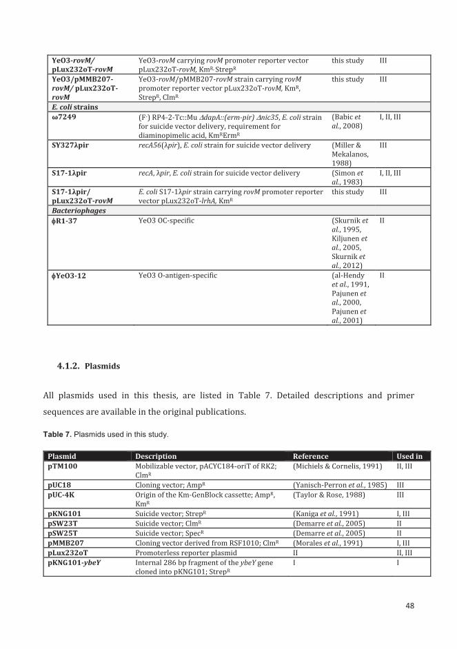
48
YeO3-rovM/ pLux232oT-rovM
YeO3-rovM carrying rovM promoter reporter vector pLux232oT-rovM, KmR, StrepR
this study III
YeO3/pMMB207-rovM/ pLux232oT-rovM
YeO3-rovM/pMMB207-rovM strain carrying rovM promoter reporter vector pLux232oT-rovM, KmR, StrepR, ClmR
this study III
E. coli strains ω7249 (F-) RP4-2-Tc::Mu dapA::(erm-pir) nic35, E. coli strain
for suicide vector delivery, requirement for diaminopimelic acid, KmRErmR
(Babic et al., 2008)
I, II, III
SY327λpir recA56(λpir), E. coli strain for suicide vector delivery (Miller & Mekalanos, 1988)
III
S17-1λpir recA, λpir, E. coli strain for suicide vector delivery (Simon et al., 1983)
I, II, III
S17-1λpir/ pLux232oT-rovM
E. coli S17-1λpir strain carrying rovM promoter reporter vector pLux232oT-lrhA, KmR
this study III
Bacteriophages R1-37 YeO3 OC-specific (Skurnik et
al., 1995, Kiljunen et al., 2005, Skurnik et al., 2012)
II
YeO3-12 YeO3 O-antigen-specific (al-Hendy et al., 1991, Pajunen et al., 2000, Pajunen et al., 2001)
II
4.1.2. Plasmids
All plasmids used in this thesis, are listed in Table 7. Detailed descriptions and primer
sequences are available in the original publications.
Table 7. Plasmids used in this study.
Plasmid Description Reference Used in pTM100 Mobilizable vector, pACYC184-oriT of RK2;
ClmR (Michiels & Cornelis, 1991) II, III
pUC18 Cloning vector; AmpR (Yanisch-Perron et al., 1985) III pUC-4K Origin of the Km-GenBlock cassette; AmpR,
KmR (Taylor & Rose, 1988) III
pKNG101 Suicide vector; StrepR (Kaniga et al., 1991) I, III pSW23T Suicide vector; ClmR (Demarre et al., 2005) II pSW25T Suicide vector; SpecR (Demarre et al., 2005) II pMMB207 Cloning vector derived from RSF1010; ClmR (Morales et al., 1991) I, III pLux232oT Promoterless reporter plasmid II II, III pKNG101-ybeY Internal 286 bp fragment of the ybeY gene
cloned into pKNG101; StrepR I I

49
pMMB207-ybeY Overexpression plasmid; the complete ybeY gene cloned into pMMB207; ClmR
I I
pSW23T-rfaH Internal 201 bp fragment of the rfaH gene cloned into pSW23T; ClmR
II II
pSW25T-rfaH Internal 201 bp fragment of the rfaH gene cloned into pSW25T; SpecR
II II
pTM100-rfaH Full rfaH gene with the upstream promoter cloned into pTM100; ClmR
II II
pLux232oT-POC OC gene cluster promoter fragment cloned into the promoter reporter vector pLux232oT; KmR
II II
pLux232oT-PECA ECA gene cluster promoter fragment cloned into the promoter reporter vector pLux232oT; KmR
II II
pLux232oT-POP1 O-antigen gene cluster promoter 1 fragment cloned into the promoter reporter vector pLux232oT; KmR
II II
pLux232oT-POP2 O-antigen gene cluster promoter 2 fragment cloned into the promoter reporter vector pLux232oT; KmR
II II
pUC18-hfq The hfq gene with flanking regions cloned into pUC18; AmpR
III III
pUC18-hfq::Km pUC18-hfq derivative with the internal part of hfq gene replaced with Km-GenBlock; KmR, AmpR
III III
pKNG101-hfq::km hfq::Km-GenBlock fragment of pUC18-hfq::Km cloned into BamHI site of pKNG101; KmR,, StrepR
III III
pKNG101-rovM Internal rovM gene fragment cloned into pKNG101; StrepR
III III
pMMB207-rovM Overexpression plasmid; the complete rovM gene cloned into pMMB207; ClmR
III III
pTM100-hfq Full hfq gene with the upstream promoter cloned into pTM100; ClmR
III III
pLux232oT-rovM rovM promoter fragment cloned into the promoter reporter vector pLux232oT; KmR
III III
4.1.3. Antibodies and antisera
Table 8. Antibodies and antisera used in this study.
Antibody/Antiserum
Description Source or reference Used in
Primary antisera and antibodies α-YopM Polyclonal rabbit anti-YopM antiserum (Trulzsch et al., 2004) I α-YopH Polyclonal rabbit anti-YopH antiserum (Trulzsch et al., 2004) I α-YopE Polyclonal rabbit anti-YopE antiserum (Trulzsch et al., 2004) I α-YopD Polyclonal rabbit anti-YopD antiserum (Trulzsch et al., 2004) I α-LcrV Polyclonal rabbit anti-LcrV antiserum (Trulzsch et al., 2004) I 2A9 Mouse anti-YadA monoclonal antibody (Skurnik et al., 1994,
Biedzka-Sarek et al., 2008) I
α-Inv Mouse anti-Invasin monoclonal antibody Petra Dersch I Mab 898 Mouse anti-ECA monoclonal antibody (Meier-Dieter et al., 1989) II
50
TomA6 Mouse anti- Y. enterocolitica O:3 O-antigen monoclonal antibody
(Pekkola-Heino et al., 1987) II
2B5 Mouse anti- Y. enterocolitica O:3 OC monoclonal antibody
(Pekkola-Heino et al., 1987) II
15D8 Mouse anti-flagellin monoclonal antibody (Feng et al., 1990) III α-RpoS Polyclonal rabbit anti-RpoS antiserum (Coynault et al., 1996) III
Secondary antisera P0447 Polyclonal goat peroxidase-conjugated anti-mouse
immunoglobulin antibodies Dako I, II, III
P0217 Polyclonal swine peroxidase-conjugated anti-rabbit immunoglobulin antibodies
Dako I, III
4.1.4. Bioinformatics resources used
The following web resources were used to obtain reference sequences and annotation
information: NCBI National Center for Biotechnology Information
(http://www.ncbi.nlm.nih.gov), UniProt (http://www.uniprot.org) and RCSB PDB Protein Data
Bank (http://www.rcsb.org/pdb).
4.2. Methods
4.2.1. Bacterial cultivation
Cultures were grown aerobically in Lysogeny Broth (LB) (Bertani, 2004), in brain heart infusion
(BHI) (Fluka) or on Yersinia selective agar (cefsulodin, irgasan and novobiocin supplemented;
CIN-agar, Oxoid, UK) at either 37°C or at room temperature (RT, ca. 22°C). LB plates were
prepared by adding 15 g of bacto agar to 1 L of LB. For electron microscopy, flagellin production
and motility evaluation bacteria were grown in tryptone broth (TB) (1% tryptone, 0.5% NaCl)
and on motility trypton agar plates (0.3% TB plates) (1% tryptone, 0.5% NaCl, 0.3% bacto agar)
at 22°C. Additionally, for biofilm formation test M9 (2mM MgSO4, 0.1 mM CaCl2, 1x M9 salts,
0.4% glucose) and MedECa (Skurnik, 1985) media were used. Antibiotics were used when
needed at the following concentrations: streptomycin (Str) 50 μg/ml, chloramphenicol (Clm)
20 μg/ml, kanamycin (Km) 100 μg/ml, ampicillin (Amp) 50 μg/ml. Expression from plasmid
pMMB207 was induced with 1 mM isopropyl-β-D-thiogalactopyranoside (IPTG).

51
4.2.2. Growth curves.
Bacteria were grown overnight at RT. Subsequently, cultures were diluted in fresh medium to
an OD600 of 0.2 and 200 μl aliquots were distributed into honeycomb plate wells (Growth Curves
Ab Ltd). The growth experiments were carried out at selected temperatures using the
Bioscreen C incubator (Growth Curves Ab Ltd) with continuous shaking. The OD600 values were
measured at 10 or 15 min intervals. The average values were calculated from 9 or 10 parallel
results.
4.2.3. SDS-PAGE.
Proteins were separated using sodium dodecyl sulfate polyacrylamide gel electrophoresis
(SDS-PAGE) with 5% stacking and 12% separating gels. After the electrophoresis the material
was either visualized using InstantBlue (Expedon), by silver staining (Mortz et al., 2001), or
transferred onto nitrocellulose membrane (Protran, Whatman, pore size 0.45 μm). Transfer of
the proteins from the SDS-PAGE gel onto the membrane was done using the semi-dry apparatus
(Thermo Scientific Owl, USA).
4.2.4. Preparation of LPS samples.
LPS samples were prepared using the modified protocol of Hitchcock and Brown (Hitchcock &
Brown, 1983) as described previously (Pinta et al., 2012).
4.2.5. Detection of O-antigen in culture supernatants.
Bacteria were grown in 5 ml LB overnight at 22°C or at 37°C. Subsequently the overnight
cultures were pelleted and 50 μl of the supernatant was mixed with 50 μl of DOC lysis buffer.
The mixture was boiled for 10 min, cooled down and incubated for 4 h at 55˚C with proteinase
K. Altogether 3, 6, 9 and 12 μl of the samples in 3 μl portions were spotted onto nitrocellulose
membranes and left at RT to air-dry. The membrane was blocked and incubated with the O-
antigen specific mAb TomA6 as described below (see immunoblotting).
4.2.6. DOC-PAGE.
LPS was separated using deoxycholate–polyacrylamide gel electrophoresis (DOC-PAGE) with
4% stacking and 12% separating as described previously (Krauss et al., 1988, Zhang & Skurnik,
1994, Skurnik et al., 1995). After the run the material was either visualized by silver staining
(Al-Hendy et al., 1991) or transferred for immunoblotting onto nitrocellulose membrane
(Protran, Whatman, pore size 0.45 μm) using the semi-dry apparatus (Thermo Scientific Owl,

52
USA). For dot-blotting 5 μl aliquots of serial 1:2 dilutions of the LPS samples were applied
directly to the membrane and left at room temperature to air-dry.
4.2.7. Immunoblotting
The membrane was blocked using 5% fat-free milk in TBST buffer (50 mM Tris-HCl, 150 mM
NaCl, 0.05% Tween 20, pH7.6) for 1h at RT. Subsequently, the membrane was incubated for
16h at 4°C with primary antibodies diluted in blocking buffer. After washing 3 times with TBST
buffer, the membrane was incubated for 1h at RT with suitable peroxidase-conjugated
secondary antibodies (dilution 1:2000 in blocking buffer). Subsequently membrane was
washed in TBST as before and incubated in ECL solution (0.1M Tris-HCl pH 8.5, 1.25 mM
luminol, 0.2 mM cumaric acid, 5.3 mM hydrogen peroxide). After draining excess fluid, the
membrane was exposed to light sensitive film (Kodak, USA).
4.2.8. Total RNA extraction.
The total RNA of bacteria was isolated using the SV Total RNA Isolation System (Promega)
following the instructions provided by the manufacturer. The quality of the isolated RNA was
verified using the Bioanalyzer (Agilent).
4.2.9. RNA-sequencing.
The RNA-seq was performed at the FIMM Technology Centre Sequencing Unit
(www.fimm.fi). The ribosomal RNA was removed using Ribo-ZeroTM rRNA Removal Kit for
Gram-negative Bacteria (Epicentre). Paired-end sequencing was completed on Illumina
HISeq2000 sequencer (Illumina) producing the read length of 90 nucleotides. The obtained
sequencing reads were filtered for quality and aligned against the Y. enterocolitica strain Y11
genome (accession number FR729477) using the TopHat read aligner (Langmead et al., 2009).
Subsequently, the Cufflinks program (Trapnell et al., 2013) was used to obtain the fragments
per kilobase of gene per million aligned fragments (FPKM) values for differential expression.
The genes were considered differentially expressed if the fold change (FC) of the average values
was >2, and the Student’s T-test p-value was <0.01. The data for RfaH project was analyzed
using the edgeR differential expression analysis package (Robinson et al., 2010). The RNA
sequence data has been deposited to Gene Expression Omnibus (Acc. no GSE66516 and
GSE62601).

53
4.2.10. Quantitative RT-PCR.
The extracted total RNA was diluted to the final concentration of 25 ng/μl. The quantitative RT-
PCR was performed using the GoTaq 1-step RT-qPCR System (Promega). The results were
calculated using the Unit Mass method. The gene-specific primers used are listed in the
respective studies.
4.2.11. Quantitative proteomics.
After growing the bacteria overnight at RT in 3 ml of LB cultures were diluted 1:10 in fresh LB
and incubated for another 4h at either RT or 37°C Afterwards, bacterial cells were pelleted by
centrifugation at 3000 x g, washed with sterile phosphate-buffered saline (PBS) and adjusted
to 2.5 x 10⁸ cfu/ml. Subsequently, 1 ml of each culture was pelleted, resuspended in lysis buffer
(100 mM ammoniumbicarbonate, 8M urea, 0.1% RapiGestTM), sonicated for 3 min (Branson
Sonifier 450, pulsed mode 30%, loading level 2) and stored at -70°C. Samples were prepared in
3 parallels. The proteins in the samples were reduced with Tris(2-carboxyethyl)phosphine
(TCEP), alkylated with iodoacetamide and digested with trypsin. The obtained digests were
purified by C18 reversed-phase chromatography columns. Afterwards, the MS analysis was
completed on an Orbitrap Elite ETD mass spectrometer (Thermo Scientific), with Xcalibur
version 2.7.1 coupled to an Thermo Scientific nLCII nanoflow HPLC system. Both the peak
extraction and the protein identification was performed using Proteome Discoverer software
(Thermo Scientific). Calibrated peak files were used to search against the Y. enterocolitica O:3
strain Y11 proteins (Uniprot) using a SEQUEST search engine. Error tolerances on the
precursor and fragment ions were ±15 ppm and ±0.6 Da, respectively. A stringent cut-off (0.5
% false discovery rate) was used for peptide identification. Spectral counts for each protein in
each sample were extracted and used in relative quantitation of protein abundance alterations
for label-free quantification.
4.2.12. Resistance assays
Thermotolerance was tested by diluting the overnight cultures to obtain ca. 1,000
bacterial cells in 10 μl of PBS. The dilutions were incubated in a thermoblock heated to 55°C.
Serial 10-fold dilutions were prepared before the incubation and after 5 min of incubation. The
number of viable bacteria was determined by plating the 10-fold dilutions.
Acid tolerance was tested as described earlier (De Koning-Ward & Robins-Browne, 1995)
with modifications. Briefly, the overnight cultures were diluted to obtain ca. 1,000 bacterial
cells in 10 μl of PBS. Aliquots of 10 μl of freshly prepared bacterial cultures were added to 90 μl

54
of PBS pH 2.5 or pH 2.0 supplemented with 1.4 mM urea. The mixtures were incubated for 2h
at 37°C. 10-fold dilutions were prepared before and after the incubation and plated on LA plates
to determine the number of bacteria.
4.2.13. Urease test.
The production of urease was verified in urea broth (0.1% peptone, 0.1% glucose, 0.5%
NaCl, 0.2% KH2PO4, 0.00012% phenol red, 2% urea) (Stuart et al., 1945). The broth was
inoculated using the overnight cultures (1:20) and incubated at RT or 37°C with shaking. The
test result was considered negative if the final color was yellow and positive if the medium
changed the color from orange to red.
4.2.14. Sugar utilization. The production of acid from different carbohydrates was determined in phenol red broth (10 g
peptone, 5 g NaCl and 0.018 g phenol red per liter supplemented with the selected carbohydrate
to a final concentration of 1%) (Iversen et al., 2006). Bacterial overnight cultures were pelleted
and diluted in phenol red broth. Dilutions 1:10 were used to inoculate 3 ml of medium
supplemented with selected sugars. Following carbohydrates were studied: glucose, sucrose,
maltose, mannitol, arabinose, galactose, mannose, and myo-inositol.
4.2.15. Motility and biofilm assays.
After growing bacteria overnight in 5 ml of tryptone broth at RT with gentle shaking, 5 μl of
each culture was applied in the middle of the tryptone motility plates (1% tryptone, 0.5% NaCl
and 0.35% agar). Motility plates were incubated for 24h at RT. Subsequently, the radius of the
bacterial growth was measured and images of the motility plates were taken (GelLogic 200
Imaging System, Kodac).
Biofilm formation was tested as described earlier (Blumer et al., 2005) with modifications.
Overnight cultures of bacteria grown in TB, M9 or MedECa were diluted 1:10 into the same
medium. The 200 μl aliquots of the cultures were transferred to the wells of 96-well polystyrene
microtiter plate (Nunc). After 72h of incubation at RT the wells were emptied, washed three
times with sterile PBS and drained. Subsequently, wells were washed with 200 μl of methanol
and left overnight to dry in order to fix the biofilm. Adhered cells were stained using 200 μl of
0.1 % crystal violet solution. Non-bound dye was removed by rinsing the wells three times with
distilled water. To solubilize the crystal violet the wells were filled with 200 μl of 96% ethanol

55
and incubated 30 min at RT. Subsequently, the absorbance of the dye was measured at 560 nm
using the Labsystems iEMS Reader MF.
4.2.16. Luminescence assay.
The activities of the promoters cloned into the luciferase reporter plasmid pLux232oT were
measured using a Chameleon microplate reader (Hidex). Bacteria were grown in black 96-well
plates (PerkinElmer) in a total volume of 200 μl of LB per well with continuous shaking. The
luminescence emission was measured at indicated time points. The relative light unit (RLU)
values were normalized using the obtained OD600 values of the cultures.
4.2.17. Bacteriophage sensitivity.
Bacterial cultures were grown overnight at either RT or 37˚C with vigorous shaking.
Afterwards, 100 μl of saturated culture was mixed with 3 ml of 0.3% soft agar (LB broth with
0.3% agar, solubilized by boiling and cooled to 50°C) and poured over a sterile LB plate. After
solidification of soft agar, 5 μl aliquots of indicated phage dilutions prepared in LB broth were
applied on the plate. The bacteria were considered sensitive if a clear lysis zone was visible
after 24 h of incubation. Bacteriophages used in this experiment are listed in the Table 6, study
II.
4.2.18. Electron microscopy.
Bacteria grown overnight on tryptone motility plates were collected from the migration area,
washed with sterile PBS, pelleted and resuspended in 0.1 M ammonium acetate. Subsequently,
the bacterial cells were allowed to sediment on carbon coated grids for 1 min and stained
negatively using 1% uranyl acetate. Samples were examined with JEOL JEM1400 transmission
electron microscope. Pictures were taken using the Olympus Morada CCD camera with the
iTEM software.
4.2.19. Serum killing assay.
The serum was prepared and the killing assay was performed as described previously
(Biedzka-Sarek et al., 2005). Normal human serum (NHS) was obtained from healthy
individuals lacking anti-Yersinia antibodies (Biedzka-Sarek et al., 2005). Overnight bacterial
cultures were diluted to obtain ca. 1,000 bacterial cells in 10 μl. 10 μl bacterial aliquots were
incubated for 30 minutes at 37°C with (i) 20 μl of NHS, (ii) 20 μl of heat-inactivated serum (HIS)
or (iii) 20 μl of EGTA serum (10 mM EGTA, 5 mM MgCl2). Afterwards, 70 μl of ice-cold brain
heart infusion broth (BHI) was added to stop the activity of complement and the tubes were
56
incubated on ice. Subsequently, the mixtures were plated on LB plates supplemented with
proper antibiotics. The bactericidal effect of the serum was calculated as the survival
percentage taking the CFU values obtained for bacteria incubated with HIS as 100%. The
experiment was carried out in triplicates, starting from individual colonies. Two separate
experiments were carried out on two separate occasions.
4.2.20. Cell culture infection assay.
The experiments were performed as described earlier (Schulte et al., 2000). HeLa cell cultures
were grown in Dulbecco's Modified Eagle's Medium (DMEM) supplemented with 10% fetal
bovine serum (FBS), 2 mM L-glutamine, 50 μg/ml of streptomycin and 100 μg/ml of penicillin.
The cells were kept at 37°C in a humidified 5% CO2 atmosphere. After incubation with trypsin
and harvesting, cells were diluted 1:3 in fresh DMEM and transferred into the 6-well plates.
Following the 24 h incubation the medium was changed into DMEM with 10% FBS and 2 mM
L-glutamine without the antibiotic supplementation. The cells were incubated at 37°C for
further 2 h. Meanwhile bacterial overnight cultures were diluted in LB and incubated for 3h at
RT. Subsequently, bacterial cells were washed in PBS, pelleted and resuspended in DMEM. The
dilutions of bacterial cultures were adjusted to infect the eukaryotic cells with 100:1 bacterium-
to-cell ratio. The number of bacteria was assessed by plating and the host cell number was
estimated using a hemacytometer. Following the inoculation of the plates, bacteria were
centrifuged down onto the cells (5 min, 750 rpm) and incubated at 37°C in a humidified 5% CO2
atmosphere for 1h. After the incubation monolayers were washed with PBS. Subsequently,
either DMEM or DMEM supplemented with 100 μl/ml gentamycin was added to the wells to
determine total cell-associated bacteria and the intracellular bacteria, respectively. Following
the 1h incubation, HeLa cells were lysed using 1% Triton X-100 in PBS. 100 μl aliquots of the
serial 10-fold dilutions were plated on LB plates in order to determine the number of released
viable bacteria.
4.2.21. Mouse experiments.
Animal experiments were performed under the permit from the Animal Experiment Board in
Finland (permit no ESAVI/5893/04.10.03/2012). The 35 inbred 6-8 week old female BALB/c
mice were purchased from Envigo (Blackthorn, UK). Upon arrival the mice were allowed to
adjust to the new housing conditions for 1 week.
57
Bacteria were prepared as described earlier (Skurnik et al., 1999) with modifications. Briefly,
bacteria were grown overnight at RT in LB supplemented with appropriate antibiotics (Km,
Clm, Strep). Afterwards, the bacterial cells were centrifuged down, resuspended in 10 ml of PBS,
and mixed thoroughly. Three 1 ml aliquots were pelleted and after removal of the supernatant
the weight of wet pellet mass was determined. Based on assumption that 100 mg (300 mg for
YeO3-hfq::Km bacteria) of the bacterial pellet contains about 1011 bacterial cells, the bacterial
suspensions were diluted to 1010 or 108 CFU per ml. The number of viable bacteria was further
confirmed by plating. For the coinfection experiments the suspensions of wild type and YeO3-
hfq::Km bacteria were mixed at the ratio of approximately. 1:1. Samples from subsequent 10-
fold dilutions were plated in order to determine the exact bacterial counts.
Mice were kept without food for 4-h before the infections. The bacterial suspensions (100 μl for
the single infection and a total of 200 μl for the co-infection model) were administered either
intraperitoneally using a 25G needle or intragastrically using a 20 gauge stainless-steel ball-
tipped catheter. After mice were sacrificed, the Peyer’s patches, spleen, and liver were removed,
weighted and homogenized in 0.5, 0.5 and 1 ml of PBS, respectively, using the Ultra-Turrax T8
homogenizer (IKA Labortechnik, Staufen, Germany). The number of viable bacterial cells was
determined by plating serial dilutions on CIN agar plates without antibiotics. Afterwards, for
the co-infection experiments the ratio of wild type to YeO3-hfq::Km colonies was determined
by patching the colonies on CIN agar plates supplemented with kanamycin.

58
5. RESULTS
5.1. YbeY, Hfq and RfaH are involved in the gene expression and protein
synthesis
In order to detect genes and proteins that are affected by the inactivation of the ybeY, hfq and
rfaH genes the transcriptomes and proteomes of the wild type bacteria and mutant strains
grown to logarithmic phase at RT and 37°C were determined.
5.1.1. Transcriptomics
The transcriptomic profiling revealed that the hfq mutation had the highest impact on gene
expression. At RT and 37°C resulted in differential expression of respectively 364 and 541
genes (Tables 2, S2 and S4, study III). The mutation in ybeY led to a similar profound change in
the transcriptomic profile, affecting the expression of over 300 genes at growth in both of the
studied temperatures (Table S2 and S3, study II). In contrast, loss of rfaH had less pronounced
effect on the transcriptomic profile, as the rfah mutation led to alteration of expression of 102
genes at either RT or 37°C (Table 1, study II). The number of genes affected directly or indirectly
by the selected mutations is presented in Table 9.
Table 9. Number of genes differentially expressed in ybeY, rfaH and hfq mutants at RT and 37°C.
Mutant strain YeO3- ybeY YeO3- rfaH YeO3-hfq::Km
RT 350 77 364 • 229 down-regulated
• 121 up-regulated • 32 down-regulated • 45 up-regulated
• 216 down-regulated • 132 up-regulated
37°C 334 44 541 • 286 down-regulated
• 48 up-regulated • 30 down-regulated • 14 up-regulated
• 96 down-regulated • 445 up-regulated
The RNA sequencing analysis furthermore revealed that the YeO3- ybeY and YeO3-hfq::Km
strains had alterations in abundance of sRNAs indicating the role of both Hfq and YbeY in sRNA
regulatory network. The conducted analysis showed that five sRNAs were affected by the loss
of YbeY under at least one studied condition (Fig. 4, study I). Two of them, CsrB and CsrC were
downregulated, whereas GcvB, RtT and 4.5S RNA were up-regulated. The analysis of sRNA
abundance in YeO3-hfq::Km showed that six RNA species were affected by the lack of the Hfq
RNA chaperone. Similarily to ybeY mutant, CsrB and CsrC were downregulated, and GcvB, FnrS,
RrpA and SroB were upregulated under at least one condition (Fig. S2, study III).

59
Moreover, both the YeO3- ybeY and YeO3-hfq::Km displayed down-regulation of urease gene
cluster.
Interestingly, the transcriptome of YeO3- ybeY strain grown at RT revealed global upregulation
of the pYV genes, which normally are repressed under these conditions (Fig. 2C, study I).
Moreover, the inactivation of ybeY gene resulted at both temperatures in gene expression
pattern typical for the cold shock response (Table 2, study I).
The analysis of YeO3-hfq::Km transcriptome showed that differentially expressed genes were
scattered among different functional classes (Fig. 1, study III). In all functional classes, except
for the motility and biofilm class where all the genes were down-regulated, both up- and down-
regulation patterns were observed. In addition, the study revealed that the loss of Hfq resulted
in changes in expression of several other transcriptional regulators (Table 3, study III),
including a pronounced overexpression of RovM regulator (Fig. 2A, study III).
The comparison of YeO3-rovM and YeO3/pMMB207-rovM transcriptomes showed that in Y.
enterocolitica 24 genes are under the regulation of RovM (Table 4, study III). The elevated levels
of RovM resulted in higher levels of several transcript including outer membrane protein X, L-
asparaginase and two members of the phosphotransferase system (PTS). Moreover, the
overexpression of RovM regulator caused significant reduction of rovA transcript abundance.
The functional analysis of genes differentially expressed in YeO3- rfaH mutant showed that
most of the affected genes belong to the following functional classes: membrane, metabolism,
cytoplasm and LPS biosynthesis (Fig. 3, study II). Moreover, the RNA sequencing data showed
that the transcription of O-antigen and OC gene clusters was down-regulated up to 10-fold
(Table 1 and Fig. 4, study II). The comparison of transcriptomic data of YeO3- rfaH and YeO3-
c-R1 strains cultivated at RT revealed that 22 out of 102 genes were similarly differentially
expressed in both the strains (Table 1, study II).
5.1.2. Proteomics
Altogether, 2631 proteins could be reliably identified in the proteomics study, accounting for
ca. 60% of all proteins annotated for the Y. enterocolitica Y11. The biggest impact on the
proteome was observed for the YeO3- ybeY and YeO3-hfq::Km strains, where abundance of
around 100 proteins was affected at both the temperatures. The rfaH mutation resulted in
alterations in expression of 31 and 14 proteins at RT and 37°C, respectively (Table 10).
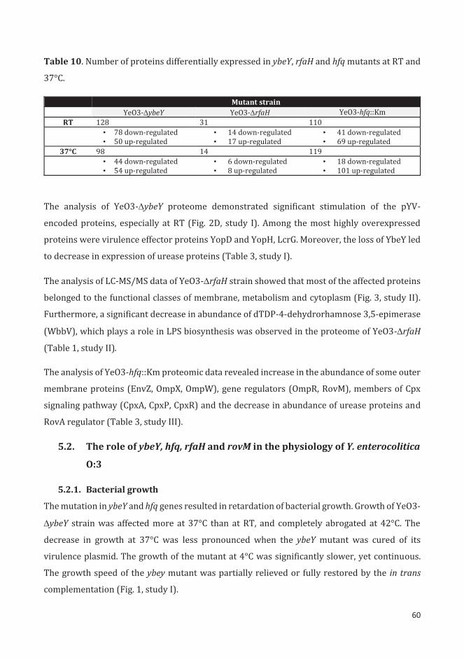
60
Table 10. Number of proteins differentially expressed in ybeY, rfaH and hfq mutants at RT and
37°C.
Mutant strain YeO3- ybeY YeO3- rfaH YeO3-hfq::Km
RT 128 31 110 • 78 down-regulated
• 50 up-regulated • 14 down-regulated • 17 up-regulated
• 41 down-regulated • 69 up-regulated
37°C 98 14 119 • 44 down-regulated
• 54 up-regulated • 6 down-regulated • 8 up-regulated
• 18 down-regulated • 101 up-regulated
The analysis of YeO3- ybeY proteome demonstrated significant stimulation of the pYV-
encoded proteins, especially at RT (Fig. 2D, study I). Among the most highly overexpressed
proteins were virulence effector proteins YopD and YopH, LcrG. Moreover, the loss of YbeY led
to decrease in expression of urease proteins (Table 3, study I).
The analysis of LC-MS/MS data of YeO3- rfaH strain showed that most of the affected proteins
belonged to the functional classes of membrane, metabolism and cytoplasm (Fig. 3, study II).
Furthermore, a significant decrease in abundance of dTDP-4-dehydrorhamnose 3,5-epimerase
(WbbV), which plays a role in LPS biosynthesis was observed in the proteome of YeO3- rfaH
(Table 1, study II).
The analysis of YeO3-hfq::Km proteomic data revealed increase in the abundance of some outer
membrane proteins (EnvZ, OmpX, OmpW), gene regulators (OmpR, RovM), members of Cpx
signaling pathway (CpxA, CpxP, CpxR) and the decrease in abundance of urease proteins and
RovA regulator (Table 3, study III).
5.2. The role of ybeY, hfq, rfaH and rovM in the physiology of Y. enterocolitica
O:3
5.2.1. Bacterial growth
The mutation in ybeY and hfq genes resulted in retardation of bacterial growth. Growth of YeO3-
ybeY strain was affected more at 37°C than at RT, and completely abrogated at 42°C. The
decrease in growth at 37°C was less pronounced when the ybeY mutant was cured of its
virulence plasmid. The growth of the mutant at 4°C was significantly slower, yet continuous.
The growth speed of the ybey mutant was partially relieved or fully restored by the in trans
complementation (Fig. 1, study I).
61
In line with previous findings in Yersinia species (Schiano et al., 2010, Kakoschke et al., 2014),
inactivation of hfq gene in Y. enterocolitica O:3 resulted in growth defect at all tested
temperatures (4, 22, 37 and 42°C). The growth retardation was most prominent at 4 and 42°C.
The in trans complementation restored the growth almost completely. Moreover, the knock-
out of rovM gene of the YeO3-hfq::Km strain resulted in improved growth under all studied
conditions (Fig 3, study III). The YeO3- rfaH, YeO3- rovM and YeO3/pMMB207-rovM strains
showed no growth defects (data not shown).
5.2.2. Cell and colony morphology
The inactivation of the hfq gene in Y. enterocolitica caused alterations in colony morphology.
After 48h of incubation on CIN agar plates at RT the YeO3-hfq::Km mutant strain formed small,
dry and dark colonies surrounded by dark violet halo. Similar effect was observed upon
overexpression of rovM in YeO3/pMMB207-rovM strain. Moreover, this phenotype was
revoked after the knock-out of rovM gene in hfq mutant, indicating the role of RovM regulator
in alterations of colony morphology.
The analysis of cell morphology conducted using the electron microscopy (EM) showed that the
YeO3-hfq::Km bacterial cells were significantly elongated (Fig. 4A and S5, study III). Moreover,
the same bacteria were visibly less aggregated when compared with wild type (data not
shown).
5.2.3. Susceptibility to environmental stresses
All the mutations introduced to Y. enterocolitica in this study resulted in decreased resistance
to environmental stresses. The YeO3- ybeY strain presented highly impaired thermotolerance
presented by decreased survival upon incubation at 50°C. While 95.26 ± 0.86% of YeO3-wt
bacteria survived 5 min incubation under this condition, only 2.38 ± 3.78% of ybeY mutant
bacteria survived. The in trans complementation restored the survival to the level of 87.17
±6.44%. Moreover, the loss of YbeY decreased the ability of bacteria to survive in acidified
environment, an ability of Y. enterocolitica that depends on the degradation of urea. The YeO3-
ybeY strain was killed upon incubation in PBS pH=2.5 with 1.4 mM urea, while 112.42 ± 8.78%
of YeO3-wt bacteria survived these conditions. In trans complementation resulted in 78.75 ±
9.31% survival rate.
Similarly to ybeY mutation, the depletion of Hfq caused decrease in survival rates during
incubation at 55°C and in the acidified medium with 1.4 mM urea (Fig. 7, study III). Only 3.39 ±

62
1.41% of YeO3-hfq::Km bacteria survived 5 min incubation at 55°C, while the survival rate of
YeO3-wt under same conditions reached 41.12 ±8.12%. The ability to survive in PBS pH=2.0
was decreased from 26.35 ± 2.06% recorded for wild-type bacteria to 4.17 ± 1.03%. The trans-
complementation restored both the heat- and acid-resistance.
The analyses of YeO3- rfaH mutant revealed increased susceptibility to SDS and polymyxin B
(Fig. 6, study II), while no significant difference between the rfaH mutant and wild type strains
were observed in tolerance to H2O2 nor to osmotic stress (data not shown).
5.2.4. Motility and biofilm formation
In line with previous studies (reviewed in (Chao & Vogel, 2010)) the loss of Hfq resulted in
impaired swimming motility. Our study showed, that the deterioration of motility is due to
decrease in synthesis of flagellin. The flagellation defect was abolished by both the in trans
complementation and rovM knock-out. Moreover, the YeO3-hfq::Km strain formed significantly
less biofilm that the wild type counterpart. Inactivation of rovM in the hfq negative background
increased the production of biofilm. Additionally, the trans-complemented strain YeO3-
hfq::Km/phfq showed overproduction of biofilm, presumably due to the hfq copy number effect
(Fig. 5, study III).
Interestingly, unlike in some other species the rfaH mutant of Y. enterocolitica did not show any
alterations in motility (data not shown). Also the YeO3- ybeY strain presented same pattern of
motility as the wild type bacteria.
5.2.5. Other physiological features affected by the mutations
In prokaryotes the 16S rRNA undergoes maturation process. The original 17S rRNA transcript
is cleaved from both 5’ and 3’ ends by different endonucleases (RNase E, RNase G, RNase R and
YbeY) with help of ribosome maturation factors (i.e. Era, KsgA, RbfA and RsgA) (Jacob et al.,
2013). The comparison of rRNA profiles of wild type and mutant strains grown at RT and 37°C
revealed remarkable differences in the structure of ybeY mutant rRNA profile. A strong
decrease in the amount of 16S rRNA species produced by YeO3- ybeY was observed (Fig. 3,
study I). The impairment in rRNA processing was more pronounced when YeO3- ybeY bacteria
were incubated at 45°C (data not shown). The other mutant strains, YeO3-hfq::Km and YeO3-
rfaH, presented normal rRNA patterns.

63
The transcriptomics and proteomics data indicated that the mutations in ybeY and hfq genes
altered the bacterial metabolism (Fig. S2 and S3, study I; Table S2-S5, study III). Further
research revealed a decreased ability of YeO3- ybeY to utilize arabinose and galactose.
Moreover, the same strain was not able to grow in the medium containing 1% myo-inositol as
a carbon source (Table S5, study I). The YeO3-hfq::Km strain presented alteration in expression
of several genes from the PTS, that optimize the utilization of carbohydrates present in the
environment. The analysis of hfq mutant transcriptome showed up-regulation of β-glucoside,
fructose, glucitol/sorbitol, glucose, mannitol, mannose and N-acetylgalactosamine-specific
enzymes, whereas cellobiose and chitobiose-specific PTS systems were down-regulated under
at least one incubation condition (Table S2 and S3, study III). Furthermore, both YeO3-hfq::Km
and YeO3/pMMB207-rovM when grown on CIN agar plates formed dark colonies surrounded
by dark violet halo indicating alterations in metabolism (Fig. S4B, study III). The same strains,
when grown on mannitol plates displayed larger halo surrounding the bacterial growth
suggesting higher rates of acidification of the medium (Fig S3B, study III).
5.3. The effect of ybeY, hfq, and rfaH mutations on the virulence and
virulence-associated traits of Y. enterocolitica O:3
5.3.1. Production of Yop effector proteins
The analyses of YeO3- ybeY transcriptome and proteome showed a significant upregulation of
factors encoded on pYV (Fig. 2, study I) at RT. Among the overexpressed genes were yopE, yopH,
yopO, yopM, as well as the injectisome genes yscN, yscO, yscP, yscB and yscD (Fig. 2C, study I).
Consistently, the proteomic study showed the overexpression of pYV-encoded factors,
especially at RT. Among the most highly expressed proteins were YopD, YopH and the secretion
chaperone SycE (Fig 2D, study I). The overproduction of the effector proteins was examined in
three different ways. First, the overnight incubation of ybeY mutant in liquid LB at 37°C resulted
in strong aggregation, an effect known to be caused by Yop secretion (Fig 2A, study I). Secondly,
the amount of proteins secreted by the wild type and YeO3- ybeY bacteria was assessed on
SDS-PAGE gel (Fig. 2B, study I) revealing increased secretion levels. In addition, the abundance
of virulence plasmid encoded proteins was evaluated with immunoblotting (Fig. 5A, study I).
Also this experiment indicated the overexpression of YopE and YopH at RT, as well as increase
in expression of YopH and YadA in DMEM medium at 37°C. Finally, immunoblotting of samples
prepared from the growth media showed the presence of Yops only after incubation at 37°C
and did not allow detection of any Yops in the supernatants of bacteria grown at RT (data not

64
shown). Therefore, it is likely that the mutation in ybeY in Y. enterocolitica O:3 leads to a
significant upregulation of factors encoded on pYV.
In line with previous results obtained for Y. enterocolitica O:8 (Kakoschke et al., 2014), we did
not observe changes in the expression of Yops in YeO3-hfq::Km strain (Table S8, study III).
Similarly, no significant changes in Yops expression was observed in YeO3- rfaH mutant.
5.3.2. LPS synthesis
Both silver staining and immunobloting of DOC-PAGE gels showed a significant decrease in
production of O-antigen and OC in YeO3- rfaH mutant strain at both RT and 37°C. The in trans
complementation resulted not only in full restoration, but also slight overproduction of O-
antigen and OC (Fig. 1, study II). The possibility that the decrease in amount of these LPS
structures was due to shedding of the O-antigen and OC from the bacterial surface was excluded
(Fig. 2, study II). Thus, these results indicated impairment in biosynthesis of O-antigen and OC
structures upon loss of RfaH. The rfaH mutation did not cause any significant changes in the
amount of ECA. These results were supported by findings from transcriptomic and proteomic
study (described earlier), and further verified by the analysis of promoter activities (Fig. 5,
study II). The conducted analyses of LPS structures in YeO3- ybeY and YeO3-hfq::Km did not
reveal any significant differences (data not shown).
5.3.3. Production of other virulence factors
The transcriptomics and quantitative proteomics analyses indicated decrease in the expression
of urease genes in both YeO3- ybeY (Table 3, study I) and YeO3-hfq::Km strains (Table 3, study
III). The urease test showed gave negative result for hfq mutant strain (Fig. 7B, study III), while
ybeY strain presented positive results in urea broth, but only after prolonged incubation. In
coherence with reduced urease activity, both the strains presented increased sensitivity to low
pH medium (described in more details in 5.2.3.).
In addition, the YeO3- ybeY strain presented a significant decrease in production of Ail, a
protein encoded by the attachment invasion locus. This result was verified using the RNA
sequencing, quantitative proteomics, as well as quantitative RT-PCR (Fig. 5B and Table S3,
study I).
Similarly, both YeO3-hfq::Km and YeO3- ybeY strains showed alterations in expression of
OmpR and RovA regulators (Table 3, study I; Table 3, study III). Furthermore, the loss of YbeY

65
resulted in decrease in abundance of such virulence factors as Hfq, Inv and H-NS, and increase
of VirF, YmoA (Table 3, study I). The knock-down of hfq gene caused alterations in expression
of several regulators implicated in bacterial virulence (i.e. PhoB, RovM, RpoS) as well as changes
in the structure of bacterial outer membrane (Cpx signaling pathway and outer membrane
structures) (Table 3, study III).
5.3.4. Resistance to normal human serum
Due to the contribution of LPS, Ail and YadA for serum resistance was previously shown
(Biedzka-Sarek et al., 2005), the susceptibility of the mutants to NHS was assessed. The test
conditions probed for the classical, lectin and alternative complement activation pathways.
The loss of ybeY resulted in significantly (p=0.023) increased resistance to NHS under studied
conditions. The ybeY mutation increased the resistance to NHS by over 3-fold (Fig. 7). Also the
loss of rfaH resulted in a slight but statistically significant (p=0.048) increase in resistance to
NHS killing (Fig. 5; Fig. 6C, study II). No significant difference was observed in resistance of
YeO3- ybeY and YeO3- rfaH strains to EGTA/Mg2+ serum (data not shown), where only the
alternative pathway can be activated. On the other hand, the lack of Hfq chaperone caused
significant (p=0.018) decrease in resistance to NHS (Fig. 7).
Figure 7. Resistance to normal human serum (NHS). The bacteria were incubated for 30 min in 66% NHS and in heat inactivated serum (HIS). The bactericidal effect was calculated as the survival percentage taking the bacterial counts obtained with bacteria incubated in HIS as 100 %. The columns show the mean ± SD serum bactericidal effect when compared with the wild type strain. The experiments were performed in triplicates.
0
50
100
150
200
250
300
350
400
YeO3 YeO3-hfq::Km YeO3-rfaH YeO3-ybeYsurv
ival
com
pare
d to
wild
type
[%]
strain
Resistance to NHS
66
5.3.5. Virulence in cell and animal models
The importance of YbeY for the virulence of Y. enterocolitica O:3 was evaluated in HeLa cell
infection model. The experiment showed decrease in the ability of the bacteria to infect the
eukaryotic cells (Fig. 5C, study I). The most prominent effect was visible during the stage of
adhesion (Fig. 5D - association, study I). The ybeY mutant bacteria displayed no apparent
alterations during the stage of invasion and the rate of invasion of associated bacteria was even
higher than the wild type bacteria (Fig. 5E, study I). Due to highly impaired growth and
increased susceptibility to environmental stresses observed for YeO3- ybeY, the virulence was
not evaluated in any animal model.
The impact of rfaH mutation on the intracellular growth of Y. enterocolitica in human intestinal
epithelial and mouse macrophage cell lines has been studied previously (Nagy et al., 2006).
Moreover, the role of O-antigen and OC in virulence of Y. enterocolitica was also assessed in
mouse experiments, showing 50- to 100-fold decrease in LD50 for the LPS mutant strains (al-
Hendy et al., 1992, Zhang et al., 1997, Skurnik et al., 1999).
The virulence of YeO3-hfq::Km was tested in experimental mouse infection models, where both
oral and intraperitoneal routes of infection were investigated. In the co-infection model mice
were infected with a mixture of wild type and YeO3-hfq::Km bacteria, and after mice were killed,
the bacterial counts in different organs were performed, and the percentage of Km-resistant
(KmR) colonies was determined (Table 5, study III). The presence of KmR bacteria was detected
only in Peyer’s patches. Moreover, the average percentage of KmR bacteria decreased over time,
from 50 % to 5.9 % two days post infection, and to 0.5 % nine days post infection. Among the
mice that were orally infected with one strain at time, a statistically significant (p=0.0064)
reduction in the number of bacteria recovered from mouse organs five days post infection was
observed (Fig. 7 and Table S8, study III) for the YeO3-hfq::Km strain. The infection with the
complemented strain YeO3-hfq::Km/phfq increased the number of recovered bacteria, while
the infection with the double mutant YeO3-rovM-hfq::Km had no significant effect on the
bacterial counts. Interestingly, infected intraperitoneally, both the single hfq and double rovM-
hfq mutants caused the death of two out of three mice within two days post infection (Table S9,
study III), whereas none of the mice infected with the wild-type strain died prematurely. The
number of bacteria recovered from organs of mice that were infected with YeO3 and YeO3-
hfq::Km and survived till the fifth day post infection did not present any significant changes.
Further investigation revealed that the amount of LPS released to the medium is increased in

67
YeO3-hfq::Km and YeO3-rovM-hfq::Km cultures (Fig S7, study III). Moreover, the hfq mutants
were characterized by increased susceptibility to SDS (Fig. S8, study III) indicating that the
structure of the outer membrane is compromised in the hfq and rovM-hfq mutant strains.
Furthermore, the analysis of the lipid A revealed alterations in the structure of this molecule in
the hfq negative bacteria grown at 37°C (Fig. S9, study III).
68
6. DISCUSSION
In this thesis two global approaches, RNA sequencing and quantitative proteomics were
combined in order to better understand the role of selected factors in gene regulation and their
impact on physiology and virulence of Y. enterocolitica. Two of the studies showed that the
deletion of hfq and ybeY genes caused highly pleiotropic effects (study I and III). The mutants
presented significant growth defects and impairment of many physiological functions, as well
as deterioration of features related to virulence and survival inside of the host organism.
Moreover, this thesis demonstrates that RfaH acts as a highly specific antiterminator in Y.
enterocolitica O:3 and affects the expression of LPS O-antigen and OC gene clusters (study II).
6.1. The impact of studied mutations on the gene expression
The most profound alterations in gene expression were caused by hfq and ybeY mutations. A
very high percentage of genes being under Hfq regulation have also been previously observed
in numerous other bacterial species, including the closely related was S. enterica and Y. pestis
(Sittka et al., 2008, Geng et al., 2009). A significant impact of ybeY mutation on the gene
expression has also been reported for E. coli (Pandey et al., 2014). On the other hand, the loss
of RfaH do cause downregulation of gene clusters harboring the ops element located
downstream in the cis-regulated promoter region (Bailey et al., 1997). In this study, the
comparison of wild type and YeO3- rfaH transcriptomes revealed low number of differentially
expressed genes, an indication of the narrow specificity of this regulator.
The comparison of results obtained through RNA-sequencing and LC-MS/MS showed general
coherence. In the case of YeO3-hfq::Km mutant a large number of proteins with altered
abundance while the corresponding genes did not show differential expression in
transcriptomic analyses. That likely originates from the role of Hfq in post-transcriptional
regulation.
The analyses of the transcriptome of the mutants indicated that the mutations caused changes
in the gene expression. First of all, RfaH is known to bind to specific ops sequences present
upstream of long operons. The main function of RfaH is to allow the polymerase to overcome
the terminators (Bailey et al., 1997). In this study the rfaH mutation caused decrease in
abundance of transcripts preceded by ops sequence, indicating the direct effect on these genes.
Subsequently, the changes in LPS biosynthesis was most likely reflected as indirect response to
69
rough phenotype. Another indication of indirect effect was observed for Hfq. The hfq mutation
changed the expression pattern of several other regulators that affected the expression of their
target genes. In this study it was shown that the overexpression of RovM in the YeO3-hfq::Km
mutant was responsible for several of the phenotypic changes observed in the hfq mutant.
Moreover, some changes in gene expression might be a response to changes in metabolism. This
study showed also that both hfq and ybeY mutations presented alterations in carbohydrate
metabolism as well as in abundance in Csr-RNAs.
6.2. YbeY
This study shows, that ybeY is not an essential gene in Y. enterocolitica O:3, but its loss affects
the growth and sensitizes the bacterium to multiple environmental stresses. In line with
previous results and the fact that ybeY was first discovered as a heat shock gene, the effect of
the ybeY mutation was most profound at elevated temperatures (Rasouly et al., 2009, Grinwald
& Ron, 2013). Similar to E. coli (Jacob et al., 2013), in Y. enterocolitica YbeY plays a role in rRNA
maturation process. In this study, the electropherograms revealed misprocessing of 16S rRNA
with the biggest impact observed when bacteria were incubated at 45°C. The impairment in
maturation may be caused by a defect in 17S rRNA cleavage and/or its degradation, as
immature form of 16S rRNA shows low stability (Jacob et al., 2013). The changes in rRNA
pattern can be also caused by a polarized expression of rrn operons that was previously
observed in ybeY mutants (Grinwald & Ron, 2013).
Results obtained in study I indicated overexpression of virulence plasmid genes under all
studied conditions. This happens plausibly due to the increase in plasmid copy number,
alterations in sRNA regulation network or changes in the supercoiling of the pYV. The latter one
can be a result of changes in expression of histone-like proteins and/or indirectly through
alterations in stability of sRNAs that play a role in virulence gene regulation.
The loss of YbeY affected the abundance of several sRNA species. Two of them, CsrB and CsrC,
are Hfq-independent and belong to the carbon storage regulatory system (Liu & Romeo, 1997,
Schiano & Lathem, 2012). In Y. pestis GcvB binds to Hfq and represses dppA, a component of the
dipeptide transport system (Koo et al., 2011). These results indicate, that both the Hfq-
dependent and Hfq-independent sRNA regulation pathways were affected in the YeO3- ybeY
mutant.
70
Moreover, the YeO3- ybeY mutant revealed the presence of transcripts typical for cold shock
response (Annamalai & Venkitanarayanan, 2005). It is known that changes in temperature
result in dissociation of polysomes, accumulation of free ribosomes and their subunits and the
state of ribosomes is one of the main sensors of environmental conditions that in E. coli elicit
the heat and cold shock responses (VanBogelen & Neidhardt, 1990, Jones et al., 1992, Golovlev,
2003). Moreover, cold shock response can be induced artificially using different antibiotics like
chloramphenicol, erythromycin, spiramycin and tetracycline, all inhibitors of protein synthesis
(VanBogelen & Neidhardt, 1990). It was previously noticed that mutation in ybeY gene results
in significant alterations in the structure of ribosomes (Rasouly et al., 2009, Rasouly et al., 2010,
Jacob et al., 2013). Therefore it is highly possible, that the cold-shock like response in YeO3-
ybeY mutant occurs due to accumulation of defective ribosomes. The defect in the translation
machinery together with accumulation of cspA transcript are factors decreasing the growth rate
of the mutant.
In Y. enterocolitica urease is considered to be an important virulence factor that allows bacteria
to survive in low pH environment and therefore to pass through the stomach during the course
of infection (De Koning-Ward & Robins-Browne, 1995, Gripenberg-Lerche et al., 2000). The
decrease in urease production observed in YeO3- ybeY mutant resulted in reduced ability to
survive in acidic conditions. Previous studies showed that the loss of acid resistance leads to
severe decrease in bacterial virulence in mouse models (De Koning-Ward & Robins-Browne,
1995, Gripenberg-Lerche et al., 2000). Moreover, ybeY mutant presented decreased ability
adhere to the HeLa cells that was most probably caused by alterations in expression of Ail
(Felek & Krukonis, 2009, Kolodziejek et al., 2010). It is known, that the ability of Y. enterocolitica
to invade eukaryotic cells in vitro correlates with its virulence (Lee et al., 1977, Une, 1977).
Taken together, the mutation in ybeY gene resulted in significant alterations in gene expression
of Y. enterocolitica O:3. The results obtained in study I showed that YbeY is required for proper
functioning of sRNA regulation network, the control of virulence gene expression and its loss
induce the expression of cold-shock genes. Such profound changes in the control of gene
expression resulted in a pleiotropic effect including an impairment of many virulence-related
features.
71
6.3. RfaH
In study II of this thesis it was shown that RfaH serves as a functional antiterminator in Y.
enterocolitica O:3. In coherence with results obtained for other bacterial species, the loss of
RfaH resulted in downregulation of long operons harboring the ops element located in the cis-
regulated promoter region (Bailey et al., 2000). The same study showed the importance of the
correct location of the ops element for the RfaH recruitment, as the ops element located far
upstream of the ECA gene cluster promoter region was not functional (Fig. 4, study II).
Disruption of rfaH caused decrease in abundance of transcripts of several genes from the LPS
biosynthesis pathway and resulted in loss of O-antigen and OC structures.
The comparison of YeO3- rfaH and wild type transcriptome profiles revealed general
similarity, indicating a narrow specificity of the regulator. Moreover, a correlation between the
transcriptome pattern of rfaH mutant and spontaneous rough strain was detected. Several
RfaH-dependent changes observed in study II were likely to be the indirect responses to the
loss of LPS. Both the YeO3- rfaH and YeO3-c-R1 strains showed changes in the expression of
Cpx envelope stress system indicating a response to changes in the cell wall integrity (Hunke et
al., 2012, Vogt & Raivio, 2012). Moreover, the alterations in amount of transport ATPase
proteins, sialic acid permease and phosphate ABC transporters were observed (Table 1, study
II) that was previously correlated with a response to decreased LPS production and
consequential reduced demand for sugars and energy (Nagy et al., 2006).
In line with previous results (Biedzka-Sarek et al., 2005), loss of RfaH resulted in increased
resistance to NHS. It is believed that the loss of LPS structure from the surface exposed the YadA
and Ail, two other factors conferring to the complement resistance (Biedzka-Sarek et al., 2005).
Similarly as in E. coli and S. enterica (Yethon et al., 2000, Nagy et al., 2006), reduction in O-
antigen and OC expression resulted in decreased resistance to SDS and polymyxin B (Fig. 6,
study II), but no impairment in resistance to hydrogen peroxide was observed in this study.
Previously it was demonstrated that the O-antigen and OC are essential for full virulence of Y.
eneterocolitica serotype O:3 (al-Hendy et al., 1992, Skurnik et al., 1999). Experiment conducted
on mice showed that the oral LD50 decreased 50- to 100 fold for the LPS mutant strains (al-
Hendy et al., 1992, Zhang et al., 1997, Skurnik et al., 1999). The impairment in virulence occurs
due to the importance of the O-antigen for the early stages of infection, the resistance to
environment of gastrointestinal track, and for the immune system evasion (al-Hendy et al.,

72
1992, Biedzka-Sarek et al., 2005). Furthermore, the impact of rfaH mutation on intracellular net
growth of Y. enterocolitica has been already evaluated by Nagy et al. (Nagy et al., 2006). In their
study the comparison of the wild type and YeO3- rfaH strain yield was determined in cultured
human intestinal epithelial (INT407) and mouse macrophage (RAW264.7) cell lines . The study
revealed that rfaH mutant exhibits reduced intracellular yield when compared to the wild type
bacterium.
It was previously demonstrated that a rough mutant of Y. enterocolitica O:8 exhibited
alterations in YadA function, loss of Ail and downregulation of inv expression. Moreover, due to
upregulation of flhDC, the flagellar master regulatory operon, the mutant presented
hypermotility (Bengoechea et al., 2004). In study II no significant changes in the production of
these virulence factors were observed. Furthermore, the wild type and YeO3- rfaH strains
presented similar motility. These discrepancies may be explained by differences underlying the
serotypes (differences in motility and flagellation between the serotype O:8 and O:3 were
described previously (Uliczka et al., 2011)). Furthermore, the rfaH mutation does not cause full
abrogation of O-antigen and OC production and thus some functional LPS is still present on the
surface of bacterial cell what may result in less severe phenotype than the one of rough mutant.
Taken together, study II of this thesis showed that RfaH serves as a highly specific regulator
that most likely affects only genes preceded by a functional ops element. Despite the small
number of affected genes, the disruption of rfaH gene led to profound changes in the bacterial
physiology. Decrease in the amount of LPS on the cell surface results in indirect changes in the
transcriptome, stress response, and lower resistance to some environmental cues. Moreover,
based on the previous results (al-Hendy et al., 1992, Zhang et al., 1997, Skurnik et al., 1999,
Nagy et al., 2006), it can be assumed that YeO3- rfaH has decreased virulence.
6.4. Hfq
The study III of this thesis demonstrated the importance of Hfq in the post-transcriptional
regulation in Y. enterocolitica O:3. The results showed, that similar as in the case of S. enterica
and Y. pestis, a high percentage of genes stay under the regulation of Hfq. Moreover, the fact that
numerous proteins displayed alterations in abundance, even though their coding genes did not
show differential expression affirms the importance of Hfq for post-transcriptional regulation.
Due to the fact that Hfq is a crucial element of the sRNA regulation network, it is assumed that
apart from its direct effect, it can also affect the expression of genes through changes in
73
abundance and activity of other regulators. The study III reported differential expression of
several regulators (Table 3, study III). In accordance with changes in the abundance of these
regulators, the alteration in expression of their target genes were also observed. Analysis
revealed differential expression of genes belonging to the RpoS, RovA and OmpR regulons
(Patten et al., 2004, Cathelyn et al., 2007, Hu et al., 2009). This study also revealed a significant
derepression of RovM regulator in YeO3-hfq::Km mutant (Fig. 2, study III).
Study III also affirmed the requirement of Hfq presence for proper expression of the RpoS sigma
factor in Y. enterocolitica. The decrease in abundance of RpoS factor was also observed
previously in different bacterial species like E. coli and S. enterica upon the Hfq depletion
(Brown & Elliott, 1996, Muffler et al., 1996). RpoS confers the optimal growth during the
stationary phase and under stress conditions (Tanaka et al., 1993, Badger & Miller, 1995) and
therefore reduction in its activity can contribute to increased susceptibility of YeO3-hfq::Km
mutant to environmental stresses. Out of four sRNA species known to regulate the translation
of RpoS in E. coli, namely DsrA, RprA, ArcZ and OxyS (Sledjeski et al., 1996, Zhang et al., 1998,
Majdalani et al., 2002, Mandin & Gottesman, 2010), study III identified two homologues in the
genome of Y. enterocolitica (RprA and ArcZ). sRNAs are known to enhance the translation of
RpoS by binding to the 5’ leader region of the mRNA what results in better availability of the
Shine-Dalgarno site (Brown & Elliott, 1997). The analysis of sRNA expression in YeO3-hfq::Km
mutant revealed alterations in abundance in RprA. Moreover, it is known that these sRNA
species require binding to Hfq for full functionality (Sledjeski et al., 1996, Zhang et al., 1998,
Majdalani et al., 2002, Mandin & Gottesman, 2010). Therefore, it is assumed that the loss of Hfq
in Y. enterocolitica disrupts the sRNA regulation pathway and results in lower abundance of
RpoS protein.
In line with the results obtained for other pathogenic Yersinia species, the loss of hfq in Y.
enterocolitica resulted in a growth defect. The growth curves of YeO3-hfq::Km showed similar
pattern as serotype O:8, displaying an intermediate phenotype between strongly impaired
characteristics of Y. pestis and the marginally affected Y. pseudotuberculosis (Geng et al., 2009,
Schiano et al., 2010, Kakoschke et al., 2014). Furthermore, in coherence with results obtained
for the serotype O:8, alterations in the bacterial metabolism, including changes in the utilization
of mannitol that resulted in different growth on CIN agar and mannitol plates were observed
(Kakoschke et al., 2014). Moreover, study III showed that Hfq of Y. enterocolitica O:3 is involved
in resistance to environmental stresses, including elevated temperature and acidic
74
environment. Similar to the case of serotype O:8, alterations in expression of urease genes were
observed (Kakoschke et al., 2014). In addition, YeO3-hfq::Km displayed impairment in motility
and biofilm formation.
The mouse infection experiments conducted in study III showed that the YeO3-hfq::Km
bacteria, similarly to other Yersinia hfq mutants (Geng et al., 2009, Schiano et al., 2010) were
characterized by decreased virulence. Virulence is a remarkably multifactorial feature of Y.
enterocolitica and therefore it is believed that different alterations caused by the loss of hfq can
contribute to impairment in virulence. Although rpoS mutant was not attenuated in mouse
infection experiments (Badger & Miller, 1995), the repression of rpoS may have a synergistic
effect. In order to pass through the stomach during the course of infection, Y. enterocolitica must
be able to survive in low pH environment (De Koning-Ward & Robins-Browne, 1995,
Gripenberg-Lerche et al., 2000), therefore reduction in the urease activity is likely one of the
shortcomings that attenuates YeO3-hfq::Km in the oral infection model. Furthermore, the
motility was also implicated in bacterial virulence (Young et al., 2000). Interestingly, two out of
three mice infected intraperitoneally with the YeO3-hfq::Km or YeO3-rovM-hfq::Km mutant
died prematurely. The decreased resistance to SDS, increased release of the LPS molecule to the
culture supernatant together with alterations in the abundance of membrane proteins indicate
that the structure of the bacterial outer membrane of hfq mutant might be compromised. The
importance of Hfq and sRNAs regulation for the biogenesis of the outer membrane was
previously established (Guillier et al., 2006, Van Puyvelde et al., 2013). Therefore in the study
III it is suggested, that the death observed in mice infected intraperitoneally with either YeO3-
hfq::Km or YeO3-rovM-hfq::Km occurred due to the septic shock caused by the alterations in the
structure of lipid A molecule and/or the increased release of the LPS from the mutant bacterial
cells.
Taken together, study III demonstrated that the loss of Hfq in Y. enterocolitica leads to profound
changes in the transcriptome and proteome affirming the importance of Hfq for gene regulation
control. The YeO3-hfq::Km mutant showed alterations in abundance of several transcriptional
regulators and sRNA species. Moreover, the loss of Hfq resulted in highly pleiotropic changes
in the phenotype including abrogation of virulence.

75
6.5. RovM
In study III of this thesis it was shown that some of the phenotypic features of YeO3-hfq::Km
mutant are due to derepression of RovM regulator. Inactivation of the rovM gene in the hfq
mutant resulted in full or partial restoration of several characteristics.
Knocking out rovM gene in YeO3-hfq::Km mutant resulted in increased growth rate, even
though did not restore it to the level presented by the wild type bacteria. Moreover, the colony
morphology observed before by Kakoschke et al. (Kakoschke et al., 2014), was fully reversed
after the loss of RovM. In turn, overexpression of rovM in YeO3/pMMB207-rovM resulted in the
same colony morphology on CIN agar and mannitol plates, strongly implicating the role of RovM
in the regulation of mannitol metabolism in Y. enterocolitica.
Study III showed also that in contrast to majority of the LysR-type regulators, but similar to
rovM of Y. pseudotuberculosis and hexA, the YeO3 rovM was also under positive autoregulation
(Harris et al., 1998, Heroven & Dersch, 2006). Moreover, most of the LysR-type regulators
interact with effectors that ultimately steer their regulatory propensities. Therefore it is
possible that RovM in Y. enterocolitica also requires additional cofactors for its full function
(Schell, 1993). Additionally, the expression of rovM in Y. pseudotuberculosis is regulated by the
Csr system (Heroven et al., 2008).
Furthermore, study III demonstrated that RovM functions as a motility regulator that represses
the flagella biosynthesis. It is likely, that in line with the results obtained in E. coli, this takes
place through binding of RovM to flhDC promoter region (Blumer et al., 2005). Study III showed
that RovM influences the biosynthesis of flagellin and the YeO3- rovM mutant is hypermotile.
However, the knock-out of rovM in hfq negative background did not restore full motility, and
the overexpression in pMMB207-rovM did not result in total abrogation of motility. Therefore
it is believed that other factors still influence the motility in this system. In Y. enterocolitica YenS
sRNA was found to be a positive regulator of motility that acts through the modulation of YenI
production. Interestingly, the interplay between the levels of yenI and yenS may lead to either
hypo- or hypermotility. (Tsai & Winans, 2011).
In contrast to previous findings in E. coli (Peterson et al., 2006), overexpression of RovM in
YeO3 increased the RpoS levels in the stationary phase cells. Yet, the overexpression of RovM
did not increase the abundance of RpoS in the YeO3-hfq::Km strain (Fig. 6, study III). Therefore,
in study III it was concluded that in Y. enterocolitica RovM positively regulates the levels of RpoS
76
in Hfq-dependent manner. It was previously observed that sRNAs allow proper translation of
RpoS (described in subchapter 6.4), therefore the positive regulation of RovM might be visible
only when certain sRNA species are functional.
In line with previous results obtained for Y. pseudotuberculosis and Y. enterocolitica O:8, RovM
represses the expression of rovA also in Y. enterocolitica O:3 (Heroven & Dersch, 2006, Lawrenz
& Miller, 2007). In Y. enterocolitica serotype O:8 it controls the expression of inv, however, in
serotype O:3 this regulation is prevented due to presence of an insertion sequence in the inv
regulatory region (Uliczka et al., 2011).
Taken together, in the study III of this thesis the effect of RovM overexpression in the hfq
negative background was analyzed. Inactivation of rovM in the YeO3-hfq::Km mutant prevented
the subsequent overexpression of this regulator and allowed to elucidate the role of RovM in
the Hfq deficient strain. This study demonstrated that the growth defect, mannitol utilization
and motility alterations observed for YeO3-hfq::Km were mediated by the overexpression of
RovM.
77
7. CONCLUSIONS AND FUTURE CONSIDERATIONS
In conclusion, all four studied genes, ybeY, rfaH, hfq and rovM, had a profound impact on gene
regulation of Y. enterocolitica O:3. The deletion of hfq and ybeY genes caused wide disturbances
in gene expression and highly pleiotropic effects. Both the mutants presented significant
growth defects and impairment of many physiological functions, as well as deterioration of
features related to virulence and survival inside of the host organism. On the other hand, RfaH
was shown to function as a highly specific antiterminator in Y. enterocolitica O:3 that affects
only the expression of LPS O-antigen and OC gene clusters and most likely leads to an indirect
response to rough phenotype. The RovM regulator was characterized by highest specificity, and
affected the expression of 24 genes in Y. enterocolitica O:3.
The conducted experiments demonstrated that the studied mutations (ybeY, rfaH and hfq)
compromised the bacterial virulence. The mutants showed significant decrease in resistance to
different environmental conditions that are normally encountered during the course of
infection. Moreover, the loss of studied genes resulted also in growth defect, impairment of
motility and biofilm formation, changes in carbohydrates metabolism, and alterations in
production of different virulence factors that also contributes to vitality and ability to establish
infection in host organism.
Due to the fact that all the mutant strains demonstrated compromised virulence, the studied
proteins might be regarded as potential targets for new drug development. Furthermore, the
strains with established avirulence might be used as sources for vaccines. This thesis revealed
the general function of RfaH in Y. enterocolitica O:3, yet the role of RfaH in the in vivo regulation
of LPS expression is still unknown. The potential role of RfaH in in vivo controlling of tissue-
and temperature-specific expression of LPS should be further investigated. The future
experiments should be also aimed at explaining the details of the mechanism behind the
overexpression of pYV genes in YeO3- ybeY mutant.
Moreover, the results of high-throughput analyses can be further used in studies aiming at
deciphering the role of yet unknown genes and proteins. This study revealed a large number of
uncharacterized genes that stay under the YbeY, RfaH, Hfq and RovM regulation. They
involvement in the regulatory pathways, as well as potential role is an interesting research
question.

78
8. ACKNOWLEDGEMENTS
This thesis work was performed in the Immunobiology Research Program at the Department
of Bacteriology and Immunology of Faculty of Medicine, at the University of Helsinki, during
2011-2016. I have been financially supported by the Doctoral Programme in Biomedicine
(DPBM), the Research Foundation of the University of Helsinki, the Emil Aaltonen Foundation,
CIMO, the BioCentrum Helsinki and Maud Kuistila Memorial Foundation. All are sincerely
acknowledged.
I wish to express my sincere and deep gratitude to my supervisor Professor Mikael Skurnik,
who guided me through this work by sharing his vast knowledge and creativeness. I have been
amazingly fortunate to have a supervisor who gave me freedom to explore my own ideas, while
at the same time patiently thought me the necessary skills. I highly treasure Mikael’s kindness
and optimism, present even in the most difficult times. I appreciate that he always had time to
discuss the details of this project, despite his busy schedule. I would also like to express my
gratitude for allowing me to get involved in different side projects and collaborations that
widen my knowledge. His involvement in this work was tremendous, but his impact on my
scientific development goes way beyond this thesis.
I would like to thank Professor Virginia Miller for accepting the role of my opponent. To the
official reviewers of this thesis, Professor Mikael Rhen and Doctor Arto Pulliainen, I am grateful
for their time, interest and constructive comments that helped to improve this work.
I am grateful to the members of my thesis committee, Doctor Eija Korpelainen and Docent
Pentti Kuusela for support and advice on my work throughout these years.
I acknowledge my collaborators Markku Varjosalo, Pirkko Mattila, José A. Bengoechea, Zhilin
Li, and Chun-Mei Li, and Helena Fernández-Carrasco for sharing their time, knowledge and
material.
I wish to thank all the present and past colleagues at the Department of Bacteriology and
Immunology for creating such a good work atmosphere. I want to thank the members of
Yersinia-lab, especially Juha Laitinen, Anu Wicklund, Saija Kiljunen, Maria Pajunen, Mabruka
Salem, Julia Moroz, Anita Lijegren and Laura Kalin for their various form of support during my
studies. I also want to thank Juha Laitinen and Anu Wicklund not only for all their skillful help
in the laboratory, but also the support in private life. Furthermore, I would like to thank all the

79
members of Immunobiology Research Program, especially Hanne Ahola, Hanne Amdahl, Arnab
Bhattacharjee, Neeta Datta, Karita Haapasalo, Ayman Khattab, Seppo Meri, Marcel Messing, Eija
Nissilä, Anna-Helena Saariaho and Päivi Saavalainen. I would also like to express my special
thanks to Judith Klievink, Satu Hyvärinen and Anita Wasik for all the work-related and work-
unrelated conversations we had during these years.
I am grateful for all my life-long friends, who were always there to support me. Anna
Byczkowska, Maciej Rubacha, Krzysztof Paszkowski, Michal Grodecki, Michal Pietrzak and
Jakub Bierzgalski have always found time for me and supported me greatly in different phases
of my life. They have always been there for me, no matter what. Without them I would not be
where I am now.
Finally, and most of all, I want to thank my fantastic family to whom I dedicate this thesis. I
would like to thank my daughter, Amanda, who gives me inestimable joy every time she smiles
and who accepted scientific literature as a bed time story. I am grateful for the love and endless
support of my husband, Heikki Leskinen, who has been always there for me, sharing all the good
and the difficult moments with me. He always encouraged me and supported my career plans.
I would like to express my deepest gratitude for my parents, Danuta Knapska and Maciej
Knapski for all their love, constant care and for believing in me. They always led me by example,
thought me the value of hard work and determination, and raised me a resourceful person.
Their support made it possible for me to follow my dreams and live a life the way I wanted. I
would like to thank my grandmother, Genowefa Wiczel for her big love and support throughout
my whole life. I wish to express my special thanks to my parents-in-law, Arja and Tapio
Leskinen, for their acceptance, constant kindness and patience. Without the love and support
from my family I could not have made it to here.
Katarzyna Leskinen
Helsinki, 2016

80
9. REFERENCES
Ackers ML, Schoenfeld S, Markman J, Smith MG, Nicholson MA, DeWitt W, Cameron DN, Griffin PM & Slutsker L (2000) An outbreak of Yersinia enterocolitica O:8 infections associated with pasteurized milk. The Journal of infectious diseases 181: 1834-1837.
Akerley BJ, Rubin EJ, Novick VL, Amaya K, Judson N & Mekalanos JJ (2002) A genome-scale analysis for identification of genes required for growth or survival of Haemophilus influenzae. Proceedings of the National Academy of Sciences of the United States of America 99: 966-971.
Akopyan K, Edgren T, Wang-Edgren H, Rosqvist R, Fahlgren A, Wolf-Watz H & Fallman M (2011) Translocation of surface-localized effectors in type III secretion. Proceedings of the National Academy of Sciences USA 108: 1639-1644.
Al-Hendy A (1992) Lipopolysaccharide of Yersinia enterocolitica: Molecular genetics and role in virulence. PhD thesis. University of Turku, Turku, Finland.
al-Hendy A, Toivanen P & Skurnik M (1991) Expression cloning of Yersinia enterocolitica O:3 rfb gene cluster in Escherichia coli K12. Microbial pathogenesis 10: 47-59.
Al-Hendy A, Toivanen P & Skurnik M (1991) Rapid method for isolation and staining of bacterial lipopolysaccharide. Microbiology and immunology 35: 331-333.
Al-Hendy A, Toivanen P & Skurnik M (1992) Lipopolysaccharide O side chain of Yersinia enterocolitica O:3 is an essential virulence factor in an orally infected murine model. Infection and immunity 60: 870-875.
Annamalai T & Venkitanarayanan K (2005) Expression of major cold shock proteins and genes by Yersinia enterocolitica in synthetic medium and foods. Journal of food protection 68: 2454-2458.
Artsimovitch I & Landick R (2002) The transcriptional regulator RfaH stimulates RNA chain synthesis after recruitment to elongation complexes by the exposed nontemplate DNA strand. Cell 109: 193-203.
Asadishad B, Ghoshal S & Tufenkji N (2013) Role of cold climate and freeze-thaw on the survival, transport, and virulence of Yersinia enterocolitica. Environmental science & technology 47: 14169-14177.
Atkinson S, Chang CY, Sockett RE, Camara M & Williams P (2006) Quorum sensing in Yersinia enterocolitica controls swimming and swarming motility. Journal of bacteriology 188: 1451-1461.
Autenrieth IB, Bohn E, Ewald JH & Heesemann J (1995) Deferoxamine B but not deferoxamine G1 inhibits cytokine production in murine bone marrow macrophages. Journal of Infectious Diseases 172: 490-496.
Autenrieth IB, Reissbrodt R, Saken E, Berner R, Vogel U, Rabsch W & Heesemann J (1994) Desferrioxamine-promoted virulence of Yersinia enterocolitica in mice depends on both desferrioxamine type and mouse strain. The Journal of infectious diseases 169: 562-567.
Babic A, Guerout AM & Mazel D (2008) Construction of an improved RP4 (RK2)-based conjugative system. Research in microbiology 159: 545-549.
Badger JL & Miller VL (1995) Role of RpoS in survival of Yersinia enterocolitica to a variety of environmental stresses. Journal of bacteriology 177: 5370-5373.
Bailey MJ, Hughes C & Koronakis V (1997) RfaH and the ops element, components of a novel system controlling bacterial transcription elongation. Molecular microbiology 26: 845-851.
Bailey MJ, Hughes C & Koronakis V (2000) In vitro recruitment of the RfaH regulatory protein into a specialised transcription complex, directed by the nucleic acid ops element. Molecular & general genetics : MGG 262: 1052-1059.
Bailey MJ, Koronakis V, Schmoll T & Hughes C (1992) Escherichia coli HlyT protein, a transcriptional activator of haemolysin synthesis and secretion, is encoded by the rfaH (sfrB) locus required for expression of sex factor and lipopolysaccharide genes. Molecular microbiology 6: 1003-1012.
Baker CS, Eory LA, Yakhnin H, Mercante J, Romeo T & Babitzke P (2007) CsrA inhibits translation initiation of Escherichia coli hfq by binding to a single site overlapping the Shine-Dalgarno sequence. Journal of bacteriology 189: 5472-5481.
81
Barua S, Yamashino T, Hasegawa T, Yokoyama K, Torii K & Ohta M (2002) Involvement of surface polysaccharides in the organic acid resistance of Shiga Toxin-producing Escherichia coli O157:H7. Molecular microbiology 43: 629-640.
Barz C, Abahji TN, Trulzsch K & Heesemann J (2000) The Yersinia Ser/Thr protein kinase YpkA/YopO directly interacts with the small GTPases RhoA and Rac-1. FEBS letters 482: 139-143.
Baumler AJ & Hantke K (1992) Ferrioxamine uptake in Yersinia enterocolitica: characterization of the receptor protein FoxA. Molecular microbiology 6: 1309-1321.
Beaume M, Hernandez D, Farinelli L, Deluen C, Linder P, Gaspin C, Romby P, Schrenzel J & Francois P (2010) Cartography of methicillin-resistant S. aureus transcripts: detection, orientation and temporal expression during growth phase and stress conditions. PloS one 5: e10725.
Beauregard A, Smith EA, Petrone BL, Singh N, Karch C, McDonough KA & Wade JT (2013) Identification and characterization of small RNAs in Yersinia pestis. RNA biology 10: 397-405.
Bellows LE, Koestler BJ, Karaba SM, Waters CM & Lathem WW (2012) Hfq-dependent, co-ordinate control of cyclic diguanylate synthesis and catabolism in the plague pathogen Yersinia pestis. Molecular microbiology 86: 661-674.
Belogurov GA, Vassylyeva MN, Svetlov V, Klyuyev S, Grishin NV, Vassylyev DG & Artsimovitch I (2007) Structural basis for converting a general transcription factor into an operon-specific virulence regulator. Molecular cell 26: 117-129.
Bengoechea JA, Najdenski H & Skurnik M (2004) Lipopolysaccharide O antigen status of Yersinia enterocolitica O:8 is essential for virulence and absence of O antigen affects the expression of other Yersinia virulence factors. Molecular microbiology 52: 451-469.
Bertani G (2004) Lysogeny at mid-twentieth century: P1, P2, and other experimental systems. Journal of bacteriology 186: 595-600.
Biedzka-Sarek M, Venho R & Skurnik M (2005) Role of YadA, Ail, and Lipopolysaccharide in Serum Resistance of Yersinia enterocolitica Serotype O:3. Infection and immunity 73: 2232-2244.
Biedzka-Sarek M, Salmenlinna S, Gruber M, Lupas AN, Meri S & Skurnik M (2008) Functional mapping of YadA- and Ail-mediated binding of human factor H to Yersinia enterocolitica serotype O:3. Infection and immunity 76: 5016-5027.
Bilusic I, Popitsch N, Rescheneder P, Schroeder R & Lybecker M (2014) Revisiting the coding potential of the E. coli genome through Hfq co-immunoprecipitation. RNA biology 11: 641-654.
Birge EA (2006) Transcription and Translation: Processes and Basic Regulation. Bacterial and Bacteriophage Genetics,p.^pp. 107-148. Springer, New York.
Black DS & Bliska JB (1997) Identification of p130Cas as a substrate of Yersinia YopH (Yop51), a bacterial protein tyrosine phosphatase that translocates into mammalian cells and targets focal adhesions. EMBO Journal 16: 2730-2744.
Black DS, Montagna LG, Zitsmann S & Bliska JB (1998) Identification of an amino-terminal substrate-binding domain in the Yersinia tyrosine phosphatase that is required for efficient recognition of focal adhesion targets. Molecular microbiology 29: 1263-1274.
Bleves S, Marenne MN, Detry G & Cornelis GR (2002) Up-regulation of the Yersinia enterocolitica yop regulon by deletion of the flagellum master operon flhDC. Journal of bacteriology 184: 3214-3223.
Blumer C, Kleefeld A, Lehnen D, et al. (2005) Regulation of type 1 fimbriae synthesis and biofilm formation by the transcriptional regulator LrhA of Escherichia coli. Microbiology 151: 3287-3298.
Boelaert JR, van Landuyt HW, Valcke YJ, Cantinieaux B, Lornoy WF, Vanherweghem JL, Moreillon P & Vandepitte JM (1987) The role of iron overload in Yersinia enterocolitica and Yersinia pseudotuberculosis bacteremia in hemodialysis patients. The Journal of infectious diseases 156: 384-387.
Bohme K, Steinmann R, Kortmann J, et al. (2012) Concerted actions of a thermo-labile regulator and a unique intergenic RNA thermosensor control Yersinia virulence. PLoS pathogens 8: e1002518.
Boland A & Cornelis GR (1998) Role of YopP in suppression of tumor necrosis factor alpha release by macrophages during Yersinia infection. Infection and immunity 66: 1878-1884.
82
Boland A, Sory MP, Iriarte M, Kerbourch C, Wattiau P & Cornelis GR (1996) Status of YopM and YopN in the Yersinia Yop virulon: YopM of Y.enterocolitica is internalized inside the cytosol of PU5-1.8 macrophages by the YopB, D, N delivery apparatus. EMBO Journal 15: 5191-5201.
Bolin I, Forsberg A, Norlander L, Skurnik M & Wolf-Watz H (1988) Identification and mapping of the temperature-inducible, plasmid-encoded proteins of Yersinia spp. Infection and immunity 56: 343-348.
Boni IV, Isaeva DM, Musychenko ML & Tzareva NV (1991) Ribosome-messenger recognition: mRNA target sites for ribosomal protein S1. Nucleic acids research 19: 155-162.
Bottone EJ (1997) Yersinia enterocolitica: the charisma continues. Clinical microbiology reviews 10: 257-276.
Boudvillain M, Figueroa-Bossi N & Bossi L (2013) Terminator still moving forward: expanding roles for Rho factor. Current opinion in microbiology 16: 118-124.
Brennan RG & Link TM (2007) Hfq structure, function and ligand binding. Current opinion in microbiology 10: 125-133.
Brown KL & Hughes KT (1995) The role of anti-sigma factors in gene regulation. Molecular microbiology 16: 397-404.
Brown L & Elliott T (1996) Efficient translation of the RpoS sigma factor in Salmonella typhimurium requires host factor I, an RNA-binding protein encoded by the hfq gene. Journal of bacteriology 178: 3763-3770.
Brown L & Elliott T (1997) Mutations that increase expression of the rpoS gene and decrease its dependence on hfq function in Salmonella typhimurium. Journal of bacteriology 179: 656-662.
Brzostek K & Raczkowska A (2007) The YompC protein of Yersinia enterocolitica: molecular and physiological characterization. Folia microbiologica 52: 73-80.
Brzostek K, Skorek K & Raczkowska A (2012) OmpR, a central integrator of several cellular responses in Yersinia enterocolitica. Advances in experimental medicine and biology 954: 325-334.
Brzostkowska M, Raczkowska A & Brzostek K (2012) OmpR, a response regulator of the two-component signal transduction pathway, influences inv gene expression in Yersinia enterocolitica O9. Frontiers in cellular and infection microbiology 2: 153.
Cain JA, Solis N & Cordwell SJ (2014) Beyond gene expression: the impact of protein post-translational modifications in bacteria. Journal of proteomics 97: 265-286.
Carniel E (2001) The Yersinia high-pathogenicity island: an iron-uptake island. Microbes and infection / Institut Pasteur 3: 561-569.
Carter HD, Svetlov V & Artsimovitch I (2004) Highly divergent RfaH orthologs from pathogenic proteobacteria can substitute for Escherichia coli RfaH both in vivo and in vitro. Journal of bacteriology 186: 2829-2840.
Carter JA, Blondel CJ, Zaldivar M, Alvarez SA, Marolda CL, Valvano MA & Contreras I (2007) O-antigen modal chain length in Shigella flexneri 2a is growth-regulated through RfaH-mediated transcriptional control of the wzy gene. Microbiology 153: 3499-3507.
Castelli ME, Fedrigo GV, Clementin AL, Ielmini MV, Feldman MF & Garcia Vescovi E (2008) Enterobacterial common antigen integrity is a checkpoint for flagellar biogenesis in Serratia marcescens. Journal of bacteriology 190: 213-220.
Cathelyn JS, Ellison DW, Hinchliffe SJ, Wren BW & Miller VL (2007) The RovA regulons of Yersinia enterocolitica and Yersinia pestis are distinct: evidence that many RovA-regulated genes were acquired more recently than the core genome. Molecular microbiology 66: 189-205.
Chambers CE & Sokol PA (1994) Comparison of siderophore production and utilization in pathogenic and environmental isolates of Yersinia enterocolitica. Journal of clinical microbiology 32: 32-39.
Chao Y & Vogel J (2010) The role of Hfq in bacterial pathogens. Current opinion in microbiology 13: 24-33. Chao Y, Papenfort K, Reinhardt R, Sharma CM & Vogel J (2012) An atlas of Hfq-bound transcripts reveals 3'
UTRs as a genomic reservoir of regulatory small RNAs. EMBO Journal 31: 4005-4019. Chavez RG, Alvarez AF, Romeo T & Georgellis D (2010) The physiological stimulus for the BarA sensor kinase.
Journal of bacteriology 192: 2009-2012.
83
Chen Y & Anderson DM (2011) Expression hierarchy in the Yersinia type III secretion system established through YopD recognition of RNA. Molecular microbiology 80: 966-980.
Cornelis GR (2002) The Yersinia Ysc-Yop virulence apparatus. International journal of medical microbiology : IJMM 291: 455-462.
Cornelis GR & Wolf-Watz H (1997) The Yersinia Yop virulon: a bacterial system for subverting eukaryotic cells. Molecular microbiology 23: 861-867.
Cotter PA & Miller JF (1998) In vivo and ex vivo regulation of bacterial virulence gene expression. Current opinion in microbiology 1: 17-26.
Cover TL & Aber RC (1989) Yersinia enterocolitica. The New England journal of medicine 321: 16-24. Coynault C, Robbe-Saule V & Norel F (1996) Virulence and vaccine potential of Salmonella typhimurium
mutants deficient in the expression of the RpoS (sigma S) regulon. Molecular microbiology 22: 149-160.
Dahiya I & Stevenson RM (2010) The UvrY response regulator of the BarA-UvrY two-component system contributes to Yersinia ruckeri infection of rainbow trout (Oncorhynchus mykiss). Archives of Microbiology 192: 541-547.
Dale J & Park S (2010) Regulation of Gene Expression. Molecular Genetics of Bacteria,p. xiii, 388 p. Wiley-Blackwell, Chichester, West Sussex, England.
Danese PN, Oliver GR, Barr K, Bowman GD, Rick PD & Silhavy TJ (1998) Accumulation of the enterobacterial common antigen lipid II biosynthetic intermediate stimulates degP transcription in Escherichia coli. Journal of bacteriology 180: 5875-5884.
Davies BW & Walker GC (2008) A highly conserved protein of unknown function is required by Sinorhizobium meliloti for symbiosis and environmental stress protection. J Bacteriol 190: 1118-1123.
De Koning-Ward TF & Robins-Browne RM (1995) Contribution of urease to acid tolerance in Yersinia enterocolitica. Infection and immunity 63: 3790-3795.
de Koning-Ward TF & Robins-Browne RM (1997) A novel mechanism of urease regulation in Yersinia enterocolitica. FEMS microbiology letters 147: 221-226.
de Smit MH & van Duin J (1990) Secondary structure of the ribosome binding site determines translational efficiency: a quantitative analysis. Proceedings of the National Academy of Sciences of the United States of America 87: 7668-7672.
Delor I & Cornelis GR (1992) Role of Yersinia enterocolitica Yst toxin in experimental infection of young rabbits. Infection and immunity 60: 4269-4277.
Demarre G, Guerout AM, Matsumoto-Mashimo C, Rowe-Magnus DA, Marliere P & Mazel D (2005) A new family of mobilizable suicide plasmids based on broad host range R388 plasmid (IncW) and RP4 plasmid (IncPalpha) conjugative machineries and their cognate Escherichia coli host strains. Research in microbiology 156: 245-255.
Denecker G, Totemeyer S, Mota LJ, Troisfontaines P, Lambermont I, Youta C, Stainier I, Ackermann M & Cornelis GR (2002) Effect of low- and high-virulence Yersinia enterocolitica strains on the inflammatory response of human umbilical vein endothelial cells. Infection and immunity 70: 3510-3520.
Dewoody RS, Merritt PM & Marketon MM (2013) Regulation of the Yersinia type III secretion system: traffic control. Frontiers in cellular and infection microbiology 3: 4.
Dornenburg JE, Devita AM, Palumbo MJ & Wade JT (2010) Widespread antisense transcription in Escherichia coli. mBio 1.
Dorrell N, Li SR, Everest PH, Dougan G & Wren BW (1998) Construction and characterisation of a Yersinia enterocolitica O:8 ompR mutant. FEMS microbiology letters 165: 145-151.
Dukuzumuremyi JM, Rosqvist R, Hallberg B, Akerstrom B, Wolf-Watz H & Schesser K (2000) The Yersinia protein kinase A is a host factor inducible RhoA/Rac-binding virulence factor. The Journal of biological chemistry 275: 35281-35290.
EFSA (2014) Trends and sources of zoonoses and zoonotic agents in humans, foodstuffs, animals and feedingstuffs in 2013.
EFSA (2014) The European Union summary report on trends and sources of zoonoses, zoonotic agents and food-borne outbreaks in 2013. European Food Safety Authority.
84
El Tahir Y & Skurnik M (2001) YadA, the multifaceted Yersinia adhesin. International journal of medical microbiology : IJMM 291: 209-218.
Ellison DW, Lawrenz MB & Miller VL (2004) Invasin and beyond: regulation of Yersinia virulence by RovA. Trends in microbiology 12: 296-300.
Erental A, Kalderon Z, Saada A, Smith Y & Engelberg-Kulka H (2014) Apoptosis-like death, an extreme SOS response in Escherichia coli. mBio 5: e01426-01414.
Erhardt M & Dersch P (2015) Regulatory principles governing Salmonella and Yersinia virulence. Frontiers in microbiology 6: 949.
Ewing WH, Ross AJ, Brenner DJ & Fanning GR (1978) Yersinia ruckeri sp. nov., the Redmouth (RM) Bacterium. International Journal of Systematic Bacteriology 28: 37-44.
Fallman M, Persson C & Wolf-Watz H (1997) Yersinia proteins that target host cell signaling pathways. The Journal of clinical investigation 99: 1153-1157.
Fang FC & Rimsky S (2008) New insights into transcriptional regulation by H-NS. Current Opinion Microbiology 11: 113-120.
Felek S & Krukonis ES (2009) The Yersinia pestis Ail protein mediates binding and Yop delivery to host cells required for plague virulence. Infection and immunity 77: 825-836.
Feng P, Sugasawara RJ & Schantz A (1990) Identification of a common enterobacterial flagellin epitope with a monoclonal antibody. Journal of general microbiology 136: 337-342.
Forsberg A, Viitanen AM, Skurnik M & Wolf-Watz H (1991) The surface-located YopN protein is involved in calcium signal transduction in Yersinia pseudotuberculosis. Molecular microbiology 5: 977-986.
Franze de Fernandez MT, Eoyang L & August JT (1968) Factor fraction required for the synthesis of bacteriophage Qbeta-RNA. Nature 219: 588-590.
Garrity GM (2005) Bergey's Manual of Systematic Bacteriology. Springer, New York, USA, Vol. Volume 2: The Proteobacteria p. 838-848.
Gemski P, Lazere JR & Casey T (1980) Plasmid associated with pathogenicity and calcium dependency of Yersinia enterocolitica. Infection and immunity 27: 682-685.
Geng J, Song Y, Yang L, et al. (2009) Involvement of the post-transcriptional regulator Hfq in Yersinia pestis virulence. PloS one 4: e6213.
Georg J & Hess WR (2011) cis-antisense RNA, another level of gene regulation in bacteria. Microbiology and molecular biology reviews : MMBR 75: 286-300.
Gil R, Silva FJ, Pereto J & Moya A (2004) Determination of the core of a minimal bacterial gene set. Microbiol Mol Biol Rev 68: 518-537.
Goguen JD, Yother J & Straley SC (1984) Genetic analysis of the low calcium response in Yersinia pestis mu d1(Ap lac) insertion mutants. Journal of bacteriology 160: 842-848.
Golovlev EL (2003) [Bacterial cold shock response at the level of DNA transcription, translation and chromosome dynamics]. Mikrobiologiia 72: 5-13.
Gong H, Vu GP, Bai Y, Chan E, Wu R, Yang E, Liu F & Lu S (2011) A Salmonella small non-coding RNA facilitates bacterial invasion and intracellular replication by modulating the expression of virulence factors. PLoS pathogens 7: e1002120.
Gottesman S & Storz G (2011) Bacterial small RNA regulators: versatile roles and rapidly evolving variations. Cold Spring Harbor perspectives in biology 3.
Goverde RL, Kusters JG & Huis in 't Veld JH (1994) Growth rate and physiology of Yersinia enterocolitica; influence of temperature and presence of the virulence plasmid. The Journal of applied bacteriology 77: 96-104.
Grinwald M & Ron EZ (2013) The Escherichia coli translation-associated heat shock protein YbeY is involved in rRNA transcription antitermination. PloS one 8: e62297.
Gripenberg-Lerche C, Zhang L, Ahtonen P, Toivanen P & Skurnik M (2000) Construction of urease-negative mutants of Yersinia enterocolitica serotypes O:3 and O:8: role of urease in virulence and arthritogenicity. Infection and immunity 68: 942-947.
Groisman EA (2001) The pleiotropic two-component regulatory system PhoP-PhoQ. Journal of bacteriology 183: 1835-1842.

85
Grutzkau A, Hanski C, Hahn H & Riecken EO (1990) Involvement of M cells in the bacterial invasion of Peyer's patches: a common mechanism shared by Yersinia enterocolitica and other enteroinvasive bacteria. Gut 31: 1011-1015.
Guell M, van Noort V, Yus E, et al. (2009) Transcriptome complexity in a genome-reduced bacterium. Science 326: 1268-1271.
Guillier M, Gottesman S & Storz G (2006) Modulating the outer membrane with small RNAs. Genes & Development 20: 2338-2348.
Harris SJ, Shih YL, Bentley SD & Salmond GP (1998) The hexA gene of Erwinia carotovora encodes a LysR homologue and regulates motility and the expression of multiple virulence determinants. Molecular microbiology 28: 705-717.
Hayashi-Nishino M, Fukushima A & Nishino K (2012) Impact of hfq on the intrinsic drug resistance of Salmonella enterica serovar typhimurium. Frontiers in microbiology 3: 205.
Heesemann J & Laufs R (1983) Construction of a mobilizable Yersinia enterocolitica virulence plasmid. Journal of bacteriology 155: 761-767.
Heesemann J, Algermissen B & Laufs R (1984) Genetically manipulated virulence of Yersinia enterocolitica. Infection and immunity 46: 105-110.
Herbst K, Bujara M, Heroven AK, Opitz W, Weichert M, Zimmermann A & Dersch P (2009) Intrinsic thermal sensing controls proteolysis of Yersinia virulence regulator RovA. PLoS pathogens 5: e1000435.
Heroven AK & Dersch P (2006) RovM, a novel LysR-type regulator of the virulence activator gene rovA, controls cell invasion, virulence and motility of Yersinia pseudotuberculosis. Molecular microbiology 62: 1469-1483.
Heroven AK & Dersch P (2014) Coregulation of host-adapted metabolism and virulence by pathogenic yersiniae. Frontiers in cellular and infection microbiology 4: 146.
Heroven AK, Bohme K & Dersch P (2012) The Csr/Rsm system of Yersinia and related pathogens: a post-transcriptional strategy for managing virulence. RNA biology 9: 379-391.
Heroven AK, Bohme K, Rohde M & Dersch P (2008) A Csr-type regulatory system, including small non-coding RNAs, regulates the global virulence regulator RovA of Yersinia pseudotuberculosis through RovM. Molecular microbiology 68: 1179-1195.
Heroven AK, Sest M, Pisano F, Scheb-Wetzel M, Steinmann R, Bohme K, Klein J, Munch R, Schomburg D & Dersch P (2012) Crp induces switching of the CsrB and CsrC RNAs in Yersinia pseudotuberculosis and links nutritional status to virulence. Frontiers in cellular and infection microbiology 2: 158.
Hitchcock PJ & Brown TM (1983) Morphological heterogeneity among Salmonella lipopolysaccharide chemotypes in silver-stained polyacrylamide gels. Journal of bacteriology 154: 269-277.
Hoe NP & Goguen JD (1993) Temperature sensing in Yersinia pestis: translation of the LcrF activator protein is thermally regulated. Journal of bacteriology 175: 7901-7909.
Hoffman J, Lindberg B & Brubaker RR (1980) Structural studies of the O-specific side-chains of the lipopolysaccharide from Yersinia enterocolitica Ye 128. Carbohydrate research 78: 212-214.
Hoiczyk E & Blobel G (2001) Polymerization of a single protein of the pathogen Yersinia enterocolitica into needles punctures eukaryotic cells. Proceedings of the National Academy of Sciences of the United States of America 98: 4669-4674.
Hu Y, Lu P, Wang Y, Ding L, Atkinson S & Chen S (2009) OmpR positively regulates urease expression to enhance acid survival of Yersinia pseudotuberculosis. Microbiology 155: 2522-2531.
Hughes KT & Mathee K (1998) The anti-sigma factors. Annual review of microbiology 52: 231-286. Hunke S, Keller R & Muller VS (2012) Signal integration by the Cpx-envelope stress system. FEMS
microbiology letters 326: 12-22. Hurst MR, Becher SA, Young SD, Nelson TL & Glare TR (2011) Yersinia entomophaga sp. nov., isolated from
the New Zealand grass grub Costelytra zealandica. International journal of systematic and evolutionary microbiology 61: 844-849.
Isberg RR & Leong JM (1990) Multiple beta 1 chain integrins are receptors for invasin, a protein that promotes bacterial penetration into mammalian cells. Cell 60: 861-871.
Iversen C, Waddington M, Farmer JJ, 3rd & Forsythe SJ (2006) The biochemical differentiation of Enterobacter sakazakii genotypes. BMC Microbiology 6: 94.
86
Jacob AI, Kohrer C, Davies BW, RajBhandary UL & Walker GC (2013) Conserved bacterial RNase YbeY plays key roles in 70S ribosome quality control and 16S rRNA maturation. Molecular cell 49: 427-438.
Jacob F (1997) L'opéron 25 ans après. Comptes rendus de l'Academie des sciences Serie III, Sciences de la vie 320: 199-206.
Jacob WF, Santer M & Dahlberg AE (1987) A single base change in the Shine-Dalgarno region of 16S rRNA of Escherichia coli affects translation of many proteins. Proceedings of the National Academy of Sciences of the United States of America 84: 4757-4761.
Jones PG, Cashel M, Glaser G & Neidhardt FC (1992) Function of a relaxed-like state following temperature downshifts in Escherichia coli. Journal of bacteriology 174: 3903-3914.
Kaberdin VR, Singh D & Lin-Chao S (2011) Composition and conservation of the mRNA-degrading machinery in bacteria. Journal of biomedical science 18: 23.
Kajimura J, Rahman A & Rick PD (2005) Assembly of cyclic enterobacterial common antigen in Escherichia coli K-12. Journal of bacteriology 187: 6917-6927.
Kajitani M, Kato A, Wada A, Inokuchi Y & Ishihama A (1994) Regulation of the Escherichia coli hfq gene encoding the host factor for phage Q beta. Journal of bacteriology 176: 531-534.
Kakoschke T, Kakoschke S, Magistro G, Schubert S, Borath M, Heesemann J & Rossier O (2014) The RNA chaperone Hfq impacts growth, metabolism and production of virulence factors in Yersinia enterocolitica. PloS one 9: e86113.
Kaniga K, Delor I & Cornelis GR (1991) A wide-host-range suicide vector for improving reverse genetics in gram-negative bacteria: inactivation of the blaA gene of Yersinia enterocolitica. Gene 109: 137-141.
Kapatral V, Campbell JW, Minnich SA, Thomson NR, Matsumura P & Pruss BM (2004) Gene array analysis of Yersinia enterocolitica FlhD and FlhC: regulation of enzymes affecting synthesis and degradation of carbamoylphosphate. Microbiology 150: 2289-2300.
Kazmierczak MJ, Wiedmann M & Boor KJ (2005) Alternative sigma factors and their roles in bacterial virulence. Microbiology and molecular biology reviews : MMBR 69: 527-543.
Khemici V & Linder P (2016) RNA helicases in bacteria. Current opinion in microbiology 30: 58-66. Kiljunen S, Hakala K, Pinta E, Huttunen S, Pluta P, Gador A, Lonnberg H & Skurnik M (2005) Yersiniophage
phiR1-37 is a tailed bacteriophage having a 270 kb DNA genome with thymidine replaced by deoxyuridine. Microbiology 151: 4093-4102.
Kobayashi K, Ehrlich SD, Albertini A, et al. (2003) Essential Bacillus subtilis genes. Proceedings of the National Academy of Sciences of the United States of America 100: 4678-4683.
Kolodziejek AM, Schnider DR, Rohde HN, Wojtowicz AJ, Bohach GA, Minnich SA & Hovde CJ (2010) Outer membrane protein X (Ail) contributes to Yersinia pestis virulence in pneumonic plague and its activity is dependent on the lipopolysaccharide core length. Infection and immunity 78: 5233-5243.
Koo JT, Alleyne TM, Schiano CA, Jafari N & Lathem WW (2011) Global discovery of small RNAs in Yersinia pseudotuberculosis identifies Yersinia-specific small, noncoding RNAs required for virulence. Proceedings of the National Academy of Sciences of the United States of America 108: E709-717.
Koo JT, Alleyne TM, Schiano CA, Jafari N & Lathem WW (2011) Global discovery of small RNAs in Yersinia pseudotuberculosis identifies Yersinia-specific small, noncoding RNAs required for virulence. Proceedings of the National Academy of Sciences of the USA 108: E709-717.
Koster M, Bitter W, de Cock H, Allaoui A, Cornelis GR & Tommassen J (1997) The outer membrane component, YscC, of the Yop secretion machinery of Yersinia enterocolitica forms a ring-shaped multimeric complex. Molecular microbiology 26: 789-797.
Krauss JH, Weckesser J & Mayer H (1988) Electrophoretic analysis of lipopolysaccharides of purple nonsulfur bacteria. International Journal of Systematic Bacteriology 38: 157-163.
Kudla G, Murray AW, Tollervey D & Plotkin JB (2009) Coding-sequence determinants of gene expression in Escherichia coli. Science 324: 255-258.
Kuhn HM, Meier-Dieter U & Mayer H (1988) ECA, the enterobacterial common antigen. FEMS Microbiology Review 4: 195-222.
Lahtinen P, Brzezinska A & Skurnik M (2003) Temperature and growth phase regulate the transcription of the O-antigen gene cluster of Yersinia enterocolitica O:3. Advances in experimental medicine and biology 529: 289-292.
87
Lambert de Rouvroit C, Sluiters C & Cornelis GR (1992) Role of the transcriptional activator, VirF, and temperature in the expression of the pYV plasmid genes of Yersinia enterocolitica. Molecular microbiology 6: 395-409.
Landini P, Egli T, Wolf J & Lacour S (2014) sigmaS, a major player in the response to environmental stresses in Escherichia coli: role, regulation and mechanisms of promoter recognition. Environmental microbiology reports 6: 1-13.
Langmead B, Trapnell C, Pop M & Salzberg SL (2009) Ultrafast and memory-efficient alignment of short DNA sequences to the human genome. Genome biology 10: R25.
Lathem WW, Schroeder JA, Bellows LE, Ritzert JT, Koo JT, Price PA, Caulfield AJ & Goldman WE (2014) Posttranscriptional regulation of the Yersinia pestis cyclic AMP receptor protein Crp and impact on virulence. mBio 5: e01038-01013.
Lawrenz MB & Miller VL (2007) Comparative analysis of the regulation of rovA from the pathogenic Yersiniae. Journal of bacteriology 189: 5963-5975.
Lee WH, McGrath PP, Carter PH & Eide EL (1977) The ability of some Yersinia enterocolitica strains to invade HeLa cells. Canadian journal of microbiology 23: 1714-1722.
LeGrand K, Petersen S, Zheng Y, Liu KK, Ozturk G, Chen JY & Young GM (2015) CsrA impacts survival of Yersinia enterocolitica by affecting a myriad of physiological activities. BMC microbiology 15: 31.
Lehnen D, Blumer C, Polen T, Wackwitz B, Wendisch VF & Unden G (2002) LrhA as a new transcriptional key regulator of flagella, motility and chemotaxis genes in Escherichia coli. Molecular microbiology 45: 521-532.
Leung KY, Reisner BS & Straley SC (1990) YopM inhibits platelet aggregation and is necessary for virulence of Yersinia pestis in mice. Infection and immunity 58: 3262-3271.
Liang JC, Bloom RJ & Smolke CD (2011) Engineering biological systems with synthetic RNA molecules. Molecular cell 43: 915-926.
Liarzi O, Barak R, Bronner V, Dines M, Sagi Y, Shainskaya A & Eisenbach M (2010) Acetylation represses the binding of CheY to its target proteins. Molecular microbiology 76: 932-943.
Lindberg AA & Hellerqvist CG (1980) Rough mutants of Salmonella typhimurium: immunochemical and structural analysis of lipopolysaccharides from rfaH mutants. Journal of general microbiology 116: 25-32.
Liu H, Wang X, Wang HD, et al. (2012) Escherichia coli noncoding RNAs can affect gene expression and physiology of Caenorhabditis elegans. Nature communications 3: 1073.
Liu J, Thanikkal EJ, Obi IR & Francis MS (2012) Elevated CpxR~P levels repress the Ysc-Yop type III secretion system of Yersinia pseudotuberculosis. Research in microbiology 163: 518-530.
Liu J, Zhou W, Liu G, et al. (2015) The conserved endoribonuclease YbeY is required for chloroplast ribosomal RNA processing in Arabidopsis. Plant physiology 168: 205-221.
Liu JM, Livny J, Lawrence MS, Kimball MD, Waldor MK & Camilli A (2009) Experimental discovery of sRNAs in Vibrio cholerae by direct cloning, 5S/tRNA depletion and parallel sequencing. Nucleic acids research 37: e46.
Liu MY & Romeo T (1997) The global regulator CsrA of Escherichia coli is a specific mRNA-binding protein. Journal of bacteriology 179: 4639-4642.
Long C, Jones TF, Vugia DJ, Scheftel J, Strockbine N, Ryan P, Shiferaw B, Tauxe RV & Gould LH (2010) Yersinia pseudotuberculosis and Y. enterocolitica infections, FoodNet, 1996-2007. Emerging infectious diseases 16: 566-567.
Lugowski C, Kulakowska M & Romanowska E (1983) Enterobacterial common antigen-tetanus toxoid conjugate as immunogen. Infection and immunity 42: 1086-1091.
Majdalani N, Hernandez D & Gottesman S (2002) Regulation and mode of action of the second small RNA activator of RpoS translation, RprA. Molecular microbiology 46: 813-826.
Mandin P & Gottesman S (2010) Integrating anaerobic/aerobic sensing and the general stress response through the ArcZ small RNA. EMBO Journal 29: 3094-3107.
Marjai E, Kalman M, Kajary I, Belteky A & Rodler M (1987) Isolation from food and characterization by virulence tests of Yersinia enterocolitica associated with an outbreak. Acta microbiologica Hungarica 34: 97-109.
88
Martinez-Antonio A & Collado-Vides J (2003) Identifying global regulators in transcriptional regulatory networks in bacteria. Current opinion in microbiology 6: 482-489.
Meier-Dieter U, Acker G & Mayer H (1989) Detection of enterobacterial common antigen on bacterial cell surfaces by colony-immunoblotting: effect of its linkage to lipopolysaccharide. FEMS microbiology letters 50: 215-219.
Mejia E, Bliska JB & Viboud GI (2008) Yersinia controls type III effector delivery into host cells by modulating Rho activity. PLoS pathogens 4: e3.
Merhej V, Adekambi T, Pagnier I, Raoult D & Drancourt M (2008) Yersinia massiliensis sp. nov., isolated from fresh water. International journal of systematic and evolutionary microbiology 58: 779-784.
Michaux C, Verneuil N, Hartke A & Giard JC (2014) Physiological roles of small RNA molecules. Microbiology 160: 1007-1019.
Michiels T & Cornelis GR (1991) Secretion of hybrid proteins by the Yersinia Yop export system. Journal of bacteriology 173: 1677-1685.
Mikulecky PJ, Kaw MK, Brescia CC, Takach JC, Sledjeski DD & Feig AL (2004) Escherichia coli Hfq has distinct interaction surfaces for DsrA, rpoS and poly(A) RNAs. Nature structural & molecular biology 11: 1206-1214.
Miller HK, Kwuan L, Schwiesow L, et al. (2014) IscR is essential for Yersinia pseudotuberculosis type III secretion and virulence. PLoS pathogens 10: e1004194.
Miller VL & Falkow S (1988) Evidence for two genetic loci in Yersinia enterocolitica that can promote invasion of epithelial cells. Infection and immunity 56: 1242-1248.
Miller VL & Mekalanos JJ (1988) A novel suicide vector and its use in construction of insertion mutations: osmoregulation of outer membrane proteins and virulence determinants in Vibrio cholerae requires toxR. Journal of bacteriology 170: 2575-2583.
Miller VL, Farmer JJ, 3rd, Hill WE & Falkow S (1989) The ail locus is found uniquely in Yersinia enterocolitica serotypes commonly associated with disease. Infection and immunity 57: 121-131.
Miller VL, Beer KB, Heusipp G, Young BM & Wachtel MR (2001) Identification of regions of Ail required for the invasion and serum resistance phenotypes. Molecular microbiology 41: 1053-1062.
Mills SD, Boland A, Sory MP, van der Smissen P, Kerbourch C, Finlay BB & Cornelis GR (1997) Yersinia enterocolitica induces apoptosis in macrophages by a process requiring functional type III secretion and translocation mechanisms and involving YopP, presumably acting as an effector protein. Proceedings of the National Academy of Sciences of the United States of America 94: 12638-12643.
Montagner C, Arquint C & Cornelis GR (2011) Translocators YopB and YopD from Yersinia enterocolitica form a multimeric integral membrane complex in eukaryotic cell membranes. Journal of bacteriology 193: 6923-6928.
Moores A, Chipper-Keating S, Sun L, McVicker G, Wales L, Gashi K & Blomfield IC (2014) RfaH suppresses small RNA MicA inhibition of fimB expression in Escherichia coli K-12. Journal of bacteriology 196: 148-156.
Morales VM, Backman A & Bagdasarian M (1991) A series of wide-host-range low-copy-number vectors that allow direct screening for recombinants. Gene 97: 39-47.
Mortz E, Krogh TN, Vorum H & Gorg A (2001) Improved silver staining protocols for high sensitivity protein identification using matrix-assisted laser desorption/ionization-time of flight analysis. Proteomics 1: 1359-1363.
Mouslim C & Hughes KT (2014) The effect of cell growth phase on the regulatory cross-talk between flagellar and Spi1 virulence gene expression. PLoS pathogens 10: e1003987.
Muffler A, Fischer D & Hengge-Aronis R (1996) The RNA-binding protein HF-I, known as a host factor for phage Qbeta RNA replication, is essential for rpoS translation in Escherichia coli. Genes & development 10: 1143-1151.
Mukherjee A, Cui Y, Ma W, Liu Y & Chatterjee AK (2000) hexA of Erwinia carotovora ssp. carotovora strain Ecc71 negatively regulates production of RpoS and rsmB RNA, a global regulator of extracellular proteins, plant virulence and the quorum-sensing signal, N-(3-oxohexanoyl)-L-homoserine lactone. Environmental microbiology 2: 203-215.

89
Murros-Kontiainen A, Fredriksson-Ahomaa M, Korkeala H, Johansson P, Rahkila R & Bjorkroth J (2011) Yersinia nurmii sp. nov. International journal of systematic and evolutionary microbiology 61: 2368-2372.
Murros-Kontiainen A, Johansson P, Niskanen T, Fredriksson-Ahomaa M, Korkeala H & Bjorkroth J (2011) Yersinia pekkanenii sp. nov. International journal of systematic and evolutionary microbiology 61: 2363-2367.
Muszynski A, Rabsztyn K, Knapska K, Duda KA, Duda-Grychtol K, Kasperkiewicz K, Radziejewska-Lebrecht J, Holst O & Skurnik M (2013) Enterobacterial common antigen and O-specific polysaccharide coexist in the lipopolysaccharide of Yersinia enterocolitica serotype O:3. Microbiology 159: 1782-1793.
Nagy G, Dobrindt U, Kupfer M, Emody L, Karch H & Hacker J (2001) Expression of hemin receptor molecule ChuA is influenced by RfaH in uropathogenic Escherichia coli strain 536. Infection and immunity 69: 1924-1928.
Nagy G, Dobrindt U, Schneider G, Khan AS, Hacker J & Emody L (2002) Loss of regulatory protein RfaH attenuates virulence of uropathogenic Escherichia coli. Infection and immunity 70: 4406-4413.
Nagy G, Danino V, Dobrindt U, Pallen M, Chaudhuri R, Emody L, Hinton JC & Hacker J (2006) Down-regulation of key virulence factors makes the Salmonella enterica serovar Typhimurium rfaH mutant a promising live-attenuated vaccine candidate. Infection and immunity 74: 5914-5925.
Navasa N, Rodriguez-Aparicio LB, Ferrero MA, Monteagudo-Mera A & Martinez-Blanco H (2014) Transcriptional control of RfaH on polysialic and colanic acid synthesis by Escherichia coli K92. FEBS letters 588: 922-928.
Neubauer H, Aleksic S, Hensel A, Finke EJ & Meyer H (2000) Yersinia enterocolitica 16S rRNA gene types belong to the same genospecies but form three homology groups. International journal of medical microbiology : IJMM 290: 61-64.
Neyt C & Cornelis GR (1999) Insertion of a Yop translocation pore into the macrophage plasma membrane by Yersinia enterocolitica: requirement for translocators YopB and YopD, but not LcrG. Molecular microbiology 33: 971-981.
Nieto JM, Bailey MJ, Hughes C & Koronakis V (1996) Suppression of transcription polarity in the Escherichia coli haemolysin operon by a short upstream element shared by polysaccharide and DNA transfer determinants. Molecular microbiology 19: 705-713.
Nilles ML, Fields KA & Straley SC (1998) The V antigen of Yersinia pestis regulates Yop vectorial targeting as well as Yop secretion through effects on YopB and LcrG. Journal of bacteriology 180: 3410-3420.
Oganesyan V, Busso D, Brandsen J, Chen S, Jancarik J, Kim R & Kim SH (2003) Structure of the hypothetical protein AQ_1354 from Aquifex aeolicus. Acta Crystallographica Section D Biological Crystallography 59: 1219-1223.
Okan NA, Bliska JB & Karzai AW (2006) A Role for the SmpB-SsrA system in Yersinia pseudotuberculosis pathogenesis. PLoS pathogens 2: e6.
Opdyke JA, Kang JG & Storz G (2004) GadY, a small-RNA regulator of acid response genes in Escherichia coli. Journal of bacteriology 186: 6698-6705.
Orth K, Xu Z, Mudgett MB, Bao ZQ, Palmer LE, Bliska JB, Mangel WF, Staskawicz B & Dixon JE (2000) Disruption of signaling by Yersinia effector YopJ, a ubiquitin-like protein protease. Science 290: 1594-1597.
Oyston PC, Dorrell N, Williams K, Li SR, Green M, Titball RW & Wren BW (2000) The response regulator PhoP is important for survival under conditions of macrophage-induced stress and virulence in Yersinia pestis. Infection and immunity 68: 3419-3425.
Pajunen M, Kiljunen S & Skurnik M (2000) Bacteriophage phiYeO3-12, specific for Yersinia enterocolitica serotype O:3, is related to coliphages T3 and T7. Journal of bacteriology 182: 5114-5120.
Pajunen MI, Kiljunen SJ, Soderholm ME & Skurnik M (2001) Complete genomic sequence of the lytic bacteriophage phiYeO3-12 of Yersinia enterocolitica serotype O:3. Journal of bacteriology 183: 1928-1937.
Pandey SP, Minesinger BK, Kumar J & Walker GC (2011) A highly conserved protein of unknown function in Sinorhizobium meliloti affects sRNA regulation similar to Hfq. Nucleic acids research 39: 4691-4708.
90
Pandey SP, Winkler JA, Li H, Camacho DM, Collins JJ & Walker GC (2014) Central role for RNase YbeY in Hfq-dependent and Hfq-independent small-RNA regulation in bacteria. BMC genomics 15: 121.
Parmley JL & Hurst LD (2007) How do synonymous mutations affect fitness? BioEssays : news and reviews in molecular, cellular and developmental biology 29: 515-519.
Patten CL, Kirchhof MG, Schertzberg MR, Morton RA & Schellhorn HE (2004) Microarray analysis of RpoS-mediated gene expression in Escherichia coli K-12. Molecular genetics and genomics : MGG 272: 580-591.
Pekkola-Heino K, Viljanen MK, Stahlberg TH, Granfors K & Toivanen A (1987) Monoclonal antibodies reacting selectively with core and O-polysaccharide of Yersinia enterocolitica O:3 lipopolysaccharide. Acta pathologica, microbiologica, et immunologica Scandinavica Section C, Immunology 95: 27-34.
Pepe JC & Miller VL (1993) Yersinia enterocolitica invasin: a primary role in the initiation of infection. Proceedings of the National Academy of Sciences of the United States of America 90: 6473-6477.
Perez-Rueda E & Collado-Vides J (2000) The repertoire of DNA-binding transcriptional regulators in Escherichia coli K-12. Nucleic acids research 28: 1838-1847.
Persson C, Carballeira N, Wolf-Watz H & Fallman M (1997) The PTPase YopH inhibits uptake of Yersinia, tyrosine phosphorylation of p130Cas and FAK, and the associated accumulation of these proteins in peripheral focal adhesions. EMBO Journal 16: 2307-2318.
Petersen S & Young GM (2002) Essential role for cyclic AMP and its receptor protein in Yersinia enterocolitica virulence. Infection and Immunology 70: 3665-3672.
Peterson CN, Carabetta VJ, Chowdhury T & Silhavy TJ (2006) LrhA regulates rpoS translation in response to the Rcs phosphorelay system in Escherichia coli. Journal of bacteriology 188: 3175-3181.
Pin C, Baranyi J & de Fernando GG (2000) Predictive model for the growth of Yersinia enterocolitica under modified atmospheres. Journal of applied microbiology 88: 521-530.
Pinta E, Li Z, Batzilla J, Pajunen M, Kasanen T, Rabsztyn K, Rakin A & Skurnik M (2012) Identification of three oligo-/polysaccharide-specific ligases in Yersinia enterocolitica. Molecular microbiology 83: 125-136.
Poncet S, Milohanic E, Maze A, et al. (2009) Correlations between carbon metabolism and virulence in bacteria. Contributions to Microbiology 16: 88-102.
Qu Y, Bi L, Ji X, et al. (2012) Identification by cDNA cloning of abundant sRNAs in a human-avirulent Yersinia pestis strain grown under five different growth conditions. Future microbiology 7: 535-547.
Quade N, Dieckmann M, Haffke M, Heroven AK, Dersch P & Heinz DW (2011) Structure of the effector-binding domain of the LysR-type transcription factor RovM from Yersinia pseudotuberculosis. Acta crystallographica Section D, Biological crystallography 67: 81-90.
Quade N, Mendonca C, Herbst K, Heroven AK, Ritter C, Heinz DW & Dersch P (2012) Structural basis for intrinsic thermosensing by the master virulence regulator RovA of Yersinia. Journal of Biological Chemistry 287: 35796-35803.
Raczkowska A, Skorek K, Bielecki J & Brzostek K (2011) OmpR controls Yersinia enterocolitica motility by positive regulation of flhDC expression. Antonie van Leeuwenhoek 99: 381-394.
Raczkowska A, Skorek K, Brzostkowska M, Lasinska A & Brzostek K (2011) Pleiotropic effects of a Yersinia enterocolitica ompR mutation on adherent-invasive abilities and biofilm formation. FEMS microbiology letters 321: 43-49.
Radziejewska-Lebrecht J, Shashkov AS, Stroobant V, Wartenberg K, Warth C & Mayer H (1994) The inner core region of Yersinia enterocolitica Ye75R (0:3) lipopolysaccharide. European journal of biochemistry / FEBS 221: 343-351.
Rahn A, Drummelsmith J & Whitfield C (1999) Conserved organization in the cps gene clusters for expression of Escherichia coli group 1 K antigens: relationship to the colanic acid biosynthesis locus and the cps genes from Klebsiella pneumoniae. Journal of bacteriology 181: 2307-2313.
Ramachandran R & Stevens AM (2013) Proteomic analysis of the quorum-sensing regulon in Pantoea stewartii and identification of direct targets of EsaR. Applied and environmental microbiology 79: 6244-6252.
Ramachandran R, Burke AK, Cormier G, Jensen RV & Stevens AM (2014) Transcriptome-based analysis of the Pantoea stewartii quorum-sensing regulon and identification of EsaR direct targets. Applied and environmental microbiology 80: 5790-5800.

91
Ramos-Morales F, Prieto AI, Beuzon CR, Holden DW & Casadesus J (2003) Role for Salmonella enterica enterobacterial common antigen in bile resistance and virulence. Journal of bacteriology 185: 5328-5332.
Rasouly A, Davidovich C & Ron EZ (2010) The heat shock protein YbeY is required for optimal activity of the 30S ribosomal subunit. Journal of bacteriology 192: 4592-4596.
Rasouly A, Schonbrun M, Shenhar Y & Ron EZ (2009) YbeY, a heat shock protein involved in translation in Escherichia coli. Journal of bacteriology 191: 2649-2655.
Redon E, Loubiere P & Cocaign-Bousquet M (2005) Role of mRNA stability during genome-wide adaptation of Lactococcus lactis to carbon starvation. The Journal of biological chemistry 280: 36380-36385.
Reines M, Llobet E, Llompart CM, Moranta D, Perez-Gutierrez C & Bengoechea JA (2012) Molecular basis of Yersinia enterocolitica temperature-dependent resistance to antimicrobial peptides. Journal of bacteriology 194: 3173-3188.
Reines M, Llobet E, Dahlstrom KM, Perez-Gutierrez C, Llompart CM, Torrecabota N, Salminen TA & Bengoechea JA (2012) Deciphering the acylation pattern of Yersinia enterocolitica lipid A. PLoS pathogens 8: e1002978.
Revell PA & Miller VL (2000) A chromosomally encoded regulator is required for expression of the Yersinia enterocolitica inv gene and for virulence. Molecular microbiology 35: 677-685.
Revell PA & Miller VL (2001) Yersinia virulence: more than a plasmid. FEMS microbiology letters 205: 159-164.
Robinson MD, McCarthy DJ & Smyth GK (2010) edgeR: a Bioconductor package for differential expression analysis of digital gene expression data. Bioinformatics 26: 139-140.
Rohde JR, Luan XS, Rohde H, Fox JM & Minnich SA (1999) The Yersinia enterocolitica pYV virulence plasmid contains multiple intrinsic DNA bends which melt at 37 degrees C. Journal of bacteriology 181: 4198-4204.
Ronnebaumer K, Sander G, Shutinoski B, Schmidt MA & Heusipp G (2009) Controlled activation of the Cpx system is essential for growth of Yersinia enterocolitica. FEMS microbiology letters 296: 274-281.
Rosner BM, Stark K, Hohle M & Werber D (2012) Risk factors for sporadic Yersinia enterocolitica infections, Germany 2009-2010. Epidemiology and infection 140: 1738-1747.
Rosqvist R, Forsberg A & Wolf-Watz H (1991) Intracellular targeting of the Yersinia YopE cytotoxin in mammalian cells induces actin microfilament disruption. Infection and immunity 59: 4562-4569.
Rutberg B (1997) Antitermination of transcription of catabolic operons. Molecular microbiology 23: 413-421.
Ryan RP (2013) Cyclic di-GMP signalling and the regulation of bacterial virulence. Microbiology 159: 1286-1297.
Saari KM, Laitinen O, Leirisalo M & Saari R (1980) Ocular inflammation associated with Yersinia infection. American journal of ophthalmology 89: 84-95.
Sanderson KE & Stocker BA (1981) Gene rfaH, which affects lipopolysaccharide core structure in Salmonella typhimurium, is required also for expression of F-factor functions. Journal of bacteriology 146: 535-541.
Sarker MR, Neyt C, Stainier I & Cornelis GR (1998) The Yersinia Yop virulon: LcrV is required for extrusion of the translocators YopB and YopD. Journal of bacteriology 180: 1207-1214.
Sauter C, Basquin J & Suck D (2003) Sm-like proteins in Eubacteria: the crystal structure of the Hfq protein from Escherichia coli. Nucleic acids research 31: 4091-4098.
Schell MA (1993) Molecular biology of the LysR family of transcriptional regulators. Annual review of microbiology 47: 597-626.
Schesser K, Spiik AK, Dukuzumuremyi JM, Neurath MF, Pettersson S & Wolf-Watz H (1998) The yopJ locus is required for Yersinia-mediated inhibition of NF-kappaB activation and cytokine expression: YopJ contains a eukaryotic SH2-like domain that is essential for its repressive activity. Molecular microbiology 28: 1067-1079.
Schiano CA & Lathem WW (2012) Post-transcriptional regulation of gene expression in Yersinia species. Frontiers in cellular and infection microbiology 2: 129.

92
Schiano CA, Bellows LE & Lathem WW (2010) The small RNA chaperone Hfq is required for the virulence of Yersinia pseudotuberculosis. Infection and immunity 78: 2034-2044.
Schluter JP, Reinkensmeier J, Daschkey S, Evguenieva-Hackenberg E, Janssen S, Janicke S, Becker JD, Giegerich R & Becker A (2010) A genome-wide survey of sRNAs in the symbiotic nitrogen-fixing alpha-proteobacterium Sinorhizobium meliloti. BMC genomics 11: 245.
Schmiel DH, Wagar E, Karamanou L, Weeks D & Miller VL (1998) Phospholipase A of Yersinia enterocolitica contributes to pathogenesis in a mouse model. Infection and immunity 66: 3941-3951.
Schulte R, Grassl GA, Preger S, Fessele S, Jacobi CA, Schaller M, Nelson PJ & Autenrieth IB (2000) Yersinia enterocolitica invasin protein triggers IL-8 production in epithelial cells via activation of Rel p65-p65 homodimers. FASEB Journal 14: 1471-1484.
Schumacher MA, Pearson RF, Moller T, Valentin-Hansen P & Brennan RG (2002) Structures of the pleiotropic translational regulator Hfq and an Hfq-RNA complex: a bacterial Sm-like protein. EMBO Journal 21: 3546-3556.
Sedgwick AK & Tilton RC (1971) Biochemical and serological characteristics of a Yersinia enterocolitica isolate. Applied microbiology 21: 383-384.
Serganov A & Patel DJ (2007) Ribozymes, riboswitches and beyond: regulation of gene expression without proteins. Nature reviews Genetics 8: 776-790.
Sesto N, Wurtzel O, Archambaud C, Sorek R & Cossart P (2013) The excludon: a new concept in bacterial antisense RNA-mediated gene regulation. Nature Reviews Microbiology 11: 75-82.
Shalem O, Dahan O, Levo M, Martinez MR, Furman I, Segal E & Pilpel Y (2008) Transient transcriptional responses to stress are generated by opposing effects of mRNA production and degradation. Molecular systems biology 4: 223.
Sihvonen LM, Hallanvuo S, Haukka K, Skurnik M & Siitonen A (2011) The ail gene is present in some Yersinia enterocolitica biotype 1A strains. Foodborne Pathogens and Disease 8: 455-457.
Simon R, Priefer U & Puhler A (1983) A Broad Host Range Mobilization System for Invivo Genetic-Engineering - Transposon Mutagenesis in Gram-Negative Bacteria. Journal of Biotechnology 1: 784-791.
Sittka A, Lucchini S, Papenfort K, Sharma CM, Rolle K, Binnewies TT, Hinton JC & Vogel J (2008) Deep sequencing analysis of small noncoding RNA and mRNA targets of the global post-transcriptional regulator, Hfq. PLoS genetics 4: e1000163.
Skrzypek E, Cowan C & Straley SC (1998) Targeting of the Yersinia pestis YopM protein into HeLa cells and intracellular trafficking to the nucleus. Molecular microbiology 30: 1051-1065.
Skurnik M (1984) Lack of correlation between the presence of plasmids and fimbriae in Yersinia enterocolitica and Yersinia pseudotuberculosis. The Journal of applied bacteriology 56: 355-363.
Skurnik M (1985) Expression of antigens encoded by the virulence plasmid of Yersinia enterocolitica under different growth conditions. Infection and Immunology 47: 183-190.
Skurnik M & Bengoechea JA (2003) The biosynthesis and biological role of lipopolysaccharide O-antigens of pathogenic Yersiniae. Carbohydrate research 338: 2521-2529.
Skurnik M, Venho R, Toivanen P & al-Hendy A (1995) A novel locus of Yersinia enterocolitica serotype O:3 involved in lipopolysaccharide outer core biosynthesis. Molecular microbiology 17: 575-594.
Skurnik M, Venho R, Bengoechea JA & Moriyon I (1999) The lipopolysaccharide outer core of Yersinia enterocolitica serotype O:3 is required for virulence and plays a role in outer membrane integrity. Molecular microbiology 31: 1443-1462.
Skurnik M, Bolin I, Heikkinen H, Piha S & Wolf-Watz H (1984) Virulence plasmid-associated autoagglutination in Yersinia spp. Journal of bacteriology 158: 1033-1036.
Skurnik M, el Tahir Y, Saarinen M, Jalkanen S & Toivanen P (1994) YadA mediates specific binding of enteropathogenic Yersinia enterocolitica to human intestinal submucosa. Infection and immunity 62: 1252-1261.
Skurnik M, Hyytiainen HJ, Happonen LJ, et al. (2012) Characterization of the genome, proteome, and structure of yersiniophage phiR1-37. Journal of virology 86: 12625-12642.
Sledjeski DD, Gupta A & Gottesman S (1996) The small RNA, DsrA, is essential for the low temperature expression of RpoS during exponential growth in Escherichia coli. EMBO Journal 15: 3993-4000.

93
Snyder L & Champness W (2007) Bacterial Gene Expression: Transcription, Translation, and Protein Folding Molecular genetics of bacteria,p. 71-137. ASM Press, Washington, D.C.
Sprague LD & Neubauer H (2005) Yersinia aleksiciae sp. nov. International journal of systematic and evolutionary microbiology 55: 831-835.
Sprague LD, Scholz HC, Amann S, Busse HJ & Neubauer H (2008) Yersinia similis sp. nov. International journal of systematic and evolutionary microbiology 58: 952-958.
Stazic D, Lindell D & Steglich C (2011) Antisense RNA protects mRNA from RNase E degradation by RNA-RNA duplex formation during phage infection. Nucleic acids research 39: 4890-4899.
Stevens MP, Hanfling P, Jann B, Jann K & Roberts IS (1994) Regulation of Escherichia coli K5 capsular polysaccharide expression: evidence for involvement of RfaH in the expression of group II capsules. FEMS microbiology letters 124: 93-98.
Straley SC, Plano GV, Skrzypek E, Haddix PL & Fields KA (1993) Regulation by Ca2+ in the Yersinia low-Ca2+ response. Molecular microbiology 8: 1005-1010.
Stuart CA, Van Stratum E & Rustigian R (1945) Further Studies on Urease Production by Proteus and Related Organisms. Journal of bacteriology 49: 437-444.
Surgey N, Robert-Baudouy J & Condemine G (1996) The Erwinia chrysanthemi pecT gene regulates pectinase gene expression. Journal of bacteriology 178: 1593-1599.
Szymanski CM, Burr DH & Guerry P (2002) Campylobacter protein glycosylation affects host cell interactions. Infection and immunity 70: 2242-2244.
Tanaka K, Takayanagi Y, Fujita N, Ishihama A & Takahashi H (1993) Heterogeneity of the principal sigma factor in Escherichia coli: the rpoS gene product, sigma 38, is a second principal sigma factor of RNA polymerase in stationary-phase Escherichia coli. Proceedings of the National Academy of Sciences of the United States of America 90: 3511-3515.
Tauxe RV, Vandepitte J, Wauters G, Martin SM, Goossens V, De Mol P, Van Noyen R & Thiers G (1987) Yersinia enterocolitica infections and pork: the missing link. Lancet 1: 1129-1132.
Taylor LA & Rose RE (1988) A correction in the nucleotide sequence of the Tn903 kanamycin resistance determinant in pUC4K. Nucleic acids research 16: 358.
Thompson JS & Gravel MJ (1986) Family outbreak of gastroenteritis due to Yersinia enterocolitica serotype 0:3 from well water. Canadian journal of microbiology 32: 700-701.
Toledo-Arana A, Repoila F & Cossart P (2007) Small noncoding RNAs controlling pathogenesis. Current opinion in microbiology 10: 182-188.
Trapnell C, Hendrickson DG, Sauvageau M, Goff L, Rinn JL & Pachter L (2013) Differential analysis of gene regulation at transcript resolution with RNA-seq. Nature biotechnology 31: 46-53.
Travers AA (1987) Structure and function of E. coli promoter DNA. CRC critical reviews in biochemistry 22: 181-219.
Trulzsch K, Sporleder T, Igwe EI, Russmann H & Heesemann J (2004) Contribution of the major secreted yops of Yersinia enterocolitica O:8 to pathogenicity in the mouse infection model. Infection and immunity 72: 5227-5234.
Tsai CS & Winans SC (2011) The quorum-hindered transcription factor YenR of Yersinia enterocolitica inhibits pheromone production and promotes motility via a small non-coding RNA. Molecular microbiology 80: 556-571.
Tuller T, Waldman YY, Kupiec M & Ruppin E (2010) Translation efficiency is determined by both codon bias and folding energy. Proceedings of the National Academy of Sciences of the United States of America 107: 3645-3650.
Uliczka F, Pisano F, Schaake J, et al. (2011) Unique cell adhesion and invasion properties of Yersinia enterocolitica O:3, the most frequent cause of human Yersiniosis. PLoS pathogens 7: e1002117.
Une T (1977) Studies on the pathogenicity of Yersinia enterocolitica. II. Interaction with cultured cells in vitro. Microbiology and immunology 21: 365-377.
Wagner RW (1994) Gene inhibition using antisense oligodeoxynucleotides. Nature 372: 333-335. Van Assche E, Van Puyvelde S, Vanderleyden J & Steenackers HP (2015) RNA-binding proteins involved in
post-transcriptional regulation in bacteria. Frontiers in microbiology 6: 141.

94
Van Ossel C & Wauters G (1990) Asymptomatic Yersinia enterocolitica infections during an outbreak in a day-nursery. European journal of clinical microbiology & infectious diseases : official publication of the European Society of Clinical Microbiology 9: 148.
Van Puyvelde S, Steenackers HP & Vanderleyden J (2013) Small RNAs regulating biofilm formation and outer membrane homeostasis. RNA biology 10: 185-191.
VanBogelen RA & Neidhardt FC (1990) Ribosomes as sensors of heat and cold shock in Escherichia coli. Proceedings of the National Academy of Sciences of the USA 87: 5589-5593.
Wassarman KM (2007) 6S RNA: a regulator of transcription. Molecular microbiology 65: 1425-1431. Waters LS & Storz G (2009) Regulatory RNAs in bacteria. Cell 136: 615-628. Wauters G, Kandolo K & Janssens M (1987) Revised biogrouping scheme of Yersinia enterocolitica.
Contributions to microbiology and immunology 9: 14-21. Weiberg A, Bellinger M & Jin H (2015) Conversations between kingdoms: small RNAs. Current opinion in
biotechnology 32: 207-215. Vercruysse M, Kohrer C, Davies BW, Arnold MF, Mekalanos JJ, RajBhandary UL & Walker GC (2014) The
highly conserved bacterial RNase YbeY is essential in Vibrio cholerae, playing a critical role in virulence, stress regulation, and RNA processing. PLoS pathogens 10: e1004175.
Wilkinson RG & Stocker BA (1968) Genetics and cultural properties of mutants of Salmonella typhimurium lacking glucosyl or galactosyl lipopolysaccharide transferases. Nature 217: 955-957.
Wilusz CJ & Wilusz J (2005) Eukaryotic Lsm proteins: lessons from bacteria. Nature structural & molecular biology 12: 1031-1036.
Vogel J & Luisi BF (2011) Hfq and its constellation of RNA. Nature reviews Microbiology 9: 578-589. Vogt SL & Raivio TL (2012) Just scratching the surface: an expanding view of the Cpx envelope stress
response. FEMS microbiology letters 326: 2-11. Yan Y, Su S, Meng X, et al. (2013) Determination of sRNA expressions by RNA-seq in Yersinia pestis grown
in vitro and during infection. PloS one 8: e74495. Yanisch-Perron C, Vieira J & Messing J (1985) Improved M13 phage cloning vectors and host strains:
nucleotide sequences of the M13mp18 and pUC19 vectors. Gene 33: 103-119. Yethon JA, Vinogradov E, Perry MB & Whitfield C (2000) Mutation of the lipopolysaccharide core
glycosyltransferase encoded by waaG destabilizes the outer membrane of Escherichia coli by interfering with core phosphorylation. Journal of bacteriology 182: 5620-5623.
Young GM (2004) Flagella: organelles for motility and protein secretion. Yersinia: Molecular and Cellular Biology,(Carniel E. HJB, ed.) p. 243-256. Horizon Bioscience, Wymondham, United Kingdom.
Young GM, Amid D & Miller VL (1996) A bifunctional urease enhances survival of pathogenic Yersinia enterocolitica and Morganella morganii at low pH. Journal of bacteriology 178: 6487-6495.
Young GM, Schmiel DH & Miller VL (1999) A new pathway for the secretion of virulence factors by bacteria: the flagellar export apparatus functions as a protein-secretion system. Proceedings of the National Academy of Sciences of the United States of America 96: 6456-6461.
Young GM, Badger JL & Miller VL (2000) Motility is required to initiate host cell invasion by Yersinia enterocolitica. Infection and immunity 68: 4323-4326.
Young GM, Smith MJ, Minnich SA & Miller VL (1999) The Yersinia enterocolitica motility master regulatory operon, flhDC, is required for flagellin production, swimming motility, and swarming motility. Journal of bacteriology 181: 2823-2833.
Zhan C, Fedorov EV, Shi W, Ramagopal UA, Thirumuruhan R, Manjasetty BA, Almo SC, Fiser A, Chance MR & Fedorov AA (2005) The ybeY protein from Escherichia coli is a metalloprotein. Acta crystallographica Section F, Structural biology and crystallization communications 61: 959-963.
Zhang A, Altuvia S, Tiwari A, Argaman L, Hengge-Aronis R & Storz G (1998) The OxyS regulatory RNA represses rpoS translation and binds the Hfq (HF-I) protein. EMBO Journal 17: 6061-6068.
Zhang A, Wassarman KM, Rosenow C, Tjaden BC, Storz G & Gottesman S (2003) Global analysis of small RNA and mRNA targets of Hfq. Molecular microbiology 50: 1111-1124.
Zhang L & Skurnik M (1994) Isolation of an R- M+ mutant of Yersinia enterocolitica serotype O:8 and its application in construction of rough mutants utilizing mini-Tn5 derivatives and lipopolysaccharide-specific phage. Journal of bacteriol 176: 1756-1760.
95
Zhang L, Al-Hendy A, Toivanen P & Skurnik M (1993) Genetic organization and sequence of the rfb gene cluster of Yersinia enterocolitica serotype O:3: Similarities to the dTDP-L-rhamnose biosynthesis pathway of Salmonella and to the bacterial polysaccharide transport systems. Molecular microbiology 9: 309-321.
Zhang L, Radziejewska-Lebrecht J, Krajewska-Pietrasik D, Toivanen P & Skurnik M (1997) Molecular and chemical characterization of the lipopolysaccharide O-antigen and its role in the virulence of Yersinia enterocolitica serotype O:8. Molecular microbiology 23: 63-76.
Crystallogr Zheng H, Sun Y, Lin S, Mao Z & Jiang B (2008) Yersinia enterocolitica infection in diarrheal patients. European journal of clinical microbiology & infectious diseases : official publication of the European Society of Clinical Microbiology 27: 741-752.
Zhou D & Yang R (2006) Global analysis of gene transcription regulation in prokaryotes. Cellular and molecular life sciences : CMLS 63: 2260-2290.
Zumbihl R, Aepfelbacher M, Andor A, Jacobi CA, Ruckdeschel K, Rouot B & Heesemann J (1999) The cytotoxin YopT of Yersinia enterocolitica induces modification and cellular redistribution of the small GTP-binding protein RhoA. The Journal of biological chemistry 274: 29289-29293.
The Roles of YbeY, RfaH, and Hfq in Gene Regulation and Virulence of Yersinia enterocolitica O:3
RESEARCH PROGRAMS UNIT, IMMUNOBIOLOGYDEPARTMENT OF BACTERIOLOGY AND IMMUNOLOGYFACULTY OF MEDICINEDOCTORAL PROGRAMME IN BIOMEDICINEUNIVERSITY OF HELSINKI
KATARZYNA LESKINEN
dissertationes scholae doctoralis ad sanitatem investigandam universitatis helsinkiensis 49/2016
49/2016
Helsinki 2016 ISSN 2342-3161 ISBN 978-951-51-2341-1
KA
TA
RZ
YN
A L
ES
KIN
EN
Th
e Roles of Y
beY, R
faH, an
d H
fq in
Gen
e Regu
lation an
d V
irulen
ce of Yersin
ia en
terocolitica O
:3
Recent Publications in this Series
28/2016 Sandra SöderholmPhosphoproteomic Characterization of Viral Infection29/2016 Mariann LasseniusBacterial Endotoxins in Type 1 Diabetes30/2016 Mette IlanderT and NK Cell Mediated Immunity in Chronic Myeloid Leukaemia31/2016 Ninja KarikoskiThe Prevalence and Histopathology of Endocrinopathic Laminitis in Horses32/2016 Michael BacklundRegulation of Angiotensin II Type 1 Receptor by Its Messenger RNA-Binding Proteins33/2016 Stanislav RozovCircadian and Histaminergic Regulation of the Sleep-Wakefulness Cycle34/2016 Bárbara Herranz BlancoMulti-Approach Design and Fabrication of Hybrid Composites for Drug Delivery and Cancer Therapy35/2016 Siri TähtinenCombining Oncolytic Immunotherapies to Break Tumor Resistance36/2016 Katri SääksjärviDiet, Lifestyle Factors, Metabolic Health and Risk of Parkinson’s Disease – A Prospective Cohort Study37/2016 Alexandre BorrelDevelopment of Computational Methods to Predict Protein Pocket Druggability and Profile Ligands using Structural Data38/2016 Gonçalo BarretoInnate Immunity in Osteoarthritis: The Role of Toll-Like Receptors and Cartilage Derived Mediators in the Disease Progression39/2016 Alexey YukinAnimal Models of Early Brain Disorders: Behavioural and Electrophysiological Approaches40/2016 Satu ValoWestern Diet and Genetic Predisposition as Risk Factors of Colon Cancer41/2016 Riccardo SiligatoHormonal Regulation of Primary and Secondary Growth in the Root of Arabidopsis thaliana42/2016 Janne TynellVirus-Host Interactions of Emerging Respiratory Pathogens43/2016 Katri KorpelaIntestinal Microbiota Development in Childhood: Implications for Health and Disease44/2016 Mikko MuonaIdentification of New Genetic Syndromes with Epilepsy by Whole-Exome Sequencing45/2016 Ana Cathia Dias MagalhãesOn the Mechanisms of Neural Development in the Ventral Telencephalon46/2016 Petri ArvilommiTreatment, Adherence and Disability in Bipolar Disorder47/2016 Annika ThomsonFinnish Pretrial Male Firesetters: Mortality, Suicidality, Psychopathy and Morbidity of Schizophrenia48/2016 Sami SvanbäckToward Accurate High-Throughput Physicochemical Profiling using Image-Based Single-Particle Analysis