Embed Size (px)
Citation preview

Barbara Gorgoni
works at the Human Genetics
Unit of the Medical Research
Council in Edinburgh, Scotland
and is an AICR post-doctoral
fellow.
Nicola K. Gray
works at the Human Genetics
Unit of the Medical Research
Council in Edinburgh, Scotland
and is funded by an MRC
Career Development Award.
Keywords: PABP, mRNAtranslation, mRNA stability,poly(A), development,RNA�protein interactions
Nicola K. Gray,
MRC Human Genetics Unit,
Western General Hospital,
Crewe Road,
Edinburgh,
EH4 2XU, UK
Tel: +44 (0) 131 332 2471
Fax: +44 (0) 131 467 8456
E-mail: [email protected]
The roles of cytoplasmicpoly(A)-binding proteins inregulating gene expression:A developmental perspectiveBarbara Gorgoni and Nicola K. GrayReceived: 10th June 2004
Abstract
Poly(A)-binding proteins (PABPs) are central to the regulation of messenger RNA (mRNA)
translation and stability; however, the roles and contributions of different PABP family
members in controlling gene expression are not yet fully understood. In this paper, the current
state of knowledge of the different cytoplasmic PABP proteins and their function in animal cells
will be summarised, with particular reference to their roles in development. Possible
regulatory mechanisms and potential new roles for these proteins in the control of specific
mRNAs are also highlighted.
INTRODUCTIONPoly(A)-binding proteins (PABPs) are a
family of proteins characterised by their
ability to bind to poly(A) RNA with a KD
of approximately 2�7 nM.1–3 PABPs
require 12 adenosines to bind, but protect
between 25 and 27 adenosine residues and
can multimerise along a poly(A) tract.1,4,5
These proteins are present in yeast, plants
and animals but are not conserved in
prokaryotes. PABPs have been divided
into two broad categories, nuclear and
cytoplasmic, based on intracellular
location and phylogeny. Nuclear PABPs
(PABPN1) bear little resemblance to their
cytoplasmic counterparts (see Figure 1),
function in the adenylation and
maturation of pre-messenger RNAs (pre-
mRNAs) and are beyond the scope of this
paper (recently reviewed in refs. 6 and 7).
Metazoa typically contain several genes
encoding cytoplasmic PABPs and the
function of one of these proteins, PABP1,
has been intensively studied in many
organisms. In humans, three additional
family members — testis PABP (tPABP),
inducible PABP (iPABP) and PABP5 —
have been identified (Figure 1). Recently,
two novel PABP proteins — embryonic
PABP (ePABP) and ePABP2 — were
identified in Xenopus laevis,8,9 which may
play a specific role during development.
Although the presence of multiple PABP
pseudogenes complicates their
identification in the human genome, at
least one of these (ePABP2) appears to
have a human homologue (Figure 1).
PHENOTYPESASSOCIATED WITH PABPsThe function of PABPs was first addressed
in yeast, where they were found to be
essential for viability.1 To date,
surprisingly few studies in higher
eukaryotes have used genetic
manipulations to analyse the role of
PABPs in development. Drosophila
encodes only one cytoplasmic PABP
(pAbp) and its deletion leads to
embryonic lethality. P-element insertions
into the untranslated regions (UTRs) of
pAbp result in a meiotic defect during
spermatogenesis10 and in a neuromuscular
junction phenotype.11 In Caenorhabditis
elegans, two cytoplasmic PABPs are
present and RNA interference (RNAi)
phenotypes have been reported for one of
these (Pab-1), including embryonic
lethality at 50�80 per cent, sterile
progeny and slow growth.12,13
& HENRY STEWART PUBLICATIONS 1477-4062. BR IEF INGS IN FUNCTIONAL GENOMICS AND PROTEOMICS . VOL 3. NO 2. 125–141. AUGUST 2004 1 2 5
Downloaded from https://academic.oup.com/bfg/article-abstract/3/2/125/251394by gueston 03 April 2018

PABP1PABP1 (also referred to as PABP, PAB1,
PAB and PABPC1) appears to be
ubiquitously expressed and is the only
PABP whose function in mRNA
translation and stability has been
extensively addressed. PABP1 is
predominantly cytoplasmic,2 but has been
reported to shuttle to the nucleus14 —
although the biological relevance of this
awaits clarification. PABP1 is composed
of four non-identical RNA recognition
motifs (RRMs) and a C-terminal domain
that does not bind RNA (Figure 1).
RRMs 1�2 bind poly(A) with high
affinity and, when bound, form a globular
structure composed of four-stranded anti-
parallel �-sheets backed by two Æ-helices.This structure allows one face of the
domain to bind poly(A), leaving the other
face free for protein�protein
interactions.15 RRMs 3�4 also have the
capacity to bind RNA and, while this
binding is often described as non-
specific,3,16–18 recent reports suggest that
at least one preferential binding sequence
is AU rich.19,20 The C-terminus of
PABP1 is composed of a less-conserved
proline-rich linker region and a carboxyl
terminal domain. The proline-rich region
is predicted to be relatively unstructured
and is implicated in PABP1
homodimerisation,3,21 which may
facilitate ordered binding to poly(A)
stretches.3,5 The carboxyl terminal
domain, broadly referred to as PABC, is
composed of five Æ-helices.22,23
Intriguingly, it bears homology to a subset
of HECT E3 (homologous to E6AP
carboxyl terminus E3) ubiquitin-protein
ligases,23 although no function in protein
degradation has been ascribed. This
domain is, however, important in
promoting protein�protein
interactions.24–29
OTHER CYTOPLASMICPABPstPABP (PABPC2 in mouse and PABPC3
in human) is encoded by an intron-free
gene that appears to have arisen from
PABP1 by retrotransposition.30,31
Consequently, tPABP is highly related to
PABP1, maintaining the same overall
structure (Figure 1). Amino acid
substitutions within the RRM regions
may underlie the modestly reduced
affinity of tPABP for poly(A).31,32
Compared with PABP1, the proline-rich
region of tPABP contains several small
deletions31 but their significance remains
unclear. In mouse and human, tPABP
mRNA is only abundant in the testis31,32
and can be detected in particular types of
male germ cells, suggesting that it may
play an important role in
spermatogenesis.31,32 The close homology
of tPABP to PABP1 makes it likely that it
functions in mRNA translation and/or
stability.
iPABP (also called PABPC4 or APP-1)
was identified separately as a protein
whose mRNA expression is upregulated
in activated T cells33 and as a protein
expressed on the surface of activated
platelets.34 Its mRNA is also expressed in
a wide variety of other tissues.33 iPABP is
PABP1 is composed offour non-identical RRMsand a C-terminaldomain that does notbind RNA
The close homology oftPABP and iPABP toPABP1 makes it likelythat these proteinsfunction in mRNAtranslation and/orstability
Figure 1: General structure of human poly(A)-binding protein (PABP)family members. PABP1 (PABP, PABPC1, PAB1, PAB) has four non-identical RNA recognition motifs (RRMs) linked by an unstructuredproline-rich region (P-rich) to a globular C-terminal domain (PABC).tPABP (PABPC2 in mouse and PABPC3 in humans) and iPABP (PABPC4or APP-1) maintain a similar structure. By contrast, PABP5 (PABPC5)lacks the proline-rich linker and PABC. PABPN1 (oculopharyngealmuscular dystrophy (OPMD), PAB2, PABP2, PABPII) and ePABP2 containonly one RRM and have a long acidic N-terminus and a short arginine-richC-terminus. Chromosomal locations of the genes are indicated on theright of the figure
1 2 6 & HENRY STEWART PUBLICATIONS 1477-4062. BRIEF INGS IN FUNCTIONAL GENOMICS AND PROTEOMICS . VOL 3. NO 2. 125–141. AUGUST 2004
Gorgoni and Gray
Downloaded from https://academic.oup.com/bfg/article-abstract/3/2/125/251394by gueston 03 April 2018

closely related to PABP1 (Figure 1) but
has a number of amino acid substitutions
in the RRM motifs and shows
considerable divergence in the proline-
rich linker region.33 iPABP has a
predominantly cytoplasmic location and
binds to poly(A)33 and eukaryotic release
factor (eRF)3,27 but no function in
translation or stability has been
established.
PABP5 (also known as PABPC5) is
encoded by a gene on the X chromosome
in humans and mice.35 Its mRNA is
present at low levels in a variety of human
tissues and at a slightly higher level in the
ovary.35 In contrast to PABP1, tPABP
and iPABP, PABP5 lacks the proline-rich
linker region and the PABC domain
(Figure 1).35 As yet, PABP5 has not been
shown to bind poly(A), to be localised in
the cytoplasm or to function in translation
or stability; however, since the C-
terminus of PABP1 is not absolutely
required for viability in yeast or
translation in Xenopus, PABP5 may be a
functional PABP protein.1,24
ePABP was identified in Xenopus as a
protein that binds AU-rich sequences8
and as an eRF3-interacting protein.36
Like Xenopus PABP1, ePABP binds
poly(A)36 and protects mRNAs from
deadenylation.8 ePABP maintains the
same general structure as PABP1 but
shows considerable divergence, especially
in RRM3 and the proline-rich linker
region.8 ePABP is present at higher levels
than PABP1 during most of oogenesis and
early embryogenesis,8,36 its levels
decreasing as PABP1 levels increase at the
onset of zygotic transcription.36 Thus, it
may play a specific developmental role in
protecting mRNA from deadenylation
and/or in poly(A)-mediated translation,
although a function in translation has yet
to be described. While no ePABP gene
has been identified in mammals, searches
of the database reveal potential open
reading frames (ORFs) with homology to
Xenopus ePABP.
A second embryonic PABP (ePABP2),
with an expression pattern reminiscent of
ePABP, has recently been identified in
Xenopus.9 It appears to have mammalian
homologues and its mRNA expression
pattern in mouse is similar to that in
Xenopus.9 ePABP2 binds to poly(A) and is
predominantly cytoplasmic despite its
resemblance to nuclear rather than
cytoplasmic PABPs (Figure 1).9 The
function of this protein remains
enigmatic; future work will determine if
it plays a role similar to nuclear PABPs in
determining the length of the poly(A) tail
added to mRNAs, during cytoplasmic
rather than nuclear polyadenylation.6,7
Alternatively, ePABP2 may function in
mRNA translation or in protecting
mRNAs from deadenylation despite its
divergence from PABP1.
In conclusion, many cell types appear
to express multiple PABP proteins;
however, the reasons for this remain
unclear. Several possibilities can be
envisaged: (1) their basic functions may be
divergent; (2) the activities of these
proteins may be differentially regulated;
(3) some cells may require elevated levels
of PABPs to achieve high levels of gene
expression; and (4) different PABPs may
preferentially regulate specific subsets of
mRNAs.
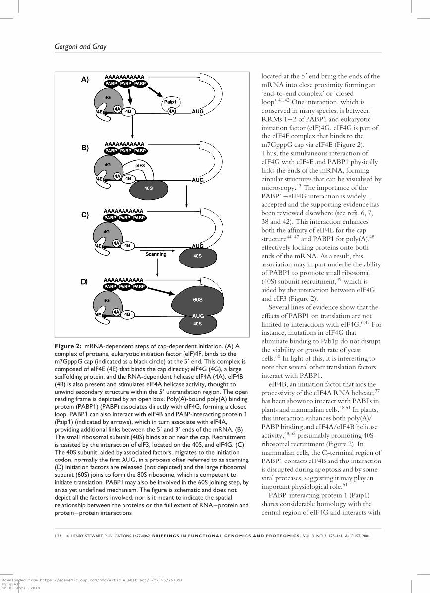
PABP1 IN TRANSLATIONINITIATIONTranslation initiation is a complex event
that ends in the formation of a ribosome
competent to begin elongation at the
initiation codon. It has been described in
detail elsewhere (see ref. 37). A simplified
version of the mRNA-dependent steps
highlighting the factors discussed below
can been seen in Figure 2. While the
poly(A) tail is not absolutely required for
translation,38 polyadenylated mRNAs are
translated with a much greater efficiency
and it is now generally accepted that the
poly(A) tail functions synergistically with
the 59 cap to promote the initiation of
translation.39,40
The function of PABP1 in translation
has been investigated in many species,
both in vivo and in vitro, resulting in a
model in which interactions between
PABP1 on the poly(A) tail and factors
Many cell types appearto express differentPABPs but it is unclearwhether these proteinsare functionallyredundant
Interactions betweenPABP1 on the poly(A)tail and factors locatedat the 59 end bring theends of the mRNA intoclose proximity formingan ‘end-to-end complex’or ‘closed loop’
& HENRY STEWART PUBLICATIONS 1477-4062. BR IEF INGS IN FUNCTIONAL GENOMICS AND PROTEOMICS . VOL 3. NO 2. 125–141. AUGUST 2004 1 2 7
The roles of cytoplasmic poly(A)-binding proteins in regulating gene expression
Downloaded from https://academic.oup.com/bfg/article-abstract/3/2/125/251394by gueston 03 April 2018
located at the 59 end bring the ends of the
mRNA into close proximity forming an
‘end-to-end complex’ or ‘closed
loop’.41,42 One interaction, which is
conserved in many species, is between
RRMs 1�2 of PABP1 and eukaryotic
initiation factor (eIF)4G. eIF4G is part of
the eIF4F complex that binds to the
m7GpppG cap via eIF4E (Figure 2).
Thus, the simultaneous interaction of
eIF4G with eIF4E and PABP1 physically
links the ends of the mRNA, forming
circular structures that can be visualised by
microscopy.43 The importance of the
PABP1�eIF4G interaction is widely
accepted and the supporting evidence has
been reviewed elsewhere (see refs. 6, 7,
38 and 42). This interaction enhances
both the affinity of eIF4E for the cap
structure44–47 and PABP1 for poly(A),48
effectively locking proteins onto both
ends of the mRNA. As a result, this
association may in part underlie the ability
of PABP1 to promote small ribosomal
(40S) subunit recruitment,49 which is
aided by the interaction between eIF4G
and eIF3 (Figure 2).
Several lines of evidence show that the
effects of PABP1 on translation are not
limited to interactions with eIF4G.6,42 For
instance, mutations in eIF4G that
eliminate binding to Pab1p do not disrupt
the viability or growth rate of yeast
cells.50 In light of this, it is interesting to
note that several other translation factors
interact with PABP1.
eIF4B, an initiation factor that aids the
processivity of the eIF4A RNA helicase,37
has been shown to interact with PABPs in
plants and mammalian cells.48,51 In plants,
this interaction enhances both poly(A)/
PABP binding and eIF4A/eIF4B helicase
activity,48,52 presumably promoting 40S
ribosomal recruitment (Figure 2). In
mammalian cells, the C-terminal region of
PABP1 contacts eIF4B and this interaction
is disrupted during apoptosis and by some
viral proteases, suggesting it may play an
important physiological role.51
PABP-interacting protein 1 (Paip1)
shares considerable homology with the
central region of eIF4G and interacts with
Figure 2: mRNA-dependent steps of cap-dependent initiation. (A) Acomplex of proteins, eukaryotic initiation factor (eIF)4F, binds to them7GpppG cap (indicated as a black circle) at the 59 end. This complex iscomposed of eIF4E (4E) that binds the cap directly; eIF4G (4G), a largescaffolding protein; and the RNA-dependent helicase eIF4A (4A). eIF4B(4B) is also present and stimulates eIF4A helicase activity, thought tounwind secondary structure within the 59 untranslation region. The openreading frame is depicted by an open box. Poly(A)-bound poly(A) bindingprotein (PABP1) (PABP) associates directly with eIF4G, forming a closedloop. PABP1 can also interact with eIF4B and PABP-interacting protein 1(Paip1) (indicated by arrows), which in turn associate with eIF4A,providing additional links between the 59 and 39 ends of the mRNA. (B)The small ribosomal subunit (40S) binds at or near the cap. Recruitmentis assisted by the interaction of eIF3, located on the 40S, and eIF4G. (C)The 40S subunit, aided by associated factors, migrates to the initiationcodon, normally the first AUG, in a process often referred to as scanning.(D) Initiation factors are released (not depicted) and the large ribosomalsubunit (60S) joins to form the 80S ribosome, which is competent toinitiate translation. PABP1 may also be involved in the 60S joining step, byan as yet undefined mechanism. The figure is schematic and does notdepict all the factors involved, nor is it meant to indicate the spatialrelationship between the proteins or the full extent of RNA�protein andprotein�protein interactions
1 2 8 & HENRY STEWART PUBLICATIONS 1477-4062. BRIEF INGS IN FUNCTIONAL GENOMICS AND PROTEOMICS . VOL 3. NO 2. 125–141. AUGUST 2004
Gorgoni and Gray
Downloaded from https://academic.oup.com/bfg/article-abstract/3/2/125/251394by gueston 03 April 2018

PABP1 via RRMs 1�2 and PABC,24,25
at a 1:1 stochiometry.25 Paip1, like
eIF4G, interacts with eIF4A (Figure 2)
but, by contrast, does not contact eIF4E,
and an interaction with eIF3 remains to
be proven.53 Thus, a role for Paip1 in
PABP1-mediated translation is less clear
than for eIF4G.24,53
Several lines of evidence suggest that
other factors may also promote PABP1-
mediated translation. First, RRMs 3�4 of
PABP1 are sufficient to stimulate
translation in tethered function assays
even though they are not implicated in
binding any of the factors discussed.24
Moreover, effects of PABP/poly(A) on
60S ribosomal subunit joining have also
been reported.54,55 Future work will be
required to understand further the factors
and mechanisms involved and whether
the different factors are utilised in
different cell types.
LINKS BETWEENTRANSLATIONTERMINATION AND PABP1The PABC domain can interact with
eRF3 in both yeast and mammals.26–28
eRF3 is a GTPase that enhances the
activity of eRF1, which catalyses
translation termination.56 The eRF3/
PABP1 interaction may promote
recycling of terminating ribosomes from
the 39 to 59 end, facilitating multiple
rounds of initiation on an mRNA.28
Alternatively, it may link translation to
mRNA decay, as eRF3 appears to
interfere with the ability of PABP1 to
multimerise on poly(A),26 potentially
leading to PABP1 dissociation,
deadenylation and, ultimately, turnover.
PABP1 IN mRNA STABILITYPABPs also play an important role in
mRNA stability. The majority of mRNA
decay studies have been carried out in
yeast, although the pathways appear to be
conserved in higher eukaryotes.57,58
Consequently, the roles of PABP1 in
mRNA stability are less defined than in
translation. Nonetheless, several lines of
evidence suggest that PABP1 can
influence both the deadenylation-
dependent decapping pathway and
exosome-mediated 39!59 degradation.58
Deadenylation appears to be the initial
step in both these decay pathways and, in
many cases, seems to be rate limiting.58,59
PABP1 can protect mRNAs from
deadenylation by inhibiting the action of
deadenylases such as poly(A) ribonuclease
(PARN),60–64 and incremental
deadenylation products equivalent to the
size of PABP footprints can be
detected.62,65
One simple model for explaining the
role of PABP1 in protecting mRNAs
from decay is the formation of the
eIF4E�eIF4G�PABP1 complex (Figure
2). By linking proteins tightly to both
ends of the mRNA, this complex can
simultaneously prevent the access of
deadenylases and can prevent decapping.
As discussed earlier, eRF3 may play a role
in disrupting these complexes, linking
translation to decay.26 Once translation
has ceased, PABP1 has been proposed to
protect mRNAs independently of eIF4E
by binding directly to the 59 cap;66,67
however, in vivo clarification of this latter
observation awaits. PABP1 may,
therefore, have additional functions in
stability, influencing steps subsequent to
deadenylation, such as remodelling of
mRNPs,6,7 and may even promote the
activity of certain deadenylases.68 A more
detailed knowledge of the factors that
mediate decay in vertebrate cells may first
be required in order to gain a fuller
insight into the role of PABP1 in mRNA
stability.
PABPs may also be involved in
regulating mRNAs containing specific
sequences that control their stability, such
as AU-rich elements (AREs) and mCRD
elements (see later, c-fos). AREs can be
bound by stabilising or destabilising
factors.69 Some destabilising factors
recruit components of the decay
machinery such as the exosome and
PARN;70-72 however, others might
function by disrupting interactions
between PABP1 and the poly(A) tail or
factors at the 59 end. Interestingly, some
PABPs may alsoregulate the stability ofspecific mRNAs
PABP1 may influenceboth the deadenylation-dependent decappingpathway and exosome-mediated 39 to 59degradation
& HENRY STEWART PUBLICATIONS 1477-4062. BR IEF INGS IN FUNCTIONAL GENOMICS AND PROTEOMICS . VOL 3. NO 2. 125–141. AUGUST 2004 1 2 9
The roles of cytoplasmic poly(A)-binding proteins in regulating gene expression
Downloaded from https://academic.oup.com/bfg/article-abstract/3/2/125/251394by gueston 03 April 2018
PABPs appear to bind AU-rich
sequences, suggesting a more direct role
in the stability of these mRNAs,8,20
although it is not clear whether they can
discriminate between AREs and other
AU-containing RNAs.19 In a few cases,
PABP1 has also been implicated in the
regulation of endonucleolytic cleavage —
a pathway that is responsible for the
degradation of a relatively small number
of mRNAs (see later, Æ–globin) —although, in most cases, cleavage is
independent of poly(A)/PABP function
(eg see ref. 73).
REGULATED EXPRESSIONOF PABP PROTEINSPABP1 mRNA is expressed at different
levels in many tissues (eg see ref. 32);
furthermore, the expression of PABP1
protein is tightly regulated by two
independent mechanisms. A 59-terminal
oligopyrimidine tract (59-TOP) controls
expression in response to cell growth.74
TOPs are also present in other
components of the translational
machinery and allow for coordinated
growth regulation (reviewed in ref. 75).
Additionally, PABP1 autoregulates its
own mRNA by binding an A-rich
sequence within its 59UTR,76,77 leading
to repression when PABP1 levels are high.
The translation of other PABP
mRNAs may also be subject to
autoregulation. Some iPABP
complementary DNA (cDNA) sequences
appear to have an A-rich stretch in their
59 UTR.78 Interestingly, iPABP seems to
have more than one promoter,79
suggesting that transcription from
alternate promoters may allow differential
regulation by generating a subset of
mRNAs without the poly(A) stretch.
tPABP mRNAs do not appear to undergo
autoregulation,31 but polysome analysis in
mouse suggests that they may be subject
to translational control by an as yet
unidentified mechanism.32
Database analysis of cDNAs reveals
potential alternative splice variants that
alter the protein-coding region of iPABP
and PABP1, often in the proline-rich
region.78 This raises the possibility that
additional PABP isoforms may be
generated by alternative splicing, further
increasing the complexity of PABPs
produced. Future work will be required
to elucidate whether these isoforms are
expressed, and, if they are, what their
importance is during development.
REGULATION OF PABPPROTEINSProtein modificationPABP proteins can be subject to both
phosphorylation and methylation.
Phosphorylation of translation factors,
including eIF4E and eIF2, is a well-
established means of controlling global
translation (reviewed in refs. 80�82).
Multiple phosphorylation forms of PABPs
have been described in plants, yeast and
sea urchins,83,84 but the functional effects
of these phosphorylation events are best
understood in plants. Phosphorylation of
wheat germ PABP enhances its
cooperative binding to poly(A),85
suggesting that its ability to homodimerise
is increased. Moreover, the ability of
eIF4G and eIF4B to promote PABP/
poly(A) interactions appears to be
differentially influenced by the
phosphorylation state of PABP.85
Strong evidence for phosphorylation of
PABPs in vertebrates is still lacking;
however, serum stimulation, which
activates a variety of kinase pathways,
enhances the PABP1�eIF4G interaction
in cultured Xenopus kidney cells86 and
human PABP1 can be phosphorylated by
the p38 mitogen-activated protein kinase
pathway in vitro.20 While phosphorylation
of PABPs has not been extensively
analysed during development, a
preliminary analysis in Xenopus eggs
suggests that neither PABP1 nor ePABP is
phosphorylated.36
PABP1 is also a substrate for
coactivator-associated arginine
methyltransferase (CARM1), which
methylates arginines in the proline-rich
linker region in vitro and in HeLa cells.87
The function of this modification is
unclear but it may affect
Additional PABPisoforms may begenerated byalternative splicing,further increasing thecomplexity of PABPproteins
PABP proteins can besubject to bothphosphorylation andmethylation
Expression of PABP1protein is tightlyregulated
1 3 0 & HENRY STEWART PUBLICATIONS 1477-4062. BRIEF INGS IN FUNCTIONAL GENOMICS AND PROTEOMICS . VOL 3. NO 2. 125–141. AUGUST 2004
Gorgoni and Gray
Downloaded from https://academic.oup.com/bfg/article-abstract/3/2/125/251394by gueston 03 April 2018

homodimerisation, RNA or protein
interactions or even its intracellular
localisation.
Protein regulatorsOther proteins can also affect the activity
and functions of PABP proteins. While
this form of regulation was first identified
in viruses, cellular proteins that utilise this
strategy have now been identified.
Rotavirus non structural protein 3
(NSP3) was the first protein shown to
modulate PABP1 activity by competing
for binding to eIF4G. This disrupts the
PABP1�eIF4G interaction and excludes
adenylated cellular mRNAs from
translation.88 Interestingly, NSP3 is
required for the translation of viral
mRNAs as it binds their non-adenylated
39 end, leading to circularisation by
mimicking PABP1 function.89
The cellular protein Paip2 acts as a
negative regulator of PABP1 by binding
to sites within RRMs 2�3 and PABC,29
inhibiting its interaction with poly(A) and
Paip1.90 A recent study points to a role
for Paip2 in development, showing that
Drosophila Paip2 inhibits cell growth in
various tissues in larvae and adults by
inhibiting translation.91 Interestingly,
recent reports also suggest that Paip2 may
have an additional role in mRNA
stability.92,93
PABPs may also be subject to
proteolytic cleavage during viral
infections and apoptosis. This has been
characterised in enteroviruses where
protease 3Cpro cleaves PABP1 at three
sites within the proline-rich linker region.
Cleavage releases the PABC domain from
the RRMs,51,94 leading to the inhibition
of poly(A)-dependent translation.95 Since
cleaved PABP1 retains its interaction with
eIF4G and with the poly(A) tail, these
observations confirm the importance of
interaction with other factors or with
itself in PABP-mediated translational
stimulation.
During apoptosis, caspase-mediated
cleavage of several translation factors
correlates with a partial inhibition of
translation (reviewed in ref. 96). Cleavage
of eIF4G and eIF4B disrupts their
interaction with PABP1 and formation of
the end-to-end complex. PABP1
degradation during apoptosis has also
recently been suggested, although it
appears not to be caspase mediated97 and
its relative contribution to translational
inhibition during apoptosis remains to be
determined.
CYTOPLASMIC POLY(A)TAIL LENGTH CHANGES INDEVELOPMENTIn animal cells, mRNAs initially receive a
poly(A) tail of between 200 and 250
nucleotides in the nucleus. Upon entry to
the cytoplasm, the poly(A) tail is normally
slowly removed and this eventually signals
the mRNA for degradation. During early
development, transcription is often
quiescent; thus, changes in the pattern of
protein synthesis rely on the activation,
repression or destruction of pre-existing
maternal mRNAs.98 These changes are
often accompanied by dramatic changes
in poly(A) tail length which occur in the
cytoplasm.38,99 In general, mRNAs are
stored with short poly(A) tails and are
polyadenylated concomitantly with their
translational activation, while
deadenylation is associated with
translational silencing.
Cytoplasmic polyadenylation requires
specific 39 UTR sequences that have been
defined during oocyte maturation
(cytoplasmic polyadenylation elements
[CPEs]100,101 or adenylation control
elements [ACEs]102) and embryogenesis
(embryonic CPEs).103,104 The importance
of cytoplasmic polyadenylation has been
demonstrated in a number of species.
Gene inactivation of mouse CPE-binding
protein-1 (CPEB-1) or testis poly(A)
polymerase (tPAP), which promote
cytoplasmic polyadenylation, blocks
oogenesis and/or spermatogenesis.105,106
Elegant experiments also underline the
important role of polyadenylating
individual mRNAs during development.
For instance, polyadenylation of c-mos in
Xenopus and mouse is essential for oocyte
maturation.107,108 Similarly, in embryos,
Paip2 acts as a negativeregulator of PABP1 byinhibiting its interactionwith poly(A) and Paip1
During earlydevelopment, mRNAsare stored with shortpoly(A) tails and arepolyadenylatedconcomitantly withtheir translationalactivation
PABPs may be subjectto proteolytic cleavageduring viral infectionsand apoptosis
& HENRY STEWART PUBLICATIONS 1477-4062. BR IEF INGS IN FUNCTIONAL GENOMICS AND PROTEOMICS . VOL 3. NO 2. 125–141. AUGUST 2004 1 3 1
The roles of cytoplasmic poly(A)-binding proteins in regulating gene expression
Downloaded from https://academic.oup.com/bfg/article-abstract/3/2/125/251394by gueston 03 April 2018

polyadenylation of bicoid is required for
the establishment of anterior�posterior
axis formation in Drosophila,109 and
polyadenylation of activin mRNA is
required for mesoderm formation in
Xenopus.110
It is widely accepted that the function
of the poly(A) tail is to bind PABP
proteins to mRNAs; however, the
mechanism by which cytoplasmic
polyadenylation promotes translation is
less clear.38 In some cases, it may enhance
the translation of mRNAs that are already
being translated at a low level,or it may
activate the translation of silent
mRNAs.38,98 This may be, in part,
determined by the PABP status of the
poly(A) tails. For instance, PABPs may be
bound to the short poly(A) tails and the
role of polyadenylation may be to recruit
additional PABP molecules. Alternatively,
the short tails may be devoid of PABPs
either because PABP binding is blocked
or because only long poly(A) tails are
sufficient to compete for limited amounts
of PABPs.
An additional layer of complexity arises
because the length of polyadenylation
varies between mRNAs. For instance,
polyadenylation of cyclin B1 and c-mos
results in an increase from 30 to 250 and
from 50 to 120 residues, respectively.111
The extent to which polyadenylation
affects the translation of individual
mRNAs is also determined by other
factors.38 First, the basal translatability of
mRNAs may be important, such that
poorly translated mRNAs may be
stimulated to the highest degree by
PABPs. In support of this, mRNAs
containing structured 59 UTRs were
found to be more sensitive to levels of
PABP1 than mRNAs containing
unstructured 59 ends.112 Secondly, the
presence of repressor proteins can
influence the apparent effects of
polyadenylation.38 Several models to
explain the relationship between
polyadenylation and 39 UTR repressors
exist.38,113 For instance, loss of a repressor
protein and polyadenylation may occur
independently and, while the two events
are not linked, their additive effects are
responsible for the overall change in
translation. Alternatively, the repressor
protein may exert a dominant effect over
the poly(A) tail by preventing the binding
of PABPs or by interfering with their
function. Loss of the repressor would
allow the poly(A) tail to participate in
end-to-end complexes. Finally,
polyadenylation may precede and be
required for the loss of the repressor
protein (see later, maskin).
Thus, the effects of poly(A)/PABPs on
the translation of mRNAs during early
development are complex and need to be
defined for individual mRNAs.
Cytoplasmic polyadenylation also occurs
in other cell types, such as neurones,114,115
and studies of early development provide
a paradigm for understanding its function.
During development, subsets of
mRNAs undergo deadenylation at
specific times.99 This process is linked to
translational silencing, since PABP1
overexpression prevents both silencing
and deadenylation.64 While deadenylation
has been studied in a number of species,
including Drosophila116 and mice,102 it has
perhaps been most extensively
characterised in Xenopus.117 During
oocyte maturation, mRNAs that lack a
CPE, such as ribosomal proteins and
actin,118,119 undergo deadenylation. This
is termed ‘default deadenylation’ since
specific sequences are not required and it
is, in part, promoted by the release of
PARN from the nucleus.120 In contrast to
deadenylation in somatic cells, mRNAs
that undergo default deadenylation can
remain stable and be degraded later in
development.117 It is unclear whether
deadenylation causes translational
silencing or whether silencing by an
unknown mechanism initiates
deadenylation. In the latter scenario,
deadenylation would promote loss of
PABP proteins, resulting in further
silencing and deadenylation.
A second type of deadenylation
requires specific cis-acting sequences and
has been best characterised in the early
embryo.99 Several elements have been
The mechanism bywhich cytoplasmicpolyadenylationpromotes translation isnot clear
During development,subsets of mRNAsundergo deadenylationand translationalsilencing at specifictimes
Polyadenylation doesnot affect thetranslation of allmRNAs equally
1 3 2 & HENRY STEWART PUBLICATIONS 1477-4062. BRIEF INGS IN FUNCTIONAL GENOMICS AND PROTEOMICS . VOL 3. NO 2. 125–141. AUGUST 2004
Gorgoni and Gray
Downloaded from https://academic.oup.com/bfg/article-abstract/3/2/125/251394by gueston 03 April 2018

identified and appear to recruit different
39 UTR-binding proteins.117 Two main
models have been proposed to explain
how these elements function.117 In the
first, 39 UTR-binding proteins recruit or
promote the activity of a deadenylase
other than PARN.8,117,121 In the second
model, binding of the regulatory protein
leads to translational repression and this,
in turn, leads to deadenylation. While the
role of PABP proteins in mRNA specific
deadenylation may be primarily to protect
the poly(A) tail, they may also have a
more active role, since ePABP was first
identified as a protein that can bind to
ARE motifs.8
REGULATION OF SPECIFICmRNAs BY PABPsIn addition to their roles in translation
described above, PABP proteins are also
involved in the regulation of specific
mRNAs. Few cases have been
documented; however, the mechanisms
described suggest a more widespread role
for PABP as a specific regulator.
One well-studied example is cyclin B1,
which is part of the maturation-
promoting factor (MPF) required for
oocyte maturation in Xenopus. Prior to
maturation, cyclin B1 mRNA has a short
poly(A) tail and is maintained in a
translationally repressed state by a
complex of proteins. Surprisingly,
repression requires CPE elements and
CPEB,122,123 which later direct
polyadenylation. CPEB at the 39 end and
eIF4E at the 59 end are bound in an
inactive complex by the bridging protein
maskin (Figure 3).124 This prevents the
association of eIF4E and eIF4G, blocking
translation initiation.124 Upon
progesterone stimulation, cyclin B1
mRNA undergoes cytoplasmic
polyadenylation,111,125 leading to the
increased binding of PABPs and
eIF4G.126 The PABP�eIF4G complex
displaces maskin from eIF4E, allowing the
initiation of translation. More recent
work suggests that Xenopus Pumilio may
also be involved in repression of cyclin B1
in oocytes by interacting with CPEB to
block polyadenylation.127
Repression of cyclin B1mRNA is overcomewhen the PABP–e1F4Gcomplex displaces therepressor maskin frome1F4E
Figure 3: Model of maskin-mediated repression/derepression. (A) In oocytes, cyclin B1mRNA has a relatively short poly(A) tail and is translationally silent. The cytoplasmicpolyadenylation location in the 39 UTR recruits CPE binding protein (CPEB), which associateswith maskin. Maskin, in turn, interacts with eukaryotic initiation factor (eIF)4E, precluding itsaccess to eIF4G and inhibiting the formation of the initiation complex. The cleavage andpolyadenylation specificity factor (CPSF) may be loosely associated with its target elementAAUAAA and with CPEB. The poly(A) tail may bind a limited number of poly(A)-bindingproteins (PABPs). For simplicity, the role of Pumilio is not depicted. (B) During oocytematuration, the kinase aurora (also known as Eg2) is activated and phosphorylates (-P) CPEB.This leads to stabilisation of the CPEB/CPSF complex and recruitment of poly(A) polymerase(PAP) to the end of the mRNA where it directs polyadenylation. Subsequently, the elongatedpoly(A) tail recruits one or more PABP molecules, which in turn associates with eIF4G. PABP-bound eIF4G displaces maskin from eIF4E and translation is enhanced. The figure is schematicand is not meant to indicate the spatial relationship between the proteins or the full extent ofRNA�protein and protein�protein interactions
& HENRY STEWART PUBLICATIONS 1477-4062. BR IEF INGS IN FUNCTIONAL GENOMICS AND PROTEOMICS . VOL 3. NO 2. 125–141. AUGUST 2004 1 3 3
The roles of cytoplasmic poly(A)-binding proteins in regulating gene expression
Downloaded from https://academic.oup.com/bfg/article-abstract/3/2/125/251394by gueston 03 April 2018

Translational repression by maskin also
occurs during early embryogenesis, where
cyclin B1 synthesis is tightly regulated by
polyadenylation to ensure cell-cycle
progression.128 Similarly, in neuronal
dendrites, CPEB and maskin are
suggested to repress mRNAs such as ÆCa2þ-calmodulin-dependent protein
kinase II (ÆCAMKII) prior to synaptic
activation.129,130 Consequently, PABPs
may mediate relief of repression in these
as well as in other cells.
PABP proteins do not always seem to
perform their function through the
poly(A) tail.24,131 For instance, in vitro
experiments suggest that translation of
YB-1 mRNA, encoding a cold-shock
domain-containing protein, is enhanced
by the binding of PABP1 to an A-rich
sequence within its 39 UTR,132
presumably by promoting end-to-end
complexes.
By contrast, when PABP1 is bound to
an A-rich sequence within its own 59
UTR, it acts as a repressor by stalling the
migration of the 40S subunit.133
Repression requires the proline-rich
region21 implicated in homodimerisation
and cooperative binding to poly(A),
raising the possibility that PABP1 must
multimerise on the 59 UTR to impede
ribosome scanning.
PABP1 has also been implicated in the
repression of other mRNAs, such as the
neuropeptide vasopressin, prior to
dendrite activation. PABP1 associates
with a dendritic localiser sequence (DLS)
that spans part of the coding region and
the 39 UTR of this mRNA.134 DLS-
bound PABP1 is proposed to interfere
with PABP1 molecules on the poly(A)
tail, either directly or indirectly, inhibiting
the formation of the end-to-end complex
(reviewed in ref. 135) by an undefined
mechanism.
The role of PABPs in regulating
specific mRNAs is not only limited to
translation but it is also important in
stability. This appears to be the case
with Æ-globin mRNA, which is
stabilised by the binding of Æ complex
protein (ÆCP)1 (heterogenous nuclear,
ribonucleoprotein (hnRNP) E1) and ÆCP2 (hnRNP E2) proteins to its 39
UTR.136 Interaction of ÆCP1 and
ÆCP2 with PABP1 leads to an increase
in their affinity for the mRNA,
precluding access to an endonuclease
(ErEn).137 ÆCP1 and ÆCP2 can, in turn,
stabilise the association of PABP1 with
the poly(A) tail, protecting the transcript
from deadenylation.137 Thus, PABP1
simultaneously protects Æ-globin mRNA
from both endonucleolytic cleavage and
39!59 degradation.
Stability of c-fos mRNA, which
encodes a transcription factor, is also
modulated by PABP1. This transcript
contains a major protein-coding region
determinant of instability (mCRD) in the
ORF that directs accelerated
deadenylation prior to degradation.138
The mCRD interacts with a protein
complex that contains PABP1, Paip1,
hnRNPD, NS1-associated protein 1
(NSAP1) and upstream of N-ras (Unr),
which is proposed to bring the mCRD
and poly(A) tail in to close proximity,
preventing deadenylation by stabilising
the PABP/poly(A)tail interaction.139
During translation, ribosomal movement
across the mCRD displaces or reorganises
the complex, and is proposed to
destabilise PABP1 binding to the poly(A)
tail, exposing the 39 end to nuclease
attack.
PERSPECTIVESGiven the wide variety of roles played by
PABPs in regulating specific mRNAs, it is
expected that additional examples and
mechanisms will continue to be
uncovered. This is supported by several
observations. First, several PABP proteins
can bind AU-rich sequences8,19,20 and the
affinity for AU of one of these, iPABP, is
only 2-fold lower than its affinity for
poly(A), suggesting that some PABP
proteins may regulate mRNAs containing
AU-rich sequences. Secondly, tethering
PABP1 to the 39 UTR promotes
translation,24 raising the possibility that
indirect recruitment of PABPs to the
mRNA by PABP-binding proteins could
PABP1 can stabilise Æ-globin mRNA bysimultaneouslyprotecting it fromendonucleolyticcleavage and 39 to 59degradation
PAB1 binding to an A-rich sequence within itsown 59 UTR causesrepression of PABP1translation
1 3 4 & HENRY STEWART PUBLICATIONS 1477-4062. BRIEF INGS IN FUNCTIONAL GENOMICS AND PROTEOMICS . VOL 3. NO 2. 125–141. AUGUST 2004
Gorgoni and Gray
Downloaded from https://academic.oup.com/bfg/article-abstract/3/2/125/251394by gueston 03 April 2018

be a novel way to control translation.
Lastly, an extensive list of proteins
containing the PABP-interaction motif 2
(PAM2), known to bind the PABC
domain, has recently been published.140
Many of these proteins do not have a
described role in translation or stability, or
even a proven interaction with PABP.
Nonetheless, it is appealing to envisage
that additional factors may modulate
PABP’s function in specific
developmental and environmental
situations, by previously described or
novel mechanisms.
Moreover, fundamental issues in
understanding the function of PABP
proteins still remain. While the formation
of end-to-end complexes clearly seems
important, as yet researchers do not
appear to have identified all the
components, or to understand their
relative contributions or how complex
formation is regulated. Likewise, the
function of PABP proteins, other than
PABP1, remains to be directly addressed,
as does their contribution to
development. The importance of
elucidating the respective roles and
functions of each family member is
underlined by the recent implication of
two family members, PABPN17 and
PABP5,35 in human disease. Research in
the next few years is likely to focus on
these unresolved issues and may reveal
novel roles in regulating gene expression
or even in other biological processes.
Acknowledgments
We apologise to our colleagues whose work we
could not include due to space constraints. We
thank Brian Collier, Kris Dickson, Tom Van
Agtmael and Gavin Wilkie for critical reading of
the manuscript and Sandy Bruce for the
preparation of figures.
References
1. Sachs, A. B., Davis, R. W. and Kornberg,R. B. (1987), ‘A single domain of yeastpoly(A)-binding protein is necessary andsufficient for RNA binding and cell viability’,Mol. Cell. Biol., Vol. 7, pp. 3268–3276.
2. Gorlach, M., Burd, C. G. and Dreyfuss, G.(1994), ‘The mRNA poly(A)-bindingprotein: localization, abundance, and RNA-
binding specificity’, Exp. Cell Res., Vol. 211,pp. 400–407.
3. Kuhn, U. and Pieler, T. (1996), ‘Xenopuspoly(A) binding protein: functional domainsin RNA binding and protein-proteininteraction’, J. Mol. Biol., Vol. 256,pp. 20–30.
4. Baer, B. W. and Kornberg, R. D. (1980),‘Repeating structure of cytoplasmic poly(A)-ribonucleoprotein’, Proc. Natl. Acad. Sci.USA, Vol. 77, pp. 1890–1892.
5. Baer, B. W. and Kornberg, R. D. (1983),‘The protein responsible for the repeatingstructure of cytoplasmic poly (A)-ribonucleoprotein’, J. Cell Biol., Vol. 96, pp.717–721.
6. Mangus, D. A., Evans, M. C. and Jacobson,A. (2003), ‘Poly(A)-binding proteins:multifunctional scaffolds for the post-transcriptional control of gene expression’,Genome Biol., Vol. 4, p. 223.
7. Kuhn, U. and Wahle, E. (2004), ‘Structureand function of poly(A) binding proteins’,Biochim. Biophys. Acta, Vol. 1678, pp. 67–84.
8. Voeltz, G. K., Ongkasuwan, J., Standart, N.et al. (2001), ‘A novel embryonic poly(A)binding protein, ePAB, regulates mRNAdeadenylation in Xenopus egg extracts’, GenesDev., Vol. 15, pp. 774–788.
9. Good, P. J., Abler, L., Herring, D. andSheets, M. D. (2004), ‘Xenopus embryonicpoly(A) binding protein 2 (ePABP2) defines anew family of cytoplasmic poly(A) bindingproteins expressed during the early stages ofvertebrate development’, Genesis, Vol. 38,pp. 166–175.
10. Fasulo, B., Becattini, R., Cenci, G. et al.(1999), ‘Doppio fuso (duo), a gene requiredfor spindle pole assembly during Drosophilamale meiosis’, Flybase, URL:http://fly.ebi.ac.uk:7081/.
11. Sigrist, S. J., Thiel, P. R., Reiff, D. F. et al.(2000), ‘Postsynaptic translation affects theefficacy and morphology of neuromuscularjunctions’, Nature, Vol. 405, pp. 1062–1065.
12. Simmer, F., Moorman, C., van der Linden,A. M. et al. (2003), Wormbase,http://www.wormbase.org/.
13. Kamath, R. S., Fraser, A. G. and Ahringer,J. A. (2003), Wormbase,http://www.wormbase.org/.
14. Afonina, E., Stauber, R. and Pavlakis, G. N.(1998), ‘The human poly(A)-binding protein1 shuttles between the nucleus and thecytoplasm’, J. Biol. Chem., Vol. 273, pp.13015–13021.
15. Deo, R. C., Bonanno, J. B., Sonenberg, N.et al. (1999), ‘Recognition of polyadenylateRNA by the poly(A)-binding protein’, Cell,Vol. 98, pp. 835–845.
& HENRY STEWART PUBLICATIONS 1477-4062. BR IEF INGS IN FUNCTIONAL GENOMICS AND PROTEOMICS . VOL 3. NO 2. 125–141. AUGUST 2004 1 3 5
The roles of cytoplasmic poly(A)-binding proteins in regulating gene expression
Downloaded from https://academic.oup.com/bfg/article-abstract/3/2/125/251394by gueston 03 April 2018
16. Burd, C. G., Matunis, E. L. and Dreyfuss, G.(1991), ‘The multiple RNA-binding domainsof the mRNA poly(A)-binding protein havedifferent RNA-binding activities’, Mol. CellBiol., Vol. 11, pp. 3419–3424.
17. Nietfeld, W., Mentzel, H. and Pieler, T.(1990), ‘The Xenopus laevis poly(A) bindingprotein is composed of multiple functionallyindependent RNA binding domains’,EMBO J., Vol. 9, pp. 3699–3705.
18. Deardorff, J. A. and Sachs, A. B. (1997),‘Differential effects of aromatic and chargedresidue substitutions in the RNA bindingdomains of the yeast poly(A)-bindingprotein’, J. Mol. Biol., Vol. 269, pp. 67–81.
19. Sladic, R. T., Lagnado, C. A., Bagley, C. J.and Goodall, G. J. (2004), ‘Human PABPbinds AU-rich RNA via RNA-bindingdomains 3 and 4’, Eur. J. Biochem., Vol. 271,pp. 450–457.
20. Bollig, F., Winzen, R., Gaestel, M. et al.(2003), ‘Affinity purification of ARE-bindingproteins identifies polyA-binding protein 1 asa potential substrate in MK2-induced mRNAstabilization’, Biochem. Biophys. Res.Commun., Vol. 301, pp. 665–670.
21. Melo, E. O., Dhalia, R., Martins de Sa, C.et al. (2003), ‘Identification of a C-terminalpoly(A)-binding protein (PABP)-PABPinteraction domain: role in cooperativebinding to poly (A) and efficient cap distaltranslational repression’, J. Biol. Chem., Vol.278, pp. 46357–46368.
22. Kozlov, G., Trempe, J. F., Khaleghpour, K.et al. (2001), ‘Structure and function of theC-terminal PABC domain of humanpoly(A)-binding protein’, Proc. Natl. Acad.Sci. USA, Vol. 98, pp. 4409–4413.
23. Deo, R. C., Sonenberg, N. and Burley, S. K.(2001), ‘X-ray structure of the humanhyperplastic discs protein: an ortholog of theC-terminal domain of poly(A)-bindingprotein’, Proc. Natl. Acad. Sci. USA, Vol. 98,pp. 4414�94419.
24. Gray, N. K., Coller, J. M., Dickson, K. S.et al. (2000), ‘Multiple portions of poly(A)-binding protein stimulate translation in vivo’,EMBO J., Vol. 19, pp. 4723–4733.
25. Roy, G., De Crescenzo, G., Khaleghpour, K.et al. (2002), ‘Paip1 interacts with poly(A)binding protein through two independentbinding motifs’, Mol. Cell Biol., Vol. 22, pp.3769–3782.
26. Hoshino, S., Imai, M., Kobayashi, T. et al.(1999), ‘The eukaryotic polypeptide chainreleasing factor (eRF3/GSPT) carrying thetranslation termination signal to the 39-poly(A) tail of mRNA. Direct association oferf3/GSPT with polyadenylate-bindingprotein’, J. Biol. Chem., Vol. 274, pp.16677–16680.
27. Cosson, B., Berkova, N., Couturier, A. et al.(2002), ‘Poly(A)-binding protein and eRF3are associated in vivo in human and Xenopuscells’, Biol. Cell, Vol. 94, pp. 205–216.
28. Uchida, N., Hoshino, S. I., Imataka, H. et al.(2002), ‘A novel role of the mammalianGSPT/eRF3 associating with poly(A)-binding protein in cap/poly(A)-dependenttranslation’, J. Biol. Chem., Vol. 277, pp.50286–50292.
29. Khaleghpour, K., Kahvejian, A., DeCrescenzo, G. et al. (2001), ‘Dual interactionsof the translational repressor Paip2 withpoly(A) binding protein’, Mol. Cell Biol., Vol.21, pp. 5200–5213.
30. Kleene, K. C., Mulligan, E., Steiger, D. et al.(1998), ‘The mouse gene encoding the testis-specific isoform of poly(A) binding protein(Pabp2) is an expressed retroposon:intimations that gene expression inspermatogenic cells facilitates the creation ofnew genes’, J. Mol. Evol., Vol. 47, pp.275–281.
31. Feral, C., Guellaen, G. and Pawlak, A.(2001), ‘Human testis expresses a specificpoly(A)-binding protein’, Nucleic Acids Res.,Vol. 29, pp. 1872–1883.
32. Kleene, K. C., Wang, M. Y., Cutler, M. et al.(1994), ‘Developmental expression of poly(A)binding protein mRNAs duringspermatogenesis in the mouse’, Mol. Reprod.Dev., Vol. 39, pp. 355–364.
33. Yang, H., Duckett, C. S. and Lindsten, T.(1995), ‘iPABP, an inducible poly(A)-bindingprotein detected in activated human T cells’,Mol. Cell Biol., Vol. 15, pp. 6770–6776.
34. Houng, A. K., Maggini, L., Clement, C. Y.and Reed, G. L. (1997), ‘Identification andstructure of activated-platelet protein-1, aprotein with RNA-binding domain motifsthat is expressed by activated platelets’, Eur. J.Biochem., Vol. 243, pp. 209–218.
35. Blanco, P., Sargent, C. A., Boucher, C. A.et al. (2001), ‘A novel poly(A)-bindingprotein gene (PABPC5) maps to an X-specific subinterval in the Xq21.3/Yp11.2homology block of the human sexchromosomes’, Genomics, Vol. 74, pp. 1–11.
36. Cosson, B., Couturier, A., Le Guellec, R.et al. (2002), ‘Characterization of the poly(A)binding proteins expressed during oogenesisand early development of Xenopus laevis’, Biol.Cell, Vol. 94, pp. 217–231.
37. Hershey, J. W. B. and Merrick, W. C.(2000), ‘Pathway and mechanism of initiationof protein synthesis’, in Sonenberg, N.,Hershey, J. W. B. and Mathews, M. B. (Eds),‘Translational Control of Gene Expression’,Cold Spring Harbor Laboratory Press, NewYork, NY, pp. 33–88.
38. Gray, N. K. and Wickens, M. P. (1998),
1 3 6 & HENRY STEWART PUBLICATIONS 1477-4062. BRIEF INGS IN FUNCTIONAL GENOMICS AND PROTEOMICS . VOL 3. NO 2. 125–141. AUGUST 2004
Gorgoni and Gray
Downloaded from https://academic.oup.com/bfg/article-abstract/3/2/125/251394by gueston 03 April 2018

‘Control of translation initiation in animals’,Ann. Rev. Cell Dev. Biol., Vol. 14, pp.399–458.
39. Gallie, D. R. (1998), ‘A tale of two termini: afunctional interaction between the termini ofan mRNA is a prerequisite for efficienttranslation initiation’, Gene, Vol. 216,pp. 1–11.
40. Sachs, A. (2000), ‘Physical and functionalinteractions between the mRNA capstructure and the poly(A) tail’, in Sonenberg,N., Hershey, J. W. B. and Mathews, M. B.(Eds), ‘Translational Control of GeneExpression’, Cold Spring Harbor LaboratoryPress, New York, NY, pp. 447–466.
41. Jacobson, A. (1996), ‘Poly(A) metabolism andtranslation: the closed-loop model’ inHershey, J. W. B., Mathews, M. B. andSonenberg, N. (Eds), ‘Translational Control’,Cold Spring Harbor Laboratory Press, NewYork, NY, pp. 451–480.
42. Wilkie, G. S., Dickson, K. S. and Gray,N. K. (2003), ‘Regulation of mRNAtranslation by 59- and 39-UTR-bindingfactors’, Trends Biochem. Sci., Vol. 28, pp.182–188.
43. Wells, S. E., Hillner, P. E., Vale, R. D. et al.(1998), ‘Circularization of mRNA byeukaryotic translation initiation factors’,Mol.Cell, Vol. 2, pp. 135–140.
44. Haghihat, A. and Sonenberg, N. (1997),‘eIF4G dramatically enchances the binding ofeIF4E to the mRNA 5’-cap structure’, Biol.Chem., Vol. 272, pp. 21677–21680.
45. Ptushkina, M., von der Haar, T., Vasilescu, S.et al. (1998), ‘Cooperative modulation byeIF4G of eIF4E-binding to the mRNA 59cap in yeast involves a site partially shared byp20’, EMBO J., Vol. 17, pp. 4798–4808.
46. Borman, A. M., Michel, Y. M. and Kean,K. M. (2000), ‘Biochemical characterisationof cap-poly(A) synergy in rabbit reticulocytelysates: the eIF4G�PABP interactionincreases the functional affinity of eIF4E forthe capped mRNA 59-end’, Nucleic AcidsRes., Vol. 28, pp. 4068–4075.
47. Luo, Y. and Goss, D. J. (2001), ‘Homeostasisin mRNA initiation: wheat germ poly(A)-binding protein lowers the activation energybarrier to initiation complex formation’,J. Biol. Chem., Vol. 276, pp. 43083–43086.
48. Le, H., Tanguay, R. L., Balasta, M. L. et al.(1997), ‘Translation initiation factors eIF-iso4G and eIF-4B interact with the poly(A)-binding protein and increase its RNAbinding activity’, J. Biol. Chem., Vol. 272, pp.16247–16255.
49. Tarun, S. Z. and Sachs, A. B. (1995), ‘Acommon function for mRNA 59 and 39 endsin translation initiation in yeast’, Genes Dev.,Vol. 9, pp. 2997–3007.
50. Tarun, S. Z., Wells, S. E., Deardorff, J. A.et al. (1997), ‘Translation initiation factoreIF4G mediates in vitro poly(A) tail-dependent translation’, Proc. Natl. Acad. Sci.USA, Vol. 94, pp. 9046–9051.
51. Bushell, M., Wood, W., Carpenter, G. et al.(2001), ‘Disruption of the interaction ofmammalian protein synthesis eukaryoticinitiation factor 4B with the poly(A)-bindingprotein by caspase- and viral protease-mediated cleavages’, J. Biol. Chem., Vol. 276,pp. 23922–23928.
52. Bi, X. and Goss, D. J. (2000), ‘Wheat germpoly(A)-binding protein increases the ATPaseand the RNA helicase activity of translationinitiation factors eIF4A, eIF4B, and eIF-iso4F.’ J. Biol. Chem., Vol. 275, pp.17740–17746.
53. Craig, A. W., Haghighat, A., Yu, A. T. et al.(1998), ‘Interaction of polyadenylate-bindingprotein with the eIF4G homologue PAIPenhances translation’, Nature, Vol. 392, pp.520�9523.
54. Munroe, D. and Jacobson, A. (1990),‘mRNA poly (A) tail, a 39 enhancer oftranslational initiation’, Mol. Cell Biol., Vol.10, pp. 3441–3455.
55. Searfoss, A., Dever, T. E. and Wickner, R.(2001), ‘Linking the 39 poly(A) tail to thesubunit joining step of translation initiation:relations of Pab1p, eukaryotic translationinitiation factor 5b (Fun12p), and Ski2p-Slh1p’, Mol. Cell Biol., Vol. 21, pp.4900–4908.
56. Welch, E. M., Wang, W. and Peltz, S. W.(2000), ‘Translation termination: it’s not theend of the story’, in Sonenberg, N., Hershey,J. W. B. and Mathews, M. B. (Eds),‘Translational Control Of Gene Expression’,Cold Spring Harbor Laboratory Press, NewYork, NY, pp. 467–486.
57. Schwartz, D. C. and Parker, R. (2000),‘Interaction of mRNA translation andmRNA degradation in S. cerevisiae’, inSonenberg, N., Hershey, J. W. B. andMathews, M. B. (Eds), ‘Translational ControlOf Gene Expression’, Cold Spring HarborLaboratory Press, New York, NY,pp. 807–825.
58. Tourriere, H., Chebli, K. and Tazi, J. (2002),‘mRNA degradation machines in eukaryoticcells’, Biochimie, Vol. 84, pp. 821–837.
59. Ross, J. (1995), ‘mRNA stability inmammalian cells’, Microbiol. Rev., Vol. 59, pp.423–450.
60. Bernstein, P., Peltz, S. W. and Ross, J.(1989), ‘The poly(A)-poly(A)-bindingprotein complex is a major determinant ofmRNA stability in vitro’, Mol. Cell Biol.,Vol. 9, pp. 659–670.
61. Ford, L. P., Bagga, P. S. and Wilusz, J.
& HENRY STEWART PUBLICATIONS 1477-4062. BR IEF INGS IN FUNCTIONAL GENOMICS AND PROTEOMICS . VOL 3. NO 2. 125–141. AUGUST 2004 1 3 7
The roles of cytoplasmic poly(A)-binding proteins in regulating gene expression
Downloaded from https://academic.oup.com/bfg/article-abstract/3/2/125/251394by gueston 03 April 2018
(1997), ‘The poly(A) tail inhibits the assemblyof a 39-to-59 exonuclease in an in vitro RNAstability system’, Mol. Cell Biol., Vol. 17, pp.398–406.
62. Korner, C. G. and Wahle, E. (1997),‘Poly(A) tail shortening by a mammalianpoly(A)-specific 39-exoribonuclease’, J. Biol.Chem., Vol. 272, pp. 10448–10456.
63. Korner, C. G., Wormington, M.,Muckenthaler, M. et al. (1998), ‘Thedeadenylating nuclease (DAN) is involved inpoly(A) tail removal during the meioticmaturation of Xenopus oocytes’, EMBO J.,Vol. 17, pp. 5427–5437.
64. Wormington, M., Searfoss, A. M. andHurney, C. A. (1996), ‘Overexpression ofpoly(A) binding protein prevents maturation-specific deadenylation and translationalinactivation in Xenopus oocytes’, EMBO J.,Vol. 15, pp. 900–909.
65. Wang, Z., Day, N., Trifillis, P. and Kiledjian,M. (1999), ‘An mRNA stability complexfunctions with poly(A)-binding protein tostabilize mRNA in vitro’, Mol. Cell Biol., Vol.19, pp. 4552–4560.
66. Gao, M., Wilusz, C. J., Peltz, S. W. andWilusz, J. (2001), ‘A novel mRNA-decapping activity in HeLa cytoplasmicextracts is regulated by AU-rich elements’,EMBO J., Vol. 20, pp. 1134–1143.
67. Khanna, R. and Kiledjian, M. (2004),‘Poly(A)-binding-protein-mediatedregulation of hDcp2 decapping in vitro’,EMBO J., Vol. 23, pp. 1968–1976.
68. Uchida, N., Hoshino, S. and Katada, T.(2004), ‘Identification of a humancytoplasmic poly(A) nuclease complexstimulated by poly(A)-binding protein’,J. Biol. Chem., Vol. 279, pp. 1383–1391.
69. Bevilacqua, A., Ceriani, M. C., Capaccioli, S.et al. (2003), ‘Post-transcriptional regulationof gene expression by degradation ofmessenger RNAs’, J. Cell Physiol., Vol. 195,pp. 356–372.
70. Chen, C. Y., Gherzi, R., Ong, S. E. et al.(2001), ‘AU binding proteins recruit theexosome to degrade ARE-containingmRNAs’, Cell, Vol. 107, pp. 451–464.
71. Lai, W. S., Kennington, E. A. andBlackshear, P. J. (2003), ‘Tristetraprolin andits family members can promote the cell-freedeadenylation of AU-rich element-containing mRNAs by poly(A) ribonuclease’,Mol. Cell Biol., Vol. 23, pp. 3798–3812.
72. Gherzi, R., Lee, K. Y., Briata, P. et al.(2004), ‘A KH domain RNA bindingprotein, KSRP, promotes ARE-directedmRNA turnover by recruiting thedegradation machinery’, Mol. Cell, Vol. 14,pp. 571–583.
73. Binder, R., Horowitz, J. A., Basilion, J. P.
et al. (1994), ‘Evidence that the pathway oftransferrin receptor mRNA degradationinvolves an endonucleolytic cleavage withinthe 39 UTR and does not involve poly(A) tailshortening’, EMBO J., Vol. 13, pp. 1969–1980.
74. Hornstein, E., Git, A., Braunstein, I. et al.(1999), ‘The expression of poly(A)-bindingprotein gene is translationally regulated in agrowth-dependent fashion through a 59-terminal oligopyrimidine tract motif’, J. Biol.Chem., Vol. 274, pp. 1708–1714.
75. Meyuhas, O. and Hornstein, E. (2000),‘Translational control of TOP mRNAs’, inSonenberg, N., Hershey, J. W. B. andMathews, M. B. (Eds), ‘Translational Controlof Gene Expression’, Cold Spring HarborLaboratory Press, New York, NY,pp. 671–694.
76. Wu, J. and Bag, J. (1998), ‘Negative controlof the poly(A)-binding protein mRNAtranslation is mediated by the adenine-richregion of its 59-untranslated region’, J. Biol.Chem., Vol. 273, pp. 34535–34542.
77. De Melo Neto, O. P., Standart, N. andMartins De Sa, C. (1995), ‘Autoregulation ofpoly(A)-binding protein synthesis in vitro.’Nucleic Acids Res., Vol. 23, pp. 2198–2205.
78. Strausberg, R. L., Feingold, E. A., Grouse,L. H. et al. (2002), ‘Generation and initialanalysis of more than 15,000 full-lengthhuman and mouse cDNA sequences’, Proc.Natl. Acad. Sci. USA, Vol. 99, pp.16899–16903.
79. Thierry-Mieg, D., Thierry-Mieg, J.,Potdevin, M. et al. (2003), ‘Identification andfunctional annotation of cDNA-supportedgenes in higher organisms using AceView’,URL: http://www.ncbi.nlm.nih.gov/IEB/Research/Acembly.
80. Raught, B., Gingras, A. and Sonenberg, N.(2000), ‘Regulation of ribosomal recruitmentin eukaryotes’, in Sonenberg, N., Hershey, J.W. B. and Mathews, M. B. (Eds),‘Translational Control of Gene Expression’,Cold Spring Harbor Laboratory Press, NewYork, NY, pp. 245–293.
81. Scheper, G. C. and Proud, C. G. (2002),‘Does phosphorylation of the cap-bindingprotein eIF4E play a role in translationinitiation?’, Eur. J. Biochem., Vol. 269, pp.5350–5359.
82. Clemens, M. J. (2001), ‘Initiation factor eIF2alpha phosphorylation in stress responses andapoptosis’, Prog. Mol. Subcell. Biol., Vol. 27,pp. 57–89.
83. Drawbridge, J., Grainger, J. L. and Winkler,M. M. (1990), ‘Identification andcharacterization of the poly(A)-bindingproteins from the sea urchin: a quantitativeanalysis’, Mol. Cell Biol., Vol. 10, pp.3994–4006.
1 3 8 & HENRY STEWART PUBLICATIONS 1477-4062. BRIEF INGS IN FUNCTIONAL GENOMICS AND PROTEOMICS . VOL 3. NO 2. 125–141. AUGUST 2004
Gorgoni and Gray
Downloaded from https://academic.oup.com/bfg/article-abstract/3/2/125/251394by gueston 03 April 2018

84. Gallie, D. R., Le, H., Caldwell, C. et al.(1997), ‘The phosphorylation state oftranslation initiation factors is regulateddevelopmentally and following heat shock inwheat’, J. Biol. Chem., Vol. 272, pp.1046–1053.
85. Le, H., Browning, K. S. and Gallie, D. R.(2000), ‘The phosphorylation state ofpoly(A)-binding protein specifies its bindingto poly(A) RNA and its interaction witheukaryotic initiation factor (eIF) 4F, eIFiso4F,and eIF4B’, J. Biol. Chem., Vol. 275, pp.17452–17462.
86. Fraser, C. S., Pain, V. M. and Morley, S. J.(1999), ‘The association of initiation factor 4Fwith poly(A)-binding protein is enhanced inserum-stimulated Xenopus kidney cells’,J. Biol. Chem., Vol. 274, pp. 196–204.
87. Lee, J. and Bedford, M. T. (2002), ‘PABP1identified as an arginine methyltransferasesubstrate using high-density protein arrays’,EMBO Rep., Vol. 3, pp. 268–273.
88. Piron, M., Vende, P., Cohen, J. et al. (1998),‘Rotavirus RNA-binding protein NSP3interacts with eIF4GI and evicts the poly(A)binding protein from eIF4F’, EMBO J., Vol.17, pp. 5811–5821.
89. Vende, P., Piron, M., Castagne, N. et al.(2000), ‘Efficient translation of rotavirusmRNA requires simultaneous interaction ofNSP3 with the eukaryotic translationinitiation factor eIF4G and the mRNA 39end’, J. Virol., Vol. 74, pp. 7064–7071.
90. Khaleghpour, K., Svitkin, Y. V., Craig, A.W. et al. (2001), ‘Translational repression by anovel partner of human poly(A) bindingprotein, Paip2’, Mol. Cell, Vol. 7, pp.205–216.
91. Roy, G., Miron, M., Khaleghpour, K. et al.(2004), ‘The Drosophila poly(A) bindingprotein-interacting protein, dPaip2, is a noveleffector of cell growth’, Mol. Cell Biol., Vol.24, pp. 1143–1154.
92. Gouyon, F., Onesto, C., Dalet, V. et al.(2003), ‘Fructose modulates GLUT5 mRNAstability in differentiated Caco-2 cells: role ofcAMP-signalling pathway and PABP(polyadenylated-binding protein)-interactingprotein (Paip) 2’, Biochem. J., Vol. 375, pp.167–174.
93. Onesto, C., Berra, E., Grepin, R. et al. (inpress), ‘Poly (A) binding protein-interactingprotein 2, a strong regulator of vascularendothelial growth factor mRNA’, J. Biol.Chem.
94. Kuyumcu-Martinez, N. M., Joachims, M.and Lloyd, R. E. (2002), ‘Efficient cleavageof ribosome-associated poly(A)-bindingprotein by enterovirus 3C protease’, J. Virol.,Vol. 76, pp. 2062–2074.
95. Kuyumcu-Martinez, N. M., Van Eden, M.
E., Younan, P. et al. (2004), ‘Cleavage ofpoly(A)-binding protein by poliovirus 3Cprotease inhibits host cell translation: a novelmechanism for host translation shutoff’, Mol.Cell Biol., Vol. 24, pp. 1779–1790.
96. Clemens, M. J., Bushell, M., Jeffrey, I. W.et al. (2000), ‘Translation initiation factormodifications and the regulation of proteinsynthesis in apoptotic cells’, Cell. DeathDiffer., Vol. 7, pp. 603–615.
97. Marissen, W. E., Triyoso, D., Younan, P.et al. (2004), ‘Degradation of poly(A)-bindingprotein in apoptotic cells and linkage totranslation regulation’, Apoptosis, Vol. 9, pp.67–75.
98. Mendez, R. and Richter, J. D. (2001),‘Translational control by CPEB: a means tothe end’, Nat. Rev. Mol. Cell Biol., Vol. 2, pp.521–529.
99. Richter, J. D. (1996), ‘Dynamics of poly (A)addition and removal during development’,in Hershey, J. W. B., Mathews, M. B. andSonenberg, N. (Eds), ‘Translational Control’,Cold Spring Harbor Laboratory Press, NewYork, NY, pp. 481�9503.
100. Fox, C. A., Sheets, M. D. and Wickens,M. P. (1989), ‘Poly (A) addition duringmaturation of frog oocytes: distinct nuclearand cytoplasmic activities and regulation bythe sequence UUUUUAU’, Genes Dev., Vol.3, pp. 2151–2162.
101. McGrew, L. L., Dworkin-Rastl, E.,Dworkin, M. B. et al. (1989), ‘Poly (A)elongation during Xenopus oocyte maturationis required for translational recruitment and ismediated by a short sequence element’, GenesDev., Vol. 3, pp. 803–815.
102. Huarte, J., Stutz, A., O’Connell, M. L. et al.(1992), ‘Transient translational silencing byreversible mRNA deadenylation’, Cell, Vol.69, pp. 1021–1030.
103. Simon, R., Tassan, J. and Richter, J. D.(1992), ‘Translational control by poly(A)elongation during Xenopus development:differential repression and enhancement by anovel cytoplasmic polyadenylation element’,Genes Dev., Vol. 6, pp. 2580–2591.
104. Simon, R. and Richter, J. D. (1994), ‘Furtheranalysis of cytoplasmic polyadenylation inXenopus embryos and identification ofembryonic cytoplasmic polyadenylationelement-binding proteins’, Mol. Cell. Biol.,Vol. 14, pp. 7867–7875.
105. Tay, J. and Richter, J. D. (2001), ‘Germ celldifferentiation and synaptonemal complexformation are disrupted in CPEB knockoutmice’, Dev. Cell, Vol. 1, pp. 201–213.
106. Kashiwabara, S., Noguchi, J., Zhuang, T.et al. (2002), ‘Regulation of spermatogenesisby testis-specific, cytoplasmic poly(A)
& HENRY STEWART PUBLICATIONS 1477-4062. BR IEF INGS IN FUNCTIONAL GENOMICS AND PROTEOMICS . VOL 3. NO 2. 125–141. AUGUST 2004 1 3 9
The roles of cytoplasmic poly(A)-binding proteins in regulating gene expression
Downloaded from https://academic.oup.com/bfg/article-abstract/3/2/125/251394by gueston 03 April 2018

polymerase TPAP’, Science, Vol. 298, pp.1999–2002.
107. Gebauer, F., Xu, W., Cooper, G. M.et al. (1994), ‘Translational control bycytoplasmic polyadenylation c-mos mRNA isnecessary for oocyte maturation in themouse’, EMBO J., Vol. 13, pp. 5712–5720.
108. Sheets, M. D., Wu, M. and Wickens, M. D.(1995), ‘Polyadenylation of c-mos as a controlpoint in Xenopus meiotic maturation’, Nature,Vol. 374, pp. 511–516.
109. Salles, F. J., Lieberfarb, M. E., Wreden, C.et al. (1994), ‘Regulated polyadenylation ofmaternal mRNAs allows coordinate initiationof Drosophila development’, Science, Vol. 266,pp. 1996�91998.
110. Simon, R., Wu, L. and Richter, J. D. (1996),‘Cytoplasmic polyadenylation of activinreceptor mRNA and the control of patternformation in Xenopus development’, Dev.Biol., Vol. 179, pp. 239–250.
111. Sheets, M. D., Fox, C. A., Hunt, T. et al.(1994), ‘The 39-untranslated regions of c-mosand cyclin mRNAs stimulate translation byregulating cytoplasmic polyadenylation’,Genes Dev., Vol. 8, pp. 926–938.
112. Gallie, D. R., Ling, J., Niepel, M. et al.(2000), ‘The role of 59-leader length,secondary structure and PABP concentrationon cap and poly(A) tail function duringtranslation in Xenopus oocytes’, Nucleic AcidsRes., Vol. 28, pp. 2943–2953.
113. Wickens, M., Goodwin, E. B., Kimble, J.et al. (2000), ‘Translational control ofdevelopmental decisions’, in Hershey, J. W.B., Mathews, M.B. and Sonenberg, N. (Eds),‘Translational Control Of Gene Expression’,Cold Spring Harbor Laboratory Press, NewYork, NY, pp. 295–370.
114. Richter, J. D. (2000), ‘Influence ofpolyadenylation-induced translation onmetazoan development and neural synapticfunction’, in Sonenberg, N., Hershey, J. W.B. and Mathews, M. B. (Eds), ‘TranslationalControl of Gene Expression’, Cold SpringHarbor Laboratory Press, New York, NY,pp. 785–805.
115. Steward, O. and Schuman, E. M. (2003),‘Compartmentalized synthesis anddegradation of proteins in neurons’, Neuron,Vol. 40, pp. 347–359.
116. Wreden, C., Verotti, A. C., Schisa, J. A. et al.(1997), ‘Nanos and pumilio establishembryonic polarity in Drosophila bypromoting posterior deadenylation ofhunchback mRNA’, Development, Vol. 124,pp. 3015–3023.
117. Paillard, L. and Osborne, H. B. (2003), ‘Eastof EDEN was a poly(A) tail’, Biol. Cell, Vol.95, pp. 211–219.
118. Fox, C. A. and Wickens, M. (1990), ‘Poly(A)
removal during oocyte maturation: a defaultreaction selectively prevented by specificsequences in the 39 UTR of certain maternalmRNAs’, Genes Dev., Vol. 4, pp.2287–2298.
119. Varnum, S. M. and Wormington, W. M.(1990), ‘Deadenylation of maternal mRNAsduring Xenopus oocyte maturation does notrequire specific cis-sequences: a defaultmechanism for translational control’, GenesDev., Vol. 4, pp. 2278–2286.
120. Copeland, P. R. and Wormington, M.(2001), ‘The mechanism and regulation ofdeadenylation: identification andcharacterization of Xenopus PARN’, RNA,Vol. 7, pp. 875–886.
121. Paillard, L., Omilli, F., Legagneux, V. et al.(1998), ‘EDEN and EDEN-BP, a cis elementand an associated factor that mediatesequence-specific mRNA deadenylation inXenopus embryos’, EMBO J., Vol. 17, pp.278–287.
122. Barkoff, A. F., Dickson, K. S., Gray, N. K.et al. (2000), ‘Translational control of cyclinB1 mRNA during meiotic maturation:coordinated repression and cytoplasmicpolyadenylation’, Dev. Biol., Vol. 220, pp.97–109.
123. de Moor, C. H. and Richter, J. D. (1999),‘Cytoplasmic polyadenylation elementsmediate masking and unmasking of cyclin B1mRNA’, EMBO J., Vol. 18, pp. 2294–2303.
124. Stebbins-Boaz, B., Cao, Q., deMoor, C. H.et al. (1999), ‘Maskin is a CPEB-associatedfactor that transiently interacts with eIF-4E’,Mol. Cell, Vol. 4, pp. 1017–1027.
125. Mendez, R., Murthy, K. G., Ryan, K. et al.(2000), ‘Phosphorylation of CPEB by Eg2mediates the recruitment of CPSF into anactive cytoplasmic polyadenylation complex’,Mol. Cell, Vol. 6, pp. 1253–1259.
126. Cao, Q. and Richter, J. D. (2002),‘Dissolution of the maskin-eIF4E complex bycytoplasmic polyadenylation and poly(A)-binding protein controls cyclin B1 mRNAtranslation and oocyte maturation’, EMBO J.,Vol. 21, pp. 3852–3862.
127. Nakahata, S., Kotani, T., Mita, K. et al.(2003), ‘Involvement of Xenopus Pumilio inthe translational regulation that is specific tocyclin B1 mRNA during oocyte maturation’,Mech. Dev., Vol. 120, pp. 865–880.
128. Groisman, I., Jung, M. Y., Sarkissian, M.et al. (2002), ‘Translational control of theembryonic cell cycle’, Cell, Vol. 109, pp.473–483.
129. Huang, Y. S., Jung, M. Y., Sarkissian, M.et al. (2002), ‘N-methyl-D-aspartate receptorsignaling results in Aurora kinase-catalyzedCPEB phosphorylation and alpha CaMKII
1 4 0 & HENRY STEWART PUBLICATIONS 1477-4062. BRIEF INGS IN FUNCTIONAL GENOMICS AND PROTEOMICS . VOL 3. NO 2. 125–141. AUGUST 2004
Gorgoni and Gray
Downloaded from https://academic.oup.com/bfg/article-abstract/3/2/125/251394by gueston 03 April 2018

mRNA polyadenylation at synapses’, EMBOJ., Vol. 21, pp. 2139–2148.
130. Huang, Y. S., Carson, J. H., Barbarese, E. etal. (2003), ‘Facilitation of dendritic mRNAtransport by CPEB’, Genes Dev., Vol. 17, pp.638–653.
131. Coller, J. M., Gray, N. K. and Wickens,M. P. (1998), ‘mRNA stabilization bypoly(A)-binding protein is independent ofpoly(A) and requires translation’, Genes Dev.,Vol. 12, pp. 3226–3235.
132. Skabkina, O. V., Skabkin, M. A., Popova,N. V. et al. (2003), ‘Poly(A)-binding proteinpositively affects YB-1 mRNA translationthrough specific interaction with YB-1mRNA’, J. Biol. Chem., Vol. 278, pp.18191–18198.
133. Bag, J. (2001), ‘Feedback inhibition ofpoly(A)-binding protein mRNA translation.A possible mechanism of translation arrest bystalled 40S ribosomal subunits’, J. Biol. Chem.,Vol. 276, pp. 47352–47360.
134. Mohr, E., Prakash, N., Vieluf, K. et al.(2001), ‘Vasopressin mRNA localization innerve cells: characterization of cis-actingelements and trans-acting factors’, Proc. Natl.Acad. Sci. USA, Vol. 98, pp. 7072–7079.
135. Mohr, E., Kachele, I., Mullin, C. et al.(2002),‘Rat vasopressin mRNA: a model system to
characterize cis-acting elements and trans-acting factors involved in dendritic mRNAsorting’, Prog. Brain Res., Vol. 139, pp.211–224.
136. Wang, Z., Day, N., Trifillis, P. andKiledjvan, M. (1999), ‘An mRNA stabilitycomplex functions with poly(A)-bindingprotein to stabilize mRNA in vitro’, Mol. Cell.Biol., Vol. 19, pp. 4552–4560.
137. Wang, Z. and Kiledjian, M. (2000), ‘Thepoly(A)-binding protein and an mRNAstability protein jointly regulate anendoribonuclease activity’, Mol. Cell Biol.,Vol. 20, pp. 6334–6341.
138. Shyu, A. B., Belasco, J. G. and Greenberg,M. E. (1991), ‘Two distinct destabilizingelements in the c-fos message triggerdeadenylation as a first step in rapid mRNAdecay’, Genes Dev., Vol. 5, pp. 221–231.
139. Grosset, C., Chen, C. Y., Xu, N. et al.(2000), ‘A mechanism for translationallycoupled mRNA turnover: interactionbetween the poly(A) tail and a c-fos RNAcoding determinant via a protein complex’,Cell, Vol. 103, pp. 29–40.
140. Albrecht, M. and Lengauer, T. (2004),‘Survey on the PABC recognition motifPAM2’, Biochem. Biophys. Res. Commun.,Vol. 316, pp. 129–138.
& HENRY STEWART PUBLICATIONS 1477-4062. BR IEF INGS IN FUNCTIONAL GENOMICS AND PROTEOMICS . VOL 3. NO 2. 125–141. AUGUST 2004 1 4 1
The roles of cytoplasmic poly(A)-binding proteins in regulating gene expression
Downloaded from https://academic.oup.com/bfg/article-abstract/3/2/125/251394by gueston 03 April 2018


![Structure and Function of Nuclear and Cytoplasmic ......mRNAs conserve the 5’- (cap) and Y- [typically a poly(A) tail; werman 1981] ends of the respective pre-mRNAs and contain an](https://img.pdfslide.us/doc/110x75/60dda831a383ad55922d0c27/structure-and-function-of-nuclear-and-cytoplasmic-mrnas-conserve-the-5a-.jpg)