Embed Size (px)
Citation preview

1
The role of the mucosa in normal and abnormal bladder function 1
2
Christopher H Fry and 1Bahareh Vahabi 3
4
School of Physiology, Pharmacology & Neuroscience, University of Bristol, UK and 5
1Department of Biological, Biomedical and Analytical Sciences, University of the West of 6
England, Bristol, UK 7
8
9
Short title: The mucosa and bladder function 10
11
12
Key words: Bladder, mucosa, spontaneous contractions, sensory signaling, sensory 13
mediators. 14
15
16
17
Address for Correspondence 18
Christopher Fry 19
School of Physiology, Pharmacology & Neuroscience, Faculty of Biomedical Sciences, 20
University of Bristol BS8 1TD, UK 21
Email: [email protected] 22
23
2
Abstract 1
The internal face of the detrusor smooth muscle wall of the urinary bladder is covered by a 2
mucosa, separating muscle from the hostile environment of urine. However, the mucosa is 3
more than a very low permeability structure and offers a sensory structure that monitors 4
the extent of bladder filling and composition of the urine. The mucosa may be considered as 5
a single functional structure and comprises a tight epithelial layer under which is a basement 6
membrane and lamina propria. The latter region itself is a complex of afferent nerves, blood 7
vessels, interstitial cells and in some species including humans a muscularis mucosae. Stress 8
on the bladder wall through physical or chemical stressors elicits release of chemicals, such 9
as ATP, acetylcholine, prostaglandins and nitric oxide, that modulate the activity of either 10
afferent nerves or the muscular components of the bladder wall. The release and responses 11
are graded so that the mucosa forms a dynamic sensory structure and there is evidence that 12
the gain of this system is increased in pathologies such as overactive bladder and bladder 13
pain syndrome. This system therefore potentially provides a number of drug targets against 14
these conditions, once a number of fundamental questions are answered. These include: 15
how is mediator release regulated; what are the intermediate roles of interstitial cells that 16
surround afferent nerves and blood vessels; and what is the mode of communication 17
between urothelium and muscle – by diffusion of mediators or by cell-to-cell 18
communication? 19
20

3
Introduction 1
The urinary bladder has two functions: to store urine, up to 500 ml in the normal adult, and 2
to completely void its content when expeditious. Storage is associated with very little 3
increase of intravesical pressure and low bladder wall tension; whilst voiding occurs with a 4
sustained rise of pressure, sufficient to overcome outflow resistance, due to contraction of 5
detrusor smooth muscle. This two-state system is controlled by the central nervous system 6
but modulated by interaction between different cell types in the layers of the bladder wall. 7
In pathological conditions such as overactive bladder this on-off process may be disrupted 8
by uncontrolled activity that could elicit unpleasant sensations of urinary urgency or pain 9
and also contractions that may be powerful enough to cause involuntary loss of urine. It is 10
therefore important to understand how storage and voiding modalities of the bladder are 11
controlled to provide therapies that minimize these pathologies. 12
13
Structure of the bladder wall 14
The smooth muscle (detrusor) of the bladder wall is protected by an external serosa and on 15
the vesical face overlain by a mucosa that itself consists of a tight transitional epithelium 16
(urothelium), basement membrane and lamina propria (LP; Figure 1A). The urothelium 17
itself is covered by a mucopolysaccharide glycocalyx that offers protection for the 18
urothelium from the hostile medium of urine. The urothelium is made up of three layers: a 19
basal cell layer attached to a basement membrane, an intermediate layer and a superficial or 20
apical layer composed of large hexagonal cells known as the “umbrella cells”. An essential 21
function of the urothelium is to offer an effective barrier between urine and underlying 22
tissues, achieved by tight junctions between umbrella cells, severely limiting solute and 23
water movement across the barrier [1,2]. Damage to the urothelium, evident on exposure to 24
noxious agents or associated with pathologies such as spinal cord injury [3], are 25
accompanied by irritative lower urinary tract symptoms. However, the urothelium has 26
transport functions as evidenced by the development of a finite membrane potential, solute 27
and water movement and the presence of aquaporins, urea transporters, ion channels (eg 28
ENaC) and mineralocorticoid receptors [4-6]. Moreover, the different composition of urine 29
sampled from the bladder lumen and renal pelvis is consistent with post-renal urinary tract 30
salt and water exchange [7]. 31
32

4
The LP that separates the urothelium from the detrusor layer is composed of an extracellular 1
matrix containing interstitial cells, fibroblasts, adipocytes, afferent and efferent nerve 2
endings, blood vessels and, in some species including humans, a more ill-defined muscular 3
layer – the muscularis mucosae. The functional interaction of these different cells and how 4
they communicate with the urothelium and detrusor layers is crucial to understand how this 5
layer has essential roles to sense bladder filling as well as exert control over detrusor 6
contractile activity. 7
8
The detrusor layer itself constitutes the mass of the bladder wall and consists of smooth 9
muscle bundles separated by connective tissue and interstitial cells. Parasympathetic 10
postganglionic nerves provide the excitatory input. 11
12
The release of mediators and sensations arising from the bladder wall 13
Physical or chemical stressors applied to the bladder itself, isolated sections of the bladder 14
wall, strips of mucosa dissected free of detrusor, or isolated urothelial cells evoke release of 15
several small molecules including: ATP, acetylcholine (ACh), prostaglandins or nitric oxide 16
[4,8-10] - Figure 1B. The fact that all these preparations release these compounds assumes 17
that the source is the urothelium, although the contribution from other cells has not been 18
systematically evaluated. Physical stressors include longitudinal strain or tension; the rate of 19
change of these variables; transmural pressure changes; osmotic swelling; or shear stresses 20
to cells; chemical or cellular stressors include extracellular acidosis [11], noxious 21
compounds such as doxorubicin [12] and inflammatory conditions [13]. Primary sensory 22
neurons also release several neuropeptides such as calcitonin gene related peptide (CGRP) 23
and substance-P that may mediate local inflammatory responses [14]. However, this is 24
beyond the scope of this article and will not be considered further. 25
26
The pathways for release and their signaling roles have been mostly investigated for ATP 27
and ACh. Overall, their action will be largely autocrine or paracrine as extracellular ATPases 28
(eNTPDases) and cholinesterases will limit their half-time. In principle, these mediators can 29
either affect local afferent nerves to convey sensations of filling to the central nervous 30
system, regulate local blood flow by affecting vessel resistance, or modulate detrusor 31
contractile function. Mucosa afferents express a number of receptors that include: P2X and 32
P2Y purinergic families; transient receptor potential channel (TRP)-V, -M, and –A families; as 33

5
well as pituitary adenylate cyclase type-1 activating polypeptide (PACAP)-selective 1
receptors. P2X2/3 receptors are understood to mediate the excitatory effects of locally 2
released ATP. P2X3 knock-out mice showed a diminished micturition reflex whereby greater 3
stretch of the bladder wall was required to elicit a given degree of afferent signaling. 4
However, activity was not abolished [15] completely, which may suggest additional roles for 5
CGRP, TRPV1 and PACAP receptors [15,16] although their functional ligands are yet to be 6
fully elucidated. The lifetime and extent of the effect for ATP released from the urothelium 7
will be limited due to the presence of ectoATPases (E-NTPDase3) on the basal surfaces of 8
urothelial cells [17]. This would be anticipated for a dynamic sensory modulator but also 9
raises the question of the roles of ADP, AMP and adenosine in also modulating signalling 10
responses. 11
12
The quantity of ATP released during imposition of stressors alters with the age and the 13
pathology of the parent tissue, suggesting an underlying cause of pathological lower urinary 14
tract sensations. Thus, ATP release is raised in bladder wall tissue from: old animals and 15
humans compared to younger counterparts [18,19], tissue biopsies of patients with 16
overactive bladders [20] and cultured urothelial cells of patients with painful bladder 17
syndrome/ interstitial cystitis [21]. 18
19
Urothelial cells also have the capacity to synthesise and exhibit stretch-activated release of 20
acetylcholine (ACh) [22,23]. There is inconsistent evidence as to whether release is 21
enhanced [24] or diminished [18] with age, but several other agents including the cytotoxic 22
drug, doxorubicin, and lipopolysaccharide reduced ACh release stimulated by cell stretch 23
[24,25]. Comparison of ACh and ATP release reveals some interesting differences: stretch-24
activated release of ACh is much greater than ATP per unit mass of tissue; the magnitude of 25
stresses required to release ACh is much smaller, as is the dynamic range of stresses that 26
release ACh [26]. Moreover, the release of ATP is modulated by muscarinic receptor 27
activation independently of physical stressors; muscarinic receptor agonists increase ATP 28
release whilst antagonists, particularly to M2 but not M3 receptors, inhibit it. Thus, it has 29
been suggested that ACh release is the first step in a sensory transducer system that itself 30
regulates the further release of ATP with consequent downstream effects [26,27]. Two 31
observations follow which question perceived wisdom about the use of antimuscarinic 32
agents to manage overactive bladder (OAB) symptoms: firstly their site of action may not 33

6
solely be on detrusor M3 receptors at the efferent nerve/smooth muscle junction, as 1
assumed, but also on the mucosa; secondly drugs with a mixed M2/M3 profile may be more 2
effective than selective M3 receptor antagonists. Certainly, antimuscarinic agents increase 3
cystometric capacity in patients with OAB, which can be explained by their action on storage 4
rather than solely on voiding mechanisms. 5
6
Stretch-induced prostaglandin (PGE2) release from the mucosa has also been measured and 7
may exert direct effects on detrusor contractile function or, via an EP1 receptor, enhance 8
local ATP release to increase afferent activation [28]. Moreover, a positive feedback process 9
is suggested by the ability of ATP to augment PGE2 release [29]. Urothelial cells contain the 10
enzymatic machinery to synthesise nitric oxide (NO) [30] and there is evidence that it 11
suppresses afferent nerve activity [31]. Increase of NO production, as occurs for example in 12
a cat model of bladder pain syndrome, is also associated with a loss of barrier function [32] 13
that in turn will augment afferent activity by allowing noxious components of urine more 14
direct access to suburothelial structures. 15
16
Pathways for mediator release 17
Significant effort has been expended to identify the cellular routes for mediator release and 18
suggests the involvement of several pathways. ATP release has been identified via hemi-19
channels of connexin or pannexin proteins, or even through vesicles [33,34]. However, these 20
conclusions are generally based on inhibitors of hemichannel proteins or vesicular transport 21
and there is debate about the specificity of these agents (35). In addition, release is 22
enhanced by an increase of intracellular [Ca2+] that may underlie the augmentation of 23
release by TRPV1 channel activation and extracellular acidosis [11] and is attenuated by 24
extracellular Ca2+ that is consistent with involvement of connexin hemichannels. However, 25
the mode of action of P2Y receptor agonists that increase release of ATP, as well as of 26
adenosine (A1) receptor agonists that reduce release has not been clarified. Of interest is 27
that ATP release is reduced from the tissue of patients who have received botulinum toxin 28
type-A (BnTx-A) injections to reduce overactive bladder symptoms [36]. Moreover, direct 29
application of BnTx-A attenuates stress-dependent ATP release and the binding targets for 30
BnTx-A has been identified on urothelial cells [37]. This also raises the question whether 31
BnTx-A as an agent to reduce OAB contractions, does so by reducing transmitter release 32
from efferent nerves, as it has assumed to work, or by dampening the sensory responses to 33

7
bladder filling, as suggested by these observations. Release of ACh is via different routes: it 1
is unaffected by reduction of vesicular formation, blockade of hemichannels or botulinum 2
toxin. The only effective modulator identified was an inhibitor of CFTR channels, which 3
reduced release by about 50% [26]. 4
5
The mucosa and contractile functions of the bladder 6
Contractile function in the bladder exists in two modalities: phasic contractions initiated by 7
transmitters released from efferent parasympathetic fibres that evoke large contractions to 8
void urine; spontaneous contractions that are not primarily initiated by motor nerves. The 9
origin and function of the latter remain unclear but they have several properties that 10
distinguish them from nerve-mediated contractions and imply they have a physiological and 11
pathological role: 12
they are unaffected by neurotoxins, but are Ca2+-sensitive; 13
they are greatly augmented by the mucosa overlaying the detrusor; 14
they can manifest as micromotions – localised, non-propagating contractions on the 15
bladder wall – that are mirrored as small intravesical pressure fluctuations; 16
they are enhanced in pathologies that manifest as overactive bladders. 17
Their normal function may be to maintain a significant tone in the bladder wall during filling 18
to ensure it maintains a roughly spherical shape but not enough to reduce the natural 19
compliance of the bladder in this phase. Several, not mutually exclusive, theories have been 20
proposed that might also contribute to the large spontaneous contractions associated with a 21
subtype of OAB called detrusor overactivity: 22
a myogenic theory, due to intrinsic spontaneous activity of detrusor myocytes 23
a neurogenic hypothesis whereby spontaneous nervous activity initiated in the central or 24
peripheral nervous system drives contractions. 25
spontaneous release of neurotransmitters 26
a urotheliogenic theory whereby the mucosa drives spontaneous detrusor contractions. 27
the mucosa itself has significant, independent contractile function 28
Of these the urotheliogenic theory and an independently contractile mucosa are the most 29
consistent with experimental evidence, although a neurogenic origin is likely in a subset of 30
patients. However, the questions arise about the nature of the interaction between mucosa 31
and detrusor, as well as how the mucosa itself generates significant contractile activity. 32
33

8
1
The contractile properties of the mucosa 2
The mucosa, in most species, may be readily separated from the detrusor layer by blunt 3
dissection and in vitro generates spontaneous contractions, as well as tonic responses to 4
electrical field stimulation and cholinergic agonists [38-40]. Several origins, not mutually 5
exclusive, have been proposed including: interstitial cells with a contractile phenotype 6
(myofibroblasts); pericytes around blood vessels or the muscularis mucosae. It is evident 7
that the pharmacological profile of mucosa spontaneous contractions is different from that 8
of the detrusor layer, for example capsaicin augments detrusor activity whilst suppressing 9
mucosal activity [39]. This would argue against the possibility that in dissecting the 10
preparations there is residual contamination of detrusor smooth muscle. This phenomenon 11
is of significance as the mucosa thickens in several conditions associated with overactive 12
bladder [41] and this activity may be especially significant in these pathologies. There is 13
also evidence that such contractile activity may be influenced by mucosal ATP release. 14
Under resting conditions mucosal ATP release is cyclical with a periodicity of about 10 15
minutes and this is reflected in a similar periodicity of the integral of spontaneous 16
contractility but with a delay of a few minutes [39]. It might be suggested that ATP release 17
form urothelium diffuses within the mucosa to modulate contractility activity. It does not 18
identify the cellular targets except that they probably have a receptor phenotype to ATP or 19
its metabolites. The contractile behavior of the mucosal layer under various pathological 20
conditions has not yet been investigated: however, there is a change in the characteristics of 21
spontaneous contractions of this layer with ageing [42]. 22
23 Functional interactions between the mucosa, detrusor and associated vasculature 24
There is also convincing evidence of mucosa-detrusor interaction in generating spontaneous 25
activity – the urotheliogenic theory. The most straightforward observation is that an in vitro 26
bladder wall preparation of detrusor and attached mucosa generates substantial 27
spontaneous contractions and these are dramatically reduced when the mucosa is removed 28
[43,44]. This is complicated by the fact that an intact mucosa overlaying detrusor muscle 29
also exerts a tonic negative inotropic effect [45]. This complex interaction can be by 30
diffusion of mediators between the two layers or from a cellular interaction. The 31
observation that simply placing a mucosa layer over previously denuded detrusor restores 32
some contractile activity supports a role for a diffusive interaction. However, if this was the 33

9
sole mode of interaction it would be expected that the pharmacological profile of 1
spontaneous contractions would be solely determined by the phenotype of detrusor and this 2
is not the case. Apart from the opposite actions of capsaicin on mucosa and detrusor activity 3
(above), the same is true of P2Y receptor agonists such as ADP, UDP and UDP. These 4
agonists generally suppress or are at least neutral on detrusor function but they increase 5
mucosa activity [39]. Moreover they greatly enhance spontaneous contractions of bladder 6
wall preparations when mucosa and detrusor are attached [46]. Optical imaging 7
experiments that map intracellular [Ca2+] and membrane potential propagated waves across 8
the bladder wall reveal not only that an intact mucosa required for such activity but it is 9
augmented by the above P2Y agonists. Moreover, these experiments also show that such 10
propagated activity is initiated in the sub-urothelium of the mucosa and actually propagates 11
to the detrusor – again augmented by P2Y agonists [46]. These mapping experiments also 12
suggest that local diffusion of agents is insufficient alone to explain mucosa-detrusor 13
interaction as the propagation velocity of such waves is too rapid and moreover too 14
extensive over the bladder wall and suggests cellular interaction is also likely. 15
16
One potential cellular mediator of mucosa-detrusor interaction is the dense network of 17
interstitial cells in the suburothelium – a network substantially increased in pathologies 18
associated with enhanced spontaneous activity such as spinal cord injury [40]. These cells 19
tend to have their cell bodies in the suburothelium nearest to the urothelium, but 20
projections run towards the detrusor layer where much of the immunoreactivity to the gap 21
junction protein connexin-43 is found. These cells also have the attributes of forming an 22
electrical functional syncytium: they are connected by connexin-43 gap junctions; and also 23
generate spontaneous depolarisations due to activation of a large density Ca2+ activated Cl- 24
current, ICl,Ca [46]. Moreover, ICl,Ca is enhanced by interventions that accelerate Ca2+ wave 25
propagation both across the bladder wall and between mucosa and detrusor, namely P2Y 26
agonists and local reduction of pH. It may be proposed therefore that a function of 27
suburothelial interstitial cells is to provide a cellular communication between the mucosa 28
and detrusor that will augment contractile activity of the latter. The cells are ideally located 29
below the urothelium to respond to mediators released from this layer, as well as their 30
metabolites and their excitable nature means they can effectively propagate responses. 31
32

10
Moreover, interstitial cells might be involved in the local control of bladder tissue perfusion 1
as a subpopulation of these cells is associated with the microvessels in the LP [47]. It is 2
postulated that adjacent perivascular interstitial cells have a role in generating spontaneous 3
vasoconstrictions of venules, which might be beneficial in maintaining blood flow during the 4
filling phase of the micturition cycle [48]. Inadequate perfusion of the bladder and the 5
resultant ischemia can readily affect the urothelium and suburothelial cells, leading to 6
altered urothelial signaling/barrier function and detrusor smooth muscle overactivity [49]. 7
The relationship between suburothelial microvessels, interstitial cells and the urothelium 8
needs to be further studied. 9
10
Conclusions. 11
The mucosa lining the inner surface of the detrusor smooth muscle layer of the bladder has 12
crucial roles other than providing an essential barrier function to protect detrusor from the 13
unphysiological environment of urine. The urothelium acts as a sensor to bladder filling, 14
although it has to be determined what is the actual physical stressor: wall stress, transmural 15
pressure, acidosis from ischaemia, etc. The urothelium responds by releasing chemical 16
mediators that eventually activate afferent nerves and/or locally influence muscle function. 17
The role of intermediate cells, such as interstitial cells, remains to be determined. However, 18
their electrically excitable nature gives them the capacity to modulate the function of nerves, 19
detrusor muscle and even local blood vessels. Overall, the mucosa offers a dynamic sensory 20
structure that allows the bladder to respond directly to the volume and composition of urine 21
and thus optimise bladder contractile function. A major unanswered question is whether 22
pathological changes to bladder function, such as overactive bladder and bladder pain 23
syndrome, are determined by alterations to mucosa behaviour. 24
25 26

11
References 1
1 Acharya P, Beckel J, Ruiz WG, Wang E, Rojas R, Birder L, Apodaca G. Distribution of the 2
tight junction proteins ZO-1, occludin, and claudin-4, -8, and -12 in bladder epithelium. 3
Am J Physiol Renal Physiol 2004;287:F305-18. 4
2 Lewis SA. Everything you wanted to know about the bladder epithelium but were afraid 5
to ask. Am J Physiol Renal Physiol 2000;278:F867-74. 6
3 Birder LA. Role of the urothelium in urinary bladder dysfunction following spinal cord 7
injury. Prog Brain Res 2006;152:135-46. 8
4 Ferguson DR, Kennedy I, Burton TJ. ATP is released from rabbit urinary bladder 9
epithelial cells by hydrostatic pressure changes - a possible sensory mechanism? J 10
Physiol 1997;505:503-11. 11
5 Ma HP, Eaton DC. Acute regulation of epithelial sodium channel by anionic 12
phospholipids. J Am Soc Nephrol 2005;16:3182-7. 13
6 Rubenwolf PC, Georgopoulos NT, Kirkwood LA, Baker SC, Southgate J. Aquaporin 14
expression contributes to human transurothelial permeability in vitro and is modulated 15
by NaCl. PLoS One 2012;7:e45339. 16
7 Cahill DJ, Fry CH, Foxall PJ. Variation in urine composition in the human urinary tract: 17
evidence of urothelial function in situ? J Urol 2003;169:871-4. 18
8 Hanna-Mitchell AT, Beckel JM, Barbadora S, Kanai AJ, de Groat WC, Birder LA. Non-19
neuronal acetylcholine and urinary bladder urothelium. Life Sci 2007;80:2298-302. 20
9 Downie JW, Karmazyn M. Mechanical trauma to bladder epithelium liberates 21
prostanoids which modulate neurotransmission in rabbit detrusor muscle. J Pharmacol 22
Exp Ther 1984;230:445-9. 23
10 Munoz A, Gangitano DA, Smith CP, Boone TB, Somogyi GT. Removal of urothelium 24
affects bladder contractility and release of ATP but not release of NO in rat urinary 25
bladder. BMC Urol 2010;10:10. 26
11 Sadananda P1, Shang F, Liu L, Mansfield KJ, Burcher E. Release of ATP from rat urinary 27
bladder mucosa: role of acid, vanilloids and stretch. Br J Pharmacol 2009;158:1655-62. 28
12 Kang SH, McDermott C, Farr S, Chess-Williams R. Enhanced urothelial ATP release and 29
contraction following intravesical treatment with the cytotoxic drug, doxorubicin 30
Naunyn Schmiedebergs Arch Pharmacol 2015;388:773-80. 31

12
13 Smith CP, Vemulakonda VM, Kiss S, Boone TB, Somogyi GT. Enhanced ATP release from 1
rat bladder urothelium during chronic bladder inflammation: effect of botulinum toxin 2
A. Neurochem Int 2005;47:291-7. 3
14 Gonzalez EJ, Merrill L, Vizzard MA. Bladder sensory physiology: neuroactive compounds 4
and receptors, sensory transducers, and target-derived growth factors as targets to 5
improve function. Am J Physiol Regul Integr Comp Physiol 2014;306:R869-78. 6
15 Vlaskovska M, Kasakov L, Rong W, Bodin P, Bardini M, Cockayne DA et al. P2X3 knock-7
out mice reveal a major sensory role for urothelially released ATP. J Neurosci 8
2001;21:5670-7. 9
16 Yoshiyama M, de Groat WC. The role of vasoactive intestinal polypeptide and pituitary 10
adenylate cyclase-activating polypeptide in the neural pathways controlling the lower 11
urinary tract. J Mol Neurosci 2008;36:227-40. 12
17 Yu W. Polarized ATP distribution in urothelial mucosal and serosal space is 13
differentially regulated by stretch and ectonucleotidases. Am J Physiol Renal Physiol 14
2015;309:F864-72. 15
18 Daly DM, Nocchi L, Liaskos M, McKay NG, Chapple C, Grundy D. Age-related changes in 16
afferent pathways and urothelial function in the male mouse bladder. J Physiol 17
2014;592:537-49. 18
19 Yoshida M, Miyamae K, Iwashita H, Otani M, Inadome A. Management of detrusor 19
dysfunction in the elderly: changes in acetylcholine and adenosine triphosphate release 20
during aging.. Urology 2004; 63: 17-23 21
20 Munoz A, Smith CP, Boone TB, Somogyi GT. Overactive and underactive bladder 22
dysfunction is reflected by alterations in urothelial ATP and NO release. Neurochem Int 23
2011;58:295-300. 24
21 Sun Y, Chai TC. Augmented extracellular ATP signaling in bladder urothelial cells from 25
patients with interstitial cystitis. Am J Physiol Cell Physiol 2006;290:C27-34. 26
22 Hanna-Mitchell AT, Beckel JM, Barbadora S, Kanai AJ, de Groat WC, Birder LA. Non-27
neuronal acetylcholine and urinary bladder urothelium. Life Sci 2007;80:2298-302. 28
23 Yoshida M, Masunaga K, Satoji Y, Maeda Y, Nagata T, Inadome A. Basic and clinical 29
aspects of non-neuronal acetylcholine: expression of non-neuronal acetylcholine in 30
urothelium and its clinical significance. J Pharmacol Sci 2008;106:193-8. 31

13
24 McDermott C, Chess-Williams R, Mills KA, Kang SH, Farr SE, Grant GD et al. Alterations 1
in acetylcholine, PGE2 and IL6 release from urothelial cells following treatment with 2
pyocyanin and lipopolysaccharide. Toxicol In Vitro 2013;27:1693-8. 3
25 Kang SH, Chess-Williams R, Anoopkumar-Dukie S, McDermott C. Induction of 4
inflammatory cytokines and alteration of urothelial ATP, acetylcholine and 5
prostaglandin E2 release by doxorubicin. Eur J Pharmacol 2013;700:102-9. 6
26 McLatchie LM, Young JS, Fry CH. Regulation of ACh release from guinea pig bladder 7
urothelial cells: potential role in bladder filling sensations. Br J Pharmacol 8
2014;171:3394-403. 9
27 Kullmann FA, Artim DE, Birder LA, de Groat WC. Activation of muscarinic receptors in 10
rat bladder sensory pathways alters reflex bladder activity. J Neurosci. 2008;28:1977-11
87. 12
28 Wang X, Momota Y, Yanase H, Narumiya S, Maruyama T, Kawatani M. Urothelium EP1 13
receptor facilitates the micturition reflex in mice. Biomed Res 2008;29:105-11. 14
29 Nile CJ, de Vente J, Gillespie JI. Stretch independent regulation of prostaglandin E2 15
production within the isolated guinea-pig lamina propria. BJU Int 2010;105:540-8. 16
30 Birder LA, Nealen ML, Kiss S, de Groat WC, Caterina MJ, Wang E et al. Beta-adrenoceptor 17
agonists stimulate endothelial nitric oxide synthase in rat urinary bladder urothelial 18
cells. J Neurosci 22: 8063–8070, 2002. 19
31 Aizawa N, Igawa Y, Nishizawa O, Wyndaele JJ. Effects of nitric oxide on the primary 20
bladder afferent activities of the rat with and without intravesical acrolein treatment. 21
Eur Urol 2011:59;264–271, 22
32 Birder L, Andersson KE. Urothelial signaling. Physiol Rev 2013;93:653-80. 23
33 Beckel JM, Daugherty SL, Tyagi P, Wolf-Johnston AS, Birder LA, Mitchell CH, de Groat WC. 24
Pannexin 1 channels mediate the release of ATP into the lumen of the rat urinary 25
bladder. J Physiol 2015;593:1857-71 26
34 Sui G, Fry CH, Montgomery B, Roberts M, Wu R, Wu C. Purinergic and muscarinic 27
modulation of ATP release from the urothelium and its paracrine actions. Am J Physiol 28
Renal Physiol 2014;306:F286-98. 29
35 Verselis VK, Srinivas M. Connexin channel modulators and their mechanisms of action. 30
Neuropharmacology 2013; 75:517-24. 31

14
36 Smith CP, Gangitano DA, Munoz A, Salas NA, Boone TB, Aoki KR et al. Botulinum toxin 1
type A normalizes alterations in urothelial ATP and NO release induced by chronic 2
spinal cord injury. Neurochem Int 2008;52:1068-75. 3
37 Hanna-Mitchell AT, Wolf-Johnston AS, Barrick SR, Kanai AJ, Chancellor MB, de Groat WC, 4
Birder LA. Effect of botulinum toxin A on urothelial-release of ATP and expression of 5
SNARE targets within the urothelium. Neurourol Urodyn 2015;34:79-84. 6
38 Moro C, Leeds C, Chess-Williams R. Contractile activity of the bladder urothelium/ 7
lamina propria and its regulation by nitric oxide. Eur J Pharmacol 2012;674:445-9. 8
39 Kushida N, Fry CH. On the origin of spontaneous activity in the bladder. BJU Int 2015 Jul 9
24. 10
40 Moro C, Chess-Williams R. Non-adrenergic, non-cholinergic, nonpurinergic contractions 11
of the urothelium/ lamina propria of the pig bladder. Auto Autocoid Pharmacol 2012; 12
32:53–59. 13
41 Ikeda Y, Fry C, Hayashi F, Stolz D, Griffiths D, Kanai A. Role of gap junctions in 14
spontaneous activity of the rat bladder. Am J Physiol Renal Physiol 2007;293:F1018-25. 15
42 Vahabi B, Sellers D, Bijos D, Drake MJ. Phasic Contractions in Urinary Bladder from 16
Juvenile versus Adult Pigs. PLoS One 2013;8:e58611. 17
43 Sui GP, Wu C, Roosen A, Ikeda Y, Kanai AJ, Fry CH. Modulation of bladder myofibroblast 18
activity: implications for bladder function. Am J Physiol Renal Physiol 2008;295:F688-19
97. 20
44 Kanai A, Roppolo J, Ikeda Y, Zabbarova I, Tai C, Birder L, et al. Origin of spontaneous 21
activity in neonatal and adult rat bladders and its enhancement by stretch and 22
muscarinic agonists. Am J Physiol Renal Physiol 2007;292:F1065-72. 23
45 Hawthorn MH, Chapple CR, Cock M, Chess-Williams R. Urothelium-derived inhibitory 24
factor(s) influences on detrusor muscle contractility in vitro. Br J Pharmacol 25
2000;129:416-9. 26
46 Fry CH, Young JS, Jabr RI, McCarthy C, Ikeda Y, Kanai AJ. Modulation of spontaneous 27
activity in the overactive bladder: the role of P2Y agonists. Am J Physiol Renal Physiol 28
2012;302:F1447-54. 29
47 Johnston L, Woolsey S, Cunningham RM, O'Kane H, Duggan B, Keane P, McCloskey KD. 30
Morphological expression of KIT positive interstitial cells of Cajal in human bladder. J 31
Urol 2010;184:370-7. 32

15
48 Hashitani H, Takano H, Fujita K, Mitsui K, Suzuki H. Functional Properties of 1
Suburothelial microvessels in the rat bladder. J Urol 2011;185:2382-91. 2
49 Azadzoi KM, Heim VN, Tarcan T, Siroky MB. Alteration of urothelial-mediated tone in the 3
ischemic bladder: role of eicosanoids. Neurourol Urodyn 2004;23:258-64. 4

16
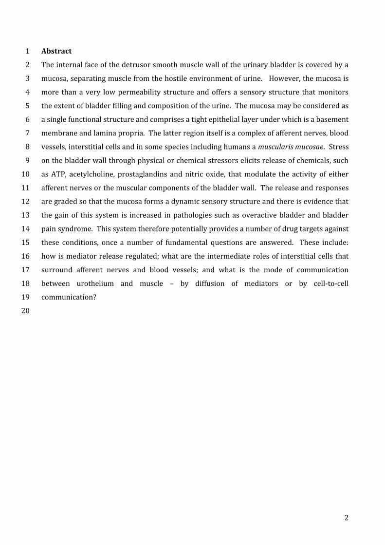
Figure 1. A: Section of the sheep bladder wall. The section shows the urothelium, sub-1
urothelium and detrusor smooth muscle layers. The suburothelium is a complex structure 2
of blood vessels, interstitial cells, afferent nerves and in this species a muscularis mucosae 3
(m.m.). External physical and chemical agents can cause release of mediators (arrows) from 4
the urothelium that could influence suburothelium structures to elicit nervous responses, 5
changes to blood vessel tone and contractile responses of detrusor and possibly muscularis 6
mucosae. Contractile responses could be mediated either by diffusion of mediators and/or 7
by cell-to-cell communication. B: a schematic drawing of the bladder wall, illustrating the 8
cell types in different layers, as well as the stresses that may induce mediator release. 9
10

17
1
2