Embed Size (px)
Citation preview


Types of Oral MucosaBy: Abdullah Akbar
Madeeha Jamil

Oral mucosa
Oral mucosa is the moist lining of the oral cavity.
COMMUNICATIONS:
- Anteriorly it is continuous with the skin of the lip through
vermilion border.
- Posteriorly it is continuous with the mucosa of the pharynx.
Anatomically it is located between the skin and GIT mucosa.

Types of oral mucosa
On the basis of structural and functional variations in different areas of the
oral cavity, oral mucosa is classified into:
1. Masticatory mucosa
2. Lining mucosa
3. Specialized mucosa

Masticatory Mucosa
Covers those areas of the oral cavity such as hard palate and gingiva, that
are exposed to masticatory forces.
Occupies almost 25% of the oral cavity.
Epithelium:
- Moderately thick
- Mostly orthokeratinized though areas of parakeratinization may be seen.
- Epithelial surface is inextensible.

Masticatory mucosa
Lamina Propria:
- Thick
- Contains closely packed bundles of collagen fibers
- Collagen fibers follow a regular course between anchoring point enabling the mucosa to resist heavy loading.
Interface between epithelium and lamina propria:
- Convoluted junction
- Numerous elongated CT papillae are present which provide good mechanical attachment and prevent the epithelium from being stripped off
under shear forces.

Masticatory mucosa
Submucosa:
- Present in fatty zone and glandular zone of hard palate
- Absent in gingiva, palatine raphe and part of hard palate adjacent to
gingiva

Masticatory mucosa

Lining mucosa
Covers the under surface of tongue, floor of mouth, inside of lips & cheeks,
alveolar processes and soft palate.
Occupies 60% of the oral cavity
Epithelium:
- Usually thin but Thicker than that of masticatory mucosa in
cheeks.
- Non-keratinized in under surface of tongue, floor of mouth, cheeks,
alveolar process and soft palate.
- Orthokeratinized in vermilion zone and parakeratinized in intermediate
zone of lips.
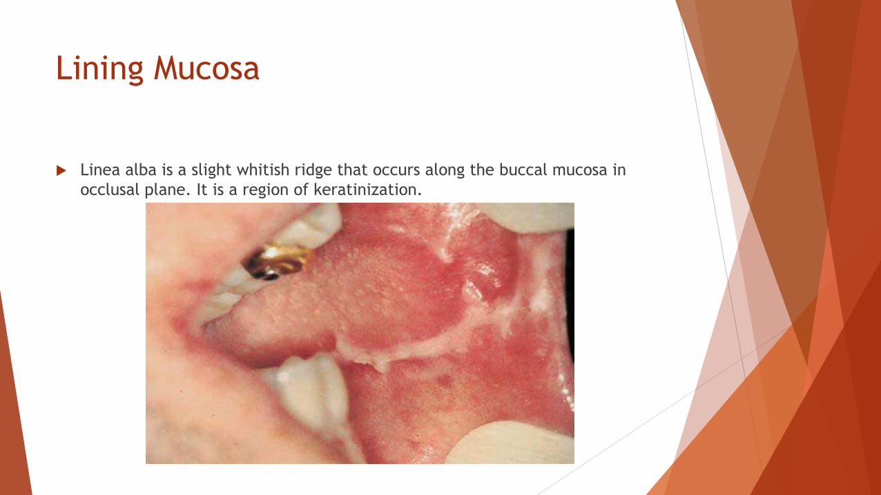
Lining Mucosa
Linea alba is a slight whitish ridge that occurs along the buccal mucosa in
occlusal plane. It is a region of keratinization.

Lining mucosa
Lamina Propria:
- Usually thicker than that of masticatory mucosa.
- Contains fewer collagen fibers which follow irregular course between
anchoring points making the mucosa stretchable to certain extent.
- Also contains elastic fibers which control the extensibility of the mucosa.
Interface between epithelium and lamina propria:
- Smooth junction
- Short CT papillae are found in soft palate, underside of tongue, floor of
mouth and alveolar processes.
- Elongated papillae are present in labial and buccal mucosa.

Lining Mucosa
Lining Mucosa
Submucosa is generally present in lining mucosa
Where lining mucosa covers muscles, it is attached by a mixture of collagen
and elastic fibers
As mucosa becomes slack during mastication, elastic fibers retract mucosa
towards the muscle and prevents the mucosa from being bitten

Lining mucosa

Specialized mucosa
Covers the dorsum of the tongue.
Occupies 15% of the oral cavity.
Although it is masticatory mucosa by function but due to its high extensibility
and lingual papillae, it is classified as “specialized mucosa”.

Specialized mucosa
Lingual Papillae:
- These are the small nipple or hair–like structures on the upper surface of
the tongue that give the tongue its characteristic rough texture.
- Four types of papillae are found on dorsum of the tongue:
1. Fungiform papillae
2. Filiform papillae
3. Foliate papillae
4. Circumvallate papillae

Specialized mucosa
Fungiform Papillae:
- fungus-like appearance
- present on tip and sides of tongue
- scattered between filiform papillae
- smooth, rounded structures covered by non-keratinized
epithelium
- appear red due to highly vascular CT
- Taste buds are present on the superior surface

Specialized mucosa
Fungiform papillae

Specialized mucosa
Filiform Papillae
- hair-like appearance
- cover entire anterior part of tongue
- cone-shaped structures covered by thick keratinized epithelium
- form a tough surface involved in compressing and breaking food when
tongue is apposed to hard palate

Specialized mucosa
Filiform papillae

Specialized mucosa
Foliate Papillae
- leaf-like appearance
- present on lateral margins of posterior part of tongue
- consist of parallel ridges that alternate with deep grooves in the mucosa
- a few taste buds are present in their lateral walls

Specialized mucosa
Foliate papillae

Specialized mucosa
Circumvallate papillae
- Arranged anterior to sulcus terminalis
- 8-12 in number
- Large structures surrounded by a deep, circular groove into which ducts of minor
salivary glands (Glands of Ebner) open
- Covered by keratinized epithelium on superior surface and non-keratinized
epithelium on lateral surface

Specialized mucosa
Circumvallate papillae

Clinical considerations
Lining mucosa is soft and pliable; gingiva and hard palate are covered by
immobile layer.
Local injections: fluid can be introduced easily into lining mucosa. Injection
into masticatory mucosa is difficult and painful.

Clinical considerations
Biopsy/wounds: Lining mucosa gapes and requires suturing; masticatory
mucosa does not.
Inflammation: accumulation of fluid is painful in masticatory mucosa; in
lining mucosa the fluid disperses and inflammation is not that evident or
painful.

Clinical considerations
Hairy tongue: buildup of keratin on filiform papillae results in their
elongation, giving the dorsum of tongue a hairy appearance.
Black discoloration is due to pigment producing bacteria and staining from
food.