Embed Size (px)
Citation preview

1 | P a g e
The Role of the geko™ device in the Prevention and Treatment of Leg Edema WHITE PAPER
Introduction Lower leg edema or peripheral edema presents itself as swelling in the leg or foot. It may be unilateral, bilateral, acute (deep vein thrombosis DVT or trauma) or chronic secondary to comorbid conditions. Calf muscle pump dysfunction contributes to lower limb edema. This article is focused on edema in the lower extremity and its role in wound care.
What is Leg Edema? Edema results in a palpable swelling in the lower extremity caused by fluid accumulation in the interstitium. The exchange of fluid in the tissues is tightly controlled and managed by hydrostatic pressure, oncotic pressure and the lymphatic system. Lower leg edema can be unilateral, bilateral, acute or chronic. Recent research has identified more specifics on how interstitial fluid is returned to the circulation.
Authors: Burrows, C., RN, BScN, MScCH (Wound Prevention & Care), Rabley-Koch, C.A. BSc, RN, NSWOC WOCC (C), Ramage, D., RN., Evans, R., MD, CCFP
Conflict of Interest The authors are Clinical Education Consultants for Perfuse Medtec Inc. Dr. R. Evans has no conflict to declare
Previously held theories of Starling law have been revised to indicate the important role of the lymphatic system in reabsorbing fluid1. A careful history and physical examination are essential to understand the underlying process. Deep vein thrombosis should be considered with acute onset unilateral edema2.
Examples of fluid accumulation in other areas of the body are: • Lung- pleural effusion • Abdomen—ascites • Heart –pericarditis
2 | P a g e
What are the Causes of Leg Edema?
Lower extremity swelling can be a result of a localized process or more systemic. Systemic volume overload can result in fluid in any area including the lower extremity. Examples of these issues are:
• Congestive heart failure • Renal disease • Liver disease • Gastroenterological specific issues • Malnutrition • Damage to lymphatics or capillaries
Three hemodynamic abnormalities associated specifically with venous disease can result in edema:
1. Valvular reflux • Obesity • Multiple pregnancies • Prolonged sitting or standing (recreation
or occupations) • History of varicose vein stripping
2. Obstruction • History of deep vein thrombosis (DVT) • May-Thurner syndrome (anatomic
variant at the left iliac vein) 3. Failure of the calf-muscle pump • Joint issues in the lower extremity (ankle
and leg) • Arthritis, surgery, trauma • Shuffling gait due to medical conditions
such as Parkinson’s disease
As with all processes in medicine the patient may have co-morbidities such as congestive heart failure, a previous DVT and a shuffling gait. Source: Evans et al. 2
What are the consequences of leg edema?
Leg and foot swelling result in:
• The inability to have functional range of
motion around the ankle joint, limiting plantar and dorsiflexion. This in turn impairs the body’s ability to pump blood using its normal muscle pump function
• Skin changes that lead to hyperkeratosis
and hemosiderin staining. These often cause stasis dermatitis resulting in the patient being more prone to contact dermatitis. Dermatitis can be itchy and/or painful
• Skin blistering and excessive drainage
predisposes the patient to skin breakdown, wounds, and infection (cellulitis). This can limit the patient’s social contact resulting in social isolation
• Increased edema can cause ill- fitting
shoes leading to mobility issues and pressure areas
• Patients experience limb heaviness,
fatigue, throbbing, cramping, burning and itchiness
• Delays in healing and local perfusion if an
ulcer is present. Source: Evans et al.2

3 | P a g e
Does calf-muscle pump failure contribute to chronic venous insufficiency?
Calf-muscle pump failure is a key contributor to chronic venous insufficiency. The function of the calf-muscle pump can be evaluated by determining the range of motion around the ankle joint, strength of the calf muscles and gait pattern.
Normal function of the calf-muscle pump to support venous return requires a mobile ankle and routine dorsiflexion beyond 90 degrees. At least 10 degrees of dorsiflexion is needed to fully activate the calf-muscle pump2. The impact of calf-muscle pump failure cannot be overestimated when addressing the treatment of venous ambulatory hypertension.
Gross et al. reported that in a group of 43 patients with leg ulcers, 60% of patients who were considered to have valve deficiency also had an impaired calf-muscle pump. In this study, 24% of the study participants had no obvious signs of valve insufficiency, but 95% of the subjects had a neuromuscular disorder that would impair calf-muscle pump function.3 As well, Meulendijks et al. have identified calf-muscle dysfunction as a strong predictor of venous leg ulcer (VLU) severity and healing4.
Competent valves ensure one-way flow of blood, while incompetent valves allow blood to return into the tissues and further distend the veins; this is seen clinically as varicosities and edema. Under normal conditions, when
the calf-muscle pump is activated there is a decrease in pressure in the veins. Ambulatory hypertension is the failure to reduce these venous pressures2.
For the calf-muscle pump to function properly, the following are required:
• Functional range of motion around the
ankle joint into plantar and dorsiflexion • Functional strength of the gastrocnemius
and soleus muscles • Proper heel-toe gait
A correctly functioning calf-muscle pump results in heartbeat-like contractions that help to push blood back toward the heart2. Therefore, walking is so important for patients. Maintaining the foot in proper position by using appropriate footwear when ambulating is also important to ensure that the calf-muscle pump can work optimally during gait.
The skeletal and muscular components must be coupled with properly functioning valves within the venous system to ensure that the blood does not flow in a retrograde fashion. The symptoms of venous disease are related to congestion in the venous system of the leg and skin changes. These symptoms may manifest as patient complaints of heaviness, fatigue, throbbing, cramping, burning/aching, itchiness of the skin, and restlessness of the legs.
Exercise can improve calf muscle pump function which results in improved
4 | P a g e
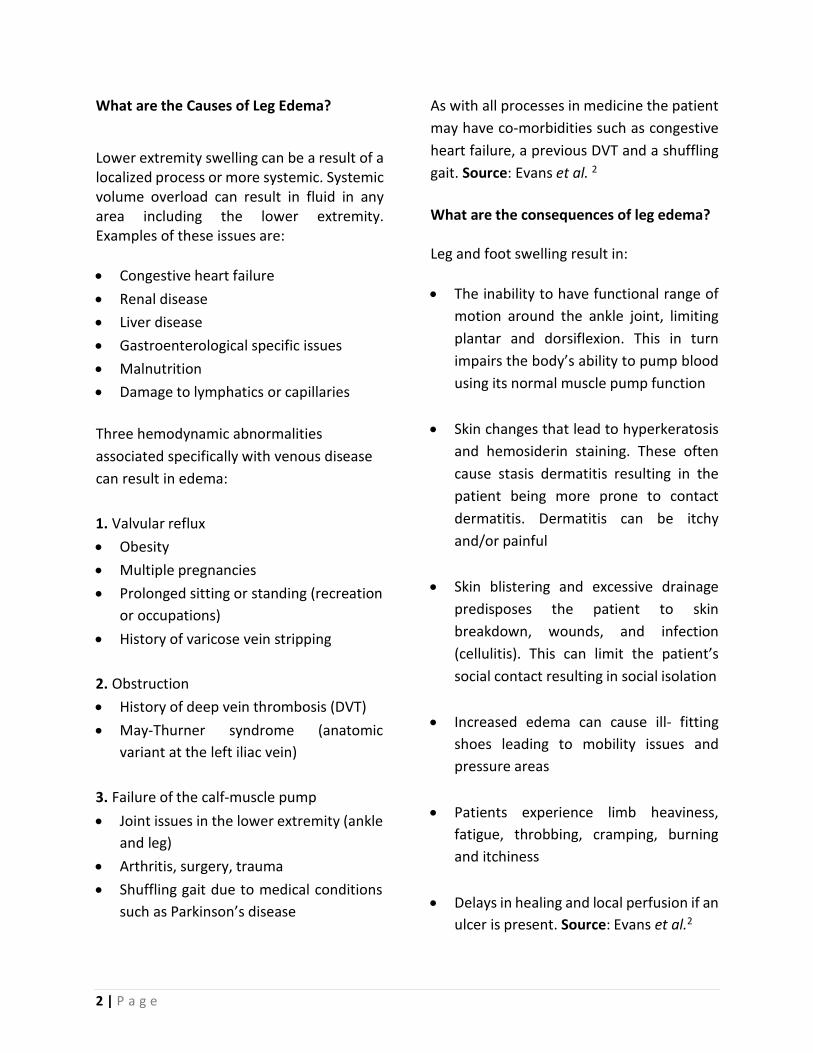
circulation, decreased edema and the risk of developing venous leg ulcers2. During activation of the calf and foot muscle pumps, the gastrocnemius muscles are in extension
when the anterior and lateral muscles like the peroneus longus and brevis are contracted (Figure 1- below).
Figure 1. Muscles affected by the geko™ device
What Is the gold standard optimal therapy? Although compression therapy remains the gold standard in the treatment and management of leg edema and VLU’s, compression therapy does present its challenges. Factors that determine appropriate compression are affordability, ease of application, comfort, non-allergenic materials and accommodation for footwear.2
As well, clinical observation reveals inconsistent application of compression bandaging resulting in variable compression
levels. Poor patient adherence due to warm weather and pain and discomfort at high levels of compression are also challenges.
Moffatt et al. reported 52% to 69% of patients cared for by community nurses had chronic edema, with 73% also having a leg ulcer.5 Green asserts that “chronic edema and leg ulceration can have a significant manual handling implication related to staff health and safety issues.
Among such safety issues are the physical strain of bending, lifting and stretching while managing skin hygiene and applying bandage systems and lifting heavy limbs,
5 | P a g e
especially if the patient is morbidly obese, wheelchair bound or physically infirm.”6 This
predisposes Home and Community care givers to work related injuries.
What are the risks and contraindications of compression therapy?
In 2020, a group of fifteen (15) leading international experts experienced in compression therapy conducted a systematic literature search of medical compression therapy-associated adverse events and contraindications.7
The expert panel developed a consensus document to outline recommendations on the contraindications and risks of Medical Compression (MC) treatment. According to Rabe et al the most common adverse events with medical compression are skin irritation, pain and discomfort. Very rare but severe adverse events, including soft tissue and nerve injury, were also identified. The authors report that low-severity skin problems, including itching, feeling cold or warm and dry skin, are frequently associated with compression therapy.
According to the Consensus Panel, “the higher incidence of itching and dryness reported with Medical Compression Stockings (MCS) when compared with Thrombo-prophylactic Stockings (TPS) may be partially explained by the skin of MCS patients already being compromised due to venous congestion. Inflammatory skin reactions are caused by skin desiccation and skin irritations due to the occlusive barrier effect of the compression material. The Consensus Panel assert that “allergic reactions to compression material are rare due to the elimination of latex based
materials.” The feeling that a compression device, MCS, TPS or compression bandage (CB) is ‘too tight’ is often reported by the patient on first application and may influence compliance. It may take several days before patients get used to wearing compression. Compression discomfort is experienced usually around the ankle or foot. Discomfort or pain below compression may be due to the new experience of ‘pressure on the leg’ but may also be due to incorrect sizing7 Roaldsen et al. in the evaluation of 22 healthy subjects, total ankle range of motion decreased 4% with compression.8
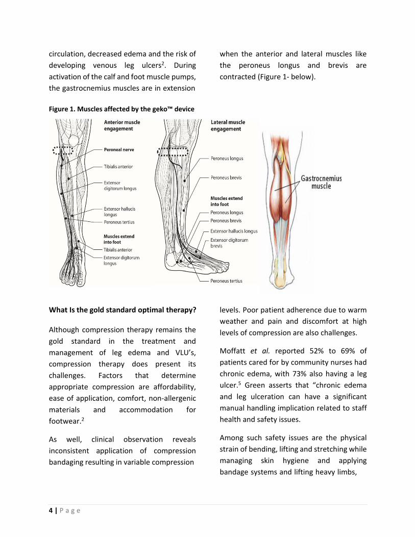
Clinical observation shows forefoot edema and edema above the compression system can occur, particularly in patients with lymphedema. In this patient population forefoot edema, can lead to possible fungal infections between the toes. Risk of skin damage and further misshaping of the limb due to bandage slippage are common issues that are encountered6.
Patients who experience redistributed swelling above the compression therapy experience pain, inability to bend the knee, and the potential for a “tourniquet” effect. Another possible problem related to the tourniquet effect is the compression therapy may slip once the fluid is redistributed. Edematous legs when compressed can lead to peri-wound maceration which can cause further skin breakdown, as well as the potential for fungal infections. (Figure 2 - below)
6 | P a g e
Figure 2.
Fluid redistribution Tourniquet Maceration
How does the geko™ device contribute to Lower Leg Muscle Pump Activation?
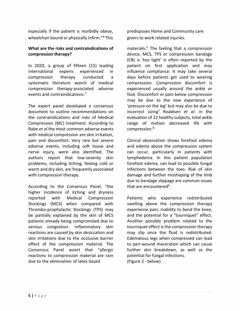
The geko™ device is an easy-to-use, light weight (10 grams) device that is fitted over the fibular head to stimulate the common peroneal nerve. With a stimulation rate of once per second, the geko™ device simultaneously activates a series of muscles
including the tibialis, peroneus longus and lateral gastrocnemius. Collectively this muscle contractions compress the venous system, efficiently evacuating blood from the deep veins of the calf9. This generates blood flow equivalent to about 60% of continuous walking and can be worn all day and every day depending upon the therapeutic objective. (Figure 3 – below)
Figure 3. The geko™ device and placement of the geko™ device
7 | P a g e
Griffin et al. in a single centre open-label intra-subject trial of 18 healthy volunteers compared blood flow in the peroneal, posterior tibial and gastrocnemial veins with and without the geko™ device.
Peak venous velocity (PV) and ejected volume (EV) per individual stimulus (VS) and volume flow (VF) was determined using ultrasound. The results demonstrated Peak Velocity (PV) increased 216% in the peroneal vein, by 112% in the posterior tibial vein and by 137% in the gastrocnemial vein (P<0.001).
Ejected volume per stimulus increased by 113% in the peroneal vein, by 38% in the posterior tibial vein and by 50% in the gastrocnemial vein (P<0.003).
Associated volume flows during the muscle contraction were increased by 36%, 25% and 17%, respectively (P=0.05)9.
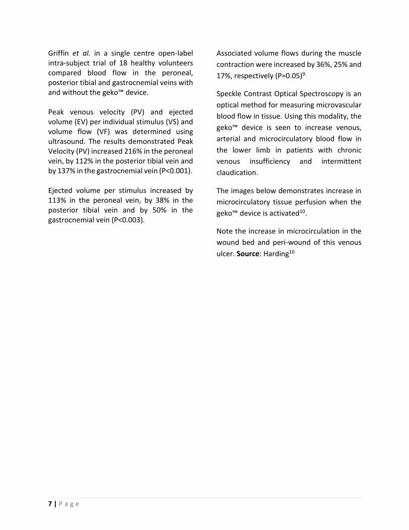
Speckle Contrast Optical Spectroscopy is an optical method for measuring microvascular blood flow in tissue. Using this modality, the geko™ device is seen to increase venous, arterial and microcirculatory blood flow in the lower limb in patients with chronic venous insufficiency and intermittent claudication.
The images below demonstrates increase in microcirculatory tissue perfusion when the geko™ device is activated10.
Note the increase in microcirculation in the wound bed and peri-wound of this venous ulcer. Source: Harding10
8 | P a g e
Figure 4. Speckle Spectroscopy with the geko™ device activated
Microcirculatory flux *with the geko™ device activated: - 225% increase in flux to the wound bed (p<0.001) - 67% increase in flux to peri-wound skin (p<0.001)
How does the geko™ device’s impact edema reduction?
The importance of the calf-muscle pump in supporting edema management is well established2,4. Exercises and activity to promote muscle pump function are necessary components of all treatment plans. Tucker et al. in a study of 30 healthy volunteers report “the geko™ devices provided 60 to 70% of venous emptying response versus the 10 sequential dorsiflexions, considered to be equal to the response seen with walking”11.
As well, change in mid-calf circumference was 55 to 70% of that achieved with full dorsiflexion (p<0.001), and volumetric flow and peak velocity in the superficial femoral vein increased by100% (p<0.01)11.
What is the clinical evidence to support the geko™ device in managing edema?
The geko™ device has been shown to effectively prevent or treat leg edema. In a randomized clinical trial (RCT) conducted at London Health Sciences Centre (LHSC), in London, Ontario two hundred twenty-one (221) patients who underwent kidney and kidney-pancreatic transplant were followed from post-op day 1-6.
The study compared the geko™ device to standard of care using Thromboembolic Deterrent (TED) stockings and Intermittent Pneumatic Compression (IPC). Some of the parameters measured included: leg edema, weight gain, urine output, high femoral velocity, length of stay, patient’s satisfaction, mobility and comfort level. Table 1 shows results as reported by Shahid et al.12
9 | P a g e
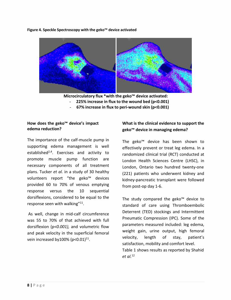
Table 1 Results of LHSC RCT Source: Shahid et al. (2019) 12
Measure Outcome Significance Demographics No significant difference None Leg Edema: Calf measured using circumference
IPC 2.3 cm vs .02 cm geko™ device P=0.001
Leg Edema: Thigh measured using circumference
IPC 2.5 cm vs 0.5 cm geko™ device P=0.001
Weight gain IPC 6.2 kg vs 2.1 kg geko™ device P=0.003 Urine output total IPC 8.8 L vs 18.4 L geko™ device P=0.003 Higher femoral vein velocity
IPC 21 cm/sec vs 29 cm/sec geko™ device P=0.001
Length of stay (104 patients)
geko™ ~ 1 less day than IPC Overall, the MPA group had a significantly shorter hospital stay compared to the TED+IPC group 8.15±3.5 days vs. 9.36±5.0 days
P=0.038
Patient satisfaction 57% would recommend IPC vs 83% geko™ device for future surgical procedures
P=0.002
Mobility (pedometer) (sitting, getting in/out of bed, walking, climbing stairs) (104 patients)
43% in IPC vs 73% geko™ device P=0.009
Comfort level Some level of discomfort: IPC 57% vs 13% geko™ device No effect on comfort: IPC 29% vs 23% geko™ device Comfort: IPC 14% vs 64% geko™ device
P=0.003
Hospital observation (outside study)
Cost savings of $2,300.00 per patient
Statistical significance was reached in all parameters with the most significant differences being leg edema, weight reduction, urinary output and high femoral velocity. Hospital length of stay was reduced in the geko™ group which equates to cost savings of $2,300.00 Cdn. as reported by the hospital.12
A study conducted by Ingves and Power looked at two patients with multifactorial and refractory leg and foot edema that did not respond to traditional treatments and best practices. The authors report that in both cases leg edema was successfully reduced at 4-16 weeks13. In addition to the edema reduction both case studies experienced a reduction of pain and improved wound healing.
10 | P a g e
Wainwright, Burgess and Middleton (2018) compared the use of Neuromuscular Electrical Stimulation (geko™ device) versus Thromboembolic Deterrent Stockings (TEDS) post Total Hip Replacement surgery. The study consisted of 40 patients, N=20 geko and N=20 for TEDS. The results showed that the TED group had significantly more edema post-operatively compared to the geko™ device(p=0.03).14
Baker et al. conducted a study to compare patients with an ankle fracture who were to undergo Open Reduction Internal Fixation (ORIF). Twenty (20) patients were compared to standard of care (bedrest, leg elevation and a backslab) versus those with the geko™ device. The results are reported as 60% of patients ready for theatre in 2 days, compared to 27% in control arm, a 122% improvement15.
Standard treatment = 3.66 days readiness to theatre (average) vs the geko™ device + plaster cast = 1.66 days readiness to theatre (average) (P=0.001)15
In a recent Case Study conducted at Northwood Long Term Care in Halifax, NS, a resident with morbid obesity and chronic leg edema was placed on the geko™ wound therapy device. The resident was started on a diuretic three weeks prior to initiating the geko™ wound therapy device.
Urinary output was measured daily over a four (4) week period. The average urinary output before the geko™ device was 600- 800 mls/day. Once the geko™ wound therapy device was initiated urine output increased to 3-4L/day. Of note, during the rest day per the protocol, urinary output declined to 800-900mls (Parsons, A., Clinical Lead Northwood, 2019. personal communication)
How can the geko™ device meet the challenges of patients managing self care?
Challenges to self care may include:
• Physical status (obesity, co-morbid
conditions, frailty) • Cognition • Motivation (lack of adherence to
compression therapy) • Geographical (remote communities with
limited access to health care)
• Skills to perform specified procedures such as compression bandaging or negative pressure wound therapy
• Complex technology • Virtual training and management
The geko™ device is “user friendly” to patients and their care givers offering a solution to these challenges. Fitting can be taught virtually to patients or their caregivers so that self care can be achieved.

11 | P a g e
What are the potential advantages of the geko™ device?
Feature The geko™ device
Compression Bandaging
Patient self-management √ x Edema prevention and treatment quantified √ x Evidence of enhanced hemodynamics quantified √ x Minimal Environmental Impact √ x Takes a few minutes to learn √ x No special skills required √ x No pain on application √ x No personal contact patient/care provider √ x Can be mailed to patient for self-care √ x Training can be done virtually √ x Evidence of cost saving for the system √ x High degree of patient adherence/satisfaction √ x Does not cause discomfort in summer √ x Takes about 60 seconds to apply √ x Lower COVID-19 risk to patient/provider due to lower human to human contact
√ x
Conclusion/Summary Edema impacts time to healing, patient safety, mobilization and quality of life. Clinical studies have demonstrated that the geko™ device reduces edema in a range of patient groups. The geko™ device activates the lower leg muscle pumps once per second to augment venous return, reduce venous congestion, and decrease leg edema. Intervention with the geko™ device may reduce pain and congestion in the limb thereby affording improved compliance with compression therapy9. The geko™ device is a simple, safe, user-friendly device that can be
used as a cost- effective adjunctive therapy or on its own to treat and manage leg edema. Innovative technology such as the geko™ device offers the solution to the challenges of edema management and self care. As clinicians consider the provision of care, virtual visits have become an option. Use of the geko™ device can be taught and self managed virtually by patients and care providers, offering an alternative for clinicians to treat lower leg edema. The geko™ device supports self care.
12 | P a g e
References:
1. Levick JR, Michel, CC. Microvascular fluid exchange and the revised Starling principle. Cardiovascular Research (2010) 87, 198-210
2. Evans R, Kuhnke J L, Burrows C, Kayssi A, Labrecque C, O’Sullivan-Drombolis D, Houghton
P. Best Practice Recommendations for the Prevention and Management of Venous Leg Ulcers. A supplement of Wound Care Canada; [2019, November]: pp 1-70. Available [on- line]: https://www.woundscanada.ca/index.php?option=com_content&view=article&id=110&c atid=12&Itemid=724#how-to-cite-a-bprs
3. Gross EA, Wood CR, Lazarus GS, Margolis DJ. Venous leg ulcers: An analysis of underlying venous disease. Br J Dermatol. 1993;129(3):270–274.
4. Meulendijks AM, de Vries FMC, van Dooren AA, Schuurmans MJ, Neumann HAM. A
systematic review on risk factors in developing a first-time venous leg ulcer. J Eur Acad Dermatol Venereol. 2018; early view.
5. Moffatt, C.J, Gaskin, R., Skyorova, M. et al. (2019). Prevalence and risk factors for chronic
edema in UK. Community Nursing Services. Lymphatic Res Biol 17(2): 147-54
6. Green, T. (2020). Challenges of managing patients with chronic oedema and leg ulceration.
7. Rabe, E., Partsch, H., Morrison, N. et al. (2020). Risks and contraindications of medical compression treatment – A critical reappraisal. An international consensus statement. Phlebology: The Journal of Venous Disease. Available [On-line] https://journals.sagepub.com/doi/full/10.1177/0268355520909066
8. Roaldsen KS., B Elfving B., Stanghelle JK.,Mattsson E. (2012) Effect of multilayer high- compression bandaging on ankle range of motion and oxygen cost of walking. Phlebology. Available [on-line]: https://www.ncbi.nlm.nih.gov/pmc/articles/PMC3257002/
9. Griffin M., Bond, D., Nicolaides A, Measurement of blood flow in the deep veins of the lower limb using the geko™ neuromuscular electro-stimulation device. International Angiology. August 2016; Issue:35[4]. Pp406-10. Available[ on-line] https://www.minervamedica.it/en/journals/international- angiology/article.php?cod=R34Y2016N04A0406
10. Harding, K. (2016). Speckle Spectroscopy. Welsh Wound Innovation Centre. Available https://www.gekodevices.com/partner/wound-therapy-2/
11. Tucker AT, Maass A, Bain DS, et al. Augmentation of venous, arterial and microvascular blood supply in the leg by isometric neuromuscular stimulation via the peroneal nerve. Int
13 | P a g e
J Angiol 2010; 19: e31–e37.11.
12. Xie W, Levine M, Aquil S, Pacoli K, Al-Ogaili R, Luke P, Sener A. Daily use of a muscle pump activator device reduces duration of hospitalization and improves early graft outcomes post-kidney transplantation: A randomized controlled trial. Can Urol Assoc J 2020 July 27; Epub ahead of print. http://dx.doi.org/10.5489/cuaj.6487
13. Ingves MV, Power AH. Two Cases of Transcutaneous Electrical Nerve Stimulation of the Common Peroneal Nerve Successfully Treating Refractory, Multifactorial Leg Oedema. Journal of Investigative Medicine High Impact Case Reports. October-December 2014: 1–4. Available [on-line] http://journals.sagepub.com/doi/abs/10.1177/2324709614559839
14. Wainwright TW, Burgess LC, Middleton RG. A feasibility randomised controlled trial to evaluate the effectiveness of a novel neuromuscular electrostimulation device in preventing the formation of edema following total hip replacement surgery. Heliyon 2018:4(7); e00697. Available [on-line] https://www.heliyon.com/article/e00697
15. Baker P, Mahmood I, Chandler H, Anwar S, Eardley W, Rangan A. A pilot clinical study comparing the geko™ neuromuscular electrostimulation device, with current standard of care to evaluate edema reduction and readiness for theatre in patients requiring open reduction internal fixation following ankle fracture. Trauma / Foot & Ankle Trauma / Surgical Treatment South Tees NHS Trust, Middlesbrough, United Kingdom. Submitted to European Federation of National Associations of Orthopaedics and Traumatology (EFORT) 2018. Available [on-line] http://gekodevices.com/wp-content/uploads/2018/04/pre- op_edema_abstract.pdf
![109 A Must-Have For Glazing Contractors · Geko 350 PV [350kg] The Geko 350 PV is the big brother of our popular Geko 250 PV robot and boasts 350kg of lifting power for effortless](https://img.pdfslide.us/doc/110x75/60109a04eb6c0d7da25b33fc/109-a-must-have-for-glazing-contractors-geko-350-pv-350kg-the-geko-350-pv-is-the.jpg)


![GERIATRIC FALL HIP INJURY PREVENTION DEVICE · PDF file[6] 1.2 Types of Hip Fractures ... The Geriatric Fall Hip Injury Prevention Device will focus on the prevention of hip injuries](https://img.pdfslide.us/doc/110x75/5ab9841b7f8b9ad5338e1021/geriatric-fall-hip-injury-prevention-device-6-12-types-of-hip-fractures-the.jpg)