Embed Size (px)
Citation preview

Eveliina Myllyluoma
Foundation for Nutrition ResearchHelsinki, Finland
Institute of BiomedicinePharmacology
University of Helsinki
ThE rolE of probioTics in Helicobacter pylori infEcTion
Helsinki 2007

Supervised by: Docent Riitta Korpela, PhD Institute of Biomedicine Pharmacology University of Helsinki, Finland
Professor (emeritus) Heikki Vapaatalo, MD Institute of Biomedicine Pharmacology University of Helsinki, Finland
Reviewed by: Docent Riitta Karttunen, MD Central Hospital of Kanta-Häme Microbiology laboratory Hämeenlinna, Finland
Professor Atte von Wright, PhD Department of Biosciences University of Kuopio, Finland
Opponent: Docent Asko Järvinen, MD Department of Medicine Division of Infectious Diseases Helsinki University Central Hospital, Finland
Layout: Oy Graaf Ab/Jani Osolanus
ISBN 978-952-92-2090-8 (pbk.)ISBN 978-952-10-3947-8 (PDF)
Helsinki University Printing HouseHelsinki 2007

To Markku, Ansuliina, and Ilmari
4
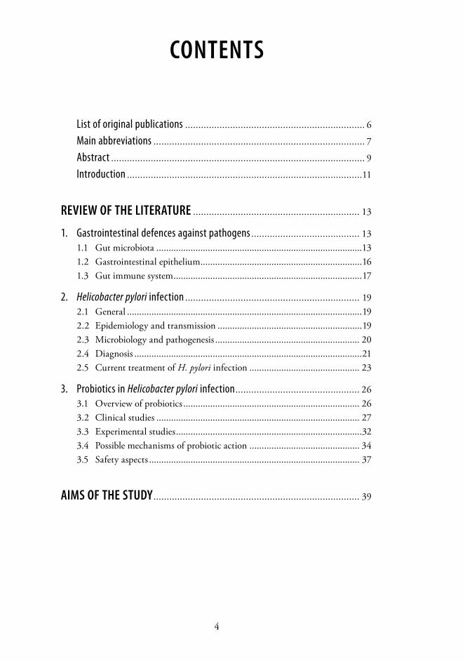
conTEnTs
List of original publications .................................................................... 6
Main abbreviations ................................................................................ 7
Abstract ................................................................................................ 9
Introduction .........................................................................................11
rEviEw of ThE liTEraTurE ............................................................... 13
1. Gastrointestinal defences against pathogens ......................................... 131.1 Gut microbiota ....................................................................................131.2 Gastrointestinal epithelium ..................................................................161.3 Gut immune system .............................................................................17
2. Helicobacter pylori infection .................................................................. 192.1 General ................................................................................................192.2 Epidemiology and transmission ...........................................................192.3 Microbiology and pathogenesis ........................................................... 202.4 Diagnosis .............................................................................................212.5 Current treatment of H. pylori infection ............................................. 23
3. Probiotics in Helicobacter pylori infection ............................................... 263.1 Overview of probiotics ........................................................................ 263.2 Clinical studies ................................................................................... 273.3 Experimental studies ............................................................................323.4 Possible mechanisms of probiotic action ............................................. 343.5 Safety aspects ...................................................................................... 37
aiMs of ThE sTudy .............................................................................. 39

5
subjEcTs, MaTErials and METhods .............................................. 41
1. Study designs and subjects ................................................................... 41
2. Ethics .................................................................................................. 42
3. Probiotic products and bacterial strains ................................................ 43
4. Methods .............................................................................................. 444.1 H. pylori infection assessment ............................................................. 444.2 Questionnaires .....................................................................................454.3 Microbiological methods .................................................................... 464.4 Morphological and functional status of the gastroduodenal mucosa ....474.5 Cell culture methods .......................................................................... 484.6 Statistical analysis ............................................................................... 50
rEsulTs ................................................................................................. 53
1. Effects of probiotics on the tolerability and efficacy of H. pylori eradication treatment ............................................................. 53
2. Effects of probiotics on microbiota following H. pylori eradication treatment ............................................................. 54
3. Effects of probiotics in untreated H. pylori infected patients ................... 56
4. Characteristics of probiotics in H. pylori infected epithelial cells .............. 57
discussion ........................................................................................... 61
1. Methodological aspects ....................................................................... 62
2. Main results ........................................................................................ 642.1 Effects of probiotics on the eradication treatment of H. pylori ............ 642.2 Effects and characteristic of probiotics in H. pylori infection .............. 66
conclusion and fuTurE prospEcTs .............................................. 69
Acknowledgements ............................................................................. 70
References .......................................................................................... 72
6
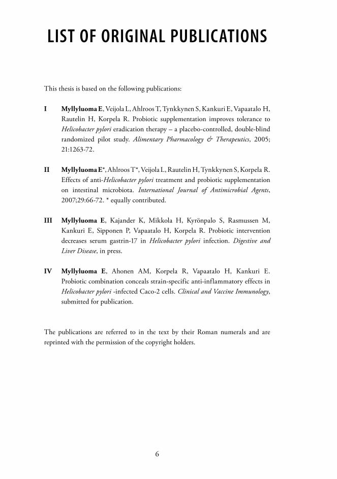
lisT of original publicaTions
This thesis is based on the following publications:
I Myllyluoma E, Veijola L, Ahlroos T, Tynkkynen S, Kankuri E, Vapaatalo H, Rautelin H, Korpela R. Probiotic supplementation improves tolerance to Helicobacter pylori eradication therapy – a placebo-controlled, double-blind randomized pilot study. Alimentary Pharmacology & Therapeutics, 2005; 21:1263-72.
II Myllyluoma E*, Ahlroos T*, Veijola L, Rautelin H, Tynkkynen S, Korpela R. Effects of anti-Helicobacter pylori treatment and probiotic supplementation on intestinal microbiota. International Journal of Antimicrobial Agents, 2007;29:66-72. * equally contributed.
III Myllyluoma E, Kajander K, Mikkola H, Kyrönpalo S, Rasmussen M, Kankuri E, Sipponen P, Vapaatalo H, Korpela R. Probiotic intervention decreases serum gastrin-17 in Helicobacter pylori infection. Digestive and Liver Disease, in press.
IV Myllyluoma E, Ahonen AM, Korpela R, Vapaatalo H, Kankuri E. Probiotic combination conceals strain-specific anti-inflammatory effects in Helicobacter pylori -infected Caco-2 cells. Clinical and Vaccine Immunology, submitted for publication.
The publications are referred to in the text by their Roman numerals and are reprinted with the permission of the copyright holders.

7
Main abbrEviaTions
Bb12 Bifidobacterium lactis Bb12Bb99 Bifidobacterium breve Bb99CFU colony forming unitCI confidence intervalsDC dendritic cellDMEM Dulbecco's modified Eagle's mediumEIA enzyme immunoassayELISA enzyme-linked immunosorbent assayFISH fluorescent in situ hybridization G gastrinH. pylori Helicobacter pyloriIg immunoglobulinIL interleukinIQR interquartile rangeLAB lactic acid bacteriaLC705 Lactobacillus rhamnosus Lc705LDH lactate dehydrogenaseLGG Lactobacillus rhamnosus GGLTB4 leukotriene B4
PBS phosphate buffered salinePCR polymerase chain reactionPG pepsinogenPGE2 prostaglandin E2
PJS Propionibacterium freudenreichii ssp. shermanii JSPPI proton pump inhibitorrRNA ribosomal RNATER transepithelial electrical resistanceTLR Toll-like receptorTNF tumor necrosis factorUBT urea breath test


9
absTracT
Helicobacter pylori (H. pylori) infection is a major cause of chronic gastritis and peptic ulcer disease, and it is also designated as a class-I carcinogen for stomach cancer. The role of probiotics in the treatment of gastrointestinal infections is increasingly documented as an alternative or complement to antibiotics, with the potential to decrease the use of antibiotics or reduce their adverse effects.
These studies were conducted to investigate the role of probiotics in the treatment of H. pylori infection. Various aspects included: an investigation of the effects of a probiotic combination consisting of Lactobacillus rhamnosus GG, L. rhamnosus LC705, Propionibacterium freudenreichii ssp. shermanii JS and Bifidobacterium breve Bb99 or B. lactis Bb12 as a supplementation to H. pylori eradication therapy, with special reference to tolerability, effectiveness, and microbiota alterations following the treatment; discovering the role of probiotics in vivo with H. pylori infected and uninfected patients, as well as with an in vitro model of H. pylori infection.
The probiotic combination therapy was able to reduce significantly the total symptom score, which takes into account both the frequency and the severity of the adverse effects, during the eradication treatment. The supplementation did not improve the success of the eradication treatment significantly, though some difference was seen in the eradication percentages (91% vs. 79%). The quantities of predominant bacterial groups were altered significantly following the triple treatment. Probiotics slightly counteracted the effects of anti-H. pylori treatment, monitored as significantly less alterations in the total numbers of aerobes and lactobacilli / enterococci group bacteria.
After probiotic intervention, L. rhamnosus GG adhered to a minority of the patients’ upper gastrointestinal mucosa, but all of the probiotics survived well through the gastrointestinal tract transit with and without antimicrobial treatment. Probiotic intervention decreased gastrin-17 levels in H. pylori infected patients and appeared to decrease the 13C-urea breath test values. In in vitro Caco-2 cell line experiments, probiotics inhibited H. pylori adhesion to intestinal epithelial cells.

10
Both L. rhamnosus strains, P. freudenreichii ssp. shermanii JS and the combination inhibited the H. pylori-induced acute cell leakage. Simultaneously, both L. rhamnosus strains and the combination transiently improved the epithelial barrier function. The pro-inflammatory effects prevailed when the probiotics were used in combination.
According to this series of studies, probiotic combination could have some potential in reducing adverse effects induced by H. pylori eradication treatment and beneficial effects on H. pylori infected subjects.

11
inTroducTion
Helicobacter pylori (H. pylori) infection is recognized as a causative agent for acute and chronic gastritis and as a predisposing factor to peptic ulcer disease, gastric cancer and gastric lymphoma. This important discovery, rewarded with the 2005 Nobel Prize in Medicine, has changed gastroenterological practice worldwide since, after its discovery, many gastroduodenal diseases became curable infectious diseases. The prevalence of H. pylori infection in the adult population of industrialized countries is estimated to be at 20–50%, and in developing countries, the rate is as high as 80% (for review, see Go 2002). The rate of carriage increases according to age group. Infection is usually acquired during childhood and persists lifelong if not treated specifically.
Only a combination of antimicrobials can be used in vivo to eradicate H. pylori, and none of the antimicrobials is effective enough to eliminate H. pylori when given as monotherapy (for review, see Kusters et al. 2006). The first-line recommended eradication treatment of H. pylori consists of a combination of two antimicrobials and an acid-suppressive drug (Malfertheiner et al. 2002). However, the triple treatment has many shortcomings, such as several adverse effects possibly leading to discontinuation of the treatment (Deltenre et al. 1998), and limited efficacy particularly because of antimicrobial resistance (for review, see Gerrits et al. 2006).
In recent years, the development of alternative anti-H. pylori treatments has been actively pursued, and investigations have been carried out to define components that could be used either as monotherapy or synergistically in combination with antimicrobials thus resulting in more effective anti-H. pylori therapy or alternative ways of controlling H. pylori infection. These novel treatments could potentially reduce the costs related to the treatment of H. pylori associated diseases. Promising results have been obtained in initial studies with several probiotic strains (for reviews, see Hamilton-Miller 2003, Gotteland et al. 2006), but there are still many open questions. As defined by FAO/WHO (2002), probiotics are live microorganisms

that, when administered in adequate amounts, confer a health benefit on the host (such as Lactobacillus spp. and Bifidobacterium spp.).
The present study aims to investigate the effects of probiotics in patients receiving the recommended eradication treatment for H. pylori infection, as well as the effects on the stomach and intestine in untreated H. pylori-positive patients, and to evaluate the characteristics of individual probiotics or the probiotic combination in an in vitro model of H. pylori infection.

13
1. gasTroinTEsTinal dEfEncEs againsT paThogEns
Natural gastrointestinal defences against pathogens can be theoretically divided into three levels of responses (for reviews, see McCracken and Lorenz 2001, Bourlioux et al. 2003). The host's microbiota provides the first level of defence against pathogens by preventing them from developing in the gastrointestinal tract, for instance, by protecting the mucosa against colonization of pathogenic microorganisms. The intestinal epithelium constitutes a second level of defence and a tight barrier against pathogens by the combined effect of the mucus layer and the epithelial cells themselves. The immune system, which constitutes the third defensive barrier against pathogenic microorganisms, can be divided into two lines of defence against pathogens: innate and adaptive immunity.
1.1 guT MicrobioTa
Composition and diversity of gut microbiotaThe human microbiota is considered to be an enormously diverse and complex ecosystem affected by host cells, ingested food and microbes (for review, see Zoetendal et al. 2006). In the past, it was commonly believed that over 400 species compose this microbiota. However, it is nowadays estimated that more than 1,000
rEviEw of ThE liTEraTurE

14
species are present in the gut (Bäckhed et al. 2005). Previously, traditional methods using culturing have been used as the gold standard for investigating bacteria. However, use of 16S ribosomal RNA (rRNA) and genome based approaches has revealed further information about microbiota. The new culture-independent techniques, such as sequencing of cloned 16S rRNA gene amplicons, 16S rRNA gene fingerprinting, quantitative fluorescent in situ hybridization (FISH), and quantitative polymerase chain reaction (PCR), provide more specific methods for detailed investigations at the species and strain levels (Zoetendal et al. 2006). Also, development of high-diversity deoxyribonucleic acid (DNA) microarrays will open new possibilities in gut microbiota research (Rajilic-Stojanovic et al. 2006). Thus, new microbes in the human microbiota are continuously being discovered. It has been suggested that only 20–40% of the bacteria from intestinal samples have been characterized, due to a lack of knowledge of appropriate culture conditions (Zoetendal et al. 1998, Suau et al. 1999, Hayashi et al. 2002).
There are very few studies on the microbiota of the stomach with culture-independent techniques, and recent evidence indicates that the stomach microbiota is also more diverse than previously thought (Hill 1985, Monstein et al. 2000, Peña et al. 2002, Roos et al. 2005, Bik et al. 2006). The most common bacterial phyla found in the stomach are Proteobacteria, Firmicutes, Bacteroidetes, Actinobacteria and Fusobacteria, and the most abundant genera found are Helicobacter, Streptococcus and Prevotella (Bik et al. 2006). Thus, even in young normochlorhydric subjects, the lumen is not bacteria free, despite pH values being most of the time below 3. However, the gastric acid is buffered during a meal, allowing microbes to proliferate. Impaired gastric acid secretion, caused, for example, by chronic atrophic gastritis, prolonged use of histamine-2 receptor antagonists or proton pump inhibitors, is associated with bacterial overgrowth in the stomach and small intestine (Hill 1985, Väkeväinen et al. 2000, Sanduleanu et al. 2001, Williams and McColl 2006).
The composition of the predominant bacterial community in the gut is reported to be host-specific and stable over time in healthy adults (for review, see Zoetendal et al. 2006). Only a limited fraction of bacterial phyla compose the major intestinal microbiota (Manichanh et al. 2006). In healthy adults, 80% of phylotypes belong to four major phylogenetic groups, which are the Clostiridium leptum, Clostridium coccoides, Bacteroides and Bifidobacteria groups (Lay et al. 2005). However, a large fraction of dominant phylotypes is subject specific (Zoetendal et al. 1998, Seksik et al. 2003, Vanhoute et al. 2006). Also, recent studies have found that mucosal microbiota is stable along the distal gastrointestinal tract from ileum to rectum, but mucosa-associated microbiota is different from fecal microbiota. The difference has been estimated to be between 50–90% (Zoetendal et al. 2002, Lepage et al. 2005).

15
The importance of microbes for the hostThe intestinal microbiota has crucial physiological functions in the gut, such as metabolic capacity and the ability to ferment carbohydrates into short-chain fatty acids, which have been shown to stimulate the growth and well-being of the colonic mucosa and colonic motility (for reviews, see Mitsuoka 2000, O’Hara and Shanahan 2006). Studies of germ-free mice have revealed that gut microbiota plays an important role in the maturation of the immune system (for review, see Hooper et al. 2002). The intestine of germ-free mice has several physiological differences as compared to that of conventional mice and also a less developed immune system. Several other specific interactions between host and bacteria have also been discovered using germ-free animal models and in vitro cell models. These include antimicrobial peptide production, maintenance of intestinal homeostasis and development of vascular network in the villi after microbial colonization (Zoetendal et al. 2006). Thus host-microbe interactions can shape the immunity and maturation of the gastrointestinal tract of the host and have a further impact on the ecology of microbiota. The balance between potentially beneficial and harmful bacteria is very important (Figure 1).
Figure 1 Potentially harmful and potentially beneficial bacteria (Modified from Bourlioux et al. 2003). P., Pseudomonas; G+, Gram positive; E., Escherichia.
Colonization resistanceThe indigenous microbiota is a natural resistance factor against potential pathogenic microorganisms and provides colonization resistance, also known as gut barrier, by controlling the growth of opportunistic microorganisms (Fons et al. 2000, Vollaard
potentially harmfulClostridiaSulfate reducersVeillonellaStaphylococciProteusP. aeruginosa
††††††
potentially beneficial Bifidobacteria Lactobacilli Methanogens Fusobacteria
††††
BacteroidesEubacteria
Anaerobic G+ cocciE. coli
EnterobacteriaSulfate reducers
Fusobacteria
potentially harmfulClostridiaSulfate reducersVeillonellaStaphylococciProteusP. aeruginosa
††††††
potentially beneficial Bifidobacteria Lactobacilli Methanogens Fusobacteria
††††
BacteroidesEubacteria
Anaerobic G+ cocciE. coli
EnterobacteriaSulfate reducers
Fusobacteria

16
and Clasener 1994). It has been suggested that commensal bacteria protect their host against microbial pathogens by interfering with their adhesion and toxic effects (for reviews, see Brook 1999, Servin et al. 2004).
1.2 gasTroinTEsTinal EpiThEliuMThe tight epithelial cell barrier forms the second line of defence between the gut luminal contents and the host. Epithelial cells lining the gastrointestinal tract are able to respond to infection by initiating either nonspecific or specific host-defence response (for reviews, see Kagnoff and Eckmann 1997, Strober 1998). Bacterial adhesion to the host cell or recognition by the host cell is often an essential first stage in the disease process.
A wide range of gastrointestinal cell surface constituents, such as several glygoconjucates, can serve as receptors for bacterial adherence (Servin and Coconnier 2003, Pretzer et al. 2005). Furthermore, epithelial cells express constitutively host pattern recognition receptors, such as Toll-like receptors (TLR). These are a family of transmembrane receptors that recognize repetitive patterns, i.e. the pathogen-associated molecular patterns present in diverse microbes, including gram-positive
and gram-negative bacteria (for reviews, see Bäckhed and Hornef 2003, Takeda et al. 2003). TLRs are also found on innate immune cells, such as dendritic cells and macrophages (Vinderola et al. 2005). TLR4 recognizes lipopolysaccharide and gram-negative bacteria, while TLR2 recognizes a variety of microbial components, such as peptidoglycan and lipoteichoic acids, from gram-positive bacteria (Abreu 2003, Matsuguchi et al. 2003, Takeda et al. 2003). Also, several other TLRs with specific actions are known, such as TLR5, which responds to the bacterial flagella (Rhee et al. 2005), and TLR9, which is activated by bacterially derived short DNA fragments containing CpG sequences (Pedersen et al. 2005). Other known recognition receptors are nucleotide-binding oligomerization domain proteins, which recognize both gram-positive and gram-negative bacteria. They are located in cell cytoplasm and are implicated in the induction of defensins.
Increased epithelial barrier permeability is frequently associated with gastrointestinal disorders contributing to both disease onset and persistence (for reviews, see Lu and Walker 2001, Berkes 2003). The gatekeeper of the paracellular pathway is the tight junction, which is an apically located cell-cell junction between epithelial cells. The tight junction permits the passage of small molecules, such as ions, while restricting the movement of large molecules, such as antigens and microorganisms, which can cause inflammation. The integral membrane protein family, which are mainly claudins, occluding and zonula occludens 1, are implicated in the formation of the paracellular channels (Berkes et al. 2003).

17
The permeability of the tight junction to ions can be assessed by transepithelial electrical measurements and by paracellular ion flux assays for major extracellular ions, such as Na+, Cl–, Ca2+, and Mg2+.
Epithelial cells are also involved in a wide range of mucosal immune and inflammatory responses together with dendritic cells, macrophages, neutrophils and lymphocytes. Epithelial cells can secrete pro- and anti-inflammatory cytokines and chemokines, such as tumor necrosis factor (TNF) -α, interleukin (IL) -2, IL-6, and IL-8, which either diminish or stimulate response or provide feedback (Goodrich and McGee 1998). IL-8, a C-X-C chemokine that is transcriptionally regulated by nuclear factor-kappa B, shows potent chemotactic activity for neutrophils. Intestinal epithelial cells also secrete many other mediators involved in immune responses to potentially pathogenic microorganisms, including antimicrobial peptides, such as defensins and mucins (for review, see Servin 2004). The immunoinflammatory reaction is highly important in eliminating pathogens, but this reaction must be controlled to avoid the risk of a more widespread inflammation. Microbes differ in terms of their ability to induce inflammatory response. The commensal microbiota produces a very mild inflammation response and is thus tolerated by the mucosa, while modified microbiota induces a more marked response (for review, see O’Hara and Shanahan 2006).
1.3 guT iMMunE sysTEMThe third level of defence is the immune system, which is crucial for humans and animals in protecting the host against invading pathogens. Bacteria are the main source of antigenic materials, and the gut microbiota is the most important stimulant of the body’s immunological defence (Bourlioux et al. 2003). The immune system comprises a complex array of interacting mechanisms. It consists of local immune tissue (mucosa associated lymphoid tissue) and the systemic immune system (in blood, liver, spleen and bone marrow). Both components can be further theoretically divided into two types of response: innate (nonspecific) and adaptive (specific) immune response (for reviews, see Borregaard et al. 2000, Janeway and Medzhitov 2002). Specific immune response is usually induced by direct contact, despite an intact epithelial barrier, between the lymphoid tissue and the potentially pathogenic macromolecules or microorganisms in the intestinal lumen.
Innate or “natural” immunity is a rapidly activated host defence that recognizes conserved microbial structures not expressed by the host and mounts a nonspecific immune response against these structures (often specific carbohydrates or lipoproteins). The activated effectors of innate immunity, such as phagocytic cells, natural killer cells, and the complement system, are able to destroy the invader

18
(Suffredini et al. 1999, Borregaard et al. 2000, Janeway and Medzhitov 2002). Innate immunity also includes acid in the stomach, lysozyme, lactoferrin and antimicrobial molecules (Borregaard et al. 2000). Intracellular pathogens, like viruses, are killed by natural killer cells.
The adaptive immune system is activated by the infection if the innate immune system is insufficient (Borregaard et al. 2000). The adaptive immune system is a more specific and powerful tool against pathogens, but the primary response mounts slower than in innate immunity. Crucially, adaptive immunity develops a memory, which enables a rapid and effective response in a reinfection. Adaptive immunity recognizes antigenic structures (often peptides), not expressed in the host, as non-self. Antigens are presented to the effector cells of the adaptive immunity by antigen presenting cells. Furthermore, intestinal dendritic cells can directly sample the contents of the gut lumen by extending dendrites between epithelial cells.
Although innate and adaptive immunity represent two separate arms of immunity, a close relationship exists between them (Palucka and Banchereau 1999, Bourlioux et al. 2003). The initiation and direction (cellular or humoral) of adaptive immunity is influenced by innate immunity, which regulates its direction via cytokines, T and B cell co-stimulatory mechanisms and antigen presentation. Furthermore, pattern recognition receptors, such as TLRs, in epithelial cells, dendritic cells and macrophages are important in bridging the innate and adaptive immune responses (for review, see Abreu and Arditi 2004). Continuous formation of immunoglobulin (Ig) A in plasma cells in the lamina propria also plays an essential role in the protective function of the mucosa (Goodrich and McGee 1998). This IgA is transported to the luminal side of the mucosa and released into the bowel as secretory IgA, where it is able to neutralize potentially pathogenic bacteria and viruses.
To summarize, the host's microbiota protects the mucosa against colonization and invasion by pathogenic microorgamisms (colonization resistance). If this normal microbial habitat is damaged, ecological niches are created for pathogens. The intestinal epithelium constitutes a tight barrier against pathogens that interact with commensal microbiota. Pathogens also crosstalk with the epithelium and modify epithelial responses, e.g. to enhance their penetration across the epithelial barrier. The immune system constitutes the more specific defence against pathogenic bacteria. The innate immunity reacts immediately but unspecifically. The adaptive immunity takes longer but is specific and keeps memory of previous aggressions. A mild inflammatory state is necessary to keep the defence enabled, but the system must remain balanced.
19
2. Helicobacter pylori infEcTion
2.1 gEnEralHelicobacter pylori (H. pylori) was isolated from gastric mucosa and bacteriologically identified in 1982 (Warren and Marshall 1983). Since then, it has been reported that ulcer recurrence rates have decreased after the eradication of H. pylori from the stomach of peptic ulcer patients (Marshall et al. 1988, Hantschel et al. 1993). It has been established that H. pylori infection is a major cause of chronic gastritis and peptic ulcer disease. H. pylori was designated a class-I (definite) carcinogen for stomach cancer in 1994 after epidemiological investigation by the International Agency for Research on Cancer (IARC), a subordinate organization of the World Health Organization (IARC 1994). Furthermore, association of primary malignant gastric lymphoma with H. pylori has been reported in a large-scale cohort study (Parsonnet et al. 1994). However, the role of H. pylori as an obligate pathogen has been questioned, and it has been proposed that Helicobacter strains could be part of the indigenous microbiota of the human stomach and that H. pylori could have both pathogenic and symbiotic features (for reviews, see Blaser 1999, 2006).
2.2 EpidEMiology and TransMissionApproximately half of the world’s population is infected with H. pylori and the prevalence shows large geographical variations (for review, see Go 2002). The infection is generally acquired during childhood and usually persists indefinitely if left untreated. It has been suggested that H. pylori infection rates are associated with age, ethnicity, socio-economic status, sanitary environments and lifestyle. In Finland, the prevalence of H. pylori infection in children is 6–10% (Ashorn et al. 1995, Rehnberg-Laiho et al. 1998) and in adults 12–70% (Rehnberg-Laiho et al. 2001). Prevalence rates generally remain stable in each birth cohort, and thus the higher prevalence of infection among the elderly reflects a birth cohort effect with higher infection rates in the past (for review, see Rautelin and Kosunen 2004). The main reservoir of H. pylori is the human stomach with the most likely mode of transmission being person to person (for review, see Vaira et al. 2001). Recent epidemiological study suggests that infected mothers are the main source of H. pylori infection in their children (Weyermann et al. 2006).

20
2.3 Microbiology and paThogEnEsisThe genus Helicobacter belongs to a subdivision of Proteobacteria, order Campylobacterales, family Helicobacteraceae, and consists of over 20 recognized species which are all microaerophilic organisms (for review, see Kusters et al. 2006). Most of the helicobacter species are catalase and oxidase positive, and many but not all species are also urease positive. They can be subdivided into two major lineages: the gastric Helicobacter species and the enterohepatic (nongastric) Helicobacter species. H. pylori is a gram-negative bacterium which usually appears spiral-shaped or as a rod with several flagella (Kusters et al. 2006). H. pylori is genetically heterogeneous, suggesting a lack of clonality. This results in every H. pylori positive subject carrying a distinct strain, although differences within relatives may be small (Logan and Walker 2001).
The pathogenetic mechanisms underlying H. pylori infection are not completely understood. H. pylori is sheltered from gastric acidity in the mucus layer. The majority of H. pylori in colonized hosts are free-living, but approximately 20% bind to gastric epithelial cells (for review, see Peek and Crabtree 2006). Colonization of the gastric epithelium by H. pylori is specific in vivo and when H. pylori is found in the duodenum, it overlays gastric metaplasia (Steer 1984). Colonization of the gastric mucosa by H. pylori evokes local inflammatory responses, which result in further mucosal injury but are not able to clear the infection (for reviews, see Algood and Cover 2006, Kusters et al. 2006). H. pylori infection thus escapes the natural gastrointestinal defences, which allows it to induce chronic infection. It has been suggested that the differences in the outcome of the disease are due to various virulence factors present in different H. pylori strains. Virulence factors help bacteria to invade the host, cause disease and evade host defences. Several virulence factors of H. pylori, such as production of urease, a vacuolating cytotoxin, and the cytotoxin-associated gene A encoded protein, are associated with injury to the gastric epithelium (Dunn et al. 1997, Atherton 1998).
H. pylori induces a strong inflammatory response in the gastric mucosa and results in the expression of a wide spectrum of cytokines, chemokines and eicosanoids such as interleukin-8 (IL-8), prostaglandin E2 (PGE2) and leukotriene B4
(LTB4) (for reviews, see Bodger and Crabtree 1998, Nauman and Crabtree 2004, Kusters et al. 2006). Released from the epithelial cells, these potent pro-inflammatory mediators promote inflammation and tissue damage locally as well as induce migration and activation of neutrophils, macrophages, lymphocytes and plasma cells to the site of infection. Closely associated with inflammation and cancer (Prescott and Fitzpatrick 2000), the production of PGE2 and LTB4 has been shown to be induced in the mucosa of H. pylori positive patients (Wakabayashi et al. 1998). Moreover, H. pylori can activate epithelial cell release of arachidonic acid,

21
required for the production of eicosanoids (Pomorski et al. 2001). In accordance with promoting chronic inflammation and development of malignancy, H. pylori infection also disturbs epithelial barrier functions (Papini E et al. 1998, Terres et al. 1998, Pelicic et al. 1999) and induces epithelial cell damage (Pai et al. 1999). The presence of both local and circulating antibodies can regularly be demonstrated in infected patients. All the machinery needed for immune defence thus seems to be present, but still, spontaneous recovery is rare. In the absence of treatment, chronic H. pylori infection persists for years (Algood and Cover 2006).
H. pylori modulates also the endocrine and physiological functions of the stomach (Blaser 2006). Studies have shown that serum pepsinogen (PG) I, sPGII and gastrin (G)-17 levels are high in the presence of H. pylori infection related to non-atrophic chronic gastritis and that the sPGI, sPGII and sG-17 concentrations are found to decrease significantly after a successful H. pylori eradication (Wagner et al. 1994, Plebani et al. 1996, Perez-Paramo et al. 1997, Sokic-Milutinovic et al. 2005). The determination of sPGI and sPGII, sG-17 and IgG anti-H. pylori antibodies has been proposed as a series of non-invasive markers that reflect both the morphological and functional status of the gastric mucosa. PGI and II are both precursors of pepsin. PGI is a specific marker of corpus secretion capacity, and PGII is strongly influenced by gastric inflammation. These precursors are secreted into the gastric lumen, and thus only a minimal quantity is measurable from the blood. Gastrin is a peptide hormone, produced by endocrine cells in the antrum of the stomach, that stimulates acid secretion in the gastric corpus (for review, see Plebani 1993). It has been also suggested that H. pylori affects gastric hormones that have a role in energy homeostasis, such as leptin and ghrelin (Blaser 2006).
2.4 diagnosisH. pylori infection can be confirmed by invasive and/or noninvasive methods. Invasive tests require esophageal gastroduodenal endoscopy. During endoscopy, biopsy specimens of the stomach and duodenum are obtained, and the diagnosis of H. pylori is generally made by urease testing, histology and/or culture. All of these biopsy-based methods are unpleasant for patients, carry a small but definite risk of complications and are subject to sampling error since infection is patchy (for reviews see, Logan and Walker 2001, Gatta et al. 2003). Non-invasive tests are based on peripheral samples, and the most commonly used ones are the urea breath test, fecal antigen tests and serology. There is no single golden standard for the diagnosis of H. pylori. In research settings, a combination of at least two methods is often applied, as compared to clinical practice, where it is common to use a single test (Kusters et al. 2006). Several other tests, such as whole-blood rapid tests, saliva

22
and urine antibody tests, and PCR, are also available, both from biopsy and from stool samples (for reviews, see Gatta et al. 2003, Krogfelt et al. 2005).
Common invasive methodsIt was noted shortly after its initial isolation that H. pylori (as well as the other gastric helicobacters) produced high amounts of urease (Warren and Marshall 1983). Rapid urease tests detect this enzyme directly in biopsy samples colonized with helicobacters (Krogfelt et al. 2005). The tests usually contain 10% urea (either in agar, solution or tablet form) and phenol red as an indicator. As the urease enzyme of the gastric helicobacter hydrolyzes urea, the pH rises and a color change occurs; a positive result can be recorded in minutes or hours.
For histology, two antral and two corpus biopsies are recommended to avoid sampling error (Price 1991). The sensitivity of the histological test depends mainly on the experience of the pathologist. The sensitivities and specificities usually achieved by histology are both above 95% (Kusters et al. 2006). The benefit of histological detection of H. pylori is that it provides histological data on inflammation and atrophy, and it also allows the classification of possible gastroduodenal lesions and reveals premalignant alterations in the mucosa (Vaira et al. 2002).
The culturing of helicobacters in vitro is very demanding and special culture conditions are necessary for it to succeed. These include a microaerobic atmosphere (oxygen level of 5–7%) with high humidity, an incubation temperature of 37oC, and a rich growth medium (Goodwin and Armstrong 1990, Dunn et al. 1997). The benefit of culturing is that it enables antibiotic susceptibility testing of the strain involved (Krogfelt et al. 2005).
Common non-invasive methodsIn urea breath test (UBT), the patient is given an oral preparation of either nonradioisotope carbon-13- (13C-) labeled urea, or radioactive isotope carbon-14- (14C) labeled urea (Gatta et al. 2003). In the presence of a H. pylori infection, bacterial urease metabolizes the urea to produce labeled carbon dioxide and ammonia. The labeled carbon diffuses into the bloodstream and is excreted by the lungs. This labeled carbon dioxide can then be measured in the patient's breath to determine the presence of H. pylori. The 13C-labeled urea is detected by mass spectrometry and the 14C-labeled urea by liquid scintillation. UBT is indicated for the initial diagnosis of H. pylori infection and for follow-up of eradication therapy. The sensitivity and specificity of UBT are above 95% (Kusters et al. 2006). False negatives can result from acid suppression with proton pump inhibitors; therefore, acid suppression treatment should be withheld for two weeks prior to testing. In

23
addition, retesting for confirmation of eradication should be done at least four weeks after the completion of therapy.
Serological assays measure specific H. pylori IgG and IgA antibodies that can determine if an individual has been infected. The sensitivity and specificity of these assays generally range between 80 and 90%, depending on the assay used (Kusters et al. 2006). In Finland, very high sensitivities (97–100%) and specificities (95–99%) have been obtained for detecting H. pylori antibody titers of the IgG class (Rautelin and Kosunen 2004). Serological tests are unreliable indicators of H. pylori status if a pretreatment serum sample is not available to run in parallel.
Stool antigen testing identifies active infection and has a sensitivity and specificity above 90% (Kusters et al. 2006). In the stool antigen test, a simple enzyme-linked immunosorbent assay (ELISA) method is used to detect the presence of H. pylori antigens shed in the feces (Gatta et al. 2003). The principle of the stool antigen test is that a polyclonal or monoclonal antibody to H. pylori is adsorbed to microwells. Diluted patient samples are added to the wells and any H. pylori in the fecal sample is bound to the adsorbed antibody. A second H. pylori antibody conjugated to peroxidase is added and binds to H. pylori. After unbound material is washed off, a substrate is added that reacts with bound peroxidase enzyme to produce a yellow color, the intensity of which can be measured to estimate H. pylori levels.
2.5 currEnT TrEaTMEnT of H. pylori infEcTionThe spontaneous decline in the prevalence of H. pylori infection in developed countries to 10%–15% allows the remaining nonmalignant gastroduodenal diseases associated with infection to be addressed with antimicrobial treatment (Table 1). Triple therapy, which combines a proton pump inhibitor (PPI) or ranitidine bismuth citrate with two antibiotics (clarithromycin and amoxicillin), is the current standard of therapy for eradicating H. pylori in Europe (Malfertheiner et al. 2002). In Finland, the recommendations follow these guidelines (Finnish Gastroenterological Association 2002). A meta-regression analysis of 74 reported studies using amoxicillin and clarithromycin plus omeprazole in the eradication of H. pylori in adults found an eradication rate of 82% (Schmid et al. 1999). Rescue therapies after the failure of first-line and second-line therapies are available, but the selection of proper antimicrobial therapy should be made on a case-by-case basis and with the help of susceptibility test results (Rautelin and Kosunen 2004). However, treatment of infection is challenged by, for example, the rapid rate with which the bacteria acquire resistance to the drugs, poor compliance, an excessively high bacteria load, impaired mucosal immunity, early re-infection and the presence
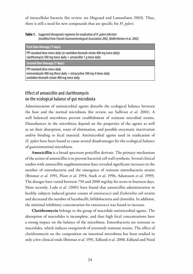
24
of intracellular bacteria (for review, see Megraud and Lamouliatte 2003). Thus, there is still a need for new compounds that are specific for H. pylori.
Table 1. Suggested therapeutic regimens for eradication of H. pylori infection (modified from Finnish Gastroenterological Association 2002, Malfertheiner et al. 2002)
First-line therapy (7 days)
PPI standard dose twice daily (or ranitidine bismuth citrate 400 mg twice daily) clarithromycin 500 mg twice daily + amoxicillin 1 g twice daily
Second-line therapy (7 days)
PPI standard dose twice dailymetronidazole 400 mg thrice daily + tetracycline 500 mg 4 times daily ranitidine bismuth citrate 400 mg twice daily
Effect of amoxicillin and clarithromycin on the ecological balance of gut microbiotaAdministration of antimicrobial agents disturbs the ecological balance between the host and the normal microbiota (for review, see Sullivan et al. 2001). A well balanced microbiota prevent establishment of resistant microbial strains. Disturbances in the microbiota depend on the properties of the agents as well as on their absorption, route of elimination, and possible enzymatic inactivation and/or binding to fecal material. Antimicrobial agents used in eradication of H. pylori have been found to cause several disadvantages for the ecological balance of gastrointestinal microbiota.
Amoxicillin is a broad spectrum penicillin derivate. The primary mechanism of the action of amoxicillin is to prevent bacterial cell wall synthesis. Several clinical studies with amoxicillin supplementation have revealed significant increases in the number of enterobacteria and the emergence of resistant enterobacteria strains (Brismar et al. 1991, Floor et al. 1994, Stark et al. 1996, Adamsson et al. 1999). The dosages have varied between 750 and 2000 mg/day for seven to fourteen days. More recently, Lode et al. (2001) have found that amoxicillin administration in healthy subjects induced greater counts of enterococci and Escherichia coli strains and decreased the number of lactobacilli, bifidobacteria and clostridia. In addition, the minimal inhibitory concentration for enterococci was found to increase.
Clarithromycin belongs to the group of macrolide antimicrobial agents. The absorption of macrolides is incomplete, and thus high fecal concentrations have a strong impact on the balance of the microbiota. Enterobacteria are resistant to macrolides, which induces overgrowth of extremely resistant strains. The effect of clarithromycin on the composition on intestinal microbiota has been studied in only a few clinical trials (Brismar et al. 1991, Edlund et al. 2000, Edlund and Nord

25
2000). The dosage regimens have been from 500 to 1000 mg/day for seven to ten days. These studies clearly demonstrate that marked reduction in the number of lactobacilli, bifidobacteria and bacteroides is evident in the anaerobic microbiota. Reduction in enterobacterial group bacteria and increased numbers of new resistant enterobacteria and enterococci strains have also been observed after treatment.
Proton pump inhibitorsProton pump inhibitors (PPIs) inhibit H+/K+-ATPase via covalent binding to cysteine residues of the proton pump, and they remain the most effective available therapy to control gastric acid secretion (for review, see Sachs et al. 2006). Omeprazole was the first clinically used PPI. Others available include lansoprazole, pantoprazole, rabeprazole and tenatoprazole. The main difference between the PPIs is the duration of inhibition of gastric acid secretion. However, this has not affected H. pylori eradication treatment efficacy when systematically compared (for review, see Bazzoli et al. 2002). Omeprazole and lansoprazole remain reference PPIs in first-line eradication treatment of H. pylori infection.
PPIs have shown an excellent safety profile, but there have been concerns that the suppression of gastric acid might alter the gastric and duodenal microbiota, which could lead to gastric cancer, enteric or other infections and malabsorptions (for review, see Williams and McColl 2006). It has been demonstrated that elevation of intragastric pH by antisecretory drugs increases bacterial growth both in mucosal biopsies and in gastric juice. Moreover, it has been found that H. pylori infected patients have greater increases of gastric pH during PPI treatment than non-infected patients (Williams and McColl 2006). The overall evidence indicates that PPIs are linked with an increased risk of enteric infections (for review, see Williams and McColl 2006) and the production of carcinogenic acetaldehyde in the upper gastrointestinal tract (Väkeväinen et al. 2000). However, there are no convincing results showing production of nitrosamines or interference with intestinal digestive processes (Williams and McColl 2006).

26
3. probioTics in Helicobacter pylori infEcTion
3.1 ovErviEw of probioTicsFermented dairy products and vegetables have been used for thousands of years. As early as 1907, Nobelist Elie Metchnikoff attributed the longevity of Bulgarian peasants to their consumption of fermented milk products (Metchnikoff 1907). He suggested that regular consumption of dairy yogurt may suppress “putrefactive” bacteria in the colon. Since then, several definitions have been used to describe these probiotics, such as substances that are produced by one microorganism and stimulate the growth of other microorganisms (Lilly and Stillwell 1965), live microbial feed supplements that beneficially affect the host animal by improving its intestinal microbial balance (Fuller 1989), and more recently, live microorganisms that, when administered in adequate amounts, confer a health benefit on the host (FAO/WHO 2002).
The most commonly used organisms in probiotic products belongs to Lactobacillus sp., and Bifidobacterium sp. (Saxelin et al. 2005). Other organisms have also been used including Bacillus sp., and yeast such as Saccharomyces boulardii. Probiotic products are commercially available in different formulations with and without prebiotics such as fructo- and galacto-oligosaccharides. The concentration of probiotics in research trials and in food or other products varies greatly, and there are no international standards regarding the levels of bacteria required (for review, see Parvez et al. 2006).
Several proposed health effects of probiotics are summarized in Figure 2. The primary clinical interest in the application of probiotics has been in the prevention and treatment of gastrointestinal infections and diseases (Parvez et al. 2006). The use of probiotics for controling chronic gastrointestinal inflammatory diseases, such as ulcerative colitis and pouchitis, has also received considerable attention. Moreover, the consumption of probiotics has been linked to improvement of a wide variety of health conditions, including lactose intolerance, high cholesterol and rheumatoid arthritis. Also, there is evidence of beneficial effects of probiotics with respect to the development of dental caries, allergy and cancer (Parvez et al. 2006). The general mechanisms by which probiotics may have an effect can be divided into three broad categories: normalization of microbiota, modulation of immune response, and metabolic functions. However, the molecular details behind these mechanisms remain mostly unknown (Marco et al. 2006).

27
3.2 clinical sTudiEsThe role of probiotics in the treatment of H. pylori infection is increasingly documented as a complement or alternative to antibiotics, and thus having the potential to decrease the use of antibiotics.
Probiotics as a complement to antibiotics may have the potential to reduce the adverse events of triple anti-Helicobacter treatment and to improve the eradication rate (Table 2). The first study provided evidence that L. acidophilus LB improved the eradication rate significantly in the probiotic group (Canducci et al. 2000). However, in this study the supplementation did not alleviate the adverse effects of the anti-Helicobacter treatment. In contrast, Armuzzi et al. (2001a, 2001b) reported in two separate studies that L. rhamnosus GG was able to reduce the occurrence of adverse effects, such as diarrhea, taste disturbance, nausea and bloating. The latter of these two studies was conducted in double-blinded fashion. Moreover, Sheu et al. (2002) reported in an open and uncontrolled trial that the L. acidophilus La5 and
Figure 2 Summary of various health effects of probiotics (modified from Parvez 2006). SCFA, short-chain fatty acid
Control of irritable bowel syndrome
Control of imflammatory bowel disease
Supression of endogenous pathogens
Prevention and alleviation of food allergy in infants
Stregthening innate immunity
Lowering serum cholesterol
Improvement of lactose tolerance
Suppression of exogenous pathogens
Reduction in risk factors for colon cancers
probioTics
normalized microbiota composition
immunomodulation
Metabolic effects
Colonization resistance
Supply of SCFA and vitamins to colonic epithelium
Lower level of toxigenic / mutagenic reactions in the gut
Lactose hydrolysis
Bile salt deconjugation and secretion
Colonization resistance

28
B. lactis Bb12 containing yogurt (AB-yogurt) was able to increase the eradication rate and also decrease several side-effects of the triple therapy. Administration of L. rhamnosus GG, Saccharomyces boulardii or combination of L. acidophilus and B. lactis for two weeks also decreased adverse events during the triple treatment (Cremonini et al. 2002). However, the effect of probiotic supplementation seemed independent of the probiotic species used. Tursi et al. (2004) also recently found that a 10-day quadruple anti-Helicobacter therapy with L. casei ssp. casei DG supplementation significantly increased the eradication rate in patients after failure of first-line eradication treatment. Similarly, L. acidophilus La5 combined with B. lactis Bb12 improved the second-line rescue therapy in patients with H. pylori resistance (Sheu et al. 2006). There are two studies conducted with dyspeptic children. In the first, L. casei DN-114 001 containing fermented milk product was effective in increasing the eradication rate of standard triple treatment (Sykora et al. 2005). More recently, L. reuteri was found to alleviate eradication treatment associated adverse effects, but it was not able to increase the eradication rate (Lionetti et al. 2006).
There are not many studies on the attenuation of microbiota disturbances with probiotics following an anti-Helicobacter triple treatment. In a pilot study, Madden et al. (2005) found that probiotic combination including two strains of L. acidophilus (CLT60 and CUL21) and two strains of B. bifidum (CUL17 and B. bifidum Rhodia) stabilized the number of facultative anaerobes. Later the same probiotic product was able to reduce the amount of antibiotic resistance among enterococci and reduce the disruption of the enterobacterial component in the re-growth population (Plummer et al. 2005). However, despite the probiotic supplementation, the microbiota in both studies was susceptible to the effects of the antibiotics administered to eradicate H. pylori.

29
Patie
nt g
roup
Stud
y des
ign
Erad
icatio
n th
erap
yPr
obio
tic st
rain
(s)
Prod
uct,
dose
, tim
eRe
sults
Refe
renc
e
120 d
yspe
ptic
adult
sO,
RCA
+ ra
bepr
azole
L. ac
idoph
ilus L
BCa
psule
, inac
tivat
ed ba
cteria
,1.5
x 10
10 CF
U/da
y,10
days
Eradic
ation
rate
↑Ad
verse
effec
ts ↔
Cand
ucci
et al.
20
00
60 as
ympt
omat
ic ad
ults
O, R
CT +
pant
opra
zole
L. rh
amno
sus G
GFre
eze-
dried
powd
er,
1.2x1
010 CF
U/da
y, 14
days
Eradic
ation
rate
↔Ad
verse
effec
ts ↓
Arm
uzzi
et al.
2001
a
120 a
sym
ptom
atic
adult
sDB
PC,R
CT +
pant
opra
zole
L. rh
amno
sus G
GFre
eze-
dried
powd
er,1.2
x1010
CFU/
day,
14
days
Eradic
ation
rate
↔Ad
verse
effec
ts ↓
Arm
uzzi
et al.
2001
b
160 d
yspe
ptic
adult
sO,
RCA
+lan
sopr
azole
L. ac
idoph
ilus L
a5 an
d B.
lactis
Bb12
Yogh
urt,
1x10
10 CF
U/da
y,
4 wee
ks
Eradic
ation
rate
↑Ad
verse
effec
ts ↓
Sheu
et al
.20
02
85 as
ympt
omat
ic ad
ults
DBPC
,RCT
+ra
bepr
azole
1. L.
rham
nosu
s GG,
2.
Sacch
arom
yces
boula
rdii,
3. L.
acido
philu
s La5
and
B. la
ctis B
b12
Freez
e-dr
ied po
wder,
1-
1.5 x
1010
CFU/
day,
2 wee
ks
Eradic
ation
rate
↔Ad
verse
effec
ts ↓
by a
ll pr
obiot
ics
Crem
onini
et al
.20
02
70 dy
spep
tic ad
ults w
ith
resis
tant
H. p
ylori
RAT
+ ra
nitidi
nebis
mut
h citr
ate+
esom
epra
zole
or pa
ntop
razo
le
L. ca
sei s
sp. c
asei
DGCa
psule
, 1.6
x1010
CFU/
day,
10 da
ys
Eradic
ation
rate
↔Ad
verse
effec
ts ↓
Tursi
et al
.20
04
86 dy
spep
tic ch
ildre
nDB
PC,R
CA +
omep
razo
leL.
case
i DN-
114 0
01Fe
rmen
ted m
ilk,
1x10
10 CF
U/da
y, 2 w
eeks
Eradic
ation
rate
↑Ad
verse
effec
ts ↔
Syko
ra et
al.
2005
40 dy
spep
tic ch
ildre
nDB
PC,R
A + om
epra
zole
5 day
s foll
owing
CT
+ om
epra
zole
5 day
sL.
reuter
i ATC
C 557
30Ca
psule
,1x
108 CF
U/da
y,20
days
Eradic
ation
rate
↔Ad
verse
effec
ts ↓
Lione
tti et
al.
2006
138 d
yspe
ptic
adult
s with
re
sista
nt H
. pylo
ri O,
RAM
+ bi
smut
h citr
ate+
omep
razo
leL.
acido
philu
s La5
and
B. lac
tis Bb
12Yo
gurt,
4x
1010
CFU/
day,
4 wee
ks be
fore
erad
icatio
n tre
atm
ent
Urea
se ac
tivity
↓ du
ring
pret
reat
men
t,Era
dicat
ion ra
te ↑
Adve
rse eff
ects
↓
Sheu
et al
.20
06
Tabl
e 2.
Clinic
al tri
als us
ing pr
obiot
ics as
a co
mple
men
t dur
ing H
. pylo
ri era
dicat
ion tr
eatm
ent.
O, op
en; R
, rand
omize
d; DB
PC, d
ouble
-blin
d plac
ebo-
cont
rolle
d; C,
clarit
hrom
ycin;
A, am
oxici
llin; T
, tini
dazo
le; M
, met
ronid
azole
; CFU
, colo
ny fo
rming
units
; ↑, in
creas
e; ↓
, dec
reas
e; ↔
, no e
ffect

30
Probiotics as an alternative to antimicrobials have also been the focus of several studies (Table 3). Administration of culture supernatant or fermented milk containing the strain of L. acidophilus La1 decreased H. pylori urease activity, measured by 13C-UBT in adults (Michetti et al. 1999) and in children (Cruchet et al. 2003), and also in two other trials by histological analysis (Felley et al. 2001, Pantoflickova et al. 2003). Furthermore, in the latter two studies, a decrease in H. pylori infection-associated inflammation was evident. However, the regular intake of L. acidophilus ( johnsonii) La1 did not eradicate H. pylori in any of the studies. Sakamoto et al. (2001) found L. gasseri OLL2716 to be effective in suppression of H. pylori and reduction in gastric mucosal inflammation as measured by 13C-UBT and assays of serum pepsinogen I. In their study, 31 subjects with H. pylori infection ingested yogurt containing L. gasseri daily for an eight-week period. L. casei was also shown to inhibit H. pylori growth and to reduce 13C-UBT values (Cats et al. 2003). Similar effects on growth of H. pylori were reported for yogurt containing L. acidophilus La5 and B. lactis Bb12 and consumed for 6 weeks by 59 human volunteers (Wang et al. 2004). However, not all clinical trials have shown effectiveness. In one open study, 27 H. pylori infected volunteers received yogurt containing three Lactobacillus spp. and one commercial starter culture for one month (Wendakoon et al. 2002). At the end of the trial 13C-UBT values remained positive in 26 of the 27 subjects. However, this study used strains that were not proven probiotics.
Studies on the effects of synbiotics (probiotics combined with prebiotics) on H. pylori infection are very scarce, and to my knowledge, no clinical studies on prebiotics exist. A randomized, open, eight-week study investigated the effects of L. acidophilus LB in comparison with antibiotics and with the synbiotic combination of probiotic yeast Saccharomyces boulardii with inulin (Gotteland et al. 2005). The eradication rate was slightly better in the S. boulardii combined with inulin study group.
To summarize, these observations suggest that consumption of certain strains of probiotics may be useful in combating H. pylori infection as a complement to the first-line or the second-line eradication therapy. Generally, complete eradication of H. pylori without anti-helicobacter therapy has not succeeded. However, regular consumption of probiotic products with specific strains as an alternative to antibiotics may have some potential in the suppression of H. pylori infection.

31
Patie
nts
Desig
nPr
obio
tic st
rain
(s)Pr
oduc
t, do
se, t
ime
Resu
ltsRe
fere
nce
20 as
ympt
omat
ic ad
ults
R, D
B, PC
L. ac
idoph
ilus (
jonhs
onii)
La1
Cultu
re su
pern
atan
t +Om
epra
zole,
dose
NA,
14 da
ys
Eradic
ation
↔,
Urea
se ac
tivity
↓, H
. pylo
ri colo
nizat
ion ↔
,In
flam
mat
ion an
d gas
tritis
↔
Mich
etti
et al.
19
99
52 as
ympt
omat
ic ad
ults
R, D
B, PC
L. ac
idoph
ilus (
jonhs
onii)
La1
Acidi
fied m
ilk +
clarit
hrom
ycin,
dose
NA,
3 wee
ks
Eradic
ation
↔,
Urea
se ac
tivity
↓, H
. pylo
ri colo
nizat
ion ↓
,In
flam
mat
ion an
d gas
tritis
↓
Felle
y et a
l.20
01
31 as
ympt
omat
ic ad
ults
PCL.
gasse
ri OLL
2716
Yogu
rt,1.8
-2.5
x 109 CF
U/da
y8 w
eeks
Seru
m pe
psino
gen I
/ II r
atio
↑,
Seru
m pe
psino
gen ↓
,Ur
ease
activ
ity ↓
Saka
mot
o et a
l. 20
01
27 as
ympt
omat
ic ad
ults
OL.
case
i 03,
L. ac
idoph
ilus 2
412
and L
. acid
ophil
us AC
D1Yo
gurt,
2.8 x
1011
CFU/
day,
30 da
ysUr
ease
activ
ity ↔
Wen
dako
on et
al.
2002
236 a
sym
ptom
atic
child
ren
DB, P
CLiv
ing an
d hea
t kille
d L.
acido
philu
s La1
or L.
pa
raca
sei S
T1
Ferm
ente
d milk
prod
ucts,
1x
1010
CFU/
day,
4 wee
ks
Eradic
ation
↔,
Urea
se ac
tivity
↓ by
live L
a1Cr
uche
t et a
l. 20
03
50 as
ympt
omat
ic ad
ults
DB, P
CL.
acido
philu
s (joh
nson
ii) La
1Ac
ified
milk
,1.2
5 x 10
9-10
CFU/
day,
16 w
eeks
Eradic
ation
↔, H
. pylo
ri colo
nizat
ion ↓
, In
flam
mat
ion ↓
and g
astri
tis ↔
Pant
oflick
ova e
t al.
2003
20 as
ympt
omat
ic ad
ults,
6 ad
ults i
n con
trol g
roup
O, C
L. ca
sei S
hirot
aM
ilk ba
sed d
rink,
1.95 x
1010
CFU/
day,
3 wee
ks
Eradic
ation
↔,
Urea
se ac
tivity
tend
ed to
↓Ca
ts et
al.
2003
70 dy
spep
tic ad
ults,
endo
s-co
py fo
r 14 s
ubjec
tsO,
CL.
acido
philu
s La5
and B
. lacti
s Bb
12Yo
gurt,
1x10
10 CF
U/da
y, 4 w
eeks
Eradic
ation
↔, U
reas
e acti
vity ↓
, Ga
striti
s and
H. p
ylori c
oloniz
ation
↓W
ang e
t al.
2004
254 a
sym
ptom
atic
child
ren
O, R
L.acid
ophil
us LB
or Sa
cchar
o-my
ces b
oular
dii w
ith in
ulin
Caps
ule or
sach
et,
LB 1x
1010
CFU/
day,
S. bo
ulard
ii 500
mg +
10 g
inulin
/day
, 8 w
eeks
Eradic
ation
↓S.
boula
rdii w
ith in
ulin m
ore e
ffecti
ve th
an L.
ac
idoph
ilus L
B
Gotte
land e
t al.
2005
Tabl
e 3.
Clinic
al tri
als us
ing pr
obiot
ics in
the t
reatm
ent o
f H. p
ylori i
nfec
tion.
R, ra
ndom
ized;
DB,
doub
le-bli
nd; P
C, pla
cebo
-con
trolle
d; O,
open
; L., L
acto
bacil
lus; C
FU, c
olony
form
ing un
its; N
A, no
t ava
ilable
; ↑, in
creas
e; ↓
, dec
reas
e; ↔
, no e
ffect

32
3.3 ExpEriMEnTal sTudiEsVarious probiotics have shown favorable effects in animal models of H. pylori infection (Table 4). The first two studies presented a highly protective and therapeutic effect of oral administration of L. salivarius on H. pylori infected gnotobiotics (animals that have been raised in germ-free environments, or contain only specific germs) BALB/c mice model (Kabir et al. 1997, Aiba et al. 1998). Similarly, Coconnier et al. (1998) reported that L. acidophilus strain LB was able to protect against H. pylori infection in conventional mice. Inhibition of stomach colonization of H. felis SC1 (a bacterium closely related to H. pylori) was observed and no evidence of gastric histopathological lesions was found. Recently, probiotic combination containing L. acidophilus R0052 and L. rhamnosus R0011 reduced the effects of H. pylori infection in a C57BL/6 mice model of infection through reducing H. pylori colonization and alleviating H. pylori-induced inflammation of the stomach (Johnson-Henry et al. 2004). Also, the same probiotic preparation has proven effective in a Mongolian gerbil model of H. pylori infection via its attenuating effect on the H. pylori colonization, the mucosal inflammation, and the impairment of the gastrin-somatostatin link (Brzozowski et al. 2006). Studies by Sgouras et al. (2004, 2005) in a C57BL/6 mice model demonstrated that L. casei strain Shirota and L. johnsonii La1, both administered in drinking water, attenuated H. pylori infection-induced gastric mucosa inflammation. However, only L. casei strain Shirota was able to down-regulate the colonization of H. pylori to gastric mucosa. Moreover, L. gasseri was found to decrease clarithromycin resistant H. pylori colonization (Ushiyama et al. 2003).
Development of an effective vaccine is also an interesting area in the prevention of H. pylori infection. However, the ability of recombinant Lactobacillus or other probiotics to be used as an antigen-delivery vehicle to induce protective immune responses has rarely been studied. In the study by Lee et al. (2001), Lactococcus lactis producing cytoplasmic urease B was shown to be unable to induce protection against H. pylori in a mouse model. In contrast, a recombinant L. plantarum strain producing H. pylori urease B subunit was found to successfully induce a partial mucosal protection against Helicobacter (Corthésy et al. 2005).

33
Mod
elSt
udy p
roto
col
Prob
iotic
stra
in(s)
Dose
, tim
eRe
sults
Refe
renc
e
Gnot
obiot
icBA
LB/c
mice
Prev
entio
n and
tre
atm
ent
L. sa
livar
ius (W
B 100
4)L.
brev
is (W
B100
5)10
9 CFU
once
for 1
wk b
efore
or 4
week
s afte
r H.
pylor
i infec
tion
Only
L. sa
livar
ius eff
ectiv
eAn
ti-H.
pylor
i IgG
titer
s ↓H.
pylor
i colo
nizat
ion ↓
Kabir
et al
. 19
97
Gnot
obiot
icBA
LB/c
mice
Treat
men
tL.
saliv
arius
(WB1
004)
L. ac
idoph
ilus (
ATCC
393)
L. ca
sei (A
TCC 4
356)
109 CF
U, fir
st we
ek 3
times
, the
n onc
e per
wee
k fo
r 3 w
eeks
Only
L. sa
livar
ius eff
ectiv
eAn
ti-H.
pylor
i IgG
titer
s ↓Ai
ba et
al.
1998
Conv
entio
nal B
ALB/
c m
iceTre
atm
ent
L. ac
idoph
ilus s
train
LB5x
108 CF
U fo
r 7 da
ysH.
felis
colon
izatio
n ↓Ga
stric
inflam
mat
ion ↓
Coco
nnier
et al
. 19
98
Gnot
obiot
icBA
LB/c
mice
Treat
men
tL.
gasse
ri OLL
2716
109 CF
U on
ce pe
r wee
k, 4 w
eeks
H. py
lori c
oloniz
ation
↓Us
hiyam
a et a
l.20
03
C57B
L/6 m
icePr
even
tion a
nd
treat
men
tL.
acido
philu
s R00
52 an
dL.
rham
nosu
s R00
1110
9 CFU/
ml (
ad lib
itum
),7 d
ays p
rior H
. pylo
ri inf
ectio
n an
d 49 d
ays a
fter
H. py
lori g
rowt
h ↓Ga
stric
inflam
mat
ion ↓
Apop
tosis
↔
John
son-
Henr
y et a
l. 20
04
C57B
L/6 m
iceTre
atm
ent
L. ca
sei s
train
Shiro
ta10
8 CFU/
ml (
ad lib
itum
), 9 m
onth
sCh
ronic
gastr
itis ↓
Anti-
H. py
lori Ig
G tit
ers ↓
H. py
lori c
oloniz
ation
↓
Sgou
ras e
t al.
2004
C57B
L/6 m
iceTre
atm
ent
L. joh
nson
ii La1
L. am
ylovo
rus C
DE 47
1L.
acido
philu
s IBB
801
1.5 x
108 CF
U /d
aily,
3 mon
ths
2.1 x
108 CF
U /d
aily,
3 mon
ths
4.6 x
108 CF
U /d
aily,
3 mon
ths
Chro
nic ga
striti
s ↓An
ti-H.
pylor
i IgG
titer
s ↓H.
pylor
i colo
nizat
ion ↔
Sgou
ras e
t al.
2005
Mon
golia
n ger
bilPr
even
tion a
nd
treat
men
tL.
acido
philu
s R00
52 an
dL.
rham
nosu
s R00
112x
109 CF
U 4 h
ours
befo
re H
. pylo
ri and
daily
for
2 wee
ksCh
ronic
gastr
itis ↓
Gastr
ic ac
id ou
tput
↔Ga
strin
↓
Brzo
zows
ki et
al.
2006
Tabl
e 4.
Prob
iotics
used
in ex
perim
enta
l H. p
ylori i
nfec
tion.
L., La
ctoba
cillus
; CFU
, colo
ny fo
rming
units
; IgG,
imm
unog
lobuli
n G; ↑
, incre
ase;
↓, d
ecre
ase;
↔, n
o effe
ct

34
3.4 possiblE MEchanisMs of probioTic acTionThe mechanisms of probiotic action on H. pylori infection are unclear, but there are a number of proposed or hypothesized possibilities from in vitro studies of host intestinal epithelial or immune cell responses to probiotic strains. A summary of these possible mechanisms is provided in Figure 3 and described here in more detail.
Generally, probiotics such as lactic acid bacteria and bifidobacteria are able to produce organic acids, hydrogen peroxide and carbon dioxide to inhibit potential pathogens. In addition, many probiotics have been found to produce more defined antimicrobial substances (for review, see Servin 2004). Coconnier et al. (1998) found that the anti-Helicobacter substance(s) in the L. acidophilus LB strain were different from lactic acid. Also, L. johnsonii La1, shown to be beneficial in several clinical and experimental studies in treatment of H. pylori infection, has been found to release non-bacteriocin antimicrobial substances (Bernet-Camard et al. 1997). Furthermore, some Bifidobacterium strains have been found to release heat-stable proteinaceous antimicrobial compounds against H. pylori in vitro (Collado et al. 2005).
The anti-infective activity of probiotics may also partly be due to coaggregation with pathogens (Cesena et al. 2001), whereby pathogens are exposed to high doses of potential growth-inhibiting factors produced by probiotics. One mechanism proposed recently is that the L. johnsonii La1 expresses cell-surface associated La1GroEL protein, and its recombinant variant, expressed in Escherichia coli, is able to induce aggregation of H. pylori, but not of other intestinal pathogens. The L. johnsonii La1 was also shown to have pro-inflammatory activity, thus favoring the activation of intestinal immunological defences (Bergonzelli et al. 2006). Adhesion of pathogens can also be inhibited by steric hindrance, where a large number of beneficial bacteria may cover receptor sites in a non-specific manner, or by competing for specific carbohydrate receptors that would otherwise be available to pathogens. Several probiotic species, such as L. salivarius, L. gasseri and L. acidophilus, have shown growth inhibition or anti-adhesion capacity against H. pylori in a gastric epithelial cell model (Midolo et al. 1995, Coconnier et al. 1998, Lorca et al. 2001, Mukai et al. 2002, Nam et al. 2002, Sgouras et al. 2004, Tsai et al. 2004).
Mukai et al. (2002) have also examined competition in the binding of nine L. reuteri strains and H. pylori to gangliotetraosylceramide (Asialo-GM1) and sulfatide, which are putative glycolipid receptor molecules of H. pylori, and identified a possible sulfatide and Asialo-GM1 binding protein of the two L. reuteri strains (JCM1081 and TM105). Moreover, several probiotics are able to inhibit adhesion of pathogenic microorganisms by modifying the glycosylation state of the receptor
35
in epithelial cells using soluble factor(s) excreted by the probiotics (for review see, Servin 2004).
Several probiotic bacteria have been shown to prevent and repair mucosal damage by inhibiting damage to tight junction proteins (Montalto et al., 2004). The probiotic strains prevented the pathogen-associated disruption of the cytoskeletal and tight junction proteins in the epithelial cells, thus improving the mucosal barrier function and preventing failure in the secretion of electrolytes (Resta-Lenert and Barrett 2002, 2003). Regular ingestion of live L. rhamnosus GG protected the integrity of gastric mucosa, as evaluated by the sucrose permeability test, against alterations by indomethacin (Gotteland et al. 2001), thus suggesting at least transient residence in the human stomach and functional effectivity. Probiotic combination of VSL#3, which is a mixture of eight different strains (L. acidophilus, L. delbruckii ssp. bulgarus, L. casei, L. plantarum, Bifidobacterium longum, B. infantis, B. breve and Streptococcus thermophilus), and soluble factors of L. rhamnosus GG were able to induce specific heat shock proteins, known for their ability to maintain actin cytoskeleton integrity (Petrof et al. 2004, Tao et al. 2006). Further, a novel mechanism of maintaining barrier function was identified by Yan and Polk (2002). These investigators showed that L. rhamnosus GG was able to prevent cytokine-induced apoptosis in intestinal epithelial cell models through the inhibition of a TNF-induced activation of the proapoptotic p38/mitogen-activated protein kinase.
Several reports suggest that probiotics are able to differentially modulate innate immune responses in both anti-inflammatory and pro-inflammatory directions. Probiotic bacteria can bind to recognition receptors, such as TLRs expressed on the surface of epithelial cells, and thus trigger a cascade of immunological defence mechanisms (for review, see Saxelin et al. 2005, Sartor 2005). TLR4 recognizes
lipopolysaccharide and gram-negative bacteria, while TLR2 recognizes a variety of microbial components, such as peptidoglycan and lipoteichoic acids, from gram-positive bacteria (for review, see Abreu 2003, Matsuguchi et al. 2003, Takeda et al. 2003). Oral CpG delivery was found to reduce significantly H. pylori colonization in a mouse model of H. pylori infection (Raghavan et al. 2003). Thus bifidobacteria, which generally contain DNA with high GC contents and hence a high fraction of unmethylated CpG sequences, could affect the immune system by interacting with TLR9, triggering the production of pro-inflammatory cytokines and promoting T helper cell -1 response to reduce H. pylori colonization (Saxelin et al. 2005). The possible mechanism underlying this effect, when regarding concomitant CpG administration, is enhanced mucosal cytokine production and immune stimulation.

36
Also, enhancement of secretory IgA production on intestinal epithelium may have a role in pathogenic bacterial defence (Viljanen et al. 2005c). Furthermore, it was shown recently that L. rhamnosus GG is able to antagonize H. pylori induced TNF-α production by murine macrophages in vitro by a contact-independent mechanism (Peña et al. 2003). Probiotics could down-regulate virulence genes in pathogenic bacteria. Furthermore, mucins, high-molecular-weight glycoproteins secreted by epithelial cells, may also bind to pathogens, thereby inhibiting their adherence to epithelial cells. Moreover, release of gastric mucin is found to be down-regulated by H. pylori (Byrd et al. 2000). Probiotics could thus interfere with the actions of H. pylori, since several strains, for example Lactobacillus plantarum 299V and L. rhamnosus GG, have induced mucin gene expression (Mack et al. 1999, Mattar et al. 2002, Mack et al. 2003).
Various microorganisms have been described as binding the dendritic cell (DC) receptor specific intercellular adhesion molecule 3-grabbing nonintegrin (DC-SIGN), including H. pylori. It has been suggested that distinct ligands from different microorganisms show different functional effects. Ligation of DC-SIGN by H. pylori induces increased levels of IL-10 in human DCs and a shift toward Th2 development (Bergman et al. 2004). In contrast, probiotics, specifically L. reuteri and L. casei, do not induce increased levels of IL-10 from DCs. Probiotics were also found to induce regulatory T cell development through modulation of DC function (Smits et al. 2005). It is currently unclear which different signals are transduced by DC-SIGN ligation. Probiotic bacteria such as L. rhamnosus induced maturation of dendritic cells, but resulted in a different cytokine profile than pathogenic bacteria (Braat et al. 2004). The probiotic product VSL#3 upregulated IL-10 and down-regulated IL-12 production by dendritic cells derived from human blood and the lamina propria. The main effect of this combination is found to come from bifidobacteria (Hart et al. 2004). Also, the Bifidobacterium strain's DNA from the same combination, VSL#3, is effective in enhancing the production of IL-1 and IL-10 (Lammers et al. 2003).

37
3.5 safETy aspEcTsData on the safety of probiotics suggest that probiotic therapy is generally considered safe (Boyle 2006, Hammerman et al. 2006). Lactobacilli and bifidobacteria are part of normal gastrointestinal microbiota, and systemic infections with these bacteria may thus occur naturally, unrelated to the ingestion of probiotics. Accordingly, increased probiotic use of L. rhamnosus GG in Finland has not led to an increase in Lactobacillus bacteremia (Salminen et al. 2002) and is also regarded as safe in immunocompromised HIV-infected patients (Salminen et al. 2004). Bifidobacteria are found to be even safer because no cases of sepsis related to probiotic ingestions have been reported. Propionibacteria are also regarded as
Figure 3 Schematic presentation of possible mechanisms of probiotics to interfere with H. pylori induced functional effects (modified from Marco et al. 2006). Probiotics potentially interfere with H. pylori by production of antimicrobial substances (1), inducing aggregation (2) and competing with host-cell-binding sites (3). They could modulate inflammatory responses (4) and strengthen the mucosal barrier (5). Probiotics with a high fraction of CpG sequences can possibly reduce H. pylori colonization (6). Differential modulation of cytokine production recognised by Toll-like receptors (TLR) (7), enhancement of secretory immunoglobulin A (Ig) A production (8) and decreased tumor necrosis factor (TNF)-α production by macrophages could have a role in H. pylori infection (9). Probiotics could down-regulate the production of virulence factors of H. pylori (10) and induce mucin release from epithelial cells (11). They could compete with H. pylori in binding to dendritic cell (DC) receptor specific intercellular adhesion molecule 3-grabbing nonintegrin (SIGN) (12) and modulate DC function and regulatory T (Tr) cell development (13). B, B cell (plasma cell); DC, dendritic cell; Hsp, heat shock protein; IL, interleukin; NF, nuclear factor; Th, T helper cell

38
safe to use, mainly because of their long history of safe use in Emmental cheese manufacturing. P. freudenreichii ssp. shermanii has consequently been proposed for “Qualified Presumption of Safety” status by the European Food Safety Authority (EFSA 2007).
Safety considerations regarding antimicrobial resistance are also recognized among probiotics. There are questions about the possibility of resistance transfer both from probiotics to pathogenic bacteria and from commensal microbiota to probiotics (for review, see Courvalin et al. 2006). Probiotics strains are generally susceptible to the majority of antibiotics, but a recent clinical study shows that they can survive gastrointestinal transit quite well during antibiotic treatment (Saarela et al. 2007). However, this did not lead to transfer of resistance genes from original microbiota to the ingested probiotics. In any case, different strains of probiotics have different safety profiles, which should be taken into account, and generalizations concerning all probiotics should be avoided (Hammermann et al. 2006).

39
The aim of the present study was to discover the role of probiotics in the treatment of Helicobacter pylori infection and to evaluate the mechanisms of action.
The specific aims of this study were:
To clarify the importance of probiotics as an adjuvant to the current recom-mended treatment of H. pylori infection (Study I).
To investigate the effects of H. pylori eradication treatment and probiotics on intestinal microbiota alterations during the treatment of H. pylori infected sub-jects (Study II).
To find out the recovery and the effects of probiotics in gastric and duodenal mucosa in H. pylori infected and uninfected patients (Study III)
To characterize the actions of probiotics in H. pylori infected intestinal epithe-lial cells (Study IV).
°
°
°
°
aiMs of ThE sTudy


41
The subjects, materials and methods are described in detail in the original studies. A summary of the subjects is shown in Table 5.
1. sTudy dEsigns and subjEcTsStudies I and II were conducted in a randomized, double-blind, placebo-controlled design. All participants received a seven-day triple therapy [lansoprazole (30 mg), clarithromycin (500 mg) and amoxicillin (1 g) twice daily; Helipak K, Orion Pharma, Espoo, Finland] and were randomized to receive either a milk-based drink containing a total of 1x10⁹ colony-forming units (CFU) / ml of probiotics or the same drink without the probiotics. Randomization was carried out according to a computer-generated blocked randomization list using a block size of four. The subjects consumed the drink twice a day during the eradication treatment and once a day for the following three weeks. For one month before the study and during the intervention (4 weeks) and the follow-up period (6 weeks), all other probiotic products were forbidden. Exclusion criteria were antibiotic treatment during the previous two months, the use of H2-receptor antagonists, bismuth or proton pump inhibitors (PPI) during the previous two weeks, H. pylori eradication treatment during the previous five years, use of probiotic products during the previous month, gastric surgery, any diagnosed chronic gastrointestinal disease, allergies to drugs used in the study, medication for fungal infection, and pregnancy or lactation.
subjEcTs, MaTErials and METhods

42
A paired study design in which each subject served as his/her own control was used in Study III. The study interventions lasted a total of 56 days (8 weeks) ± 3 days. All patients underwent gastroduodenoscopy, and biopsy samples were taken by an experienced endoscopist. The probiotic preparation intake was started on the morning after the first endoscopic examination, and the last probiotic drink was taken on the evening of the day before the second endoscopic examination. The volunteers fasted over the night preceding each gastroduodenoscopy. Exclusion criteria were upper gastrointestinal tract lesions, a history of upper gastrointestinal surgery, chronic severe atrophy in the gastric or duodenal mucosa and were otherwise similar to Studies I and II. All the subjects were asked to abstain from commercial products containing probiotic microorganisms two weeks prior to the intervention and during the intervention.
Table 5. A summary of the subjects.
Study n Gendermale/female
Age, ymean (range)
Description of study groups
i H. pylori infected 47 18/29 56 (24–69) H. pylori infected with triple eradication treatment
ii Total H. pylori infected Uninfected controls
583919
18/4015/243/16
57 (34–69)44 (26–64)
H. pylori infected with triple eradication treatment and uninfected controls
iii Total H. pylori infected Uninfected controls
1376
1/121/6 0/6
51 (40–69)48 (41–58)
H. pylori infected and uninfected patients referred for gastroduodenoscopy
2. EThicsAll subjects gave their written informed consent (Studies I, II, III). The Ethics Committee of the Joint Authority for the Hospital District of Helsinki and Uusimaa approved the protocols for Studies I and II. The Ethics Committee of the Pirkanmaa Hospital District (Tampere University Hospital, Tampere, Finland) and the research licensing committee at the City of Tampere approved Study III.

43
3. probioTic producTs and bacTErial sTrains
Probiotic products (Studies I, II, III)The probiotic products consisted of four different strains: Lactobacillus rhamnosus GG (ATCC 53103), Lactobacillus rhamnosus LC705 (DSM 7061), Propionibacterium freudenreichii ssp. shermanii JS (DSM 7076), and Bifidobacterium breve Bb99 in Studies I and II or B. lactis Bb12 in Study III (Chr. Hansen, Horsholm, Denmark). This probiotic combination was used because LGG has been found beneficial as a supplement in H. pylori eradication treatment (Armuzzi et al. 2001a, 2001b), and LC705 and Propionibacterium freudenreichii ssp. shermanii JS (PJS) have shown anti-pathogenic effects (Suomalainen and Mäyrä-Mäkinen 1999, Hatakka et al. 2007). Moreover, Bb12 has shown beneficial effects in H. pylori infection in vitro and in vivo (Cremonini et al. 2002, Wang et al. 2004, Sheu et al. 2006). In Studies I and II, two batches of the milk-based drinks were used during the clinical study to maintain product quality and to prevent loss of microbial activity in the products. In Study III, several batches of the drink were used during the study period. The daily dose of the drink was 250 ml, and the amount of each probiotic bacteria in the drink was on average 107 CFU/ml. Microbial quality of the products was regularly assessed during the interventions by microbial plating methods.
Bacterial cultures (Study IV)The lyophilized H. pylori was grown on Brucella agar enriched with horse serum at +37°C for 4 to 6 days under microaerophilic conditions. The bacterial suspension was stored at -80°C. Before the experiments, H. pylori was subcultured twice on Brucella agar as described above. For the experiments, the bacteria were aseptically harvested and suspended in Dulbecco's modified Eagle's medium (DMEM) and kept under microaerophilic conditions until use. H. pylori concentrations were determined by plating methods.
Both L. rhamnosus GG (LGG) and L. rhamnosus LC705 (LC705) were grown in de Man, Rogosa and Sharpe broth at +37°C under aerobic conditions for 18–20 h. PJS was grown in broth for propionibacteria strains at +30°C for 48 h. Bifidobacterium breve Bb99 (Bb99) was grown in de Man, Rogosa and Sharpe broth broth enriched with 1% L-cysteine hydrocloride monohydrate under anaerobic conditions at +37°C for 24 h. E. coli strain DH5α was grown in Luria-Bertani broth under aerobic conditions at +37°C for 18 h. All probiotic bacteria were subcultured three times before harvesting for the experiments. Harvested bacteria

44
were centrifuged, washed with phosphate buffered saline (PBS) and resuspended at 109 CFU/ml, as estimated by plating methods, in DMEM.
Table 6. Summary of bacterial strains used in study IV.
Srain Origin
L. rhamnosus GG, ATCC 53103 Isolated from adult human
L. rhamnosus LC705, DSM 7061 Isolated from milk
Propionibacterium freudenreichii ssp. shermanii JS, DSM 7076
Isolated from cheese
Bifidobacterium breve Bb99, DSM 13692
Isolated from a 3-month-old breast-fed infant
Helicobacter pylori, NCTC 11637 Isolated from human, gastric antrum (cytotoxin-as-sociated gene A -positive strain)
Escherichia coli, DH5α General laboratory strain
4. METhods4.1 H. pylori infEcTion assEssMEnT13C-urea breath test (Studies I, II, III)After an overnight fast, subjects gave duplicate breath samples before and ten minutes after swallowing the 13C-labeled urea tablets with a glass of water (Diabact UBT, Diabact AB, Uppsala, Sweden). The breath samples were analyzed using isotope ratio mass spectrometry, and delta over baseline values over 4 per mil (‰) was considered confirmation of a positive H. pylori infection.
Serological tests (Studies I, II, III)For each volunteer (studies I and II), rapid whole-blood tests (Pyloriset Screen II, Orion Diagnostica, Espoo, Finland, and Biocard Helicobacter pylori IgG, AniBiotech Ltd, Helsinki, Finland) were carried out on fingertip blood according to the manufacturers’ instructions. A blood sample was obtained from each subject, and the sera were stored at -20°C until analysis. Serum samples collected at the baseline were determined by an in-house EIA, together with samples collected at 4 months after therapy, for H. pylori antibodies of the IgG and IgA classes. As an indicator of successful eradication therapy, antibody titers of the IgG class had fallen by more than 40% from the pre-treatment level (Studies I and II). In Study III, serum samples were stored before analysis at -20 °C for determination of IgG-H. Pylori levels by ELISA according to the manufacturer's instructions (Biohit
45
Ltd., Helsinki, Finland). The normal reference value for IgG-H. Pylori titers was <44 IU.
Histology (Study III)The presence and colonization of H. pylori infection was determined by modified
Giemsa staining. Gastric biopsies (two from the antrum and two from the corpus)
were obtained from each patient for analysis. Biopsy samples were fixed in 4% paraformaldehyde, stained with hematoxylin-eosin and Giemsa, and subsequently
evaluated by an experienced pathologist.
Biopsy-based rapid urease test (Study III)A rapid urease test was performed according to the manufacturer’s instructions (Biohit) on at least one biopsy sample from either the gastric antrum or the corpus. The biopsy sample was placed on gel containing urea and phenol red as an indicator. The color change was monitored after 2 and after 30 minutes.
4.2 QuEsTionnairEs
Treatment related symptoms (Study I)The participants recorded their daily symptoms in a standardized questionnaire, modified from de Boer et al. (1996), at baseline and during the entire intervention period, five weeks in all. Baseline symptoms were recorded daily for one week before the intervention. The following symptoms were included in the total symptom score: epigastric pain, bloating, flatulence, taste disturbance, loss of appetite, nausea, vomiting, heartburn, constipation, and skin rash. The symptoms were scored from 0 to 3: absent (0), mild (1), moderate (2) and severe (3).
Bowel function (Study I)The participants also filled in a daily questionnaire on their bowel function (frequency and consistency). Stool consistency was graded from hard (0) to watery (5). Diarrhea was defined as at least three watery or loose stools per day for a minimum of two consecutive days.
Probiotic drink consumption (Studies I, II, III)Compliance regarding consumption of the study drink was evaluated by questionnaires in all clinical studies.

46
4.3 Microbiological METhods
Molecular methodsFor FISH analysis, fecal samples were diluted, homogenized and centrifuged to remove the coarse material. The supernatant was mixed 1:4 with fresh 4% paraformaldehyde and fixed. The fixed bacterial cells were collected, washed and resuspended in PBS with added 94% ethanol. The cell suspension was diluted 1:20 with hybridization buffer and the fluorescent probe was added (Table 7). Hybridization was carried out overnight; washing and filtering were performed according to Franks et al. (1998). Filters were mounted on a slide with AntiFade reagent and covered with a glass. Cells were counted visually with an epifluorescence microscope. DAPI dye (4',6-diamidino-2-phenylindole) was used for the detection of total bacteria counts.
Table 7. Cy3-labeled oligonucleotide probes used in FISH analysis.
Bacteria group Probe Reference
Bacteroides fragilis Bfra602 Franks et al. 1998
Bacteroides distasonis Bdis656 Langendijk et al. 1995
Bifidobacteria Bif164 Franks et al. 1998
Clostridium histolyticum Chis150 Harmsen et al. 1999
Eubacterium rectale - Clostridium coccoides Erec482 Suau et al. 2001
Lactobacilli and enterococci Lab158 Harmsen et al. 1999
Faecalibacterium prausnitzii Fprau0645 Suau et al. 2001
The concentration of LGG in the feces was analyzed with real-time quantitative PCR (Study II). A strain-specific quantitative PCR method was used for LGG using LightCycler FRET technique (Halme et al. 2002). DNA for PCR was isolated from the feces using a Wizard® Genomic DNA purification kit following the manufacturer’s instructions (Promega, Madison, WI, USA), except that lysozyme and mutanolysin were used during the 1 h incubation at +37°C. The standard DNAs for quantification were isolated from the dilutions of the known concentrations (CFU/ml) of LGG. The concentration of LGG in the culture was determined by plating, and a tenfold dilution series was made for DNA isolation. The isolation steps were the same for the standards and for the fecal DNAs.
For quantification of the PJS strains, fecal samples were melted and ten-fold dilutions were made in Wilkins-Chalgren broth (study II). The dilutions were plated on buffered propionibacterium agar and incubated anaerobically at +30°C for seven days. Typical PJS-like colonies, light yellow in color, were counted and the cell morphology was verified by microscopy. Isolates were further identified

47
by biochemical tests (nitrate reduction and lactose fermentation) and random amplification of polymorphic DNA analysis. PCR amplification was performed with a DyNAzyme® DNA Polymerase kit (Finnzyme, Espoo, Finland) according to the instructions of the manufacturer. PCR was performed straight from a fresh colony. PJS was used as a positive control in all the analyses. The preliminarily count of the amount of PJS strains was corrected according to RAPD-verified PJS isolates.
In Study III, a strain-specific real-time PCR assay was developed for quantification of each strain using a LightCycler FRET technique similarly to that in Study II (Halme et al. 2002, Mikkola et al. 2006), except that the concentration of Bb12-strain was determined with a B. lactis-specific primer pair (Ventura et al. 2001) with the SYBR Green and melting curve analysis method. Each volunteer’s two biopsy samples from the same locus were combined and immersed, vortexed vigorously and cooled on ice. Bacterial genomic DNA was extracted from the biopsy and fecal samples, and pure cultures were isolated with the Wizard® genomic DNA purification kit (Promega) following the manufacturer’s instructions. In addition, lactic acid bacteria (LAB) of the genera Lactobacillus, Pediococcus, Leuconostoc, and Weissella were analyzed with group-specific primers using SYBR Green in real-time PCR to confirm successful DNA isolation (Walter et al. 2001). The standard curves used for quantification of the strains consisted of a series of 10-fold dilutions of the target species genomic DNA.
Culture methodsFor microbial populations analyzed by cultivation (Study II), the fecal samples were thawed in an anaerobic chamber and diluted 1:10 with Wilkins-Chalgren medium. The mixture was homogenized and dilutions were plated on agar plates. Total aerobic bacteria were cultivated on BHI agar and anaerobic bacteria on the same agar reduced before used. Enterobacteria were grown on Violet red bile glucose agar. YGC agar was used for yeast and molds. Propionic acid bacteria were grown on buffered propionic agar.
4.4 Morphological and funcTional sTaTus of ThE gasTroduodEnal Mucosa
Histological evaluation of gastric and duodenal biopsies Six gastric biopsies (two from the antrum, two from the corpus and two from the duodenum) were obtained from each patient for histological analysis. Biopsy samples were fixed in 4% paraformaldehyde, stained with hematoxylin-eosin and

48
subsequently evaluated by an experienced pathologist. The degree of inflammation present in the histological specimens was classified according to the updated Sydney system (Dixon et al. 1996). A grading scale from 0 to 3 (none, mild, moderate, or severe) was assigned for four histological variables: chronic inflammation (mononuclear cell infiltration), activity (polymorphonuclear neutrophil infiltration), intestinal metaplasia, and glandular atrophy for the antrum and corpus separately.
Determination of serum pepsinogens and gastrin-17Serum samples were stored before analysis at −20°C for determination of serum pepsinogen (sPG) I, sPGII and gastrin-17 levels by ELISA, according to the manufacturer's instructions (Biohit). Normal reference values were as follows: sPGI: 25–100 μg/l; sPGII: 2–10 μg/l; and sG-17: 2.5–7.5 pmol/l.
4.5 cEll culTurE METhods
Caco-2 cell culture Caco-2 cells (HTB 37) obtained from the American Type Culture Collection (Manassas, VA, USA) were cultured at +37°C in DMEM supplemented with 10% fetal calf serum (Biological Industries, Kibbutz Beit Haemek, Israel) and antibiotics (penicillin G 100 U/ml, amphotericin B 250 ng/ml, and streptomycin 100 μg/ml, GIBCO). Cells were seeded (1x10⁵ cells/cm2) on microporous inserts as presented in Figure 4 (Transwell, Corning Costar, Corning, NY, USA) or on 12-well plates (Corning Costar). Confluent monolayers were differentiated for 15 to 21 days and were cultured in medium without antibiotics for 24 h before each experiment.
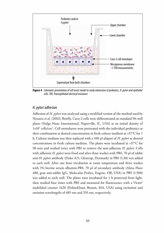
49
H. pylori adhesionAdhesion of H. pylori was analyzed using a modified version of the method used by Nozawa et al. (2002). Briefly, Caco-2 cells were differentiated on standard 96-well plates (Nalge Nunc International, Naperville, IL, USA) at an initial density of 1x10⁵ cells/cm2. Cell monolayers were pretreated with the individual probiotics or their combination at desired concentration in fresh culture medium at +37°C for 1 h. Culture medium was then replaced with a 100 μl aliquot of H. pylori at desired concentrations in fresh culture medium. The plates were incubated at +37°C for 90 min and washed twice with PBS to remove the non-adherent H. pylori. Cells with adherent H. pylori were fixed and after three washes with PBS, 70 μl of rabbit anti-H. pylori antibody (Dako A/S, Glostrup, Denmark) in PBS (1:30) was added to each well. After one hour incubation at room temperature and three washes with 1% bovine serum albumin-PBS, 70 μl of secondary antibody (Alexa Fluor 488, goat anti-rabbit IgG, Molecular Probes, Eugene, OR, USA) in PBS (1:500) was added to each well. The plates were incubated for 1 h protected from light, then washed four times with PBS and measured for fluorescence with a Victor2 multilabel counter 1420 (PerkinElmer, Boston, MA, USA) using excitation and emission wavelengths of 485 nm and 535 nm, respectively.
Probiotics and/orH.pylori
Upper chamber
Lower chamber
Caco-2 cell monolayer
Microporous membrane → TER measurements
Supernatant from both chambers
Figure 4 Schematic presentation of cell insert model to study interaction of probiotics, H. pylori and epithelial cells. TER, Transepithelial electrical resistance
50
Epithelial cell integrityBacteria were added in various concentrations to the apical compartments of cell-culture inserts, and the cultures were incubated at +37°C for 42 h. Transepithelial electrical resistance (TER), as an index of epithelial integrity, was measured with the EVOM™ Epithelial Voltohmeter using “chopstick” electrodes (World Precision Instruments, Stevenage, UK). TER across monolayers was measured at the indicated time points during 42 h incubation. Measurements are expressed in Ω/cm2 after subtracting the mean resistance of cell-free inserts. Prior to experimentation, the Caco-2 monolayers were differentiated for 21 days to acquire a mean baseline TER of 950 Ω/cm2.
Measurement of epithelial cell leakage and apoptosisLactate dehydrogenase (LDH) release indicating cell membrane damage and caspase-3 indicating apoptosis were measured at 8 and 24 h after H. pylori infection. Release of LDH into the culture medium was quantified using a kit from Roche Molecular Biochemicals (Mannheim, Germany). Cell culture supernatants were collected at 8 or 24 h after H. pylori infection, centrifuged to remove particulate matter, and assayed according to the manufacturer’s instructions. Activation of caspase-3 was measured with a kit from Molecular Probes. Cells from 12-well experiments were lysed and assayed according to the manufacturer’s instructions. Fluorescence intensities were measured at excitation and emission wavelenghts of 355 nm and 460 nm, respectively, after one hour incubation at room temperature.
Cytokine and eicosanoid measurementsInterleukins IL-8 and IL-10, and PGE2 and LTB4 were analyzed from culture supernatants by enzyme-linked immunosorbent assay (ELISA) kits according to manufacturer s instructions. The assays’ detection limits were 1 pg/ml for the IL-8 and IL-10 ELISAs (both from CLB, Sanquin, Amsterdam, The Netherlands), and 15 pg/ml and 6 pg/ml for the PGE2 and LTB4 assays (both from Cayman Chemical, Ann Arbor, MI, USA), respectively.
4.6 sTaTisTical analysisIn Study I, the results are expressed as means, 95 percent confidence intervals (CI), medians or interquartile ranges (IQR). Statistical comparison was made using the t test, the Mann-Whitney U test or the Hodges-Lehmann estimation of the shift of medians. Median regression analysis (also known as the least absolute value model) was used to estimate the difference between adjusted medians with the baseline
51
value as a covariate. Measures with a discrete distribution are expressed as counts (%) and analyzed by the chi-square test, Fischer’s exact test or the Fisher-Freeman-Halton test. Hochberg’s adjustments were performed to correct significance levels for the multiple tests.
In Studies II and III, Wilcoxon’s signed rank test for paired samples was used for the comparison of alterations among groups. Two-way Student’s t test was used in Study II to analyze the microbial results between the placebo and probiotic groups. Logarithmic transformation was performed when appropriate. A p value <0.05 was considered significant. In Study IV, statistical differences were analyzed by one-way analyses of variance (ANOVA) and Bonferroni multiple comparisons testing with p values <0.05 considered significant. GraphPad Prism version 3.0 (GraphPad, SanDiego, CA, USA) was used for statistical analyses of the data in Studies II-IV.


53
The main results of Studies I-IV are presented in the following chapters.
1. EffEcTs of probioTics on ThE TolErabiliTy and Efficacy of H. pylori EradicaTion TrEaTMEnT
The probiotic combination therapy significantly reduced the total symptom score during H. pylori eradication treatment. During the eradication week, the baseline-adjusted median symptom score decreased in the probiotic group, whereas an increase was seen in the placebo group, -3 (95% CI: -7 to 0) vs. 3 (95% CI: -1 to 6) respectively, p = 0.038 between the two groups. The median total symptom score was 22 (IQR 15, 34) in the probiotic group and 32 (IQR 13, 46) in the placebo group. As to individual symptoms, major differences in epigastric pain and bloating were observed between the groups.
The probiotic combination therapy did not significantly increase the eradication rate of the treatment even though the eradication percentages appeared to differ. Eradication efficacy was evaluated at four weeks (13C-UBT) and at four months (EIA serology) after completion of the course of triple therapy. H. pylori eradication was verified by both 13C-UBT and serology in 21 out of 23 subjects [91% (95% CI: 72 to 99)] in the probiotic therapy group and 19 out of 24 [79% (95% CI: 56 to 93)] in the placebo group (p = 0.42).
rEsulTs

54
No significant differences were observed in defecation frequencies nor in diarrhea between the placebo group and the probiotic group.
To prove the viability and survival of the probiotic bacteria during antibiotic treatment and to test patient compliance objectively, concentrations of two probiotic bacteria (LGG and PJS) were analyzed from fecal samples. Despite the triple treatment for H. pylori eradication, the mean fecal concentration of LGG increased in the probiotic therapy group by 2.1 log10 CFU/g (p < 0.001) and attained a maximal increase of 2.35 log10 CFU/g (p < 0.001) by the end of the intervention compared to the placebo group. The mean fecal concentration of PJS showed an even greater increase, of 3.7 log10 CFU/g (p < 0.001) after the first week, and a 2.8 log10 CFU/g (p < 0.001) increase at the end of the trial, compared to the placebo group.
To sum up, the probiotic combination was able to reduce the subjects’ total adverse symptoms during the eradication week. The fecal recovery of LGG and PJS showed that these probiotics are able to survive gastrointestinal transit during intense antimicrobial treatment for H. pylori eradication.
2. EffEcTs of probioTics on MicrobioTa following H. pylori EradicaTion TrEaTMEnT
During eradication treatment, the total bacterial counts decreased significantly in the fecal samples of both the placebo and the probiotic group. The counts of bifidobacteria, lactobacilli/enterococci and clostridia also decreased significantly. Furthermore, the fecal species F. prausnitzii and E. rectale – C. coccoides and the total anaerobe population were strongly suppressed in both study groups. In contrast to the placebo group, where aerobic bacteria significantly decreased after antimicrobial treatment, the number of total aerobes clearly increased during the treatment and intervention when compared to the baseline in the probiotic group.
The ratio of anaerobes/aerobes was significantly increased in the placebo group during the eradication treatment. In the probiotic group, there was no major change in the ratio of anaerobes/aerobes during the trial. An increase was found in the number and prevalence of fecal yeasts in both groups after H. pylori eradication treatment.

55
In the post-eradication period, the counts of bifidobacteria and lactobacilli/enterococci groups were clearly lower at end of the intervention than at the baseline. When the bacterial populations in the two groups were compared, the number of total aerobes and lactobacilli/enterococci between Weeks 1 to 4 was found to be significantly higher in the probiotic group than in the placebo group (Figure 5, A and B). After the antimicrobial treatment, a significant decrease was also found in the numbers of Enterobacteriaceae in both study groups.
There were no major differences in the microbiota of H. pylori infected patients and healthy control volunteers. Only the number of total anaerobes and clostridia were slightly, but significantly lower in H. pylori infected patients.
To summarize, significant reductions in the amount of total bacteria, bifidobacteria, lactobacillus/enterococcus group bacteria, bacteroides, C. histolyticum group, E. rectale - C. coccoides group, F. prausnitzii, total anaerobes and enterobacteria were seen in the fecal samples of both the placebo and probiotic group during the anti-H. pylori treatment and in the post-treatment period. Probiotic combination slightly stabilized the microbial disturbances during H. pylori eradication treatment since the number of lactobacilli/enterococci alterations decreased and the total amount of aerobes increased.
L actobacilli / enterococci
0 1 2 3 4 5 6 7 8 9 10
7.0
7.5
8.0
8.5
9.0Probiotics
Placebo
Time (weeks)
log (10) CFU/g
*
B
T otal aerobes
0 1 2 3 4 5 6 7 8 9 10
5.0
5.5
6.0
6.5
7.0
7.5
8.0
ProbioticsPlacebo
Time (weeks)
log (10) CFU/g
****
ATotal aerobes Lactobacilli / enterococci
log (1
0) CF
U/g
a b
log (1
0) CF
U/g
Figure 5 Fecal concentrations (CFU/g feces) of total aerobes (A) and lactobacilli/enterobacteria group bacteria (B) in the two study groups. Values are expressed as mean (± SEM), n = 19–20/group. * p<0.05 and *** p<0.001 when compared to the placebo group.

56
3. EffEcTs of probioTics in unTrEaTEd H. pylori infEcTEd paTiEnTs
Before probiotic ingestion, at the baseline, a detectable level of LGG was found in the fecal samples of eight of the 12 subjects. LC705 was detected in five, PJS in two and Bb12 in three of the subjects. The amount of all four probiotics in feces increased significantly from baseline values during the intervention in all patients.
The recovery of individual probiotic strains and lactic acid bacteria in gastric and duodenal mucosal biopsies was analyzed from a total of 72 samples. At the baseline, LGG was detected in the duodenum of one of the 12 subjects. At the end of the intervention, three of the subjects were found to have LGG: two in the corpus and one in the duodenum. Other probiotic strains, LC705, PJS and Bb12, were not found in any of the biopsy samples before or after the intervention.
Histologically chronic inflammation in the antrum and corpus level remained the same in all patients during the intervention. In contrast, inflammation activity decreased in two subjects’ antrums and also in one subject’s corpus, but these effects did not achieve statistical significance. All H. pylori uninfected patients had healthy, non-inflamed gastric and duodenal mucosa.
During the eight week probiotic treatment, the 13C-UBT values appeared to decrease between baseline and Week 8 (p=0.063). Mean values for baseline and after probiotic treatment were 22.3 (range 13.8–44.5) and 16.3 (range 6.2–34.1), respectively.
The effect of the probiotic combination intervention on several non-invasive gastric serum markers was determined. Serum gastrin-17 decreased significantly from the baseline level in the H. pylori infected group (p=0.046). Other markers studied, e.g. pepsinogens I, II and the pepsinogen I/II ratio, remained stable throughout the intervention. Marked differences were found between H. pylori positive and H. pylori negative subjects in all serum markers evaluated.
To sum up, the individual bacterial strains in the probiotic combination, when administered twice daily for 8 weeks, could be recovered in all of the subjects’ fecal samples. Of the probiotics used, LGG showed marginal ability to adhere to the upper gastrointestinal tract mucosa. Furthermore, the probiotic intervention decreased serum gastrin-17 levels and appeared to lower 13C-UBT values.
57
4. characTErisTics of probioTics in H. pylori infEcTEd EpiThElial cElls
A summary of the in vitro results is shown in Table 8.
AdhesionLGG inhibited H. pylori adhesion by half already at the concentration of 107 CFU/ml. At this concentration, none of the other probiotics studied inhibited H. pylori adherence. LC705, Bb99, and the combination were effective at one order of magnitude greater concentrations, whereas PJS inhibited H. pylori adherence only with the highest concentration of 109 CFU/ml. Maximal inhibitory effects were seen with 109 CFU/ml concentrations of LGG and PJS.
Barrier functionWhen H. pylori-infected epithelial cells were pretreated with LGG, LC705, or the combination, an increase in transepithelial resistance (TER) (p<0.001) was observed during the first 8 h, suggesting barrier function enhancement. After 18 h of incubation, however, this effect was counteracted with a TER decline and the potentiation of H. pylori-induced barrier deterioration.
H. pylori increased LDH release from epithelial cells when measured at 8 h, suggesting induction of acute cell membrane leakage. Pretreatment with LGG, LC705, PJS, or the combination counteracted this H. pylori-induced effect. At 8 h of incubation, no significant effects on caspase-3 activity were observed, suggesting that neither H. pylori nor the probiotic pretreatments promoted acute apoptotic effects. After 24 h of incubation, probiotic bacteria and their combination potentiated LDH-release from H. pylori-infected Caco-2 cells, suggesting that the probiotics induced infected cells to cause membrane damage.
Immunoinflammatory responsesPretreatment with LGG, LC705, or PJS inhibited IL-8 secretion from H. pylori infected cells, but had no effect in uninfected cells. In contrast, Bb99 and the combination of probiotics induced a massive increase in IL-8 secretion as compared to the control cell monolayer or to the H. pylori infected monolayer. The Bb99 strain also substantially potentiated IL-8 release from H. pylori infected cells. Of all probiotic strains, only the effect of Bb99, although diminished, persisted when the probiotics were used as a combination. A weak measurable release of IL-10 was detected after the cells’ exposure to H. pylori (1.9 pg/ml at concentration 108 CFU/ml and 2.0 pg/ml at 107 CFU/ml) and also after exposure to Bb99-pretreated

58
H. pylori-infected cell monolayer (2.6 pg/ml at concentration 108 and 3.5 pg/ml at concentration 107). This anti-inflammatory effect was lost when the probiotics were used as a combination.
H. pylori dose-dependently increased PGE2 and LTB4 release from Caco-2 cells. Treatment of H. pylori-uninfected cells with LGG, LC705, and PJS also induced the production of PGE2. This effect was not seen with Bb99 or the combination. However, pretreatment of H. pylori-infected cells with LC705, PJS, or Bb99 attenuated H. pylori-induced PGE2 production. An opposite and enhancing effect was observed with LGG and the combination treatment.
A decrease in the release of H. pylori-induced chemotactic proinflammatory LTB4 was evident by pretreatment with LC705. In contrast, Bb99 produced a 1.7-fold increase and the combination pretreatments a 3.5-fold increase in LTB4 release from cells infected with the lower concentration of H. pylori. The anti-inflammatory effect of LC705 was lost when the probiotics were combined, and the combination persistently increased LTB4 release also from cells infected with the higher concentration of H. pylori. Probiotics had no effect on uninfected cells’ LTB4 release.
To sum up, all strains and their combination inhibited adherence of H. pylori to Caco-2 cells. The acute H. pylori-induced membrane damage was alleviated by LGG, LC705, PJS and the combination. This was in accordance with the epithelial cell barrier strengthening effect found with LGG, LC705 and the combination. Strain-specific pro-inflammatory effects were shown to dominate over the anti-inflammatory components when probiotic bacteria were combined.

59
H. py
lori
LGG
LC70
5PJ
SBb
99Co
mbi
natio
n
Mea
sure
men
tAc
ute
Dela
yed
Acut
eDe
laye
dAc
ute
Dela
yed
Acut
eDe
laye
dAc
ute
Dela
yed
Acut
eDe
laye
d
Adhe
sion
100%
--
--
-
Trans
epith
elial
resis
tanc
e↓
↓+
-+
-0
00
0+
-
Lacta
te
dehy
drog
enas
e↑
(↑)
-+
-+
-+
0+
-+
Casp
ase-
3↔
↔0
-0
-0
00
00
-
Inte
rleuk
in-8
↑-
--
++
Pros
tagla
ndin
E 2↑
+-
--
+
Leuk
otrie
ne B 4
↑0
-0
++
Tabl
e 8.
Sum
mar
y of t
he eff
ects
of pr
obiot
ics in
H. p
ylori i
nfec
tion i
n vitr
o.
LGG,
L. rh
amno
sus G
G; LC
705,
L. rh
amno
sus L
c705
; PJS
, P. fr
eude
nreic
hii ss
p. sh
erman
ii Js;
Bb99
, B. b
reve B
b99.
The e
ffects
of H
. pylo
ri are
mea
sure
d aga
inst u
ninfec
ted c
ontro
l Cac
o-2 c
ells:
↑, in
creas
ed ac
itivit
y; ↓
, dec
reas
ed ac
tivity
; ↔, n
o effe
ct on
the a
ctivit
y; ( )
, slig
ht.
The e
ffects
of pr
obiot
ics ar
e mea
sure
d aga
inst H
. pylo
ri inf
ecte
d Cac
o-2 c
ells:+
, incre
ased
aciti
vity;
-, de
creas
ed ac
tivity
; 0, n
o effe
ct on
the a
ctivit
y.


61
During H. pylori eradication treatment, many patients experience antibiotic-associated adverse events, such as bloating, epigastric pain and diarrhea. Even if the short duration of the treatment helps to prevent most discontinuations, the usual eradication rates in clinical trials remain at approximately 80%. Previous studies investigating anti-H. pylori regimens have revealed that the gut microbiota is significantly altered during eradication treatment, and specifically the concentrations of lactobacilli and bifidobacteria decrease substantially.
As probiotic bacteria have been shown to have beneficial effects on several disorders of the gastrointestinal tract, it was investigated whether a probiotic mixture of four strains (L. rhamnosus GG, L. rhamnosus LC705, P. freudenreichii JS and B. breve 99 or B. lactis Bb12) would be able to alleviate the adverse effects associated with anti-Helicobacter treatment and increase the efficacy of the eradication treatment. Furthermore, the mechanisms of action by which probiotics provide protection from H. pylori infection or promote therapy still remain more or less obscure. Thus, the effects of probiotics on upper gastrointestinal tract mucosa were assessed in H. pylori infected and uninfected patients, and the cross-talk between intestinal epithelial cells, probiotic bacteria and H. pylori was investigated in an in vitro model of infection.
discussion

62
1. METhodological aspEcTsSelection of the probiotic strainsThe justification for the use of the chosen probiotic strains and combination may be questioned. However, two earlier studies by Armuzzi et al. (2001a, 2001b) have found that LGG is effective in decreasing the adverse effects related to H. pylori eradication treatment, but does not increase the H. pylori eradication rate. Also, it was shown that L. rhamnosus GG is able to antagonize H. pylori induced TNF-α production by murine macrophages in vitro (Peña et al. 2003). L. rhamnosus LC705 (LC705) and Propioniobacterium freudenreichii ssp. shermanii JS (PJS) have been shown to antagonize yeasts in the manufacture of sour milk products (Suomalainen and Mäyrä-Mäkinen 1999). Furthermore, the combination of LGG, LC705 and PJS has reduced the prevalence of oral Candida infection in the elderly (Hatakka et al. 2007). B. lactis Bb12 combined with L. acidophilus La5 have turned out to be beneficial in H. pylori infection with and without anti-helicobacter treatment (Cremonini et al. 2002, Wang et al. 2004, Sheu et al. 2006). Furthermore, the combination of probiotics have been observed to be effective in the treatment of irritable bowel syndrome (IBS) in two double-blind, randomized, placebo-controlled long-term clinical studies (Kajander et al. 2005, Kajander et al. 2006). Moreover, it has been demonstrated that this probiotic combination and LGG have opposite immunological effects in infants with atopic eczema-dermatitis syndrome (Pohjavuori et al. 2004, Viljanen et al. 2005a, 2005b).
Assessment of H. pylori infection statusThere is no single standard for the assessment of H. pylori infection status. Therefore, the presence of H. pylori infection was evaluated by several methods in the present study. In Study I, the invasive methods were not possible since volunteers did not have indications for gastroscopy, and thus non-invasive urea breath test (UBT) served as the standard. The limitation of this UBT method is that the gastric microbiota of some subjects may also produce urease without any H. pylori infection. To increase the reliability of the diagnoses, all positive screening results were confirmed also with an in-house EIA-based serological method that is well-validated locally (Rautelin and Kosunen 2004). The same subjects’ blood samples before and after therapy were run in parallel. In the third study, H. pylori infection was identified by both invasive methods (histology and rapid biopsy tests) and non-invasive ones (UBT and serology).

63
Evaluation of adverse eventsA standardized and validated questionnaire, modified from de Boer et al. (1996), was used to evaluate adverse effects in Study I. In this questionnaire, 10 treatment-related symptoms were systematically recorded daily for one week before the eradication treatment (baseline), during the treatment (eradication week) and also for three weeks thereafter (follow-up intervention). Correct diary completion was checked at the study meetings after baseline, during the intervention and at the end of the intervention. Diarrhea was evaluated using the criteria recommended by the World Health Organization. In order to gain a solid understanding of “background” symptoms, an evaluation period for baseline symptoms prior to the study was considered important because participants commonly have, for example, questionnaire-induced greater subjective awareness of symptoms. Most of the previous studies have not considered the symptoms before treatment, and thus all of the symptoms recorded during or after treatment were regarded as adverse events in those studies (Canduzzi et al. 2000, Armuzzi et al. 2001a, 2001b, Sheu et al. 2002, Cremonini et al. 2002, Tursi et al. 2004, Sykora et al. 2005). However, part of these baseline symptoms could be related to an ongoing H. pylori infection or undiagnosed dyspepsia. In any case, this fact does not significantly interfere with the conclusions drawn from our study since there were no significant differences in baseline symptom scores between the two study groups and all analyses were made with baseline as a covariate. We also used median values instead of means to better accommodate for the small sample size and rather skewed data.
Microbiological analysisThere are several methodological difficulties in investigating intestinal microbiota alterations. The traditional fecal culture method has been regarded as the standard for studying bacteria. However, microbes may fail to grow on artificial agar, the selectivity of culture media used may be poor, microbes may even prevent the growth of others, or some species may be misidentified (for review, see Tannock 2001). To obtain a more accurate microbial profile, we have used novel molecular techniques based on FISH and quantitive PCR, which provide rapid quantitative and qualitative information on the composition of the intestinal microbiota, in addition to the traditional culture methods (for review, see Zoetendal et al. 2006).
Survival of probioticsTo investigate survival of the probiotic bacteria during antibiotic treatment and probiotic intervention, and to test patient compliance objectively, concentrations of two probiotic bacteria (LGG and PJS) were analyzed from fecal samples in Study

64
I and all four individual strains in Study III. The amount of probiotic bacterial strains recovery from feces increased markedly following the probiotic intervention in both Studies I and III, indicating excellent survival of gastrointestinal transit and excellent patient compliance during the studies.
LGG was the only probiotic strain recovered from biopsies, which is consistent with several in vitro studies indicating that LGG is a very well adhering strain (Tuomola and Salminen 1998, Tuomola et al. 2000). Host genetic factors and the original bacterial community adapted to this upper gastrointestinal niche (Bik et al. 2006) probably explain why only a minority of the patients had detectable levels of LGG in the biopsies.
Epithelial cell modelThe relevance of the choice of a single intestinal cell line, Caco-2, for studying the effects of probiotics in H. pylori infection may be questioned, and this selection may to some extent limit the drawing of conclusions. However, none of the gastric epithelial cell lines available to date (e.g. AGS, MKN-28) are able to grow as a tightly sealed polarized monolayer. Therefore, the Caco-2 cell line which displays a well organized brush border with good transepithelial resistance and a capacity to release several inflammatory mediators upon treatment with H. pylori (Hidalgo et al. 1989, Kim et al. 2002) was chosen. Also, it provides a suitable and highly cited method for studying the effects of infectious agents as well as probiotic bacteria (for reviews, see Servin and Coconnier 2003). Moreover, a single well-characterized experimental gut epithelial cell model was chosen to enable unequivocal head-to-head comparison of probiotics’ effects on the level of cell responses to infection with a live pathogen, H. pylori. Comparison of responses by different cell models was not the aim of this study.
2. Main rEsulTs 2.1 EffEcTs of probioTics on
ThE EradicaTion TrEaTMEnT of H. pyloriGenerally 20–30% of first-line anti-H. pylori treatments fail (Fischbach et al. 2002), and one of the important causes for the failure in clinical practice is the high prevalence of adverse effects (Deltenre et al. 1998). In the present study, the probiotic combination was effective in alleviating the adverse effects of H. pylori eradication treatment. The tolerability of the treatment was assessed primarily by total symptom score, which takes into account both the frequency and the
65
severity of the symptoms with baseline symptoms as a covariate. The hypothesized mechanisms of probiotics in the attenuation of symptoms are largely unknown. It can be speculated that probiotic supplementation may resist alterations of microbiota or diminish the antibiotic-induced overgrowth of potentially harmful microorganisms. In support of this hypothesis, probiotic combination stabilized disturbances in Lactobacillus and Enterococcus group bacteria and the total aerobes following H. pylori eradication treatment. Furthermore, probiotics, specifically LGG and PJS, were able to survive in the gastrointestinal tract during eradication treatment. It may be that these particular species are, to some extent, resistant to antimicrobial therapy, because concentrations of lactobacilli and bifidobacteria in general have been shown to decrease during the eradication of H. pylori (Bühling et al. 2001, Madden et al. 2005, Plummer et al. 2005) Thus, the stabilization of the microbiota by probiotic therapy could explain, at least in part, the alleviation of symptoms in the present study.
The eradication rate, exceeding 90% in the probiotics group, is considered high. However, the small sample size of Study I does not allow one to draw firm conclusions, although our results are in line with previous studies with other probiotics (for reviews, see Hamilton-Miller 2003, Gotteland et al. 2005). It is of interest that while the H. pylori infection of two volunteers receiving probiotics in this study was not successfully eradicated, their 13C-UBT value decreased to borderline levels. In the placebo group, none of the five treatment failures had borderline results from 13C-UBT. 13C-UBT values have been reported to correlate positively with H. pylori colonization (Perri et al. 1998, Zagari et al. 2005). This suggests that probiotic therapy could have a colonization-lowering effect on H. pylori infection during eradication treatment as measured by 13C-UBT. This finding is also supported by the results from Study III, since probiotic intervention appeared to decrease bacterial load when administered to untreated patients.
Although the numbers of bifidobacteria, lactobacilli and enterococci, bacteroides and F. prausnitzii increased after the cessation of the triple therapy, they did not reach baseline levels even after the 9-week follow-up period. These quite long-term disturbances are partly in accordance with earlier studies of anti-H. pylori treatment with clarithromycin and metronidazole, showing that the amounts of lactobacilli and bifidobacteria increase but do not reach the baseline level 4 weeks after the treatment (Bühling et al. 2001). It can be speculated that long-term changes in the microbiota could contribute to the long-term persistence of resistant enterococcal populations after anti-Helicobacter treatment as demonstrated by others; these resistant bacteria groups could serve as reservoirs of resistance genes (Sjölund et al. 2003).

66
2.2 EffEcTs and characTErisTic of probioTics in H. pylori infEcTion
Probiotic intervention decreased gastrin-17 levels in H. pylori infected patients. H. pylori infection is associated with increased basal and stimulated plasma gastrin concentrations and acid output, especially in patients with duodenal ulcers (Schubert 1999). Elevations in gastrin-17 levels have also been associated with an increased risk for gastric atrophy and cancer (Fox and Wang 2002). The exact mechanisms mediating the effects are not known, but it is suggested that both bacteria products and the inflammatory infiltrate are able to stimulate gastrin and acid secretion (Schubert 1999). Our study is the first, as far as we know, to show that probiotics decrease serum gastrin-17. Furthermore, probiotic intervention appeared to reduce 13C-UBT values. The density of H. pylori in mucosa is strongly related to the gastric inflammation and secretion capacity (Di Mario et al. 2006), and the 13C-UBT is an indirect indicator of H. pylori density in gastric mucosa (Perri et al. 1998, Zagari et al. 2005). A moderate reduction in values (approximately 27%), as shown in the present study, suggests that the total amount of Helicobacter in the stomach decreases during the intervention. Support for this conclusion is found in some recent studies in which certain probiotic strains or combinations were seen to reduce H. pylori density (Michetti et al. 1999, Sakamoto et al. 2001, Cruchet et al. 2003, Wang et al. 2004, Sykora et al. 2005). Our results are also consistent with these findings, indicating that probiotics are not able to eradicate H. pylori infection.
The mechanisms behind the decreased gastrin-17 seem to be unrelated to the adherence of probiotics in the gastric mucosa. Several probiotic strains have been shown to decrease IL-8 and TNF-α (Morita et al. 2002, Peña et al. 2003, Zhang et al. 2005), and this could hypothetically be one mechanism in which probiotics interfere with gastrin-17 release, since during H. pylori-induced inflammation these mediators have been implicated in increasing gastrin production from G cells (for review, see Walter 2006). However, based on the present study, a more logical explanation for the reduced gastrin-17 after probiotic consumption could be the decreased Helicobacter amount in the stomach as indicated by 13C-UBT measurements, and this could possibly lead to decreased release of pro-inflammatory cytokines in the stomach.
After probiotic intervention, L. rhamnosus GG adhered to a minority of patients’ upper gastrointestinal mucosas, but all probiotics survived well through the gastrointestinal tract transit. This is supported by the in vitro study (IV) since probiotics affected barrier function and immunoinflammatory mediators upon H. pylori infection by mechanisms unrelated to their ability to inhibit pathogen adhesion. Recent results indicate that beneficial effects of probiotics could be, at

67
least partially, mediated by their own DNA rather than their ability to colonize the gut (for review, see Mottet and Michetti 2005). On the other hand, adhesion of probiotics at the target sites would possibly result in an enhanced exposure to probiotics at the place of action, perhaps achieving the desired responses, such as immune modulation at a lower dosage.
Probiotics exerted strain-dependent biphasic effects on the barrier function of H. pylori infected Caco-2 cells. In the present study, the monolayer’s resistance, TER, as an indicator of epithelial barrier function, was acutely tightened by the two Lactobacillus rhamnosus strains (LGG and LC705), whereas after longer exposure, these treatments potentiated the decline in barrier function in H. pylori infected epithelial cells. In conjunction with measuring transepithelial resistance, we also evaluated the morphological integrity of all cell layers, apoptosis and cell leakage. No cell detachment indicating necrosis was observed. In accordance with TER results, we found that both of the Lactobacillus strains studied, LGG and LC705, inhibited significantly caspase-3 when cells were infected with H. pylori. Similar acute enhancement has been reported for the probiotic mixture VSL#3 (Otte et al. 2004), and a decreased integrity of the epithelial cell monolayer under inflammatory conditions has been reported for probiotics (Menard et al. 2004). Our results show a time-dependent dual effect of probiotics on intestinal barrier function and thus present a novel rationale to explain the varying outcomes of previous reports.
For evaluation of the effects of probiotics on the different immunoinflammatory responses, we measured the release of IL-8, IL-10, PGE2, and LTB4 from the epithelial cells in vitro. Both the Lactobacillus strains (LGG and LC705) and the propionibacteria PJS were able to reduce H. pylori-induced IL-8 production. These results are in accordance with reports on non-pathogenic bacteria suppressing IL-8 production in colon epithelial cells stimulated with TNF-α (Hidalgo et al. 1989, Kim et al. 2002, Otte et al. 2004).
H. pylori dose-dependently increased PGE2 and LTB4 release from epithelial cells. The present findings support the role of intestinal PGE2 and LTB4 in mediating mucosal inflammation in H. pylori infection. LC705, PJS, and Bb99 were able to suppress the PGE2 release induced by H. pylori, despite the fact that LC705 and PJS alone activated PGE2 production. Pretreatment with LC705 evidently reduced the H. pylori-induced release of chemotactic pro-inflammatory LTB4. In contrast, LTB4 release from cells infected with the lower concentration of H. pylori was increased 1.7-fold with Bb99 and 3.5-fold with the combination pretreatments. Probiotics had no effect on uninfected cells’ LTB4 release.

68
Taken together, the present results suggest some degree of beneficial effect of this probiotic combination on H. pylori infected subjects, at least in terms of decreasing gastrin-17 levels. Moreover, these results show that probiotics exert their effects via mechanisms unrelated to their adherence to the gastric mucosa. The combination does not necessarily provide additional immunomodulatory benefit due to the varying and counterbalancing effects of the individual components. However, the immune-stimulatory effects of the probiotic combination may have beneficial indirect effects on H. pylori infected patients, since the persistence of the H. pylori infection suggests that the host immune response is not effective in eliminating the infection.

69
The present study investigated the effects of a probiotic combination supplementing recommended H. pylori eradication treatment and probiotic intervention in H. pylori infected and uninfected patients. Furthermore, the effects of individual probiotics and the combination were characterized using an in vitro model of H. pylori infection.
In conclusion, the probiotic combination alleviated the adverse events associated with anti-H. pylori therapy following eradication treatment and stabilized the microbiota. The multispecies probiotic combination evidently had some degree of beneficial effect on H. pylori infected subjects, at least in terms of decreasing gastrin-17 levels, possibly by decreasing the H. pylori load in the stomach, as indicated by 13C-UBT measurements. The results suggest that the regular intake of probiotics does not eradicate H. pylori, but could be considered as an adjuvant to the conventional antibiotic therapy of H. pylori infection. Probiotics exert their effects via mechanisms unrelated to their adherence to the gastric mucosa, and thus the interference exerted by this probiotic combination on H. pylori could be due to the probiotics' indirect immunomodulating properties.
In order to further evaluate the value of this probiotic combination in H. pylori infection, studies comprising larger numbers of patients are necessary. Cutting-edge approaches, such as DNA microarray, would also be of interest to define the effects of anti-Helicobacter pylori treatment and probiotics on gut microbiota and to create molecular maps of host-pathogen interactions. The goal of the research should be to provide H. pylori infection eradication treatment with optimal efficacy and tolerability to avoid excess use of antimicrobials and to offer alternative ways of controlling H. pylori infection if eradication treatment is not recommended.
conclusion and fuTurE prospEcTs

70
acknowlEdgEMEnTs
This work was carried out during the years 2002–2007 in the Institute of Biomedicine, Pharmacology, at the University of Helsinki and in Research and Development, Nutrition Research, at Valio Ltd., Helsinki, Finland.
I have had the priviledge to work under the supervision of Professor (emeritus) Heikki Vapaatalo, MD and Docent Riitta Korpela, PhD. I greatly appreciate Professor Heikki Vapaatalo for his profound scientific discussions over these years and for introducing me to the traditions of the scientific world. Docent Riitta Korpela’s enthusiasm and energy in the field of science is admirable. She has always been willing to spare time and encourage when needed. Riitta is the person I credit with getting my scientific work started in the first place.
Professor Esa Korpi, the Head of the Institute of Biomedicine, is sincerely acknowledged for the opportunity to work at Institute of Biomedicine, Pharmacology.
I owe my respectful thanks to the official reviewers of this thesis; Docent Riitta Karttunen, MD and Professor Atte von Wright, for their constructive criticism and for giving me valuable suggestions for improving my thesis.
Esko Kankuri, MD, has played a key role in my last study. Esko’s expertise in cell line research has been very helpful. It has been a pleasure to work with him. I am also grateful to Lahja Eurajoki for her skilled laboratory work and for the pleasant times in the lab. Pirkko Möller and Eeva Harju are warmly thanked for the technical assistance and help in a variety of tasks over these years.
I cordially wish to thank all my other co-authors for their time and expertise: Lea Veijola MD and Docent Hilpi Rautelin MD at Haartman Institute; Soile Tynkkynen PhD, Anna-Maija Ahonen MSc, Kajsa Kajander MSc, Terhi Ahlroos MSc and Heli Mikkola MSc, all at Valio Ltd; Martin Rasmussen MD and Sinikka Kyrönpalo MD at Hatanpää Health Centre, Tampere; and Professor Pentti Sipponen MD at Helsinki University Central Hospital.
Our Nutraceutical (former Gastrointestinal) research group at the time of the studies — Taru Pilvi, MSc, Riina Kekkonen, MSc, Tiina Jauhiainen, MSc, Pertteli Salmenperä, MSc, and Katja Hatakka, MSc — have given me support and advice. Also, I want to thank my colleague and friend from beginning of our MSc studies, Laura Piirainen, PhD, for her support during these years. Also my special thanks are due to Maija Saxelin, PhD, for her advice and sincere interest in my work and to Hannu Kautiainen, MSc, for the statistical advice.

71
I wish to express my gratitude to Valio Ltd. for providing me with the facilities and resources for the clinical studies and providing all the bacteria preparations and study drinks used in this thesis. I am grateful to Minna Hietala for her assistance in the first clinical study. The personnel assisting in preparing the drinks and analyzing the microbiological content of the drinks and study samples are greatly appreciated. Also I wish to thank Biohit Ltd. for determination of gastric mucosa markers.
I express my warmest gratitude to my family: my mother Aila and father Veli, who have always supported me in numerous ways; my sister Jaana and her family, and my brother Jani, for sharing life outside of research.
Finally, but not least, I wish to express my deepest gratitude to my dear husband Markku, not only for his loving support, but also for his never ending adaptability. In addition, I am grateful to our little daughter Ansuliina and baby boy Ilmari for bringing more happiness and love than I could ever imagine.
This thesis was primarily supported by the Foundation for Nutrition Research, Helsinki, Finland. It was also supported by grants from the Sandelin Fund of the Finnish Cultural Foundation, the Finnish Cultural Foundation of Satakunta, the Finnish Association of Academic Agronomists, the Finnish-Norwegian Medical Foundation, the Biomedicum Helsinki Foundation, the Research Foundation of Orion Corporation, the Association of Clinical and Public Health Nutritionists in Finland, and the Foundation for Food Research of the Finnish Food and Drink Industries’ Federation.
Töölö, May 2007
Eveliina Myllyluoma
72
rEfErEncEsAbreu MT. Immunologic regulation of toll-like receptors in gut epithelium. Curr Opin
Gastroenterol 2003;19:559-64.
Abreu MT, Arditi M. Innate immunity and toll-like receptors: clinical implications of basic science research. J Pediatr 2004;144:421-9.
Adamsson I, Nord CE, Lundquist P, Sjöstedt S, Edlund C. Comparative effects of omeprazole, amoxicillin plus metronidazole versus omeprazole, clarithromycin plus metronidazole on the oral, gastric and intestinal microflora in Helicobacter pylori-infected patients. J Antimicrob Chemother 1999;44:629-40.
Aiba Y, Suzuki N, Kabir AM, Takagi A, Koga Y. Lactic acid-mediated suppression of Helicobacter pylori by the oral administration of Lactobacillus salivarius as a probiotic in a gnotobiotic murine model. Am J Gastroenterol 1998;93:2097-101.
Algood HMS, Cover TL. Helicobacter pylori persistence: an overview of interactions between H. pylori and host immune defenses. Clin Microbiol Rev 2006;19:597-613.
Armuzzi A, Cremonini F, Bartolozzi F, Canducci F, Candelli M, Ojetti V, Cammarota G, Anti M, De Lorenzo A, Pola P, Gasbarrini G, Gasbarrini A. The effect of oral administration of Lactobacillus GG on antibiotic-associated gastrointestinal side-effects during Helicobacter pylori eradication therapy. Aliment Pharmacol Ther 2001a;15:163-9.
Armuzzi A, Cremonini F, Ojetti V, Bartolozzi F, Canducci F, Candelli M, Santarelli L, Cammarota G, De Lorenzo A, Pola P, Gasbarrini G, Gasbarrini A. Effect of Lactobacillus GG supplementation on antibiotic-associated gastrointestinal side effects during Helicobacter pylori eradication therapy: a pilot study. Digestion 2001b;63:1-7.
Ashorn M, Mäki M, Hallström M, Uhari M, Åkerblom HK, Viikari J, Miettinen A. Helicobacter pylori infection in Finnish children and adolescents. A serologic cross-sectional and follow-up study. Scand J Gastroenterol 1995;30:876-9.
Atherton J. Helicobacter pylori virulence factors. Br Med Bull 1998;54:105-20.
Bazzoli F, Pozzato P, Rokkas T. Helicobacter pylori: the challenge in therapy. Helicobacter 2002;7:S43-9.
Bergman MP, Engering A, Smits HH, van Vliet SJ, van Bodegraven AA, Wirth HP, Kapsenberg ML, Vandenbroucke-Grauls CM, van Kooyk Y, Appelmelk BJ. Helicobacter pylori modulates the T helper cell 1/T helper cell 2 balance through phase-variable interaction between lipopolysaccharide and DC-SIGN. J Exp Med 2004;200:979-90.
Bergonzelli GE, Granato D, Pridmore RD, Marvin-Guy LF, Donnicola D, Corthésy-Theulaz IE. GroEL of Lactobacillus johnsonii La1 (NCC 533) is cell surface associated: potential role in interactions with the host and the gastric pathogen Helicobacter pylori. Infect Immun 2006;74:425-34.
Berkes J, Viswanathan VK, Savkovic SD, Hecht G. Intestinal epithelial responses to enteric pathogens: effects on the tight junction barrier, ion transport, and inflammation. Gut 2003;52:439-51.

73
Bernet-Camard MF, Lievin V, Brassart D, Neeser JR, Servin AL, Hudault S. The human Lactobacillus acidophilus strain LA1 secretes a nonbacteriocin antibacterial substance(s) active in vitro and in vivo. Appl Environ Microbiol 1997;63:2747-53.
Bik EM, Eckburg PB, Gill SR, Nelson KE, Purdom EA, Francois F, Perez-Perez G, Blaser MJ, Relman DA. Molecular analysis of the bacterial microbiota in the human stomach. PNAS 2006;103:732-7.
Blaser MJ. Hypothesis: the changing relationships of Helicobacter pylori and humans: implications for health and disease. J Infect Dis 1999;179:1523-30.
Blaser MJ. Who are we? Indigenous microbes and the ecology of human diseases. EMBO Rep 2006;7:956-60.
Bodger K, Crabtree JE. Helicobacter pylori and gastric inflammation. Br Med Bull 1998;54:139-50.
Bourlioux P, Koletzko B, Guarner F, Braesco V. The intestine and its microflora are partners for the protection of the host: report on the Danone Symposium “The Intelligent Intestine,” held in Paris, June 14, 2002. Am J Clin Nutr 2003;78:675-83.
Borregaard N, Elsbach P, Ganz T, Garred P, Svejgaar A. Innate immunity: from plants to humans. Immunol Today 2000;21:68-70.
Boyle RJ, Robins-Browne RM, Tang ML. Probiotic use in clinical practice: what are the risks? Am J Clin Nutr 2006;83:1256-64.
Braat H, van den Brande J, van Tol E, Hommes D, Peppelenbosch M, van Deventer S. Lactobacillus rhamnosus induces peripheral hyporesponsiveness in stimulated CD4+ T cells via modulation of dendritic cell function. Am J Clin Nutr 2004;80:1618-25.
Brismar B, Edlund C, Nord CE. Comparative effects of clarithromycin and erythromycin on the normal intestinal microflora. Scand J Infect Dis 1991;23:635-42.
Brook I. Bacterial interference. Crit Rev Microbiol 1999;25:155-72.
Brzozowski T, Konturek PC, Mierzwa M, Drozdowicz D, Bielanski W, Kwiecien S, Konturek SJ, Pawlik WW, Hahn EG. Effect of probiotics and triple eradication therapy on the cyclooxygenase (COX)-2 expression, apoptosis, and functional gastric mucosal impairment in Helicobacter pylori -infected Mongolian gerbils. Helicobacter 2006;11:10-20.
Bühling A, Radun D, Müller WA, Malferhteiner P. Influence of anti-Helicobacter triple-therapy with metronidazole, omeprazole and clarithromycin on intestinal microflora. Aliment Pharmacol Ther 2001;15:1445-52.
Byrd JC, Yunker CK, Xu QS, Sternberg LR, Bresalier RS. Inhibition of gastric mucin synthesis by Helicobacter pylori. Gastroenterology 2000;118:1072-9.
Bäckhed F, Hornef M. Toll-like receptor 4-mediated signaling by epithelial surfaces: necessity or threat? Microbes Infect 2003;5:951-9.
Bäckhed F, Ley RE, Sonnenbug JL, Peterson DA, Gordon JI. Host-bacterial mutualism in the human intestine. Science 2005;307:1915-20.
Canducci F, Armuzzi A, Cremonini F, Cammarota G, Bartolozzi F, Pola P, Gasbarrini G, Gasbarrini A. A lyophilized and inactivated culture of Lactobacillus acidophilus increases Helicobacter pylori eradication rates. Aliment Pharmacol Ther 2000;14:1625-9.

74
Cats A, Kuipers EJ, Bosschaert MA, Pot RG, Vandenbroucke-Grauls CM, Kusters JG. Effect of frequent consumption of a Lactobacillus casei -containing milk drink in Helicobacter pylori -colonized subjects. Aliment Pharmacol Ther 2003;17:429-35.
Cesena C, Morelli L, Alander M, Siljander T, Tuomola E, Salminen S, Mattila-Sandholm T, Vilpponen-Salmela T, von Wright A. Lactobacillus crispatus and its nonaggregating mutant in human colonization trials. J Dairy Sci 2001;84:1001-10.
Coconnier MH, Lievin V, Hemery E, Servin AL. Antagonistic activity against Helicobacter infection in vitro and in vivo by the human Lactobacillus acidophilus strain LB. Appl Environ Microbiol 1998;64:4573-80.
Collado MC, González A, González R, Hernández M, Ferrús MA, Sanz Y. Antimicrobial peptides are among the antagonistic metabolites produced by Bifidobacterium against Helicobacter pylori. Int J Antimicrob Agents 2005;25:385-91.
Corthésy B, Boris S, Isler P, Grangette C, Mercenier A. Oral immunization of mice with lactic acid bacteria producing Helicobacter pylori urease B subunit partially protects against challenge with Helicobacter felis. J Infect Dis 2005;192:1441-9.
Courvalin P. Antibiotic resistance: the pros and cons of probiotics. Dig Liver Dis 2006;38:S261-5.
Cremonini F, Di Caro S, Covino M, Armuzzi A, Gabrielli M, Santarelli L, Nista EC, Cammarota G, Gasbarrini G, Gasbarrini A. Effect of different probiotic preparations on anti-Helicobacter pylori therapy-related side effects: a parallel group, triple blind, placebo-controlled study. Am J Gastroenterol 2002;97:2744-9.
Cruchet S, Obregon MC, Salazar G, Diaz E, Gotteland M. Effect of the ingestion of a dietary product containing Lactobacillus johnsonii La1 on Helicobacter pylori colonization in children. Nutrition 2003;19:716-21.
De Boer WA, Thys JC, Borody TJ, et al. Proposal for use of a standard side effect scoring system in studies exploring Helicobacter pylori treatment regimens. Eur J Gastroenterol Hepatol 1996;8:641-3.
Deltenre M, Ntounda R, Jonas C, De Koster E. Eradication of Helicobacter pylori: why does it fail? Ital J Gastroenterol Hepatol 1998;30:S326-8.
Di Mario F, Cavallaro LG, Moussa AM, Caruana P, Merli R, Maini A, Bertolini S, B o ND, Rugge M, Cavestro GM, Aragona G, Plebani M, Franz e A, Nervi G. Usefulness of serum pepsinogens in Helicobacter pylori chronic gastritis: Relationship with inflammation, activity, and density of the bacterium. Dig Dis Sci 2006;51:1791–5.
Dixon MF, Genta R, Yardley JH, Correa P. Classification and grading of gastritis: the updated Sydney System. Am J Surg Pathol 1996:20:1161−81.
Dunn BE, Cohen H, Blaser MJ. Helicobacter pylori. Clin Microbiol Rev 1997;10:720-41.
Edlund C, Alvan G, Barkholt L, Vacheron F, Nord CE. Pharmacokinetics and comparative effects of telithromycin (HMR 3647) and clarithromycin on the oropharyngeal and intestinal microflora. J Antimicrob Chemother 2000;46:741-9.
Edlund C, Nord CE. Effect of the human normal microflora of oral antibiotics for treatment of urinary tract infections. J Antimicrob Chemother 2000;46:S41-8.

75
European Food Safety Authority (EFSA), 2007. EFSA public consultation on the Qualified Presumption of Safety (QPS) approach for the safety assessment of microorganisms deliberately added to food and feed, Annex 3: Assessment of Gram Positive Non-Sporulating Bacteria with respect to a Qualified Presumption of Safety.
FAO/WHO, 2002. Guidelines for the evaluation of probiotics in food. Report of a joint FAO/WHO working group on drafting quidelines for the evaluation of probiotics in food. World Health Organization, London Ontario, Canada.
Felley CP, Corthesy-Theulaz I, Rivero JL, Sipponen P, Kaufmann M, Bauerfeind P, Wiesel PH, Brassart D, Pfeifer A, Blum AL, Michetti P. Favourable effect of an acidified milk (LC-1) on Helicobacter pylori gastritis in man. Eur J Gastroenterol Hepatol 2001;13:25-9.
Floor M, van Akkeren F, Rozenberg-Arska M, Visser M, Kolsters A, Beumer H, Verhoef J. Effect of loracarbef and amoxicillin on the oropharyngeal and intestinal microflora of patients with bronchitis. Scand J Infect Dis 1994;26:191-7.
Finnish Gastroenterological Association. Diagnostics and treatment of helicobacter infection. Treatment recommendation from Finnish Gastroenterological Association. Duodecim 2002;118:1533-5.
Fischbach LA, Goodman KJ, Feldman M, Aragaki C. Sources of variation of Helicobacter pylori treatment success in adults worldwide: a meta-analysis. Int J Epidemiol 2002;31:128-39.
Fons M, Gomez A, Karjalainen T. Mechanisms of colonization and colonization resistance of the digestive tract. Microbial Ecol Health Dis 2000; 2:S240-6.
Fox JG, Wang TC. Helicobacter pylori: pathogenesis. Curr Opin Gastroenterol 2002;18:15-25.
Franks AH, Harmsen HJM, Raangs GC, Jansen GJ, Schut F, Welling GW. Variations of bacterial populations in human feces measured by fluorescent in situ hybridization with group-specific 16S rRNA-targeted oligonucleotide probes. Appl Environ Microbiol 1998;64:3336-45.
Fuller R. Probiotics in man and animals. J Appl Bacteriol 1989;66:365-78.
Gatta L, Ricci C, Tampieri A, Vaira D. Non-invasive techniques for the diagnosis of Helicobacter pylori infection. Clin Microbiol Infect 2003;9:489-96.
Gerrits M, van Vliet AHM, Kuipers EJ, Kusters JG. Helicobacter pylori and antimicrobial resistance: molecular mechanisms and clinical implications. Lancet Infect Dis 2006;6:699–709.
Go M. Natural history and epidemiology of Helicobacter pylori infection. Aliment Pharmacol Ther 2002;16:S3-15.
Goodrich ME, McGee DW. Regulation of mucosal B cell immunoglobulin secretion by intestinal epithelial cell-derived cytokines. Cytokine 1998;10:948-55.
Goodwin CS, Armstrong JA. Microbiological aspects of Helicobacter pylori (Campylobacter pylori). Eur J Clin Microbiol Infect Dis 1990;9:1-13.
Gotteland M, Brunser O, Cruchet S. Systematic review: are probiotics useful in controlling gastric colonization by Helicobacter pylori? Aliment Pharmacol Ther 2006;23:1077-86.
76
Gotteland M, Cruchet S, Verbeke S. Effect of Lactobacillus ingestion on the gastrointestinal mucosal barrier alterations induced by indometacin in humans. Aliment Pharmacol Ther 2001;15:11-7.
Gotteland M, Poliak L, Cruchet S, Brunser. Effect of regular ingestion of Saccharomyces boulardii plus inulin or Lactobacillus acidophilus LB in children colonized by Helicobacter pylori. Act Pediatr 2005;94:1747-51.
Halme T, Suoniemi A, Tynkkynen S. Strain specific identification of Lactobacillus rhamnosus GG and LC705 from faecal samples [abstract]. FEMS Seventh Symposium on Lactic Acid Bacteria – Genetics, Metabolism and Applications. 2002;B41.
Hamilton-Miller JM. The role of probiotics in the treatment and prevention of Helicobacter pylori infection. Int J Antimicrob Agents 2003;22:360-6.
Hammerman C, Bin-Nun A, Kaplan M. Safety of probiotics: comparison of two popular strains. BMJ 2006;333:1006-8.
Hantschel E, Brandstatter G, Dragosics G, Hirschl AM, Nemec H, Schultze K, Taufer M, Wurzer H. Effect of ranitidine and amoxicillin plus metronidazole on the recurrence of duodenal ulcer. New Engl J Med 1993;328:308–12.
Harmsen HJM, Elfferich P, Schut F, Welling GW. A 16S rRNA-targeted probe for detection of lactobacilli and enterococci in faecal samples by fluorescent in situ hybridization. Microb Ecol Health Dis 1999;11:3-12.
Hart AL, Lammers K, Brigidi P, Vitali B, Rizzello F, Gionchetti P, Campieri M, Kamm MA, Knight SC, Stagg AJ. Modulation of human dendritic cell phenotype and function by probiotic bacteria. Gut 2004;53:1602-9
Hatakka K, Ahola AJ, Yli-Knuuttila H, Richardson M, Poussa T, Meurman JH, Korpela R. Probiotics reduce the prevalence of oral candida in the elderly - a randomized controlled trial. J Dent Res 2007;86:125-30.
Hayashi H, Sakamoto M, Benno Y. Phylogenetic analysis of the human gut microbiota using 16S rDNA clone libraries and strictly anaerobic culture-based methods. Microbiol Immunol 2002;46:535-48.
Hidalgo IJ, Raub TJ, Borchardt RT. Characterization of the human colon carcinoma cell line (Caco-2) as model system for intestinal epithelial permeability. Gastroenterology 1989;96:736-49.
Hill M. Normal and pathological microbial flora of the upper gastrointestinal tract. Scand J Gastroenterol 1985;111:S1-6.
Hooper LV, Midtwedt T, Gordon JI. How host-microbial interactions shape the nutrient environment of the mammalian intestine. Annu Rev Nutr 2002;22:283-307.
International Agency for Research on Cancer. Schistosomes, liver flukes, and Helicobacter pylori. IARC Monogr Eval Carcinog Risks Human 1994;61:177–240.
Janeway CA, Medzhitov R. Innate immune recognition. Annu Rev Immunol 2002;20:197-216.
Johnson-Henry KC, Mitchell DJ, Avitzur Y, Galindo-Mata E, Jones NL and Sherman PM. Probiotics reduce bacterial colonization and gastric inflammation in H. pylori-infected mice. Dig Dis Sci 2004;49:1095-102.

77
Kabir AM, Aiba Y, Takagi A, Kamiya S, Miwa T, Koga Y. Prevention of Helicobacter pylori infection by lactobacilli in a gnotobiotic murine model. Gut 1997;41:49-55.
Kagnoff MF, Eckmann L. Epithelial cells as sensors for microbial infection. J Clin Invest 1997;100:6-10.
Kajander K, Hatakka K, Poussa T, Färkkilä M, Korpela R. A probiotic mixture alleviates symptoms in irritable bowel syndrome patients: a controlled 6-month intervention. Aliment Pharmacol Ther 2005;22:387-94.
Kajander K, Kyrönpalo S, Rasmussen M, Myllyluoma E, Ristimäki A, Vapaatalo H, Korpela R. A probiotic combination effectively reduces abdominal pain and bloating in irritable bowel syndrome [abstract]. SSA GutImpact: the 2nd Platform Meeting on Foods for Intestinal health, Tallinn, Estonia. 2006:47.
Kim JM, Kim JS, Jung HC, Song IS, Kim CY. Up-regulation of inducible nitric oxide synthase and nitric oxide in Helicobacter pylori-infected human gastric epithelial cells: possible role of interferon-gamma in polarized nitric oxide secretion. Helicobacter 2002;7:116-28.
Krogfelt KA, Lehours P, Megraud F. Diagnosis of Helicobacter pylori Infection. Helicobacter 2005;10:S5-13.
Kusters JG, van Vliet AH, Kuipers EJ. Pathogenesis of Helicobacter pylori infection. Clin Microbiol Rev 2006;19:449-90.
Lammers KM, Brigidi P, Vitali B, Gionchetti P, Rizzello F, Caramelli E, Matteuzzi D, Campieri M. Immunomodulatory effects of probiotic bacteria DNA: IL-1 and IL-10 response in human peripheral blood mononuclear cells. FEMS Immunol Med Microbiol 2003;38:165-72.
Langendijk PS, Schut F, Jansen GJ, Raangs GC, Kamphuis GR, Wilkinson MH, Welling GW. Quantitative fluorescence in situ hybridization of Bifidobacterium spp. with genus-specific 16S rRNA-targeted probes and its application in faecal samples. Appl Environ Microbiol 1995;61:3069-75.
Lay C, Rigottier-Gois L, Holmström K, Rajillic M, Vaughan EE, de Vos WM, Collins MD, Thiel R, Namsolleck P, Blaut M, Dore J. Colonic microbiota signatures across five northern European countries. Appl Environ Microbiol 2005;71:4153-5.
Lee MH, Roussel Y, Wilks M, Tabaqchali S. Expression of Helicobacter pylori urease subunit B gene in Lactococcus lactis MG1363 and its use as a vaccine delivery system against H. pylori infection in mice. Vaccine 2001;19:3927-35.
Lepage P, Seksik P, Sutren M, de la Cochetiere MF, Jian R, Marteau P, Dore J. Biodiversity of the mucosa-associated microbiota is stable along the distal digestive tract in healthy individuals and patients with IBD. Inflamm Bowel Dis 2005;11:473-80.
Lilly DM, Stillwell RH. Probiotics: growth-promoting factors produced by microorganims. Science 1965;147:747-8.
Lionetti E, Miniello VL, Castellaneta SP, Magista AM, de Canio A, Maurogiovanni G, Lerardi E, Cavallo L, Francavilla R. Lactobacillus reuteri therapy to reduce side-effects during anti-Helicobacter pylori treatment in children: a randomized placebo controlled trial. Aliment Pharmacol Ther 2006;24:1461-8.

78
Lode H, Von der Hoh N, Ziege S, Borner K, Nord CE. Ecological effects of linezolid versus amoxicillin/clavulanic acid on the normal intestinal microflora. Scand J Infect Dis 2001;33:899-8.
Logan RP, Walker MM. ABC of the upper gastrointestinal tract: Epidemiology and diagnosis of Helicobacter pylori infection. BMJ 2001;323:920-2.
Lorca GL, Wadström T, Valdez GF, Ljungh Å. Lactobacillus acidophilus autolysins inhibit Helicobacter pylori in vitro. Curr Microbiol 2001;42:39-44.
Lu L, Walker WA. Pathologic and physiologic interactions of bacteria with the gastrointestinal epithelium. Am J Clin Nutr 2001;73:1124S-30S.
Mack DR, Ahrne S, Hyde L, Wei S, Hollingsworth MA. Extracellular MUC3 mucin secretion follows adherence of Lactobacillus strains to intestinal epithelial cells in vitro. Gut 2003;52:827-33.
Mack DR, Michail S, Wei S, McDougall L, Hollingsworth MA. Probiotics inhibit enteropathogenic E. coli adherence in vitro by inducing intestinal mucin gene expression. Am J Physiol 1999;276:G941-50.
Madden JAJ, Plummer SF, Tang J, Garaiova I, Plummer NT, Herbison M, Hunter JO, Shimada T, Cheng L, Shirakawa T. Effect of probiotics on preventing disruption of the intestinal microflora following antibiotic therapy: A double-blind, placebo-controlled pilot study. Int Immunopharmacol 2005;5:1091-7.
Malfertheiner P, Megraud F, O'Morain C, Hungin AP, Jones R, Axon A, Graham DY, Tytgat G, European Helicobacter Pylori Study Group (EHPSG). Current concepts in the management of Helicobacter pylori infection - the Maastricht 2-2000 Consensus Report. Aliment Pharmacol Ther 2002;16:167-80.
Manichanh C, Rigottier-Gois L, Bonnaud E, Gloux K, Pelletier E, Frangeul L, Nalin R, Jarrin C, Chardon P, Marteau P, Roca J, Dore J. Reduced diversity of faecal microbiota in Crohn's disease revealed by a metagenomic approach. Gut 2006;55:205-11.
Marco ML, Pavan S, Kleerebezem M. Towards understanding molecular modes of probiotic action. Curr Opin Biotechnol 2006;17:204-10.
Marshall BJ, Goodwin CS, Warren JR, Murray R, Blincow ED, Blackbourn SJ, Phillips M, Waters TE, Sanderson CR. Prospective double-blind trial of duodenal ulcer relapse after eradication of Campylobacter pylori. Lancet 1988;2:1437–42.
Matsuguchi T, Takagi A, Matsuzaki T, Nagaoka M, Ishikawa K, Yokokura T, Yoshikai Y. Lipoteichoic acids from Lactobacillus strains elicit strong tumor necrosis factor alpha-inducing activities in macrophages through Toll-like receptor 2. Clin Diagn Lab Immunol 2003;10:259-66.
Matute AJ, Schurink CA, Krijnen RM, Florin A, Rozenberg-Arska M, Hoepelman IM. Double-blind, placebo-controlled study comparing the effect of azithromycin with clarithromycin on oropharyngeal and bowel microflora in volunteers. Eur J Clin Microbiol Infect Dis 2002;21:427-31.
Mattar AF, Teitelbaum DH, Drongowski RA, Yongyi F, Harmon CM, Coran AG. Probiotics up-regulare MUC-2 mucin gene expression in a Caco-2 cell-culture model. Pediatr Surg Int 2002;18:586-90.

79
McCracken VJ, Lorenz RG. The gastrointestinal ecosystem: a precarious alliance among epithelium, immunity and microbiota. Cell Microbiol 2001;3:1-11.
Megraud F, Lamouliatte H. Review article: the treatment of refractory Helicobacter pylori infection. Aliment Pharmacol Ther 2003;17:1333-43.
Menard S, Candalh C, Bambou JC, Terpend K, Cerf-Bensussan N, Heyman M. Lactic acid bacteria secrete metabolites retaining anti-inflammatory properties after intestinal transport. Gut 2004;53:821-8.
Metchinkoff E. The prolongation of life. Optimistic studies. 1907. Butterworth-Heinemann, London.
Michetti P, Dorta G, Wiesel PH, Brassart D, Verdu E, Herranz M, Felley C, Porta N, Rouvet M, Blum AL, Corthesy-Theulaz I. Effect of whey-based culture supernatant of Lactobacillus acidophilus ( johnsonii) La1 on Helicobacter pylori infection in humans. Digestion 1999;60:203-9.
Midolo PD, Lambert JR, Hull R, Luo F, Grayson ML. In vitro inhibition of Helicobacter pylori NCTC 11637 by organic acids and lactic acid bacteria. J Appl Bacteriol 1995;79:475-9.
Mikkola H, Myllyluoma E, Ahlroos T, Tynkkynen S. Quantification of probiotic strains in faecal samples by real-time PCR [abstract]. IUFOST 13th World Congress of Food Science and Technology - Food is Life 2006;286.
Mitsuoka T. Significance of dietary modulation of intestinal flora and intestinal environment. Biosc Microflora 2000; 19:15-25.
Monstein HJ, Tiveljung A, Kraft CH, Borch K, Jonasson J. Profiling of bacterial flora in gastric biopsies from patients with Helicobacter pylori-associated gastritis and histologically normal control individuals by temperature gradient gel electrophoresis and 16S rDNA sequence analysis. J Med Microbiol 2000;49:817-22.
Montalto M, Maggiano N, Ricci R, Curigliano V, Santoro L, Di Nicuolo F, Vecchio FM, Gasbarrini A, Gasbarrini G. Lactobacillus acidophilus protects tight junctions from aspirin damage in HT-29 cells. Digestion 2004;69:225-8.
Morita H, He F, Fuse T, Ouwehand AC, Hashimoto H, Hosoda M, Mizumachi K, Kurisaki JI. Adhesion of lactic acid bacteria to Caco-2 cells and their effect on cytokine secretion. Microbiol Immunol 2002;46:293-7.
Mottet C, Michetti P. Probiotics: wanted dead or alive. Dig Liver Dis 2005;37:3-6.
Mukai T, Asasaka T, Sato E, Mori K, Matsumoto M and Ohori H. Inhibition of binding of Helicobacter pylori to the glycolipid receptors by probiotic Lactobacillus reuteri. FEMS Immunol Med Microbiol 2002;32:105-10.
Nam H, Ha M, Bae O, Lee Y. Effect of Weissella confusa strain PL9001 on the adherence and growth of Helicobacter pylori. Appl Environ Microbiol 2002;68:4642-5.
Naumann M, Crabtree J. Helicobacter pylori –induced epithelial cell signaling in gastric carcinogenesis. Trends Microbiol 2004;12:29-36.
Nozawa Y, Nishihara K, Peek RM, Nakano N, Uji T, Ajioka H, Matsuura N, Miyake H. Identification of a signalling cascade for interleukin-8 production by Helicobacter pylori in human gastric epithelial cells. Biochem Pharmacol 2002;64:21-30.

80
O’Hara Am, Shanahan F. The gut flora as a forgotten organ. EMBO Rep 2006;7:688-93.
Otte JM, Podolsky DK. Functional modulation of enterocytes by gram-positive and gram-negative microorganisms. Am J Physiol Gastrointestinal Liver Physiol 2004;286:G613-26.
Pai R, Cover TL, Tarnawski AS. Helicobacter pylori vacuolating cytotoxin (VacA) disorganizes the cytoskeletal architecture of gastric epithelial cells. Biochem Biophys Res Commun 1999;262:245-50.
Palucka K, Banchereau J. Linking innate and adaptive immunity. Nat Med 1999;5:868-70.
Pantoflickova D, Corthesy-Theulaz I, Dorta G, Stolte M, Isler P, Rochat F, Enslen M, Blum AL. Favourable effect of regular intake of fermented milk containing Lactobacillus johnsonii on Helicobacter pylori associated gastritis. Aliment Pharmacol Ther 2003;18:805-13.
Papini E, Satin B, Norais N, de Bernard M, Telford JL, Rappuoli R. Selective increase of the permeability of polarized epithelial cell monolayers by Helicobacter pylori vacuolating toxin. J Clin Invest 1998;102:813-20.
Parsonnet J, Hansen S, Rodriguez L, Gelb AB, Warnke RA, Jellum E, Orentreich N, Vogelman JH and Freidman GD. Helicobacter pylori infection and gastric lymphoma. New Engl J Med 1994;330:1267–71.
Parvez S, Malik KA, Ah Kang S, Kim HY. Probiotics and their fermented food products are beneficial for health. J Appl Microbiol 2006;100:1171-85.
Pedersen G, Andresen L, Matthiessen MW, Rask-Madsen J, Brynskov J. Expression of Toll-like receptor 9 and response to bacterial CpG oligodeoxynucleotides in human intestinal epithelium. Clin Exp Immunol 2005;141:298-306.
Peek RM, Crabtree JE. Helicobacter infection and gastric neoplasia. J Pathol 2006;208:233-48.
Pelicic V, Reyrat JM, Sartori L, Pagliaccia C, Rappuoli R, Telford JL, Montecucco C, Papini E. Helicobacter pylori VacA cytotoxin associated with the bacteria increases epithelial permeability independently of its vacuolating activity. Microbiology 1999;145:2043-50.
Peña JA, McNeil K, Fox JG, Versalovic J. Molecular evidence of Helicobacter cinaedi organisms in human gastric biopsy specimens. J Clin Microbiol 2002;40:1511-3.
Peña JA, Versalovic J. Lactobacillus rhamnosus GG decreases TNF-α production in lipopolysaccharide-activated murine macrophages by a contact-independent mechanism. Cell Microbiol 2003;5:277-85.
Perez-Paramo M, Albillos A, Calleja JL, Salas C, Marin MC, Marcos ML, Cacho G, Escartin P, Ortiz-Berrocal J. Changes in gastrin and serum pepsinogens in monitoring of Helicobacter pylori response to therapy. Dig Dis Sci 1997;42:1734-40.
Perri F, Clemente R, Pastore M, Quitadamo M, Festa V, Bisceglia M, Li Bergoli M, Lauriola G, Leandro G, Ghoos Y, Rutgeers P, Andriulli A. The 13C-urea breath test as a predictor of intragastric bacterial load and severity of Helicobacter pylori gastritis. Scan J Clin Lab Invest 1998;58:19-28.

81
Petrof EO, Kojima K, Ropeleski MJ, Musch MW, Tao Y, De Simone C, Chang EB. Probiotics inhibit nuclear factor-kappaB and induce heat shock proteins in colonic epithelial cells through proteasome inhibition. Gastroenterology 2004;127:1474-87.
Plebani M. Pepsinogens in health and disease. Crit Rev Clin Lab Sci 1993;30:273-32.
Plebani M, Basso D, Cassaro M, Brigato L, Scrigner M, Toma A, Di Mario F, Rugge. Helicobacter pylori serology in patients with chronic gastritis. Am J Gastroenterol 1996;91:954-8.
Plummer SF, Garaiova I, Sarvotham T, Cottrell SL, Le Scouiller S, Weaver MA, Tang J, Dee P, Hunter J. Effects of probiotics on the composition of the intestinal microbiota following antibiotic therapy. Int J Antimicrob Agents 2005;26:69-74.
Pretzer G, Snel J, Molenaar D, Wiersma A, Bron PA, Lambert J, de Vos WM, ven der Meer R, Smits MA, Kleerebezem M. Biodiversity-based identification and functional characterization of the mannose-specific adhesin of Lactobacillus plantarum. J Bacteriol 2005;187:6128-36.
Price AB. The Sydney System: histological division. J Gastroenterol Hepatol 1991;6:209-22.
Pohjavuori E, Viljanen M, Korpela R, Kuitunen M, Tiittanen M, Vaarala O, Savilahti E. Lactobacillus GG effect in increasing IFN-gamma production in infants with cow's milk allergy. J Allergy Clin Immunol 2004;114:131-6.
Pomorski T, Meyer TF, Naumann M. Helicobacter pylori-induced prostaglandin E(2) synthesis involves activation of cytosolic phospholipase A(2) in epithelial cells. J Biol Chem 2001;276:804-10.
Prescott SM, Fitzpatrick FA. Cyclooxygenase-2 and carcinogenesis. Biochim Biophys Acta 2000;1470:M69-78.
Raghavan S, Nyström J, Fredriksson M, Holmgren J, Harandi AM. Orally administered CpG oligodeoxynucleotide induces production of CXC and CC chemokines in the gastric mucosa and suppresses bacterial colonization in a mouse model of Helicobacter pylori infection. Infect Immun 2003;71:7014-22.
Rajilic-Stojanovic M, Heilig HGHJ, Zoetendal E, Smidt H, Molenaar D, de Vos WM. Development and application of HITchip – diversity microarray for the human intestinal microbiota [abstract]. 2nd Platform Meeting on Foods for Intestinal Health – Scientific impact, consumer needs and future developments. 2006;39.
Rautelin H, Kosunen TU. Helicobacter pylori infection in Finland. Ann Med 2004;36:82-8.
Rehnberg-Laiho L, Rautelin H, Koskela P, Sarna S, Pukkala E, Aromaa A, Knekt P, Kosunen TU. Decreasing prevalence of helicobacter antibodies in Finland, with reference to the decreasing incidence of gastric cancer. Epidemiol Infect 2001;126:37-42.
Rehnberg-Laiho L, Rautelin H, Valle M, Kosunen TU. Persisting Helicobacter antibodies in Finnish children and adolescents between two and twenty years of age. Pediatr Infect Dis J 1998;17:796-9.

82
Resta-Lenert S, Barrett KE. Enteroinvasive bacteria alter barrier and transport properties of human intestinal epithelium: role of iNOS and COX-2. Gastroenterology 2002;122:1070-87.
Resta-Lenert S, Barrett KE. Live probiotics protect intestinal epithelial cells from the effects of infection with enteroinvasive Escherichia coli (EIEC). Gut. 2003;52:988-97.
Rhee SH, Im E, Riegler M, Kokkotou E, O’Brien M, Pothoulakis C. Pathophysiological role of Toll-like receptor 5 engagement by bacterial flagellin in colonic inflammation. Proc Natl Acad Sci 2005;102:13610-5.
Roos S, Engstrand L, Johnsson H. Lactobacillus gastricus sp. nov., Lactobacillus antri sp. nov., Lactobacillus kalixensis sp. nov. and Lactobacillus ultunensis sp. nov., isolated from human stomach mucosa. Int J Syst Evol Microbiol 2005;55:77-82.
Saarela M, Maukonen J, von Wright A, Vilpponen-Salmela T, Patterson AJ, Scott KP, Hämynen H, Mätto J. Tetracycline susceptibility of the ingested Lactobacillus acidophilus LaCH-5 and Bifidobacterium animalis subsp. lactis Bb-12 strains during antibiotic/probiotic intervention. Int J Antimicrob Agents 2007;29:271-80.
Sachs G, Shin JM, Howden CW. Review article: the clinical pharmacology of proton pump inhibitors. Aliment Pharmacol Ther 2006;23:S2-8.
Sakamoto I, Igarashi M, Kimura K, Takagi A, Miwa T and Koga Y. Suppressive effect of Lactobacillus gasseri OLL 2716 (LG21) on Helicobacter pylori infection in humans. J Antimicrob Chemother 2001;47:709-10.
Salminen MK, Rautelin H, Tynkkynen S, Poussa T, Saxelin M, Valtonen V, Järvinen A. Lactobacillus bacteremia, clinical significance, and patient outcome, with special focus on probiotic L. rhamnosus GG. Clin Infect Dis 2004;38:62-9.
Salminen MK, Tynkkynen S, Rautelin H, Saxelin M, Vaara M, Ruutu P, Sarna S, Valtonen V, Järvinen A. Lactobacillus bacteremia during a rapid increase in probiotic use of Lactobacillus rhamnosus GG in Finland. Clin Infect Dis 2002;35:1155-60.
Sanduleanu S, Jonkers D, De Bruine A, Hameeteman W, Stockbrugger RW. Non-Helicobacter pylori bacterial flora during acid-suppressive therapy: differential findings in gastric juice and gastric mucosa. Aliment Pharmacol Ther 2001;15:379-88.
Sartor RB. Probiotic therapy of intestinal inflammation and infections. Curr Opin Gastroenterol 2005;21:44-50.
Saxelin M, Tynkkynen S, Mattila-Sandholm T, de Vos WM. Probiotic and other functional microbes: from markets to mechanisms. Curr Opin Biotechnol 2005;16:204-11.
Schmid CH, Whiting G, Cory D, Ross SD, Chalmers TC. Omeprazole plus antibiotics in the eradication of Helicobacter pylori infection: a meta-regression analysis of randomized, controlled trials. Am J Ther 1999;6:25-36.
Schubert ML. Regulation of gastric acid secretion. Curr Opin Gastroenterol 1999;15:457.
Seksik P, Rigottier-Gois L, Gramet G, Sutren M, Pochart P, Marteau P, Jian R, Dore J. Alterations of the dominant faecal bacterial groups in patients with Crohn's disease of the colon. Gut 2003;52:237-42.
Servin AL. Antagonistic activities of lactobacilli and bifidobacteria against microbial pathogens. FEMS Microbiol Rev 2004;28:405-40.

83
Servin AL, Coconnier MH. Adhesion of probiotic strains to the intestinal mucosa and interaction with pathogens. Best Pract Res Clin Gastroenterol 2003;17:741-54.
Sgouras D, Maragkoudakis P, Petraki K, Martinez-Gonzalez B, Eriotou E, Michopoulos S, Kalantzopoulos G, Tsakalidou E, Mentis A. In vitro and in vivo inhibition of Helicobacter pylori by Lactobacillus casei strain shirota. Appl Environ Microbiol 2004;70:518-26.
Sgouras DN, Panayotopoulou EG, Martinez-Gonzalaz B, Petraki K, Michopoulos S and Mentis A. Lactobacillus johnsonii La1 attenuates Helicobacter pylori -associated gastritis and reduces levels of proinflammatory chemokines in C57BL/6 mice. Clin Diag Lab Immunol 2005;12:1378-86.
Sheu BS, Cheng HC, Kao AW, Wang ST, Yang YJ, Yang HB and Wu JJ. Pretreatment with Lactobacillus- and Bifidobacterium-containing yogurt can improve the efficacy of quadruple therapy in eradicating residual Helicobacter pylori infection after failed triple therapy. Am J Clin Nutr 2006;83:864-9.
Sheu BS, Wu JJ, Lo CY, Wu HW, Chen JH, Lin YS, Lin MD. Impact of supplement with Lactobacillus- and Bifidobacterium-containing yogurt on triple therapy for Helicobacter pylori eradication. Aliment Pharmacol Ther 2002;16:1669-75.
Sjölund M, Wreiber K, Andersson DI, Blaser MJ, Engstrand L. Long-term persistence of resistant Enterococcus species after antibiotics to eradicate Helicobacter pylori. Ann Intern Med 2003;139:483-7.
Smits HH, Engering A, van der Kleij D, de Jong EC, Schipper K, van Capel TM, Zaat BA, Yazdanbakhsh M, Wierenga EA, van Kooyk Y, Kapsenberg ML. Selective probiotic bacteria induce IL-10-producing regulatory T cells in vitro by modulating dendritic cell function through dendritic cell-specific intercellular adhesion molecule 3-grabbing nonintegrin. J Allergy Clin Immunol 2005;115:1260-7.
Sokic-Milutinovic A, Todorovic V, Milosavljevic T, Micev M, Drndarevic N, Mitrovic O. Gastrin and antral G cells in course of Helicobacter pylori eradication: Six months follow up study. World J Gastroenterol 2005;11:4140-7.
Stark CA, Adamsson I, Endlund C, Sjöstedt S, Seensalu R, Wikström B, Nord CE. Effects of omeprazole and amoxicillin on the human oral and gastrointestinal microflora in patients with Helicobacter pylori infection. J Antimicrob Chemother 1996;38:927-39.
Steer HW. Surface morphology of the gastroduodenal mucosa in duodenal ulceration. Gut 1984;25:1203-10.
Strober W. Interactions between epithelial cells and immune cells in the intestine. Ann N Y Acad Sci 1998;859:37-45.
Suau A, Bonnet R, Sutren M, Godon JJ, Gibson GR, Collins MD, Dore J. Direct analysis of genes encoding 16S rRNA from complex communities reveals many novel molecular species within the human gut. Appl Environ Microbiol 1999;65:4799-807.
Suau A, Rochet V, Sghir A, Gramet G, Brewaeys S, Sutren M, Rigottier-Gois L, Dore J. Fusobacterium prausnitzii and related species represent a dominant group within the human fecal flora. System Appl Microbiol 2001;24:139-45.
Suffredini AF, Fantuzzi G, Badolato R, Oppenheim JJ, O´Grady NP. New insights into the biology of the acute phase response. J Clin Immunol 1999;19:203-14.

84
Sullivan Å, Edlund C, Nord CE. Effect of antimicrobial agents on the ecological balance of human microflora. Lancet 2001;1:101-14.
Suomalainen TH, Mäyrä-Mäkinen AM. Propionic acid bacteria as protective cultures in fermented milks and breads. Lait 1999;79:165-74.
Sykora J, Valecková K, Amlerová J, Siala K, Decek P, Watkins S, Varvarovska J, Stozický F, Pazdiora P and Schawarz. Effects of a specially designed fermented milk product containing probiotic Lactobacilli casei DN-114 001 and the eradication of H. pylori in children. J Clin Gastroenterol 2005;39:692-8.
Takeda K, Kaisho T, Akira S. Toll-like receptors. Annu Rev Immunol 2003;21:335-76.
Tannock GW. Molecular assessment of intestinal microflora. Am J Clin Nutr 2001;73: S410-4.
Tao Y, Drabik KA, Waypa TS, Musch MW, Alverdy JC, Schneewind O, Chang EB, Petrof EO. Soluble factors from Lactobacillus GG activate MAPKs and induce cytoprotective heat shock proteins in intestinal epithelial cells. Am J Physiol Cell Physiol 2006;290:C1018-30.
Terrés AM, Pajares JM, Hopkins AM, Murphy A, Moran A, Baird AW, Kelleher D. Helicobacter pylori disrupts epithelial barrier function in a process inhibited by protein kinase C activators. Infect Immun 1998;66:2943-50.
Tuomola EM, Ouwehand AC, Salminen SJ. Chemical, physical and enzymatic pre-treatments of probiotic lactobacilli alter their adhesion to human intestinal mucus glycoproteins. Int J Food Microbiol 2000;60:75–81.
Tuomola EM, Ouwehand AC, Salminen SJ. The effect of probiotic bacteria on the adhesion of pathogens to human intestinal mucus. FEMS Immunol Med Microbiol 1999;26:137-42.
Tuomola EM, Salminen SJ. Adhesion of some probiotic and dairy Lactobacillus strains to Caco-2 cell cultures. Int J Food Microbiol 1998;41:45–51
Tursi A, Brandimarte G, Giorgetti GM and Modeo ME. Effect of Lactobacillus casei supplementation on the effectiveness and tolerability of a new second-line 10-day quadruple therapy after failure of a first attempt to cure Helicobacter pylori infection. Med Sci Monit 2004;10:662-6.
Tsai CC, Huang LF, Lin CC, Tsen HY. Antagonistic activity against Helicobacter pylori infection in vitro by a strain of Enterococcus faecium TM39. Int J Food Microbiol 2004;96:1-12.
Ushiyama A, Tanaka K, Aiba Y, Shiba T, Takagi A, Mine T, Koga Y. Lactobacillus gasseri OLL2716 as a probiotic in clarithromycin-resistant Helicobacter pylori infection. J Gastroenterol Hepatol 2003;18:986-91.
Vaira D, Gatta L, Ricci C, Miglioli M. Review article: diagnosis of Helicobacter pylori infection. Aliment Pharmacol Ther 2002;16:S16-23.
Vaira D, Holton J, Ricci C, Menegatti M, Gatta L, Berardi S, Tampieri A, Miglioli M. Review article: the transmission of Helicobacter pylori from stomach to stomach. Aliment Pharmacol Ther 2001;15:S33-43.

85
Valeur N, Engel P, Carbajal N, Connolly E, Ladefoged K. Colonization and immunomodulation by Lactobacillus reuteri ATCC 55730 in the human gastrointestinal tract. Appl Environ Microbiol 2004;70:1176-81.
Vanhoutte T, De Preter V, De Brandt E, Verbeke K, Swings J, Huys G. Molecular monitoring of the fecal microbiota of healthy human subjects during administration of lactulose and Saccharomyces boulardii. Appl Environ Microbiol 2006;72:5990-7.
Ventura M, Reniero R, Zink R. Specific identification and targeted characterization of Bifidobacterium lactis from different environmental isolates by a combined multiplex-PCR approach. Appl Environ Microbiol 2001;67:2760-5.
Viljanen M, Kuitunen M, Haahtela T, Juntunen-Backman K, Korpela R, Savilahti E. Probiotic effects on faecal inflammatory markers and on faecal IgA in food allergic atopic eczema/dermatitis syndrome infants. Pediatr Allergy Immunol 2005a;16:65-71.
Viljanen M, Pohjavuori E, Haahtela T, Korpela R, Kuitunen M, Sarnesto A, Vaarala O, Savilahti E. Induction of inflammation as a possible mechanism of probiotic effect in atopic eczema-dermatitis syndrome. J Allergy Clin Immunol 2005b;115:1254-9.
Viljanen M, Savilahti E, Haahtela T, Juntunen-Backman K, Korpela R, Poussa T, Tuure T, Kuitunen M. Probiotics in the treatment of atopic eczema/dermatitis syndrome in infants: a double-blind placebo-controlled trial. Allergy 2005c;60:494-500.
Vinderola G, Matar C, Perdigon G. Role of intestinal epithelial cells in immune effects mediated by gram-positive probiotic bacteria: involvement of toll-like receptors. Clin Diagn Lab Immunol 2005;12:1075-84.
Vollaard EJ, Clasener HAL. Colonization resistance. Antimicrob Agents Chemother 1994;38:409-14.
Väkeväinen S, Tillonen J, Salaspuro M, Jousimies-Somer H, Nuutinen H, Färkkilä M. Hypochlorhydria induced by a proton pump inhibitor leads to intragastric microbial production of acetaldehyde from ethanol. Aliment Pharmacol Ther 2000;14:1511-8.
Wagner S, Haruma K, Gladziwa U, Soudah B, Gebel M, Bleck J, Schmidt H, Manns M. Helicobacter pylori infection and serum pepsinogen A, pepsinogen C, and gastrin in gastritis and peptic ulcer: significance of inflammation and effect of bacterial eradication. Am J Gastroenterol 1994;89:1211-8.
Wakabayashi H, Orihara T, Nakawa A, Mivamoto A, Watanabe A. Effect of Helicobacter pylori infection on gastric mucosal phospholipid contents and their fatty acid composition. J Gastroenterol Hepatol 1998;13:566-71.
Walter J, Hertel C, Tannock GW, Lis CM, Munro K, Hammes WP. Detection of Lactobacillus, Pediococcus, Leuconostoc, and Weissella species in human feces by using group-specific PCR primers and denaturing gradient gel electrophoresis. Appl Environ Microbiol 2001;67:2578-85.
Walter SA, Grabowska AM, El-Zaatari M, Takhar A. Gastrin – active participant or bystander in gastric carcinogenesis? Nat Rev Cancer 2006;6:936-46.
Wang KY, Li SN, Liu CS, Perng DS, Su YC, Wu DC, Jan CM, Lai CH, Wang TN, Wang WM. Effects of ingesting Lactobacillus- and Bifidobacterium-containing yogurt in subjects with colonized Helicobacter pylori. Am J Clin Nutr 2004;80:737-41.
86
Warren JR and Marshall BJ. Unidentified curved bacilli on gastric epithelium in active chronic gastritis. Lancet 1983;1:1273-5.
Wendakoon CN, Thomson AB and Ozimek L. Lack of therapeutic effect of a specially designed yogurt for the eradication of Helicobacter pylori infection. Digestion 2002;65:16-20.
Weyermann M, Adler G, Brenner H, Rothenbacher D. The mother as source of Helicobacter pylori infection. Epidemiology 2006;17:332-4.
Williams C, McColl KE. Review article: proton pump inhibitors and bacterial overgrowth. Aliment Pharmacol Ther 2006;23:3-10.
Yan F, Polk B. Probiotic bacterium prevents cytokine-induced apoptosis in intestinal epithelial cells. J Biol Chem 2002;277:50959-65.
Zagari RM, Pozzato P, Martuzzi C, Fuccio L, Martinelli G, Roda E, Bazzoli F. 13C-Urea breath test to assess Helicobacter pylori bacterial load. Helicobacter 2005;10:615-9.
Zhang L, Li N, Caicedo R, Neu J. Alive and dead Lactobacillus rhamnosus GG decrease tumor necrosis factor-alpha-induced interleukin-8 production in Caco-2 cells. J Nutr 2005;135:1752-6.
Zoetendal EG, Akkermans ADL, de Vos WM. Temperature gradient gel electrophoresis analysis of 16S rRNA from human fecal samples reveals stable and host-specific communities of actibe bacteria. Appl Environ Microbio 1998;64:3854-9.
Zoetendal EG, Vaughan EE, de Vos WM. A microbial world within us. Mol Microbiol 2006;59:1639-50.
Zoetendal EG, von Wright A, Vilpponen-Salmela T, Ben-Amor K, Akkermans ADL, de Vos WM. Mucosa-associated bacteria in the human gastrointestinal tract are uniformly distributed along the colon and differ from the community recovered from feces. Appl Environ Microbiol 2002;68:3401–7.