Embed Size (px)
Citation preview

NE35CH17-Lumsden ARI 14 May 2012 14:25
The Role of Organizersin Patterning theNervous SystemClemens Kiecker and Andrew LumsdenMedical Research Council (MRC) Center for Developmental Neurobiology, King’sCollege, London SE1 1UL, United Kingdom; email: [email protected],[email protected]
Annu. Rev. Neurosci. 2012. 35:347–67
First published online as a Review in Advance onMarch 29, 2012
The Annual Review of Neuroscience is online atneuro.annualreviews.org
This article’s doi:10.1146/annurev-neuro-062111–150543
Copyright c© 2012 by Annual Reviews.All rights reserved
0147-006X/12/0721-0347$20.00
Keywords
morphogen signaling, gradients, Spemann, competence, neural tube,cell lineage restriction boundaries
Abstract
The foundation for the anatomical and functional complexity of thevertebrate central nervous system is laid during embryogenesis. AfterSpemann’s organizer and its derivatives have endowed the neural platewith a coarse pattern along its anteroposterior and mediolateral axes,this basis is progressively refined by the activity of secondary organizerswithin the neuroepithelium that function by releasing diffusible sig-naling factors. Dorsoventral patterning is mediated by two organizerregions that extend along the dorsal and ventral midlines of the entireneuraxis, whereas anteroposterior patterning is controlled by severaldiscrete organizers. Here we review how these secondary organizersare established and how they exert their signaling functions. Organizersignals come from a surprisingly limited set of signaling factor families,indicating that the competence of target cells to respond to those signalsplays an important part in neural patterning.
347
Ann
u. R
ev. N
euro
sci.
2012
.35:
347-
367.
Dow
nloa
ded
from
ww
w.a
nnua
lrev
iew
s.or
gby
Uni
vers
ity o
f M
inne
sota
- T
win
Citi
es o
n 03
/25/
13. F
or p
erso
nal u
se o
nly.
NE35CH17-Lumsden ARI 14 May 2012 14:25
Contents
INTRODUCTION. . . . . . . . . . . . . . . . . 348SPEMANN’S ORGANIZER
AND EARLY NEURALPATTERNING . . . . . . . . . . . . . . . . . 348
DORSOVENTRALPATTERNING . . . . . . . . . . . . . . . . . 349The Notochord . . . . . . . . . . . . . . . . . . 351The Floor Plate . . . . . . . . . . . . . . . . . . 351Ventral Neural Patterning by
Shh: A Paradigm forMorphogen Signaling . . . . . . . . . 352
Ventral Patterning in theHindbrain and Midbrain . . . . . . . 352
Organizers of Ventral ForebrainDevelopment . . . . . . . . . . . . . . . . . 353
The Roof Plate . . . . . . . . . . . . . . . . . . . 353Dorsal Patterning in the Anterior
Hindbrain . . . . . . . . . . . . . . . . . . . . 354The Cortical Hem. . . . . . . . . . . . . . . . 354
ANTEROPOSTERIORPATTERNING . . . . . . . . . . . . . . . . . 355The Midbrain-Hindbrain
Boundary . . . . . . . . . . . . . . . . . . . . . 355The Anterior Neural
Boundary/Commissural Plate . . 356
The Zona LimitansIntrathalamica . . . . . . . . . . . . . . . . . 356
Rhombomere Boundaries . . . . . . . . . 357COMMON FEATURES OF
NEUROEPITHELIALORGANIZERS . . . . . . . . . . . . . . . . . . 357Organizers Form Along Cell
Lineage Restriction Boundaries 357Positive Feedback Maintains
Organizers . . . . . . . . . . . . . . . . . . . . 357Intrinsic Factors Regulate
Differential Responses toOrganizer Signals . . . . . . . . . . . . . 358
Hes Genes Prevent Neurogenesisin Organizer Regions . . . . . . . . . . 358
NEUROEPITHELIALORGANIZERS ALSOREGULATEPROLIFERATION,NEUROGENESIS, ANDAXON GUIDANCE. . . . . . . . . . . . . 358
NEUROEPITHELIALORGANIZERS INEVOLUTION. . . . . . . . . . . . . . . . . . . 359
CONCLUSIONS. . . . . . . . . . . . . . . . . . . 360
Dorsal blastoporelip: a group of dorsalmesodermal cells ofthe amphibian embryowhere the involutionof mesoderm andendoderm starts,marking the onsetof gastrulation
INTRODUCTION
Although it remains astounding, even to theexperienced neurobiologist, that a structure ascomplex as the human brain can arise from asingle cell, work in different vertebrate modelorganisms has started to reveal a network oftissue and genetic interactions that engineerthis extraordinary feat. During embryogenesis,the primordial neuroepithelium progressivelysubdivides into distinct regions in a patterningprocess governed by small groups of cellsthat regulate cell fate in surrounding tissuesby releasing signaling factors. These localsignaling centers are called organizers toreflect their ability to confer identity onneighboring tissues in a nonautonomousfashion.
SPEMANN’S ORGANIZER ANDEARLY NEURAL PATTERNING
In 1935 Hans Spemann received the NobelPrize in Medicine for his work with Hilde Man-gold showing that transplantation of a smallgroup of cells from the dorsal blastopore lipof a donor embryo to the ventral side of ahost embryo is sufficient to induce a secondarybody axis (reviewed in De Robertis & Kuroda2004, Niehrs 2004, Stern 2001). Differentlypigmented salamander embryos were used asdonors and hosts, allowing for an easy distinc-tion between cells of graft and host origin. Sur-prisingly, most tissues in the induced secondaxis were derived from the host, suggesting thatthe graft had induced surrounding tissue toform axial structures. Thus, Spemann named
348 Kiecker · Lumsden
Ann
u. R
ev. N
euro
sci.
2012
.35:
347-
367.
Dow
nloa
ded
from
ww
w.a
nnua
lrev
iew
s.or
gby
Uni
vers
ity o
f M
inne
sota
- T
win
Citi
es o
n 03
/25/
13. F
or p
erso
nal u
se o
nly.

NE35CH17-Lumsden ARI 14 May 2012 14:25
AP: anteroposterior
DV: dorsoventral
Bone morphogeneticprotein (BMP):subfamily of thetransforming growthfactor β superfamilyof secreted signalingfactors; initiallyidentified by theirpromotion of bone andcartilage formation
Fibroblast growthfactor (FGF):secreted signalingmolecules that signalvia tyrosine kinasereceptors
Wnts: secretedlipid-modifiedglycoproteins thatregulate multipleaspects ofembryogenesis andadult homeostasis byactivating severaldifferent signalingpathways
AME: axialmesendoderm
the dorsal blastopore lip the organizer, and tis-sues with comparable inductive activity havesince been identified in all vertebrate model or-ganisms and more recently also in some nonver-tebrates (Darras et al. 2011, Meinhardt 2006,Nakamoto et al. 2011). Nowadays the term or-ganizer is used more widely to describe groupsof cells that can determine the fate of neigh-boring cell populations by emitting molecularsignals.
The ectopic twin induced in Spemann’sexperiment contained a complete CNS thatwas properly patterned along its anteropos-terior (AP, head-to-tail) and dorsoventral(DV, back-to-belly) axes, indicating that theorganizer harbors both neural-inducing andneural-patterning activities. More recently, alarge number of factors that are expressed inSpemann’s organizer have been identified, andseveral were found to be secreted inhibitorsof bone morphogenetic proteins (BMPs). Incombination with other findings in frog andfish embryos, this led to a model wherebySpemann’s organizer induces the neural platein the dorsal ectoderm by inhibiting BMPs,whereas the ventral ectoderm forms epidermisbecause it remains exposed to BMPs (DeRobertis & Kuroda 2004, Munoz-Sanjuan& Brivanlou 2002). Experiments in chickembryos have since added complexity tothis default model for neural induction byimplicating other signaling proteins such asfibroblast growth factors (FGFs) and Wnts asadditional neural inducers (Stern 2006).
During gastrulation, the organizer re-gion stretches out and gives rise to the axialmesendoderm (AME), which comes to under-lie the midline of the neural plate along itsAP axis. Otto Mangold found that differentAP regions of the AME induced differentparts of the embryonic axis when graftedinto host embryos, leading to the idea ofregionally specific inductions by the organizer(Niehrs 2004). This model was challenged inthe 1950s when Nieuwkoop and others pro-posed that the CNS is patterned by a gradientof a transformer that travels within the planeof the neural plate and induces different neural
fates in a dose-dependent manner such thatforebrain, midbrain, hindbrain, and spinal cordform at increasing levels of this transformer(Stern 2001). FGFs (Mason 2007), retinoicacid (Maden 2007), and Wnts all posteriorizeneuroectoderm dose-dependently, but Wntsappear to be the best candidates to fulfill thisrole in a manner consistent with Nieuwkoop’smodel (Kiecker & Niehrs 2001a). Spemann’sorganizer also secretes inhibitors of Wnts, inaddition to BMP antagonists, and these factorsremain expressed in the anterior AME butare absent from the posterior AME duringgastrulation (Kiecker & Niehrs 2001b). Thus,the Spemann-Mangold model of regionallyspecific inductions and Nieuwkoop’s gradient-based model turn out to be two sides of thesame coin: The anterior AME induces theforebrain by acting as a sink for posteriorizingWnts.
DORSOVENTRAL PATTERNING
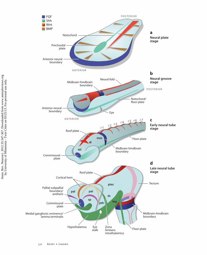
The ectoderm that surrounds the neural plateexpresses BMPs while Spemann’s organizer andthe extending AME express BMP inhibitors.Experiments in zebrafish embryos have sug-gested that this generates a gradient of BMP ac-tivity that defines mediolateral positions withinthe neural plate (Barth et al. 1999). Hence, aCartesian coordinate system of two orthogonalgradients is established—a Wnt gradient alongthe AP axis and a BMP gradient along the medi-olateral axis—and the AME defines the originof this system by secreting BMP and Wnt an-tagonists (Figure 1a) (Meinhardt 2006, Niehrs2010). It is clear that such global mechanismscan establish only a crude initial pattern, andwe argue below that this pattern is increasinglyrefined through the establishment of local (orsecondary) organizers in the neuroepithelium.
Gastrulation is followed by neurulation dur-ing which the lateral folds of the neural plateroll up and fuse to form the neural tube(Figure 1b,c). Thus, the initial mediolateralpattern is transposed into DV polarity: Cellsthat are medial in the plate end up ventral in
www.annualreviews.org • Organizers and the Nervous System 349
Ann
u. R
ev. N
euro
sci.
2012
.35:
347-
367.
Dow
nloa
ded
from
ww
w.a
nnua
lrev
iew
s.or
gby
Uni
vers
ity o
f M
inne
sota
- T
win
Citi
es o
n 03
/25/
13. F
or p
erso
nal u
se o
nly.
NE35CH17-Lumsden ARI 14 May 2012 14:25
aNeural platestage
bNeural groovestage
cEarly neural tubestage
dLate neural tubestage
ANTERIOR
ANTERIOR
r1r2 r3
r4 r5 r6 r7
POSTERIOR
POSTERIOR
Notochord
Prechordalplate
Eye
Eyestalk
Zonalimitansintrathalamica
Hypothalamus
th
teg
Midbrain-hindbrainboundary
Midbrain-hindbrainboundary
Floor plate
Floor plate
Notochord/floor plate
Tectumptec
Anterior neuralboundary
Neural foldMidbrain-hindbrain
boundary
Anterior neuralboundary
Roof plate
Roof plate
Commissuralplate
Commissuralplate
Pallial-subpallialboundary/
antihem
Cortical hem
Medial ganglionic eminence/lamina terminalis
di
mes
pth
pal
sp
pal
tel
FGFShh
BMPWnt
350 Kiecker · Lumsden
Ann
u. R
ev. N
euro
sci.
2012
.35:
347-
367.
Dow
nloa
ded
from
ww
w.a
nnua
lrev
iew
s.or
gby
Uni
vers
ity o
f M
inne
sota
- T
win
Citi
es o
n 03
/25/
13. F
or p
erso
nal u
se o
nly.

NE35CH17-Lumsden ARI 14 May 2012 14:25
Motor neurons:efferent neurons thatcontrol muscle activity
Interneurons:neurons that connectafferent and efferentneurons inmultisynapticpathways
Shh: a secretedsignaling factor; itsactive form is lipidmodified andproteolyticallyprocessed
the tube, whereas lateral neural plate cells endup dorsal.
The Notochord
During gastrulation and neurulation a largeportion of the AME narrows to a thin rod,the notochord, which underlies the midline ofthe neural plate and later most of the neuraltube (Figure 1a–c). The prechordal plate, theanterior end of the AME that lies beneath theprospective anterior forebrain, is a bit widerthan the notochord. The ventralmost cells ofthe neural tube that reside directly above thenotochord form the floor plate on either side ofwhich form motor neurons and various types ofventral interneurons.
Although not a neural structure, the noto-chord was one of the first tissues shown to act asa local organizer of CNS development. Micro-surgical experiments in chick embryos revealedthat the notochord is both necessary and suffi-cient to induce ventral neural identity: Sectionsof the spinal cord from which the notochordhad been removed developed without a floorplate or motor neurons; conversely, the trans-plantation of pieces of notochord beneath thelateral neural plate resulted in the induction ofan ectopic floor plate and motor neurons abovethe graft (Placzek et al. 1990, van Straaten et al.1985).
A breakthrough in understanding this actionof the notochord came with the finding thatSonic hedgehog (Shh), a vertebrate ortholog ofthe Drosophila segment polarity gene hedgehog,is expressed in the notochord and a bit later alsoin the floor plate. Shh encodes a secreted sig-naling factor and is thus a prime candidate forthe inductive signal released by the notochord.
Overexpression of Shh in mouse, zebrafish,and frog, or coculture of rat neuroectodermwith Shh-expressing cells result in ectopic floorplate, motor neuron, and ventral interneuroninduction, indicating that Shh mimics theeffect of notochord grafts (Echelard et al. 1993,Krauss et al. 1993, Roelink et al. 1994). Con-versely, mice carrying a mutation in the Shhgene fail to form a floor plate and lack multipleventral neural cell types (Chiang et al. 1996).Taken together, these data strongly suggestthat Shh mediates the organizer function of thenotochord.
The Floor Plate
The floor plate is a strip of wedge-shapedglial cells along the ventral midline of theneural tube. Like the notochord, the floorplate expresses Shh and is therefore likely tocontribute to the induction of ventral cellidentities. Fate-mapping studies in the chickembryo in combination with detailed examina-tions of cellular morphologies and marker geneexpression have revealed that the floor plateconsists of different cell populations alongboth its AP and mediolateral axes (Placzek& Briscoe 2005). For example, whereas thefloor plate is devoid of neural progenitors inthe spinal cord and hindbrain, dopaminergicneurons are generated in the floor plate of themidbrain (Ono et al. 2007, Puelles et al. 2004).
The origin of the floor plate remains contro-versial. The ablation and grafting experimentsdescribed above strongly suggest that it is in-duced by Shh signaling from the notochord;however, others have argued that the floorplate and notochord originate from a commonprecursor population in Spemann’s organizer
←−−−−−−−−−−−−−−−−−−−−−−−−−−−−−−−−−−−−−−−−−−−−−−−−−−−−−−−−−−−−−−−−−−−−−−−−−−−−−−−−−−−−−−−−−−Figure 1Main stages of neural development in a schematized amniote embryo. (a) Neural plate stage. Mediolateral gradients of BMP (brownwedges) and Shh (green) activity together with an anteroposterior gradient of Wnt activity (red wedge) establish a quasi-Cartesiancoordinate system of positional information across the neural plate. (b) Neural groove stage. The interplay between Wnts andANB-derived Wnt inhibitors patterns the area between the presumptive forebrain and midbrain (red wedge). (c) Early neural tube stage.(d ) Late neural tube stage. Colors: FGF expression (blue), Shh expression ( green), BMP expression (brown), Wnt expression(orange/red ). Abbreviations: di, diencephalon; mes, mesencephalon; pal, pallium; ptec, pretectum; pth, prethalamus; r, rhombomere;sp, subpallium; teg, tegmentum; tel, telencephalon; th, thalamus.
www.annualreviews.org • Organizers and the Nervous System 351
Ann
u. R
ev. N
euro
sci.
2012
.35:
347-
367.
Dow
nloa
ded
from
ww
w.a
nnua
lrev
iew
s.or
gby
Uni
vers
ity o
f M
inne
sota
- T
win
Citi
es o
n 03
/25/
13. F
or p
erso
nal u
se o
nly.
NE35CH17-Lumsden ARI 14 May 2012 14:25
Morphogen: factorthat is released locally,forms a concentrationgradient within atissue, and inducesdifferent cell fatesdose-dependently
and that floor plate cells are inserted into themidline of the neural plate as the AP axis of theembryo extends (Le Douarin & Halpern 2000).These views are likely not entirely incompat-ible: The common lineage of notochord andfloor plate may endow them with shared prop-erties, and both tissues are capable of inducinghomeogenetic responses in neuroepithelium.
Ventral Neural Patterning by Shh: AParadigm for Morphogen Signaling
The specification of multiple cell types inthe ventral neural tube is arguably one of themost thoroughly studied examples of neuralpatterning. Considerable evidence gatheredover the past 15 years indicates that Shhfunctions as a true morphogen in this process;i.e., it is released from a local source (noto-chord, floor plate) and forms a concentrationgradient that specifies different cell fates in adose-dependent fashion (Dessaud et al. 2008,Lupo et al. 2006). In mouse embryos that weregenetically engineered to express fluorescentlylabeled Shh protein (Shh-GFP) from the Shhlocus, a declining ventral-to-dorsal gradientof fluorescence is detectable within the ventralneural tube (Chamberlain et al. 2008).
The morphogen model is intuitively ap-pealing because it explains a complex processpattern formation, drawing on a simple chemi-cal activity—the diffusion of a single substancefrom a localized source. However, trying to un-derstand the cellular mechanism of morphogensignaling raises a number of difficult issues. Forexample, how are different concentrations of amorphogen translated into distinct cell fates?In vertebrates, Shh activates the transcriptionalactivators Gli1 and Gli2 and antagonizes therepressor Gli3. Thus, the extracellular gradientof Shh is translated into opposing gradients ofintracellular Gli1/2 and Gli3 activity along theDV axis of the neural tube (Fuccillo et al. 2006,Lei et al. 2004, Stamataki et al. 2005). Theseoverlapping activities regulate two classes oftranscriptional control genes: Class I genessuch as Pax6, Pax7, and Irx3 are repressed,and class II genes, including Foxa2, Nkx2.2,
Olig2, Nkx6.1, Dbx1, and Dbx2, are induced byShh-Gli signaling. Different thresholds ofShh signaling are required for the repressionor activation of individual class I and class IIgenes, resulting in a nested expression patternof these genes along the DV axis of the spinalcord. Furthermore, several class I and class IIgenes cross-repress each other, resulting in asharpening of the boundaries between theirexpression domains (Briscoe et al. 2000). Ulti-mately, the combinatorial expression of class Iand class II genes at a specific DV location de-termines which type of neural progenitor willform.
Dessaud et al. (2008) recently suggested thatthe duration of exposure, in addition to theextracellular concentration of Shh, determinesthe fate of the receiving cell. This model is likelyto reflect the gradual buildup of the Shh gradi-ent in vivo better than would a static gradientmodel.
Ventral Patterning in the Hindbrainand Midbrain
Early in its development, the hindbrain be-comes subdivided into a series of seven to eightsegments called rhombomeres (r1–8) (reviewedin Kiecker & Lumsden 2005). Nevertheless,the topological organization of neurons alongthe DV axis of the hindbrain is similar to thatof the spinal cord. The induction of hindbrainmotor neurons (which contribute to theIVth–XIIth cranial nerves) also depends onShh signaling from the notochord/floor plateand on the nested expression of various classI and class II genes (Osumi et al. 1997, Pattynet al. 2003, Takahashi & Osumi 2002).
The ventral part of the midbrain (andof the posterior forebrain) gives rise to thetegmentum where neural progenitors are or-ganized in a series of morphologically visiblearcs that are characterized by periodic gene ex-pression patterns (Sanders et al. 2002). Gain-and loss-of-function experiments in chick em-bryos have provided evidence that this patternis also controlled by Shh signaling (Agarwalaet al. 2001, Bayly et al. 2007).
352 Kiecker · Lumsden
Ann
u. R
ev. N
euro
sci.
2012
.35:
347-
367.
Dow
nloa
ded
from
ww
w.a
nnua
lrev
iew
s.or
gby
Uni
vers
ity o
f M
inne
sota
- T
win
Citi
es o
n 03
/25/
13. F
or p
erso
nal u
se o
nly.

NE35CH17-Lumsden ARI 14 May 2012 14:25
Organizers of Ventral ForebrainDevelopment
Like the notochord, the prechordal plate ex-presses Shh, and grafts of this AME tissue tothe lateral forebrain result in ectopic expres-sion of the ventral forebrain marker Nkx2.1 inchick embryos. However, this effect cannot bemimicked by implanting Shh-producing cells,suggesting that the inductive capacity of theprechordal plate reaches beyond mere secretionof Shh (Pera & Kessel 1997). A good candi-date factor to mediate this difference is BMP7,which is expressed in the prechordal mesodermbut not in the posterior notochord and syn-ergizes with Shh in inducing ventral forebrainidentity (Dale et al. 1997). Furthermore, potentWnt inhibitors are expressed in the prechordalplate, and these likely contribute to forebraininduction and ventralization (Kiecker & Niehrs2001b).
The vertebrate forebrain becomes dividedinto the telencephalon anteriorly and the di-encephalon posteriorly (Figure 1c). The dor-sal part of the telencephalon, the pallium, givesrise to the hippocampus and the cerebral cortex(or functionally equivalent structures in non-mammalian species), whereas the ventral part,the subpallium, gives rise to the basal gan-glia. The earliest distinction between pallial andsubpallial identity is established at the neuralplate stage and is mediated by Shh from theprechordal plate (Gunhaga et al. 2000). How-ever, prechordal plate-derived Shh also inducesa new domain of Shh expression in the mostventral part of the subpallium, the presump-tive medial ganglionic eminence (MGE), in amanner that is very similar to the inductionof the floor plate by Shh from the notochord.This telencephalic Shh domain is likely to actas a secondary organizer that refines the DVpattern of the telencephalon (Figure 1d )(Sousa & Fishell 2010).
The Roof Plate
Another specialized cell population, the roofplate, forms along the dorsal midline of theentire neural tube. Similar to the floor plate,
the roof plate is induced by inductive signalsfrom a nonneural tissue—in this case by BMPsand Wnts expressed in the ectoderm flankingthe neural plate (Chizhikov & Millen 2004b).Neural tube closure brings the roof plate pro-genitors from either side of the neural plate to-gether to form the dorsal midline of the neuraltube. Identifying a specific genetic pathway forroof plate formation has been complicated bythe fact that two other cell groups, the neuralcrest and some dorsal interneuron progenitors,also arise from the dorsal midline.
However, there is good evidence that theroof plate functions as an organizer of dorsalpatterning in the spinal cord. Six populationsof dorsal interneurons (dI1–6) are generatedin the dorsal half of the spinal cord, and thecoculture of roof plate and naıve neural platetissue leads to the induction of at least two ofthose (Liem et al. 1997). Genetic ablation ofthe roof plate in mice results in the lack of dI1–3 and expansion of dI4–6 interneurons, con-firming that the roof plate is not only sufficientbut also required for dorsal spinal cord pat-terning (K.J. Lee et al. 2000). Lmx1a mutantmice in which roof plate formation is disruptedshow a similar, if somewhat milder phenotype(Millonig et al. 2000). Overexpression ofLmx1a—which encodes a LIM homeodomaintranscription factor—in the chick spinal cordresults in ectopic induction of dI1 at the ex-pense of dI2–6 interneurons in the vicinity ofthe electroporated cells, suggesting that a dif-fusible signal is secondarily induced (Chizhikov& Millen 2004a).
Identification of the signal that mediatesthe organizer function of the roof plate hasbeen more problematic than identifying that ofthe floor plate. The roof plate expresses severalWnts and a large number of BMPs, and thegenetic inactivation of individual factors oftenresults in entirely normal spinal cord devel-opment, probably owing to compensation byother family members. The BMP-type factorGDF7 is an exception because Gdf7−/− mutantmice lack dI1 interneurons (Lee et al. 1998).Zebrafish mutants with defects in the BMPpathway fail to form Rohon-Beard neurons,
www.annualreviews.org • Organizers and the Nervous System 353
Ann
u. R
ev. N
euro
sci.
2012
.35:
347-
367.
Dow
nloa
ded
from
ww
w.a
nnua
lrev
iew
s.or
gby
Uni
vers
ity o
f M
inne
sota
- T
win
Citi
es o
n 03
/25/
13. F
or p
erso
nal u
se o
nly.
NE35CH17-Lumsden ARI 14 May 2012 14:25
Boundary: aninterface between twoadjacent tissues thatprevents interminglingbetween cells fromeither side of theboundary
indicating that BMPs are required for theformation of at least some dorsal cell types(Nguyen et al. 2000). Wnt1/Wnt3a doublemutant mice show a severe reduction of dI1–3and a concomitant expansion of dI4 and dI5interneurons and are therefore phenotypicallysimilar to roof plate–ablated mice, althoughless severely so (Muroyama et al. 2002).However, the reduction of dI1–3 interneuronscould be due to defective proliferation ratherthan patterning because Wnts act as mitogensin gain-of-function experiments in chick(Megason & McMahon 2002).
Do roof plate signals act in a dose-dependentfashion, similar to Shh on the ventral side ofthe neural tube? Some evidence points towardgraded effects by both BMPs and Wnts in thedorsal spinal cord (Liem et al. 1997, Megason &McMahon 2002, Timmer et al. 2002); however,the picture is far less conclusive than for Shhand it remains possible that qualitative as wellas quantitative mechanisms are at work (i.e., in-dividual BMPs or Wnts may specifically inducecertain subpopulations of dorsal interneurons).
Dorsal Patterning in theAnterior Hindbrain
The dorsal part of the anterior hindbrain (r1)undergoes a series of complex morphologicalchanges that result in the formation of the cere-bellum. Granule cells, the most prevailing celltype in the cerebellum, are generated from therhombic lip, a germinal zone at the interface be-tween the roof plate and the r1 neuroepithelium(Hatten & Roussel 2011, Wingate 2001). Theroof plate of r1, like that of the spinal cord, ex-presses several BMPs, and these are sufficientto initiate granule cell formation when addedectopically to r1 neuroepithelium (Alder et al.1999). The locus coeruleus, the major nora-drenergic nucleus of the brain, is also induced inthe dorsal half of r1 before its neurons migrateventrally to reach their final destination in thelateral floor of the IVth ventricle. Applicationof BMP antagonists to the anterior hindbrainof chick embryos results in the disappearanceof or a dorsal shift of locus coeruleus neurons,
suggesting that the role of BMPs in the induc-tion of these neurons may be dose dependent(Vogel-Hopker & Rohrer 2002).
The Cortical Hem
In the telencephalon, the roof plate sinks be-tween the two cortical hemispheres and givesrise to the monolayered choroid plexus mediallyand to the cortical hem laterally. Immediatelyadjacent to the hem, which expresses BMPs andWnts, the hippocampus is induced and the cere-bral cortex forms next to that. Genetic ablationof the telencephalic roof plate in mice resultsin a severe undergrowth of the cortical pri-mordium, suggesting a nonautonomous effectof the hem on cortical specification (Monukiet al. 2001).
Ectopic application of BMPs to thedeveloping chick telencephalon leads to holo-prosencephaly (a failure to separate the cerebralhemispheres), but this is likely to be a resultof increased cell death rather than a change inpatterning (Golden et al. 1999). A requirementfor BMP signaling in telencephalic patterninghas been tested by genetically disrupting a BMPreceptor gene in the mouse forebrain. Thesemice fail to differentiate the choroid plexus,but all other telencephalic subdivisions developnormally, arguing against a role for BMPs asan organizer signal (Hebert et al. 2002).
Wnt signaling specifies dorsal identity atthe earliest stages of telencephalic development(Backman et al. 2005, Gunhaga et al. 2003), andthe Wnt antagonists secreted by the prechordalplate likely help to set up early DV polarity byprotecting the ventral forebrain from the dor-salizing activity of Wnts. At later stages, Wntsignaling is required for hippocampus differen-tiation (Galceran et al. 2000, S.M. Lee et al.2000, Machon et al. 2003). The boundary be-tween the pallium and the subpallium (PSB)begins to express Wnt inhibitors, raising thepossibility that a gradient of Wnt activity is es-tablished across the pallium between the hemand the PSB (which has also been called theantihem) (Assimacopoulos et al. 2003, Froweinet al. 2002). However, there is little evidence
354 Kiecker · Lumsden
Ann
u. R
ev. N
euro
sci.
2012
.35:
347-
367.
Dow
nloa
ded
from
ww
w.a
nnua
lrev
iew
s.or
gby
Uni
vers
ity o
f M
inne
sota
- T
win
Citi
es o
n 03
/25/
13. F
or p
erso
nal u
se o
nly.
NE35CH17-Lumsden ARI 14 May 2012 14:25
MHB: midbrain-hindbrain boundary
for a later patterning function for Wnts beyondhippocampus induction (Chenn & Walsh 2002,Hirabayashi et al. 2004, Hirsch et al. 2007,Ivaniutsin et al. 2009, Machon et al. 2007,Muzio et al. 2005). The PSB also expressesseveral members of the epidermal growth fac-tor family, transforming growth factor α andFGF7, but their roles in telencephalic develop-ment remain unknown (Assimacopoulos et al.2003).
In summary, two major signaling centers or-ganize DV patterning of the neural tube: theroof plate dorsally, which secretes BMPs andWnts, and the floor plate ventrally, which se-cretes Shh. Both are induced by the same setsof molecular signals from the epidermis and thenotochord, respectively. The notochord itselfis an organizer that can mediate ventral neuralpatterning. In the telencephalon, an additionalpotential organizer, the PSB, is located midwayalong the DV axis.
ANTEROPOSTERIORPATTERNING
In contrast with the DV axis, which is patternedby two signaling centers at opposite poles of theneural tube, the AP axis is patterned by severaldiscrete local organizers.
The Midbrain-Hindbrain Boundary
The boundary between the midbrain andhindbrain (MHB) is characterized by a mor-phological constriction of the neural tube and istherefore also called the isthmus (Figure 1c,d ).The first experimental evidence that the MHBfunctions as an organizer came from micro-surgical studies conducted in chick embryos:Transplantation of anterior hindbrain orposterior midbrain tissue into the posteriorforebrain of a host embryo induced the forma-tion of an ectopic isthmus and ectopic midbraintissue around the graft (Bloch-Gallego et al.1996, Gardner & Barald 1991, Martinez et al.1991). Experimental rotation of the entiremidbrain vesicle formed a double-posteriormidbrain and induced ectopic midbrain and
cerebellar structures in the posterior forebrain(Marın & Puelles 1994). Demonstrating arequirement for the MHB in the patterningof the surrounding tissues by microsurgeryturned out to be less feasible because theisthmic organizer rapidly regenerates aftersurgical removal (Irving & Mason 1999).
Two signaling factors, Wnt1 and FGF8, areexpressed on the anterior and posterior sidesof the MHB, respectively. Both factors are re-quired for midbrain-hindbrain development asWnt1 mutations in the mouse, and mutationsin Fgf8 in both mouse and zebrafish embryosresult in defects in midbrain patterning andcerebellum formation (McMahon et al. 1992,Meyers et al. 1998, Picker et al. 1999, Reiferset al. 1998, Thomas et al. 1991). However, ingain-of-function experiments only FGF8 canmimic MHB organizer function (Crossley et al.1996, Irving & Mason 2000, Lee et al. 1997,Martinez et al. 1999), whereas Wnt1 seems topromote cell growth and proliferation withoutaffecting patterning (Panhuysen et al. 2004).Thus, FGF8 is the main organizer factor se-creted from the MHB.
What determines the AP position of MHBformation? Many studies have uncovereda genetic module, including FGF8, Wnt1,and several transcription factors that stabilizeMHB gene expression in a network of positivemaintenance loops and mutually repressiveinteractions (Liu & Joyner 2001, Wurst &Bally-Cuif 2001). In particular, the homeo-domain transcription factors Otx2 and Gbx2play a central role in MHB positioning. Otx2is expressed in the forebrain and midbrainand Gbx2 in the anterior hindbrain, and theinterface between the two expression domainspresages the position of the MHB from neuralplate stages onward. Various gene targetingexperiments in the mouse have demonstratedthat an experimental shift of this interfacealways results in a concomitant repositioningof the MHB (Acampora et al. 1997, Broccoliet al. 1999, Millet 1999, Wassarman et al.1997). Knockdown and cell transplantationexperiments in the fish have revealed that theotx/gbx interface is regulated by Wnt8 at the
www.annualreviews.org • Organizers and the Nervous System 355
Ann
u. R
ev. N
euro
sci.
2012
.35:
347-
367.
Dow
nloa
ded
from
ww
w.a
nnua
lrev
iew
s.or
gby
Uni
vers
ity o
f M
inne
sota
- T
win
Citi
es o
n 03
/25/
13. F
or p
erso
nal u
se o
nly.
NE35CH17-Lumsden ARI 14 May 2012 14:25
ANB: anterior neuralborder
ZLI: zona limitansintrathalamica
neural plate stage (Rhinn et al. 2005). Thus,the position of the MHB is directly definedby the early gradient of Wnt signaling thatestablishes the initial AP polarity of the neuralplate. Similar to the mutual repression of classI and class II genes in the spinal cord, Otx2 andGbx2 repress each other, thereby stabilizing thebinary cell fate choice around the MHB (Liu& Joyner 2001, Wurst & Bally-Cuif 2001).
The Anterior NeuralBoundary/Commissural Plate
Elegant cell ablation and transplantation exper-iments in zebrafish revealed an organizing func-tion of the anterior border of the neural plate(ANB) (Houart et al. 1998). A Wnt inhibitor ofthe secreted Frizzled-related protein family isexpressed in the ANB, and overexpression anddepletion of this factor phenocopy the effects ofANB transplantation and removal, respectively(Houart et al. 2002). Wnt8B is expressed in thepresumptive midbrain and posterior forebrainat the stages when ANB signaling is requiredand is therefore likely to be the main antagonistof the ANB. Thus, after the global gradient ofWnt activity has established general AP polarityin the neural plate, Wnts regulate AP identityin a more localized fashion in the prospectiveforebrain-midbrain region (Figure 1b).
In the mouse, a role for Wnt inhibition fromthe ANB has yet to be demonstrated; however,FGF8, which is also expressed there, can mimicthe anteriorizing effects of ANB in explants invitro (Shimamura & Rubenstein 1997). Stud-ies in both mouse and zebrafish embryos havedemonstrated a need for FGFs in forebrain pat-terning (Meyers et al. 1998, Walshe & Mason2003).
After neural tube closure, the ANB becomesa patch of cells at the anterior end of the neuraltube that will eventually form the commissuralplate (CP), a scaffold for the formation of fore-brain commissures at later stages. The CP con-tinues to express FGF8, and impressive in uteroelectroporation experiments in the mouse haverevealed that FGF8 promotes anterior at theexpense of posterior cortical fates. An ectopic
source of FGF8 at the posterior end of the cor-tex resulted in a partial mirrored duplication ofanterior cortical areas (Fukuchi-Shimogori &Grove 2001). These experiments identified theCP as an organizer of cortical patterning viaits secretion of FGF8. Toyoda et al. (2010) re-cently showed that FGF8 acts directly and ata long range during this process, i.e., as a truemorphogen.
The Zona Limitans Intrathalamica
The zona limitans intrathalamica (ZLI) is anarrow stripe of Shh-expressing cells in the alarplate of the diencephalon, transecting the neu-raxis between the presumptive prethalamus andthe thalamus (Kitamura et al. 1997, Shimamuraet al. 1995, Zeltser et al. 2001). Gain- and loss-of-function experiments in chick, zebrafish, andmouse embryos have revealed that the ZLI actsas an organizer of diencephalic developmentby secreting Shh (Kiecker & Lumsden 2004,Scholpp et al. 2006, Vieira et al. 2005, Vueet al. 2009). At least in the thalamus, the activityof Shh appears to be dose dependent, withhigher levels of signaling inducing the gamma-aminobutyric acid (GABA)-ergic rostralthalamus and lower levels inducing the gluta-matergic caudal thalamus (Hashimoto-Toriiet al. 2003, Vue et al. 2009; but see Jeong et al.2011).
Fgf8 is expressed in a small patch in the dor-sal ZLI and contributes to regulating the fatedecision between the rostral and caudal tha-lamus (Kataoka & Shimogori 2008). Further-more, a plethora of Wnts shows sharp bordersof expression at the ZLI, suggesting that Wntsignaling may also be involved in regulatingthe regionalization and/or proliferation of thediencephalic primordium (Bluske et al. 2009,Quinlan et al. 2009).
The apposition of any neural tissue anteriorto the ZLI with any tissue between the ZLIand the MHB results in induction of Shh,indicating that planar interactions are sufficientto induce the ZLI organizer (Guinazu et al.2007, Vieira et al. 2005). The ZLI forms atthe interface between the expression domains
356 Kiecker · Lumsden
Ann
u. R
ev. N
euro
sci.
2012
.35:
347-
367.
Dow
nloa
ded
from
ww
w.a
nnua
lrev
iew
s.or
gby
Uni
vers
ity o
f M
inne
sota
- T
win
Citi
es o
n 03
/25/
13. F
or p
erso
nal u
se o
nly.

NE35CH17-Lumsden ARI 14 May 2012 14:25
Neurogenesis: theprocess by whichproliferating neuralprogenitors exit thecell cycle anddifferentiate intofunctional neurons
of two classes of transcription factors: zincfinger proteins of the Fez family anteriorly andhomeodomain proteins of the Irx family poste-riorly (Hirata et al. 2006, Kobayashi et al. 2002,Rodrıguez-Seguel et al. 2009, Scholpp et al.2007). Expression of Irx3 in chick is inducedby Wnt signaling, suggesting that, as for Gbx2at the MHB, the early Wnt signal that poste-riorizes the neural plate directly positions theZLI (Braun et al. 2003).
Rhombomere Boundaries
Segmentation of the hindbrain into rhom-bomeres is controlled by graded retinoic acidsignaling and by the reiterated and nestedexpression of tyrosine kinases and transcriptionfactors, many of which are vertebrate orthologsof Drosophila gap and Hox genes (Kiecker &Lumsden 2005, Maden 2007). In zebrafish,the boundaries between rhombomeres expressseveral Wnts (Figure 1c), and the knockdownof these factors results in disorganized neuro-genesis adjacent to the boundaries, suggestingthat they may function as organizers, althoughno patterning defects have been demonstratedwithin the rhombomeres of such embryos(Amoyel et al. 2005, Riley et al. 2004).
COMMON FEATURESOF NEUROEPITHELIALORGANIZERS
As discussed above, organizers influence cellfate in surrounding tissues by secretingdiffusible signaling factors that often act in amorphogen-like fashion—that is, they inducedifferent responses in receiving cells at differ-ent distances from the source. In addition tothis defining feature of organizers, several othercommonalities have been observed regardingthe establishment, maintenance, and signalingproperties of neuroepithelial organizers.
Organizers Form Along Cell LineageRestriction Boundaries
One of the hallmarks of hindbrain segmenta-tion is the formation of cell lineage–restricted
boundaries that prevent cells from moving be-tween adjacent rhombomeres (Fraser et al.1990, Jimenez-Guri et al. 2010). This findingprompted a search for boundaries in other partsof the neural tube, which led to the discoverythat such boundaries often coincide with orga-nizers (Kiecker & Lumsden 2005): Cell lineagerestriction at the MHB has been demonstratedby sophisticated time-lapse imaging in zebrafishembryos and genetic fate mapping in the mouse(Langenberg & Brand 2005, Zervas et al. 2004);the ZLI is flanked by boundaries on either side(Zeltser et al. 2001); and signaling functionshave been reported for rhombomere bound-aries (see above). Lineage restriction has notbeen tested at the ANB, but it seems unlikelythat cells intermingle freely across the neural-epidermal border. All three DV organizers—floor plate, PSB, and roof plate—also showsome degree of lineage restriction (Awatramaniet al. 2003, Fishell et al. 1993, Fraser et al. 1990,Jimenez-Guri et al. 2010).
The molecular mechanisms underlyingboundary formation in the neural tube are notwell understood. However, specific signalingpathways have been implicated: Eph-ephrinsignaling is essential for segmentation in thehindbrain (Cooke et al. 2005, Kemp et al. 2009,Xu et al. 1999), and the Notch pathway ap-pears to be involved in boundary formation atthe ZLI, at rhombomere boundaries, and at theMHB (Cheng et al. 2004, Tossell et al. 2011,Zeltser et al. 2001).
Cell lineage restriction at organizers prob-ably serves a dual function. First, boundariestend to minimize contact between flanking cellpopulations, which may help to keep organizersin a straight line, facilitating the generation ofa consistent diffusion gradient. Second, cells oneither side of the organizer are kept in separateimmiscible pools, thereby stabilizing a patternafter it has been induced.
Positive Feedback MaintainsOrganizers
Another feature shared by several neuroep-ithelial organizers is that their maintenance
www.annualreviews.org • Organizers and the Nervous System 357
Ann
u. R
ev. N
euro
sci.
2012
.35:
347-
367.
Dow
nloa
ded
from
ww
w.a
nnua
lrev
iew
s.or
gby
Uni
vers
ity o
f M
inne
sota
- T
win
Citi
es o
n 03
/25/
13. F
or p
erso
nal u
se o
nly.

NE35CH17-Lumsden ARI 14 May 2012 14:25
depends on the signal they produce. Both thefloor plate and the roof plate are induced bytheir own signals, BMP and Shh; the ZLIdepends on ongoing Shh signaling (Kiecker& Lumsden 2004, Zeltser 2005); and MHBintegrity depends on FGF signaling (Sunmonuet al. 2011, Trokovic et al. 2005). Alan Turing’sclassical model for pattern formation postulateda chemical network of local self-enhancementand long-range inhibition, and the autoinduc-tion of neuroepithelial organizers fits the localself-enhancement component of this modelrather well (Meinhardt 2009).
Intrinsic Factors Regulate DifferentialResponses to Organizer Signals
FGF signaling from the MHB establishesthe tectum anteriorly and the cerebellumposteriorly, and Shh from the ZLI inducesprethalamic gene expression anteriorly andpatterns the thalamus posteriorly. How can onesignal induce such asymmetric responses oneither side of an organizer? Two orthologs ofthe Drosophila competence factor iroquois, Irx2and Irx3, are expressed posterior to the MHBand ZLI, respectively. Ectopic expression ofthese factors anterior to the organizer resultsin a conversion of tectum into cerebellumand of prethalamus into thalamus (Kiecker &Lumsden 2004, Matsumoto et al. 2004). Theseeffects are dependent on the organizer signalsFGF and Shh, suggesting that Irx2 and Irx3are not patterning factors themselves but thatthey convey a prepattern that determines thecompetence of different subdivisions of theneural tube to respond to secreted signals.
FGF-soaked beads induce ectopic mid-brain and hindbrain structures to form fromposterior forebrain tissue, but in the anteriorforebrain FGFs anteriorize the pallium. Sim-ilarly, Shh from the ventral midline inducesthe hypothalamus marker Nkx2.1 anteriorly,whereas it induces Nkx6.1 posteriorly. Thelimit between these two regions of differentialcompetence to respond to FGFs coincideswith the ZLI, and the homeobox genes Irx3and Six3 were shown to mediate posterior
versus anterior competence (Kobayashi et al.2002).
Taken together, intrinsic factors establish aprepattern in the developing CNS that regu-lates the cellular response to organizer signals.These factors are often induced by the earliestsignals that pattern the neural plate—for ex-ample, Irx3 is induced and Six3 is repressedby posteriorizing Wnt signaling (Braun et al.2003)—thereby linking early and late stages ofneural patterning. This does not mean that or-ganizer signals are merely permissive triggersthat determine the timing and extent of regionalspecialization, the identity of which is prepat-terned; they are also responsible for evokingdifferent responses within the same field (as ex-emplified by the induction of GABAergic versusglutamatergic neurons by different doses of Shhwithin the Irx3-positive thalamus).
Hes Genes Prevent Neurogenesisin Organizer Regions
Organizers typically coincide with boundariesthat are characterized by slower prolifera-tion and a delay or absence of neurogenesis(Guthrie et al. 1991, Lumsden & Keynes 1989).Transcription factors of the Hes family thatmediate Notch signaling are required to inhibitneurogenesis at the MHB of zebrafish and frogembryos (Geling et al. 2004, Ninkovic et al.2005, Takada et al. 2005). All neural progeni-tors express Hes genes, but they usually becomedownregulated when cells undergo neurogene-sis. An analysis in mouse embryos has revealedthat in boundary regions Hes genes remainexpressed and that it is this strong persistentexpression that sets boundaries apart and allowsorganizer regions to form (Baek et al. 2006).
NEUROEPITHELIALORGANIZERS ALSOREGULATE PROLIFERATION,NEUROGENESIS, AND AXONGUIDANCE
Many organizer signals also function as mito-gens, suggesting that growth, in addition to
358 Kiecker · Lumsden
Ann
u. R
ev. N
euro
sci.
2012
.35:
347-
367.
Dow
nloa
ded
from
ww
w.a
nnua
lrev
iew
s.or
gby
Uni
vers
ity o
f M
inne
sota
- T
win
Citi
es o
n 03
/25/
13. F
or p
erso
nal u
se o
nly.

NE35CH17-Lumsden ARI 14 May 2012 14:25
Commissural axons:nerve fibers that crossthe midline of thenervous system
patterning, is modulated by organizers. For ex-ample, Shh mutant mice show not only pat-terning defects, but also a structural lack ofmany ventral neural tissues (Chiang et al. 1996).Wnt1 promotes growth in the MHB region(Panhuysen et al. 2004), and Wnts from theroof plate and cortical hem are known to reg-ulate proliferation of the spinal cord and pal-lium (Chenn & Walsh 2002, Ivaniutsin et al.2009, Megason & McMahon 2002, Muzio et al.2005). FGFs from the MHB promote growth ofthe midbrain and cerebellum (Partanen 2007),but they also serve as survival factors in the mid-brain (Basson et al. 2008). Similarly, FGFs se-creted from the CP prevent apoptosis and pro-mote growth in the telencephalon (Paek et al.2009, Thomson et al. 2009). Contrary to theproliferative effects of FGFs, Shh, and Wnts,the BMP pathway often induces apoptosis whenectopically activated (Anderson et al. 2002, Limet al. 2005, Liu et al. 2004).
To complicate the picture even further,some organizer factors promote cell cycleexit and neurogenesis (Fischer et al. 2011,Hirabayashi et al. 2004, Machon et al. 2007,Munji et al. 2011, Xie et al. 2011). Theseseemingly contradictory effects of the sameclasses of signals may be explained by temporalchanges in the competence of the target cells(Hirsch et al. 2007); however, in some casesdifferent members of the same protein familyexert opposing effects on the balance betweenproliferation and differentiation (Borelloet al. 2008, David 2010). Thus, by releasinggrowth-promoting and growth-inhibiting cuesfrom localized sources, organizers help to moldthe increasingly complex shape of the neuraltube and coordinate the temporal progressionof neurogenesis in defined subdivisions of theneural tube (Scholpp et al. 2009).
Once their regional identity has beenestablished, differentiated neurons need towire up precisely to form functional networks.Organizers also play a role at this stage of CNSformation, for example, by expressing axonguidance factors such as the chemoattractantnetrin, which is secreted by the floor plateto guide commissural axons (Dickson 2002,
Tessier-Lavigne & Goodman 1996). Morerecently, many of the classical morphogensthat are secreted by organizers have beenfound to double as axon guidance molecules atlater stages (Charron & Tessier-Lavigne 2005,Osterfield et al. 2003, Sanchez-Camanchoet al. 2005, Zou & Lyuksyutova 2007). Shhfrom the floor plate cooperates with netrin inattracting commissural axons, whereas BMPsfrom the roof plate repel them (Augsburgeret al. 1999, Charron et al. 2003). After theseaxons have crossed the midline, Shh signalingrepels them via Hedgehog-interacting protein(Bourikas et al. 2005). Wnts are expressed inan AP gradient in the floor plate and guide thesame axons anteriorly after they have crossedthe midline (Lyuksyutova et al. 2003), whereascorticospinal axons are directed posteriorly bya repulsive interaction between Wnts and theatypical Wnt receptor Ryk (Liu et al. 2005).Thus, organizers influence CNS formation notonly at early patterning stages, but also at laterstages when functional circuits are established.
NEUROEPITHELIALORGANIZERS IN EVOLUTION
Vertebrates possess the most complex of allbrains; even their closest relatives, the tunicatesand hemichordates, have relatively simple ner-vous systems (Meinertzhagen et al. 2004). Setsof transcription factors that mark AP and DVsubdivisions of the neural tube are conserved farbeyond the chordate phylum (Irimia et al. 2010,Lowe et al. 2003, Reichert 2005, Tomer et al.2010, Urbach & Technau 2008), and severalorthologs of AP marker genes are even foundalong the head-to-foot axis of the coelenterateHydra, suggesting an ancient origin of the ge-netic modules that regulate neural patterning(Technau & Steele 2011). By contrast, local or-ganizers appear to be far less conserved: Forexample, an equivalent of the MHB is presentin the urochordate Ciona (Imai et al. 2009) butnot in the cephalochordate Amphioxus (Hol-land 2009). Both Ciona and Amphioxus seemto lack an equivalent of the ZLI, whereas acomparable region has been identified in the
www.annualreviews.org • Organizers and the Nervous System 359
Ann
u. R
ev. N
euro
sci.
2012
.35:
347-
367.
Dow
nloa
ded
from
ww
w.a
nnua
lrev
iew
s.or
gby
Uni
vers
ity o
f M
inne
sota
- T
win
Citi
es o
n 03
/25/
13. F
or p
erso
nal u
se o
nly.

NE35CH17-Lumsden ARI 14 May 2012 14:25
hemichordate Saccoglossus (C. Lowe, personalcommunication). Some organizers are evenmissing in lower vertebrates; no hedgehog ex-pression or MGE-like differentiation has beenfound in the telencephalon of the lamprey, sug-gesting that this ventroanterior organizer is agnathostome invention (Sugahara et al. 2011).
These observations indicate that localorganizers are more recent innovations thanthe basic AP/DV patterning network and thatthey show some evolutionary flexibility thatmay provide a driving force for morphologicalchange. This idea is supported by the recentfinding that differences in forebrain morphol-ogy among cichlid fishes from Lake Malawiare correlated with subtle changes in signalstrength, timing of signal production, andthe position of forebrain organizers (Sylvesteret al. 2010). Similarly, loss of eyesight in acave-dwelling morph of the tetra Astyanax wasshown to be caused by changes in the forebrainexpression of fgf8 and shh (Pottin et al. 2011).Thus, although the basic subdivisions of thebrain are likely to have developed a long timeago, organizers are a more recent acquisitionthat may have been imposed on the underlying
pattern and allow evolutionary adaptation toecological niches.
CONCLUSIONS
Almost 90 years have passed since Spemanndiscovered the amphibian gastrula organizer;however, the organizer concept is more topicalthan ever, in particular in the developing ver-tebrate CNS where multiple organizers regu-late patterning, proliferation, neurogenesis, celldeath, and axon pathfinding. Neural organizersare generated by inductive signaling events be-tween neighboring tissues, and they often formalong, or are stabilized by, cell lineage restric-tion boundaries. The pattern induced by an or-ganizer results in the formation of different cellpopulations that can potentially form furtherorganizers at their interfaces, thereby subdivid-ing the neuroepithelium into increasingly morespecialized regions. In many ways, neural de-velopment can be regarded as a self-organizingprocess: Once initial polarity has been estab-lished, all the interactions necessary to form afunctional CNS occur within the neuroepithe-lium itself.
DISCLOSURE STATEMENT
The authors are not aware of any affiliations, memberships, funding, or financial holdings thatmight be perceived as affecting the objectivity of this review.
ACKNOWLEDGMENTS
We apologize to the many researchers whose work we could not cite due to space constraints.
LITERATURE CITED
Acampora D, Avantaggiato V, Tuorto F, Simeone A. 1997. Genetic control of brain morphogenesis throughOtx gene dosage requirement. Development 124:3639–50
Agarwala S, Sanders TA, Ragsdale CW. 2001. Sonic hedgehog control of size and shape in midbrain patternformation. Science 291:2147–50
Alder J, Lee KJ, Jessell TM, Hatten ME. 1999. Generation of cerebellar granule neurons in vivo by trans-plantation of BMP-treated neural progenitor cells. Nat. Neurosci. 2:535–40
Amoyel M, Cheng YC, Jiang YJ, Wilkinson DG. 2005. Wnt1 regulates neurogenesis and mediates lateralinhibition of boundary cell specification in the zebrafish hindbrain. Development 132:775–85
Anderson RM, Lawrence AR, Stottmann RW, Bachiller D, Klingensmith J. 2002. Chordin and noggin promoteorganizing centers of forebrain development in the mouse. Development 129:4975–87
360 Kiecker · Lumsden
Ann
u. R
ev. N
euro
sci.
2012
.35:
347-
367.
Dow
nloa
ded
from
ww
w.a
nnua
lrev
iew
s.or
gby
Uni
vers
ity o
f M
inne
sota
- T
win
Citi
es o
n 03
/25/
13. F
or p
erso
nal u
se o
nly.
NE35CH17-Lumsden ARI 14 May 2012 14:25
Assimacopoulos S, Grove EA, Ragsdale CW. 2003. Identification of a Pax6-dependent epidermal growth factorfamily signaling source at the lateral edge of the embryonic cerebral cortex. J. Neurosci. 23:6399–403
Augsburger A, Schuchardt A, Hoskins S, Dodd J, Butler S. 1999. BMPs as mediators of roof plate repulsionof commissural neurons. Neuron 24:127–41
Awatramani R, Soriano P, Rodriguez C, Mai JJ, Dymecki SM. 2003. Cryptic boundaries in roof plate andchoroid plexus identified by intersectional gene activation. Nat. Genet. 35:70–75
Backman M, Machon O, Mygland L, van den Bout CJ, Zhong W, et al. 2005. Effects of canonical Wntsignaling on dorso-ventral specification of the mouse telencephalon. Dev. Biol. 279:155–68
Baek JH, Hatakeyama J, Sakamoto S, Ohtsuka T, Kageyama R. 2006. Persistent and high levels of Hes1expression regulate boundary formation in the developing central nervous system. Development 133:2467–76
Barth KA, Kishimoto Y, Rohr KB, Seydler C, Schulte-Merker S, Wilson SW. 1999. Bmp activity establishesa gradient of positional information throughout the entire neural plate. Development 126:4977–87
Basson MA, Echevarria D, Ahn CP, Sudarov A, Joyner AL, et al. 2008. Specific regions within the embryonicmidbrain and cerebellum require different levels of FGF signaling during development. Development135:889–98
Bayly RD, Ngo M, Aglyamova GV, Agarwala S. 2007. Regulation of ventral midbrain patterning by Hedgehogsignaling. Development 134:2115–24
Bloch-Gallego E, Millet S, Alvarado-Mallart RM. 1996. Further observations on the susceptibility of dien-cephalic prosomeres to En-2 induction and on the resulting histogenetic capabilities. Mech. Dev. 58:51–63
Bluske KK, Kawakami Y, Koyano-Nakagawa N, Nakagawa Y. 2009. Differential activity of Wnt/β-cateninsignaling in the embryonic mouse thalamus. Dev. Dyn. 238:3297–309
Borello U, Cobos I, Long JE, McWhirter JR, Murre C, Rubenstein JL. 2008. FGF15 promotes neurogenesisand opposes FGF8 function during neocortical development. Neural Dev. 3:17
Bourikas D, Pekarik V, Baeriswyl T, Grunditz A, Sadhu R, et al. 2005. Sonic hedgehog guides commissuralaxons along the longitudinal axis of the spinal cord. Nat. Neurosci. 8:297–304
Braun MM, Etheridge A, Bernard A, Robertson CP, Roelink H. 2003. Wnt signaling is required at distinctstages of development for the induction of the posterior forebrain. Development 130:5579–87
Briscoe J, Pierani A, Jessell TM, Ericson J. 2000. A homeodomain protein code specifies progenitor cellidentity and neuronal fate in the ventral neural tube. Cell 101:435–45
Broccoli V, Boncinelli E, Wurst W. 1999. The caudal limit of Otx2 expression positions the isthmic organizer.Nature 401:164–68
Chamberlain CE, Jeong J, Guo C, Allen BL, McMahon AP. 2008. Notochord-derived Shh concentrates inclose association with the apically positioned basal body in neural target cells and forms a dynamic gradientduring neural patterning. Development 135:1097–106
Charron F, Stein E, Jeong J, McMahon AP, Tessier-Lavigne M. 2003. The morphogen sonic hedgehog isan axonal chemoattractant that collaborates with netrin-1 in midline axon guidance. Cell 113:11–23
Charron F, Tessier-Lavigne M. 2005. Novel brain wiring functions for classical morphogens: a role as gradedpositional cues in axon guidance. Development 132:2251–62
Cheng YC, Amoyel M, Qiu X, Jiang YJ, Xu Q, Wilkinson DG. 2004. Notch activation regulates the segre-gation and differentiation of rhombomere boundary cells in the zebrafish hindbrain. Dev. Cell 6:539–50
Chenn A, Walsh CA. 2002. Regulation of cerebral cortical size by control of cell cycle exit in neural precursors.Science 297:365–69
Chiang C, Litingtung Y, Lee E, Young KE, Corden JL, et al. 1996. Cyclopia and defective axial patterning inmice lacking Sonic hedgehog gene function. Nature 383:407–13
Chizhikov VV, Millen KJ. 2004a. Control of roof plate formation by Lmx1a in the developing spinal cord.Development 131:2693–705
Chizhikov VV, Millen KJ. 2004b. Mechanisms of roof plate formation in the vertebrate CNS. Nat. Rev.Neurosci. 5:808–12
Cooke JE, Kemp HA, Moens CB. 2005. EphA4 is required for cell adhesion and rhombomere-boundaryformation in the zebrafish. Curr. Biol. 15:536–42
Crossley PH, Martinez S, Martin GR. 1996. Midbrain development induced by FGF8 in the chick embryo.Nature 380:66–68
www.annualreviews.org • Organizers and the Nervous System 361
Ann
u. R
ev. N
euro
sci.
2012
.35:
347-
367.
Dow
nloa
ded
from
ww
w.a
nnua
lrev
iew
s.or
gby
Uni
vers
ity o
f M
inne
sota
- T
win
Citi
es o
n 03
/25/
13. F
or p
erso
nal u
se o
nly.

NE35CH17-Lumsden ARI 14 May 2012 14:25
Dale JK, Vesque C, Lints TJ, Sampath TK, Furley A, et al. 1997. Cooperation of BMP7 and SHH in theinduction of forebrain ventral midline cells by prechordal mesoderm. Cell 90:257–69
Darras S, Gerhart J, Terasaki M, Kirschner M, Lowe CJ. 2011. ß-catenin specifies the endomesoderm anddefines the posterior organizer of the hemichordate Saccoglossus kowalevskii. Development 138:959–70
David MD. 2010. Wnt-3a and Wnt-3 differently stimulate proliferation and neurogenesis of spinal neuralprecursors and promote neurite outgrowth by canonical signaling. J. Neurosci. Res. 88:3011–23
De Robertis EM, Kuroda H. 2004. Dorsal-ventral patterning and neural induction in Xenopus embryos. Annu.Rev. Cell Dev. Biol. 20:285–308
Dessaud E, McMahon AP, Briscoe J. 2008. Pattern formation in the vertebrate neural tube: a sonic hedgehogmorphogen-regulated transcriptional network. Development 135:2489–503
Dickson BJ. 2002. Molecular mechanisms of axon guidance. Science 298:1959–64Echelard Y, Epstein DJ, St-Jacques B, Shen L, Mohler J, et al. 1993. Sonic hedgehog, a member of a family
of putative signaling molecules, is implicated in the regulation of CNS polarity. Cell 75:1417–30Fischer T, Faus-Kessler T, Welzl G, Simeone A, Wurst W, Prakash N. 2011. Fgf15-mediated control of
neurogenic and proneural gene expression regulates dorsal midbrain neurogenesis. Dev. Biol. 350:496–510
Fishell G, Mason CA, Hatten ME. 1993. Dispersion of neural progenitors within the germinal zones of theforebrain. Nature 362:636–38
Fraser S, Keynes R, Lumsden A. 1990. Segmentation in the chick embryo hindbrain is defined by cell lineagerestrictions. Nature 344:431–35
Frowein J, Campbell K, Gotz M. 2002. Expression of Ngn1, Ngn2, Cash1, Gsh2 and Sfrp1 in the developingchick telencephalon. Mech. Dev. 110:249–52
Fuccillo M, Joyner AL, Fishell G. 2006. Morphogen to mitogen: the multiple roles of hedgehog signalling invertebrate neural development. Nat. Rev. Neurosci. 7:772–83
Fukuchi-Shimogori T, Grove EA. 2001. Neocortex patterning by the secreted signaling molecule FGF8.Science 294:1071–74
Galceran J, Miyashita-Lin EM, Devaney E, Rubenstein JL, Grosschedl R. 2000. Hippocampus developmentand generation of dentate gyrus granule cells is regulated by LEF1. Development 127:469–82
Gardner CA, Barald KF. 1991. The cellular environment controls the expression of engrailed-like protein inthe cranial neuroepithelium of quail-chick chimeric embryos. Development 113:1037–48
Geling A, Plessy C, Rastegar S, Strahle U, Bally-Cuif L. 2004. Her5 acts as a prepattern factor that blocksneurogenin1 and coe2 expression upstream of Notch to inhibit neurogenesis at the midbrain-hindbrainboundary. Development 131:1993–2006
Golden JA, Bracilovic A, McFadden KA, Beesley JS, Rubenstein JL, Grinspan JB. 1999. Ectopic bone mor-phogenetic proteins 5 and 4 in the chicken forebrain lead to cyclopia and holoprosencephaly. Proc. Natl.Acad. Sci. USA 96:2439–44
Guinazu MF, Chambers D, Lumsden A, Kiecker C. 2007. Tissue interactions in the developing chick dien-cephalon. Neural Dev. 2:25
Gunhaga L, Jessell TM, Edlund T. 2000. Sonic hedgehog signaling at gastrula stages specifies ventral telen-cephalic cells in the chick embryo. Development 127:3283–93
Gunhaga L, Marklund M, Sjodal M, Hsieh JC, Jessell TM, Edlund T. 2003. Specification of dorsal telen-cephalic character by sequential Wnt and FGF signaling. Nat. Neurosci. 6:701–7
Guthrie S, Butcher M, Lumsden A. 1991. Patterns of cell division and interkinetic nuclear migration in thechick embryo hindbrain. J. Neurobiol. 22:742–54
Hashimoto-Torii K, Motoyama J, Hui CC, Kuroiwa A, Nakafuku M, Shimamura K. 2003. Differential ac-tivities of Sonic hedgehog mediated by Gli transcription factors define distinct neuronal subtypes in thedorsal thalamus. Mech. Dev. 120:1097–111
Hatten ME, Roussel MF. 2011. Development and cancer of the cerebellum. Trends Neurosci. 34:134–42Hebert JM, Mishina Y, McConnell SK. 2002. BMP signaling is required locally to pattern the dorsal telen-
cephalic midline. Neuron 35:1029–41Hirabayashi Y, Itoh Y, Tabata H, Nakajima K, Akiyama T, et al. 2004. The Wnt/β-catenin pathway directs
neuronal differentiation of cortical neural precursor cells. Development 131:2791–801
362 Kiecker · Lumsden
Ann
u. R
ev. N
euro
sci.
2012
.35:
347-
367.
Dow
nloa
ded
from
ww
w.a
nnua
lrev
iew
s.or
gby
Uni
vers
ity o
f M
inne
sota
- T
win
Citi
es o
n 03
/25/
13. F
or p
erso
nal u
se o
nly.
NE35CH17-Lumsden ARI 14 May 2012 14:25
Hirata T, Nakazawa M, Muraoka O, Nakayama R, Suda Y, Hibi M. 2006. Zinc-finger genes Fez and Fez-likefunction in the establishment of diencephalon subdivisions. Development 133:3993–4004
Hirsch C, Campano LM, Wohrle S, Hecht A. 2007. Canonical Wnt signaling transiently stimulates prolifer-ation and enhances neurogenesis in neonatal neural progenitor cultures. Exp. Cell Res. 313:572–87
Holland LZ. 2009. Chordate roots of the vertebrate nervous system: expanding the molecular toolkit.Nat. Rev. Neurosci. 10:736–46
Houart C, Caneparo L, Heisenberg C, Barth K, Take-Uchi M, Wilson S. 2002. Establishment of the telen-cephalon during gastrulation by local antagonism of Wnt signaling. Neuron 35:255–65
Houart C, Westerfield M, Wilson SW. 1998. A small population of anterior cells patterns the forebrain duringzebrafish gastrulation. Nature 391:788–92
Imai KS, Stolfi A, Levine M, Satou Y. 2009. Gene regulatory networks underlying the compartmentalizationof the Ciona central nervous system. Development 136:285–93
Irimia M, Pineiro C, Maeso I, Gomez-Skarmeta JL, Casares F, Garcia-Fernandez J. 2010. Conserved devel-opmental expression of Fezf in chordates and Drosophila and the origin of the zona limitans intrathalamica(ZLI) brain organizer. Evodevo 1:7
Irving C, Mason I. 1999. Regeneration of isthmic tissue is the result of a specific and direct interaction betweenrhombomere 1 and midbrain. Development 126:3981–89
Irving C, Mason I. 2000. Signalling by FGF8 from the isthmus patterns anterior hindbrain and establishes theanterior limit of Hox gene expression. Development 127:177–86
Ivaniutsin U, Chen Y, Mason JO, Price DJ, Pratt T. 2009. Adenomatous polyposis coli is required for early eventsin the normal growth and differentiation of the developing cerebral cortex. Neural Dev. 16:3
Jeong Y, Dolson DK, Waclaw RR, Matise MP, Sussel L, et al. 2011. Spatial and temporal requirements forsonic hedgehog in the regulation of thalamic interneuron identity. Development 138:531–41
Jimenez-Guri E, Udina F, Colas JF, Sharpe J, Padron-Barthe L, et al. 2010. Clonal analysis in mice underlinesthe importance of rhombomeric boundaries in cell movement restriction during hindbrain segmentation.PLoS One 5:e10112
Kataoka A, Shimogori T. 2008. Fgf8 controls regional identity in the developing thalamus. Development135:2873–81
Kemp HA, Cooke JE, Moens CB. 2009. EphA4 and EfnB2a maintain rhombomere coherence by indepen-dently regulating intercalation of progenitor cells in the zebrafish neural keel. Dev. Biol. 327:313–26
Kiecker C, Lumsden A. 2004. Hedgehog signaling from the ZLI regulates diencephalic regional identity.Nat. Neurosci. 7:1242–49
Kiecker C, Lumsden A. 2005. Compartments and their boundaries in vertebrate brain development. Nat. Rev.Neurosci. 6:553–64
Kiecker C, Niehrs C. 2001a. A morphogen gradient of Wnt/β-catenin signalling regulates anteroposteriorneural patterning in Xenopus. Development 128:4189–201
Kiecker C, Niehrs C. 2001b. The role of prechordal mesendoderm in neural patterning. Curr. Opin. Neurobiol.11:27–33
Kitamura K, Miura H, Yanazawa M, Miyashita T, Kato K. 1997. Expression patterns of Brx1 (Rieg gene),Sonic hedgehog, Nkx2.2, Dlx1 and Arx during zona limitans intrathalamica and embryonic ventral lateralgeniculate nuclear formation. Mech. Dev. 67:83–96
Kobayashi D, Kobayashi M, Matsumoto K, Ogura K, Nakafuku M, Shimamura K. 2002. Early subdivisionsin the neural plate define distinct competence for inductive signals. Development 129:83–93
Krauss S, Concordet JP, Ingham PW. 1993. A functionally conserved homolog of the Drosophila segmentpolarity gene hh is expressed in tissues with polarizing activity in zebrafish embryos. Cell 75:1431–44
Langenberg T, Brand M. 2005. Lineage restriction maintains a stable organizer cell population at the zebrafishmidbrain-hindbrain boundary. Development 132:3209–16
Le Douarin NM, Halpern ME. 2000. Discussion point. Origin and specification of the neural tube floor plate:insights from the chick and zebrafish. Curr. Opin. Neurobiol. 10:23–30
Lee KJ, Dietrich P, Jessell TM. 2000. Genetic ablation reveals that the roof plate is essential for dorsalinterneuron specification. Nature 403:734–40
www.annualreviews.org • Organizers and the Nervous System 363
Ann
u. R
ev. N
euro
sci.
2012
.35:
347-
367.
Dow
nloa
ded
from
ww
w.a
nnua
lrev
iew
s.or
gby
Uni
vers
ity o
f M
inne
sota
- T
win
Citi
es o
n 03
/25/
13. F
or p
erso
nal u
se o
nly.
NE35CH17-Lumsden ARI 14 May 2012 14:25
Lee KJ, Mendelsohn M, Jessell TM. 1998. Neuronal patterning by BMPs: a requirement for GDF7 in thegeneration of a discrete class of commissural interneurons in the mouse spinal cord. Genes Dev. 12:3394–407
Lee SM, Danielian PS, Fritzsch B, McMahon AP. 1997. Evidence that FGF8 signalling from the midbrain-hindbrain junction regulates growth and polarity in the developing midbrain. Development 124:959–69
Lee SM, Tole S, Grove EA, McMahon AP. 2000. A local Wnt-3a signal is required for development of themammalian hippocampus. Development 127:457–67
Lei Q, Zelman AK, Kuang E, Li S, Matise MP. 2004. Transduction of graded Hedgehog signaling by acombination of Gli2 and Gli3 activator functions in the developing spinal cord. Development 131:3593–604
Liem KF Jr, Tremml G, Jessell TM. 1997. A role for the roof plate and its resident TGFβ-related proteinsin neuronal patterning in the dorsal spinal cord. Cell 91:127–38
Lim Y, Cho G, Minarcik J, Golden J. 2005. Altered BMP signaling disrupts chick diencephalic development.Mech. Dev. 122:603–20
Liu A, Joyner AL. 2001. Early anterior/posterior patterning of the midbrain and cerebellum. Annu. Rev.Neurosci. 24:869–96
Liu Y, Helms AW, Johnson JE. 2004. Distinct activities of Msx1 and Msx3 in dorsal neural tube development.Development 131:1017–28
Liu Y, Shi J, Lu CC, Wang ZB, Lyuksyutova AI, et al. 2005. Ryk-mediated Wnt repulsion regulates posterior-directed growth of corticospinal tract. Nat. Neurosci. 8:1151–59
Lowe CJ, Wu M, Salic A, Evans L, Lander E, et al. 2003. Anteroposterior patterning in hemichordates andthe origins of the chordate nervous system. Cell 113:853–65
Lumsden A, Keynes R. 1989. Segmental patterns of neuronal development in the chick hindbrain. Nature337:424–28
Lupo G, Harris WA, Lewis KE. 2006. Mechanisms of ventral patterning in the vertebrate nervous system.Nat. Rev. Neurosci. 7:103–14
Lyuksyutova AI, Lu CC, Milanesio N, King LA, Guo N, et al. 2003. Anterior-posterior guidance of commis-sural axons by Wnt-frizzled signaling. Science 302:1984–88
Machon O, Backman M, Machonova O, Kozmik Z, Vacik T, et al. 2007. A dynamic gradient of Wnt signalingcontrols initiation of neurogenesis in the mammalian cortex and cellular specification in the hippocampus.Dev. Biol. 311:223–37
Machon O, van den Bout CJ, Backman M, Kemler R, Krauss S. 2003. Role of β-catenin in the developingcortical and hippocampal neuroepithelium. Neuroscience 122:129–43
Maden M. 2007. Retinoic acid in the development, regeneration and maintenance of the nervous system.Nat. Rev. Neurosci. 8:755–65
Marın F, Puelles L. 1994. Patterning of the embryonic avian midbrain after experimental inversions: a polar-izing activity from the isthmus. Dev. Biol. 163:19–37
Martinez S, Crossley PH, Cobos I, Rubenstein JL, Martin GR. 1999. FGF8 induces formation of an ec-topic isthmic organizer and isthmocerebellar development via a repressive effect on Otx2 expression.Development 126:1189–200
Martinez S, Wassef M, Alvarado-Mallart RM. 1991. Induction of a mesencephalic phenotype in the 2-day-oldchick prosencephalon is preceded by the early expression of the homeobox gene en. Neuron 6:971–81
Mason I. 2007. Initiation to end point: the multiple roles of fibroblast growth factors in neural development.Nat. Rev. Neurosci. 8:583–96
Matsumoto K, Nishihara S, Kamimura M, Shiraishi T, Otoguro T, et al. 2004. The prepattern transcriptionfactor Irx2, a target of the FGF8/MAP kinase cascade, is involved in cerebellum formation. Nat. Neurosci.7:605–12
McMahon AP, Joyner AL, Bradley A, McMahon JA. 1992. The midbrain-hindbrain phenotype ofWnt-1−/Wnt-1− mice results from stepwise deletion of engrailed-expressing cells by 9.5 days postcoitum.Cell 69:581–95
Megason SG, McMahon AP. 2002. A mitogen gradient of dorsal midline Wnts organizes growth in the CNS.Development 129:2087–98
364 Kiecker · Lumsden
Ann
u. R
ev. N
euro
sci.
2012
.35:
347-
367.
Dow
nloa
ded
from
ww
w.a
nnua
lrev
iew
s.or
gby
Uni
vers
ity o
f M
inne
sota
- T
win
Citi
es o
n 03
/25/
13. F
or p
erso
nal u
se o
nly.

NE35CH17-Lumsden ARI 14 May 2012 14:25
Meinertzhagen IA, Lemaire P, Okamura Y. 2004. The neurobiology of the ascidian tadpole larva: recentdevelopments in an ancient chordate. Annu. Rev. Neurosci. 27:453–85
Meinhardt H. 2006. Primary body axes of vertebrates: generation of a near-Cartesian coordinate system andthe role of Spemann-type organizer. Dev. Dyn. 235:2907–19
Meinhardt H. 2009. Models for the generation and interpretation of gradients. Cold Spring Harb. Perspect. Biol.1:a001362
Meyers EN, Lewandowski M, Martin GR. 1998. An Fgf8 mutant allelic series generated by Cre- and Flp-mediated recombination. Nat. Genet. 18:136–41
Millet S. 1999. A role for Gbx2 in repression of Otx2 and positioning the mid/hindbrain organizer. Nature401:161–64
Millonig JH, Millen KJ, Hatten ME. 2000. The mouse Dreher gene Lmx1a controls formation of the roofplate in the vertebrate CNS. Nature 403:764–69
Monuki ES, Porter FD, Walsh CA. 2001. Patterning of the dorsal telencephalon and cerebral cortex by a roofplate-Lhx2 pathway. Neuron 32:591–604
Munji RN, Choe Y, Li G, Siegenthaler JA, Pleasure SJ. 2011. Wnt signaling regulates neuronal differentiationof cortical intermediate progenitors. J. Neurosci. 31:1676–87
Munoz-Sanjuan I, Brivanlou AH. 2002. Neural induction, the default model and embryonic stem cells.Nat. Rev. Neurosci. 3:271–80
Muroyama Y, Fujihara M, Ikeya M, Kondoh H, Takada S. 2002. Wnt signaling plays an essential role inneuronal specification of the dorsal spinal cord. Genes Dev. 16:548–53
Muzio L, Soria JM, Pannese M, Piccolo S, Mallamaci A. 2005. A mutually stimulating loop involving emx2and canonical wnt signalling specifically promotes expansion of occipital cortex and hippocampus. Cereb.Cortex 15:2021–28
Nakamoto A, Nagy LM, Shimizu T. 2011. Secondary embryonic axis formation by transplantation of Dquadrant micromeres in an oligochaete annelid. Development 138:283–90
Nguyen VH, Trout J, Connors SA, Andermann P, Weinberg E, Mullins MC. 2000. Dorsal and intermediateneuronal cell types of the spinal cord are established by a BMP signaling pathway. Development 127:1209–20
Niehrs C. 2004. Regionally specific induction by the Spemann-Mangold organizer. Nat. Rev. Genet. 5:425–34Niehrs C. 2010. On growth and form: a Cartesian coordinate system of Wnt and BMP signaling specifies
bilaterian body axes. Development 137:845–57Ninkovic J, Tallafuss A, Leucht C, Topczewski J, Tannhauser B, et al. 2005. Inhibition of neurogenesis
at the zebrafish midbrain-hindbrain boundary by the combined and dose-dependent activity of a newhairy/E(spl) gene pair. Development 132:75–88
Ono Y, Nakatani T, Sakamoto Y, Mizuhara E, Minaki Y, et al. 2007. Differences in neurogenic potentialin floor plate cells along an anteroposterior location: midbrain dopaminergic neurons originate frommesencephalic floor plate cells. Development 134:3213–25
Osterfield M, Kirschner MW, Flanagan JG. 2003. Graded positional information: interpretation for both fateand guidance. Cell 113:425–28
Osumi N, Hirota A, Ohuchi H, Nakafuku M, Iimura T, et al. 1997. Pax-6 is involved in the specification ofhindbrain motor neuron subtype. Development 124:2961–72
Paek H, Gutin G, Hebert JM. 2009. FGF signaling is strictly required to maintain early telencephalic precursorcell survival. Development 136:2457–65
Panhuysen M, Vogt Weisenhorn DM, Blanquet V, Brodski C, Heinzmann U, et al. 2004. Effects of Wnt1signaling on proliferation in the developing mid-/hindbrain region. Mol. Cell. Neurosci. 26:101–11
Partanen J. 2007. FGF signalling pathways in development of the midbrain and anterior hindbrain.J. Neurochem. 101:1185–93
Pattyn A, Vallstedt A, Dias JM, Sander M, Ericson J. 2003. Complementary roles for Nkx6 and Nkx2 classproteins in the establishment of motoneuron identity in the hindbrain. Development 130:4149–59
Pera EM, Kessel M. 1997. Patterning of the chick forebrain anlage by the prechordal plate. Development124:4153–62
www.annualreviews.org • Organizers and the Nervous System 365
Ann
u. R
ev. N
euro
sci.
2012
.35:
347-
367.
Dow
nloa
ded
from
ww
w.a
nnua
lrev
iew
s.or
gby
Uni
vers
ity o
f M
inne
sota
- T
win
Citi
es o
n 03
/25/
13. F
or p
erso
nal u
se o
nly.

NE35CH17-Lumsden ARI 14 May 2012 14:25
Picker A, Brennan C, Reifers F, Clarke JD, Holder N, Brand M. 1999. Requirement for the zebrafish mid-hindbrain boundary in midbrain polarisation, mapping and confinement of the retinotectal projection.Development 126:2967–78
Placzek M, Briscoe J. 2005. The floor plate: multiple cells, multiple signals. Nat. Rev. Neurosci. 6:230–40Placzek M, Tessier-Lavigne M, Yamada T, Jessell T, Dodd J. 1990. Mesodermal control of neural cell identity:
floor plate induction by the notochord. Science 250:985–88Pottin K, Hinaux H, Retaux S. 2011. Restoring eye size in Astyanax mexicanus blind cavefish embryos through
modulation of the Shh and Fgf8 forebrain organising centres. Development 138:2467–76Puelles E, Annino A, Tuorto F, Usiello A, Acampora D, et al. 2004. Otx2 regulates the extent, identity and
fate of neuronal progenitor domains in the ventral midbrain. Development 131:2037–48Quinlan R, Graf M, Mason I, Lumsden A, Kiecker C. 2009. Complex and dynamic patterns of Wnt pathway
gene expression in the developing chick forebrain. Neural Dev. 4:35Reichert H. 2005. A tripartite organization of the urbilaterian brain: developmental genetic evidence from
Drosophila. Brain Res. Bull. 66:491–94Reifers F, Bohli H, Walsh EC, Crossley PH, Stainier DY, Brand M. 1998. Fgf8 is mutated in zebrafish
acerebellar (ace) mutants and is required for maintenance of midbrain-hindbrain boundary developmentand somitogenesis. Development 125:2381–95
Rhinn M, Lun K, Luz M, Werner M, Brand M. 2005. Positioning of the midbrain-hindbrain boundaryorganizer through global posteriorization of the neuroectoderm mediated by Wnt8 signaling. Development132:1261–72
Riley BB, Chiang MY, Storch EM, Heck R, Buckles GR, Lekven AC. 2004. Rhombomere boundaries are Wntsignaling centers that regulate metameric patterning in the zebrafish hindbrain. Dev. Dyn. 231:278–91
Rodrıguez-Seguel E, Alarcon P, Gomez-Skarmeta JL. 2009. The Xenopus Irx genes are essential for neuralpatterning and define the border between prethalamus and thalamus through mutual antagonism withthe anterior repressors Fezf and Arx. Dev. Biol. 329:258–68
Roelink H, Augsburger A, Heemskerk J, Korzh V, Norlin S, et al. 1994. Floor plate and motor neuroninduction by vhh-1, a vertebrate homolog of hedgehog expressed by the notochord. Cell 76:761–75
Sanchez-Camancho C, Rodrıguez J, Ruiz JM, Trousse F, Bovolenta P. 2005. Morphogens as growth conesignalling molecules. Brain Res. Brain Res. Rev. 49:242–52
Sanders TA, Lumsden A, Ragsdale CW. 2002. Arcuate plan of chick midbrain development. J. Neurosci.22:10742–50
Scholpp S, Delogu A, Gilthorpe J, Peukert D, Schindler S, Lumsden A. 2009. Her6 regulates the neurogeneticgradient and neuronal identity in the thalamus. Proc. Natl. Acad. Sci. USA 106:19895–900
Scholpp S, Foucher I, Staudt N, Peukert D, Lumsden A, Houart C. 2007. Otx1l, Otx2 and Irx1b establish andposition the ZLI in the diencephalon. Development 134:3167–76
Scholpp S, Wolf O, Brand M, Lumsden A. 2006. Hedgehog signalling from the zona limitans intrathalamicaorchestrates patterning of the zebrafish diencephalon. Development 133:855–64
Shimamura K, Hartigan DJ, Martinez S, Puelles L, Rubenstein JL. 1995. Longitudinal organization of theanterior neural plate and neural tube. Development 121:3923–33
Shimamura K, Rubenstein JL. 1997. Inductive interactions direct early regionalization of the mouse forebrain.Development 124:2709–18
Sousa VH, Fishell G. 2010. Sonic hedgehog functions through dynamic changes in temporal competence inthe developing forebrain. Curr. Opin. Genet. Dev. 20:391–99
Stamataki D, Ulloa F, Tsoni SV, Mynett A, Briscoe J. 2005. A gradient of Gli activity mediates graded Sonichedgehog signaling in the neural tube. Genes Dev. 19:626–41
Stern CD. 2001. Initial patterning of the central nervous system: how many organizers? Nat. Rev. Neurosci.2:92–98
Stern CD. 2006. Neural induction: 10 years on since the ‘default model’. Curr. Opin. Cell Biol. 18:692–97Sugahara F, Aota S, Kuraku S, Murakami Y, Takio-Ogawa Y, et al. 2011. Involvement of Hedgehog and FGF
signalling in the lamprey telencephalon: evolution of regionalization and dorsoventral patterning of thevertebrate forebrain. Development 138:1217–26
Sunmonu NA, Li K, Guo Q, Li JY. 2011. Gbx2 and Fgf8 are sequentially required for formation of themidbrain-hindbrain compartment boundary. Development 138:725–34
366 Kiecker · Lumsden
Ann
u. R
ev. N
euro
sci.
2012
.35:
347-
367.
Dow
nloa
ded
from
ww
w.a
nnua
lrev
iew
s.or
gby
Uni
vers
ity o
f M
inne
sota
- T
win
Citi
es o
n 03
/25/
13. F
or p
erso
nal u
se o
nly.

NE35CH17-Lumsden ARI 14 May 2012 14:25
Sylvester JB, Rich CA, Loh YH, van Staaden MJ, Fraser GJ, Streelman JT. 2010. Brain diversity evolves viadifferences in patterning. Proc. Natl. Acad. Sci. USA 107:9718–23
Takada H, Hattori D, Kitayama A, Ueno N, Taira M. 2005. Identification of target genes for the XenopusHes-related protein XHR1, a prepattern factor specifying the midbrain-hindbrain boundary. Dev. Biol.283:253–67
Takahashi M, Osumi N. 2002. Pax6 regulates specification of ventral neurone subtypes in the hindbrain byestablishing progenitor domains. Development 129:1327–38
Technau U, Steele RE. 2011. Evolutionary crossroads in developmental biology: Cnidaria. Development138:1447–58
Tessier-Lavigne M, Goodman CS. 1996. The molecular biology of axon guidance. Science 274:1123–33Thomas KR, Musci TS, Neumann PE, Capecchi MR. 1991. Swaying is a mutant allele of the proto-oncogene
Wnt-1. Cell 67:969–76Thomson RE, Kind PC, Graham NA, Etherson ML, Kennedy J, et al. 2009. Fgf receptor 3 activation promotes
selective growth and expansion of occipitotemporal cortex. Neural Dev. 4:4Timmer JR, Wang C, Niswander L. 2002. BMP signaling patterns the dorsal and intermediate neural tube
via regulation of homeobox and helix-loop-helix transcription factors. Development 129:2459–72Tomer R, Denes AS, Tessmar-Raible K, Arendt D. 2010. Profiling by image registration reveals common
origin of annelid mushroom bodies and vertebrate pallium. Cell 142:800–9Tossell K, Kiecker C, Wizenmann A, Lang E, Irving C. 2011. Notch signalling stabilises boundary formation
at the midbrain-hindbrain organiser. Development 138:3745–57Toyoda R, Assimacopoulos S, Wilcoxon J, Taylor A, Feldman P, et al. 2010. FGF8 acts as a classic diffusible
morphogen to pattern the neocortex. Development 137:3439–48Trokovic R, Jukkola T, Saarimaki J, Peltopuro P, Naserke T, et al. 2005. Fgfr1-dependent boundary cells
between developing mid- and hindbrain. Dev. Biol. 278:428–39Urbach R, Technau GM. 2008. Dorsoventral patterning of the brain: a comparative approach. Adv. Exp. Med.
Biol. 628:42–56van Straaten HW, Hekking JW, Thors F, Wiertz-Hoessels EL, Drukker J. 1985. Induction of an additional
floor plate in the neural tube. Acta Morphol. Neerl. Scand. 23:91–97Vieira C, Garda AL, Shimamura K, Martinez S. 2005. Thalamic development induced by Shh in the chick
embryo. Dev. Biol. 284:351–63Vogel-Hopker A, Rohrer H. 2002. The specification of noradrenergic locus coeruleus (LC) neurones depends
on bone morphogenetic proteins (BMPs). Development 129:983–91Vue TY, Bluske K, Alishani A, Yang LL, Koyano-Nakagawa N, et al. 2009. Sonic hedgehog signaling controls
thalamic progenitor identity and nuclei specification in mice. J. Neurosci. 29:4484–97Walshe J, Mason I. 2003. Unique and combinatorial functions of Fgf3 and Fgf8 during zebrafish forebrain
development. Development 130:4337–49Wassarman KM, Lewandowski M, Campbell K, Joyner AL, Rubenstein JL, et al. 1997. Specification of the
anterior hindbrain and establishment of a normal mid/hindbrain organizer is dependent on Gbx2 genefunction. Development 124:2923–34
Wingate RJ. 2001. The rhombic lip and early cerebellar development. Curr. Opin. Neurobiol. 11:82–88Wurst W, Bally-Cuif L. 2001. Neural plate patterning: upstream and downstream of the isthmic organizer.
Nat. Rev. Neurosci. 2:99–108Xie Z, Chen Y, Li Z, Bai G, Zhu Y, et al. 2011. Smad6 promotes neuronal differentiation in the intermediate
zone of the dorsal neural tube by inhibition of the Wnt/β-catenin pathway. Proc. Natl. Acad. Sci. USA108:12119–24
Xu Q, Mellitzer G, Robinson V, Wilkinson DG. 1999. In vivo cell sorting in complementary segmentaldomains mediated by Eph receptors and ephrins. Nature 399:267–71
Zeltser LM. 2005. Shh-dependent formation of the ZLI is opposed by signals from the dorsal diencephalon.Development 132:2023–33
Zeltser LM, Larsen CW, Lumsden A. 2001. A new developmental compartment in the forebrain regulatedby Lunatic fringe. Nat. Neurosci. 4:683–84
Zervas M, Millet S, Ahn S, Joyner AL. 2004. Cell behaviors and genetic lineages of the mesencephalon andrhombomere 1. Neuron 43:345–57
Zou Y, Lyuksyutova AI. 2007. Morphogens as conserved axon guidance cues. Curr. Opin. Neurobiol. 17:22–28
www.annualreviews.org • Organizers and the Nervous System 367
Ann
u. R
ev. N
euro
sci.
2012
.35:
347-
367.
Dow
nloa
ded
from
ww
w.a
nnua
lrev
iew
s.or
gby
Uni
vers
ity o
f M
inne
sota
- T
win
Citi
es o
n 03
/25/
13. F
or p
erso
nal u
se o
nly.

NE35-FrontMatter ARI 21 May 2012 11:24
Annual Review ofNeuroscience
Volume 35, 2012Contents
The Neural Basis of EmpathyBoris C. Bernhardt and Tania Singer � � � � � � � � � � � � � � � � � � � � � � � � � � � � � � � � � � � � � � � � � � � � � � � � � � � � � � � � 1
Cellular Pathways of Hereditary Spastic ParaplegiaCraig Blackstone � � � � � � � � � � � � � � � � � � � � � � � � � � � � � � � � � � � � � � � � � � � � � � � � � � � � � � � � � � � � � � � � � � � � � � � � � � � � � �25
Functional Consequences of Mutations in Postsynaptic ScaffoldingProteins and Relevance to Psychiatric DisordersJonathan T. Ting, Joao Peca, and Guoping Feng � � � � � � � � � � � � � � � � � � � � � � � � � � � � � � � � � � � � � � � � � �49
The Attention System of the Human Brain: 20 Years AfterSteven E. Petersen and Michael I. Posner � � � � � � � � � � � � � � � � � � � � � � � � � � � � � � � � � � � � � � � � � � � � � � � � � � �73
Primary Visual Cortex: Awareness and BlindsightDavid A. Leopold � � � � � � � � � � � � � � � � � � � � � � � � � � � � � � � � � � � � � � � � � � � � � � � � � � � � � � � � � � � � � � � � � � � � � � � � � � � � �91
Evolution of Synapse Complexity and DiversityRichard D. Emes and Seth G.N. Grant � � � � � � � � � � � � � � � � � � � � � � � � � � � � � � � � � � � � � � � � � � � � � � � � � � � 111
Social Control of the BrainRussell D. Fernald � � � � � � � � � � � � � � � � � � � � � � � � � � � � � � � � � � � � � � � � � � � � � � � � � � � � � � � � � � � � � � � � � � � � � � � � � � 133
Under Pressure: Cellular and Molecular Responses During Glaucoma,a Common Neurodegeneration with AxonopathyRobert W. Nickells, Gareth R. Howell, Ileana Soto, and Simon W.M. John � � � � � � � � � � � 153
Early Events in Axon/Dendrite PolarizationPei-lin Cheng and Mu-ming Poo � � � � � � � � � � � � � � � � � � � � � � � � � � � � � � � � � � � � � � � � � � � � � � � � � � � � � � � � � � 181
Mechanisms of Gamma OscillationsGyorgy Buzsaki and Xiao-Jing Wang � � � � � � � � � � � � � � � � � � � � � � � � � � � � � � � � � � � � � � � � � � � � � � � � � � � � � 203
The Restless Engram: Consolidations Never EndYadin Dudai � � � � � � � � � � � � � � � � � � � � � � � � � � � � � � � � � � � � � � � � � � � � � � � � � � � � � � � � � � � � � � � � � � � � � � � � � � � � � � � � 227
The Physiology of the Axon Initial SegmentKevin J. Bender and Laurence O. Trussell � � � � � � � � � � � � � � � � � � � � � � � � � � � � � � � � � � � � � � � � � � � � � � � � 249
Attractor Dynamics of Spatially Correlated Neural Activity in theLimbic SystemJames J. Knierim and Kechen Zhang � � � � � � � � � � � � � � � � � � � � � � � � � � � � � � � � � � � � � � � � � � � � � � � � � � � � � 267
Neural Basis of Reinforcement Learning and Decision MakingDaeyeol Lee, Hyojung Seo, and Min Whan Jung � � � � � � � � � � � � � � � � � � � � � � � � � � � � � � � � � � � � � � � � � 287
vii
Ann
u. R
ev. N
euro
sci.
2012
.35:
347-
367.
Dow
nloa
ded
from
ww
w.a
nnua
lrev
iew
s.or
gby
Uni
vers
ity o
f M
inne
sota
- T
win
Citi
es o
n 03
/25/
13. F
or p
erso
nal u
se o
nly.

NE35-FrontMatter ARI 21 May 2012 11:24
Critical-Period Plasticity in the Visual CortexChristiaan N. Levelt and Mark Hubener � � � � � � � � � � � � � � � � � � � � � � � � � � � � � � � � � � � � � � � � � � � � � � � � � 309
What Is the Brain-Cancer Connection?Lei Cao and Matthew J. During � � � � � � � � � � � � � � � � � � � � � � � � � � � � � � � � � � � � � � � � � � � � � � � � � � � � � � � � � � 331
The Role of Organizers in Patterning the Nervous SystemClemens Kiecker and Andrew Lumsden � � � � � � � � � � � � � � � � � � � � � � � � � � � � � � � � � � � � � � � � � � � � � � � � � � � 347
The Complement System: An Unexpected Role in Synaptic PruningDuring Development and DiseaseAlexander H. Stephan, Ben A. Barres, and Beth Stevens � � � � � � � � � � � � � � � � � � � � � � � � � � � � � � � � 369
Brain Plasticity Through the Life Span: Learning to Learn and ActionVideo GamesDaphne Bavelier, C. Shawn Green, Alexandre Pouget, and Paul Schrater � � � � � � � � � � � � � 391
The Pathophysiology of Fragile X (and What It Teaches Us aboutSynapses)Asha L. Bhakar, Gul Dolen, and Mark F. Bear � � � � � � � � � � � � � � � � � � � � � � � � � � � � � � � � � � � � � � � � � � 417
Central and Peripheral Circadian Clocks in MammalsJennifer A. Mohawk, Carla B. Green, and Joseph S. Takahashi � � � � � � � � � � � � � � � � � � � � � � � � 445
Decision-Related Activity in Sensory Neurons: Correlations AmongNeurons and with BehaviorHendrikje Nienborg, Marlene R. Cohen, and Bruce G. Cumming � � � � � � � � � � � � � � � � � � � � � � 463
Compressed Sensing, Sparsity, and Dimensionality in NeuronalInformation Processing and Data AnalysisSurya Ganguli and Haim Sompolinsky � � � � � � � � � � � � � � � � � � � � � � � � � � � � � � � � � � � � � � � � � � � � � � � � � � � 485
The Auditory Hair Cell Ribbon Synapse: From Assembly to FunctionSaaid Safieddine, Aziz El-Amraoui, and Christine Petit � � � � � � � � � � � � � � � � � � � � � � � � � � � � � � � � 509
Multiple Functions of Endocannabinoid Signaling in the BrainIstvan Katona and Tamas F. Freund � � � � � � � � � � � � � � � � � � � � � � � � � � � � � � � � � � � � � � � � � � � � � � � � � � � � � � 529
Circuits for Skilled Reaching and GraspingBror Alstermark and Tadashi Isa � � � � � � � � � � � � � � � � � � � � � � � � � � � � � � � � � � � � � � � � � � � � � � � � � � � � � � � � � � 559
Indexes
Cumulative Index of Contributing Authors, Volumes 26–35 � � � � � � � � � � � � � � � � � � � � � � � � � � � 579
Cumulative Index of Chapter Titles, Volumes 26–35 � � � � � � � � � � � � � � � � � � � � � � � � � � � � � � � � � � � 583
Errata
An online log of corrections to Annual Review of Neuroscience articles may be found athttp://neuro.annualreviews.org/
viii Contents
Ann
u. R
ev. N
euro
sci.
2012
.35:
347-
367.
Dow
nloa
ded
from
ww
w.a
nnua
lrev
iew
s.or
gby
Uni
vers
ity o
f M
inne
sota
- T
win
Citi
es o
n 03
/25/
13. F
or p
erso
nal u
se o
nly.