Embed Size (px)
Citation preview
cancers
Review
The Role of Long Non-Coding RNA NNT-AS1 inNeoplastic Disease
Cong Zhou and Shiwei Duan *
Medical Genetics Center, School of Medicine, Ningbo University, Ningbo 315211, Zhejiang, China;[email protected]* Correspondence: [email protected]
Received: 19 September 2020; Accepted: 18 October 2020; Published: 23 October 2020�����������������
Simple Summary: Nicotinamide nucleotide transhydrogenase-antisense 1 (NNT-AS1), which is anewly-discovered long non-coding RNA (lncRNA), has been found to be dysregulated in a variety ofneoplastic diseases. With the accumulation of studies on NNT-AS1 in recent years, the mechanism ofNNT-AS1 and its significance for tumor occurrence and progression are constantly being updatedand improved. Thus, this paper aims to summarize the abnormal expression of NNT-AS1 and itsprognostic values in different neoplastic diseases. In addition, the detailed competing endogenousRNA networks and subsequent biology behaviors, as well as the role of NNT-AS1 in mediatingcisplatin resistance are revealed in this paper. This review not only summarizes the past research ofNNT-AS1, but also provides some ideas for future research in this field.
Abstract: Studies have shown that non-coding RNAs (ncRNAs), especially long non-coding RNAs(lncRNAs), play an important regulatory role in the occurrence and development of human cancer.Nicotinamide nucleotide transhydrogenase-antisense 1 (NNT-AS1) is a newly-discovered cytoplasmiclncRNA. Many studies have shown that it has abnormally-high expression levels in malignant tumors,but there are also a few studies that have reported low expression levels of NNT-AS1 in gastric cancer,breast cancer, and ovarian cancer. At present, the regulatory mechanism of NNT-AS1 as a miRNAsponge, which may be an important reason affecting tumor cell proliferation, invasion, metastasis,and apoptosis is being studied in-depth. In addition, NNT-AS1 has been found to be related tocisplatin resistance. In this review, we summarize the abnormal expression of NNT-AS1 in a variety ofneoplastic diseases and its diagnostic and prognostic value, and we explain the mechanism by whichNNT-AS1 regulates cancer progression by competing with miRNAs. In addition, we also revealthe correlation between NNT-AS1 and cisplatin resistance and the potential clinical applicationsof NNT-AS1.
Keywords: lncRNA; NNT-AS1; cancer; miRNA; sponge
1. Introduction
The Encyclopedia of DNA Elements (ENCODE) project has shown that at least 76% of thehuman genome is transcribed, but that only ~1.2% of RNAs are protein-coding, while the remainderhave no obvious protein-coding potential (referred to as non-coding RNAs, ncRNAs) [1,2]. Initially,ncRNAs were considered transcriptional “noises”, but with the deepening of molecular biologyresearch, more and more studies have clarified the important role of ncRNAs in various biologicalactivities [3,4] In addition, mutations and disorders of ncRNAs, especially long non-coding RNAs(lncRNAs), have been found to be closely related to cancer [5,6]. LncRNAs were initially considered tobe transcripts longer than 200 nucleotides without a functional open reading frame (ORF >100 aa) [7].However, many recent studies have found that some transcripts previously annotated as lncRNAs
Cancers 2020, 12, 3086; doi:10.3390/cancers12113086 www.mdpi.com/journal/cancers
Cancers 2020, 12, 3086 2 of 14
contain small ORFs and can be translated into small peptides or micro-peptides, some of which have abiological function [8–10]. According to LNCipedia 5 (a public database for lncRNA sequence andannotation), current numbers of annotated lncRNAs have reached 127,802 [11]. In the past few decades,a large number of studies have shown that lncRNAs play an important role in the progression ofvarious cancers and drug resistance to chemotherapy [12,13]. Therefore, abnormally-expressed lncRNAis expected to become a novel biomarker or a new target for cancer treatment.
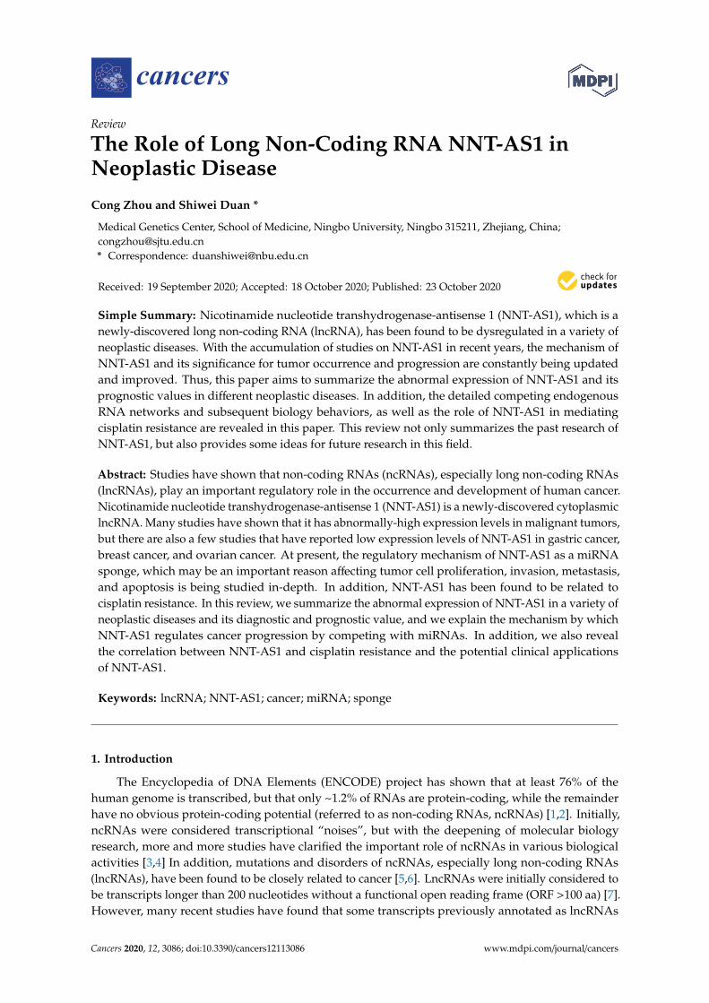
Nicotinamide nucleotide transhydrogenase-antisense 1 (NNT-AS1) is a newly-discovered lncRNAmainly distributed in the cytoplasm [14]. NNT-AS1 is located in 5p12 and has three exons.The transcription directions of NNT-AS1 and the adjacent protein-coding gene nicotinamide nucleotidetranshydrogenase (NNT) are opposite and there is no overlapping region between NNT and NNT-AS1(Figure 1). In a study of multiple sclerosis, there was no significant difference in NNT-AS1 levels inperipheral blood samples between the case group and the healthy group [15]. However, in all thestudies on tumorous diseases that have been published so far, the expression level of NNT-AS1 intumor tissues or tumor cells has been abnormal, which indicates the potential correlation betweenNNT-AS1 dysregulation and tumorous diseases. This review summarizes the abnormal expressionlevels of NNT-AS1 in tumors and describes its regulation of a large number of downstream genes bycompeting with miRNAs to affect multiple signaling pathways. We also summarize the diagnostic andprognostic values of NNT-AS1 in cancer and present its role in the resistance of antitumor drugs.
Figure 1. Genome position of long non-coding RNA (lncRNA) nicotinamide nucleotidetranshydrogenase-antisense 1 (NNT-AS1). NNT-AS1 is mapped to chromosome 5. Referenceinformation comes from the University of California Santa Cruz (UCSC) Genome Browser (http://genome.ucsc.edu/).
2. The Aberrant Expression of NNT-AS1 in Cancer
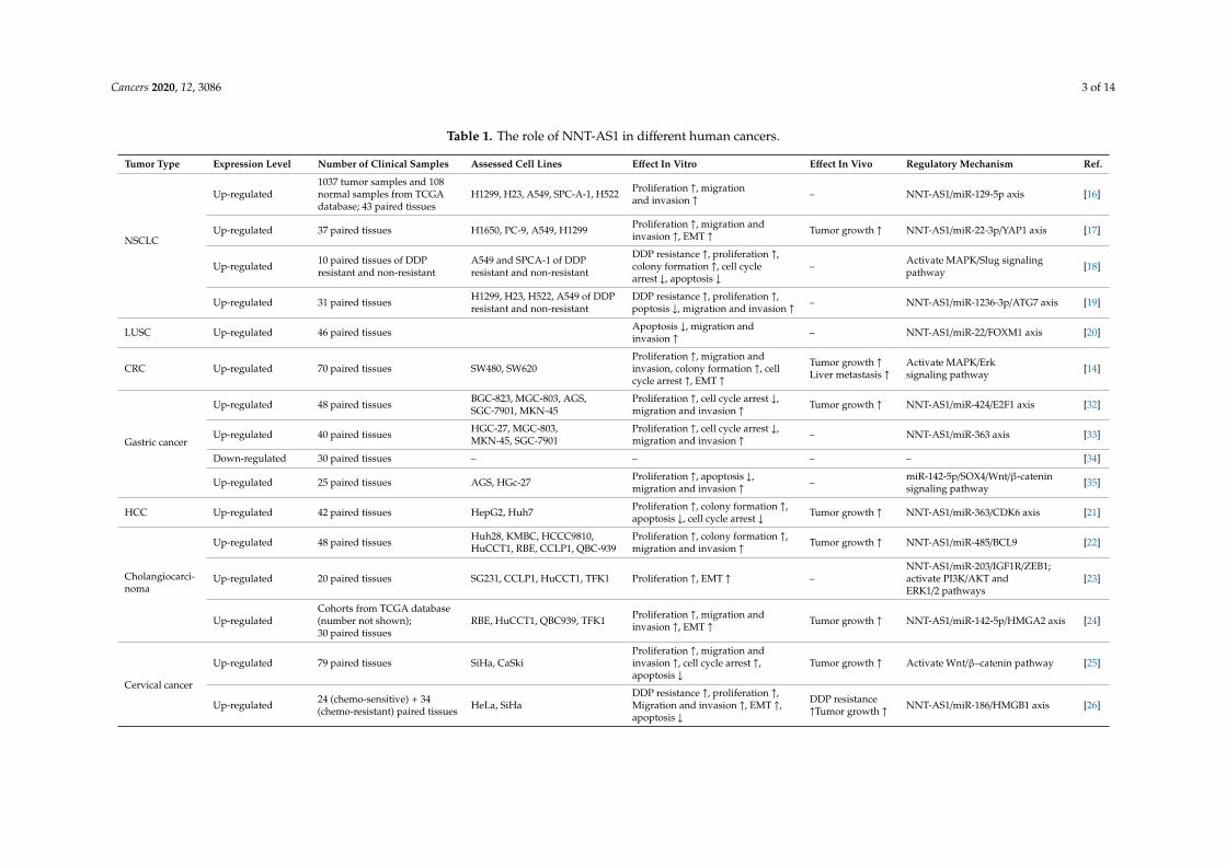
Current research shows that NNT-AS1 is highly expressed in most tumors, including non-small-celllung cancer (NSCLC) [16–19], lung squamous cell carcinoma (LUSC) [20], colorectal cancer (CRC) [14],hepatocellular carcinoma (HCC) [21], cholangiocarcinoma (CAA) [22–24], cervical cancer [25,26],osteosarcoma [27,28], bladder cancer [29,30], and glioma [31]. These results have been verified in thecorresponding tumor tissues or tumor cells. In subsequent functional experiments on cells, these studiesfurther revealed the cancer-promoting effects of high levels of NNT-AS1, including induction ofproliferation, promotion of invasion and metastasis, and inhibition of apoptosis. In addition,some studies have used mouse models to confirm the tumor growth and liver metastasis effectsof NNT-AS1 (Table 1).
Cancers 2020, 12, 3086 3 of 14
Table 1. The role of NNT-AS1 in different human cancers.
Tumor Type Expression Level Number of Clinical Samples Assessed Cell Lines Effect In Vitro Effect In Vivo Regulatory Mechanism Ref.
NSCLC
Up-regulated1037 tumor samples and 108normal samples from TCGAdatabase; 43 paired tissues
H1299, H23, A549, SPC-A-1, H522 Proliferation ↑, migrationand invasion ↑ – NNT-AS1/miR-129-5p axis [16]
Up-regulated 37 paired tissues H1650, PC-9, A549, H1299 Proliferation ↑, migration andinvasion ↑, EMT ↑ Tumor growth ↑ NNT-AS1/miR-22-3p/YAP1 axis [17]
Up-regulated 10 paired tissues of DDPresistant and non-resistant
A549 and SPCA-1 of DDPresistant and non-resistant
DDP resistance ↑, proliferation ↑,colony formation ↑, cell cyclearrest ↓, apoptosis ↓
– Activate MAPK/Slug signalingpathway [18]
Up-regulated 31 paired tissues H1299, H23, H522, A549 of DDPresistant and non-resistant
DDP resistance ↑, proliferation ↑,poptosis ↓, migration and invasion ↑ – NNT-AS1/miR-1236-3p/ATG7 axis [19]
LUSC Up-regulated 46 paired tissues Apoptosis ↓, migration andinvasion ↑ – NNT-AS1/miR-22/FOXM1 axis [20]
CRC Up-regulated 70 paired tissues SW480, SW620Proliferation ↑, migration andinvasion, colony formation ↑, cellcycle arrest ↑, EMT ↑
Tumor growth ↑Liver metastasis ↑
Activate MAPK/Erksignaling pathway [14]
Gastric cancer
Up-regulated 48 paired tissues BGC-823, MGC-803, AGS,SGC-7901, MKN-45
Proliferation ↑, cell cycle arrest ↓,migration and invasion ↑ Tumor growth ↑ NNT-AS1/miR-424/E2F1 axis [32]
Up-regulated 40 paired tissues HGC-27, MGC-803,MKN-45, SGC-7901
Proliferation ↑, cell cycle arrest ↓,migration and invasion ↑ – NNT-AS1/miR-363 axis [33]
Down-regulated 30 paired tissues – – – – [34]
Up-regulated 25 paired tissues AGS, HGc-27 Proliferation ↑, apoptosis ↓,migration and invasion ↑ – miR-142-5p/SOX4/Wnt/β-catenin
signaling pathway [35]
HCC Up-regulated 42 paired tissues HepG2, Huh7 Proliferation ↑, colony formation ↑,apoptosis ↓, cell cycle arrest ↓ Tumor growth ↑ NNT-AS1/miR-363/CDK6 axis [21]
Cholangiocarci-noma
Up-regulated 48 paired tissues Huh28, KMBC, HCCC9810,HuCCT1, RBE, CCLP1, QBC-939
Proliferation ↑, colony formation ↑,migration and invasion ↑ Tumor growth ↑ NNT-AS1/miR-485/BCL9 [22]
Up-regulated 20 paired tissues SG231, CCLP1, HuCCT1, TFK1 Proliferation ↑, EMT ↑ –NNT-AS1/miR-203/IGF1R/ZEB1;activate PI3K/AKT andERK1/2 pathways
[23]
Up-regulatedCohorts from TCGA database(number not shown);30 paired tissues
RBE, HuCCT1, QBC939, TFK1 Proliferation ↑, migration andinvasion ↑, EMT ↑ Tumor growth ↑ NNT-AS1/miR-142-5p/HMGA2 axis [24]
Cervical cancer
Up-regulated 79 paired tissues SiHa, CaSkiProliferation ↑, migration andinvasion ↑, cell cycle arrest ↑,apoptosis ↓
Tumor growth ↑ Activate Wnt/β–catenin pathway [25]
Up-regulated 24 (chemo-sensitive) + 34(chemo-resistant) paired tissues HeLa, SiHa
DDP resistance ↑, proliferation ↑,Migration and invasion ↑, EMT ↑,apoptosis ↓
DDP resistance↑Tumor growth ↑ NNT-AS1/miR-186/HMGB1 axis [26]
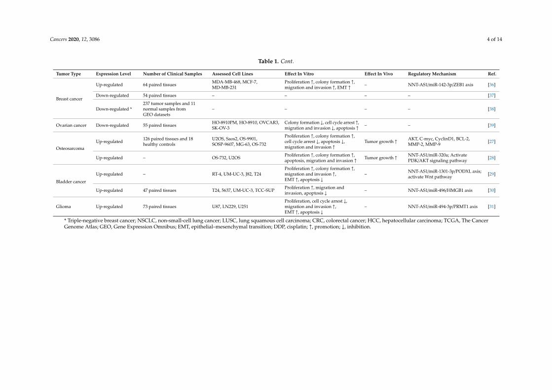
Cancers 2020, 12, 3086 4 of 14
Table 1. Cont.
Tumor Type Expression Level Number of Clinical Samples Assessed Cell Lines Effect In Vitro Effect In Vivo Regulatory Mechanism Ref.
Breast cancer
Up-regulated 64 paired tissues MDA-MB-468, MCF-7,MD-MB-231
Proliferation ↑, colony formation ↑,migration and invasion ↑, EMT ↑ – NNT-AS1/miR-142-3p/ZEB1 axis [36]
Down-regulated 54 paired tissues – – – – [37]
Down-regulated *237 tumor samples and 11normal samples fromGEO datasets
– – – – [38]
Ovarian cancer Down-regulated 55 paired tissues HO-8910PM, HO-8910, OVCAR3,SK-OV-3
Colony formation ↓, cell cycle arrest ↑,migration and invasion ↓, apoptosis ↑ – – [39]
OsteosarcomaUp-regulated 126 paired tissues and 18
healthy controlsU2OS, Saos2, OS-9901,SOSP-9607, MG-63, OS-732
Proliferation ↑, colony formation ↑,cell cycle arrest ↓, apoptosis ↓,migration and invasion ↑
Tumor growth ↑ AKT, C-myc, CyclinD1, BCL-2,MMP-2, MMP-9 [27]
Up-regulated – OS-732, U2OS Proliferation ↑, colony formation ↑,apoptosis, migration and invasion ↑ Tumor growth ↑ NNT-AS1/miR-320a; Activate
PI3K/AKT signaling pathway [28]
Bladder cancer
Up-regulated – RT-4, UM-UC-3, J82, T24Proliferation ↑, colony formation ↑,migration and invasion ↑,EMT ↑, apoptosis ↓
– NNT-AS1/miR-1301-3p/PODXL axis;activate Wnt pathway [29]
Up-regulated 47 paired tissues T24, 5637, UM-UC-3, TCC-SUP Proliferation ↑, migration andinvasion, apoptosis ↓ – NNT-AS1/miR-496/HMGB1 axis [30]
Glioma Up-regulated 73 paired tissues U87, LN229, U251Proliferation, cell cycle arrest ↓,migration and invasion ↑,EMT ↑, apoptosis ↓
– NNT-AS1/miR-494-3p/PRMT1 axis [31]
* Triple-negative breast cancer; NSCLC, non-small-cell lung cancer; LUSC, lung squamous cell carcinoma; CRC, colorectal cancer; HCC, hepatocellular carcinoma; TCGA, The CancerGenome Atlas; GEO, Gene Expression Omnibus; EMT, epithelial–mesenchymal transition; DDP, cisplatin; ↑, promotion; ↓, inhibition.
Cancers 2020, 12, 3086 5 of 14
However, studies on the expression of NNT-AS1 in gastric, breast, and ovarian cancers seem tobe contradictory. In gastric cancer, there have been three studies that have reported high levels ofNNT-AS1 in tumor tissues and cell samples, and the performance of NNT-AS1 in subsequent in vivoand in vitro tests also verified its cancer-promoting function [32,33,35]. However, Farbod Esfandi etal. found that compared with the adjacent samples, the NNT-AS1 levels detected in gastric cancersamples were overall down-regulated [34]. Their further stratified analysis showed that compared withtumors without lymphatic/vascular invasion, tumors with lymphatic/vascular invasion had higherlevels of NNT-AS1, which seems to be consistent with the malignant manifestations of NNT-AS1 [34].The authors attributed these contradictions to ethnic differences [34]. However, it is worth noting thatthe sample size of this study was relatively small and lacked follow-up in vivo and in vitro experiments.Therefore, more samples and a more complete experimental system are needed for further verification.For breast cancer, Li et al. first reported the high expression of NNT-AS1 and confirmed it as a motivatorfor breast cancer progression in functional experiments [36]. In contrast, Saleh Gargari S. et al. reportedthe low expression of NNT-AS1 in breast cancer samples, and in subgroup analysis, the expressionof NNT-AS1 in estrogen receptor (ER)-negative tumor samples was higher than ER-positive tumorsamples [37]. This suggests that the expression of NNT-AS1 may be related to sex hormone receptors.A recent bioinformatics study based on Gene Expression Omnibus (GEO) data analyzed the expressionof lncRNA in triple-negative breast cancer (including ER-negative), in which NNT-AS1 was also foundto be down-regulated [38]. In addition, in a study on ovarian cancer, the low level of NNT-AS1 wasconfirmed as well [39].
Interestingly, it has been reported that, unlike Erα-receptor-negative ovarian cancer cells, treatmentof Erα-receptor-positive ovarian cancer cells with estrogen (E2) will cause a large number of lncRNAdisorders and changes in some protein levels, such as elevated TC0101441, which was found topromote the invasion and metastasis of ovarian cancer cells [40]. This suggests that sex hormones andsex hormone receptors may affect lncRNA and tumor progression. In Erα-receptor-positive breastcancer cells, when E2 is monitored or E2 is given, the levels of miR-424 and E2F1 also increase [41,42].In a gastric cancer study, the regulatory axis of NNT-AS1/miR-424/E2F1 was confirmed [32]. Therefore,the low expression of NNT-AS1 up-regulates miR-424 and E2F1, and the abnormal E2 level is likely to becorrelated. Although the current evidence is not enough to determine the causal relationship, this maybe one of the reasons for the low expression of NNT-AS1 and the progression of sex-hormone-relatedtumors. In addition, in a study of papillary thyroid carcinoma, lncRNA-H19 was found to inducethe up-regulation of ERβ and promote cancer-stem-like properties [43]. These findings suggest thepossibility that NNT-AS1 may have a similar ability to regulate ER receptors and contribute to tumorprogression. These models are worthy of further exploration in future research.
3. The Diagnostic or Prognostic Value of Aberrant NNT-AS1 Expression
Some studies have analyzed the diagnostic and prognostic value of abnormal expression ofNNT-AS1 (Table 2). The results of Farbod Esfandi et al. showed that the area under the curve(AUC) of NNT-AS1 expression level for gastric cancer diagnosis was 0.63, sensitivity was 73.3%,specificity was 70%, and p = 0.09, which suggests that NNT-AS1 is not suitable as a diagnostic markerfor gastric cancer [34]. For breast cancer, the NNT-AS1 expression level showed lower sensitivity(56.6%) and similar specificity (71.2%) [37]. These two studies indicated that the diagnostic efficiency ofthe down-regulated NNT-AS1 may be unsatisfactory. In addition, the diagnostic value of up-regulatedNNT-AS1 in cancer has not been reported.
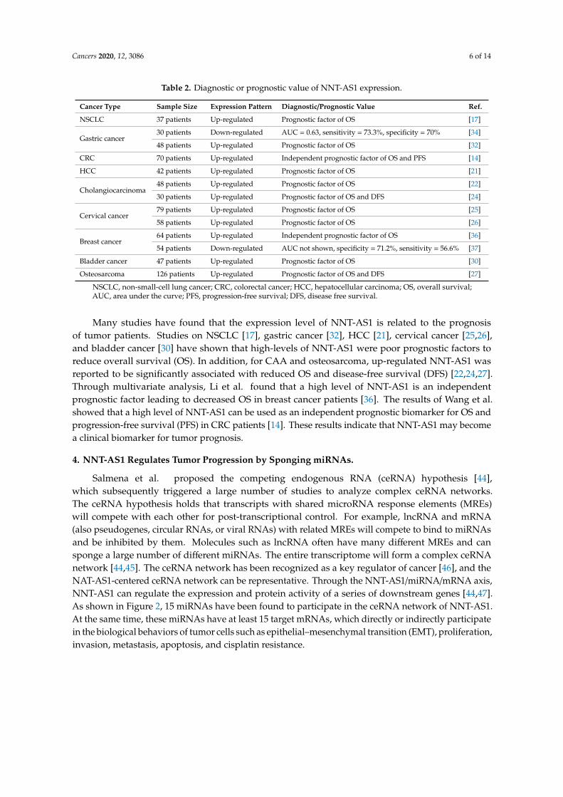
Cancers 2020, 12, 3086 6 of 14
Table 2. Diagnostic or prognostic value of NNT-AS1 expression.
Cancer Type Sample Size Expression Pattern Diagnostic/Prognostic Value Ref.
NSCLC 37 patients Up-regulated Prognostic factor of OS [17]
Gastric cancer30 patients Down-regulated AUC = 0.63, sensitivity = 73.3%, specificity = 70% [34]
48 patients Up-regulated Prognostic factor of OS [32]
CRC 70 patients Up-regulated Independent prognostic factor of OS and PFS [14]
HCC 42 patients Up-regulated Prognostic factor of OS [21]
Cholangiocarcinoma48 patients Up-regulated Prognostic factor of OS [22]
30 patients Up-regulated Prognostic factor of OS and DFS [24]
Cervical cancer79 patients Up-regulated Prognostic factor of OS [25]
58 patients Up-regulated Prognostic factor of OS [26]
Breast cancer64 patients Up-regulated Independent prognostic factor of OS [36]
54 patients Down-regulated AUC not shown, specificity = 71.2%, sensitivity = 56.6% [37]
Bladder cancer 47 patients Up-regulated Prognostic factor of OS [30]
Osteosarcoma 126 patients Up-regulated Prognostic factor of OS and DFS [27]
NSCLC, non-small-cell lung cancer; CRC, colorectal cancer; HCC, hepatocellular carcinoma; OS, overall survival;AUC, area under the curve; PFS, progression-free survival; DFS, disease free survival.
Many studies have found that the expression level of NNT-AS1 is related to the prognosisof tumor patients. Studies on NSCLC [17], gastric cancer [32], HCC [21], cervical cancer [25,26],and bladder cancer [30] have shown that high-levels of NNT-AS1 were poor prognostic factors toreduce overall survival (OS). In addition, for CAA and osteosarcoma, up-regulated NNT-AS1 wasreported to be significantly associated with reduced OS and disease-free survival (DFS) [22,24,27].Through multivariate analysis, Li et al. found that a high level of NNT-AS1 is an independentprognostic factor leading to decreased OS in breast cancer patients [36]. The results of Wang et al.showed that a high level of NNT-AS1 can be used as an independent prognostic biomarker for OS andprogression-free survival (PFS) in CRC patients [14]. These results indicate that NNT-AS1 may becomea clinical biomarker for tumor prognosis.
4. NNT-AS1 Regulates Tumor Progression by Sponging miRNAs.
Salmena et al. proposed the competing endogenous RNA (ceRNA) hypothesis [44],which subsequently triggered a large number of studies to analyze complex ceRNA networks.The ceRNA hypothesis holds that transcripts with shared microRNA response elements (MREs)will compete with each other for post-transcriptional control. For example, lncRNA and mRNA(also pseudogenes, circular RNAs, or viral RNAs) with related MREs will compete to bind to miRNAsand be inhibited by them. Molecules such as lncRNA often have many different MREs and cansponge a large number of different miRNAs. The entire transcriptome will form a complex ceRNAnetwork [44,45]. The ceRNA network has been recognized as a key regulator of cancer [46], and theNAT-AS1-centered ceRNA network can be representative. Through the NNT-AS1/miRNA/mRNA axis,NNT-AS1 can regulate the expression and protein activity of a series of downstream genes [44,47].As shown in Figure 2, 15 miRNAs have been found to participate in the ceRNA network of NNT-AS1.At the same time, these miRNAs have at least 15 target mRNAs, which directly or indirectly participatein the biological behaviors of tumor cells such as epithelial–mesenchymal transition (EMT), proliferation,invasion, metastasis, apoptosis, and cisplatin resistance.
Cancers 2020, 12, 3086 7 of 14
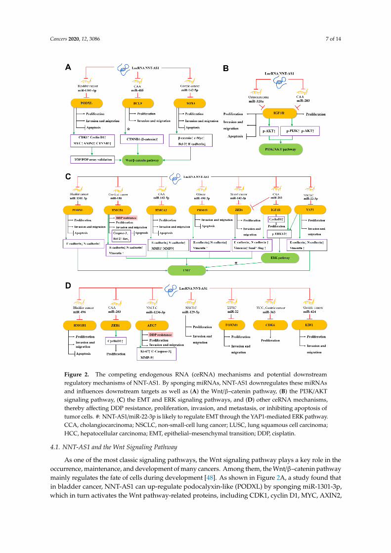
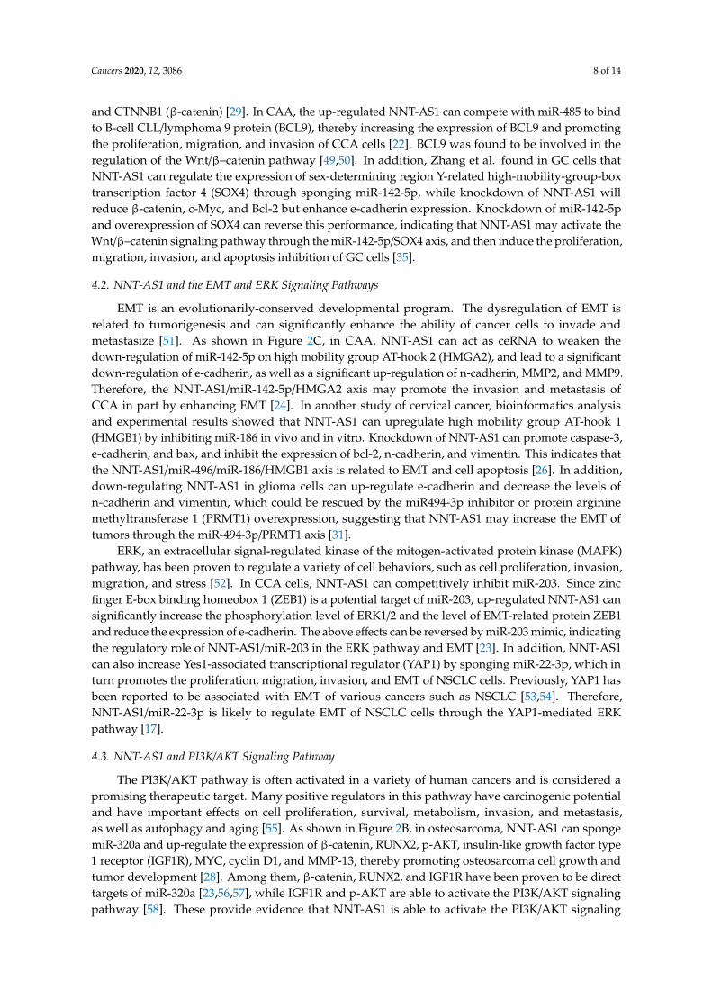
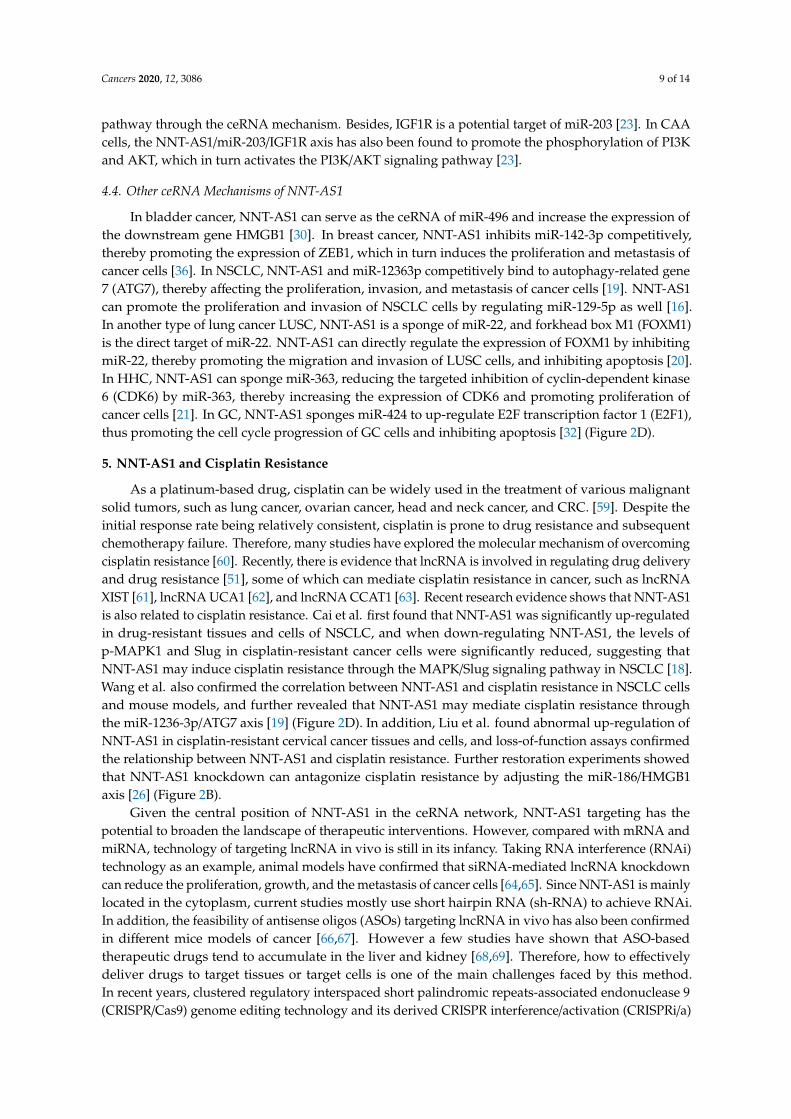
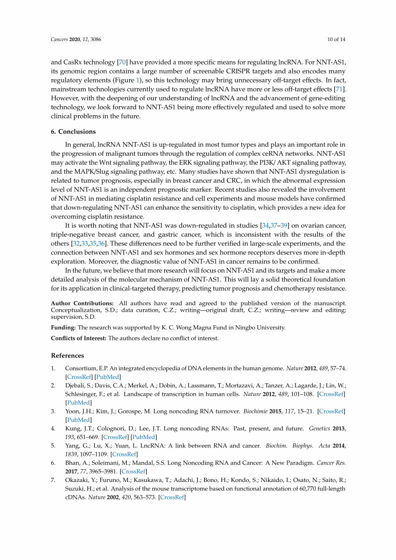
Figure 2. The competing endogenous RNA (ceRNA) mechanisms and potential downstreamregulatory mechanisms of NNT-AS1. By sponging miRNAs, NNT-AS1 downregulates these miRNAsand influences downstream targets as well as (A) the Wnt/β–catenin pathway, (B) the PI3K/AKTsignaling pathway, (C) the EMT and ERK signaling pathways, and (D) other ceRNA mechanisms,thereby affecting DDP resistance, proliferation, invasion, and metastasis, or inhibiting apoptosis oftumor cells. #: NNT-AS1/miR-22-3p is likely to regulate EMT through the YAP1-mediated ERK pathway.CCA, cholangiocarcinoma; NSCLC, non-small-cell lung cancer; LUSC, lung squamous cell carcinoma;HCC, hepatocellular carcinoma; EMT, epithelial–mesenchymal transition; DDP, cisplatin.
4.1. NNT-AS1 and the Wnt Signaling Pathway
As one of the most classic signaling pathways, the Wnt signaling pathway plays a key role in theoccurrence, maintenance, and development of many cancers. Among them, the Wnt/β–catenin pathwaymainly regulates the fate of cells during development [48]. As shown in Figure 2A, a study found thatin bladder cancer, NNT-AS1 can up-regulate podocalyxin-like (PODXL) by sponging miR-1301-3p,which in turn activates the Wnt pathway-related proteins, including CDK1, cyclin D1, MYC, AXIN2,
Cancers 2020, 12, 3086 8 of 14
and CTNNB1 (β-catenin) [29]. In CAA, the up-regulated NNT-AS1 can compete with miR-485 to bindto B-cell CLL/lymphoma 9 protein (BCL9), thereby increasing the expression of BCL9 and promotingthe proliferation, migration, and invasion of CCA cells [22]. BCL9 was found to be involved in theregulation of the Wnt/β–catenin pathway [49,50]. In addition, Zhang et al. found in GC cells thatNNT-AS1 can regulate the expression of sex-determining region Y-related high-mobility-group-boxtranscription factor 4 (SOX4) through sponging miR-142-5p, while knockdown of NNT-AS1 willreduce β-catenin, c-Myc, and Bcl-2 but enhance e-cadherin expression. Knockdown of miR-142-5pand overexpression of SOX4 can reverse this performance, indicating that NNT-AS1 may activate theWnt/β–catenin signaling pathway through the miR-142-5p/SOX4 axis, and then induce the proliferation,migration, invasion, and apoptosis inhibition of GC cells [35].
4.2. NNT-AS1 and the EMT and ERK Signaling Pathways
EMT is an evolutionarily-conserved developmental program. The dysregulation of EMT isrelated to tumorigenesis and can significantly enhance the ability of cancer cells to invade andmetastasize [51]. As shown in Figure 2C, in CAA, NNT-AS1 can act as ceRNA to weaken thedown-regulation of miR-142-5p on high mobility group AT-hook 2 (HMGA2), and lead to a significantdown-regulation of e-cadherin, as well as a significant up-regulation of n-cadherin, MMP2, and MMP9.Therefore, the NNT-AS1/miR-142-5p/HMGA2 axis may promote the invasion and metastasis ofCCA in part by enhancing EMT [24]. In another study of cervical cancer, bioinformatics analysisand experimental results showed that NNT-AS1 can upregulate high mobility group AT-hook 1(HMGB1) by inhibiting miR-186 in vivo and in vitro. Knockdown of NNT-AS1 can promote caspase-3,e-cadherin, and bax, and inhibit the expression of bcl-2, n-cadherin, and vimentin. This indicates thatthe NNT-AS1/miR-496/miR-186/HMGB1 axis is related to EMT and cell apoptosis [26]. In addition,down-regulating NNT-AS1 in glioma cells can up-regulate e-cadherin and decrease the levels ofn-cadherin and vimentin, which could be rescued by the miR494-3p inhibitor or protein argininemethyltransferase 1 (PRMT1) overexpression, suggesting that NNT-AS1 may increase the EMT oftumors through the miR-494-3p/PRMT1 axis [31].
ERK, an extracellular signal-regulated kinase of the mitogen-activated protein kinase (MAPK)pathway, has been proven to regulate a variety of cell behaviors, such as cell proliferation, invasion,migration, and stress [52]. In CCA cells, NNT-AS1 can competitively inhibit miR-203. Since zincfinger E-box binding homeobox 1 (ZEB1) is a potential target of miR-203, up-regulated NNT-AS1 cansignificantly increase the phosphorylation level of ERK1/2 and the level of EMT-related protein ZEB1and reduce the expression of e-cadherin. The above effects can be reversed by miR-203 mimic, indicatingthe regulatory role of NNT-AS1/miR-203 in the ERK pathway and EMT [23]. In addition, NNT-AS1can also increase Yes1-associated transcriptional regulator (YAP1) by sponging miR-22-3p, which inturn promotes the proliferation, migration, invasion, and EMT of NSCLC cells. Previously, YAP1 hasbeen reported to be associated with EMT of various cancers such as NSCLC [53,54]. Therefore,NNT-AS1/miR-22-3p is likely to regulate EMT of NSCLC cells through the YAP1-mediated ERKpathway [17].
4.3. NNT-AS1 and PI3K/AKT Signaling Pathway
The PI3K/AKT pathway is often activated in a variety of human cancers and is considered apromising therapeutic target. Many positive regulators in this pathway have carcinogenic potentialand have important effects on cell proliferation, survival, metabolism, invasion, and metastasis,as well as autophagy and aging [55]. As shown in Figure 2B, in osteosarcoma, NNT-AS1 can spongemiR-320a and up-regulate the expression of β-catenin, RUNX2, p-AKT, insulin-like growth factor type1 receptor (IGF1R), MYC, cyclin D1, and MMP-13, thereby promoting osteosarcoma cell growth andtumor development [28]. Among them, β-catenin, RUNX2, and IGF1R have been proven to be directtargets of miR-320a [23,56,57], while IGF1R and p-AKT are able to activate the PI3K/AKT signalingpathway [58]. These provide evidence that NNT-AS1 is able to activate the PI3K/AKT signaling
Cancers 2020, 12, 3086 9 of 14
pathway through the ceRNA mechanism. Besides, IGF1R is a potential target of miR-203 [23]. In CAAcells, the NNT-AS1/miR-203/IGF1R axis has also been found to promote the phosphorylation of PI3Kand AKT, which in turn activates the PI3K/AKT signaling pathway [23].
4.4. Other ceRNA Mechanisms of NNT-AS1
In bladder cancer, NNT-AS1 can serve as the ceRNA of miR-496 and increase the expression ofthe downstream gene HMGB1 [30]. In breast cancer, NNT-AS1 inhibits miR-142-3p competitively,thereby promoting the expression of ZEB1, which in turn induces the proliferation and metastasis ofcancer cells [36]. In NSCLC, NNT-AS1 and miR-12363p competitively bind to autophagy-related gene7 (ATG7), thereby affecting the proliferation, invasion, and metastasis of cancer cells [19]. NNT-AS1can promote the proliferation and invasion of NSCLC cells by regulating miR-129-5p as well [16].In another type of lung cancer LUSC, NNT-AS1 is a sponge of miR-22, and forkhead box M1 (FOXM1)is the direct target of miR-22. NNT-AS1 can directly regulate the expression of FOXM1 by inhibitingmiR-22, thereby promoting the migration and invasion of LUSC cells, and inhibiting apoptosis [20].In HHC, NNT-AS1 can sponge miR-363, reducing the targeted inhibition of cyclin-dependent kinase6 (CDK6) by miR-363, thereby increasing the expression of CDK6 and promoting proliferation ofcancer cells [21]. In GC, NNT-AS1 sponges miR-424 to up-regulate E2F transcription factor 1 (E2F1),thus promoting the cell cycle progression of GC cells and inhibiting apoptosis [32] (Figure 2D).
5. NNT-AS1 and Cisplatin Resistance
As a platinum-based drug, cisplatin can be widely used in the treatment of various malignantsolid tumors, such as lung cancer, ovarian cancer, head and neck cancer, and CRC. [59]. Despite theinitial response rate being relatively consistent, cisplatin is prone to drug resistance and subsequentchemotherapy failure. Therefore, many studies have explored the molecular mechanism of overcomingcisplatin resistance [60]. Recently, there is evidence that lncRNA is involved in regulating drug deliveryand drug resistance [51], some of which can mediate cisplatin resistance in cancer, such as lncRNAXIST [61], lncRNA UCA1 [62], and lncRNA CCAT1 [63]. Recent research evidence shows that NNT-AS1is also related to cisplatin resistance. Cai et al. first found that NNT-AS1 was significantly up-regulatedin drug-resistant tissues and cells of NSCLC, and when down-regulating NNT-AS1, the levels ofp-MAPK1 and Slug in cisplatin-resistant cancer cells were significantly reduced, suggesting thatNNT-AS1 may induce cisplatin resistance through the MAPK/Slug signaling pathway in NSCLC [18].Wang et al. also confirmed the correlation between NNT-AS1 and cisplatin resistance in NSCLC cellsand mouse models, and further revealed that NNT-AS1 may mediate cisplatin resistance throughthe miR-1236-3p/ATG7 axis [19] (Figure 2D). In addition, Liu et al. found abnormal up-regulation ofNNT-AS1 in cisplatin-resistant cervical cancer tissues and cells, and loss-of-function assays confirmedthe relationship between NNT-AS1 and cisplatin resistance. Further restoration experiments showedthat NNT-AS1 knockdown can antagonize cisplatin resistance by adjusting the miR-186/HMGB1axis [26] (Figure 2B).
Given the central position of NNT-AS1 in the ceRNA network, NNT-AS1 targeting has thepotential to broaden the landscape of therapeutic interventions. However, compared with mRNA andmiRNA, technology of targeting lncRNA in vivo is still in its infancy. Taking RNA interference (RNAi)technology as an example, animal models have confirmed that siRNA-mediated lncRNA knockdowncan reduce the proliferation, growth, and the metastasis of cancer cells [64,65]. Since NNT-AS1 is mainlylocated in the cytoplasm, current studies mostly use short hairpin RNA (sh-RNA) to achieve RNAi.In addition, the feasibility of antisense oligos (ASOs) targeting lncRNA in vivo has also been confirmedin different mice models of cancer [66,67]. However a few studies have shown that ASO-basedtherapeutic drugs tend to accumulate in the liver and kidney [68,69]. Therefore, how to effectivelydeliver drugs to target tissues or target cells is one of the main challenges faced by this method.In recent years, clustered regulatory interspaced short palindromic repeats-associated endonuclease 9(CRISPR/Cas9) genome editing technology and its derived CRISPR interference/activation (CRISPRi/a)
Cancers 2020, 12, 3086 10 of 14
and CasRx technology [70] have provided a more specific means for regulating lncRNA. For NNT-AS1,its genomic region contains a large number of screenable CRISPR targets and also encodes manyregulatory elements (Figure 1), so this technology may bring unnecessary off-target effects. In fact,mainstream technologies currently used to regulate lncRNA have more or less off-target effects [71].However, with the deepening of our understanding of lncRNA and the advancement of gene-editingtechnology, we look forward to NNT-AS1 being more effectively regulated and used to solve moreclinical problems in the future.
6. Conclusions
In general, lncRNA NNT-AS1 is up-regulated in most tumor types and plays an important role inthe progression of malignant tumors through the regulation of complex ceRNA networks. NNT-AS1may activate the Wnt signaling pathway, the ERK signaling pathway, the PI3K/ AKT signaling pathway,and the MAPK/Slug signaling pathway, etc. Many studies have shown that NNT-AS1 dysregulation isrelated to tumor prognosis, especially in breast cancer and CRC, in which the abnormal expressionlevel of NNT-AS1 is an independent prognostic marker. Recent studies also revealed the involvementof NNT-AS1 in mediating cisplatin resistance and cell experiments and mouse models have confirmedthat down-regulating NNT-AS1 can enhance the sensitivity to cisplatin, which provides a new idea forovercoming cisplatin resistance.
It is worth noting that NNT-AS1 was down-regulated in studies [34,37–39] on ovarian cancer,triple-negative breast cancer, and gastric cancer, which is inconsistent with the results of theothers [32,33,35,36]. These differences need to be further verified in large-scale experiments, and theconnection between NNT-AS1 and sex hormones and sex hormone receptors deserves more in-depthexploration. Moreover, the diagnostic value of NNT-AS1 in cancer remains to be confirmed.
In the future, we believe that more research will focus on NNT-AS1 and its targets and make a moredetailed analysis of the molecular mechanism of NNT-AS1. This will lay a solid theoretical foundationfor its application in clinical-targeted therapy, predicting tumor prognosis and chemotherapy resistance.
Author Contributions: All authors have read and agreed to the published version of the manuscript.Conceptualization, S.D.; data curation, C.Z.; writing—original draft, C.Z.; writing—review and editing;supervision, S.D.
Funding: The research was supported by K. C. Wong Magna Fund in Ningbo University.
Conflicts of Interest: The authors declare no conflict of interest.
References
1. Consortium, E.P. An integrated encyclopedia of DNA elements in the human genome. Nature 2012, 489, 57–74.[CrossRef] [PubMed]
2. Djebali, S.; Davis, C.A.; Merkel, A.; Dobin, A.; Lassmann, T.; Mortazavi, A.; Tanzer, A.; Lagarde, J.; Lin, W.;Schlesinger, F.; et al. Landscape of transcription in human cells. Nature 2012, 489, 101–108. [CrossRef][PubMed]
3. Yoon, J.H.; Kim, J.; Gorospe, M. Long noncoding RNA turnover. Biochimie 2015, 117, 15–21. [CrossRef][PubMed]
4. Kung, J.T.; Colognori, D.; Lee, J.T. Long noncoding RNAs: Past, present, and future. Genetics 2013,193, 651–669. [CrossRef] [PubMed]
5. Yang, G.; Lu, X.; Yuan, L. LncRNA: A link between RNA and cancer. Biochim. Biophys. Acta 2014,1839, 1097–1109. [CrossRef]
6. Bhan, A.; Soleimani, M.; Mandal, S.S. Long Noncoding RNA and Cancer: A New Paradigm. Cancer Res.2017, 77, 3965–3981. [CrossRef]
7. Okazaki, Y.; Furuno, M.; Kasukawa, T.; Adachi, J.; Bono, H.; Kondo, S.; Nikaido, I.; Osato, N.; Saito, R.;Suzuki, H.; et al. Analysis of the mouse transcriptome based on functional annotation of 60,770 full-lengthcDNAs. Nature 2002, 420, 563–573. [CrossRef]
Cancers 2020, 12, 3086 11 of 14
8. Chen, J.; Brunner, A.D.; Cogan, J.Z.; Nunez, J.K.; Fields, A.P.; Adamson, B.; Itzhak, D.N.; Li, J.Y.; Mann, M.;Leonetti, M.D.; et al. Pervasive functional translation of noncanonical human open reading frames. Science2020, 367, 1140–1146. [CrossRef]
9. Nelson, B.R.; Makarewich, C.A.; Anderson, D.M.; Winders, B.R.; Troupes, C.D.; Wu, F.; Reese, A.L.;McAnally, J.R.; Chen, X.; Kavalali, E.T.; et al. A peptide encoded by a transcript annotated as long noncodingRNA enhances SERCA activity in muscle. Science 2016, 351, 271–275. [CrossRef]
10. Ji, Z.; Song, R.; Regev, A.; Struhl, K. Many lncRNAs, 5′UTRs, and pseudogenes are translated and some arelikely to express functional proteins. Elife 2015, 4, e08890. [CrossRef]
11. Volders, P.J.; Anckaert, J.; Verheggen, K.; Nuytens, J.; Martens, L.; Mestdagh, P.; Vandesompele, J. LNCipedia5: Towards a reference set of human long non-coding RNAs. Nucleic Acids Res. 2019, 47, D135–D139.[CrossRef] [PubMed]
12. Chi, Y.; Wang, D.; Wang, J.; Yu, W.; Yang, J. Long Non-Coding RNA in the Pathogenesis of Cancers. Cells2019, 8, 1015. [CrossRef] [PubMed]
13. Chen, Q.N.; Wei, C.C.; Wang, Z.X.; Sun, M. Long non-coding RNAs in anti-cancer drug resistance. Oncotarget2017, 8, 1925–1936. [CrossRef]
14. Wang, Q.; Yang, L.; Hu, X.; Jiang, Y.; Hu, Y.; Liu, Z.; Liu, J.; Wen, T.; Ma, Y.; An, G.; et al. UpregulatedNNT-AS1, a long noncoding RNA, contributes to proliferation and migration of colorectal cancer cellsin vitro and in vivo. Oncotarget 2017, 8, 3441–3453. [CrossRef] [PubMed]
15. Eftekharian, M.M.; Taheri, M.; Arsang-Jang, S.; Komaki, A.; Ghafouri-Fard, S. Nicotinamide nucleotidetranshydrogenase expression analysis in multiple sclerosis patients. Int. J. Neurosci. 2019, 129, 1256–1260.[CrossRef] [PubMed]
16. Shen, Q.; Jiang, Y. LncRNA NNT-AS1 promotes the proliferation, and invasion of lung cancer cells viaregulating miR-129-5p expression. Biomed. Pharmacother. 2018, 105, 176–181. [CrossRef] [PubMed]
17. He, W.; Zhang, Y.; Xia, S. LncRNA NNT-AS1 promotes non-small cell lung cancer progression throughregulating miR-22-3p/YAP1 axis. Thorac. Cancer 2020, 11, 549–560. [CrossRef]
18. Cai, Y.; Dong, Z.Y.; Wang, J.Y. LncRNA NNT-AS1 is a major mediator of cisplatin chemoresistance innon-small cell lung cancer through MAPK/Slug pathway. Eur. Rev. Med. Pharmacol. Sci. 2018, 22, 4879–4887.[CrossRef]
19. Wang, H.; Guo, M.; Ding, D.; Yang, F.; Chen, Z. Long Non-Coding RNA NNT-AS1 Contributes to CisplatinResistance via miR-1236-3p/ATG7 Axis in Lung Cancer Cells. Onco Targets Ther. 2020, 13, 3641–3652.[CrossRef]
20. Ma, J.; Qi, G.; Li, L. LncRNA NNT-AS1 promotes lung squamous cell carcinoma progression by regulatingthe miR-22/FOXM1 axis. Cell Mol. Biol. Lett. 2020, 25, 34. [CrossRef]
21. Lu, Y.B.; Jiang, Q.; Yang, M.Y.; Zhou, J.X.; Zhang, Q. Long noncoding RNA NNT-AS1 promotes hepatocellularcarcinoma progression and metastasis through miR-363/CDK6 axis. Oncotarget 2017, 8, 88804–88814.[CrossRef]
22. Huang, L.; Jiang, X.; Kang, P.; Wang, Z.; Leng, K.; Ji, D.; Xu, Y.; Wang, H.; Cui, Y. Long non-codingRNA NNT-AS1 functions as an oncogenic gene through modulating miR-485/BCL9 in cholangiocarcinoma.Cancer Manag. Res. 2019, 11, 7739–7749. [CrossRef]
23. Gu, Y.; Zhu, Z.; Pei, H.; Xu, D.; Jiang, Y.; Zhang, L.; Xiao, L. Long non-coding RNA NNT-AS1 promotescholangiocarcinoma cells proliferation and epithelial-to-mesenchymal transition through down-regulatingmiR-203. Aging (Albany NY) 2020, 12, 2333–2346. [CrossRef]
24. Gu, Y.; Li, C.; Xiao, L.; Li, J.; Pei, H.; Xu, D.; Jiang, Y.; Zhang, X.; Zhang, L.; Li, K.; et al. High expressionof long non-coding RNA NNT-AS1 facilitates progression of cholangiocarcinoma through promotingepithelial-mesenchymal transition. Am. J. Transl. Res. 2019, 11, 5438–5456. [PubMed]
25. Hua, F.; Liu, S.; Zhu, L.; Ma, N.; Jiang, S.; Yang, J. Highly expressed long non-coding RNA NNT-AS1promotes cell proliferation and invasion through Wnt/beta-catenin signaling pathway in cervical cancer.Biomed. Pharmacother. 2017, 92, 1128–1134. [CrossRef] [PubMed]
26. Liu, Y.; Guo, R.; Qiao, Y.; Han, L.; Liu, M. LncRNA NNT-AS1 contributes to the cisplatin resistance of cervicalcancer through NNT-AS1/miR-186/HMGB1 axis. Cancer Cell Int. 2020, 20, 190. [CrossRef]
27. Ye, H.; Lin, J.; Yao, X.; Li, Y.; Lin, X.; Lu, H. Overexpression of Long Non-Coding RNA NNT-AS1 Correlateswith Tumor Progression and Poor Prognosis in Osteosarcoma. Cell Physiol. Biochem. 2018, 45, 1904–1914.[CrossRef] [PubMed]
Cancers 2020, 12, 3086 12 of 14
28. Li, C.; Zhang, S.; Qiu, T.; Wang, Y.; Ricketts, D.M.; Qi, C. Upregulation of long non-coding RNA NNT-AS1promotes osteosarcoma progression by inhibiting the tumor suppressive miR-320a. Cancer Biol. Ther. 2019,20, 413–422. [CrossRef]
29. Liu, Y.; Wu, G. NNT-AS1 enhances bladder cancer cell growth by targeting miR-1301-3p/PODXL axis andactivating Wnt pathway. Neurourol. Urodyn. 2020, 39, 547–557. [CrossRef]
30. Wu, D.; Zhang, T.; Wang, J.; Zhou, J.; Pan, H.; Qu, P. Long noncoding RNA NNT-AS1 enhances the malignantphenotype of bladder cancer by acting as a competing endogenous RNA on microRNA-496 thereby increasingHMGB1 expression. Aging (Albany NY) 2019, 11, 12624–12640. [CrossRef]
31. Zheng, D.; Chen, D.; Lin, F.; Wang, X.; Lu, L.; Luo, S.; Chen, J.; Xu, X. LncRNA NNT-AS1 promote glioma cellproliferation and metastases through miR-494-3p/PRMT1 axis. Cell Cycle 2020, 19, 1621–1631. [CrossRef][PubMed]
32. Chen, B.; Zhao, Q.; Guan, L.; Lv, H.; Bie, L.; Huang, J.; Chen, X.B. Long non-coding RNA NNT-AS1 spongesmiR-424/E2F1 to promote the tumorigenesis and cell cycle progression of gastric cancer. J. Cell Mol. Med.2018, 22, 4751–4759. [CrossRef] [PubMed]
33. Wang, X.; Ren, M.; Li, Y.; Hu, J.; Lu, G.; Ma, W.; Guo, D.; Lu, X.; He, S. Long noncoding RNA NNT-AS1promotes gastric cancer proliferation and invasion by regulating microRNA-363 expression. J. Cell Biochem.2019, 120, 5704–5712. [CrossRef] [PubMed]
34. Esfandi, F.; Taheri, M.; Kahaei, M.S.; Omrani, M.D.; Kholghi Oskooei, V.; Ghafouri-Fard, S. Downregulationof nicotinamide nucleotide transhydrogenase and its naturally occurring antisense RNA in gastric cancer.Asia Pac. J. Clin. Oncol. 2019, 15, e191–e196. [CrossRef] [PubMed]
35. Zhang, J.; Zhang, K.; Hou, Y. Long noncoding RNA NNTAS1 knockdown represses the progression ofgastric cancer via modulating the miR1425p/SOX4/Wnt/betacatenin signaling pathway. Mol. Med. Rep. 2020,22, 687–696. [CrossRef]
36. Li, Y.; Lv, M.; Song, Z.; Lou, Z.; Wang, R.; Zhuang, M. Long non-coding RNA NNT-AS1 affects progression ofbreast cancer through miR-142-3p/ZEB1 axis. Biomed. Pharmacother. 2018, 103, 939–946. [CrossRef]
37. Saleh Gargari, S.; Taheri, M.; Kholghi Oskooei, V.; Omrani, M.D.; Ghafouri-Fard, S. Transcription Levelsof nicotinamide nucleotide transhydrogenase and Its Antisense in Breast Cancer Samples. Cell J. 2019,21, 331–336. [CrossRef]
38. Li, X.X.; Wang, L.J.; Hou, J.; Liu, H.Y.; Wang, R.; Wang, C.; Xie, W.H. Identification of Long Noncoding RNAsas Predictors of Survival in Triple-Negative Breast Cancer Based on Network Analysis. Biomed Res. Int. 2020,2020, 8970340. [CrossRef]
39. Huang, Y.; Shi, J.; Xu, Y. Long non-coding RNA NNT-AS1 contributes to cell proliferation, metastasis andapoptosis in human ovarian cancer. Oncol. Lett. 2018, 15, 9264–9270. [CrossRef]
40. Qiu, J.; Ye, L.; Ding, J.; Feng, W.; Zhang, Y.; Lv, T.; Wang, J.; Hua, K. Effects of oestrogen on long noncodingRNA expression in oestrogen receptor alpha-positive ovarian cancer cells. J. Steroid Biochem. Mol. Biol. 2014,141, 60–70. [CrossRef]
41. Cicatiello, L.; Mutarelli, M.; Grober, O.M.; Paris, O.; Ferraro, L.; Ravo, M.; Tarallo, R.; Luo, S.; Schroth, G.P.;Seifert, M.; et al. Estrogen receptor alpha controls a gene network in luminal-like breast cancer cellscomprising multiple transcription factors and microRNAs. Am. J. Pathol. 2010, 176, 2113–2130. [CrossRef][PubMed]
42. Baran-Gale, J.; Purvis, J.E.; Sethupathy, P. An integrative transcriptomics approach identifies miR-503 as acandidate master regulator of the estrogen response in MCF-7 breast cancer cells. RNA 2016, 22, 1592–1603.[CrossRef] [PubMed]
43. Li, M.; Chai, H.F.; Peng, F.; Meng, Y.T.; Zhang, L.Z.; Zhang, L.; Zou, H.; Liang, Q.L.; Li, M.M.; Mao, K.G.; et al.Estrogen receptor beta upregulated by lncRNA-H19 to promote cancer stem-like properties in papillarythyroid carcinoma. Cell Death Dis. 2018, 9, 1120. [CrossRef] [PubMed]
44. Salmena, L.; Poliseno, L.; Tay, Y.; Kats, L.; Pandolfi, P.P. A ceRNA hypothesis: The Rosetta Stone of a hiddenRNA language? Cell 2011, 146, 353–358. [CrossRef]
45. Thomson, D.W.; Dinger, M.E. Endogenous microRNA sponges: Evidence and controversy. Nat. Rev. Genet.2016, 17, 272–283. [CrossRef]
46. Chan, J.J.; Tay, Y. Noncoding RNA:RNA Regulatory Networks in Cancer. Int. J. Mol. Sci 2018, 19. [CrossRef]47. Anastasiadou, E.; Jacob, L.S.; Slack, F.J. Non-coding RNA networks in cancer. Nat. Rev. Cancer 2018, 18, 5–18.
[CrossRef]
Cancers 2020, 12, 3086 13 of 14
48. Duchartre, Y.; Kim, Y.M.; Kahn, M. The Wnt signaling pathway in cancer. Crit Rev. Oncol. Hematol. 2016,99, 141–149. [CrossRef]
49. Takada, K.; Zhu, D.; Bird, G.H.; Sukhdeo, K.; Zhao, J.J.; Mani, M.; Lemieux, M.; Carrasco, D.E.; Ryan, J.;Horst, D.; et al. Targeted disruption of the BCL9/beta-catenin complex inhibits oncogenic Wnt signaling.Sci. Transl. Med. 2012, 4, 148ra117. [CrossRef]
50. Chen, J.; Rajasekaran, M.; Xia, H.; Kong, S.N.; Deivasigamani, A.; Sekar, K.; Gao, H.; Swa, H.L.; Gunaratne, J.;Ooi, L.L.; et al. CDK1-mediated BCL9 phosphorylation inhibits clathrin to promote mitotic Wnt signalling.EMBO J. 2018, 37, e99395. [CrossRef]
51. Mittal, V. Epithelial Mesenchymal Transition in Tumor Metastasis. Annu. Rev. Pathol. 2018, 13, 395–412.[CrossRef] [PubMed]
52. De Luca, A.; Maiello, M.R.; D’Alessio, A.; Pergameno, M.; Normanno, N. The RAS/RAF/MEK/ERK and thePI3K/AKT signalling pathways: Role in cancer pathogenesis and implications for therapeutic approaches.Expert Opin. Ther. Targets 2012, 16 (Suppl. S2), S17–S27. [CrossRef] [PubMed]
53. Yu, M.; Chen, Y.; Li, X.; Yang, R.; Zhang, L.; Huangfu, L.; Zheng, N.; Zhao, X.; Lv, L.; Hong, Y.; et al. YAP1contributes to NSCLC invasion and migration by promoting Slug transcription via the transcription co-factorTEAD. Cell Death Dis. 2018, 9, 464. [CrossRef] [PubMed]
54. Overholtzer, M.; Zhang, J.; Smolen, G.A.; Muir, B.; Li, W.; Sgroi, D.C.; Deng, C.X.; Brugge, J.S.; Haber, D.A.Transforming properties of YAP, a candidate oncogene on the chromosome 11q22 amplicon. Proc. Natl. Acad.Sci. USA 2006, 103, 12405–12410. [CrossRef] [PubMed]
55. Aoki, M.; Fujishita, T. Oncogenic Roles of the PI3K/AKT/mTOR Axis. Curr. Top. Microbiol. Immunol. 2017,407, 153–189. [CrossRef]
56. Sun, J.Y.; Huang, Y.; Li, J.P.; Zhang, X.; Wang, L.; Meng, Y.L.; Yan, B.; Bian, Y.Q.; Zhao, J.; Wang, W.Z.;et al. MicroRNA-320a suppresses human colon cancer cell proliferation by directly targeting beta-catenin.Biochem. Biophys. Res. Commun. 2012, 420, 787–792. [CrossRef]
57. Hamam, D.; Ali, D.; Vishnubalaji, R.; Hamam, R.; Al-Nbaheen, M.; Chen, L.; Kassem, M.; Aldahmash, A.;Alajez, N.M. microRNA-320/RUNX2 axis regulates adipocytic differentiation of human mesenchymal(skeletal) stem cells. Cell Death Dis. 2014, 5, e1499. [CrossRef]
58. Davaadelger, B.; Perez, R.E.; Zhou, Y.; Duan, L.; Gitelis, S.; Maki, C.G. The IGF-1R/AKT pathway hasopposing effects on Nutlin-3a-induced apoptosis. Cancer Biol. Ther. 2017, 18, 895–903. [CrossRef]
59. Dasari, S.; Tchounwou, P.B. Cisplatin in cancer therapy: Molecular mechanisms of action. Eur. J. Pharmacol.2014, 740, 364–378. [CrossRef]
60. Galluzzi, L.; Vitale, I.; Michels, J.; Brenner, C.; Szabadkai, G.; Harel-Bellan, A.; Castedo, M.; Kroemer, G.Systems biology of cisplatin resistance: Past, present and future. Cell Death Dis. 2014, 5, e1257. [CrossRef]
61. Sun, J.; Pan, L.M.; Chen, L.B.; Wang, Y. LncRNA XIST promotes human lung adenocarcinoma cells to cisplatinresistance via let-7i/BAG-1 axis. Cell Cycle 2017, 16, 2100–2107. [CrossRef]
62. Fang, Z.; Zhao, J.; Xie, W.; Sun, Q.; Wang, H.; Qiao, B. LncRNA UCA1 promotes proliferation and cisplatinresistance of oral squamous cell carcinoma by sunppressing miR-184 expression. Cancer Med. 2017,6, 2897–2908. [CrossRef] [PubMed]
63. Hu, B.; Zhang, H.; Wang, Z.; Zhang, F.; Wei, H.; Li, L. LncRNA CCAT1/miR-130a-3p axis increases cisplatinresistance in non-small-cell lung cancer cell line by targeting SOX. Cancer Biol. Ther. 2017, 18, 974–983.[CrossRef]
64. Gupta, R.A.; Shah, N.; Wang, K.C.; Kim, J.; Horlings, H.M.; Wong, D.J.; Tsai, M.C.; Hung, T.; Argani, P.;Rinn, J.L.; et al. Long non-coding RNA HOTAIR reprograms chromatin state to promote cancer metastasis.Nature 2010, 464, 1071–1076. [CrossRef]
65. Ren, S.; Liu, Y.; Xu, W.; Sun, Y.; Lu, J.; Wang, F.; Wei, M.; Shen, J.; Hou, J.; Gao, X.; et al. Long noncodingRNA MALAT-1 is a new potential therapeutic target for castration resistant prostate cancer. J. Urol. 2013,190, 2278–2287. [CrossRef] [PubMed]
66. Hu, Q.; Ye, Y.; Chan, L.C.; Li, Y.; Liang, K.; Lin, A.; Egranov, S.D.; Zhang, Y.; Xia, W.; Gong, J.; et al. OncogeniclncRNA downregulates cancer cell antigen presentation and intrinsic tumor suppression. Nat. Immunol.2019, 20, 835–851. [CrossRef] [PubMed]
67. Arun, G.; Diermeier, S.; Akerman, M.; Chang, K.C.; Wilkinson, J.E.; Hearn, S.; Kim, Y.; MacLeod, A.R.;Krainer, A.R.; Norton, L.; et al. Differentiation of mammary tumors and reduction in metastasis upon Malat1lncRNA loss. Genes Dev. 2016, 30, 34–51. [CrossRef]
Cancers 2020, 12, 3086 14 of 14
68. Geary, R.S.; Norris, D.; Yu, R.; Bennett, C.F. Pharmacokinetics, biodistribution and cell uptake of antisenseoligonucleotides. Adv. Drug Deliv. Rev. 2015, 87, 46–51. [CrossRef]
69. Crooke, S.T.; Baker, B.F.; Pham, N.C.; Hughes, S.G.; Kwoh, T.J.; Cai, D.; Tsimikas, S.; Geary, R.S.; Bhanot, S.The Effects of 2′-O-Methoxyethyl Oligonucleotides on Renal Function in Humans. Nucleic Acid. Ther. 2018,28, 10–22. [CrossRef]
70. Konermann, S.; Lotfy, P.; Brideau, N.J.; Oki, J.; Shokhirev, M.N.; Hsu, P.D. Transcriptome Engineering withRNA-Targeting Type VI-D CRISPR Effectors. Cell 2018, 173, 665–676. [CrossRef]
71. Stojic, L.; Lun, A.T.L.; Mangei, J.; Mascalchi, P.; Quarantotti, V.; Barr, A.R.; Bakal, C.; Marioni, J.C.; Gergely, F.;Odom, D.T.; et al. Specificity of RNAi, LNA and CRISPRi as loss-of-function methods in transcriptionalanalysis. Nucleic Acids Res. 2018, 46, 5950–5966. [CrossRef]
Publisher’s Note: MDPI stays neutral with regard to jurisdictional claims in published maps and institutionalaffiliations.
© 2020 by the authors. Licensee MDPI, Basel, Switzerland. This article is an open accessarticle distributed under the terms and conditions of the Creative Commons Attribution(CC BY) license (http://creativecommons.org/licenses/by/4.0/).