Embed Size (px)
Citation preview
ONLINE FIRST
This is a provisional PDF only. Copyedited and fully formatted version will be made available soon.
ISSN: 0015-5659
e-ISSN: 1644-3284
The role of congenital malformations of the thoracic egress inthe development of the syndrome
Authors: M. Artico, M. T. Santarelli, G. Stevanato, R. Cirocchi, V. D'Andrea, A.Nicolai, G. Cialone, G. Monteleone, I. Pindinello, S. Taurone
DOI: 10.5603/FM.a2020.0152
Article type: Original article
Submitted: 2020-09-24
Accepted: 2020-12-02
Published online: 2020-12-30
This article has been peer reviewed and published immediately upon acceptance.It is an open access article, which means that it can be downloaded, printed, and distributed freely,
provided the work is properly cited.Articles in "Folia Morphologica" are listed in PubMed.
Powered by TCPDF (www.tcpdf.org)
The role of congenital malformations of the thoracic egress in the development of the
syndrome
M. Artico1*, M.T. Teresa Santarelli1*, G. Stevanato2, R. Cirocchi3, V. D’Andrea4, A. Nicolai1, G.
Cialone5, G. Monteleone6, I. Pindinello7*, S. Taurone1*
1Department of Sensory Organs, “Sapienza” University of Rome, Italy
2Neurosurgery Unit, Dell’Angelo Hospital, Mestre, Venice, Italy
3Department of Surgical Sciences, University of Perugia, Italy
4Department of Surgical Sciences, Sapienza University of Rome, Italy
5Unit of Radiology Regina Coeli, Rome, Italy
6Department of Biomedicine and Preventive Medicine, Tor Vergata University of Rome, Italy
7Department of Drug Chemistry and Technology, “Sapienza” University of Rome, Italy
Address for correspondence: Samanta Taurone, Department of Sensory Organs, “Sapienza”
University of Rome, V.le del Policlinico 155, 00161 Rome, Italy, tel/fax: 0649918054, e-mail:
*These authors equally contributed
Abstract
Thoracic Outlet Syndrome (TOS) represents a clinical condition caused by compression of the
neurovascular structures that cross the thoracic outlet. TOS can be classified in: 1) NTOS
(neurogenic TOS), 2) VTOS (venous TOS), 3) ATOS (arterial TOS). Many different causes can
determine the Syndrome: Congenital Malformations, Traumas, and Functional Impairments. This
manuscript reviews how the congenital malformations play an important role in adult age; however,
TOS also affects patients of all ages. Radiological imaging like RX (radiography), MR (Magnetic
Resonance) and CT (Computed Tomography) can provide useful information to assess TOS causes
and decide a potential surgery.79% of the patientsincluded in the first two stages of NAV staging
experienced excellent results with FKT; whereas patients included in the third and fourth stage of
NAV staging were subject to surgery.The treatment of acute forms of TOS involves thrombolysis
and anticoagulant therapy; surgery is appropriate for true neurogenic TOS, vascular TOS and in
some cases when conservative treatment fails.
Key words: brachial plexus, subclavian artery, subclavian vein, neuromuscular bundle, first
rib, anterior and middle scalene muscle, congenital malformation, clinical grading,
interscalene triangle, costoclavicular triangle
INTRODUCTION
Thoracic Outlet Syndrome (TOS) manifests with signs and symptoms that depend on the structureof
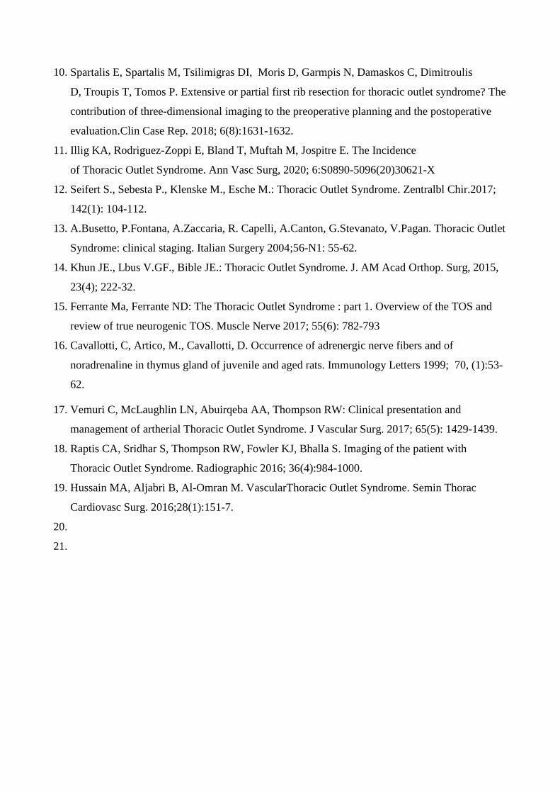
the neurovascular bundle being compressed: the brachial plexus, the subclavian artery and the
subclavian vein (Fig.1) [1,2]. The neurovascular dysfunction depends on three factors: 1) The space
between the neck and the axilla is very limited; 2) Physiological conditions may cause intermittent
compression to the neurovascular bundle; 3) Congenital malformations of bones and of muscles
may trigger the symptoms. The classic subjects affected by TOS are young asthenic women with a
thin neck and weak muscles. The structural anomalies involved in the TOS are: 1) Anomalous ribs,
2) Anomalous scalene tendon insertion, 3) Fibrous band insertion of the first rib, 4) Clavicular
abnormalities. (Fig.2) [3,4,5].
MATERIALS AND METHODS
TOS includes three different syndromes: 1) NTOS: Neurogenic TOS with compression of the
brachial plexus; 2) VTOS: Venous TOS with compression of the subclavian vein; 3) ATOS:
Arterial TOS with compression of the subclavian artery [6-9]. The estimate of a suspected TOS may
be assessed via medical history, medical examination and diagnostic tests that have, however, low
sensitivity and low specificity but may support the diagnosis. To diagnose cervical ribs and
anomalous first ribs one may use x-rays of the cervical spine and shoulder girdle and also CT, MR
and electromyography. Patients present symptoms of venous obstruction, arterial insufficiency,
paresthesia and pain [7, 10-13]. The therapy is often conservative, including exercises and physical
therapy. If it fails, it may be necessary surgical approaches like supraclavicular exposure and the
first rib resection. In this study we present a study of 181 clinical cases classified according to: 1)
Type of malformation (cervical rib, anomalous first rib, scalenus medius insertion, scalenus
minimus (Sibson's muscle) hypertrophy, Sibson’s fascia band, fibrous band arising from incomplete
cervical rib and elongated C7 transverse process, anomalous scalenus anticus insertion, anomalous
vessels, hypertrophy of little pectoral); 2) Physical structure of the patient; 3) Gender. Any single

case is classified through three parameters: Nerve (N), Artery(A) and Vein(V). There are four
grades for each parameter according to clinical and instrumental severity, Table 1 - see A.Busetto et
al, P[13].
Table 1. Pang grades
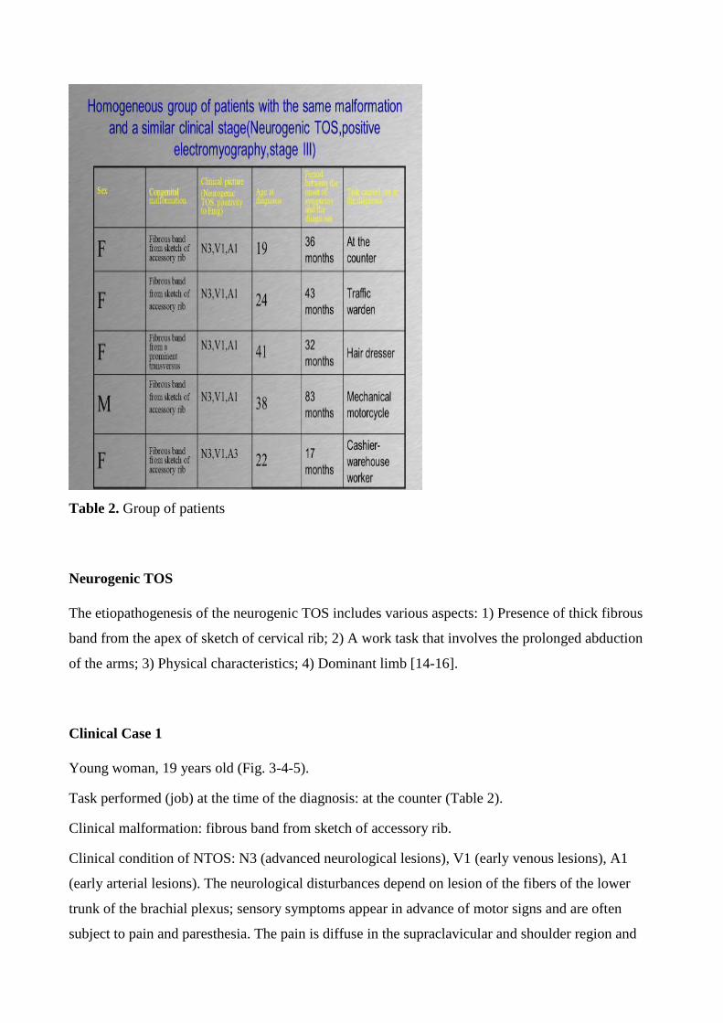
Table 2. Group of patients
Neurogenic TOS
The etiopathogenesis of the neurogenic TOS includes various aspects: 1) Presence of thick fibrous
band from the apex of sketch of cervical rib; 2) A work task that involves the prolonged abduction
of the arms; 3) Physical characteristics; 4) Dominant limb [14-16].
Clinical Case 1
Young woman, 19 years old (Fig. 3-4-5).
Task performed (job) at the time of the diagnosis: at the counter (Table 2).
Clinical malformation: fibrous band from sketch of accessory rib.
Clinical condition of NTOS: N3 (advanced neurological lesions), V1 (early venous lesions), A1
(early arterial lesions). The neurological disturbances depend on lesion of the fibers of the lower
trunk of the brachial plexus; sensory symptoms appear in advance of motor signs and are often
subject to pain and paresthesia. The pain is diffuse in the supraclavicular and shoulder region and
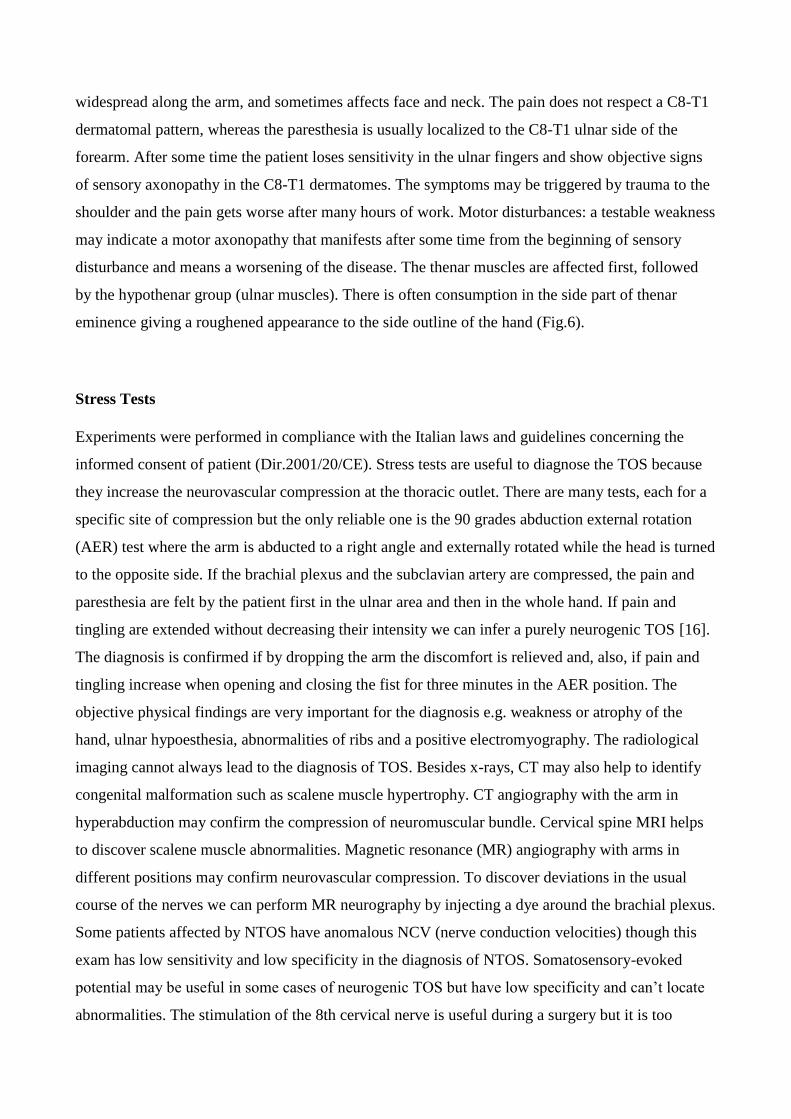
widespread along the arm, and sometimes affects face and neck. The pain does not respect a C8-T1
dermatomal pattern, whereas the paresthesia is usually localized to the C8-T1 ulnar side of the
forearm. After some time the patient loses sensitivity in the ulnar fingers and show objective signs
of sensory axonopathy in the C8-T1 dermatomes. The symptoms may be triggered by trauma to the
shoulder and the pain gets worse after many hours of work. Motor disturbances: a testable weakness
may indicate a motor axonopathy that manifests after some time from the beginning of sensory
disturbance and means a worsening of the disease. The thenar muscles are affected first, followed
by the hypothenar group (ulnar muscles). There is often consumption in the side part of thenar
eminence giving a roughened appearance to the side outline of the hand (Fig.6).
Stress Tests
Experiments were performed in compliance with the Italian laws and guidelines concerning the
informed consent of patient (Dir.2001/20/CE). Stress tests are useful to diagnose the TOS because
they increase the neurovascular compression at the thoracic outlet. There are many tests, each for a
specific site of compression but the only reliable one is the 90 grades abduction external rotation
(AER) test where the arm is abducted to a right angle and externally rotated while the head is turned
to the opposite side. If the brachial plexus and the subclavian artery are compressed, the pain and
paresthesia are felt by the patient first in the ulnar area and then in the whole hand. If pain and
tingling are extended without decreasing their intensity we can infer a purely neurogenic TOS [16].
The diagnosis is confirmed if by dropping the arm the discomfort is relieved and, also, if pain and
tingling increase when opening and closing the fist for three minutes in the AER position. The
objective physical findings are very important for the diagnosis e.g. weakness or atrophy of the
hand, ulnar hypoesthesia, abnormalities of ribs and a positive electromyography. The radiological
imaging cannot always lead to the diagnosis of TOS. Besides x-rays, CT may also help to identify
congenital malformation such as scalene muscle hypertrophy. CT angiography with the arm in
hyperabduction may confirm the compression of neuromuscular bundle. Cervical spine MRI helps
to discover scalene muscle abnormalities. Magnetic resonance (MR) angiography with arms in
different positions may confirm neurovascular compression. To discover deviations in the usual
course of the nerves we can perform MR neurography by injecting a dye around the brachial plexus.
Some patients affected by NTOS have anomalous NCV (nerve conduction velocities) though this
exam has low sensitivity and low specificity in the diagnosis of NTOS. Somatosensory-evoked
potential may be useful in some cases of neurogenic TOS but have low specificity and can’t locate
abnormalities. The stimulation of the 8th cervical nerve is useful during a surgery but it is too

invasive for outpatients. We can also use a combination of MAC (Medial Antebrachial Cutaneous
nerve conduction) and C8 nerve root stimulation tests to do the diagnosis of NTOS [13-17]. MAC
assessment is useful to reveal little alterations in the transmission of the lower trunk of the brachial
plexus. Botulinum toxin injection into the anterior scalene muscle has been used for the diagnosis of
NTOS and to reduce the symptoms.
Clinical Staging and Classification of TOS
TOS may be staged by its temporal sequence for severity and chronicity. In the first three stages
there are weakness, pain and tingling in the whole hand (symptoms of intermittent ischemia). In the
fourth stage there are persistent ischemic changes in the hands like gangrene and skin necrosis from
thromboembolism in the subclavian territory.
Management of TOS
Patients with NTOS should have a conservative treatment for three months and then a surgery can
be taken into consideration. Conservative treatment includes soft physical therapy, muscle
relaxants, anti-inflammatory drugs that determine an improvement of symptoms in many patients
with a better function and return to work. Surgery is necessary when there are neurological
dysfunctions and acute vascular insufficiency and functional impairments. Surgical procedures
concern lysis of fibrotic band, scalenectomy and first rib excision.
Arterial TOS
ATOS is the least common type of TOS and is caused by a congenital malformation that determine
a compression of subclavian artery. Possible malformations are: cervical or anomalous first rib,
fibromuscular bands, scalene muscle [18].
Clinical Case 2
Young woman, 22 years old.
Tasks performed (job) at the time of the diagnosis: cashier (Table 2).
Clinical malformation: Fibrous band from sketch of accessory rib(Table 2)
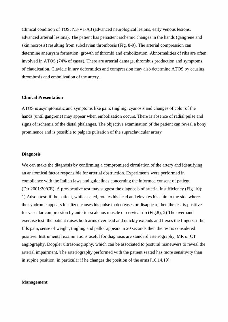
Clinical condition of TOS: N3-V1-A3 (advanced neurological lesions, early venous lesions,
advanced arterial lesions). The patient has persistent ischemic changes in the hands (gangrene and
skin necrosis) resulting from subclavian thrombosis (Fig. 8-9). The arterial compression can
determine aneurysm formation, growth of thrombi and embolization. Abnormalities of ribs are often
involved in ATOS (74% of cases). There are arterial damage, thrombus production and symptoms
of claudication. Clavicle injury deformities and compression may also determine ATOS by causing
thrombosis and embolization of the artery.
Clinical Presentation
ATOS is asymptomatic and symptoms like pain, tingling, cyanosis and changes of color of the
hands (until gangrene) may appear when embolization occurs. There is absence of radial pulse and
signs of ischemia of the distal phalanges. The objective examination of the patient can reveal a bony
prominence and is possible to palpate pulsation of the supraclavicular artery
Diagnosis
We can make the diagnosis by confirming a compromised circulation of the artery and identifying
an anatomical factor responsible for arterial obstruction. Experiments were performed in
compliance with the Italian laws and guidelines concerning the informed consent of patient
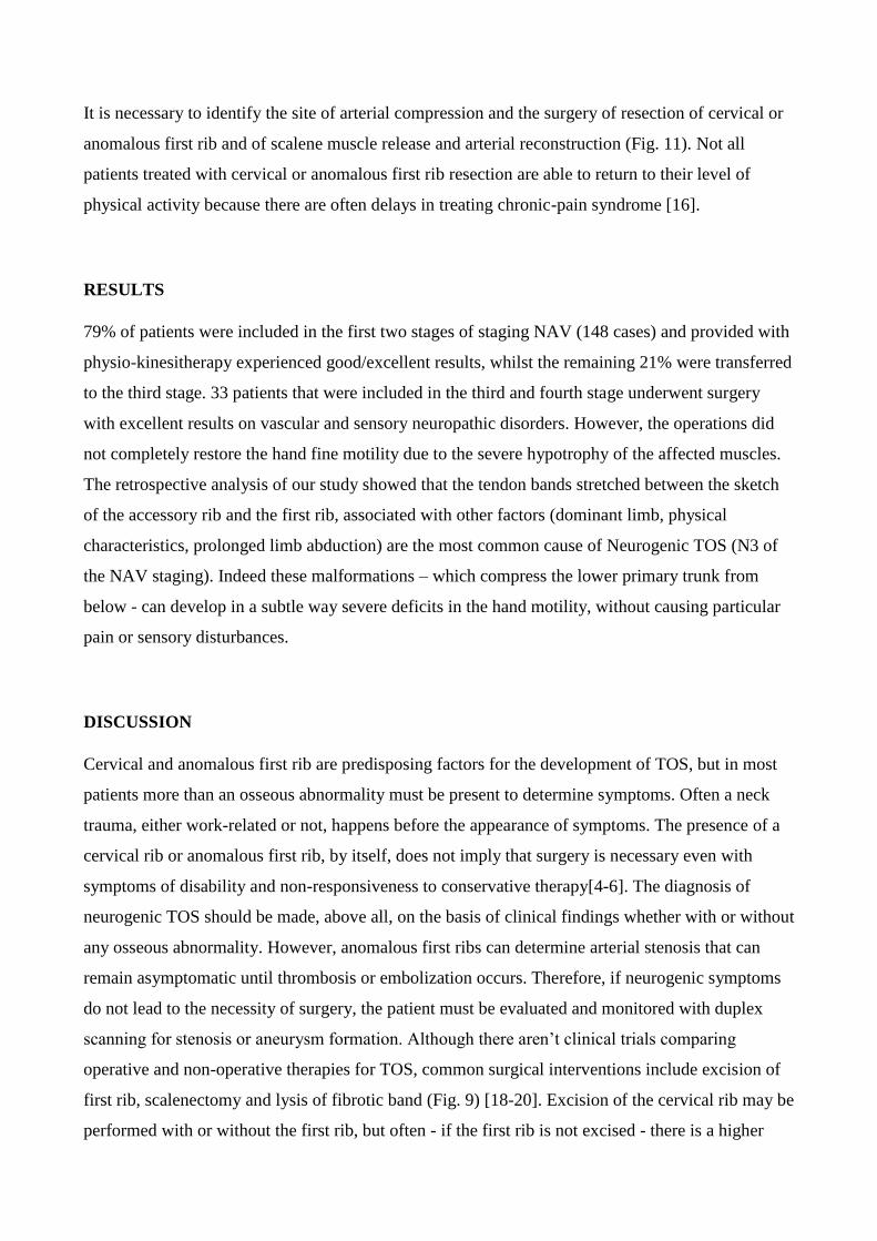
(Dir.2001/20/CE). A provocative test may suggest the diagnosis of arterial insufficiency (Fig. 10):
1) Adson test: if the patient, while seated, rotates his head and elevates his chin to the side where
the syndrome appears localized causes his pulse to decreases or disappear, then the test is positive
for vascular compression by anterior scalenus muscle or cervical rib (Fig.8); 2) The overhand
exercise test: the patient raises both arms overhead and quickly extends and flexes the fingers; if he
fills pain, sense of weight, tingling and pallor appears in 20 seconds then the test is considered
positive. Instrumental examinations useful for diagnosis are standard arteriography, MR or CT
angiography, Doppler ultrasonography, which can be associated to postural maneuvers to reveal the
arterial impairment. The arteriography performed with the patient seated has more sensitivity than
in supine position, in particular if he changes the position of the arms [10,14,19].
Management
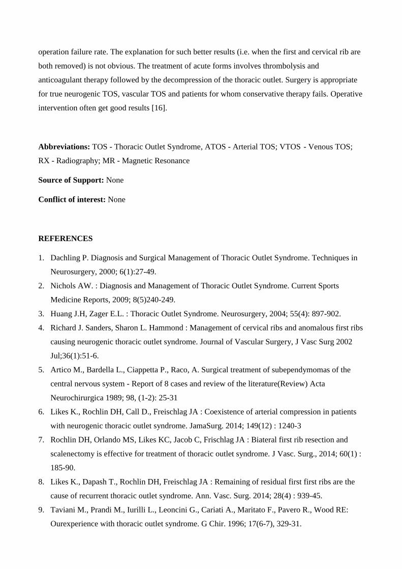
It is necessary to identify the site of arterial compression and the surgery of resection of cervical or
anomalous first rib and of scalene muscle release and arterial reconstruction (Fig. 11). Not all
patients treated with cervical or anomalous first rib resection are able to return to their level of
physical activity because there are often delays in treating chronic-pain syndrome [16].
RESULTS
79% of patients were included in the first two stages of staging NAV (148 cases) and provided with
physio-kinesitherapy experienced good/excellent results, whilst the remaining 21% were transferred
to the third stage. 33 patients that were included in the third and fourth stage underwent surgery
with excellent results on vascular and sensory neuropathic disorders. However, the operations did
not completely restore the hand fine motility due to the severe hypotrophy of the affected muscles.
The retrospective analysis of our study showed that the tendon bands stretched between the sketch
of the accessory rib and the first rib, associated with other factors (dominant limb, physical
characteristics, prolonged limb abduction) are the most common cause of Neurogenic TOS (N3 of
the NAV staging). Indeed these malformations – which compress the lower primary trunk from
below - can develop in a subtle way severe deficits in the hand motility, without causing particular
pain or sensory disturbances.
DISCUSSION
Cervical and anomalous first rib are predisposing factors for the development of TOS, but in most
patients more than an osseous abnormality must be present to determine symptoms. Often a neck
trauma, either work-related or not, happens before the appearance of symptoms. The presence of a
cervical rib or anomalous first rib, by itself, does not imply that surgery is necessary even with
symptoms of disability and non-responsiveness to conservative therapy[4-6]. The diagnosis of
neurogenic TOS should be made, above all, on the basis of clinical findings whether with or without
any osseous abnormality. However, anomalous first ribs can determine arterial stenosis that can
remain asymptomatic until thrombosis or embolization occurs. Therefore, if neurogenic symptoms
do not lead to the necessity of surgery, the patient must be evaluated and monitored with duplex
scanning for stenosis or aneurysm formation. Although there aren’t clinical trials comparing
operative and non-operative therapies for TOS, common surgical interventions include excision of
first rib, scalenectomy and lysis of fibrotic band (Fig. 9) [18-20]. Excision of the cervical rib may be
performed with or without the first rib, but often - if the first rib is not excised - there is a higher
operation failure rate. The explanation for such better results (i.e. when the first and cervical rib are
both removed) is not obvious. The treatment of acute forms involves thrombolysis and
anticoagulant therapy followed by the decompression of the thoracic outlet. Surgery is appropriate
for true neurogenic TOS, vascular TOS and patients for whom conservative therapy fails. Operative
intervention often get good results [16].
Abbreviations: TOS - Thoracic Outlet Syndrome, ATOS - Arterial TOS; VTOS - Venous TOS;
RX - Radiography; MR - Magnetic Resonance
Source of Support: None
Conflict of interest: None
REFERENCES
1. Dachling P. Diagnosis and Surgical Management of Thoracic Outlet Syndrome. Techniques in
Neurosurgery, 2000; 6(1):27-49.
2. Nichols AW. : Diagnosis and Management of Thoracic Outlet Syndrome. Current Sports
Medicine Reports, 2009; 8(5)240-249.
3. Huang J.H, Zager E.L. : Thoracic Outlet Syndrome. Neurosurgery, 2004; 55(4): 897-902.
4. Richard J. Sanders, Sharon L. Hammond : Management of cervical ribs and anomalous first ribs
causing neurogenic thoracic outlet syndrome. Journal of Vascular Surgery, J Vasc Surg 2002
Jul;36(1):51-6.
5. Artico M., Bardella L., Ciappetta P., Raco, A. Surgical treatment of subependymomas of the
central nervous system - Report of 8 cases and review of the literature(Review) Acta
Neurochirurgica 1989; 98, (1-2): 25-31
6. Likes K., Rochlin DH, Call D., Freischlag JA : Coexistence of arterial compression in patients
with neurogenic thoracic outlet syndrome. JamaSurg. 2014; 149(12) : 1240-3
7. Rochlin DH, Orlando MS, Likes KC, Jacob C, Frischlag JA : Biateral first rib resection and
scalenectomy is effective for treatment of thoracic outlet syndrome. J Vasc. Surg., 2014; 60(1) :
185-90.
8. Likes K., Dapash T., Rochlin DH, Freischlag JA : Remaining of residual first first ribs are the
cause of recurrent thoracic outlet syndrome. Ann. Vasc. Surg. 2014; 28(4) : 939-45.
9. Taviani M., Prandi M., Iurilli L., Leoncini G., Cariati A., Maritato F., Pavero R., Wood RE:
Ourexperience with thoracic outlet syndrome. G Chir. 1996; 17(6-7), 329-31.
10. Spartalis E, Spartalis M, Tsilimigras DI, Moris D, Garmpis N, Damaskos C, Dimitroulis
D, Troupis T, Tomos P. Extensive or partial first rib resection for thoracic outlet syndrome? The
contribution of three-dimensional imaging to the preoperative planning and the postoperative
evaluation.Clin Case Rep. 2018; 6(8):1631-1632.
11. Illig KA, Rodriguez-Zoppi E, Bland T, Muftah M, Jospitre E. The Incidence
of Thoracic Outlet Syndrome. Ann Vasc Surg, 2020; 6:S0890-5096(20)30621-X
12. Seifert S., Sebesta P., Klenske M., Esche M.: Thoracic Outlet Syndrome. Zentralbl Chir.2017;
142(1): 104-112.
13. A.Busetto, P.Fontana, A.Zaccaria, R. Capelli, A.Canton, G.Stevanato, V.Pagan. Thoracic Outlet
Syndrome: clinical staging. Italian Surgery 2004;56-N1: 55-62.
14. Khun JE., Lbus V.GF., Bible JE.: Thoracic Outlet Syndrome. J. AM Acad Orthop. Surg, 2015,
23(4); 222-32.
15. Ferrante Ma, Ferrante ND: The Thoracic Outlet Syndrome : part 1. Overview of the TOS and
review of true neurogenic TOS. Muscle Nerve 2017; 55(6): 782-793
16. Cavallotti, C, Artico, M., Cavallotti, D. Occurrence of adrenergic nerve fibers and of
noradrenaline in thymus gland of juvenile and aged rats. Immunology Letters 1999; 70, (1):53-
62.
17. Vemuri C, McLaughlin LN, Abuirqeba AA, Thompson RW: Clinical presentation and
management of artherial Thoracic Outlet Syndrome. J Vascular Surg. 2017; 65(5): 1429-1439.
18. Raptis CA, Sridhar S, Thompson RW, Fowler KJ, Bhalla S. Imaging of the patient with
Thoracic Outlet Syndrome. Radiographic 2016; 36(4):984-1000.
19. Hussain MA, Aljabri B, Al-Omran M. VascularThoracic Outlet Syndrome. Semin Thorac
Cardiovasc Surg. 2016;28(1):151-7.
20.
21.
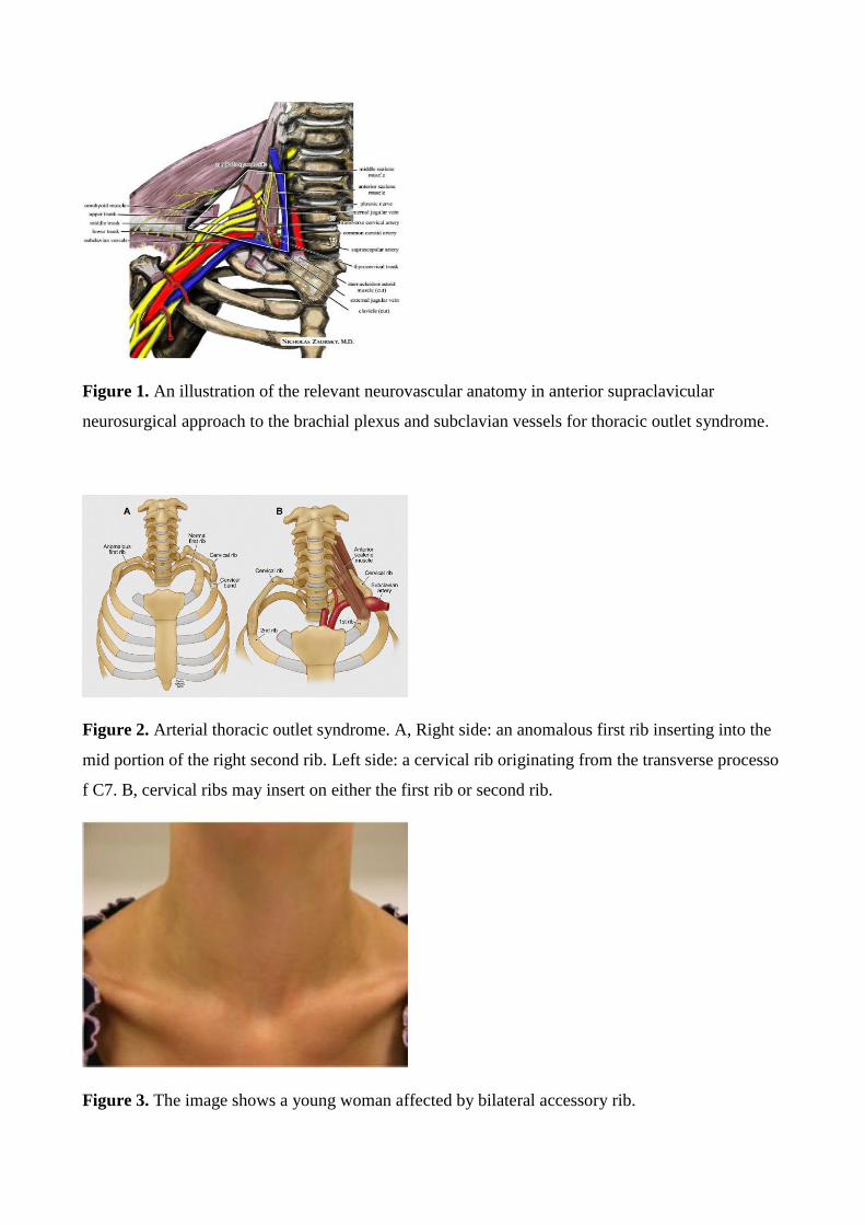
Figure 1. An illustration of the relevant neurovascular anatomy in anterior supraclavicular
neurosurgical approach to the brachial plexus and subclavian vessels for thoracic outlet syndrome.
Figure 2. Arterial thoracic outlet syndrome. A, Right side: an anomalous first rib inserting into the
mid portion of the right second rib. Left side: a cervical rib originating from the transverse processo
f C7. B, cervical ribs may insert on either the first rib or second rib.
Figure 3. The image shows a young woman affected by bilateral accessory rib.
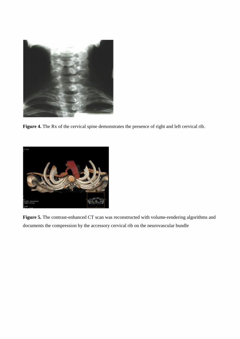
Figure 4. The Rx of the cervical spine demonstrates the presence of right and left cervical rib.
Figure 5. The contrast-enhanced CT scan was reconstructed with volume-rendering algorithms and
documents the compression by the accessory cervical rib on the neurovascular bundle

Figure 6. A-B Thenar consumption in thoracic outlet syndrome. Note the severe thenar
consumption especially with respect to the abductor pollicis brevis.
Figure 7. Antero-posterior cervical spine x-ray shows the presence of bilateral cervical accessory
rib
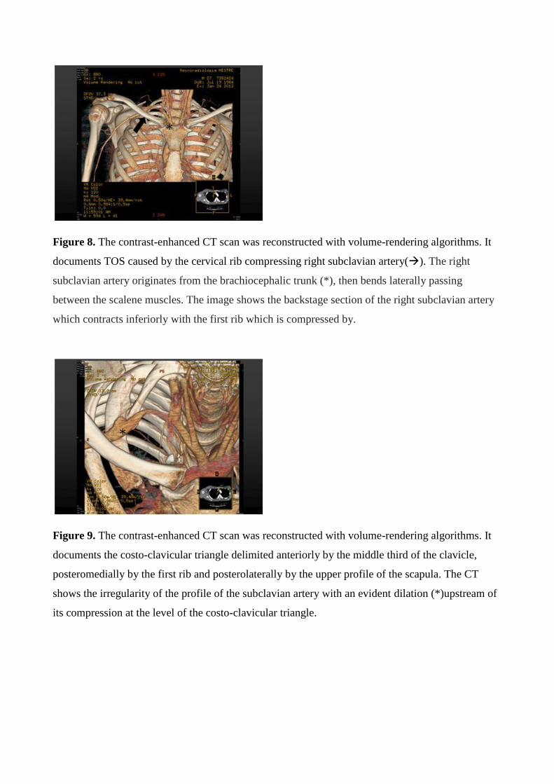
Figure 8. The contrast-enhanced CT scan was reconstructed with volume-rendering algorithms. It
documents TOS caused by the cervical rib compressing right subclavian artery(). The right
subclavian artery originates from the brachiocephalic trunk (*), then bends laterally passing
between the scalene muscles. The image shows the backstage section of the right subclavian artery
which contracts inferiorly with the first rib which is compressed by.
Figure 9. The contrast-enhanced CT scan was reconstructed with volume-rendering algorithms. It
documents the costo-clavicular triangle delimited anteriorly by the middle third of the clavicle,
posteromedially by the first rib and posterolaterally by the upper profile of the scapula. The CT
shows the irregularity of the profile of the subclavian artery with an evident dilation (*)upstream of
its compression at the level of the costo-clavicular triangle.
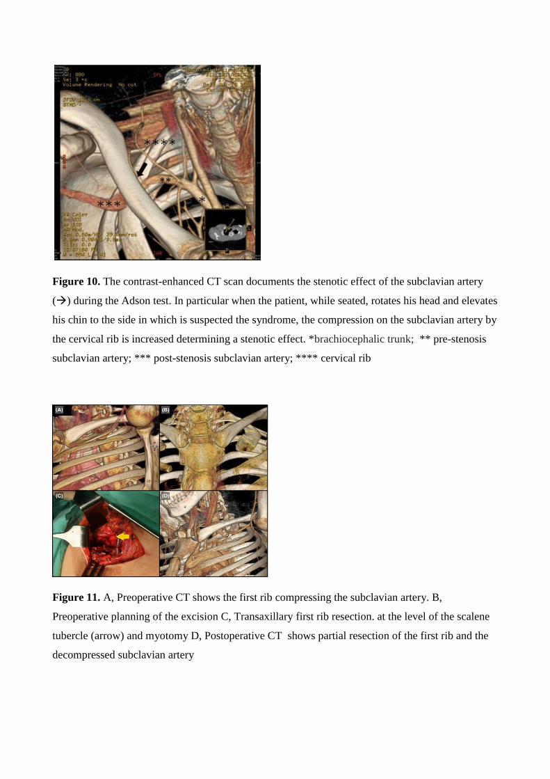
Figure 10. The contrast-enhanced CT scan documents the stenotic effect of the subclavian artery
() during the Adson test. In particular when the patient, while seated, rotates his head and elevates
his chin to the side in which is suspected the syndrome, the compression on the subclavian artery by
the cervical rib is increased determining a stenotic effect. *brachiocephalic trunk; ** pre-stenosis
subclavian artery; *** post-stenosis subclavian artery; **** cervical rib
Figure 11. A, Preoperative CT shows the first rib compressing the subclavian artery. B,
Preoperative planning of the excision C, Transaxillary first rib resection. at the level of the scalene
tubercle (arrow) and myotomy D, Postoperative CT shows partial resection of the first rib and the
decompressed subclavian artery