Embed Size (px)
Citation preview
The role of CD80 and CD86 in macrophage activation
and its regulation following LPS stimulation
Seghen Woldai
Research report submitted to the Department of
Biochemistry, Microbiology and Immunology In partial fulfillment of the requirements
for the degree Master of Science
Microbiology and Immunology
University of Ottawa
Ottawa, Ontario, Canada Aug, 2014
© Seghen Woldai, Ottawa, Canada, 2014

ii
Abstract
The binding of CD80/CD86 on the APC to CD28 on the T cell surface provides a second signal for T
cell activation. While it was once believed that this interaction represented a one-way signal,
resulting in T cell activation, recently, it has been investigated as a bidirectional signaling process.
CD80/86 activation produces IL-6 in DCs, but its role in macrophage activation is unknown.
Dysregulation of CD80/86 expression has been observed in autoimmune disorders and cancer, and
may also influence the development of immune responses including production of cytokines in
response to stimulation with TLR-4 ligand, LPS. Therefore, the focus of my project was twofold: 1) to
investigate the role of CD80/86 as signaling receptors capable of transmitting extracellular signals,
and 2) to determine the TLR-4 activated pathways that regulate CD80/86 expression in human
monocyte-derived macrophages (MDMs). Since I demonstrated that activation of CD80/86 alone did
not induce expression of the four cytokines investigated, I hypothesized that CD80/86 synergizes
with other signaling pathways. I show for the first time that CD80/86 activation synergizes with TLR-
4 signaling to produce IL-27 and IL-10 in human MDMs. Since cIAPs play a key role in TLR-4-
mediated signaling, I investigated their role in TLR-4- and CD80/86-activated production of IL-10 and
IL-27. Degradation of IAPs by SMAC mimetics inhibited LPS-induced IL-10 and IL-27 production in
MDMs. However, it did not alter the TLR-4 and CD80/86 synergistic effect on IL-10 and IL-27
production suggesting that IAPs may not play a role in CD80/86 activation of macrophages. Since I
have demonstrated this role for IAPs, I extended my studies by examining the involvement of IAPs
and other upstream signaling molecules such as SHP-1, RIP1, TRAF2, in modulating the LPS-induced
CD80/86 expression. I showed that cIAP2, SHP-1, RIP1, TRAF2 co-localize to form a complex that
regulates the LPS-induced CD80 and CD86 expression through AKT-activated p38 MAPK in human
macrophages. These findings may lead to the development of novel therapeutic interventions in the
treatment of autoimmune diseases.
iii
Acknowledgements
I would like to extend my gratitude to my supervisor Dr. Ashok Kumar for his constant support throughout my academic program. I am very grateful for his patience and mentorship and it has been a pleasure working in his lab. I would like to thank the visiting scientist, Dr. Maya Kozlowski, who provided invaluable guidance and encouragement during her stay. It has been an absolute pleasure working with you and I thank for your constant kind words of advice. To my committee members, Dr. Subash Sad and Dr. Fraser Scott, I thank you for your counsel, contribution and expertise. Thank you to all past and current lab members: Maria Blahoianu, Aurelia Busca, Ramon Caballero, Ankur Chopra, Niranjala Gajanayaka, and Jason Fernandes. Thank you for making the lab feel like a home away from home and providing troubleshooting advice for each experiment. A special thank you to Salma Iqbal, Yulia Konarski, and Jay Majithia who have offered tremendous support and friendship throughout the years. Each laugh, each word of encouragement, and each “developing party” will never be forgotten. It has been an incredible journey with the Kumar lab and I thank you all for the memories. To my mom, thank you for teaching me that the key to success is education. Thank you for understanding that yes, sometimes I just call you to complain about a failed experiment and yes, you were right, it was all going to be OK in the end. You are my inspiration and my role model and I thank you for your unconditional love. To my sisters and brother, Semhar, Sara, Senai, thank you for every laugh, every piece of advice, and every word of encouragement. You were the best cheerleaders a sister could ask for and I am forever grateful to have you in my life. To my friends, thank you for sharing in my excitement and my frustrations. To my roommates, coming home to laughter every night is the reason I survived the last few years. The days may have been stressful, but it was always nice to come home to some great friends. To Allison, you are an incredibly strong woman and you inspire me to be a better person every day. I hope you never forget what a "line up" is! To Kayla, these last 7 years have been amazing – thank you for sharing in my stress and for never letting me give up. “Without you” will always make me think of you! Finally, to Angelica, my best friend and sister, I really am not sure I could have made it without you. Thank you for reminding me of home every time we talk. Thank you for each and every laugh (way too many!) and thank you for always being there for me, day or night. We have been apart for way too long, but I’m coming home…for good this time! And finally, to Andre, thank you for being my ultimate cheerleader. Thank you for staying with me in the lab all those late nights just so I wouldn’t be all by myself – I know you were bored, but I always appreciated the company. Thank you for letting me talk about my project, and genuinely trying to understand every last detail – I think you know way too much about CD80/86 than you ever cared to learn! Thank you for celebrating my successes as if they were your own and reminding me that I can overcome every failure. Thank you for teaching me the value of patience, in research and in life. More than anything, thank you for pushing me farther than I ever thought I could go.
iv
Table of Contents
Abstract............................................................................................................................................... ii
Acknowledgements ........................................................................................................................... iii
Table of Contents............................................................................................................................... iv
List of Abbreviations .......................................................................................................................... vi
List of Figures ................................................................................................................................... viii
1.1 Introduction ..................................................................................................................................... 7
1.2 Innate immune system and inflammation .................................................................................. 7
1.3 Macrophages ............................................................................................................................... 9
1.4 Pro-inflammatory and Anti-inflammatory Cytokines ................................................................ 10
1.5 B7 receptors ............................................................................................................................... 11
1.6 The role of CD80 and CD86 in Th cell differentiation ................................................................ 14
1.7 The role of CD80 and CD86 in disease ....................................................................................... 15
1.7.1 The involvement of costimulatory molecules in cancer and in the development of
therapeutic interventions ............................................................................................................ 16
1.8 CD80/CD86 and transplant rejection ..................................................................................... 18
1.10 CD80 and CD86 as signaling molecules ................................................................................... 19
1.11 Toll like receptor signalling ...................................................................................................... 20
1.12 TLR-4 and the activation of the MAPKs and PI3K pathway ................................................. 23
1.13 Inhibitor of Apoptosis Proteins and SMAC mimetics .............................................................. 25
1.14 IAPs and NF-κB signaling ...................................................................................................... 26
1.15 IAPs and Pattern Recognition Receptor (PRR) signaling: cIAPs and TLR-4 signaling ........... 27
1.16 The role of tyrosine phosphorylation the LPS/TLR-4 pathway ................................................ 29
1.17 The regulation of CD80 and CD86 expression ......................................................................... 30
1.18 Rationale .................................................................................................................................. 33
1.19 Hypothesis ............................................................................................................................... 34
1.20 Objectives ................................................................................................................................ 34
2.1 Materials and Methods ................................................................................................................. 35
2.2 Reagents .................................................................................................................................... 35
2.3 Cell culture ................................................................................................................................. 35
2.3.1 Human Monocyte-derived Macrophages (MDMs) ............................................................ 35
2.4 MAP kinase and PI3K inhibition ................................................................................................. 36
v
2.5 Transfection of MDMs with small interfering RNA (siRNA) ....................................................... 36
2.6 Western blot analysis ................................................................................................................ 37
2.7 Flow Cytometry.......................................................................................................................... 37
2.8 RNA isolation and semi-quantitative RT-PCR analysis for CD80 and CD86 ............................... 38
2.9 Treatment with Anti-CD80 and Anti-CD86 antibodies .............................................................. 39
2.10 Cytokine measurement by ELISA (Enzyme-linked Immunosorbent assay) ............................. 39
2.11 Immunofluorescence ............................................................................................................... 40
2.12 Statistical Analysis .................................................................................................................... 40
2.13 Ethics Statement ...................................................................................................................... 40
3.1. Results ........................................................................................................................................... 41
3.2. CD80 and CD86 activation synergizes with TLR-4 signaling pathway to regulate IL-10 and IL-27
production in human MDMs ........................................................................................................... 41
3.2.1 Inhibitor of apoptosis proteins regulate LPS-induced IL-10 and IL-27 expression, but are
not involved in the CD80/CD86-induced IL-10 and IL-27 production ......................................... 45
3.3. Regulation of CD80 in LPS-induced monocyte-derived macrophages ..................................... 48
3.3.1 Inhibitor of Apoptosis Proteins (IAPs) regulate LPS-induced CD80 and CD86 surface
protein expression ....................................................................................................................... 48
3.3.2 RIP1 and TRAF2 regulate the LPS-induced CD80 surface protein expression .................... 51
3.3.3 SHP-1 positively regulates CD80 and CD86 protein in human MDMs ................................ 54
3.3.4 SHP-1, cIAP2, RIP1 and TRAF2 co-localize in human MDMs .............................................. 57
3.3.5 PI3K pathway regulates the LPS-induced CD80 and CD86 protein expression .................. 57
3.3.6 P38, but not JNK or ERK MAPK regulate the LPS-induced CD80 and CD86 expression...... 63
3.3.7 SHP-1 and IAPs regulate activation of downstream PI3K/p38 signaling axis ..................... 65
4.1 Discussion ...................................................................................................................................... 71
4.1.1 Regulation of CD80 and CD86 at the transcriptional level ................................................. 78
4.1.2 CD80 Transcription ............................................................................................................. 78
4.1.3 CD86 Transcription ............................................................................................................. 79
Concluding Remarks, Future Directions and Significance ................................................................... 82
Appendix .............................................................................................................................................. 86
References ........................................................................................................................................... 90
vi
List of Abbreviations
α- anti Akt Protein kinase B AP1 Activating protein-1 APC antigen presenting cell APS ammonium persulfate ATP adenosine triphosphate BCR B cell receptor BIR baculoviral IAP repeat BMDMs bone marrow derived macrophage cultures BMK-1 big MAPK-1 BSA bovine serum albumin cIAP cellular inhibitor of apoptosis CSF-1 colony stimulating factor 1 CTL cytotoxic T-lymphocyte DCs dendritic cells DMSO dimethyl sulphoxide ECL enhanced chemiluminescence ELISA Enzyme linked immuno-sorbent assay ERK extracellular-signal regulated kinase FLICE Fas-associated death domain (FADD)-like IL- β converting enzyme FLIP FLICE inhibitory protein FBS fetal bovine serum FITC fluorescein isothiocyanate HEPES 4-(2-hydroxyethyl)-1-piperazineethanesulfonic acid HIV human immunodeficiency virus HLA human leukocyte antigen HRP horseradish peroxidase IAP inhibitor of apoptosis IFN interferon IκB inhibitor-kappa B IL- interleukin IP immunoprecipitation IRAK IL-1R associated kinases IRF interferon regulatory factor JNK c-Jun N-terminal kinase LBP LPS binding protein LPS Lipopolysaccharide mAb monoclonal antibody/antibodies MAPK mitogen associated protein kinase MAPKK MAPK kinase MAPKKK MAPK Kinase kinase M-CSF macrophage colony-stimulating factor MD2 myeloid differentiation protein 2 MDMs monocyte-derived macrophages MHC major histocompatibility complex mTOR mammalian target of rapamycin
vii
MyD88 myeloid differentiation factor 88 NIK NF-κB inducing kinase NF-κB Nuclear factor kappa-light-chain enhancer of activated B cells NK cells Natural killer cells PAMP pathogen associated molecular pattern PBMCs peripheral blood mononuclear cells PCR polymerized chain reaction PE phycoerythrin PerCP Peridinin chlorophyll protein PFA paraformaldehyde PI propidium iodide PI3K phosphotidyl inositol-3-kinase PMA phorbol-12-myristate-13-acetate PRRs pattern recognition receptors PTK protein tyrosine kinase PTP protein tyrosine phosphatase PVDF polyvinylidene difluoride qRT-PCR semi-quantitative real time polymerase chain reaction RING really interesting new gene RIP1 receptor interacting protein 1 RNS reactive nitrogen species ROS reactive oxygen species RPM Revolutions per minute RT-PCR Reverse transcriptase polymerase chain reaction SDS-PAGE sodium dodecyl sulphate poly acrylamide gel elctrophoresis SH2 Src-homology domains 2 SHP-1 SH2 domain containing phosphatase-1 siRNA small interfering RNA SMAC small mitochondria-derived activators of caspases TBE Tris boric acid –EDTA TBS Tris buffered saline TBST Tris buffered saline and Tween-20 TCR T cell receptor TEMED N,N,N’,N’-tetramethylethylenediamine TGF-β transforming growth factor-β Th cells T helper cells Th17 T helper 17 cells TIR Toll/IL-1 receptor TIRAP Toll-interleukin 1 receptor adaptor protein TLR toll-like receptor TNF tumor necrosis factor TRAF TNF receptor associated factor TRAM TRIF related adaptor molecule TRIF TIR-domain-containing adapter-inducing interferon-β T reg T regulatory cell XIAP x-chromosome linked IAP
viii
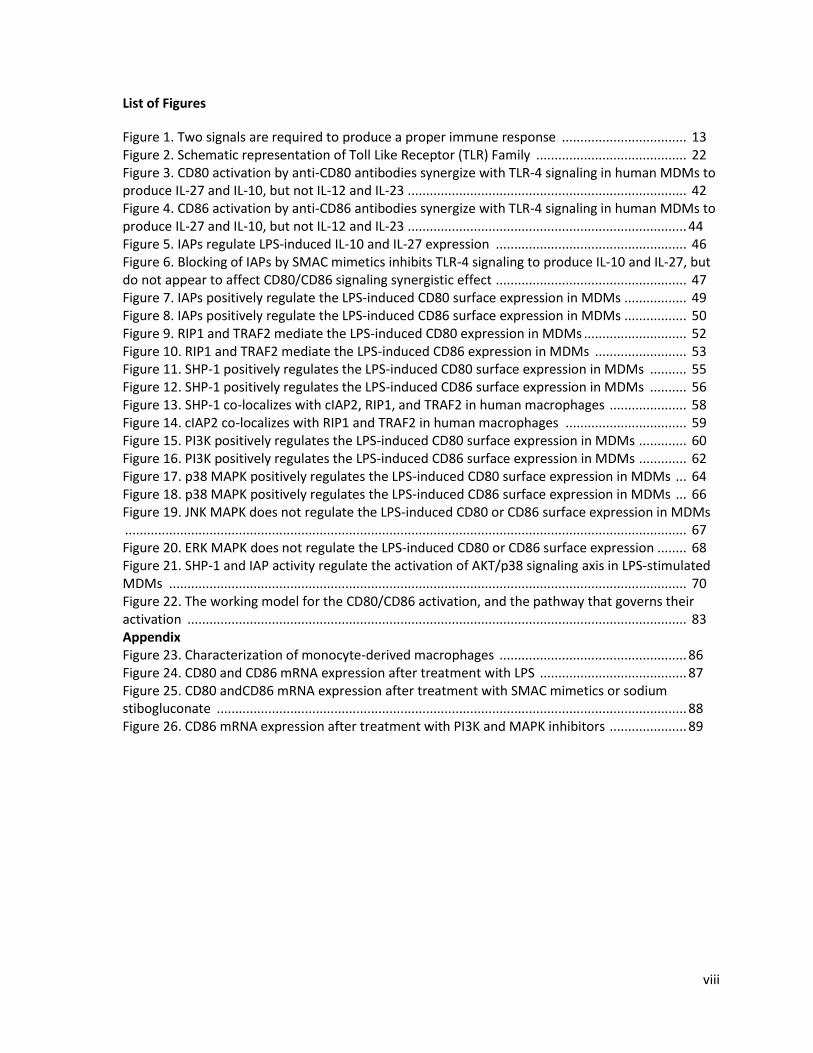
List of Figures
Figure 1. Two signals are required to produce a proper immune response .................................. 13 Figure 2. Schematic representation of Toll Like Receptor (TLR) Family ......................................... 22 Figure 3. CD80 activation by anti-CD80 antibodies synergize with TLR-4 signaling in human MDMs to produce IL-27 and IL-10, but not IL-12 and IL-23 ............................................................................ 42 Figure 4. CD86 activation by anti-CD86 antibodies synergize with TLR-4 signaling in human MDMs to produce IL-27 and IL-10, but not IL-12 and IL-23 ............................................................................ 44 Figure 5. IAPs regulate LPS-induced IL-10 and IL-27 expression .................................................... 46 Figure 6. Blocking of IAPs by SMAC mimetics inhibits TLR-4 signaling to produce IL-10 and IL-27, but do not appear to affect CD80/CD86 signaling synergistic effect .................................................... 47 Figure 7. IAPs positively regulate the LPS-induced CD80 surface expression in MDMs ................. 49 Figure 8. IAPs positively regulate the LPS-induced CD86 surface expression in MDMs ................. 50 Figure 9. RIP1 and TRAF2 mediate the LPS-induced CD80 expression in MDMs ............................ 52 Figure 10. RIP1 and TRAF2 mediate the LPS-induced CD86 expression in MDMs ......................... 53 Figure 11. SHP-1 positively regulates the LPS-induced CD80 surface expression in MDMs .......... 55 Figure 12. SHP-1 positively regulates the LPS-induced CD86 surface expression in MDMs .......... 56 Figure 13. SHP-1 co-localizes with cIAP2, RIP1, and TRAF2 in human macrophages ..................... 58 Figure 14. cIAP2 co-localizes with RIP1 and TRAF2 in human macrophages ................................. 59 Figure 15. PI3K positively regulates the LPS-induced CD80 surface expression in MDMs ............. 60 Figure 16. PI3K positively regulates the LPS-induced CD86 surface expression in MDMs ............. 62 Figure 17. p38 MAPK positively regulates the LPS-induced CD80 surface expression in MDMs ... 64 Figure 18. p38 MAPK positively regulates the LPS-induced CD86 surface expression in MDMs ... 66 Figure 19. JNK MAPK does not regulate the LPS-induced CD80 or CD86 surface expression in MDMs ......................................................................................................................................................... 67 Figure 20. ERK MAPK does not regulate the LPS-induced CD80 or CD86 surface expression ........ 68 Figure 21. SHP-1 and IAP activity regulate the activation of AKT/p38 signaling axis in LPS-stimulated MDMs ............................................................................................................................................. 70 Figure 22. The working model for the CD80/CD86 activation, and the pathway that governs their activation ........................................................................................................................................ 83 Appendix Figure 23. Characterization of monocyte-derived macrophages ................................................... 86 Figure 24. CD80 and CD86 mRNA expression after treatment with LPS ........................................ 87 Figure 25. CD80 andCD86 mRNA expression after treatment with SMAC mimetics or sodium stibogluconate ................................................................................................................................ 88 Figure 26. CD86 mRNA expression after treatment with PI3K and MAPK inhibitors ..................... 89
7
1.1 Introduction
1.2 Innate immune system and inflammation
Infectious diseases are a leading cause of morbidity and mortality worldwide. The innate
immune system is the first line of defense and the biggest contributor to acute inflammation
induced by tissue damage or microbial infection (1). Inflammation is the rapid, coordinated
response that leads to the resolution of infection, the repair of damage and the return to the
homeostatic state, with minimal damage to the host (2). The survival of the host is largely based on
its ability to coordinate a network of cells to recognize and induce the appropriate response for the
elimination of the microbes. Monocytes, macrophages, dendritic cells (DCs), neutrophils,
eosinophils, mast cells and natural killer (NK) cells represent many of the cells of the innate immune
response (3). Neutrophils are among the first cells to be recruited to the site of infection, followed
by the recruitment of monocytes, and T and B cells (4). Neutrophils possess highly cytotoxic
granules with proteases that are capable of degrading material that has undergone phagocytosis.
They also produce reactive nitrogen species (RNS) and reactive oxygen species (ROS) to induce DNA
damage, and to further denature proteins and disrupt lipid (5). Chemokines released by neutrophils
initiate the continued response by other cells, such as monocytes and macrophages which express
pattern recognition receptors (PRRs) on their cell surface (3). These PRRs recognize pathogens
associated molecular patterns (PAMPs), such as lipopolysaccharide (LPS) on the surface of Gram
negative bacteria. For example, the engagement of the toll-like receptor 4 (TLR-4), a
transmembrane protein on the surface of macrophages, with its cognate ligand, LPS, will initiate an
immune response (3). Other PAMPs include glycolipids, flagellin, lipoproteins, viral RNA and
bacterial DNA, and their engagement with PRRs will result in a specific and targeted destruction of
the infected cell or organism by phagocytosis or the release of cytotoxic agents (2).

8
Circulating monocytes constitute between 5-10% of peripheral blood leukocytes (3). They
originate from a common myeloid progenitor cell in the bone marrow that is shared with
neutrophils and are released into the bloodstream undifferentiated for 1-3 days (3). Upon
recruitment into the site of infection/inflammation, they can differentiate into tissue macrophages
(Kupffer cells in the liver; microglia in the brain) or myeloid DCs, contributing to host defence and
tissue repair (3). To recruit other inflammatory cells to the sites of infections, these cells release
cytokines and other proinflammatory mediators to induce changes in the local environment to
convert the infected tissue to an inflamed state (2). For example, the release of pro-inflammatory
cytokines, such as TNF-α, IL-1 and IL-6, will initiate leukocyte migration and infiltration, and a flow of
plasma to the site of injury (3). Macrophages and DCs are professional antigen presenting cells
(APCs) which, upon ingestion of a pathogen, migrate to draining lymph nodes and present
pathogen-associated antigens resulting in activation of the adaptive immune response. APCs use
HLA/MHC (Human Leukocyte Antigen/Major Histocompatibility Complex) class I and class II
molecules to present extracellular peptides to T cells to drive a proper immune response (6). Some
viruses prevent expression of the MHC molecule on the surface of APCs to circumvent detection and
destruction; however, human APCs that do not express the MHC molecule are removed by NK cells,
representing a vital arm of the innate immune system responsible for targeting and lysing cells that
do not express MHC molecules (7).
Generated in the thymus, T lymphocytes circulate through the bloodstream and primary
lymphoid organs, like the lymph nodes and spleen (3). After encountering APCs, T cells are activated
and begin to proliferate and differentiate. This activation requires two signals. The first signal is
antigen specific and requires binding of the T cell receptor to the antigenic peptides presented
within the context of the MHC molecules (8). This signal is insufficient to elicit an optimal immune
reaction and requires the second antigen-independent signal. This second signal is provided by the
9
binding of CD28 on the T cell surface with one of two costimulatory molecules on the APC: CD80 or
CD86 (8). Macrophages represent a large portion of the APCs that express CD80 and CD86 and
therefore, play an important role in the activation of the immune response.
1.3 Macrophages
Originating in the bone marrow from CD34+ myeloid progenitor cells, monocytes are
released into peripheral blood and enter tissue to replenish tissue macrophage population (9). They
can also differentiate into DCs and osteoclasts depending on the local inflammatory milieu (9). The
process of monocyte to macrophage differentiation is governed by the binding of the hematopoietic
growth factor, the macrophage colony stimulating factor (M-CSF) to its receptor (M-CSFR) on the
surface of monocytes. Activation of the M-CSFR results in monocyte changes, beginning with their
adherence and development of an elongated, spindle-like appearance (10). Cell cycle genes are
activated and the monocyte-differentiated macrophages gain functions such as antigen
presentation (10). As key players in the innate immune system, macrophages engulf and digest
microorganisms, dead cells, and debris during infection and produce inflammatory mediators, such
as cytokines and chemokines, to activate other cell types and kill bacteria (11). By recognizing,
processing, and presenting antigen to T cells, macrophages are critical players in bridging the gap
between innate and adaptive immunity (11).
Based on the cytokine milieu, macrophages can become polarized and gain specialized
functional properties. Similar to the Th1/Th2 nomenclature, polarized macrophages are often
characterized as M1 or M2 cells (10). M1 or classically activated macrophages are induced by IFN-γ
and TNF, and express higher levels of IL-12 and IL-23 and lower levels of IL-10 (10). They produce
antimicrobial effector molecules, such as ROS, and inflammatory cytokines, including IL-1β, TNF and
IL-6, and are potent contributors to the Th1 response, specifically mediating resistance to tumors
10
and intracellular parasites (10). Excessive M1 polarization will inhibit cell proliferation and cause
tissue damage (12). In contrast, M2 or alternatively activated macrophages, representing a broad
spectrum of cells activated by cytokines such as IL-4 or IL-13, turn off the damaging immune system
with anti-inflammatory cytokines and work towards tissue repair (12). Therefore, M2 cells play a
critical role in resolving inflammation, promoting cell growth and wound healing. M2 macrophages
display an IL-12low and IL-23low phenotype, are poorly microbicidal, but retain an important role as
immunomodulators (13). Both subsets of macrophages have important roles in the immune system,
and their activation leads to the induction of various branches of adaptive immune responses.
1.4 Pro-inflammatory and Anti-inflammatory Cytokines
Cytokines and chemokines are secreted by immune cells during an inflammatory response
as soluble messengers (14). As important cell-cell communicators, these signaling molecules are
released, among other innate immune cells, by macrophages to recruit other inflammatory cells and
as direct killers (15). Cytokines can be broadly divided into two groups: interferons and interleukins
(IL) (14). The IL-12 family of immunoregulatory cytokines play a critical role in bridging the innate
and adaptive branches of the immune response. The IL-12 family includes the structurally related,
heterodimeric cytokines IL-12, IL-23, IL-27 and IL-35. IL-12 is composed of two subunits, IL-12p40
and IL-12p35. IL-12 is crucial in promoting a Th1 response (16). Upon activation with LPS,
macrophages secrete proinflammatory cytokines including TNF and IL-6, followed by the release of
other cytokines promoting an inflammatory response, including IL-12 and IL-23 (14). IL-23, through
induction of IL-17, promotes a highly pro-inflammatory Th17 response. The role of IL-27 in an
immune response has been controversial, as it has been identified as having both a pro- and anti-
inflammatory role. However, recently IL-27 has been increasingly recognized as an anti-
inflammatory cytokine because of its ability to induce IL-10 and inhibit a Th17 response, mediated
11
by IL-23 (17). IL-10 is a homodimer that is produced mostly by macrophages, although other
immune cells also secrete this cytokine. Upon LPS stimulation, IL-10 inhibits the release of pro-
inflammatory cytokines, in particular IL-12 (18), and reduces the presentation of antigen by
downregulating the expression of MHC class II molecules (19). Release of anti-inflammatory
cytokines such as IL-10, IL-27, TGF-β and IL-4 delimit the inflammatory response resulting in
clearance of the infection. While the secretion of IL-12, IL-23, IL-27, and IL-10 are all mediated
through different mechanisms in the macrophage, they each carry an important role in mediating a
proper immune response.
1.5 B7 receptors
CD80 and CD86 are two important costimulatory molecules part of the B7 family that bridge
the gap between the innate and adaptive immune responses. As transmembrane proteins on the
surface of APCs, CD80 and CD86 have a large role in exhibiting both the recognition within the
innate response and activation of the adaptive response. CD80 and CD86 are members of the
immunoglobulin supergene family (IgSF), expressed by hemopoietic cells, including monocytes,
macrophages, DCs, and B cells (20). CD80 is a 55 kDa type I hydrophobic transmembrane
glycoprotein with a short 19 amino acid cytoplasmic domain (21). On the other hand, CD86 is a
70kDa type 1 membrane glycoprotein, composed of an extended cytoplasmic domain (21). It has
been demonstrated that at basal levels, macrophages express low levels of CD80 and constitutively
express CD86, but both of these molecules can be induced following LPS or IFN-γ stimulation (22).
CD80 and CD86 are expressed as monomers, consisting of a single amino-terminal immunoglobulin
variable (IgV)-like, one membrane proximal Ig constant (IgC)-like domain, a transmembrane domain,
and a cytoplasmic tail (23). The importance of IgV and IgC domains has been demonstrated. IgV has
been shown to modulate B7 protein activity with higher levels of IgV-CD80 transcripts during GVH
12
disease (24). In the absence of IgC domain, there was over a ten-fold reduction in binding to CD28
and CTLA-4, suggesting that both domains are required for full binding in vivo (25).
Two signals are required for the activation of T cells (Figure 1). The first is the antigen-
dependent and requires that the MHC molecule present antigen to the T cell receptor. The second is
the binding of CD80 or CD86 to CD28 on the surface of T cells (24). After the CD80/CD86-CD28
interaction, T cells are activated, proliferate and differentiate to acquire effector functions (26).
They also provide aid to other cells, such as B cells and NK cells, to initiate an adaptive immune
response (26). Activated T cells produce cytokines through induction of cytokine genes and mRNA
stabilization (27). CD80/CD86-CD28 signals also increase anti-apoptotic activity and T cell survival by
upregulating the expression of BCL-XL (B-cell lymphoma-extra large), a transmembrane molecule
that prevents the release of cytochrome c from the mitochondria and subsequent activation of the
apoptotic pathway (28). This important interaction is also responsible for lowering the threshold for
T cell activation, reducing the number of T cells required for effective cytokine production and
immune response (29). In the absence of the secondary signal between CD80/CD86 and CD28, T
cells undergo apoptosis or enter a state of anergy, making them unable to produce IL-2 required for
their proliferation, even after subsequent stimulation (30).
The careful balance between the positive and negative regulation of T cells is maintained in
part by the cytotoxic T-lymphocyte antigen 4 (CTLA-4) expressed on the surface of T cells (26). In
addition to interacting with CD28, CD80 and CD86 can also interact with CTLA-4 (also known as
CD152), initiating an inhibitory signal, downregulating the proliferation of T cells (8). CTLA-4 is not
present on the surface of T lymphocytes during resting state, but instead exists intracellularly.
Following T cell activation, the surface expression of CTLA-4 is upregulated making it readily
available to bind to CD80 or CD86 followed by induction of an inhibitory signal to prevent an over-
activation of the immune response (26). CTLA-4 shares 30% homology with CD28, but has a higher

13
Figure 1: Two signals are required to produce a proper immune response. The first signal constitutes of the presentation of an antigen by the MHC molecule through MHC interaction with the T cell receptor (TCR). The second signal is the binding of CD80 or CD86 to either CD28, resulting in proliferation and differentiation of T cells, or CTLA-4, resulting in cell cycle arrest. If CD80 or CD86 is not present on the surface of the APC, the T cell undergoes apoptosis or anergy.
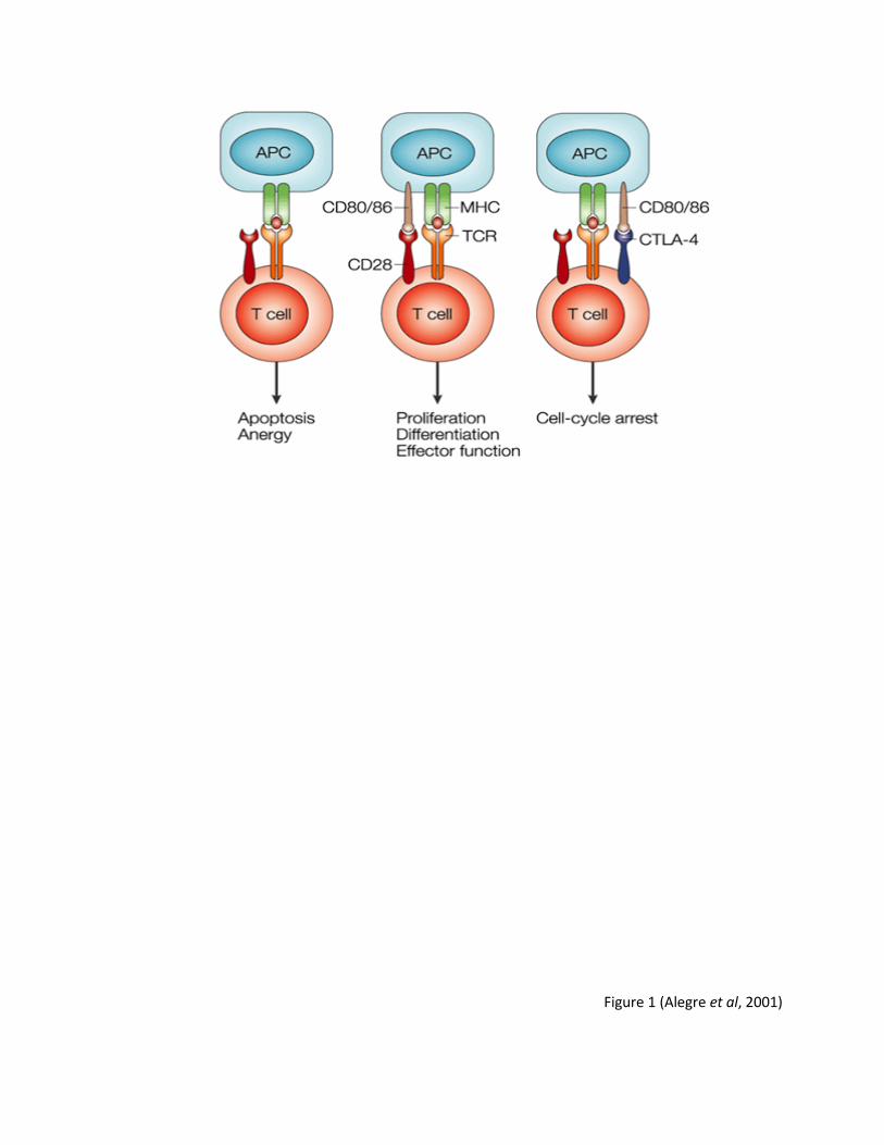
Figure 1 (Alegre et al, 2001)

14
affinity to its ligands, preferentially interacting with CD80/CD86 to terminate an immune response
(26). CTLA-4 works by reducing the production of IL-2 and expression of the IL-2 receptor, and by
arresting T cells at the G1 phase of the cell cycle (31). Although both CD80 and CD86 bind to CD28
and CTLA-4, recent research has demonstrated that their binding affinities differ; CD80 binds to
both receptors with two to three times more affinity, with slower dissociation constants and faster
binding kinetics compared to CD86 (22) .
CD80 and CD86 peripheral membrane proteins represent two major members of the B7
family. The B7 family also includes newer members: B7-DC, B7-H1, B7-H2, B7-H3, and B7-H4
expressing their own unique, but often overlapping functions in directing immune functions (32).
Preferentially expressed on DCs, B7-DC binds to its receptor PD-1 resulting in T cell proliferation and
cytokine production (33). In vivo experiments in knockout mice have demonstrated that B7-H1 and
B7-H2 have a role in effector responses, particularly in the production of antibodies, and are critical
in the tumor immune response (32). B7-H3 has been identified as a major player in cancer research
and may play a diverse role in the regulation of growth and differentiation of non-hematopoietic
cells (34). B7-H4, highly expressed in human cancers, has been shown to inhibit immune responses,
contributing to tumor escape (35). Although interest in the immunological function of these newer
members of the B7 family has increased within the past decade, the ongoing research into the
elucidation of the mechanisms regulating the expression and function of the first two B7 family
members, B7-1 and B7-2, or CD80 and CD86, has expanded tremendously, particularly in their
prospects in diagnostics and the development of immunotherapies.
1.6 The role of CD80 and CD86 in Th cell differentiation
CD4+ T helper cells can differentiate into two distinct subtypes of effector cells each with
their own set of regulatory cytokines. Th1 cells regulate cell-mediated immune responses through
15
production of IL-2, IFN-γ, and TNF-β. In contrast, Th2 cells regulate humoral immunity and produce
IL-4, IL-5, and IL-10 (36). Both cell types have important implications in the resistance and
progression of disease. Following T cell activation, precursor T cells will differentiate preferentially
down the Th2 pathway in the presence of IL-4, or down the Th1 pathway in the presence of IL-12.
Activation of these cell types by antigens further reinforces their differentiation pattern manifesting
by the production of IFN-γ by Th1 cells and IL-4 by Th2 cells (37). More recently, it has been
demonstrated that CD80 and CD86 may play a role in the differentiation of these two subtypes of
cells. Treatment of murine cells with anti-CD80 antibodies in vitro and in vivo favored the induction
of IL-4 expression and drove the immune response towards a Th2 phenotype, whereas treatment
with anti-CD86 antibodies resulted in the production of effector cells of a Th1 phenotype (38). In
contrast, another study demonstrated that the presence of CD80 actually inhibited IL-4 production
(39). These controversial results warrant further studies aimed at clarifying the role of CD80 and
CD86 in the differentiation of Th1 and Th2 cells and in their potential efficacy in the treatment of
disease and infection. Particularly, elucidating the molecular mechanisms governing the expression
and function of CD80/CD86 in APCs would represent an important step toward the design of new
therapeutic strategies for treatment of cancer, immune diseases and infection.
1.7 The role of CD80 and CD86 in disease
CD80 and CD86 have been identified as key regulators of immune activation, tolerance
regulation and the skewing of T cell responses in disease models, such as graft-vs-host diseases,
cancer, and autoimmune diseases (40).
16
1.7.1 The involvement of costimulatory molecules in cancer and in the development of therapeutic interventions
There is evidence to suggest that they also have an important role in tumor immunity. The
majority of tumor APCs are of low immunogenicity and often lack expression of costimulatory
molecules. For example, human gliomas have been shown to suppress immune responses and
proliferation of lymphocytes and these effects were attributed to a reduced expression of CD80 and
CD86. Topical expression of CD80 and CD86 in murine neuroblastoma cells was able to control
tumor growth up to doses greater than one million cells (42). Lack of CD80/CD86 expression in
glioma-infiltrating microglia/macrophages (GIMs) also led to the decreased secretion of the
cytokines required to initiate an effective innate immune response, such as TNF-α, IL-1, IL-6, and
this was in spite of the normal expression levels of TLRs (43). As a result, these GIMs were unable to
effectively costimulate and activate T cells (44). Increased T cell activity and cytokine production
were restored upon transfection of murine sarcoma cells with CD80/CD86 encoding DNA constructs
(45). Furthermore, in experiments where wild type mice were injected with the sarcoma cells
transfected with the CD80 expression constructs, half of the mice remained tumor free and
exhibited an increase in T cell population and Th1 and Th2 cytokine production, including IFN-γ,
which plays an important role in eliciting antigen-specific anti-tumor effects (45). The protective
effects of CD86 through the amplification of both local and systemic anti-tumor immunity were also
demonstrated in a study where the CD86-transfected mastocytoma P815 tumor cells were injected
into mice. More importantly, these mice were also protected against subsequent challenges with
the lethal wild-type P815 tumor and this protection was attributed primarily to the enhanced
generation of CD8+ T cells and tumor specific cytolytic activity (46). Collectively, these studies
demonstrate that induction of CD80/CD86 expression enhances anti-tumor immunity (47). In spite
of a growing body of evidence demonstrating a critical role of CD80/CD86 in cancer prevention,

17
further research is needed to delineate the regulation of CD80/CD86 expression needed to use
these molecules as anti-cancer therapeutics.
Evidence emerging from research using human and animal models clearly demonstrates
that T cells form an integral part of cancer immune surveillance, in particular, the recognition of
tumor associated antigens by CTLs (48). More recently, T cell-associated CTLA-4 molecule, which
promotes a negative signal, has been identified as a potential target for the development of anti-
cancer therapeutics. CD80/CD86 molecules interact with both CD28 and CTLA-4 expressed on T cells
and these interactions modulate the adaptive immune response. For example, the binding of
CD80/CD86 to CD28 results in the activation of T cells and the initiation of an adaptive immune
response, while the interaction with CTLA-4, which has a higher binding affinity compared to CD28,
inhibits excessive T cell activation to prevent host tissue damage and immune over-activation (21).
Upon binding to CD80 or CD86, CTLA-4 negatively regulates T cell activation, and as such, plays an
important role in T cell homeostasis, through the inhibition of IL-2 production and cell cycle
progression (49). Attempts to inhibit CTLA-4 function produced promising results demonstrating
enhanced anti-tumor response. For example, the immunopotentiating effects of CTLA-4 blockade by
anti-CTLA-4 antibodies in conjunction with CD28-CD80/CD86 stimulation was achieved in a mouse
colon cancer model (50). This treatment resulted in a reduced tumor size, particularly in
combination with irradiated tumors, and in an increased level of IFN-γ-secreting T cells and tumor-
specific CTL activity (50). Similar results were found in vivo after administration of anti-CTLA-4
antibodies which led to the rejection of pre-existing tumors (51). The long term effects of this
treatment have also been studied in mice treated with anti-CTLA-4 antibodies, in combination with
irradiated cancer cells engineered to produce granulocyte and macrophage colony stimulating
factor (GM-CSF) (52). These mice rejected previously established tumors and were found to be
refractory to secondary tumor challenge, suggesting the development of immunological memory

18
(52). Clinically, anti-CTLA-4 antibodies, such as ipilimumab and temozolomide have shown promising
results and have been approved for treatment for patients with metastatic melanoma, particularly
since they show low toxicity and can penetrate the blood-brain barrier, making them ideal for
treatment against brain cancers (53, 54).
1.8 CD80/CD86 and transplant rejection
The importance of these two costimulatory molecules was recognized in transplantation
immunology experiments. It was first thought that only antigen presentation was required to
activate lymphocytes, but upon the discovery that tissue cells of non-hemapoietic origin were not
readily rejected during transplantation, it was believed that a second signal provided by APCs was
required for lymphocyte activation and graft rejection (55).
Transplant rejection is an immune response by the recipient’s immune system that results
in T cell mediated rejection and destruction of the transplanted tissue. Graft-versus-host (GVH)
disease remains the principal risk involved in organ and bone marrow transplantation (56), and
remains the leading cause of morbidity and mortality after lung and heart transplantations (57).
Rejection of transplanted allografts is dependent on T cell activation, which requires its engagement
with the APC. CD80 and CD86 play pivotal roles in this T-cell dependent process, and attempts have
been made to modulate the CD80/CD86-CD28 signaling pathway to stop this rejection (56). Because
CTLA-4 has been identified as providing an inhibitory signal to T cells, it has been suggested as a
potential therapeutic target for graft-vs-host disease. The development of a membrane bound anti-
CTLA-4 antibody expressed on B cells in vivo demonstrated a profound inhibited T cell proliferation
and cytokine production in vitro and in vivo (58). The antibody prevented the rejection of allogeneic
tumor cells by antigen-specific CD8+ T cells in vivo (56) and protected NOD (non-obese diabetic)
mice from developing spontaneous autoimmune diabetes (59). In mice and rats, CTLA-4Ig treatment
19
prolonged the acceptance and survival of cardiac allografts (60, 61). Mice treated with CTLA-4Ig in a
xenogeneic islet transplant model prevented human pancreatic islet cell rejection (62). Taken
together, CTLA-4 and the CD80/CD86-CD28 pathway play a critical role in controlling T-cell
responses to foreign antigen and present novel targets for future research.
1.10 CD80 and CD86 as signaling molecules
The role of macrophages and the importance of the costimulatory markers CD80 and CD86
in T cell activation have been well characterized. Particularly, the studies investigating the role of
CD80 and CD86 in autoimmune diseases and cancer clearly demonstrated lack of T cell activation
and depletion of T cell numbers in the absence of CD80 or CD86 surface expression (63). While the
biological significance of CD28 ligation has been well described, more recently, CD28 has emerged
as a potential agonist ligand of B7 receptor molecules. Until now however, little is known about the
role of CD80 and CD86 as signaling molecules. Both molecules are comprised of two highly
glycosylated extracellular Ig-like domains linked to a transmembrane domain and a cytoplasmic tail
(64). The importance of the cytoplasmic tail has been highlighted in studies where interaction of
tailless CD80 molecules with CD28 was examined. These tailless molecules could not promote
antibody-induced cytoskeleton-dependent redistribution and capping in epithelial and lymphoid
cells (65). These results suggested that the cytoplasmic region of CD80 is involved in the localization
and redistribution of CD80 molecules on cell surfaces and is therefore important for effective
costimulation. Further studies by the same group identified a small, 30-kilodalton (kDa)
phosphoprotein that associated with the cytoplasmic tail of CD80 in activated cells (66), however
identification of this protein and its function have not been investigated further.
More recently, the role of CD80 and CD86 as signaling molecules has been investigated in
DCs where it has been demonstrated that CD80/CD86 transduce signals to the DC. In particular,

20
following engagement of CD80 or CD86 with CD28, DCs have been shown to produce IL-6, a
proinflammatory cytokine that is necessary for full T cell activation (67). To elucidate signal
transduction pathways activated by engagement of CD80 or CD86, Koorella et al demonstrated that
treatment of DCs with anti-CD80 and anti-CD86 antibodies, or with a recombinant fusion protein
CD28-Ig led to induction of IL-6 expression through activation of the PI3K/AKT pathway and
transcription factor, NF-κB (67). Moreover, employing murine DCs, it was also shown that anti-
CD28-Ig induced both IL-6 and IFN-γ production, and blockade of p38 MAPK activity resulted in
complete suppression of IL-6 production (68). This bidirectional signaling presents new potential
avenues for the development of therapeutic interventions targeting these costimulatory molecules.
As a result, deciphering the CD80/CD86 signaling pathway and the molecules that govern their
expression is essential. Since toll like receptors (TLRs) serve as a model for cell activation pathways,
it was of interest to study them in the context of macrophage activation.
1.11 Toll like receptor signalling
Toll like receptors (TLRs) are cognate pattern recognition receptors (PRRs) that act as guards
against invading organism that bear pathogen-associated molecular patterns (PAMPs), such as
Gram-negative bacterial LPS, and damage-associated molecular pattern molecules (DAMPs), such as
heat shock proteins (69). By recognizing specific molecular patterns that are found in a broad range
of microbial pathogens, an inflammatory response can be triggered for eradication of the pathogen
(69). These type I transmembrane glycoproteins function as homo- or heterodimers, and are
structurally characterized by extracellular leucine-rich repeats and toll/IL-1 receptor (TIR) signaling
domains (70). The TLRs are broadly divided into two classes depending on cellular component
where they engage their ligands. TLR-1, TLR-2, TLR-4, TLR-5, and TLR-6 are expressed on the cell
surface, while TLR-3, TLR-7, TLR-8, and TLR-9 bind to their cognate ligand in the endosome (71). TLR
21
signaling leads to multiple outcomes depending on the cell type responding to the stimuli; for
example, cell differentiation and proliferation, induction of inflammatory/regulatory genes,
antibody class-switching production (71). All TLRs except TLR-3 utilize MyD88 for signal
transduction, whereas TLR-3 signals through TRIF (71). Myeloid differentiation factor 88 (MyD88),
TIRAP (Toll-interleukin-1 receptor domain containing adaptor protein), TRIF (toll receptor-associated
activator of interferon), and TRAM (toll receptor associated molecule) are examples of TIR-domain
containing adaptors that are activated upon ligation of TLRs (72). Upon binding of LPS to TLR-4, the
TRIF-dependent, also known as the MyD88 independent, and MyD88 dependent pathways are
activated. MyD88 was first characterized as an essential component for activating the innate
immune system through the activation of nuclear-factor KB (NF-κB) and mitogen-activated protein
kinases (MAPKs) (72). For example, after infection, LPS or bacterial endotoxin will interact with LPS-
binding protein (LBP), which facilitates its subsequent interaction with CD14 found on the surface of
monocytes/macrophages. CD14 recruits an accessory protein MD2, which in turn binds to TLR-4
expressed on APCs (73). Activation of the TLR-4 signaling pathway is initiated following recruitment
of TIRAP to the TIR domain of TLR and MyD88; this results in the translocation of TRIF to the
complex, which also requires the recruitment of TRAM (Figure 2) (69). Such formed adaptor protein
complex activated a cascade of signalling molecules, eventually leading to the activation of the
transcription factors NF-κB and interferon regulatory factors (IRFs), which in turn induce the
expression of various inflammatory cytokines, type 1 interferons, and chemokines. In addition to the
TRIF mediated protein complexes, TLR-associated adaptor protein MyD88 also interacts with
signaling proteins forming a MyD88-dependent signaling complex. In response to stimuli, MyD88
recruits members of the interleukin-1 receptor associated kinase (IRAK) family, which are
sequentially phosphorylated and dissociated from MyD88, resulting in the activation of tumor
necrosis factor receptor-associated factor 6 (TRAF-6). This in turn activates transforming growth
22
Figure 2: Schematic representation of Toll Like Receptor (TLR) Family. Numerous pathways are
induced upon engagement of TLRs. In particular, binding of LPS to its cognate receptor TLR-4
induces NF-κB is the central regulator of immune responses.
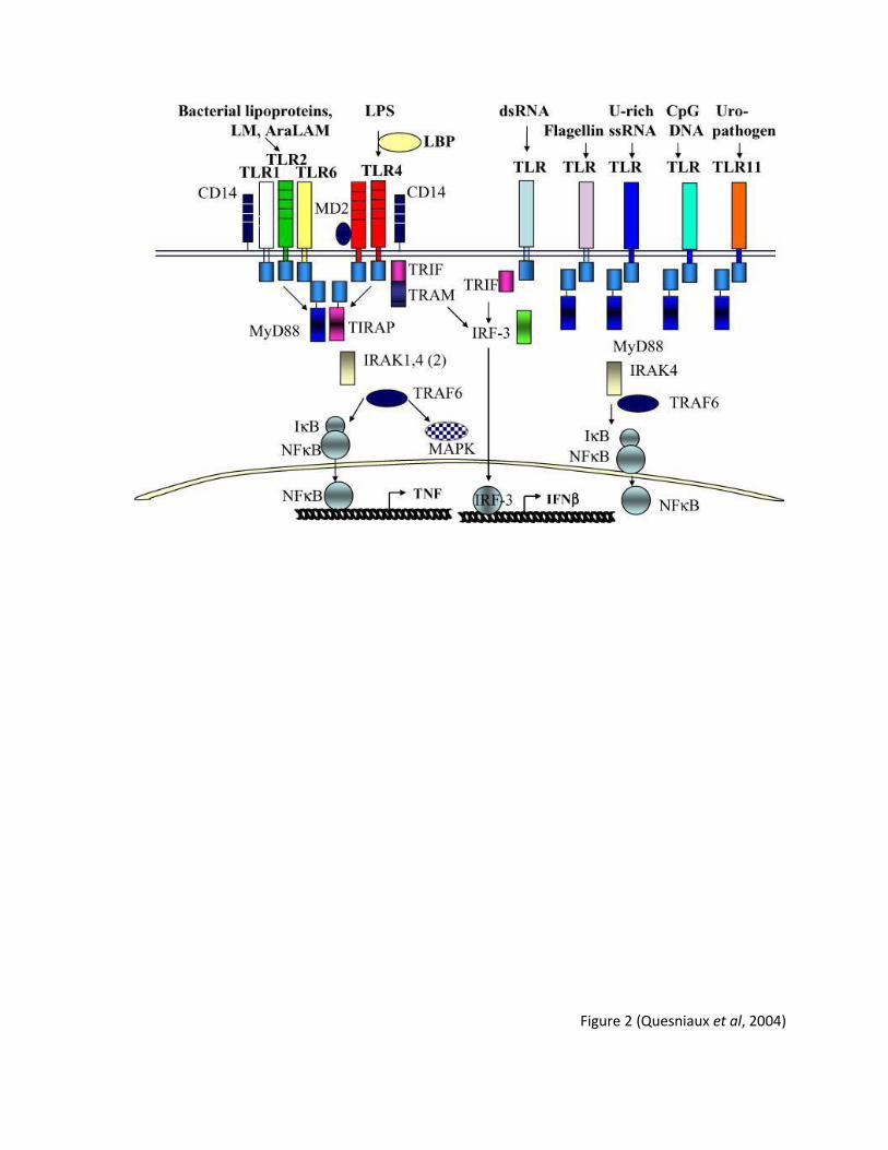
Figure 2 (Quesniaux et al, 2004)
23
factor-β-activated protein kinase 1 (TAK1), which is a member of the MAP3K family, in a ubiquitin-
dependent fashion. TAK1 then activates the IKK complex which leads to the activation of NF-KB
(Figure 2), while simultaneously activating other members of the MAP kinase family (72). NF-κB is
the central regulator of immune responses that are involved in the proliferation and survival of cells
and is responsible for inducing the expression of various cytokines and chemokines, including IL-12
(70). NF-κB is a heterodimer composed of the p65 and p50 subunits, residing in the cytoplasm in an
inactive form by interacting with inhibitor NF-κB proteins (IκB). Through the stimulation by the TLR-
4 ligand, LPS, IκB proteins are phosphorylated by an IKK complex, releasing NF-κB into the nucleus.
Activating protein-1 (AP-1) family proteins control various cellular processes including
differentiation, proliferation and apoptosis and are activated by the TLR-4 pathway. Among the AP-1
family proteins, c-Jun plays a central role in the inflammatory response, and the activation of AP-1 is
mediated by MAP kinases, such as c-Jun N-terminal kinase (JNK), p38, and extracellular signal
regulated kinase (ERK) (72). Dysregulation of TLR-4 signaling has been implicated in the
development of autoimmune diseases, such as multiple sclerosis.
1.12 TLR-4 and the activation of the MAPKs and PI3K pathway
In addition to the canonical MyD88-dependent and a MyD88-independent TRIF-mediated
pathway, engagement of TLR-4 also results in the activation of the MAPK and the phosphoinositide-
3-kinase pathways (71). The MAPKs are key players in cellular responses, such as cell growth and
survival, proliferation, differentiation, neuronal response, and apoptosis (74). This family of
serine/threonine protein kinases is highly conserved among eukaryotes and the dysregulation of
MAPK signaling has been implicated in cancer, and immune and neuronal diseases (75, 76). In
mammals, the triple kinase module consisting of a MAPK kinase kinase (MKKK) phosphorylates and
activates a MAPK kinase (MKK) that activate the terminal MAPK by dual phosphorylation of the
24
threonine and tyrosine residues (77). Six distinct groups of MAPKs have been characterized in
mammals: extracellular signal-regulated kinases (ERK1/2), jun NH2 terminal kinases (JNK1/2/3), p38
(p38 α, β, δ), ERK7/8, ERK3/4 and ERK5. To date, ERK1/2, JNKs, and p38 kinases are the best
characterized (77). Once they are activated, MAPKs can phosphorylate the target substrates, such as
transcription factors (77). In particular, activation of ERK1/2 and p38 has been implicated in the
regulation of cell proliferation and differentiation, whereas phosphorylation of JNK has been
specifically implicated in the production of cytokines, such as TNF-α (78-80). Crosstalk between
signaling cascades also contributes to the continuing complexity of the MAPK signal transduction
pathway. For example, MAPKK activates both JNK and p38α (77).
The PI3K pathway is activated by many different signaling molecules, and it is crucial for cell
growth and cell survival. PI3Ks are a family of lipid kinases consisting of three classes (class I, class II,
and class III) (71). Class IA PI3Ks, composed of p110α, p110β, and p110δ subunits, are critical in
immune cell function (81). The subunits are regulated by p85α or p85β regulatory subunits; the p85
subunit engages to phosphorylated tyrosines to release the inhibitory pressure p85 places upon the
catalytic p110 PI3K subunit (82). The PI3K complex is recruited with phosphorylated tyrosines,
bringing PI3K to the lipid membrane, in close proximity with its lipid substrate (71). The activation of
the PI3K pathway culminates in the phosphorylation and activation of AKT, a serine/threonine-
specific protein kinase that has an important role in various cellular processes, such as apoptosis,
cell proliferation and migration, and transcription (83). While it is known that LPS induces all three
types of MAPKs and the PI3K pathway, little is known about their role in the regulation of LPS-
induced CD80 and CD86 expression in macrophages.

25
1.13 Inhibitor of Apoptosis Proteins and SMAC mimetics
One of the main anti-apoptotic families of caspase inactivating proteins are the inhibitor of
apoptosis proteins (IAPs) (84). The activity of IAPs is antagonized by the release of pro-apoptotic
proteins known as a second mitochondrial activator of caspases (SMAC) from the mitochondria (84).
Since the inappropriate regulation of apoptosis is one of the hallmarks of many diseases, including
cancer, understanding the role of IAPs is an attractive strategy for developing new classes of drug
therapy.
Described over 20 years ago, the iap baculovirus gene was originally found to inhibit
apoptosis in virally infected Spodoptera frugiperda insect cells (85). They are characterized by the
presence of a variable number of highly conserved Baculoviral IAP repeat (BIR) motifs, a sequence of
~70 amino acids with three anti-parallel beta sheets and four alpha helices, allowing BIRs to mediate
protein-protein interactions (86). They can also contain additional functional regions, such as a RING
domain or caspase-associated recruitment domain (CARD), important for protein interactions and
ubiquitin ligase activity (87). Eight mammalian IAPs have been identified: cellular IAP1 (cIAP1),
cIAP2, X-chromosome linked IAP (XIAP), neuronal apoptosis inhibitory protein (NAIP), livin, survivin,
IAP-like protein 2 (ILP2) and baculovirus inhibitor of apoptosis repeat containing ubiquitin-
conjugating enzyme (BRUCE) (87). In mammals, cIAP1, cIAP2 and XIAP and their role in apoptosis
have been studied extensively. The BIR2 and BIR3 in these molecules have been identified as being
the important motifs in the binding of caspases and apoptosis-regulatory molecules (87). For
example, the common anchoring motif on BIR3 found on cIAP1 and cIAP2 selectively targets caspase
9, preventing its activation and the activation of caspase 3 and 7, whereas BIR2 prevents the
activation of caspase 3 and 7 specifically (88, 89). Recent studies have identified BIR1 as being
critical for the interaction of cIAPs and with tumor necrosis factor (TNF) associated factors (TRAFs),
in particular, TRAF2 (90). Through the association of their BIR and TRAF-N domains, the cIAP1- and
26
cIAP2-TRAF2 interaction regulates receptor-mediated apoptosis through TNF receptor 1- and 2-
associated complexes (90). The degree of cell death is tightly controlled by IAPs and their
importance can be highlighted in neurodegenerative disorders and cancer. For example, patients
with spinal muscular atrophy were found to have partly deleted NAIP (86) and pancreatic cancer has
been associated with an overexpression of cIAP2, survivin, livin and XIAP (91).
In response to apoptotic stimuli, SMAC, also known as Direct IAP Binding protein with Low
pI (DIABLO), undergoes proteolytic processing and is subsequently released from the mitochondria
into the cytosol to inactivate several IAPs, including cIAP1, cIAP2, and XIAP (92). The first four amino
acids (Ala-Val-Pro-Ile) on the N terminal of this proapoptotic molecule (the AVPI tetrapeptide
binding motif) binds to a surface groove on the BIR3 motif of IAPs, directly competing with the ATPF
tetrapeptide of caspase 9, thereby interfering with the interaction between caspase 9 and the IAP
(93, 94). Since they block apoptosis at the downstream effector phase, IAPs represent a particularly
interesting target for drug therapy. SMAC mimetics were designed to overcome the apoptosis
resistant tumor cells by preventing the interaction between caspases and IAPs (95). This small
molecule induces cell death without the mitochondrial release of apoptotic factors by mimicking the
AVPI tetrapeptide and binding to the BIR3 domain (94), leading to the rapid ubiquitination and
proteasomal degradation of cIAP1 and cIAP2, but not XIAP, although the method by which auto-
ubiquitination is induced is not entirely clear (90). Increasing the abundance of pro-apoptotic
proteins such as SMAC is a more attractive therapeutic strategy for tumors because it could limit the
toxicities associated with other therapies, such as recombinant cytokine therapy (96).
1.14 IAPs and NF-κB signaling
While the use and testing of SMAC mimetics in cancer therapeutics has demonstrated their
ability to reveal the apoptosis-opposing properties of IAPs, accumulating evidence has suggested
27
that cIAP1 and cIAP2 may also be involved in signal transduction pathways, in particular, TNF-α-
mediated NF-κB activation (87). Upon binding to its receptor, TNF-R1, TNF-α exerts its effect by
rapidly recruiting the TNFR-associated death domain (TRADD) protein and the receptor-interacting
protein 1 (RIP1), which consequently recruits TRAF2 to form a large complex (97). It is believed that
during this recruitment, cIAP1 and cIAP2 interact with TRAF2 in a TNF-dependent manner and can
bind to TNF receptors (98). TRAF2, cIAP1 and cIAP2 facilitate the ubiquitination of RIP1, a crucial
event for the propagation of the signal (99). RIP1 serves as a docking site for inhibitor of κB (IκB)
kinase (IKK) α/β/γ heterocomplex, which, when phosphorylated, will phosphorylate IκBα, an
inhibitory protein. IκBα signals for its ubiquitination and proteasomal degradation, allowing NF-κB
to translocating into the nucleus. Here, it regulates target genes, in particular, prosurvival genes
such as FLICE inhibitory protein and cIAP2, inhibiting the caspase-8-mediated apoptotic pathway
that is concomitantly engaged by TNF-α (97). In the non-canonical pathway, receptors such as CD40
activate NF-κB inducing kinase (NIK) which also results in the translocation of NF-κB to the nucleus
(87). Blocking NF-κB activation resulted in decreased TNF production and protected the cells from
SMAC mimetic-induced apoptosis (100). Therefore, TNF-α-mediated cell survival is dependent on
the proper NF-κB induction.
1.15 IAPs and Pattern Recognition Receptor (PRR) signaling: cIAPs and TLR-4 signaling
IAP proteins are critical regulators of the NF-κB signaling and the expression of genes that
control innate and adaptive immunity, inflammation, and cell survival and migration. The canonical
pathway for the activation of NF-κB is essential for the innate immune response (101), while the
non-canonical pathway is vital for the adaptive immune response and the development and
maintenance of lymphoid organs (102). After recognition of PAMPs by their respective PRRs, the
inflammatory response is activated and mediated by the NF-κB pathway and the MAP kinases
28
(MAPK), which increase the transcription of genes that encode cytokines, chemokines, adhesion
molecules, and antimicrobial peptides to recruit inflammatory and phagocytic cells to the infection
site (103). After stimulation with LPS, the MyD88 dependent pathway is activated resulting in the
recruitment of TRAF3 and TRAF6 to form the MyD88-assembled signaling complex (104). In this
complex, TRAF3 undergoes TRAF6-, cIAP1-, and cIAP2-dependent degradative ubiquitination
promoting cytosolic translocation of the entire signaling complex, activating MAPKs, and inducing in
inflammatory genes (105). Depletion of cIAP1 and cIAP2 with SMAC mimetics blocked p38 and JNK
MAPK activation via TLR-4 (105). Furthermore, depletion resulted in a reduction in LPS-induced
cytokine production, with no effect on interferon responses, indicating an absence of a role for
cIAPs in the MyD88-independent/TRIF-mediated pathway (105).
IAPs have also been identified as regulators of the cytoplasmic signaling pathway initiated
by the nucleotide-binding oligomerization domain 1 (NOD1) and NOD2 receptors, as well as the
retinoic acid-inducible gene (RIG)-1 receptor. NOD1 and NOD2 are PRRs that require cIAP1, cIAP2
(106) and XIAP (107) to elicit a proper immune signal. Upon recognition of bacterial peptidoglycan,
NOD1 and NOD2 self-oligomerize and recruit RIP2, cIAP1, cIAP2, and XIAP (106). This complex
induces the ubiquitination of RIP2 by cIAP1 and cIAP2, resulting in the downstream activation of
MAPK p38 and JNK (108). The importance of IAPs was highlighted in cIAP1-, cIAP2-, and XIAP-
deficient mice that were treated with the NOD1 and NOD2 ligands. They failed to activate NF-κB and
MAPK resulting in a reduction in immune signaling and cytokine expression, and this was attributed
to their ubiquitination of RIP2, similar to their role in TLR signaling (106, 107). Similarly, IAPs have
also been associated in the type I interferon response mediated through RIG-1, the cytoplasmic PRR
that recognizes viral double-stranded RNA through their helicase domain (109). Upon recognition,
RIG-1 is recruited to the mitochondrial antiviral signaling (MAVS) protein, which interacts with
29
TRAF3 to induce a type I interferon response (110). cIAPs ubiquitinate TRAF3 in order to enhance
interferon regulatory factor 3 (IRF-3) and NF-κB to induce an antiviral immune response (111).
In summary, there exists compelling evidence to suggest that IAPs do not simply have an
anti-apoptotic role. These proteins have important roles in NF-κB activation and MAPK signaling,
and the use of SMAC mimetics has been identified as significant antagonists that can be potentially
used in cancer therapies.
1.16 The role of tyrosine phosphorylation the LPS/TLR-4 pathway
Phosphorylation is a fundamental mechanism that provides a quick and reversible change
known to alter the function of proteins; it is important in the regulation of normal cellular
processes, such as cell growth and differentiation (112). Aberrant phosphorylation of tyrosine
residues can result in excessive cell proliferation and disease (113). Protein tyrosine kinases (PTKs)
and protein tyrosine phosphatases (PTPs) control the tyrosine phosphorylation of signalling
molecules (113). While PTKs catalyze the phosphorylation of tyrosine residues, PTPs
dephosphorylate the phosphotyrosine residues, maintaining equilibrium in biological systems (113).
More recently, the role of tyrosine phosphorylation in the LPS/TLR-4 pathway has been
investigated. Binding of LPS to TLR-4 initiated the phosphorylation of signaling molecules and the
activation of protein tyrosine kinases (PTKs) (114). The observation that a broad spectrum of
tyrosine kinase inhibitors prevented LPS-induced cytokine production and protected against septic
shock attracted considerable attention in research (115). This protection was correlated with the
ability of the inhibitors to block LPS-induced tyrosine phosphorylation of ERK2 MAPK proteins and
the subsequent release of TNF-α in murine macrophages (115). The induction of other cytokines,
such as IL-1β and IL-6, has also been shown to require the activation of PTKs (116). The SRC family of
tyrosine kinases make up the largest group of non-receptor tyrosine kinases. The 9 members of this
30
group are: Src, Hck, Fyn, Lyn, Fgr, Lck, Blk, Yes, and Yrk, each containing an N-terminal unique
domain SH3, SH2 and tyrosine kinase domain (114). They are differentially expressed in various cell
types, with members of the innate immune system being Src, Fyn, Fgr, Lyn and Hck (117). Activation
of TLR-3, TLR-4, and TLR-9 in macrophages was shown to upregulate PTP expression, and triggered
an inhibited TNF-α, IL-6 and IFN-β production, impairing NF-κB activation (118).
PTPs have been described as both positive and negative regulators of signaling processes.
For example, the SRC homology region 2 (SH2) domain-containing tyrosine phosphatase-1 (SHP-1)
negatively regulates the cellular responses in hematopoietic cells initiated by colony stimulating
factor (CSF-1), IL-3, c-Kit, erythropoietin, and IFN-α/β, but positively regulates epidermal growth
factor and IFN-γ in astrocytes (116). SHP-1 has been strongly associated with the LPS/TLR-4
pathway; it has been shown that SHP-1 regulates the activation of ERK1/2 MAPK and NF-κB in bone
marrow derived macrophages (116). The importance of SHP-1 has been highlighted in SHP-1-
deficient mice; these mice manifest autoimmune disorders and a phenotype that correlates with
autoantibody production, a consequence of the altered production of inflammatory cytokines, such
as IL-6 (119). A proximal function of SHP-1 in conjunction with PTKs has been suggested; similar to
underexpression of SHP-1, treatment with the PTK-specific inhibitor, Herbimycin A, also inhibited
LPS-induced IL-6 production (116).
1.17 The regulation of CD80 and CD86 expression
Upon binding of LPS to its cognate receptor CD14/TLR-4, a cascade of signaling events is
induced resulting in gene activation and expression of proteins. In particular, signaling molecules
and transcription factors that regulate the expression of CD80 and CD86 have been elucidated
(121). CD80 expression can be induced by a number of stimuli other than LPS, including IL-4, anti-B
cell receptor (BCR), antibody or anti-CD40 antibody, and stimulation of monocytic cells with LPS and
31
IFN-γ (20, 22, 120). CD80 and CD86 are crucial for T cell differentiation and activation of an immune
response. It is therefore likely that immunoregulatory cytokines and mitogens, such as LPS, would
modulate CD80/CD86-mediated signals to enhance the activation of T cells (22). In LPS-stimulated
human monocytes, CD80 expression has been shown to be regulated by the (interferon regulatory
factor) IRF-7 transcription factor through the activation of JNK (121). Further studies identified a
distinct CD80-responsive element corresponding to the IRF-7 binding site, located between 84 and
72 base pair region upstream of the transcription start site of the CD80 gene (121). In LPS-
stimulated human monocytes, p38 and JNK MAPK have been shown to regulate CD86 expression,
through an IL-10-dependent and IL-10-independent pathways (8). LPS stimulation of normal human
monocytes results in the downregulation of CD86 expression, mediated via endogenously produced
IL-10 involving p38 MAPK (8). However, THP-1 cells are noncompliant to the inhibitory effects of IL-
10, and CD86 expression was upregulated in an IL-10-independent manner following LPS stimulation
in these cells (8). The regulation of CD80 and CD86 in DCs has also been investigated. One group
demonstrated the role of JNK MAPK in the LPS-induced CD80 and CD86 expression in human
monocyte-derived DCs (122). Another group identified a critical role for p38 MAPK in the LPS- and
TNF-α-induced CD80/CD86 expression human monocyte-derived DCs (123).
There are conflicting studies on the functional differences between CD80 and CD86. While
some reports have demonstrated that they provide similar costimulatory signals for T cell activation
and cytokine production, others have shown contrasting effects (64). Two DNA vaccination studies
have demonstrated that CD86, but not CD80, supported an HIV peptide-specific cytotoxic T
lymphocyte response (124, 125). More recently, CD80 has been identified as a more potent ligand
for CTLA-4 based on its higher affinity and enhanced avidity, whereas CD86 is a more effective CD28
ligand (126). On the other hand, mice that were CD80 or CD86 deficient demonstrated the
overlapping nature of their functions (127). Although the reason for these conflicting studies is not
32
clear, it could be due to differences in their experimental approach, such as the ligands used, or due
to the differences in their expression kinetics. Further studies are required to elucidate the critical,
and potentially different, role for these two molecules.
Modulation of CD80 and CD86 expression on APCs may alter the development of immune
responses. While the role of MAPK in their regulation has been studied in some cell types, there has
been limited work looking into the regulation of CD80 and CD86 in macrophages. To better
understand their role as a target for therapeutics, the signaling molecules involved in the regulation
of CD80 and CD86 in macrophages must be studied extensively.
33
1.18 Rationale
The activation of T cells is a crucial requirement for the initiation of effective immune
responses, and is dependent on the engagement of T cells with CD80/CD86 on the APC. The role of
CD80/CD86 in the activation of T cells has been well characterized, but more recently these B7
molecules have been investigated as signaling receptors in DCs. It has been shown that stimulation
of DCs with anti-CD80/CD86 antibodies induced IL-6 expression through the activation of the PI3K
pathway (67). Furthermore, Jain et al demonstrated that CD86 stimulation enhanced the effect of
TLR-2 stimulation on resting B cells (41). However, the role of CD80 and CD86 in macrophage
activation remains unknown. Macrophages are a potent source of cytokines particularly anti-
inflammatory IL-10 andthe IL-12 family of cytokines including IL-12, IL-23 and IL-27. Hence
stimulation of macrophages via CD80 and/or CD86 activation alone or in concert with LPS may have
a profound effect on the production of IL-10, IL-12, IL-23 and IL-27 and consequent development of
immune responses.
Dysregulation of CD80 and CD86 expression on macrophages has been observed following
HIV infection, in autoimmune disorders, and in cancer. Therefore, dysregulated expression of CD80
and/or CD86 may also influence the production of IL-10, IL-12, IL-23 and IL-27 by macrophages in
response to stimulation with TLR-4 ligand, LPS and consequent development of immune response.
Determining signaling molecules and the signaling pathways that govern their regulation may
provide novel strategies to control diseases such as autoimmune disorders and cancer. Therefore,
the focus of this project was twofold: 1) to investigate the role of CD80 and CD86 as signaling
receptors capable of transmitting extracellular signals either alone or in synergy with LPS leading to
the production of anti-inflammatory IL-10 and the IL-12 family of cytokines including IL-12, IL-23 and
IL-27, and 2) to determine the TLR-4 activated pathways that regulate CD80/CD86 expression in
human macrophages.
34
Since cIAPs play a key role in TLR-4-mediated signaling (128), it is reasonable to hypothesize
that cIAPs may play a role in CD80 and/or CD86 mediated activation either alone or in concert with
LPS activation of MDMs. cIAPs have also been shown to interact with TRAFs in the TLR-4 mediated
signaling pathways (129). Therefore, it is possible that cIAPs either alone or in association with
TRAFs and other interacting signaling molecules such as RIP1 or SHP-1 may regulate the expression
of CD80 and CD86 in MDMs stimulated by the TLR-4 pathway.
1.19 Hypothesis
Activation of CD80 and CD86 receptors either alone or in synergy with TLR-4 enhances
expression of cytokines such as IL-10 and IL-12 family of cytokines including IL-12, IL-23 and IL-27. In
addition, IAPs which play a key role in TLR-4-mediated signaling, either alone or in association with
TRAFs and other interacting signaling molecules such as RIP-1 or SHP-1 may regulate the expression
of CD80 and CD86 in MDMs stimulated by the TLR-4 pathway.
1.20 Objectives
1. To determine whether activation of CD80 and CD86 receptors alone, or in synergy with TLR-
4 altered cytokine expression of IL-10 and IL-12 family of cytokines.
2. To determine the role of TLR-4 activated signaling molecules, such as cIAP, SHP-1, RIP1 and
TRAF2, and signaling pathways, such as MAPKs and PI3K, in LPS-induced expression of CD80
and CD86 in human MDMs.
3. To determine the role of TLR4-activated signalling molecules including MAPKs and PI3K/AKT
in the expression of CD80 and CD86 in human MDMs, and their association with the
upstream signalling molecules.

35
2.1 Materials and Methods
2.2 Reagents
Chemical inhibitors SB203580 (pp38 inhibitor), SP600125 (pJNK inhibitor), PD98059 (pERK
inhibitor), LY294002 (pAKT inhibitor), sodium stibogluconate (SHP-1 inhibitor), and SU6656 (pSrc
inhibitor) were purchased from Calbiochem (La Jolla, California). Lipopolysaccharide (LPS) was
obtained from Escherichia coli 0111:B4 (Sigma-Aldrich, St. Louis, Missouri). Second mitochondria-
derived activator of caspases (SMAC) mimetics, LN730, was a generous gift of Dr. R. Korneluk at the
Children’s Hospital of Eastern Ontario, Apoptosis Research Centre.
2.3 Cell culture
2.3.1 Human Monocyte-derived Macrophages (MDMs)
Human blood was obtained from healthy volunteers with written consent, according to a
protocol approved by the Ethics Review Committee of The Ottawa General Hospital. Peripheral
blood mononuclear cells (PBMCs) were extracted using Ficoll-Paque (GE Healthcare,
Buckinghamshire, UK) to create a density gradient following centrifugation of whole blood at 1600
RPM for 45 mins at 4° C in Allegra X-12R centrifuge (Beckman Coulter, Brea, California). The cell
layer containing mainly PBMCs was collected and counted. To generate MDMs, monocytes were
isolated via the adherence method. PBMCs were resuspended at 4 x 10⁶ cells/ml in Iscove’s
Modified DMEM 1X media (Sigma-Aldrich). The cells were seeded at 1 ml/well in 12 well
polystyrene plates (Thermoscientific, Rochester, New York) and monocytes were allowed to adhere
to the plate for 3 hrs in a humidified environment at 37° C, 6% CO2/air mixture. Non-adherent cells
were washed off and adherent cells were cultured for six days with complete media containing and
100 units/ml penicillin and 100 μg/ml gentamicin (Sigma-Aldrich) and 10% v/v fetal bovine serum
(GE Healthcare) and 10 ng/ml recombinant human macrophage colony stimulating factor (M-CSF)
36
(R&D Systems, Minneapolis, Minnesota). SnCells were washed and media with M-CSF was
replenished every two days for six days. Macrophages were characterized by Flow Cytometry, using
phycoerythrin (PE)-conjugated anti-CD11a, -CD11b, -CD11c, -CD80, -CD83, -CD16 and HLA-DR
antibodies (BD Biosciences, Franklin Lakes, New Jersey). Windows Multiple Document Interface
(WinMDI) version 2.8 software (J. Trotter, Scripps Research Institute, San Diego, California) was used
to create histograms.
2.4 MAP kinase and PI3K inhibition
To determine the effect of p38, JNK, ERK1/2, PI3K, SHP-1, and SRC on the LPS-induced
expression of CD80 and CD86, MDMs were treated for 2 hrs with their pharmacological inhibitors
SB203580, SP600125, PD98059, LY294002, sodium stibogluconate, and SU6656 (Calbiochem),
respectively, for 2 hrs, followed by LPS stimulation (1 μg/mL) for 15 mins, 4 hrs, or 24 hrs, for
Western Blot, RT-PCR, and ELISA/Flow Cytometry, respectively. MDMs were treated with 50nM of
SMAC mimetics for 24 hrs, before LPS stimulation. After various treatments, the viability of cells was
>95% viable, as tested via Flow Cytometry.
2.5 Transfection of MDMs with small interfering RNA (siRNA)
All control and specific siRNAs were purchased from Santa Cruz Biotechnology, Santa Cruz,
California. MDMs (1 x 10⁶ cells/well) were seeded in 250 μL of serum-free media. 4 μM of the
Transfection Reagent (Santa Cruz Biotechnology) and 4 μM of siRNA specific to p38, AKT, SHP-1,
SRC, RIP1, TRAF2, cIAP1 and 2, and a nonspecific control pool (siRNA control) were incubated for 30
mins, before adding this mixture to the cells at a final ratio of 40:4 of transfection reagent to siRNA.
After 24 hrs of incubation at 37° C, 200μL complete media was added to stop the transfection and

37
cells rested overnight at 37° C. The following day, cells were treated with 1 μg/ml LPS for another 15
mins or 24 hrs and then were collected for Western blotting and Flow Cytometry.
2.6 Western blot analysis
MDMs (1 x 10⁶) lysates were prepared and total protein concentration of the cell lysates
was determined using Bradford protein assay kit (Bio-Rad Laboratories, Hercules, California). Equal
amounts of proteins (30 μg) were subjected to SDS-PAGE before being transferred onto a
polyvinylidene difluoride membrane (Bio-Rad Laboratories) using a wet transfer blotting cell
apparatus (Bio-Rad Laboratories). Membranes were then probed overnight at 4° C with 1:1000
concentration of antibodies against SHP-1 (BD Biosciences), cellular IAP1 (cIAP1), cIAP2, TRAF2,
RIP1, pAKT, AKT, pp38, p38, pJNK, JNK, pERK, ERK (all from Cell Signaling, Danvers, Massachusetts).
The membranes were then washed and blocked with 5% skim milk in TBS-T, followed by 1:1000
goat anti-rabbit or goat anti-mouse secondary antibodies conjugated to horseradish peroxidase
(Bio-Rad Laboratories). To ensure equal loading, the membranes were stripped of the primary
antibodies and reprobed with antibodies specific for their unphosphorylated forms, or with a
loading control such as anti-GAP-DH (Cell Signaling). Immunoblots were visualized using the
Amersham enhanced chemiluminescence (ECL) Western Blotting detection system using the
Chemigenius Bio-imaging system and the GeneSnap software (Syngene, Cambridge, England).
2.7 Flow Cytometry
CD80 and CD86 expression on MDMs was determined by flow cytometric analysis. For FACS
staining, 0.5 x 10⁶ MDMs in a polystyrene tube (Falcon, Lincoln Park, New Jersey) were washed with
PBS and stained with 2.5 μL of PE-conjugated CD80 and CD86 antibodies (BD Biosciences) for 30
mins at 4° C. After staining, cells were washed with PBS and centrifuged at 2200 RPM for 5 mins to
38
remove excess antibodies. 10,000 events were collected using FACSCanto Flow Cytometer (BD
Biosciences) and analyzed using the WinMDI version 2.9 software (J Trotter, Scripps Institute, San
Diego, California).
2.8 RNA isolation and semi-quantitative RT-PCR analysis for CD80 and CD86
MDMs were stimulated with 1 μg/mL of LPS for 4 hrs before collecting and washing cell
pellets. Total RNA was extracted from cells using RNeasy Plus Mini kit (Qiagen, Hilden, Germany)
according to the manufacturer’s protocol. Using a spectrophotometer Nanodrop 2000C (Nanodrop
Products, Wilmington, Delaware), the concentration of purified RNA was measured and 50 ng/μL of
purified RNA was added to the master mix. To prepare the RT-PCR mastermix, the High Capacity
cDNA Reverse Transcription Kit (Applied Biosystems, Carlsbad, California) was used: 7.5 μL 10X
reverse transcriptase buffer, 3 μL dNTPs, 7.5 μL random primers, 3.75 μL reverse transcriptase and
53.25 μL ddH2O. RT-PCR was carried out using GeneAmp PCR system 2700 amplifier (Applied
Biosystems). Following this, real-time PCR reactions were prepared using 12.5 μL Taqman DNA
polymerase, 1.25 μL primer pairs and 8.75 μL ddH2O using 7500 Real Time PCR System. The
transcript expression levels were shown relative to the control, β-actin by calculation of cycle
threshold (Ct) values in amplification plots (Applied Biosystems). Primers were as follows:
CD80 sense (5’-AGT ACA AGA ACC GGA CCA TC-3’); CD80 antisense(5’-GGC GTA CAC TTT CCC TTC
TC-3’); CD86 sense(5’-AGG ACA AGG GCT TGT ATC AA-3’); CD86 antisense(5’-ATT GCT CGT AAC ATC
AGG GA-3’); β-actin sense(5’-TGA CGG GGT CAC CCA CAC TGT GCC CAT CTA-3’); β-actin antisense(5’-
CTA GAA GCA TTT GCG GTG GAG GAT GGA GGG-3’).
39
2.9 Treatment with Anti-CD80 and Anti-CD86 antibodies
Using 500 ng/mL antibodies that neutralize CD80/CD86-induced IL-2 secretion in T cells and
a monoclonal mouse IgG1 isotype control (all from R&D systems), MDMs were treated for 24 hrs in
combination with low levels of LPS (1-10 ng) for 24 hrs before IL-12p40, IL-23, IL-27 and IL-10
expression was measured by ELISA.
2.10 Cytokine measurement by ELISA (Enzyme-linked Immunosorbent assay)
MDM supernatants were collected in order to measure IL-12p40 (Invitrogen, Burlington,
Canada), IL-23 (R&D systems), IL-27 (R&D systems), and IL-10 (BD Biosciences) cytokine production.
96 well plates (Corning Incorporated, Corning, New York) were coated overnight at 4 °C with ELISA
coating buffer (4.24 g of Na2CO3 and 5.04 g NaHCO3 in 1L ddH2O, pH 8.2) and IL-12p40, IL-23, IL-27,
or IL-10 capture antibody as per the manufacturer’s instructions. The plates were washed three
times with PBS containing 0.05% Tween-20 (Bio-Rad Laboratories), and blocked for 2 hrs with PBS
containing 1% BSA or 10% FBS, according to the manufacturer’s protocol. After blocking, the plates
were washed again, and standards and test supernatants were added and incubated overnight. On
the third day, the plates were washed and biotinylated secondary antibodies for anti-IL-12p40, IL-
23, IL-27 and IL-10 were added, followed by the addition of streptavidin horse radish peroxidase.
The color reaction was carried out using 3,3’, 5,5’-tetramethylbenzibidine (TMB) substrate solution
and 450 nm liquid stop solution for TMB microwell substrates (BioFX Laboratories, Owings Mills,
Maryland). Cytokine concentrations were measured by absorbance using Bio-Rad iMark microplate
reader and data were analyzed using Microplate Manager 6 software (Bio-Rad Laboratories).
40
2.11 Immunofluorescence
MDMs were cultured and differentiated in 12 well plates containing round glass coverslips
(Electron Microscopy Sciences, Hatfield, Pennsylvania). Cells were fixed in 4 % paraformaldehyde
(Sigma-Aldrich) for 30 mins at 37° C, washed three times with PBS, before incubating with 50 mM
NH4Cl (Sigma-Aldrich) in PBS for 10 mins to quench the cells. Cells were washed again and
permeabilized with 0.1% Triton-X in PBS for 10 mins. The coverslips were then incubated overnight
at 37° C with two primary antibodies SHP-1 (BD Biosciences), RIP1 (Cell Signaling), TRAF2 (Cell
Signaling), or cIAP2 (Cell Signaling) at 1:300 ratio of 5% FBS. The next day, coverslips were washed
three times with PBS and incubated for 1 hr in AlexaFluor goat anti-rabbit (red) or anti-mouse
antibody (green) (Invitrogen). The coverslips were then washed again three times with PBS and
immersed in mounting media (Dako, Glostrup, Denmark) with DAPI (Molecular Probes, Eugene,
Oregon), before being visualized by confocal microscopy.
2.12 Statistical Analysis
Data were plotted and analysis was done using GraphPad Prism 5. Means were compared
by the Student’s t test or ANOVA and results are expressed as a mean ± standard deviation of at
least three experiments.
2.13 Ethics Statement
Informed written consent was provided by all healthy participants based on the study
protocol approved by the Research Ethics Boards of the Research Institute, Children’s Hospital of
Eastern Ontario and the Ottawa Hospital Research Institute, University of Ottawa, Ottawa, Canada.

41
3.1. Results
3.2. CD80 and CD86 activation synergizes with TLR-4 signaling pathway to regulate IL-10 and IL-27 production in human MDMs
After encountering an APC, T cells are activated by two important signals. The first antigen-
specific signal requires the TCR to bind to the MHC molecule and the second signal is provided by
the binding of CD28 with one of two costimulatory molecules on the surface of the APC (8).
Macrophages represent a large portion of the APCs that express these molecules. While the role of
CD80 and CD86 as a second signal for T cell activation is well characterized, it is not well understood
if this interaction with T cells results in macrophage activation. More recently, the role of CD80 and
CD86 as a signaling molecule has been investigated in dendritic cells. Stimulation of DCs with anti-
CD80/CD86 antibodies has been shown to induce IL-6 expression through the activation of PI3K/AKT
pathway (67). Anti-CD80 and anti-CD86 antibodies have also been shown to neutralize CD80/CD86-
induced IL-2 secretion in T cells by binding to their binding pockets and were therefore used to
represent the interaction between CD28/CTLA-4 on the surface of the T cell with CD80 and CD86.
However, little is known about this bidirectional signaling in macrophages. To explore this, human
MDMs were treated with either anti-CD80/anti-CD86 antibodies or with a control antibody IgG for
24 hours (Figure 3 and Figure 4). Antibody treatment alone resulted in no changes in the production
of these cytokines.
Innate immune cells, such as macrophages, respond to various bacterial components by
providing regulatory signals to other immune cells through the secretion of cytokines (14). Recent
evidence has demonstrated that the downstream effects of Toll like receptor activation can
combine with the effects seen after stimulation of costimulatory molecules to bolster downstream
functions (41). Jain et al demonstrated that CD86 costimulation enhanced the effect of TLR-2
stimulation on resting B cells, resulting in substantial increases in the activation, differentiation,

42
Figure 3: CD80 activation by anti-CD80 antibodies synergize with TLR-4 signaling in human MDMs
to produce IL-27 and IL-10, but not IL-12 and IL-23.
MDMs (1x10⁶ cells/ml) were treated with 500 ng/mL of the anti-CD80 antibody or isotype matched
control IgG1 control antibodies, in the presence or absence of 10 ng/mL of LPS for 24 hours.
Production of A) IL-27 (n=12), B) IL-10 (n=8), C) IL-23 (n=10), and D) IL-12p40 (n=6) protein was
measured by ELISA. Values in bar graph are means of at least three independent experiments ±SEM;
nonsignificant (ns); *p≤0.05; **p≤0.005; ***p≤0.0005.
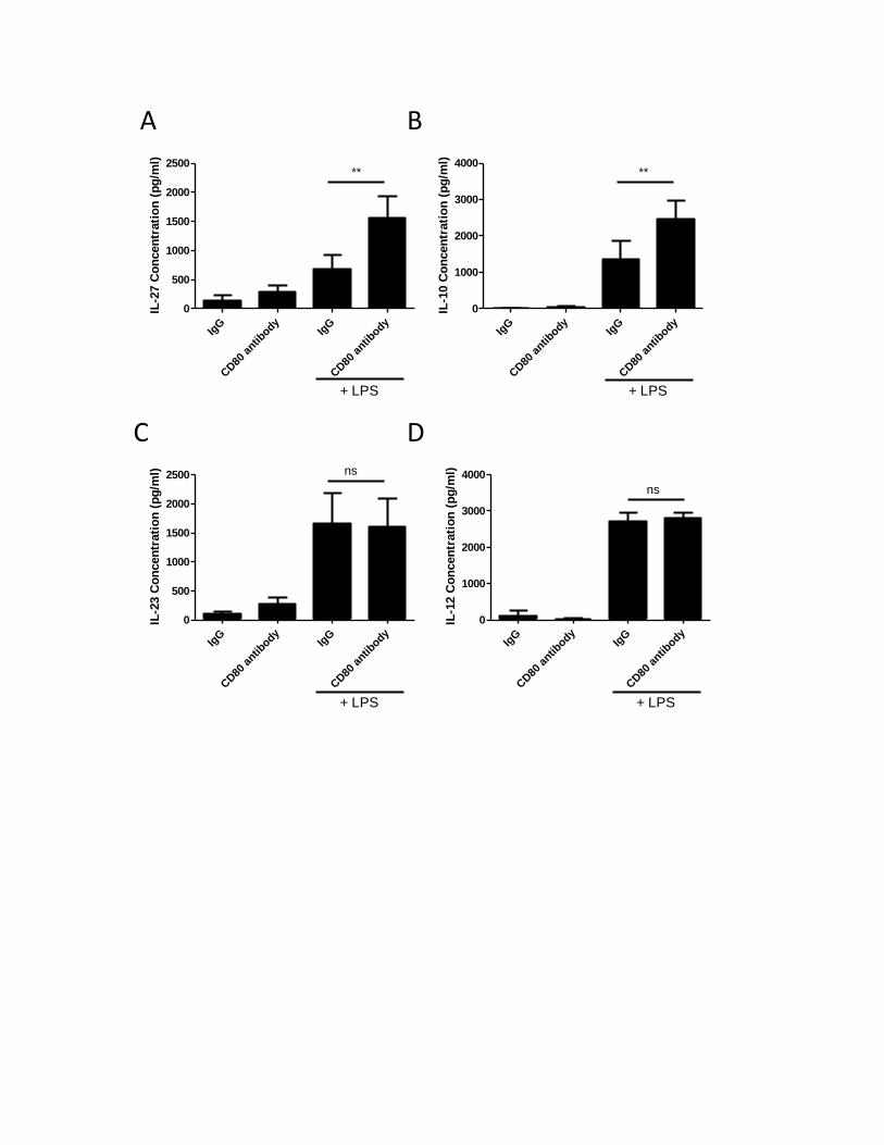
A B
IgG
CD80
antib
odyIg
G
CD80
antib
ody
0
500
1000
1500
2000
2500
+ LPS
**
IL-2
7 C
on
cen
trati
on
(p
g/m
l)
IgG
CD80
antib
odyIg
G
CD80
antib
ody
0
1000
2000
3000
4000**
+ LPS
IL-1
0 C
on
cen
trati
on
(p
g/m
l)
C D
IgG
CD80
antib
odyIg
G
CD80
antib
ody
0
500
1000
1500
2000
2500 ns
+ LPS
IL-2
3 C
on
cen
trati
on
(p
g/m
l)
IgG
CD80
antib
odyIg
G
CD80
antib
ody
0
1000
2000
3000
4000
ns
+ LPS
IL-1
2 C
on
cen
trati
on
(p
g/m
l)
43
survival, and antigen uptake capacity of these cells (41). In view of these findings, I wanted to
explore whether CD80/CD86 molecules also promote signaling via with TLR-4 pathway in MDMs. To
explore this process, human MDMs were treated simultaneously with either anti-CD80 or anti-CD86
antibody or with a control antibody, IgG, and with LPS (10 ng/mL) for 24 hours (Figure 3 and Figure
4). Various concentrations of LPS (1 ng/mL-1 ug/mL) were tested and a low dose was selected for
these experiments (10 ng/mL) (data not shown). Supernatants were collected and assessed for IL-
27, IL-10, IL-23, and IL-12p40 secretion by ELISA. The addition of LPS alone, or LPS with IgG control
antibody, induced production of all four cytokines. In contrast, treatment with LPS and anti-CD80
antibody together showed a significantly enhanced production of IL-27 and IL-10 protein (Figure 3A
and 3B). Similarly, treatment with LPS and anti-CD86 antibody together demonstrated increased IL-
27 and IL-10 production (Figure 4A and 4B). This effect, however, was not seen with IL-12p40 and IL-
23 after similar treatment with either anti-CD80 or anti-CD86 antibodies, along with LPS. There was
no significant difference in production of IL-23 or IL-12p40 in MDMs stimulated with LPS alone
versus with LPS and anti-CD80 antibody together (Figure 3C and 3D). Similarly, there was no
significant difference in IL-23 or IL-12p40 secretion following stimulation with anti-CD86 antibody
and LPS (Figure 4C and 4D). Taken together, these results suggest that engagement of CD80/CD86
promotes signals which synergize with the TLR-4 pathway to induce IL-10 and IL-27 production in
human MDMs, but not IL-12p40 and IL-23.
44
Figure 4: CD86 activation by anti-CD86 antibodies synergize with TLR-4 signaling in human MDMs
to produce IL-27 and IL-10, but not IL-12 and IL-23.
MDMs (1x10⁶ cells/ml) were treated with 500 ng/mL of the anti-CD86 antibody or isotype matched
control IgG1 control antibodies, in the presence or absence of 10 ng/mL of LPS for 24 hours.
Production of A) IL-27 (n=10), B) IL-10 (n=8), C) IL-23 (n=10), and D) IL-12p40 (n=6) protein was
measured by ELISA. Values in bar graph are means of at least three independent experiments ±SEM;
nonsignificant (ns); *p≤0.05; **p≤0.005; ***p≤0.0005.
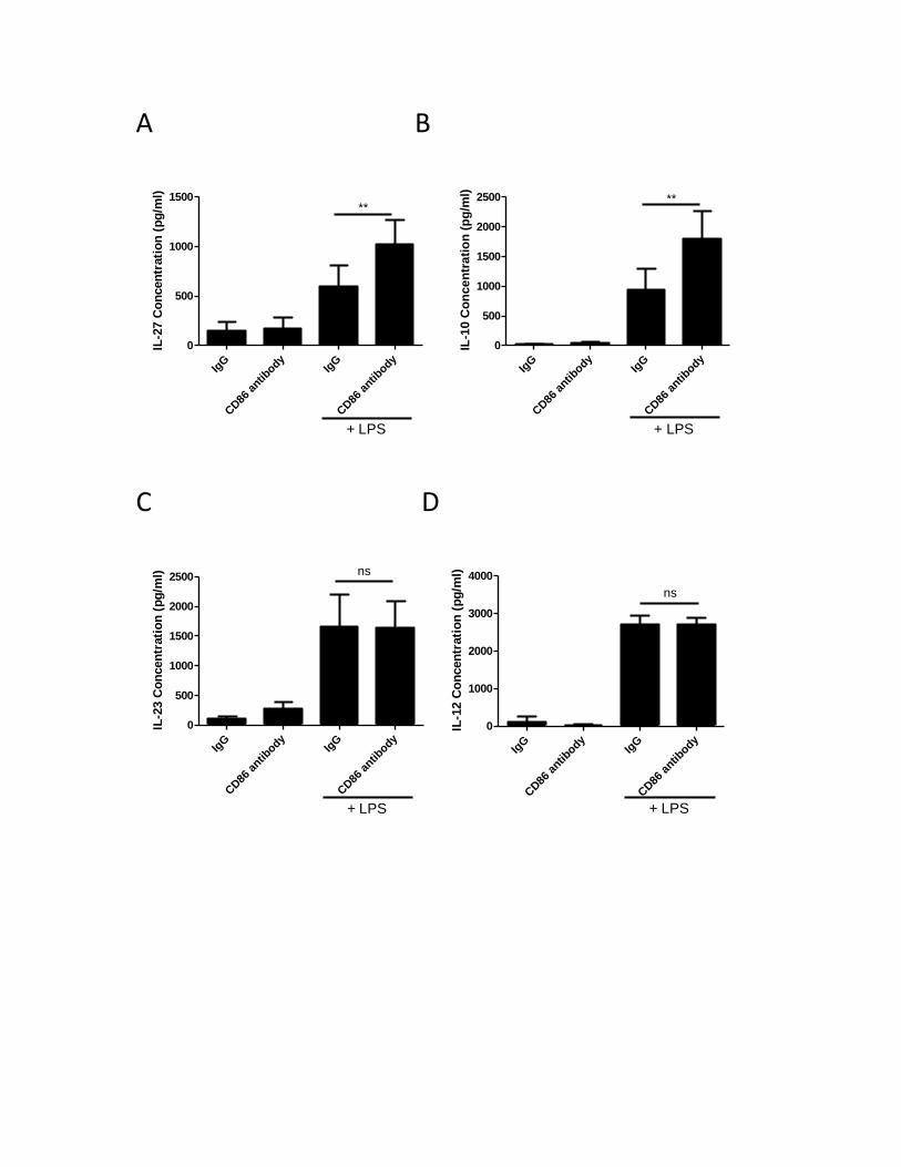
A B
IgG
CD86
antib
odyIg
G
CD86
antib
ody
0
500
1000
1500**
+ LPS
IL-2
7 C
on
cen
trati
on
(p
g/m
l)
IgG
CD86
antib
odyIg
G
CD86
antib
ody
0
500
1000
1500
2000
2500 **
+ LPS
IL-1
0 C
on
ce
ntr
ati
on
(p
g/m
l)
C D
IgG
CD86
antib
odyIg
G
CD86
antib
ody
0
500
1000
1500
2000
2500ns
+ LPS
IL-2
3 C
on
cen
trati
on
(p
g/m
l)
IgG
CD86
antib
odyIg
G
CD86
antib
ody
0
1000
2000
3000
4000
ns
+ LPS
IL-1
2 C
on
cen
trati
on
(p
g/m
l)

45
3.2.1 Inhibitor of apoptosis proteins regulate LPS-induced IL-10 and IL-27 expression, but are not involved in the CD80/CD86-induced IL-10 and IL-27 production
IAPs have emerged as important regulators of innate immune signaling downstream of TLR-
4 (128). In particular, they have been identified as having a crucial role in the activation of
downstream NF-κB, which are induced by the binding of LPS to TLR-4. Furthermore, depletion of
cIAPs was shown to specifically block LPS-induced p38 and JNK MAPK activation and the induction
of cytokine gene transcription in murine bone-marrow derived macrophages (105). Tseng et al
demonstrated that cIAPs were present in the MyD88-assembled complex and were essential for the
induction of inflammatory cytokine genes, such as TNF, IL-6 and IL-12β, in response to LPS (105). To
investigate the role of IAPs in the LPS-induced production of IL-10 and IL-27, MDMs were treated
with 50 nM/mL SMAC mimetics for 24 hours, followed by LPS stimulation for another 24 hours.
Biological activity of SMAC mimetics is demonstrated in Figure 5C. ELISA was used to measure the
protein expression levels. Treatment with SMAC mimetics alone had no effect on IL-10 and IL-27
production, but LPS-induced IL-10 and IL-27 expression was abrogated after SMAC mimetic
treatment (Figure 5A and 5B, respectively). Therefore, IAPs regulate the LPS/TLR-4-induced IL-10
and IL-27 production in human MDMs.
Since IL-10 and IL-27 expression was shown to at least regulated at least in part through the
CD80/CD86 synergistic relationship with TLR-4, I wanted to investigate whether synergism of
CD80/CD86 and TLR-4 is subject to regulation by IAPs. I pretreated the cells with 50 nM/mL SMAC
mimetics as aboveand then treated the MDMs with anti-CD80 or anti-CD86 antibody in concert with
a low dose of LPS (10 ng/mL). Consistent with the previous results, the LPS-induced IL-10 and IL-27
expression was enhanced upon addition of anti-CD80/CD86 antibody, but these preliminary results
suggested that this synergistic relationship may not be subject to regulation by IAPs (Figure 6).

46
Figure 5: IAPs regulate LPS-induced IL-10 and IL-27 expression.
MDMs (1x10⁶ cells/mL) were pretreated with SMAC mimetics (50 nM/mL) for 24 hours, following with stimulation in the presence or absence of LPS for 24 hours. A) IL-10 and B) IL-27 production was measured by ELISA. SMAC mimetics specifically block cIAP2 expression (C). Values in bar graph are means of at least three independent experiments ±SEM; *p≤0.05; **p≤0.005; ***p≤0.0005 with at least n=3.
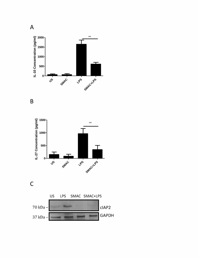
A
US
SM
AC
LPS
SM
AC+L
PS
0
500
1000
1500
2000 **IL
-10 C
on
cen
trati
on
(p
g/m
l)
B
US
SM
AC
LPS
SM
AC+L
PS
0
500
1000
1500**
IL-2
7 C
on
cen
trati
on
(p
g/m
l)
C
47
Figure 6: Blocking of IAPs by SMAC mimetics inhibits TLR-4 signaling to produce IL-10 and IL-27,
but do not appear to affect CD80 and CD86 signaling synergistic effect.
MDMs (1x10⁶ cells/mL) were pretreated with SMAC mimetics (50 nM/mL) for 24 hours, following
with cells were stimulated with 500 ng/mL of either anti-CD80 or anti-CD86 or isotype matched IgG1
control antibodies, in the presence or absence of 10 ng/mL LPS for 24 hours. IL-10 (A and B) and IL-
27 (C and D) protein secretion was measured by ELISA. Values in bar graph are means of at least
three independent experiments ±SEM; nonsignificant (ns); *p≤0.05; **p≤0.005; ***p≤0.0005 with
at least n=3.
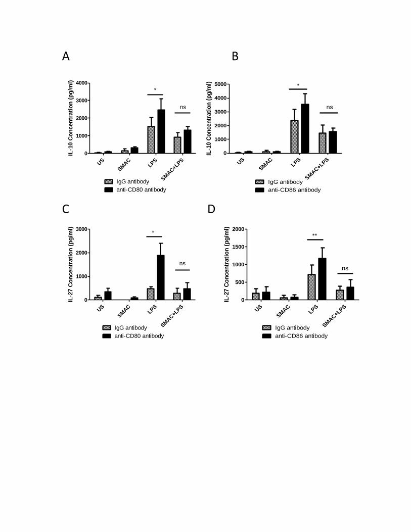
A B
US
SM
AC
LPS
SM
AC+L
PS
0
1000
2000
3000
4000
IgG antibody
anti-CD80 antibody
*
ns
IL-1
0 C
on
cen
trati
on
(p
g/m
l)
US
SMAC
LPS
SMAC+LPS
0
1000
2000
3000
4000
5000
IgG antibody
anti-CD86 antibody
*
ns
IL-1
0 C
on
cen
trati
on
(p
g/m
l)
C D
US
SM
AC
LPS
SM
AC+L
PS
0
1000
2000
3000
IgG antibody
anti-CD80 antibody
*
ns
IL-2
7 C
on
cen
trati
on
(p
g/m
l)
US
SMAC
LPS
SMAC+LPS
0
500
1000
1500
2000
IgG antibody
anti-CD86 antibody
**
ns
IL-2
7 C
on
cen
trati
on
(p
g/m
l)
48
3.3. Regulation of CD80 in LPS-induced monocyte-derived macrophages
3.3.1 Inhibitor of Apoptosis Proteins (IAPs) regulate LPS-induced CD80 and CD86 surface protein expression
In light of the influence of CD80 expression on the activation of T cells, and the importance of this
activation on immune responsiveness and disease progression, understanding the regulation of
CD80 and CD86 expression could facilitate the development of immune therapies. The regulation of
these two costimulatory molecules has been characterized in monocytes and dendritic cells. In
particular, the LPS/TLR-4-activated MAPKs and PI3K molecules have been identified as important
regulators of CD80 and CD86 expression in these cells (8, 22, 122). Recently, accumulating evidence
has suggested that IAPs may be involved in signal transduction pathways, in particular, TNF-α-
mediated NF-κB activation (87). Therefore, I investigated the role of IAPs in the regulation of CD80
and CD86 upregulation in response to LPS. Human MDMs were isolated from peripheral blood
mononuclear cells (PBMCs) and treated for 24 hours with 50 nM/mL of SMAC mimetics and LPS for
another 24 hours, and CD80 and CD86 surface protein expression was measured by Flow Cytometry.
Inhibition of cIAPs by SMAC mimetics prevented the LPS-induced CD80 protein expression (Figure
7A), suggesting that cIAPs are positive regulators of CD80 surface expression. I also investigated the
role of cIAP2 in the regulation of CD86 expression in human MDMs and obtained similar results.
CD86 protein expression was upregulated following LPS treatment, and this upregulation was
positively regulated by IAPs, as CD86 expression returned to basal levels upon treatment with SMAC
mimetics (Figure 8).To confirm the role of IAPs in the regulation of LPS-induced CD80 and CD86
surface protein expression, MDMs were transfected with cIAP2 specific siRNA for 24 hours, followed
by LPS stimulation for 24 hours. CD80 (Figure 7B) and CD86 (Figure 8B) surface expression was
measured by Flow Cytometry. As compared to the cells transfected with control siRNA, MDMs with
effective cIAP2 knockdown demonstrated a decreased CD80 and CD86 expression. Silencing of
cIAP2 was confirmed via Western Immunoblotting (Figure 7C).
49
Figure 7: IAPs positively regulate the LPS-induced CD80 surface expression in MDMs. A: SMAC mimetics inhibit LPS-induced CD80 surface expression. B and C: cIAP2 specific siRNA inhibits LPS-induced CD80 surface expression. MDMs (1x10⁶ cells/mL) were treated with 50 nM/mL of SMAC mimetics for 24 hours followed by LPS stimulation for 24 hours. CD80 surface expression was measured by Flow Cytometry (A). These results were confirmed with siRNA (B), where MDMs (1x10⁶ cells/mL) were transfected with cIAP2 or control siRNA (CSI) for 24 hours, followed by LPS stimulation for 24 hours. Confirmation of cIAP2 silencing was demonstrated by Western Immunoblotting (C).
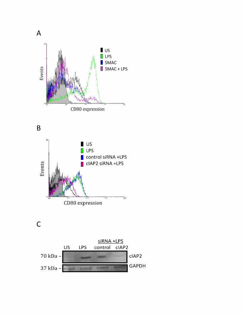
A
B
C

50
Figure 8: IAPs positively regulate the LPS-induced CD86 surface expression in MDMs.
A: SMAC mimetics inhibit LPS-induced CD86 surface expression B: cIAP2 specific siRNA inhibits LPS-induced CD86 surface expression. MDMs (1x10⁶ cells/mL) were treated with 50 nM/mL of SMAC mimetics for 24 hours followed by
LPS stimulation for 24 hours. CD86 surface expression was measured by Flow Cytometry (A). These
results were confirmed with siRNA (B), where MDMs (1x10⁶ cells/mL) were transfected with cIAP2
or control siRNA (CSI) for 24 hours, followed by LPS stimulation for 24 hours.

A
B
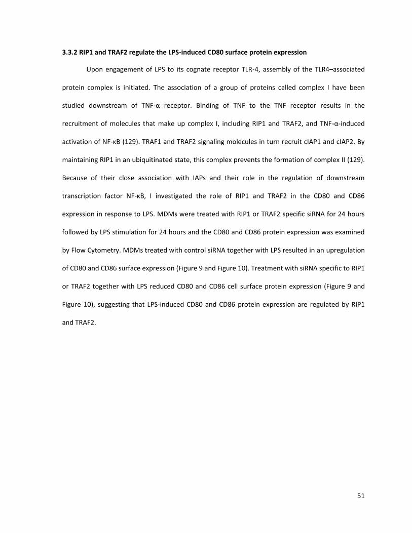
51
3.3.2 RIP1 and TRAF2 regulate the LPS-induced CD80 surface protein expression
Upon engagement of LPS to its cognate receptor TLR-4, assembly of the TLR4–associated
protein complex is initiated. The association of a group of proteins called complex I have been
studied downstream of TNF-α receptor. Binding of TNF to the TNF receptor results in the
recruitment of molecules that make up complex I, including RIP1 and TRAF2, and TNF-α-induced
activation of NF-κB (129). TRAF1 and TRAF2 signaling molecules in turn recruit cIAP1 and cIAP2. By
maintaining RIP1 in an ubiquitinated state, this complex prevents the formation of complex II (129).
Because of their close association with IAPs and their role in the regulation of downstream
transcription factor NF-κB, I investigated the role of RIP1 and TRAF2 in the CD80 and CD86
expression in response to LPS. MDMs were treated with RIP1 or TRAF2 specific siRNA for 24 hours
followed by LPS stimulation for 24 hours and the CD80 and CD86 protein expression was examined
by Flow Cytometry. MDMs treated with control siRNA together with LPS resulted in an upregulation
of CD80 and CD86 surface expression (Figure 9 and Figure 10). Treatment with siRNA specific to RIP1
or TRAF2 together with LPS reduced CD80 and CD86 cell surface protein expression (Figure 9 and
Figure 10), suggesting that LPS-induced CD80 and CD86 protein expression are regulated by RIP1
and TRAF2.

52
Figure 9: RIP1 and TRAF2 mediate LPS-induced CD80 expression in MDMs.
MDMs (1x10⁶ cells/mL) were transfected with RIP1 (A) or TRAF2 (C) siRNA, or control siRNA (CSI) for
24 hours, followed by LPS stimulation for 24 hours. CD80 surface expression was measured by Flow
Cytometry (left panel). Confirmation of RIP-1 (B) and TRAF2 (D) silencing was demonstrated by
Western Immunoblotting (right panel).
A B
C D
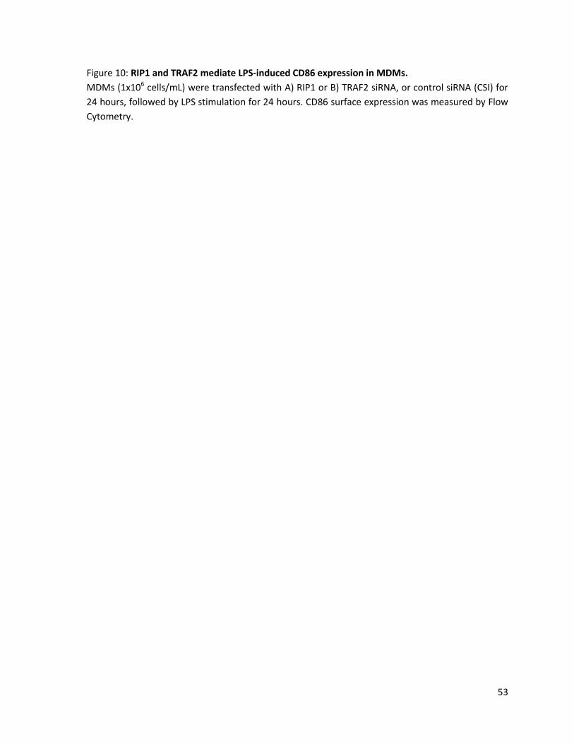
53
Figure 10: RIP1 and TRAF2 mediate LPS-induced CD86 expression in MDMs.
MDMs (1x10⁶ cells/mL) were transfected with A) RIP1 or B) TRAF2 siRNA, or control siRNA (CSI) for
24 hours, followed by LPS stimulation for 24 hours. CD86 surface expression was measured by Flow
Cytometry.
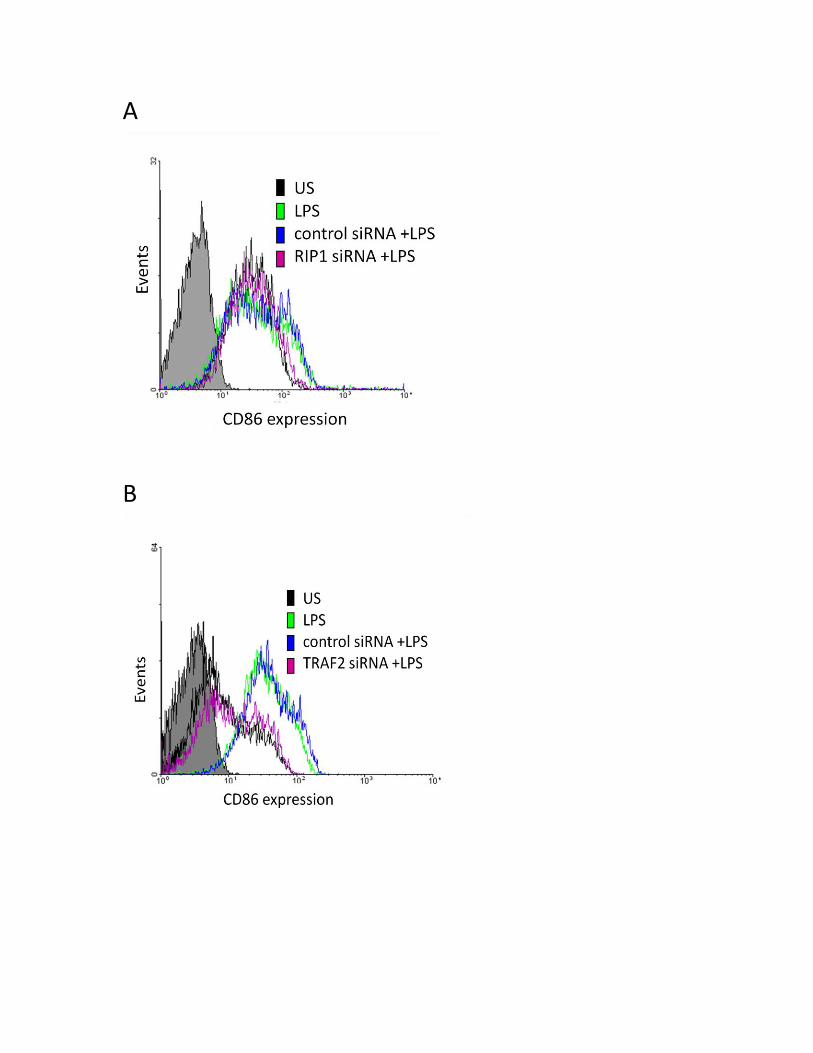
A
B
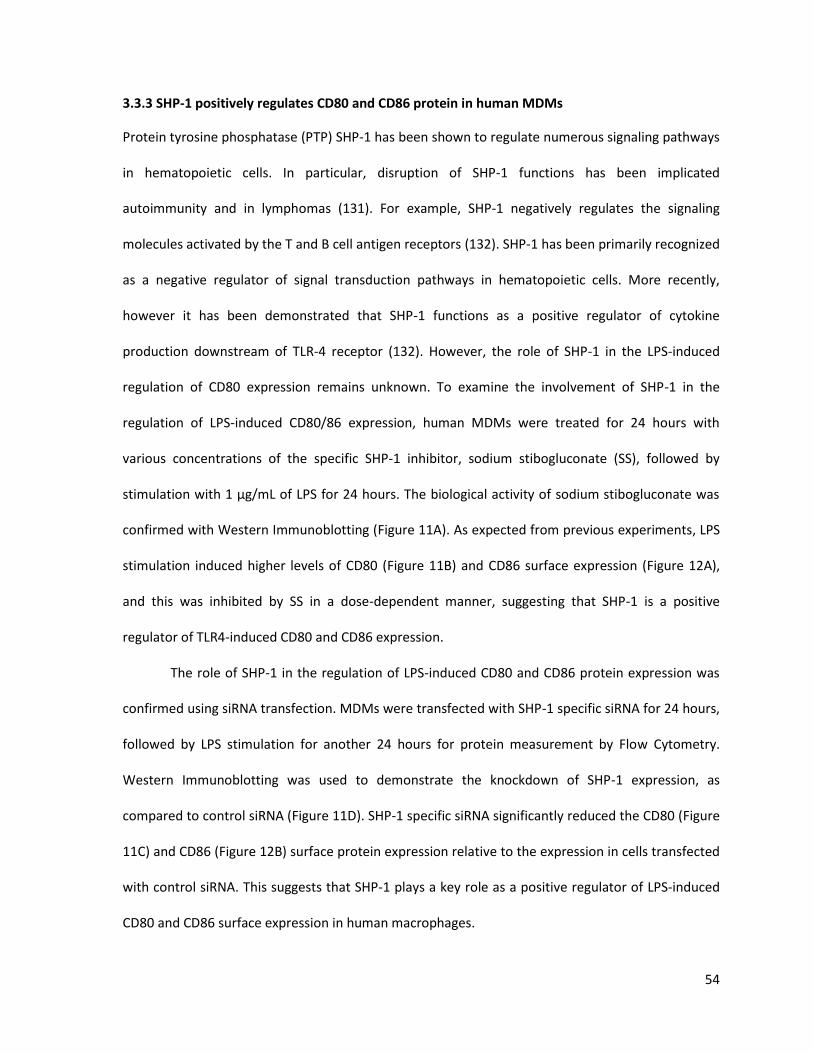
54
3.3.3 SHP-1 positively regulates CD80 and CD86 protein in human MDMs
Protein tyrosine phosphatase (PTP) SHP-1 has been shown to regulate numerous signaling pathways
in hematopoietic cells. In particular, disruption of SHP-1 functions has been implicated
autoimmunity and in lymphomas (131). For example, SHP-1 negatively regulates the signaling
molecules activated by the T and B cell antigen receptors (132). SHP-1 has been primarily recognized
as a negative regulator of signal transduction pathways in hematopoietic cells. More recently,
however it has been demonstrated that SHP-1 functions as a positive regulator of cytokine
production downstream of TLR-4 receptor (132). However, the role of SHP-1 in the LPS-induced
regulation of CD80 expression remains unknown. To examine the involvement of SHP-1 in the
regulation of LPS-induced CD80/86 expression, human MDMs were treated for 24 hours with
various concentrations of the specific SHP-1 inhibitor, sodium stibogluconate (SS), followed by
stimulation with 1 μg/mL of LPS for 24 hours. The biological activity of sodium stibogluconate was
confirmed with Western Immunoblotting (Figure 11A). As expected from previous experiments, LPS
stimulation induced higher levels of CD80 (Figure 11B) and CD86 surface expression (Figure 12A),
and this was inhibited by SS in a dose-dependent manner, suggesting that SHP-1 is a positive
regulator of TLR4-induced CD80 and CD86 expression.
The role of SHP-1 in the regulation of LPS-induced CD80 and CD86 protein expression was
confirmed using siRNA transfection. MDMs were transfected with SHP-1 specific siRNA for 24 hours,
followed by LPS stimulation for another 24 hours for protein measurement by Flow Cytometry.
Western Immunoblotting was used to demonstrate the knockdown of SHP-1 expression, as
compared to control siRNA (Figure 11D). SHP-1 specific siRNA significantly reduced the CD80 (Figure
11C) and CD86 (Figure 12B) surface protein expression relative to the expression in cells transfected
with control siRNA. This suggests that SHP-1 plays a key role as a positive regulator of LPS-induced
CD80 and CD86 surface expression in human macrophages.
55
Figure 11: SHP-1 positively regulates the LPS-induced CD80 surface expression in MDMs.
A: Sodium stibogluconate inhibits SHP-1 expression. B: Inhibition of SHP-1 reduced CD80 surface expression. C and D: SHP-1 specific siRNA inhibits LPS-induced CD80 surface expression. MDMs (1x10⁶ cells/mL) were treated with indicated concentrations of sodium stibogluconate (SS)
for 24 hours, followed by LPS stimulation for 24 hours. A) SS inhibitor decreased SHP-1 expression as
measured by Western Immunoblotting. The effect of SS on CD80 surface expression was
determined by Flow Cytometry. These results were confirmed with siRNA, where MDMs (1x10⁶
cells/mL) were transfected with siRNA specific to SHP-1 or control siRNA (CSI) for 24 hours, followed
by LPS stimulation for 24 hours. CD80 surface expression was measured by Flow Cytometry (D).
Confirmation of SHP-1 silencing was demonstrated by Western Immunoblotting (E).
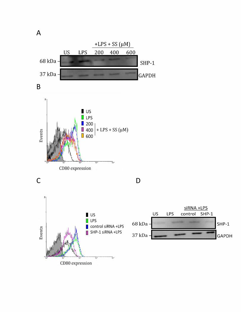
A
B
C D

56
Figure 12: SHP-1 positively regulates the LPS-induced CD86 surface expression in MDMs.
A: Inhibition of SHP-1 reduced CD86 surface expression. B: SHP-1 specific siRNA inhibits LPS-induced CD86 surface expression. MDMs (1x10⁶ cells/mL) were treated with indicated concentrations of sodium stibogluconate (SS)
for 24 hours, followed by LPS stimulation for 24 hours. CD86 surface expression was measured by
Flow Cytometry (A). These results were confirmed with siRNA (B), where MDMs (1x10⁶ cells/mL)
were transfected with SHP-1 or control siRNA (CSI) for 24 hours, followed by LPS stimulation for 24
hours. CD86 cell surface expression after transfection was measured by Flow Cytometry (B).
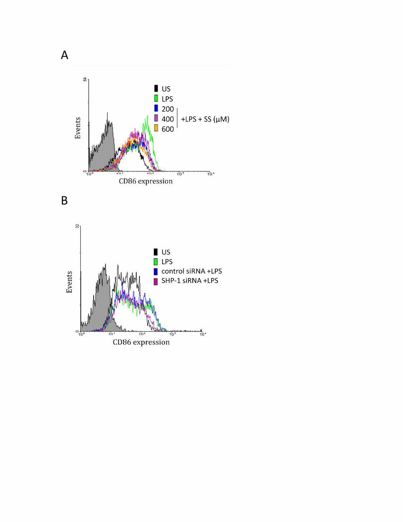
A
B

57
3.3.4 SHP-1, cIAP2, RIP1 and TRAF2 co-localize in human MDMs
Since it is known that RIP1 and TRAF2 interact to form a complex with cIAP2 (106)
(confirmed in Figure 14A and Figure 14B), I wanted to investigate whether SHP-1 also co-localized
with cIAP2. For this, MDMs on slides were fixed and permeabilized before being incubated
overnight with two primary antibodies, SHP-1 (anti-mouse), RIP1 (anti-rabbit), TRAF2 (anti-rabbit) or
cIAP2 (anti-rabbit/anti-mouse). These proteins were visualized by confocal microscopy. Figure 13A
demonstrates that these two proteins co-immunofluoresce, suggesting their co-localization within
the macrophage. Furthermore, I demonstrated that SHP-1 co-localized with RIP1 (Figure 13B) and
TRAF2 (Figure 13C). Taken together, these results suggest that SHP-1, cIAP2, RIP1 and TRAF2 co-
localize in human macrophages.
3.3.5 PI3K pathway regulates the LPS-induced CD80 and CD86 protein expression
Engagement of LPS to its receptor TLR-4 induces a host of cellular responses, including the
activation of the PI3K pathway (133). The PI3K pathway is crucial for cellular growth and cell survival
and more recently, its role in the regulation of LPS-induced CD80 and CD86 expression in B cells has
been investigated. Attridge et al have demonstrated that a potent and sustained CD86 upregulation
in murine B cells triggered by the addition of IL-21 is mediated through a PI3K-dependent
mechanism (134). Hence, I investigated whether PI3K modulated CD80 and CD86 expression in
human macrophages. Since the activation of the PI3K pathway culminates in the phosphorylation of
AKT, LY294002 a highly specific inhibitor that abolishes PI3K-dependent AKT phosphorylation, but
does not inhibit other lipid and protein kinases, such as MAPKs, was used (135). MDMs were treated
for 2 hours with LY294002 and then stimulated for 24 hours with LPS to measure CD80 and CD86
surface protein expression. Figure 15B demonstrates the dose-dependent decrease in LPS-induced
CD80 protein expression. CD80 mRNA expression was measured after treatment with LY294002
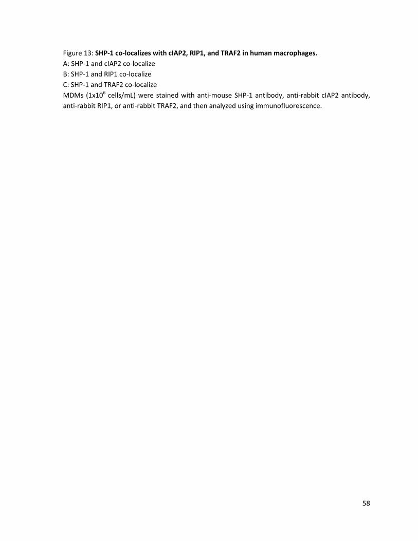
58
Figure 13: SHP-1 co-localizes with cIAP2, RIP1, and TRAF2 in human macrophages.
A: SHP-1 and cIAP2 co-localize
B: SHP-1 and RIP1 co-localize
C: SHP-1 and TRAF2 co-localize
MDMs (1x10⁶ cells/mL) were stained with anti-mouse SHP-1 antibody, anti-rabbit cIAP2 antibody,
anti-rabbit RIP1, or anti-rabbit TRAF2, and then analyzed using immunofluorescence.

A SHP-1 cIAP2 DAPI SHP-1 + cIAP2
B SHP-1 RIP1 DAPI SHP-1 + RIP1
C SHP-1 TRAF2 DAPI SHP-1 + TRAF2
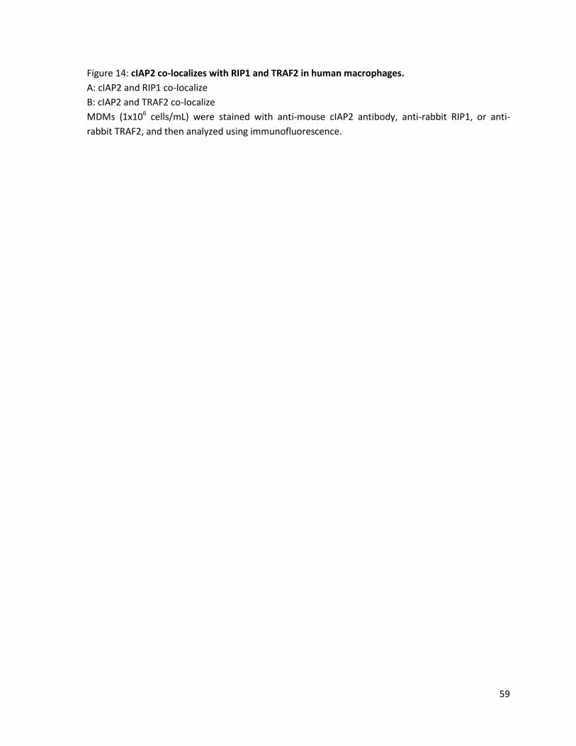
59
Figure 14: cIAP2 co-localizes with RIP1 and TRAF2 in human macrophages.
A: cIAP2 and RIP1 co-localize
B: cIAP2 and TRAF2 co-localize
MDMs (1x10⁶ cells/mL) were stained with anti-mouse cIAP2 antibody, anti-rabbit RIP1, or anti-
rabbit TRAF2, and then analyzed using immunofluorescence.
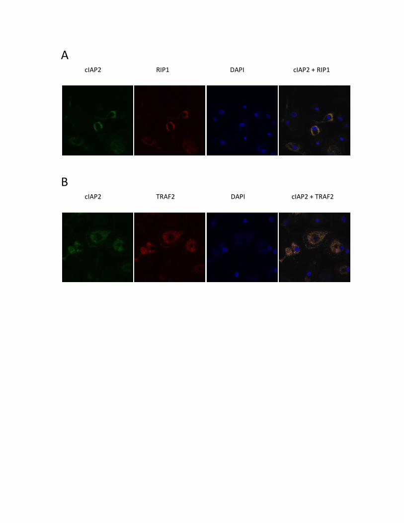
A cIAP2 RIP1 DAPI cIAP2 + RIP1
B cIAP2 TRAF2 DAPI cIAP2 + TRAF2
60
Figure 15: PI3K positively regulates the LPS-induced CD80 surface expression in MDMs.
A: LY294002 inhibits phospho-AKT expression. B and C: Inhibition of pAKT reduced CD80 surface and mRNA expression. D and E: AKT specific siRNA inhibits LPS-induced CD80 surface expression. MDMs (1x10⁶ cells/mL) were treated with indicated concentrations of LY294002 for 2 hours,
followed by LPS stimulation for A) 15 mins B) 24 hours and C) 4 hours. A) Biological activity assay of
LY inhibitor measured by Western Immunoblotting. CD80 surface and mRNA expression was
measured by Flow Cytometry (B) and qRT-PCR (C), respectively. These results were confirmed with
siRNA, where MDMs (1x10⁶ cells/mL) were transfected with AKT or control siRNA (CSI) for 24 hours,
followed by LPS stimulation for 24 hours. CD80 cell surface expression after transfection was
measured by Flow Cytometry (D). Confirmation of AKT silencing was demonstrated by Western
Immunoblotting (E). Values in bar graph are means of at least three independent experiments
±SEM; *p≤0.05; **p≤0.005; ***p≤0.0005 as measured by One-Way Anova with at least n=3.

A
B C
US
LPS 10 25 50
0
5
10
15
20
25 **
+ LPS + LY294002 (uM)
CD
80 m
RN
A f
old
ch
an
ge
D E
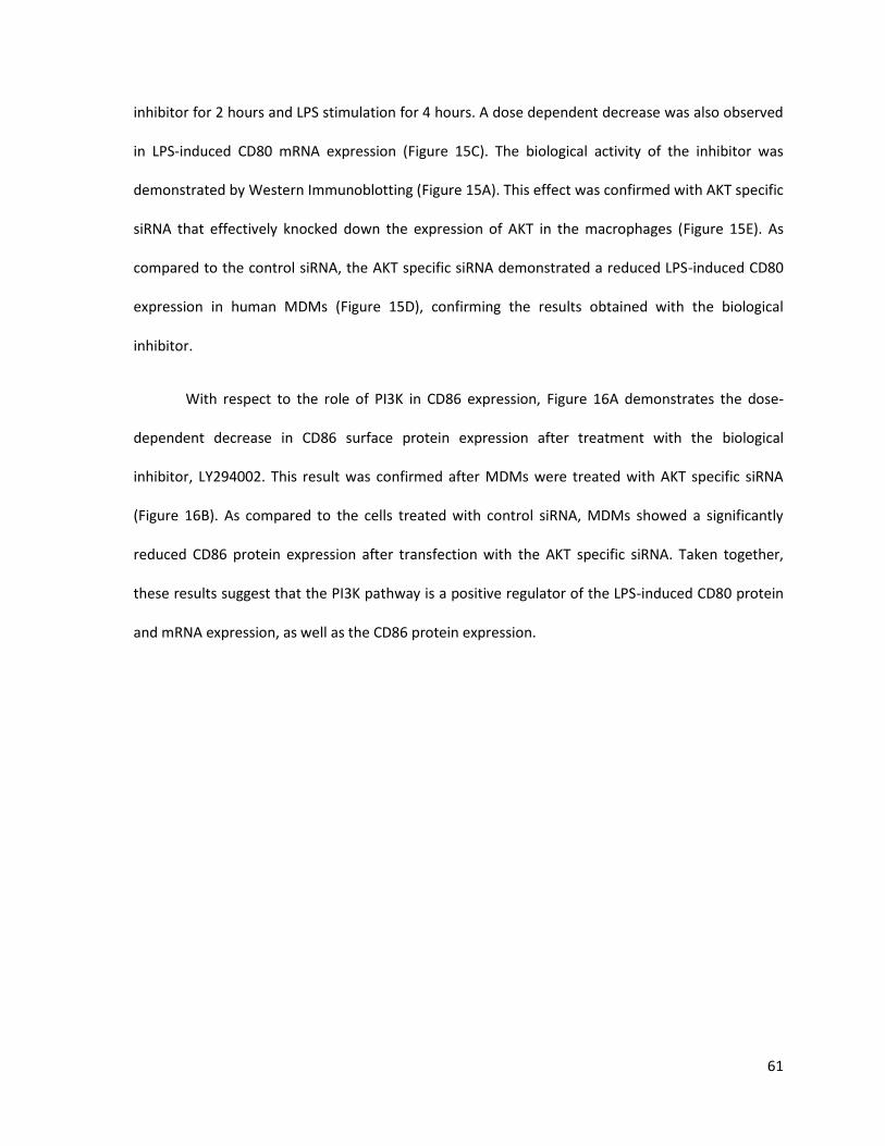
61
inhibitor for 2 hours and LPS stimulation for 4 hours. A dose dependent decrease was also observed
in LPS-induced CD80 mRNA expression (Figure 15C). The biological activity of the inhibitor was
demonstrated by Western Immunoblotting (Figure 15A). This effect was confirmed with AKT specific
siRNA that effectively knocked down the expression of AKT in the macrophages (Figure 15E). As
compared to the control siRNA, the AKT specific siRNA demonstrated a reduced LPS-induced CD80
expression in human MDMs (Figure 15D), confirming the results obtained with the biological
inhibitor.
With respect to the role of PI3K in CD86 expression, Figure 16A demonstrates the dose-
dependent decrease in CD86 surface protein expression after treatment with the biological
inhibitor, LY294002. This result was confirmed after MDMs were treated with AKT specific siRNA
(Figure 16B). As compared to the cells treated with control siRNA, MDMs showed a significantly
reduced CD86 protein expression after transfection with the AKT specific siRNA. Taken together,
these results suggest that the PI3K pathway is a positive regulator of the LPS-induced CD80 protein
and mRNA expression, as well as the CD86 protein expression.

62
Figure 16: PI3K positively regulates the LPS-induced CD86 surface expression in MDMs.
A: Inhibition of pAKT reduced CD86 surface expression. B: AKT specific siRNA inhibits LPS-induced CD86 surface expression. MDMs (1x10⁶ cells/mL) were treated with indicated concentrations of LY294002 for 2 hours,
followed by LPS stimulation for 24 hours. CD86 surface expression was measured by Flow Cytometry
(A). These results were confirmed with siRNA, where MDMs (1x10⁶ cells/mL) were transfected with
AKT specific or control siRNA (CSI) for 24 hours, followed by LPS stimulation for 24 hours. CD86 cell
surface expression after transfection was measured by Flow Cytometry (B).
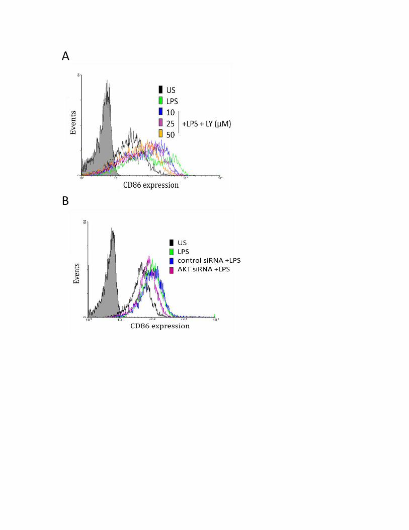
A
B

63
3.3.6 P38, but not JNK or ERK MAPK regulate the LPS-induced CD80 and CD86 expression
In recent years, significant progress has been made in elucidating members of the LPS
signaling pathways that are involved in the regulation of CD80 and CD86 expression (8, 22, 122,
123). In particular, Mitogen-activated protein kinases (MAPKs) have been shown to be key players in
regulating cellular responses such as proliferation and differentiation in human monocytes (8, 22).
Three main families of MAPKs have been shown to be activated by LPS: extracellular signal-
regulated protein kinases (ERK1 and ERK2), c-Jun N-terminal kinases (JNKs) and the p38
MAPK/stress-activated protein kinases (8). In human monocytes, the LPS-induced CD86 expression
has been shown to be regulated by p38 and JNK MAPK (8). Furthermore, regulation of CD80
expression in monocytic cells was shown to be mediated through the activation of JNK MAPK (121).
The role of MAPKs in the regulation of CD80 and CD86 expression has also been explored in DCs.
Nakahara et al and Arrighi et al have demonstrated that JNK MAPK (122) and p38 MAPK (123) are
involved in the LPS-induced CD80 and CD86 expression in human DCs (122). The modulation of
CD80 and CD86 expression by MAPKs has also been highlighted in other cell types. Therefore, it was
of interest to investigate the role of MAPKs in the expression of CD80 and CD86 in human
macrophages.
To investigate the role of p38 MAPK in the LPS-induced CD80 and CD86 expression, MDMs
were treated with SB203580, a specific inhibitor for p38 MAPK, for 2 hours and then stimulated with
LPS for 24 hours. Treatment with LPS resulted in a significant upregulation in CD80 protein
expression, and this was reduced with increasing doses of the p38 inhibitor, suggesting that p38
MAPK is a positive regulator of the LPS-induced CD80 surface protein expression (Figure 17B). These
data were confirmed using p38 specific siRNA (Figure 17D). As compared to the control siRNA,
silencing p38 resulted in a decreased CD80 surface protein expression. The effect of p38 inhibition
on CD80 mRNA expression was examined by RT-PCR. Figure 17C demonstrates the dose dependent

64
Figure 17: p38 MAPK positively regulates the LPS-induced CD80 surface expression in MDMs.
A: SB203580 inhibits phospho-p38 expression. B and C: Inhibition of pp38 reduced CD80 surface and mRNA expression. D and E: p38 specific siRNA inhibits LPS-induced CD80 surface expression. MDMs (1x10⁶ cells/mL) were treated with indicated concentrations of SB203580 for 2 hours,
followed by LPS stimulation for A) 15 mins B) 24 hours and C) 4 hours. A) Biological activity assay of
SB inhibitor measured by Western Immunoblotting. CD80 surface and mRNA expression was
measured by Flow Cytometry (B) and qRT-PCR (C), respectively. These results were confirmed with
siRNA, where MDMs (1x10⁶ cells/mL) were transfected with p38 or control siRNA (CSI) for 24 hours,
followed by LPS stimulation for 24 hours. CD80 cell surface expression after transfection was
measured by Flow Cytometry (D). Confirmation of p38 silencing was demonstrated with Western
Immunoblotting (E). Values in bar graph are means of at least three independent experiments
±SEM; *p≤0.05; **p≤0.005; ***p≤0.0005 as measured by One-Way Anova with at least n=3.

A
B C
US
LPS 10 25 50
0
5
10
15
20
25**
+ LPS + SB203580 (uM)
CD
80 m
RN
A f
old
ch
an
ge
D E

65
decrease of CD80 mRNA transcripts after treatment with the p38 inhibitor for 2 hours and LPS
stimulation for 4 hours. Collectively, this data demonstrates that CD80 surface protein and mRNA
are positively regulated by p38 MAPKinase. Similarly, I investigated the role of p38 MAPK in the
regulation of CD86 protein expression. P38 MAPK expression was required for LPS-induced CD86
expression, as the inhibition of this molecule resulted in a dose dependent decrease in CD86 protein
expression as measured by Flow Cytometry (Figure 18A). After treatment with p38 specific siRNA,
there was a marked decrease in CD86 surface protein expression (Figure 18B). Taken together, this
data demonstrates that p38 MAPK is a crucial regulator of the CD86 protein.
In parallel, I also investigated the role of JNK and ERK MAPK in the regulation of LPS-induced
CD80 and CD86 expression. Using various doses of SP600125, the specific inhibitor for JNK MAPK, I
showed that JNK MAPK did not play a role in the LPS-induced CD80 or CD86 surface protein
expression (Figure 19). To investigate the role of ERK MAPK in the regulation of LPS-induced CD80
and CD86 expression, I employed the specific inhibitor PD98059 in increasing doses and measured
its effect on CD80/CD86 surface protein and mRNA expression. Using three concentrations of
PD98059, I demonstrated that ERK MAPK is not involved in the LPS-induced CD80 surface protein
(Figure 20B) and mRNA expression (Figure 20C). Similarly, ERK MAPK was found to not be involved
in the LPS-induced CD86 surface protein (Figure 20D).
3.3.7 SHP-1 and IAPs regulate activation of downstream PI3K/p38 signaling axis
Since I identified that IAPs, SHP-1 signaling molecule, and PI3K and p38 MAPK pathways
positively regulate CD80 and CD86 expression, I wanted to elucidate the mechanism by which this
occurs. For this, I inhibited SHP-1 using sodium stibogluconate (SS) and measured the
phosphorylation of AKT and p38 molecules. I demonstrated that SHP-1 positively regulated the
activation of both PI3K signaling (Figure 21A) and p38 MAPK (Figure 21B). In addition, using SMAC

66
Figure 18: p38 MAPK positively regulates the LPS-induced CD86 surface expression in MDMs.
A: Inhibition of pp38 reduced CD86 surface expression. B: p38 specific siRNA inhibits LPS-induced CD86 surface expression. MDMs (1x10⁶ cells/mL) were treated with indicated concentrations of SB203580 for 2 hours,
followed by LPS stimulation for 24 hours. CD86 surface expression was measured by Flow Cytometry
(A). These results were confirmed with siRNA, where MDMs (1x10⁶ cells/mL) were transfected with
p38 or control siRNA (CSI) for 24 hours, followed by LPS stimulation for 24 hours. CD86 cell surface
expression was measured after transfection by Flow Cytometry (C). Values in bar graph are means
of at least three independent experiments ±SEM; *p≤0.05; **p≤0.005; ***p≤0.0005 as measured by
One-Way Anova with at least n=3.
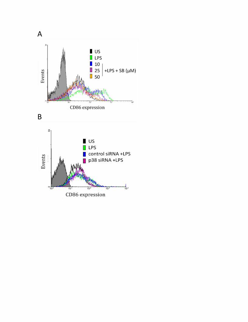
A
B
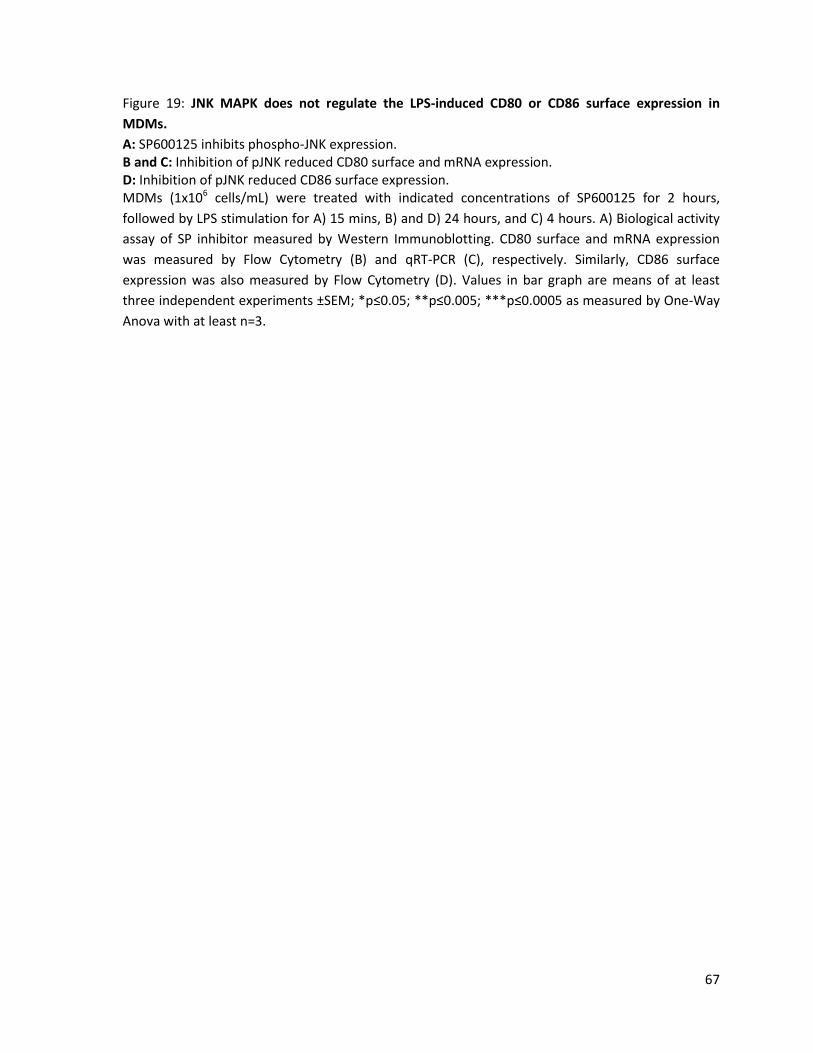
67
Figure 19: JNK MAPK does not regulate the LPS-induced CD80 or CD86 surface expression in
MDMs.
A: SP600125 inhibits phospho-JNK expression. B and C: Inhibition of pJNK reduced CD80 surface and mRNA expression. D: Inhibition of pJNK reduced CD86 surface expression. MDMs (1x10⁶ cells/mL) were treated with indicated concentrations of SP600125 for 2 hours,
followed by LPS stimulation for A) 15 mins, B) and D) 24 hours, and C) 4 hours. A) Biological activity
assay of SP inhibitor measured by Western Immunoblotting. CD80 surface and mRNA expression
was measured by Flow Cytometry (B) and qRT-PCR (C), respectively. Similarly, CD86 surface
expression was also measured by Flow Cytometry (D). Values in bar graph are means of at least
three independent experiments ±SEM; *p≤0.05; **p≤0.005; ***p≤0.0005 as measured by One-Way
Anova with at least n=3.
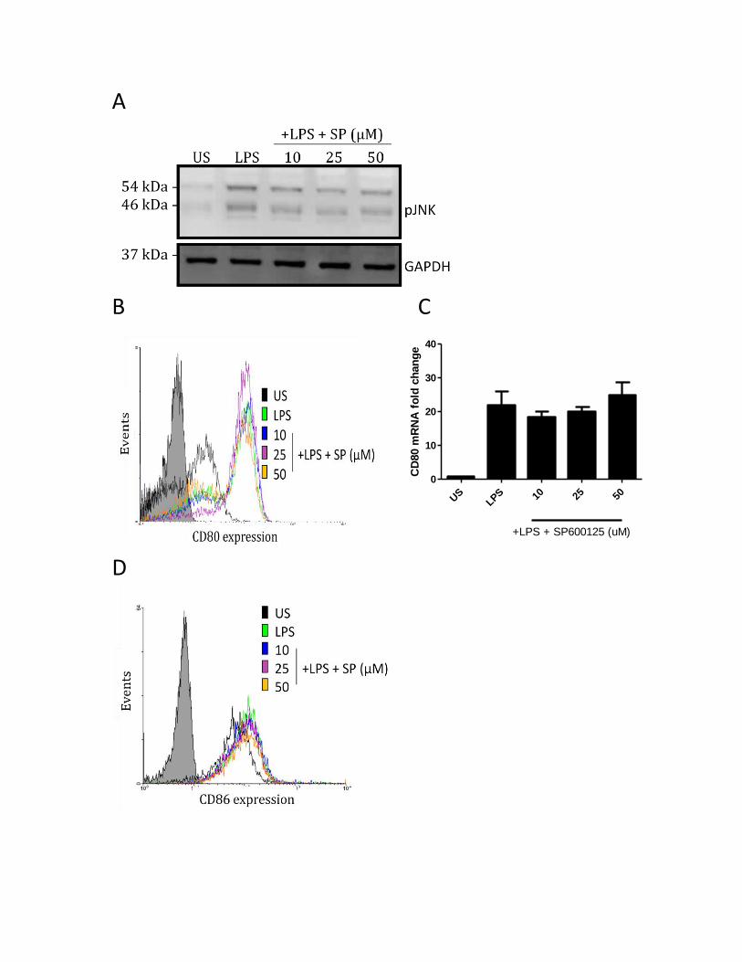
A
B C
US
LPS 10 25 50
0
10
20
30
40
+LPS + SP600125 (uM)
CD
80 m
RN
A f
old
ch
an
ge
D
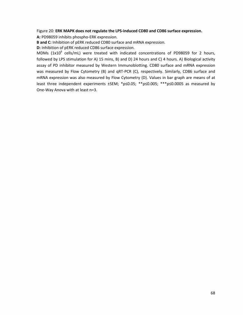
68
Figure 20: ERK MAPK does not regulate the LPS-induced CD80 and CD86 surface expression.
A: PD98059 inhibits phospho-ERK expression. B and C: Inhibition of pERK reduced CD80 surface and mRNA expression. D: Inhibition of pERK reduced CD86 surface expression. MDMs (1x10⁶ cells/mL) were treated with indicated concentrations of PD98059 for 2 hours,
followed by LPS stimulation for A) 15 mins, B) and D) 24 hours and C) 4 hours. A) Biological activity
assay of PD inhibitor measured by Western Immunoblotting. CD80 surface and mRNA expression
was measured by Flow Cytometry (B) and qRT-PCR (C), respectively. Similarly, CD86 surface and
mRNA expression was also measured by Flow Cytometry (D). Values in bar graph are means of at
least three independent experiments ±SEM; *p≤0.05; **p≤0.005; ***p≤0.0005 as measured by
One-Way Anova with at least n=3.
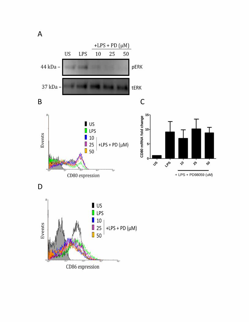
A
B C
US
LPS 10 25 50
0
5
10
15
+ LPS + PD98059 (uM)
CD
80 m
RN
A f
old
ch
an
ge
D
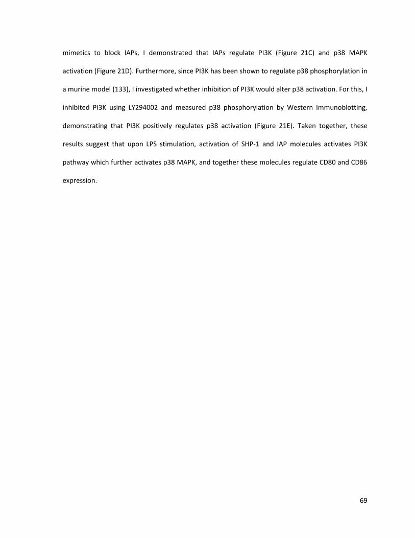
69
mimetics to block IAPs, I demonstrated that IAPs regulate PI3K (Figure 21C) and p38 MAPK
activation (Figure 21D). Furthermore, since PI3K has been shown to regulate p38 phosphorylation in
a murine model (133), I investigated whether inhibition of PI3K would alter p38 activation. For this, I
inhibited PI3K using LY294002 and measured p38 phosphorylation by Western Immunoblotting,
demonstrating that PI3K positively regulates p38 activation (Figure 21E). Taken together, these
results suggest that upon LPS stimulation, activation of SHP-1 and IAP molecules activates PI3K
pathway which further activates p38 MAPK, and together these molecules regulate CD80 and CD86
expression.

70
Figure 21: SHP-1 and IAP activity regulate the activation of AKT/p38 signalling axis in LPS-
stimulated MDMs.
MDMs (1x10⁶ cells/mL) were treated with indicated concentrations of sodium stibogluconate (SS),
SMAC mimetics, and LY294002 followed by LPS stimulation for 15 min. Phosphorylation of AKT (A)
and p38 MAPK (B) molecules after treatment with SS, and after treatment with SMAC mimetics (C
and D) were measured by Western Immunoblotting. P38 MAPK phosphorylation after LY treatment
(E) was also measured by Western Immunoblotting.

A B
C D
E
71
4.1 Discussion
CD80 and CD86 play a key role in the activation of effective immune responses, and as such
in the modulation of diseases (136). Expressed on the surface of APCs including macrophages, these
costimulatory molecules bind to their cognate receptors, CD28 and CTLA-4, resulting in the
activation, proliferation, and differentiation of T cells. In the absence of CD80 or CD86 engagement,
T cells are unable to produce IL-2 or proliferate in response to antigen, and instead enter a state of
anergy or undergo apoptosis (30). While it was once believed that the binding of B7 molecules to
CD28/CTLA-4 represented a one way signal, resulting in T cell activation, more recently, this
interaction has been investigated as a bidirectional signaling process. CD80 and CD86 were shown
to transduce signals to DCs, suggesting that these proteins act as signaling receptors (67, 68). Hence,
CD80 and CD86 induction on macrophages may impact the development of an immune response.
LPS, a TLR-4 ligand is a potent inducer of cytokines, including IL-10 and the IL-12 family of cytokines,
and a variety of surface molecules including CD80 and CD86 on macrophages. Therefore,
understanding the regulation of CD80 and CD86 expression, and decoding their potential as
signaling receptors within macrophages may provide new avenues for therapeutic treatment of
autoimmune disorders.
The focus of this project was twofold: 1) to investigate the role of CD80 and CD86 as
signaling receptors capable of transmitting extracellular signals, and 2) to determine the TLR-4
activated pathways that regulate CD80/CD86 expression in human macrophages. I show for the first
time that CD80/CD86 activation synergizes with TLR-4 signaling to produce IL-27 and IL-10 in human
MDMs. I have also demonstrated that cIAPs play a key role in TLR-4-mediated signaling, as inhibition
of IAPs with SMAC mimetics inhibited LPS-induced IL-10 and IL-27 production in MDMs, but these
preliminary results suggested that this synergistic relationship may not be subject to regulation by

72
IAPs. Furthermore, I have shown that LPS-induced CD80 and CD86 expression was regulated by
cIAP2-SHP-1-RIP1-TRAF2 complex through AKT-activated p38 MAPK in human macrophages.
To investigate the role of CD80 and CD86 as signaling receptors, I employed anti-CD80 and
anti-CD86 antibodies and measured the production of IL-10 and IL-12 family of cytokines. Anti-
CD80/anti-CD86 antibodies have been shown to neutralize CD80/CD86-induced IL-2 secretion in T
cells by binding to the CD80/CD86 binding pocket. These antibodies represent an experimental
model for the engagement of CD28/CTLA-4 by these B7 molecules. My results demonstrate that
activation of CD80/CD86 alone did not induce expression of any of the four cytokines investigated.
This is in contrast to DCs where stimulation with anti-CD80/CD86 antibodies induced IL-6 expression
through the activation of the PI3K pathway (67). This difference could be attributed to possibly
higher surface expression of these molecules exhibiting stronger activation. This could also suggest
significant differences between the responses and signaling pathways in macrophages versus DCs.
Further studies are warranted to look at other cytokines, including IL-6. Jain et al demonstrated that
CD86 stimulation enhanced the effect of TLR-2 stimulation on resting B cells (41). I, therefore,
hypothesized that CD80/CD86 synergizes with other signaling pathways activated by other ligands.
In particular, I investigated the role of LPS/TLR-4 pathway, because of its ability to activate several
signaling cascades. Since LPS is a potent mitogen, I employed suboptimal concentrations of LPS (10
ng/mL) to investigate the crosstalk between the CD80/CD86 and the TLR-4 signaling pathways.
Surprisingly, treatment of cells with anti-CD80/CD86 antibodies and LPS induced significantly higher
levels of IL-10 and IL-27 production, as compared to LPS-stimulated cells alone, without affecting
the production of IL-12 and IL-23. My results clearly suggest that although CD80/CD86 activation on
its own did not activate macrophages to induce IL-10 and IL-12 family of cytokines, CD80/CD86
activation synergizes with TLR-4 pathway to induce production of IL-10 and IL-27. TLR-dependent
synergistic activity has been recognized in murine macrophages and DCs previously. Makela et al
73
showed that stimulation of TLR-8 together with TLR-3 or TLR-4 in monocyte-derived DCs led to
synergistic IL-10, IL-12, IL-6 and TNF-α mRNA expression and cytokine production (137). Synergistic
TNF- α expression has also been seen in murine macrophages in response to TLR-9 and TLR-4
stimulation (138).
While IAPs were originally identified as having a primary role in the prevention of cell death,
IAPs have now been identified as key players in the TLR-4 pathway. cIAP1 and cIAP2 are involved in
signal transduction pathways, in particular TNF-α-mediated NF-κB activation (87). To analyze the
role of IAPs in innate immune signaling, I used SMAC mimetic chemical compounds. SMAC mimetics
were designed as a means of counteracting the activity of IAPs and inducing cell death, particularly
because of the high level of IAPs found in certain forms of malignancies, such as leukemia (139). I
have demonstrated that 50 nM/mL of SMAC mimetics is sufficient to exhibit degradation of IAPs in
human macrophages, without inducing cell death. In this study, I demonstrated for the first time
that IAPs mediate the IL-10 and IL-27 secretion in human MDMs. In murine macrophages, IAPs have
been shown to regulate the LPS induced production of proinflammatory cytokines such as IL-6 and
TNF-α (140). However, inhibition of IAPs did not alter IL-10 production in murine macrophage (105).
In contrast, my results show that IAPs positively regulate LPS-induced IL-10 expression in human
MDMs, suggesting that the role of IAPs may differ across species. The role of IAPs has also been
investigated in signaling through another class of PRRs, NOD-like receptor activated signaling
pathways (128). Downstream of NOD is RIP2, and similar to their role in TLR signaling, cIAPs are
required for ubiquitination of RIP2, which in turn is required for the activation of NF-κB. As result,
depletion of IAPs was shown to result in reduced cytokine and chemokine secretion in response to
stimulation of NOD receptors in murine cells (141).
Due to their role in TLR/NOD signaling, I investigated whether IAPs were involved in the
CD80/CD86-TLR-4 synergistic regulation of IL-10 and IL-27 production. Treatment with SMAC
74
mimetics alone did not affect the CD80/CD86 signaling pathway. Furthermore, these preliminary
results suggest that SMAC mimetics had no significant effect on the CD80/CD86-TLR-4 synergistic
pathway. Taken together, these results suggest that IAPs inhibit LPS-induced IL-10/IL-27 expression
without any impact on CD80/CD86-mediated activation of human MDMs. Further studies are
required to confirm this role for IAPs. In particular, my results could be substantiated by IAP-specific
siRNAs and by exploring the far-reaching effects of the CD80/CD86 pathway through the
measurements of other cytokines related to inflammation, such as IL-6 and TNF-α. While these
results demonstrate for the first time the existence of a CD80/CD86 signaling pathway in
macrophages, further studies are required to fully elucidate this pathway and the molecules
involved specifically with the use of Fc chimeras of CTLA-4 and CD28. This would present a more
specific model for the interaction between CD80/CD86 and CD28/CTLA-4.
Activation of the TLR-4 pathway has also been implicated in the upregulation of CD80 and
CD86 expression on the surface of monocytes and dendritic cells (8, 22, 123). Initiation of an
inflammatory response following association of LPS with TLR-4 results in the induction of cellular
responses to Gram-negative bacterial infections. In monocytic cells, LPS stimulation modulates the
expression of costimulatory molecules CD80 and CD86 (21, 121). Since these molecules play a
crucial role in the activation of T cells, the LPS-enhanced CD80 and CD86 expression may provide a
mechanism for the amplification of T cell activation, and the augmentation of the immune response.
The molecular mechanism by which the expression of CD80 and CD86 is controlled is poorly
understood in human macrophages. In particular, the role of signaling molecules, such as cIAPs,
SHP-1, RIP1 and TRAF2, and signaling pathways, such as MAPK and PI3K, have not been
investigated.
Since I have demonstrated the role of IAPs in the induction of IL-10 and IL-27 via the
LPS/TLR-4 signaling, I extended my studies by examining the involvement of IAPs in modulating the
75
LPS-induced CD80 and CD86 expression. My results showed that blocking IAP expression by either
SMAC mimetics or cIAP2-specific siRNA led to a reduced expression of CD80 and CD86 after LPS
stimulation. These results suggest that IAPs regulate LPS-induced CD80 and CD86 expression.
Since IAPs form a complex with RIP1 and TRAF2 and regulate downstream transcription
factor NF-κB (129), it was of interest to determine if these molecules also played a role in the
regulation of CD80 and CD86 expression. Using RIP1-specific and TRAF2 –specific siRNA, I
demonstrated that RIP1 and TRAF2 knockdowns demonstrated a significantly lower surface
expression of CD80 and CD86 in MDMs, as compared to MDMs transfected with control siRNAs.
Taken together, my results suggest that IAPs, RIP1, and TRAF2 regulate LPS-induced CD80 and CD86
surface expression.
It has been shown that binding of LPS to TLR-4 induces tyrosine phosphorylation of signaling
molecules and activates protein tyrosine kinases (PTKs) (113). The competing activities of protein
tyrosine phosphatases (PTPs) and PTKs regulate the phosphorylation of key signaling molecules
(113). My results demonstrate that PTP SHP-1, through the activation of the PI3K pathway and the
p38 MAPK pathway, is a required positive regulator of the LPS-induced expression of CD80 and
CD86 in MDMs. The involvement of SHP-1 was demonstrated by employing pharmacological
inhibitors, such as sodium stibogluconate and SHP-1 specific siRNAs. It should be noted however
that the relatively low transfection efficiency of siRNA shown in Western Immunoblots may explain
the incomplete abrogation of LPS-induced CD80 and CD86 expression.
Since it has been shown that cIAPs, RIP1, and TRAF2 form a complex in MDMs, I investigated
the inclusion of SHP-1 in this complex. Using confocal microscopy, I demonstrated that SHP-1 co-
localizes, as judged by co-immunofluorescence, with cIAP2, RIP1 and TRAF2. Moreover, my results
further show that cIAP2, RIP1 and TRAF2 co-immunofluoresce. These results suggest that cIAP2-
RIP1-TRAF2-SHP-1 together co-localize and may form a complex within the macrophage to regulate
76
the LPS-induced CD80 and CD86 expression. These results need to be further confirmed by co-
immunoprecipitation studies.
It is well established that LPS stimulation results in the activation of the MAPK and PI3K
pathways in most cell types including monocytes and macrophages (133). Recently, MAPKs were
identified as key modulators involved in the regulation of CD80 and CD86 expression in monocytes
and dendritic cells. For example, Kim et al demonstrated the mandatory role of p38 MAPK in the
upregulation of CD80 and CD86 expression in Vitamin C treated DCs (142). Similar results were
obtained after stimulation with LPS and TNF-α, where inhibition of p38, but not ERK MAPK was
shown to inhibit maturation of human blood-derived DCs, preventing upregulation of both CD80
and CD86 (123). In support of these results, our laboratory has also demonstrated the role of LPS-
induced MAPKs in the regulation of CD80 and CD86 expression in human monocytic cells (8, 22). Lim
et al have identified the roles for p38 MAPK in the regulation of CD86 expression in human
monocytes(121). Furthermore, JNK MAPK was shown to be involved in the LPS-induced CD80 and
CD86 expression in human promonocytic THP-1 cells (8, 121). PI3K has also been identified as a key
modulator in LPS-induced CD80/CD86 expression. In particular, PI3K was shown to upregulate CD86
expression in Epstein Barr Virus-transformed B cells after ligation with CD40/CD40 homodimer
(143). More recently, IL-21-induced CD86 expression has also been recognized as a PI3K-dependent
mechanism in B cells (144), further demonstrating the crucial role that this pathway plays. In light of
these observations, I investigated the role of the MAPK and PI3K pathways in the regulation of CD80
and CD86 expression in human macrophages in the context of upstream cIAP2-RIP1-TRAF2-SHP-1
complex. Inhibition of PI3K and p38 MAPK by LY294002 and SB203580, and by AKT-specific and p38-
specific siRNAs, respectively, significantly decreased surface expression of both CD80 and CD86 in
LPS-stimulated MDMs. Similar studies were employed to investigate the role of other MAPKs, JNK
and ERK, but inhibition of these proteins with specific inhibitors showed no effect on the CD80 or

77
CD86 surface or protein expression. These results were also confirmed using JNK1/2-specific and
ERK-specific siRNAs (data not shown), suggesting the specific role of p38 MAPK in the regulation of
CD80/CD86 surface expression. Taken together, these results suggest that the PI3K pathway and
p38 MAPK are positive regulators of the LPS-induced CD80 and CD86 surface protein expression.
PTP SHP-1 and IAPs are critical proteins for the activation of signaling molecules
downstream of the TLR-4. For example, SHP-1 has been identified as a key regulator in the
activation of ERK1/2 MAPK and NF-κB in bone marrow derived macrophages (116). Furthermore,
IAPs regulated LPS-induced p38 MAPK expression (128). I have demonstrated that in human MDMs,
IAPs, SHP-1, p38 MAPK and PI3K pathways are crucial for the LPS-induced upregulation of CD80 and
CD86 expression. To elucidate whether IAPs and SHP-1 regulate CD80/CD86 expression through p38
MAPK and PI3K activation, I inhibited SHP-1 and IAP expression using sodium stibogluconate and
SMAC mimetics respectively, and examined the level of phosphorylation of p38 MAPK and AKT by
Western Immunoblotting. I demonstrated that SHP-1 and IAPs are necessary for the LPS-induced
activation of the p38 MAPK and PI3K pathways.
The PI3K pathway has been shown to enhance LPS-induced MAPK expression in monocytes
and murine macrophages (133, 145). To determine if PI3K and p38 MAPk constitute two distinct
pathway or a single pathway to induce CD80/CD86 expression, I inhibited the PI3K pathway and
showed that p38 MAPK activation is inhibited. This suggests that the LPS-induced CD80 and CD86
activation in human MDMs occurs through a complex formed between cIAP2-RIP1-TRAF2-SHP-1,
which activates the PI3K pathway, and subsequently p38 MAPK to upregulate CD80 and CD86
surface expression.
While I recognize that the drawback of using inhibitors to demonstrate their role is their
non-specific nature and toxicity at higher concentrations, I have examined the viability of my cells
treated with various concentrations of these inhibitors by staining apoptotic cells with propidium

78
iodide (PI). I have also used low concentrations of each inhibitor and ensured that effects shown
were dose-dependent. In addition, viable cells observed using FACS analysis were gated accordingly
to eliminate apoptotic cells. I have also confirmed all of my findings with the use of specific siRNAs.
For the experiments involving SMAC mimetics, I used a very low dose (50 nM/mL) to ensure IAP
degradation, without the induction of cell death. As a result, I can conclude that none of the
inhibitors used in my studies caused apoptosis at any concentration and did not exert nonspecific
effects at the concentration used.
4.1.1 Regulation of CD80 and CD86 at the transcriptional level
Understanding the regulation of gene expression at the transcriptional level is crucial
requirement to completely elucidate the pathway by which CD80/CD86 expression is regulated. To
confirm the results obtained at the transcriptional level, I treated MDMs for 2 hours with their
respective inhibitors or 24 hours with SMAC mimetics, followed by LPS stimulation for 4 hours, the
required time for the measurement of transcripts (146-148). RNA was isolated using RNeasy Plus
Mini Kit and RT-PCR was used to measure transcript levels. The mRNA results obtained were not
consistent with the results obtained at the protein level. As suggested by my committee members, I
am incorporating these results in the appendix along with its discussion in this chapter.
4.1.2 CD80 Transcription
Treatment with LPS resulted in an increase in CD80 mRNA transcription, similar to CD80
protein levels (Figure 24A). However, IAPs and SHP-1 did not have a significant effect on LPS-
induced CD80 mRNA expression (Figure 25A and Figure 25C, respectively). On the other hand,
inhibition of PI3K and p38 MAPK in LPS-stimulated MDMs resulted in a reduced CD80 protein and
mRNA expression (Figure 15C and Figure 17C, respectively).

79
4.1.3 CD86 Transcription
Unlike the upregulation of CD86 protein expression after LPS treatment, CD86 mRNA
expression was surprisingly decreased after treatment with LPS (Figure 24B). IAPs and SHP-1
appeared to have no significant role in the CD86 mRNA expression (Figure 25B and Figure 25D,
respectively). Furthermore, using LY294002, SB203580 and PD98059, the inhibitors specific for PI3K,
p38 MAPK and ERK MAPK, respectively, were not involved in the CD86 mRNA expression (Figure
26A-D). These results were inconsistent with protein results that demonstrated that IAPs, SHP-1,
PI3K and p38 MAPK were required for CD86 surface expression (Figure 8A, Figure 12A, Figure 16A,
and Figure 18A, respectively). It is also inconsistent with previous results that demonstrated that
LPS stimulation resulted in increased CD86 surface expression. Investigating the role of JNK MAPK in
the regulation of CD86 expression demonstrated that, surprisingly, JNK MAPK negatively regulated
CD86 mRNA transcription (Figure 26C). Inhibition of JNK MAPK with SP600125 resulted in a dose
dependent increase of CD86 mRNA, but there were no significant changes in CD86 protein
expression after inhibition of JNK (Figure 19D).
The differential regulation of gene expression at the post-transcriptional or post-
translational level is widespread in eukaryotes (149). It is a fundamental process for the synthesis,
assembly and localization of the macromolecular structures in cells, and is achieved through a highly
interconnected program regulated at diverse levels (150). For example, translational repressors,
such as eukaryotic translation initiation factor 4E (eIF4E) binding proteins (4E-BPs) have been shown
to govern the generation of induced pluripotent stem cells (iPSCs) (151) through the addition of a 5’
guanine cap which significantly increased mRNA stability and translational efficiency (151). The
protein half-life is a major post-translational factor that can vary from a few seconds to a few days
and is dependent on the intrinsic protein stability, the post-translational modifications, such as
phosphorylation and ubiquitination (152).

80
More recently, the advancement in technologies related to mRNA/protein analysis has
demonstrated that the correlation between expression of mRNA and protein is relatively poor,
attributed to the differential regulation of mRNA stability, expression, and degradation (153). For
example, Fournier et al assessed the expression of proteins following rapamycin treatment (153)
and demonstrated that of the 56 proteins that exhibited increased expression, 26 proteins did not
show correlation with an increase in corresponding mRNAs, some even demonstrating a significant
decrease in mRNA expression (153). They concluded that some proteins were more stable in the
presence of rapamycin, rather than in its absence and as a result, the increased protein expression
was not attributed to an increase in mRNA. Further studies are needed to investigate this
discordance between mRNA and protein expression.
These studies can be expanded by examining the stability of the CD80 and CD86 mRNA and
protein and by determining their half-lives using actinomycin D and cycloheximide to inhibit
transcription and protein biosynthesis, respectively. The reduced CD86 mRNA expression and
increased protein expression after LPS treatment could be explained by a long half life for the CD86
protein. The lower mRNA transcription could address the moderate increase in surface protein
expression.
One of the drawbacks of using qRT-PCR is the normalization and reference genes used. In
these experiments, β-actin was used as the housekeeping gene as it remains constant despite
treatments, and all transcript levels were quantified relative to β-actin. Using expression
microarrays could present another method of assessing CD80/CD86 mRNA as it provides an
absolute transcript concentration (152). Further studies are required to better understand this
differential regulation at the protein and transcriptional level.
In summary, the focus of this project was to investigate the role of CD80 and CD86 as a
signaling molecule and to determine the proteins that regulate their expression in human

81
macrophages. I have shown for the first time that activation of the CD80 or CD86 molecules
synergizes with TLR-4 signaling to produce IL-27 and IL-10 in human macrophages. I have also
demonstrated that inhibition of IAPs by SMAC mimetics diminished LPS-induced IL-10 and IL-27
expression. Finally, I have shown that cIAP2-SHP-1-RIP1-TRAF2 complex regulate the activation of
AKT/p38 MAPK signaling, and this pathway regulates the expression of CD80 and CD86 in human
macrophages.
82
Concluding Remarks, Future Directions and Significance
The activation of T cells requires the costimulatory signals provided by CD80 and CD86
molecules on the surface of APCs (127). CD80 and CD86 have a critical role in the activation of an
effective immune response and the modulation of diseases (136). This study has demonstrated for
the first time that activation of the CD80/CD86 pathway synergizes with the TLR-4 pathway to
induce IL-10 and IL-27 production in human macrophages, but not IL-12 and IL-23 (Figure 22). I have
also shown for the first time that while IAPs regulate the TLR-4-induced IL-10 and IL-27 production,
they are not involved in the CD80/CD86-TLR-4 synergistic regulation of IL-10 and IL-27 in
macrophages. These preliminary results call for further studies employing the Fc chimeras for CTLA-
4 and CD28, which would provide a more representative model for the interaction between
CD80/CD86 and their receptors CD28/CTLA-4. Furthermore, the investigation into the role of IAPs in
the TLR-4 and CD80/CD86 pathways could be confirmed using siRNAs. The synergy between
CD80/CD86 and TLR-4 pathway did not appear to regulate IL-12 and IL-23 expression, which could
suggest that the pathway that regulates the IL-12 and IL-23 expression is independent of this
CD80/CD86-TLR-4 pathway that regulates IL-10 and IL-27 expression, but further studies would be
required to better understand how these cytokines are modulated. To explore this, studies could be
conducted looking at the role of this synergistic pathway in the modulation of other cytokines, such
as IL-6 which has been shown to be modulated by CD80 and CD86 signaling pathways in DCs (67).
Furthermore, future studies could focus on the synergistic relationship of CD80/CD86 with other
pathways. For example, TLR-4 and TLR-3 are partially analogous as both receptors trigger TRIF-
mediated signaling, resulting in Type I IFN production (154). Studies could investigate whether
activation of CD80/CD86 could synergize with TLR-3 or other TLRs to induce other cytokines.
Furthermore, it would be of interest to explore the pathways activated by CD80/CD86 molecules,
such as JAK/STAT, PI3K or MAPKs.
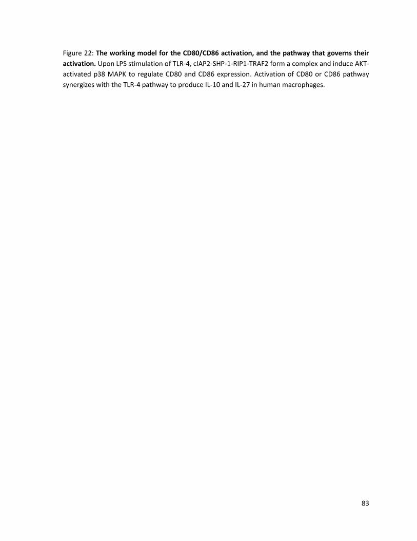
83
Figure 22: The working model for the CD80/CD86 activation, and the pathway that governs their
activation. Upon LPS stimulation of TLR-4, cIAP2-SHP-1-RIP1-TRAF2 form a complex and induce AKT-
activated p38 MAPK to regulate CD80 and CD86 expression. Activation of CD80 or CD86 pathway
synergizes with the TLR-4 pathway to produce IL-10 and IL-27 in human macrophages.

84
In light of this crucial role for CD80 and CD86 in the regulation of downstream cytokine
expression, I investigated the molecules that govern their regulation in response to LPS. Through
the use of specific inhibitors and siRNAs, I have determined that cIAP2, SHP-1, RIP1, and TRAF2
molecules are positive regulators of the CD80 and CD86 surface protein expression. Furthermore, I
have demonstrated that the p38 MAPK and PI3K pathway positively regulate the CD80 and CD86
expression in macrophages. My results have also demonstrated that JNK and ERK MAPK are not
involved in the regulation of CD80 or CD86 surface protein expression. Taken together, I have
demonstrated that macrophages employ the cIAP2-SHP-1-RIP1-TRAF2 complex to regulate
activation of the AKT/p38 signaling axis downstream of TLR-4 after LPS stimulation, and together,
this pathway positively regulates the CD80 and CD86 surface protein expression (Figure 22).
At the transcriptional level, I have demonstrated that treatment with LPS resulted in an
increase in CD80 mRNA, but IAPs and SHP-1 did not have a significant effect on LPS-induced CD80
expression. This is in contrast to CD80 protein expression, which was shown to be positively
regulated by both IAP and SHP-1. Inhibition of the PI3K and p38 MAPK pathways showed consistent
mRNA and protein expression. I have also shown that LPS stimulation resulted in increased CD86
protein levels, but surprisingly, decreased CD86 mRNA expression. I have also shown that while JNK
MAPK does not appear to regulate the expression of these costimulatory molecules at the protein
level, it does appear to negatively regulate CD86 mRNA expression. This could suggest a separate
pathway by which the expression of CD80 and CD86 is reduced. The RNA/protein discordance
suggests that further studies are required to better understand this differential regulation at the
protein and transcriptional level. It would also be of interest to identify the transcription factors
involved in the context of the pathways described.
The differential regulation of gene expression at the post-transcriptional or post-
translational level is widespread in eukaryotes (149). The protein half-life is a major post-

85
translational factor that can vary from a few seconds to a few days (152). My results can be
expanded by examining the stability of the CD80 and CD86 mRNA and protein by determining their
half-lives. Furthermore with the advancement in technologies related to mRNA/protein analysis,
expression microarrays could be used to assess CD80/CD86 mRNA as it provides an absolute
transcript concentration (152). Further studies are required to better understand this differential
regulation at the protein and transcriptional level.
In conclusion, my studies have attempted to identify the pathways that govern the CD80
and CD86 expression and the bidirectional signaling in macrophages. My results demonstrate that
activation of CD80 or CD86 pathways synergize with the LPS/TLR-4 pathway to activate IL-10 and IL-
27 expression. Furthermore, CD80 and CD86 protein expression is regulated by a cIAP2-SHP-1-RIP1-
TRAF2 complex through AKT-activated p38 signaling axis. Understanding the regulation of
CD80/CD86 molecules, and the bidirectional signaling in macrophages, could provide insight into
possible therapeutic interventions for the treatment of cancer and autoimmune diseases.

86
Appendix
Figure 23: Characterization of monocyte-derived macrophages. PBMCs (4 x 10⁶ cells/mL) were
plated and supplemented with 10 ng/mL of macrophage colony stimulating factor (M-CSF) for 6
days. Cells were washed and stained with PE-conjugated antibodies for 15 minutes. Flow Cytometry
was used to assess surface expression of CD83, CD80, CD16, CD11a, CD11b, CD11c, and HLA-DR.
Results were generated by Dr. Aurelia Busca.


87
Figure 24: CD80 and CD86 mRNA expression after treatment with LPS. MDMs were treated with
LPS for 4 hrs. qRT-PCR was used to measure CD80 (A) or CD86 (B) mRNA expression. Values in bar
graph are means of at least three independent experiments ±SEM.
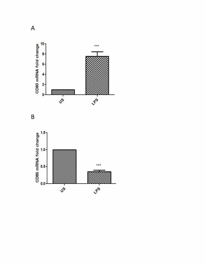
A
B
88
Figure 25: CD80/CD86 mRNA expression after treatment with SMAC mimetics or sodium
stibogluconate. MDMs were treated with their respective inhibitors for 24 hrs, followed by LPS
stimulation for 4 hrs. qRT-PCR was used to measure CD80 (left side) or CD86 (right side) mRNA
expression. Values in bar graph are means of at least three independent experiments ±SEM.

A B
US
LPS
SM
AC
SM
AC+L
PS
0
5
10
15
**
ns
CD
80 m
RN
A f
old
ch
an
ge
US
LPS
SM
AC
SM
AC+L
PS
0.0
0.5
1.0
1.5
**
CD
86 m
RN
A f
old
ch
an
ge
C D
US
LPS20
040
060
0
0
10
20
30
40
+LPS + SS (uM)
ns
CD
80 m
RN
A f
old
ch
an
ge
US
LPS20
040
060
0
0.0
0.5
1.0
1.5
+LPS + SS (uM)
ns
CD
86 m
RN
A f
old
ch
an
ge

89
Figure 26: CD86 mRNA expression after treatment with PI3K and MAPK inhibitors. MDMs were
treated with their respective inhibitors for 2 hrs, followed by LPS stimulation for 4 hrs. qRT-PCR was
used to measure CD86 mRNA expression. Values in bar graph are means of at least three
independent experiments ±SEM.
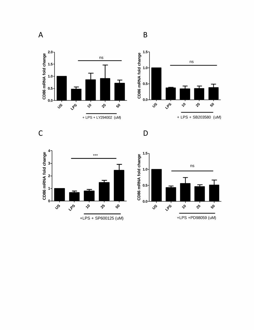
A B
US
LPS 10 25 50
0.0
0.5
1.0
1.5
2.0
+ LPS + LY294002 (uM)
CD
86 m
RN
A f
old
ch
an
ge
ns
US
LPS 10 25 50
0.0
0.5
1.0
1.5
+ LPS + SB203580 (uM)
CD
86 m
RN
A f
old
ch
an
ge
ns
C D
US
LPS 10 25 50
0
1
2
3
4
+LPS + SP600125 (uM)
***
CD
86 m
RN
A f
old
ch
an
ge
US
LPS 10 25 50
0.0
0.5
1.0
1.5
+LPS +PD98059 (uM)
CD
86 m
RN
A f
old
ch
an
ge
ns
90
References
1. Akira, S., S. Uematsu, and O. Takeuchi. 2006. Pathogen recognition and innate immunity. Cell 124: 783-801.
2. Barton, G. M. 2008. A calculated response: control of inflammation by the innate immune system. J. Clin. Invest. 118: 413-420.
3. Liaskou, E., D. V. Wilson, and Y. H. Oo. 2012. Innate immune cells in liver inflammation. Mediators Inflamm. 2012: 949157.
4. Medzhitov, R. 2007. Recognition of microorganisms and activation of the immune response. Nature 449: 819-826.
5. Nathan, C. and M. U. Shiloh. 2000. Reactive oxygen and nitrogen intermediates in the relationship between mammalian hosts and microbial pathogens. Proc. Natl. Acad. Sci. U. S. A. 97: 8841-8848.
6. van Kasteren, S. I., H. Overkleeft, H. Ovaa, and J. Neefjes. 2014. Chemical biology of antigen presentation by MHC molecules. Curr. Opin. Immunol. 26C: 21-31.
7. Jones, T. R., L. K. Hanson, L. Sun, J. S. Slater, R. M. Stenberg, and A. E. Campbell. 1995. Multiple independent loci within the human cytomegalovirus unique short region down-regulate expression of major histocompatibility complex class I heavy chains. J. Virol. 69: 4830-4841.
8. Lim, W., W. Ma, K. Gee, S. Aucoin, D. Nandan, F. Diaz-Mitoma, M. Kozlowski, and A. Kumar. 2002. Distinct role of p38 and c-Jun N-terminal kinases in IL-10-dependent and IL-10-independent regulation of the costimulatory molecule B7.2 in lipopolysaccharide-stimulated human monocytic cells. J. Immunol. 168: 1759-1769.
9. Gordon, S. and P. R. Taylor. 2005. Monocyte and macrophage heterogeneity. Nat. Rev. Immunol. 5: 953-964.
10. Martinez, F. O., S. Gordon, M. Locati, and A. Mantovani. 2006. Transcriptional profiling of the human monocyte-to-macrophage differentiation and polarization: new molecules and patterns of gene expression. J. Immunol. 177: 7303-7311.
11. Shi, C. and E. G. Pamer. 2011. Monocyte recruitment during infection and inflammation. Nat. Rev. Immunol. 11: 762-774.
12. Mills, C. D. 2012. M1 and M2 Macrophages: Oracles of Health and Disease. Crit. Rev. Immunol. 32: 463-488.
13. Benoit, M., B. Desnues, and J. L. Mege. 2008. Macrophage polarization in bacterial infections. J. Immunol. 181: 3733-3739.
14. Stow, J. L., P. C. Low, C. Offenhauser, and D. Sangermani. 2009. Cytokine secretion in macrophages and other cells: pathways and mediators. Immunobiology 214: 601-612.

91
15. Holgate, S. T. 2000. The role of mast cells and basophils in inflammation. Clin. Exp. Allergy 30 Suppl 1: 28-32.
16. Watford, W. T., M. Moriguchi, A. Morinobu, and J. J. O'Shea. 2003. The biology of IL-12: coordinating innate and adaptive immune responses. Cytokine Growth Factor Rev. 14: 361-368.
17. Stumhofer, J. S., A. Laurence, E. H. Wilson, E. Huang, C. M. Tato, L. M. Johnson, A. V. Villarino, Q. Huang, A. Yoshimura, D. Sehy, C. J. Saris, J. J. O'Shea, L. Hennighausen, M. Ernst, and C. A. Hunter. 2006. Interleukin 27 negatively regulates the development of interleukin 17-producing T helper cells during chronic inflammation of the central nervous system. Nat. Immunol. 7: 937-945.
18. Schuetze, N., S. Schoeneberger, U. Mueller, M. A. Freudenberg, G. Alber, and R. K. Straubinger. 2005. IL-12 family members: differential kinetics of their TLR4-mediated induction by Salmonella enteritidis and the impact of IL-10 in bone marrow-derived macrophages. Int. Immunol. 17: 649-659.
19. de Waal Malefyt, R., J. Haanen, H. Spits, M. G. Roncarolo, A. te Velde, C. Figdor, K. Johnson, R. Kastelein, H. Yssel, and J. E. de Vries. 1991. Interleukin 10 (IL-10) and viral IL-10 strongly reduce antigen-specific human T cell proliferation by diminishing the antigen-presenting capacity of monocytes via downregulation of class II major histocompatibility complex expression. J. Exp. Med. 174: 915-924.
20. Carreno, B. M. and M. Collins. 2002. The B7 family of ligands and its receptors: new pathways for costimulation and inhibition of immune responses. Annu. Rev. Immunol. 20: 29-53.
21. Greenfield, E. A., K. A. Nguyen, and V. K. Kuchroo. 1998. CD28/B7 costimulation: a review. Crit. Rev. Immunol. 18: 389-418.
22. Lim, W., K. Gee, S. Mishra, and A. Kumar. 2005. Regulation of B7.1 costimulatory molecule is mediated by the IFN regulatory factor-7 through the activation of JNK in lipopolysaccharide-stimulated human monocytic cells. J. Immunol. 175: 5690-5700.
23. Chen, L. and D. B. Flies. 2013. Molecular mechanisms of T cell co-stimulation and co-inhibition. Nat. Rev. Immunol. 13: 227-242.
24. Bugeon, L., R. E. Hargreaves, T. Crompton, S. Outram, A. Rahemtulla, A. C. Porter, and M. J. Dallman. 2001. Selective silencing of full-length CD80 but not IgV-CD80 leads to impaired clonal deletion of self-reactive T cells and altered regulation of immune responses. Eur. J. Immunol. 31: 118-127.
25. Peach, R. J., J. Bajorath, J. Naemura, G. Leytze, J. Greene, A. Aruffo, and P. S. Linsley. 1995. Both extracellular immunoglobin-like domains of CD80 contain residues critical for binding T cell surface receptors CTLA-4 and CD28. J. Biol. Chem. 270: 21181-21187.
26. Alegre, M. L., K. A. Frauwirth, and C. B. Thompson. 2001. T-cell regulation by CD28 and CTLA-4. Nat. Rev. Immunol. 1: 220-228.

92
27. Lindstein, T., C. H. June, J. A. Ledbetter, G. Stella, and C. B. Thompson. 1989. Regulation of lymphokine messenger RNA stability by a surface-mediated T cell activation pathway. Science 244: 339-343.
28. Boise, L. H., M. Gonzalez-Garcia, C. E. Postema, L. Ding, T. Lindsten, L. A. Turka, X. Mao, G. Nunez, and C. B. Thompson. 1993. Bcl-X, a Bcl-2-Related Gene that Functions as a Dominant Regulator of Apoptotic Cell Death. Cell 74: 597-608.
29. Itoh, Y. and R. N. Germain. 1997. Single cell analysis reveals regulated hierarchical T cell antigen receptor signaling thresholds and intraclonal heterogeneity for individual cytokine responses of CD4+ T cells. J. Exp. Med. 186: 757-766.
30. Lenschow, D. J., T. L. Walunas, and J. A. Bluestone. 1996. CD28/B7 system of T cell costimulation. Annu. Rev. Immunol. 14: 233-258.
31. Krummel, M. F. and J. P. Allison. 1995. CD28 and CTLA-4 have opposing effects on the response of T cells to stimulation. J. Exp. Med. 182: 459-465.
32. Flies, D. B. and L. Chen. 2007. The new B7s: playing a pivotal role in tumor immunity. J. Immunother. 30: 251-260.
33. Tseng, S. Y., M. Otsuji, K. Gorski, X. Huang, J. E. Slansky, S. I. Pai, A. Shalabi, T. Shin, D. M. Pardoll, and H. Tsuchiya. 2001. B7-DC, a new dendritic cell molecule with potent costimulatory properties for T cells. J. Exp. Med. 193: 839-846.
34. Saatian, B., X. Y. Yu, A. P. Lane, T. Doyle, V. Casolaro, and E. W. Spannhake. 2004. Expression of genes for B7-H3 and other T cell ligands by nasal epithelial cells during differentiation and activation. Am. J. Physiol. Lung Cell. Mol. Physiol. 287: L217-25.
35. Sica, G. L., I. H. Choi, G. Zhu, K. Tamada, S. D. Wang, H. Tamura, A. I. Chapoval, D. B. Flies, J. Bajorath, and L. Chen. 2003. B7-H4, a molecule of the B7 family, negatively regulates T cell immunity. Immunity 18: 849-861.
36. Du, C., M. Fang, Y. Li, L. Li, and X. Wang. 2000. Smac, a mitochondrial protein that promotes cytochrome c-dependent caspase activation by eliminating IAP inhibition. Cell 102: 33-42.
37. Zou, J. P., L. A. Morford, C. Chougnet, A. R. Dix, A. G. Brooks, N. Torres, J. D. Shuman, J. E. Coligan, W. H. Brooks, T. L. Roszman, and G. M. Shearer. 1999. Human glioma-induced immunosuppression involves soluble factor(s) that alters monocyte cytokine profile and surface markers. J. Immunol. 162: 4882-4892.
38. Johnson, B. D., X. Yan, D. W. Schauer, and R. J. Orentas. 2003. Dual expression of CD80 and CD86 produces a tumor vaccine superior to single expression of either molecule. Cell. Immunol. 222: 15-26.
39. Hussain, S. F., D. Yang, D. Suki, E. Grimm, and A. B. Heimberger. 2006. Innate immune functions of microglia isolated from human glioma patients. J. Transl. Med. 4: 15.

93
40. Hussain, S. F., D. Yang, D. Suki, K. Aldape, E. Grimm, and A. B. Heimberger. 2006. The role of human glioma-infiltrating microglia/macrophages in mediating antitumor immune responses. Neuro Oncol. 8: 261-279.
41. Lee, M. J., S. H. Choi, Y. H. Shim, S. H. Cho, and C. W. Kim. 2001. Tumor removal enhances immunity induced by B7.1. Pathobiology 69: 281-288.
42. Yang, G., K. E. Hellstrom, I. Hellstrom, and L. Chen. 1995. Antitumor immunity elicited by tumor cells transfected with B7-2, a second ligand for CD28/CTLA-4 costimulatory molecules. J. Immunol. 154: 2794-2800.
43. Hussain, S. F. and A. B. Heimberger. 2005. Immunotherapy for human glioma: innovative approaches and recent results. Expert Rev. Anticancer Ther. 5: 777-790.
44. Reilly, R. T., L. A. Emens, and E. M. Jaffee. 2001. Humoral and cellular immune responses: independent forces or collaborators in the fight against cancer? Curr. Opin. Investig Drugs 2: 133-135.
45. Fife, B. T. and J. A. Bluestone. 2008. Control of peripheral T-cell tolerance and autoimmunity via the CTLA-4 and PD-1 pathways. Immunol. Rev. 224: 166-182.
46. Son, C. H., J. H. Bae, D. Y. Shin, H. R. Lee, Y. J. Choi, W. S. Jo, M. Ho Jung, C. D. Kang, K. Yang, and Y. S. Park. 2014. CTLA-4 blockade enhances antitumor immunity of intratumoral injection of immature dendritic cells into irradiated tumor in a mouse colon cancer model. J. Immunother. 37: 1-7.
47. Leach, D. R., M. F. Krummel, and J. P. Allison. 1996. Enhancement of antitumor immunity by CTLA-4 blockade. Science 271: 1734-1736.
48. van Elsas, A., A. A. Hurwitz, and J. P. Allison. 1999. Combination immunotherapy of B16 melanoma using anti-cytotoxic T lymphocyte-associated antigen 4 (CTLA-4) and granulocyte/macrophage colony-stimulating factor (GM-CSF)-producing vaccines induces rejection of subcutaneous and metastatic tumors accompanied by autoimmune depigmentation. J. Exp. Med. 190: 355-366.
49. Margolin, K. A., A. M. Di Giacomo, and M. Maio. 2010. Brain metastasis in melanoma: clinical activity of CTLA-4 antibody therapy. Semin. Oncol. 37: 468-472.
50. Eggermont, A. M., A. Testori, M. Maio, and C. Robert. 2010. Anti-CTLA-4 antibody adjuvant therapy in melanoma. Semin. Oncol. 37: 455-459.
51. Lafferty, K. J., S. J. Prowse, C. J. Simeonovic, and H. S. Warren. 1983. Immunobiology of tissue transplantation: a return to the passenger leukocyte concept. Annu. Rev. Immunol. 1: 143-173.
52. Hwang, K. W., W. B. Sweatt, I. E. Brown, C. Blank, T. F. Gajewski, J. A. Bluestone, and M. L. Alegre. 2002. Cutting edge: targeted ligation of CTLA-4 in vivo by membrane-bound anti-CTLA-4 antibody prevents rejection of allogeneic cells. J. Immunol. 169: 633-637.
94
53. Belperio, J. A., S. S. Weigt, M. C. Fishbein, and J. P. Lynch 3rd. 2009. Chronic lung allograft rejection: mechanisms and therapy. Proc. Am. Thorac. Soc. 6: 108-121.
54. Griffin, M. D., D. K. Hong, P. O. Holman, K. M. Lee, M. J. Whitters, S. M. O'Herrin, F. Fallarino, M. Collins, D. M. Segal, T. F. Gajewski, D. M. Kranz, and J. A. Bluestone. 2000. Blockade of T cell activation using a surface-linked single-chain antibody to CTLA-4 (CD152). J. Immunol. 164: 4433-4442.
55. Fife, B. T., M. D. Griffin, A. K. Abbas, R. M. Locksley, and J. A. Bluestone. 2006. Inhibition of T cell activation and autoimmune diabetes using a B cell surface-linked CTLA-4 agonist. J. Clin. Invest. 116: 2252-2261.
56. Lin, H., S. F. Bolling, P. S. Linsley, R. Q. Wei, D. Gordon, C. B. Thompson, and L. A. Turka. 1993. Long-term acceptance of major histocompatibility complex mismatched cardiac allografts induced by CTLA4Ig plus donor-specific transfusion. J. Exp. Med. 178: 1801-1806.
57. Sayegh, M. H., E. Akalin, W. W. Hancock, M. E. Russell, C. B. Carpenter, P. S. Linsley, and L. A. Turka. 1995. CD28-B7 blockade after alloantigenic challenge in vivo inhibits Th1 cytokines but spares Th2. J. Exp. Med. 181: 1869-1874.
58. Lenschow, D. J., Y. Zeng, J. R. Thistlethwaite, A. Montag, W. Brady, M. G. Gibson, P. S. Linsley, and J. A. Bluestone. 1992. Long-term survival of xenogeneic pancreatic islet grafts induced by CTLA4lg. Science 257: 789-792.
59. Paul, W. E. and R. A. Seder. 1994. Lymphocyte responses and cytokines. Cell 76: 241-251.
60. Thompson, C. B. 1995. Distinct roles for the costimulatory ligands B7-1 and B7-2 in T helper cell differentiation? Cell 81: 979-982.
61. Kuchroo, V. K., M. P. Das, J. A. Brown, A. M. Ranger, S. S. Zamvil, R. A. Sobel, H. L. Weiner, N. Nabavi, and L. H. Glimcher. 1995. B7-1 and B7-2 costimulatory molecules activate differentially the Th1/Th2 developmental pathways: application to autoimmune disease therapy. Cell 80: 707-718.
62. Schweitzer, A. N. and A. H. Sharpe. 1999. Mutual regulation between B7-1 (CD80) expressed on T cells and IL-4. J. Immunol. 163: 4819-4825.
63. Subauste, C. S., R. de Waal Malefyt, and F. Fuh. 1998. Role of CD80 (B7.1) and CD86 (B7.2) in the immune response to an intracellular pathogen. J. Immunol. 160: 1831-1840.
64. Vasu, C., A. Wang, S. R. Gorla, S. Kaithamana, B. S. Prabhakar, and M. J. Holterman. 2003. CD80 and CD86 C domains play an important role in receptor binding and co-stimulatory properties. Int. Immunol. 15: 167-175.
65. Doty, R. T. and E. A. Clark. 1996. Subcellular localization of CD80 receptors is dependent on an intact cytoplasmic tail and is required for CD28-dependent T cell costimulation. J. Immunol. 157: 3270-3279.
95
66. Doty, R. T. and E. A. Clark. 1998. Two regions in the CD80 cytoplasmic tail regulate CD80 redistribution and T cell costimulation. J. Immunol. 161: 2700-2707.
67. Koorella, C., J. R. Nair, M. E. Murray, L. M. Carlson, S. K. Watkins, and K. P. Lee. 2014. Novel Regulation of CD80/CD86-induced Phosphatidylinositol 3-Kinase Signaling by NOTCH1 Protein in Interleukin-6 and Indoleamine 2,3-Dioxygenase Production by Dendritic Cells. J. Biol. Chem. 289: 7747-7762.
68. Orabona, C., U. Grohmann, M. L. Belladonna, F. Fallarino, C. Vacca, R. Bianchi, S. Bozza, C. Volpi, B. L. Salomon, M. C. Fioretti, L. Romani, and P. Puccetti. 2004. CD28 induces immunostimulatory signals in dendritic cells via CD80 and CD86. Nat. Immunol. 5: 1134-1142.
69. Rakhesh, M., M. Cate, R. Vijay, A. Shrikant, and A. Shanjana. 2012. A TLR4-interacting peptide inhibits lipopolysaccharide-stimulated inflammatory responses, migration and invasion of colon cancer SW480 cells. Oncoimmunology 1: 1495-1506.
70. Zhu, J. and C. Mohan. 2010. Toll-like receptor signaling pathways--therapeutic opportunities. Mediators Inflamm. 2010: 781235.
71. Troutman, T. D., J. F. Bazan, and C. Pasare. 2012. Toll-like receptors, signaling adapters and regulation of the pro-inflammatory response by PI3K. Cell. Cycle 11: 3559-3567.
72. Kawai, T. and S. Akira. 2008. Toll-like receptor and RIG-I-like receptor signaling. Ann. N. Y. Acad. Sci. 1143: 1-20.
73. Ulevitch, R. J. and P. S. Tobias. 1999. Recognition of gram-negative bacteria and endotoxin by the innate immune system. Curr. Opin. Immunol. 11: 19-22.
74. Yang, S. H., A. D. Sharrocks, and A. J. Whitmarsh. 2013. MAP kinase signalling cascades and transcriptional regulation. Gene 513: 1-13.
75. Wagner, E. F. and A. R. Nebreda. 2009. Signal integration by JNK and p38 MAPK pathways in cancer development. Nat. Rev. Cancer. 9: 537-549.
76. Gerits, N., S. Kostenko, and U. Moens. 2007. In vivo functions of mitogen-activated protein kinases: conclusions from knock-in and knock-out mice. Transgenic Res. 16: 281-314.
77. Krishna, M. and H. Narang. 2008. The complexity of mitogen-activated protein kinases (MAPKs) made simple. Cell Mol. Life Sci. 65: 3525-3544.
78. Wortzel, I. and R. Seger. 2011. The ERK Cascade: Distinct Functions within Various Subcellular Organelles. Genes Cancer. 2: 195-209.
79. Guma, M. and G. S. Firestein. 2012. c-Jun N-Terminal Kinase in Inflammation and Rheumatic Diseases. Open Rheumatol. J. 6: 220-231.

96
80. Oeztuerk-Winder, F. and J. J. Ventura. 2012. The many faces of p38 mitogen-activated protein kinase in progenitor/stem cell differentiation. Biochem. J. 445: 1-10.
81. Vanhaesebroeck, B., J. Guillermet-Guibert, M. Graupera, and B. Bilanges. 2010. The emerging mechanisms of isoform-specific PI3K signalling. Nat. Rev. Mol. Cell Biol. 11: 329-341.
82. Fruman, D. A. and L. C. Cantley. 2002. Phosphoinositide 3-kinase in immunological systems. Semin. Immunol. 14: 7-18.
83. Hennessy, B. T., D. L. Smith, P. T. Ram, Y. Lu, and G. B. Mills. 2005. Exploiting the PI3K/AKT pathway for cancer drug discovery. Nat. Rev. Drug Discov. 4: 988-1004.
84. Busca, A., M. Saxena, M. Kryworuchko, and A. Kumar. 2009. Anti-apoptotic genes in the survival of monocytic cells during infection. Curr. Genomics 10: 306-317.
85. Crook, N. E., R. J. Clem, and L. K. Miller. 1993. An apoptosis-inhibiting baculovirus gene with a zinc finger-like motif. J. Virol. 67: 2168-2174.
86. Wang, K. and B. Lin. 2013. Inhibitor of apoptosis proteins (IAPs) as regulatory factors of hepatic apoptosis. Cell. Signal. 25: 1970-1980.
87. Srinivasula, S. M. and J. D. Ashwell. 2008. IAPs: what's in a name? Mol. Cell 30: 123-135.
88. Eckelman, B. P., G. S. Salvesen, and F. L. Scott. 2006. Human inhibitor of apoptosis proteins: why XIAP is the black sheep of the family. EMBO Rep. 7: 988-994.
89. Shiozaki, E. N. and Y. Shi. 2004. Caspases, IAPs and Smac/DIABLO: mechanisms from structural biology. Trends Biochem. Sci. 29: 486-494.
90. Varfolomeev, E., J. W. Blankenship, S. M. Wayson, A. V. Fedorova, N. Kayagaki, P. Garg, K. Zobel, J. N. Dynek, L. O. Elliott, H. J. Wallweber, J. A. Flygare, W. J. Fairbrother, K. Deshayes, V. M. Dixit, and D. Vucic. 2007. IAP antagonists induce autoubiquitination of c-IAPs, NF-kappaB activation, and TNFalpha-dependent apoptosis. Cell 131: 669-681.
91. Lopes, R. B., R. Gangeswaran, I. A. McNeish, Y. Wang, and N. R. Lemoine. 2007. Expression of the IAP protein family is dysregulated in pancreatic cancer cells and is important for resistance to chemotherapy. Int. J. Cancer 120: 2344-2352.
92. Varfolomeev, E. and D. Vucic. 2008. (Un)expected roles of c-IAPs in apoptotic and NFkappaB signaling pathways. Cell. Cycle 7: 1511-1521.
93. Wu, G., J. Chai, T. L. Suber, J. W. Wu, C. Du, X. Wang, and Y. Shi. 2000. Structural basis of IAP recognition by Smac/DIABLO. Nature 408: 1008-1012.
94. LaCasse, E. C., D. J. Mahoney, H. H. Cheung, S. Plenchette, S. Baird, and R. G. Korneluk. 2008. IAP-targeted therapies for cancer. Oncogene 27: 6252-6275.

97
95. Sun, H., Z. Nikolovska-Coleska, C. Y. Yang, D. Qian, J. Lu, S. Qiu, L. Bai, Y. Peng, Q. Cai, and S. Wang. 2008. Design of small-molecule peptidic and nonpeptidic Smac mimetics. Acc. Chem. Res. 41: 1264-1277.
96. Beug, S. T., V. A. Tang, E. C. Lacasse, H. H. Cheung, C. E. Beauregard, J. Brun, J. P. Nuyens, N. Earl, M. St-Jean, J. Holbrook, H. Dastidar, D. J. Mahoney, C. Ilkow, F. Le Boeuf, J. C. Bell, and R. G. Korneluk. 2014. Smac mimetics and innate immune stimuli synergize to promote tumor death. Nat. Biotechnol. 32: 182-190.
97. Mahoney, D. J., H. H. Cheung, R. L. Mrad, S. Plenchette, C. Simard, E. Enwere, V. Arora, T. W. Mak, E. C. Lacasse, J. Waring, and R. G. Korneluk. 2008. Both cIAP1 and cIAP2 regulate TNFalpha-mediated NF-kappaB activation. Proc. Natl. Acad. Sci. U. S. A. 105: 11778-11783.
98. Shu, H. B., M. Takeuchi, and D. V. Goeddel. 1996. The tumor necrosis factor receptor 2 signal transducers TRAF2 and c-IAP1 are components of the tumor necrosis factor receptor 1 signaling complex. Proc. Natl. Acad. Sci. U. S. A. 93: 13973-13978.
99. Bertrand, M. J., S. Milutinovic, K. M. Dickson, W. C. Ho, A. Boudreault, J. Durkin, J. W. Gillard, J. B. Jaquith, S. J. Morris, and P. A. Barker. 2008. cIAP1 and cIAP2 facilitate cancer cell survival by functioning as E3 ligases that promote RIP1 ubiquitination. Mol. Cell 30: 689-700.
100. Petersen, S. L., L. Wang, A. Yalcin-Chin, L. Li, M. Peyton, J. Minna, P. Harran, and X. Wang. 2007. Autocrine TNFalpha signaling renders human cancer cells susceptible to Smac-mimetic-induced apoptosis. Cancer. Cell. 12: 445-456.
101. Alcamo, E., J. P. Mizgerd, B. H. Horwitz, R. Bronson, A. A. Beg, M. Scott, C. M. Doerschuk, R. O. Hynes, and D. Baltimore. 2001. Targeted mutation of TNF receptor I rescues the RelA-deficient mouse and reveals a critical role for NF-kappa B in leukocyte recruitment. J. Immunol. 167: 1592-1600.
102. Bonizzi, G. and M. Karin. 2004. The two NF-kappaB activation pathways and their role in innate and adaptive immunity. Trends Immunol. 25: 280-288.
103. Takeuchi, O. and S. Akira. 2010. Pattern recognition receptors and inflammation. Cell 140: 805-820.
104. Matsuzawa, A., P. H. Tseng, S. Vallabhapurapu, J. L. Luo, W. Zhang, H. Wang, D. A. Vignali, E. Gallagher, and M. Karin. 2008. Essential cytoplasmic translocation of a cytokine receptor-assembled signaling complex. Science 321: 663-668.
105. Tseng, P. H., A. Matsuzawa, W. Zhang, T. Mino, D. A. Vignali, and M. Karin. 2010. Different modes of ubiquitination of the adaptor TRAF3 selectively activate the expression of type I interferons and proinflammatory cytokines. Nat. Immunol. 11: 70-75.
106. Bertrand, M. J., K. Doiron, K. Labbe, R. G. Korneluk, P. A. Barker, and M. Saleh. 2009. Cellular inhibitors of apoptosis cIAP1 and cIAP2 are required for innate immunity signaling by the pattern recognition receptors NOD1 and NOD2. Immunity 30: 789-801.
98
107. Bauler, L. D., C. S. Duckett, and M. X. O'Riordan. 2008. XIAP regulates cytosol-specific innate immunity to Listeria infection. PLoS Pathog. 4: e1000142.
108. Inohara, N., T. Koseki, J. Lin, L. del Peso, P. C. Lucas, F. F. Chen, Y. Ogura, and G. Nunez. 2000. An induced proximity model for NF-kappa B activation in the Nod1/RICK and RIP signaling pathways. J. Biol. Chem. 275: 27823-27831.
109. Yoneyama, M., M. Kikuchi, T. Natsukawa, N. Shinobu, T. Imaizumi, M. Miyagishi, K. Taira, S. Akira, and T. Fujita. 2004. The RNA helicase RIG-I has an essential function in double-stranded RNA-induced innate antiviral responses. Nat. Immunol. 5: 730-737.
110. Kawai, T. and S. Akira. 2006. Innate immune recognition of viral infection. Nat. Immunol. 7: 131-137.
111. Mao, A. P., S. Li, B. Zhong, Y. Li, J. Yan, Q. Li, C. Teng, and H. B. Shu. 2010. Virus-triggered ubiquitination of TRAF3/6 by cIAP1/2 is essential for induction of interferon-beta (IFN-beta) and cellular antiviral response. J. Biol. Chem. 285: 9470-9476.
112. Johnson, P. and J. L. Cross. 2009. Tyrosine phosphorylation in immune cells: direct and indirect effects on toll-like receptor-induced proinflammatory cytokine production. Crit. Rev. Immunol. 29: 347-367.
113. Bohmer, F., S. Szedlacsek, L. Tabernero, A. Ostman, and J. den Hertog. 2013. Protein tyrosine phosphatase structure-function relationships in regulation and pathogenesis. FEBS J. 280: 413-431.
114. Page, T. H., M. Smolinska, J. Gillespie, A. M. Urbaniak, and B. M. Foxwell. 2009. Tyrosine kinases and inflammatory signalling. Curr. Mol. Med. 9: 69-85.
115. Novogrodsky, A., A. Vanichkin, M. Patya, A. Gazit, N. Osherov, and A. Levitzki. 1994. Prevention of lipopolysaccharide-induced lethal toxicity by tyrosine kinase inhibitors. Science 264: 1319-1322.
116. Rego, D., A. Kumar, L. Nilchi, K. Wright, S. Huang, and M. Kozlowski. 2011. IL-6 production is positively regulated by two distinct Src homology domain 2-containing tyrosine phosphatase-1 (SHP-1)-dependent CCAAT/enhancer-binding protein beta and NF-kappaB pathways and an SHP-1-independent NF-kappaB pathway in lipopolysaccharide-stimulated bone marrow-derived macrophages. J. Immunol. 186: 5443-5456.
117. Lowell, C. A. 2004. Src-family kinases: rheostats of immune cell signaling. Mol. Immunol. 41: 631-643.
118. Zhang, P., X. Liu, Y. Li, X. Zhu, Z. Zhan, J. Meng, N. Li, and X. Cao. 2013. Protein tyrosine phosphatase with proline-glutamine-serine-threonine-rich motifs negatively regulates TLR-triggered innate responses by selectively inhibiting IkappaB kinase beta/NF-kappaB activation. J. Immunol. 190: 1685-1694.
119. Bignon, J. S. and K. A. Siminovitch. 1994. Identification of PTP1C mutation as the genetic defect in motheaten and viable motheaten mice: a step toward defining the roles of protein tyrosine
99
phosphatases in the regulation of hemopoietic cell differentiation and function. Clin. Immunol. Immunopathol. 73: 168-179.
120. Creery, W. D., F. Diaz-Mitoma, L. Filion, and A. Kumar. 1996. Differential modulation of B7-1 and B7-2 isoform expression on human monocytes by cytokines which influence the development of T helper cell phenotype. Eur. J. Immunol. 26: 1273-1277.
121. Lim, W., K. Gee, S. Mishra, and A. Kumar. 2005. Regulation of B7.1 costimulatory molecule is mediated by the IFN regulatory factor-7 through the activation of JNK in lipopolysaccharide-stimulated human monocytic cells. J. Immunol. 175: 5690-5700.
122. Nakahara, T., H. Uchi, K. Urabe, Q. Chen, M. Furue, and Y. Moroi. 2004. Role of c-Jun N-terminal kinase on lipopolysaccharide induced maturation of human monocyte-derived dendritic cells. Int. Immunol. 16: 1701-1709.
123. Arrighi, J. F., M. Rebsamen, F. Rousset, V. Kindler, and C. Hauser. 2001. A critical role for p38 mitogen-activated protein kinase in the maturation of human blood-derived dendritic cells induced by lipopolysaccharide, TNF-alpha, and contact sensitizers. J. Immunol. 166: 3837-3845.
124. Agadjanyan, M. G., J. J. Kim, N. Trivedi, D. M. Wilson, B. Monzavi-Karbassi, L. D. Morrison, L. K. Nottingham, T. Dentchev, A. Tsai, K. Dang, A. A. Chalian, M. A. Maldonado, W. V. Williams, and D. B. Weiner. 1999. CD86 (B7-2) can function to drive MHC-restricted antigen-specific CTL responses in vivo. J. Immunol. 162: 3417-3427.
125. Kim, J. J., M. L. Bagarazzi, N. Trivedi, Y. Hu, K. Kazahaya, D. M. Wilson, R. Ciccarelli, M. A. Chattergoon, K. Dang, S. Mahalingam, A. A. Chalian, M. G. Agadjanyan, J. D. Boyer, B. Wang, and D. B. Weiner. 1997. Engineering of in vivo immune responses to DNA immunization via codelivery of costimulatory molecule genes. Nat. Biotechnol. 15: 641-646.
126. Collins, A. V., D. W. Brodie, R. J. Gilbert, A. Iaboni, R. Manso-Sancho, B. Walse, D. I. Stuart, P. A. van der Merwe, and S. J. Davis. 2002. The interaction properties of costimulatory molecules revisited. Immunity 17: 201-210.
127. Acuto, O. and F. Michel. 2003. CD28-mediated co-stimulation: a quantitative support for TCR signalling. Nat. Rev. Immunol. 3: 939-951.
128. Jain, S., S. B. Chodisetti, and J. N. Agrewala. 2013. Combinatorial signaling through TLR-2 and CD86 augments activation and differentiation of resting B cells. PLoS One 8: e54392.
129. Damgaard, R. B. and M. Gyrd-Hansen. 2011. Inhibitor of apoptosis (IAP) proteins in regulation of inflammation and innate immunity. Discov. Med. 11: 221-231.
130. Rothe, M., M. G. Pan, W. J. Henzel, T. M. Ayres, and D. V. Goeddel. 1995. The TNFR2-TRAF signaling complex contains two novel proteins related to baculoviral inhibitor of apoptosis proteins. Cell 83: 1243-1252.
100
131. Cheung, H. H., D. J. Mahoney, E. C. Lacasse, and R. G. Korneluk. 2009. Down-regulation of c-FLIP Enhances death of cancer cells by smac mimetic compound. Cancer Res. 69: 7729-7738.
132. Bohmer, F., S. Szedlacsek, L. Tabernero, A. Ostman, and J. den Hertog. 2013. Protein tyrosine phosphatase structure-function relationships in regulation and pathogenesis. FEBS J. 280: 413-431.
133. Zhang, J., A. K. Somani, and K. A. Siminovitch. 2000. Roles of the SHP-1 tyrosine phosphatase in the negative regulation of cell signalling. Semin. Immunol. 12: 361-378.
134. Zong, Y., L. Sun, B. Liu, Y. S. Deng, D. Zhan, Y. L. Chen, Y. He, J. Liu, Z. J. Zhang, J. Sun, and D. Lu. 2012. Resveratrol inhibits LPS-induced MAPKs activation via activation of the phosphatidylinositol 3-kinase pathway in murine RAW 264.7 macrophage cells. PLoS One 7: e44107.
135. Attridge, K., R. Kenefeck, L. Wardzinski, O. S. Qureshi, C. J. Wang, C. Manzotti, K. Okkenhaug, and L. S. Walker. 2014. IL-21 promotes CD4 T cell responses by phosphatidylinositol 3-kinase-dependent upregulation of CD86 on B cells. J. Immunol. 192: 2195-2201.
136. Tang, Y., M. T. Nakada, P. Rafferty, J. Laraio, F. L. McCabe, H. Millar, M. Cunningham, L. A. Snyder, P. Bugelski, and L. Yan. 2006. Regulation of vascular endothelial growth factor expression by EMMPRIN via the PI3K-Akt signaling pathway. Mol. Cancer. Res. 4: 371-377.
137. Sansom, D. M., C. N. Manzotti, and Y. Zheng. 2003. What's the difference between CD80 and CD86? Trends Immunol. 24: 314-319.
138. Makela, S. M., M. Strengell, T. E. Pietila, P. Osterlund, and I. Julkunen. 2009. Multiple signaling pathways contribute to synergistic TLR ligand-dependent cytokine gene expression in human monocyte-derived macrophages and dendritic cells. J. Leukoc. Biol. 85: 664-672.
139. Yi, A. K., J. G. Yoon, S. C. Hong, T. W. Redford, and A. M. Krieg. 2001. Lipopolysaccharide and CpG DNA synergize for tumor necrosis factor-alpha production through activation of NF-kappaB. Int. Immunol. 13: 1391-1404.
140. Wrzesien-Kus, A., P. Smolewski, A. Sobczak-Pluta, A. Wierzbowska, and T. Robak. 2004. The inhibitor of apoptosis protein family and its antagonists in acute leukemias. Apoptosis 9: 705-715.
141. Tseng, P. H., A. Matsuzawa, W. Zhang, T. Mino, D. A. Vignali, and M. Karin. 2010. Different modes of ubiquitination of the adaptor TRAF3 selectively activate the expression of type I interferons and proinflammatory cytokines. Nat. Immunol. 11: 70-75.
142. Chen, G., M. H. Shaw, Y. G. Kim, and G. Nunez. 2009. NOD-like receptors: role in innate immunity and inflammatory disease. Annu. Rev. Pathol. 4: 365-398.
143. Kim, H. W., S. I. Cho, S. Bae, H. Kim, Y. Kim, Y. I. Hwang, J. S. Kang, and W. J. Lee. 2012. Vitamin C Up-regulates Expression of CD80, CD86 and MHC Class II on Dendritic Cell Line, DC-1 Via the Activation of p38 MAPK. Immune Netw. 12: 277-283.
101
144. Reyes-Moreno, C., J. Girouard, R. Lapointe, A. Darveau, and W. Mourad. 2004. CD40/CD40 homodimers are required for CD40-induced phosphatidylinositol 3-kinase-dependent expression of B7.2 by human B lymphocytes. J. Biol. Chem. 279: 7799-7806.
145. Attridge, K., R. Kenefeck, L. Wardzinski, O. S. Qureshi, C. J. Wang, C. Manzotti, K. Okkenhaug, and L. S. Walker. 2014. IL-21 promotes CD4 T cell responses by phosphatidylinositol 3-kinase-dependent upregulation of CD86 on B cells. J. Immunol. 192: 2195-2201.
146. Guha, M. and N. Mackman. 2002. The phosphatidylinositol 3-kinase-Akt pathway limits lipopolysaccharide activation of signaling pathways and expression of inflammatory mediators in human monocytic cells. J. Biol. Chem. 277: 32124-32132.
147. Li, D. Q., N. Zhou, L. Zhang, P. Ma, and S. C. Pflugfelder. 2010. Suppressive effects of azithromycin on zymosan-induced production of proinflammatory mediators by human corneal epithelial cells. Invest. Ophthalmol. Vis. Sci. 51: 5623-5629.
148. Ito, T. and M. Kodama. 1996. Demonstration by reverse transcription-polymerase chain reaction of multiple cytokine mRNA expression in bovine alveolar macrophages and peripheral blood mononuclear cells. Res. Vet. Sci. 60: 94-96.
149. Matsumura, N., Y. Takeyama, T. Ueda, T. Yasuda, M. Shinzeki, H. Sawa, T. Nakajima, and Y. Kuroda. 2007. Decreased expression of Toll-like receptor 2 and 4 on macrophages in experimental severe acute pancreatitis. Kobe J. Med. Sci. 53: 219-227.
150. Blencowe, B., S. Brenner, T. Hughes, and Q. Morris. 2009. Post-transcriptional gene regulation: RNA-protein interactions, RNA processing, mRNA stability and localization. Pac. Symp. Biocomput. 545-548.
151. Halbeisen, R. E., A. Galgano, T. Scherrer, and A. P. Gerber. 2008. Post-transcriptional gene regulation: from genome-wide studies to principles. Cell Mol. Life Sci. 65: 798-813.
152. Tahmasebi, S., T. Alain, V. K. Rajasekhar, J. P. Zhang, M. Prager-Khoutorsky, A. Khoutorsky, Y. Dogan, C. G. Gkogkas, E. Petroulakis, A. Sylvestre, M. Ghorbani, S. Assadian, Y. Yamanaka, J. R. Vinagolu-Baur, J. G. Teodoro, K. Kim, X. J. Yang, and N. Sonenberg. 2014. Multifaceted regulation of somatic cell reprogramming by mRNA translational control. Cell. Stem Cell. 14: 606-616.
153. Maier, T., M. Guell, and L. Serrano. 2009. Correlation of mRNA and protein in complex biological samples. FEBS Lett. 583: 3966-3973.
154. Fournier, M. L., A. Paulson, N. Pavelka, A. L. Mosley, K. Gaudenz, W. D. Bradford, E. Glynn, H. Li, M. E. Sardiu, B. Fleharty, C. Seidel, L. Florens, and M. P. Washburn. 2010. Delayed correlation of mRNA and protein expression in rapamycin-treated cells and a role for Ggc1 in cellular sensitivity to rapamycin. Mol. Cell. Proteomics 9: 271-284.
155. Takeda, K. and S. Akira. 2005. Toll-like receptors in innate immunity. Int. Immunol. 17: 1-14.

102

103