Embed Size (px)
DESCRIPTION
the respiratory system anatomy notes
Citation preview
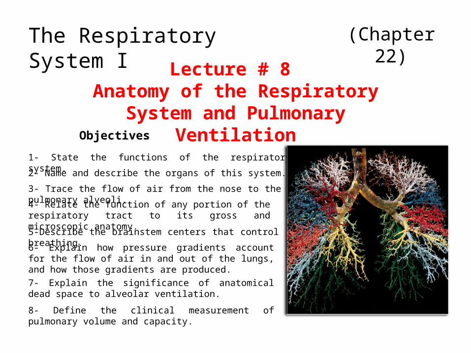
(Chapter 22)
Lecture # 8 Anatomy of the Respiratory System
and Pulmonary Ventilation
The Respiratory System I
Objectives
1- State the functions of the respiratory system.
2- Name and describe the organs of this system.
3- Trace the flow of air from the nose to the pulmonary alveoli.
4- Relate the function of any portion of the respiratory tract to its gross and microscopic anatomy.
5-Describe the brainstem centers that control breathing.
6- Explain how pressure gradients account for the flow of air in and out of the lungs, and how those gradients are produced.
7- Explain the significance of anatomical dead space to alveolar ventilation.
8- Define the clinical measurement of pulmonary volume and capacity.
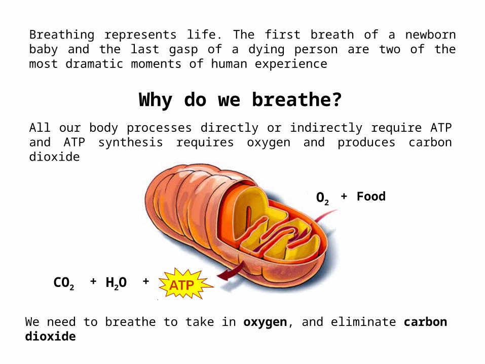
Breathing represents life. The first breath of a newborn baby and the last gasp of a dying person are two of the most dramatic moments of human experience
Why do we breathe?All our body processes directly or indirectly require ATP and ATP synthesis requires oxygen and produces carbon dioxide
FoodO2+
CO2 H2O ++
We need to breathe to take in oxygen, and eliminate carbon dioxide
Alveoli in lung
Tissue cells
O2
O2
FoodCO2
H2O
CO2
CO2
O2
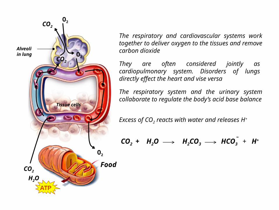
The respiratory and cardiovascular systems work together to deliver oxygen to the tissues and remove carbon dioxide
They are often considered jointly as cardiopulmonary system. Disorders of lungs directly effect the heart and vise versa
The respiratory system and the urinary system collaborate to regulate the body’s acid base balance
Excess of CO2 reacts with water and releases H+
CO2 H2O + + H+HCO3
-H2CO3
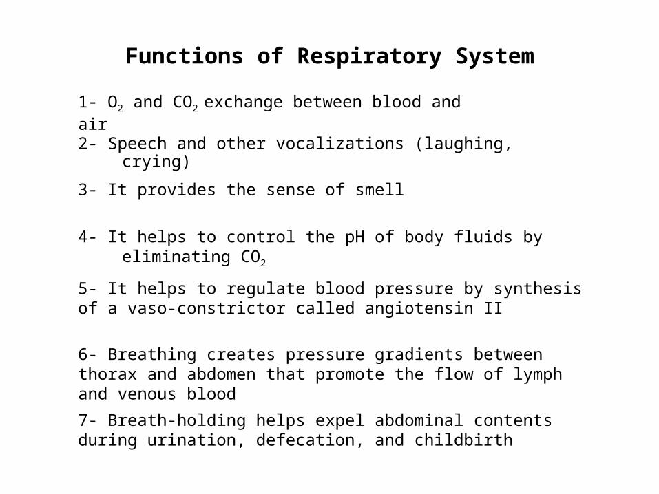
Functions of Respiratory System
1- O2 and CO2 exchange between blood and air
2- Speech and other vocalizations (laughing, crying)
3- It provides the sense of smell
4- It helps to control the pH of body fluids by eliminating CO2
5- It helps to regulate blood pressure by synthesis of a vaso-constrictor called angiotensin II
6- Breathing creates pressure gradients between thorax and abdomen that promote the flow of lymph and venous blood
7- Breath-holding helps expel abdominal contents during urination, defecation, and childbirth

Principal Organs of the Respiratory System
Trachea
Nose
Pharynx
Larynx
Lungs
Bronchi
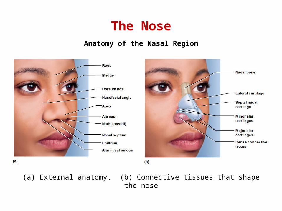
The NoseAnatomy of the Nasal Region
(a) External anatomy. (b) Connective tissues that shape the nose
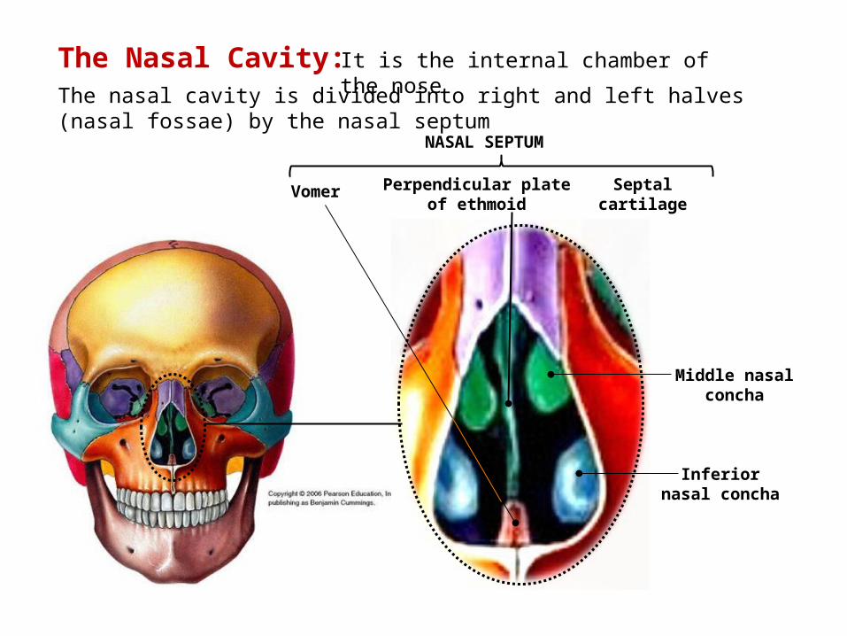
NASAL SEPTUM
It is the internal chamber of the nose
The nasal cavity is divided into right and left halves (nasal fossae) by the nasal septum
Middle nasal concha
Inferior nasal concha
Vomer Perpendicular plate of ethmoid
The Nasal Cavity:
Septal cartilage
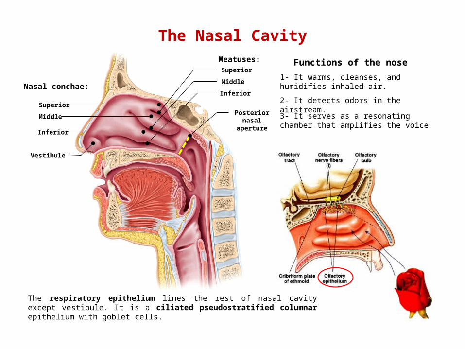
The Nasal Cavity
Nasal conchae:
Meatuses:
Superior
Middle
Inferior
Superior
Middle
Inferior
Vestibule
Functions of the nose
1- It warms, cleanses, and humidifies inhaled air.
2- It detects odors in the airstream.
3- It serves as a resonating chamber that amplifies the voice.
Posterior nasal
aperture
The respiratory epithelium lines the rest of nasal cavity except vestibule. It is a ciliated pseudostratified columnar epithelium with goblet cells.
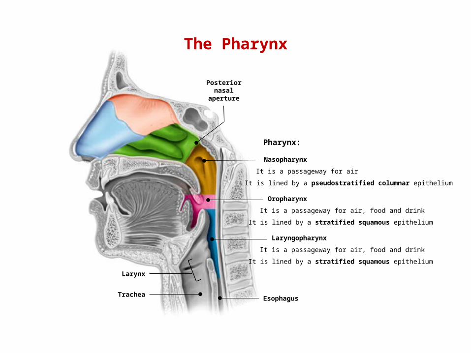
The Pharynx
EsophagusTrachea
Larynx
Posterior nasal
aperture
Nasopharynx
Oropharynx
Laryngopharynx
Pharynx:
It is a passageway for air
It is a passageway for air, food and drink
It is a passageway for air, food and drink
It is lined by a pseudostratified columnar epithelium
It is lined by a stratified squamous epithelium
It is lined by a stratified squamous epithelium
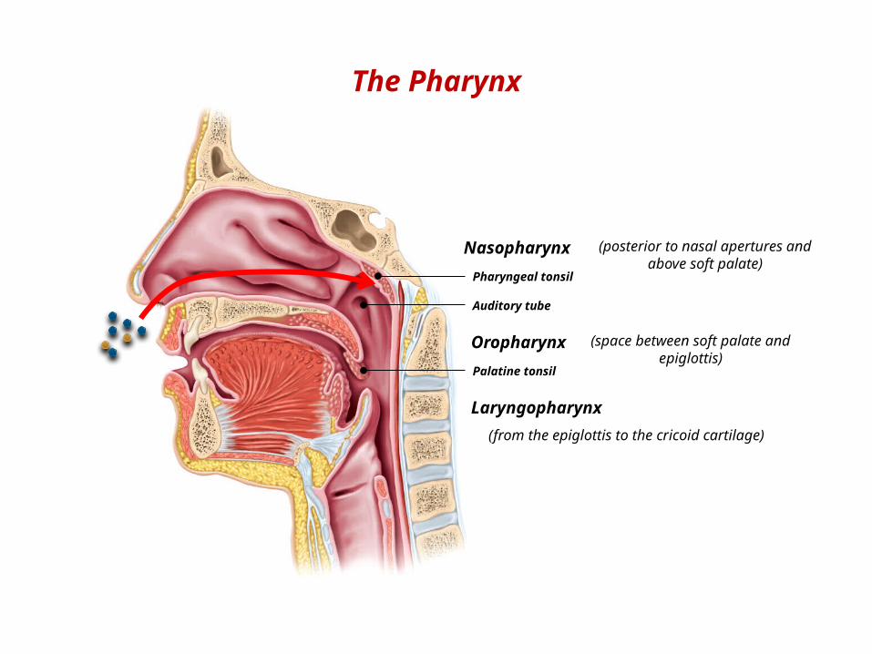
Nasopharynx
Oropharynx
Laryngopharynx
Pharyngeal tonsil
Auditory tube
Palatine tonsil
(posterior to nasal apertures and above soft palate)
(space between soft palate and epiglottis)
(from the epiglottis to the cricoid cartilage)
The Pharynx
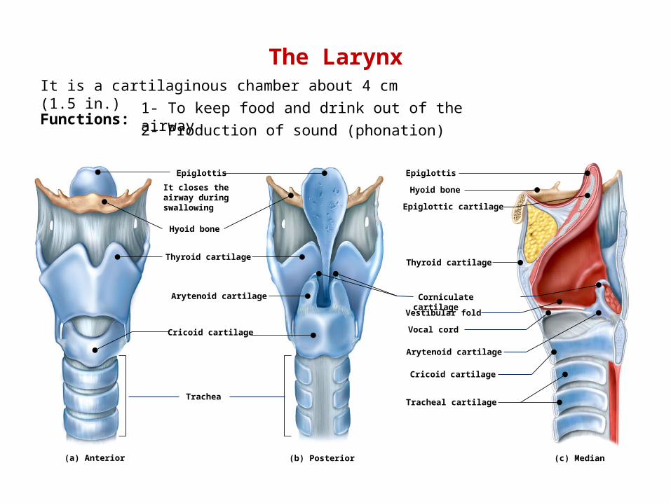
The Larynx
(a) Anterior (b) Posterior (c) Median
It is a cartilaginous chamber about 4 cm (1.5 in.)
Functions:1- To keep food and drink out of the airway
2- Production of sound (phonation)
Hyoid bone
Thyroid cartilage
Arytenoid cartilage
Cricoid cartilage
EpiglottisEpiglottis
Hyoid bone
Thyroid cartilage
Vestibular fold
Vocal cord
Arytenoid cartilage
Cricoid cartilage
Tracheal cartilage
Corniculate cartilage
It closes the airway during swallowing
Trachea
Epiglottic cartilage

Median
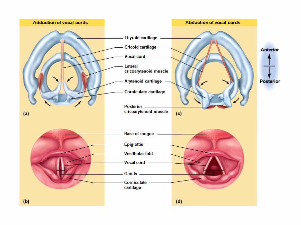
Vestibular foldThey play no role in speech but close the larynx during swallowing
Vocal cord
They produce sound when air passes between them
(from the thyroid cartilage to the arytenoid cartilage)
The Larynx
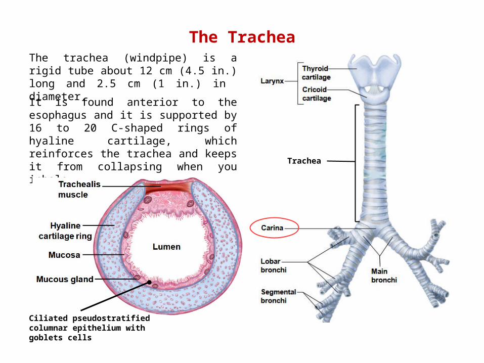
The TracheaThe trachea (windpipe) is a rigid tube about 12 cm (4.5 in.) long and 2.5 cm (1 in.) in diameter.
It is found anterior to the esophagus and it is supported by 16 to 20 C-shaped rings of hyaline cartilage, which reinforces the trachea and keeps it from collapsing when you inhale.
Trachea
Ciliated pseudostratified columnar epithelium with goblets cells
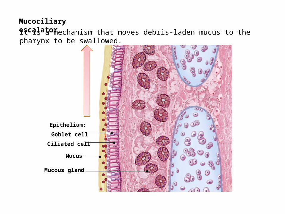
Mucociliary escalator
Epithelium:
Mucous gland
Ciliated cell
Goblet cell
Mucus
It is a mechanism that moves debris-laden mucus to the pharynx to be swallowed.
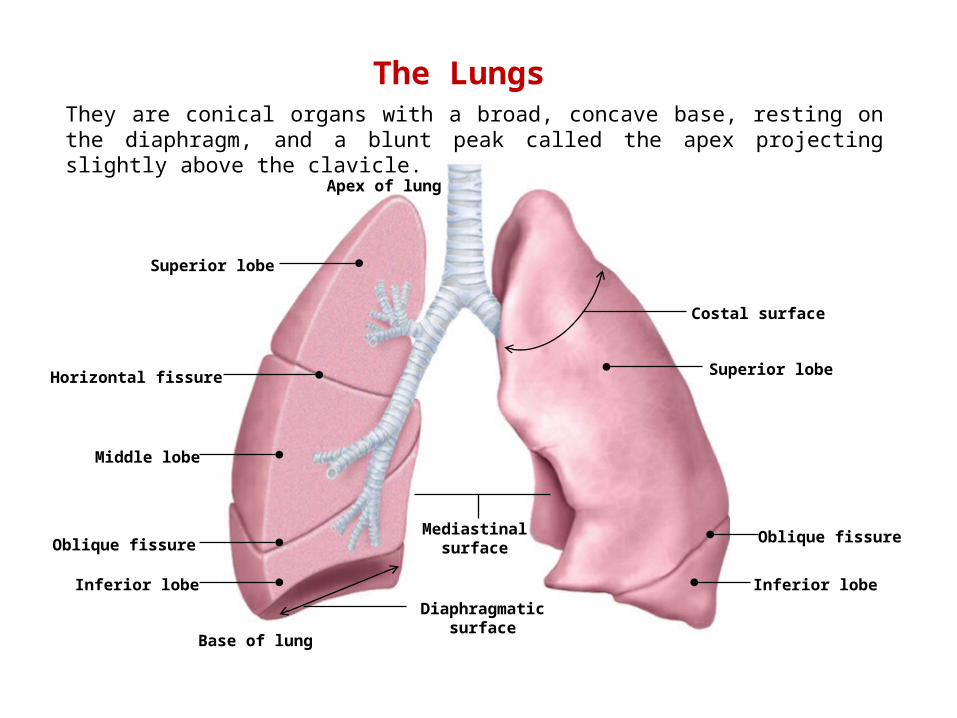
The LungsThey are conical organs with a broad, concave base, resting on the diaphragm, and a blunt peak called the apex projecting slightly above the clavicle.
Apex of lung
Base of lung
Oblique fissure
Horizontal fissure
Oblique fissure
Superior lobe
Middle lobe
Inferior lobe
Superior lobe
Inferior lobe
Costal surface
Mediastinal surface
Diaphragmatic surface
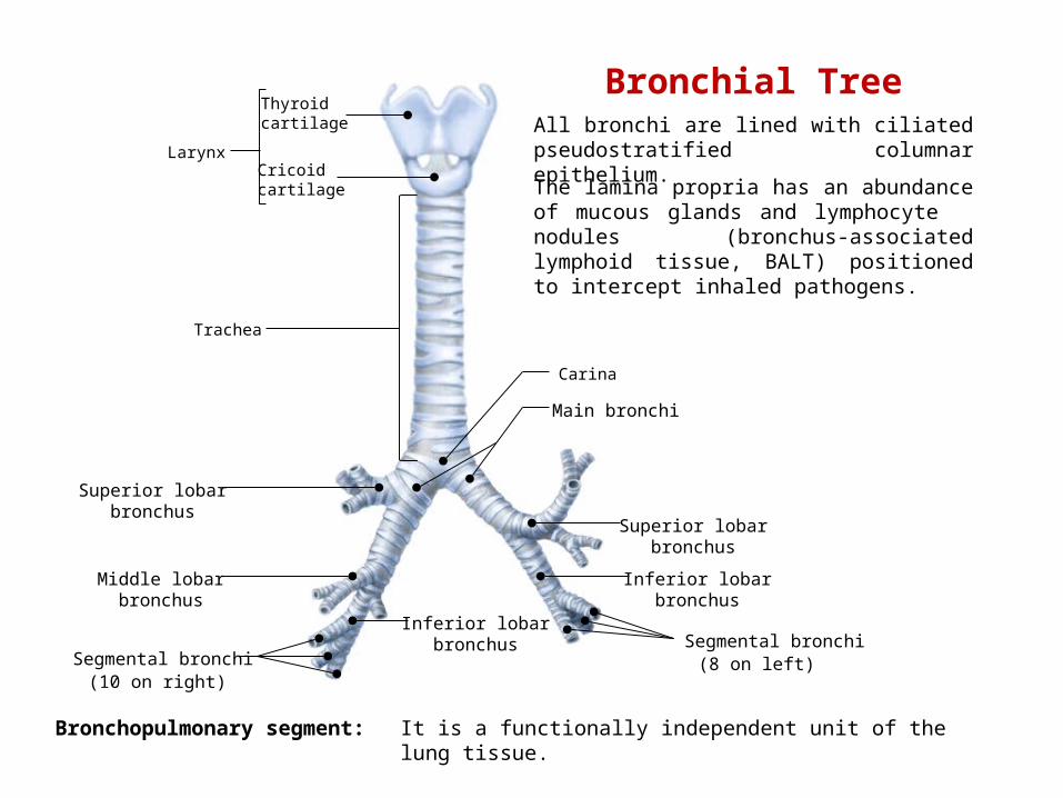
Larynx
Trachea
Carina
Thyroidcartilage
Cricoidcartilage
Main bronchi
Superior lobarbronchus
Middle lobarbronchus
Inferior lobarbronchus
Superior lobarbronchus
Inferior lobarbronchus
Segmental bronchi(10 on right)
Segmental bronchi(8 on left)
Bronchopulmonary segment: It is a functionally independent unit of the lung tissue.
Bronchial TreeAll bronchi are lined with ciliated pseudostratified columnar epithelium.
The lamina propria has an abundance of mucous glands and lymphocyte nodules (bronchus-associated lymphoid tissue, BALT) positioned to intercept inhaled pathogens.
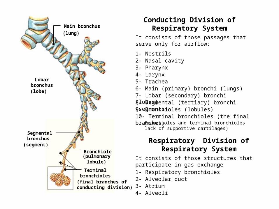
Main bronchus
Lobar bronchus
Segmental bronchus
Bronchiole
(lung)
(lobe)
(segment)
(pulmonary lobule)
(final branches of conducting division)
Terminal bronchioles
Conducting Division of Respiratory System
1- Nostrils
It consists of those passages that serve only for airflow:
2- Nasal cavity3- Pharynx4- Larynx5- Trachea6- Main (primary) bronchi (lungs)7- Lobar (secondary) bronchi (lobes)8- Segmental (tertiary) bronchi (segments)9- Bronchioles (lobules)10- Terminal bronchioles (the final branches)
Bronchioles and terminal bronchioles lack of supportive cartilages)
Respiratory Division of Respiratory System
1- Respiratory bronchioles 2- Alveolar duct
It consists of those structures that participate in gas exchange
3- Atrium4- Alveoli
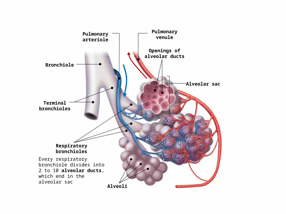
Bronchiole
Terminal bronchioles
Respiratory bronchioles
Openings of alveolar ducts
Every respiratory bronchiole divides into 2 to 10 alveolar ducts, which end in the alveolar sac
Alveolar sac
Alveoli
Pulmonary arteriole
Pulmonary venule
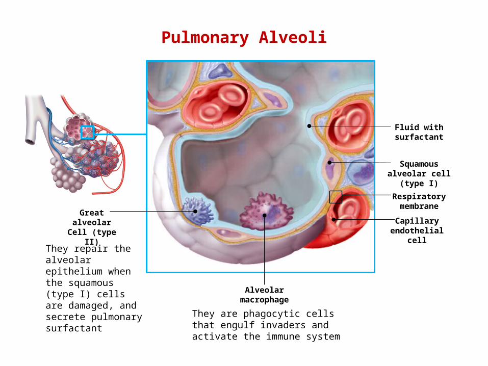
Pulmonary Alveoli
Squamous alveolar cell
(type I)
Capillary endothelial
cell
Respiratory membrane
Fluid with surfactant
Alveolarmacrophage
Greatalveolar
Cell (type II)
They repair the alveolar epithelium when the squamous (type I) cells are damaged, and secrete pulmonary surfactant
They are phagocytic cells that engulf invaders and activate the immune system
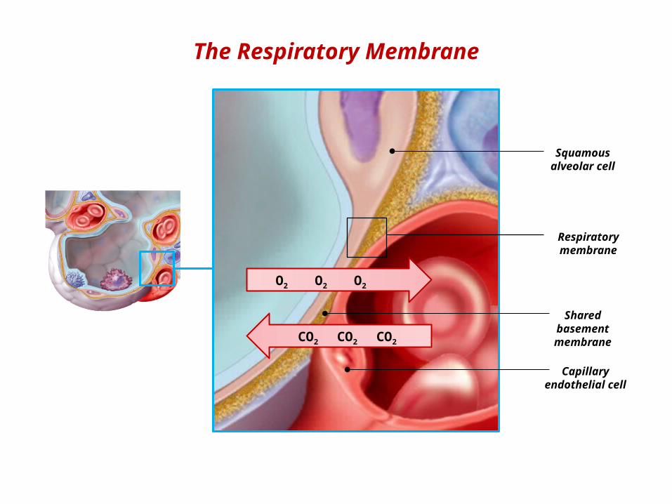
The Respiratory Membrane
Squamous alveolar cell
Capillary endothelial
cell
Respiratory membrane
Shared basement membraneCO2 CO2 CO2
O2 O2 O2
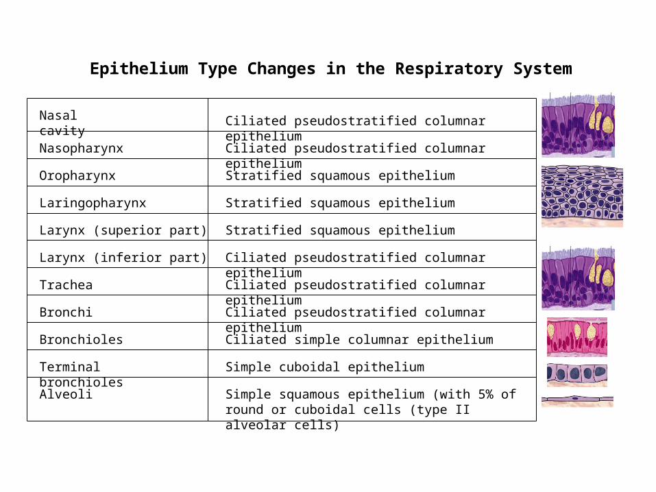
Nasal cavity
Nasopharynx
Trachea
Bronchi
Bronchioles
Terminal bronchioles
Alveoli
Oropharynx
Laringopharynx
Ciliated pseudostratified columnar epithelium
Stratified squamous epithelium
Stratified squamous epithelium
Larynx (superior part) Stratified squamous epithelium
Larynx (inferior part)
Ciliated pseudostratified columnar epithelium
Ciliated pseudostratified columnar epithelium
Ciliated pseudostratified columnar epithelium
Ciliated pseudostratified columnar epithelium
Ciliated simple columnar epithelium
Simple cuboidal epithelium
Simple squamous epithelium (with 5% of round or cuboidal cells (type II alveolar cells)
Epithelium Type Changes in the Respiratory System
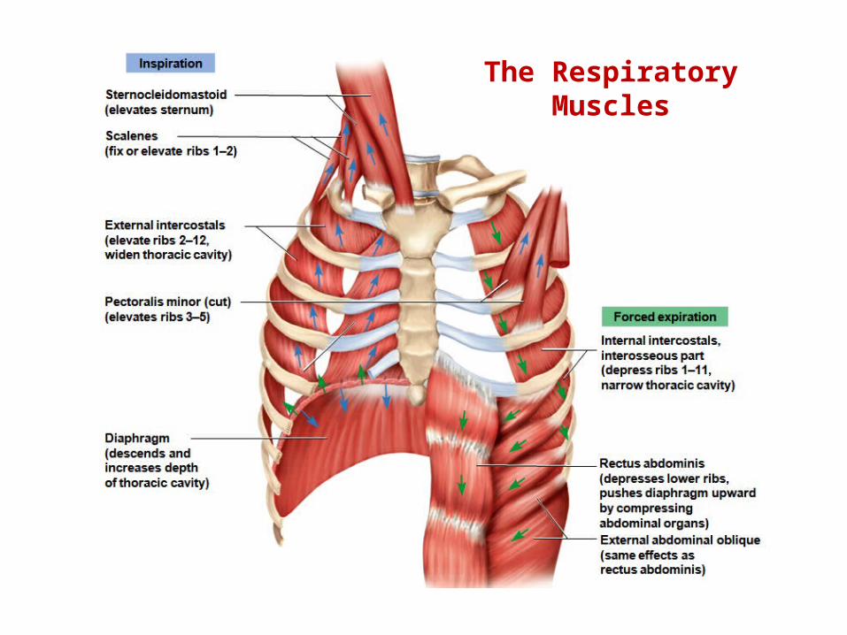
The Respiratory Muscles
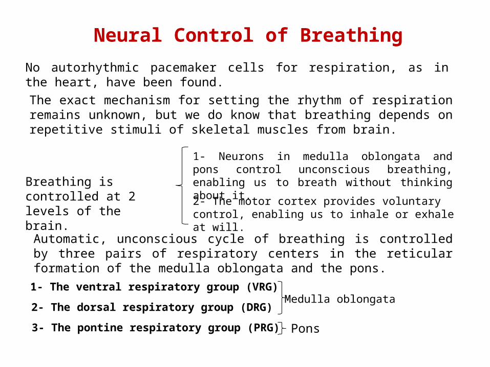
Neural Control of Breathing
No autorhythmic pacemaker cells for respiration, as in the heart, have been found.
The exact mechanism for setting the rhythm of respiration remains unknown, but we do know that breathing depends on repetitive stimuli of skeletal muscles from brain.
Breathing is controlled at 2 levels of the brain.
1- Neurons in medulla oblongata and pons control unconscious breathing, enabling us to breath without thinking about it.
2- The motor cortex provides voluntary control, enabling us to inhale or exhale at will.
Automatic, unconscious cycle of breathing is controlled by three pairs of respiratory centers in the reticular formation of the medulla oblongata and the pons.
1- The ventral respiratory group (VRG)
2- The dorsal respiratory group (DRG)
3- The pontine respiratory group (PRG)
Medulla oblongata
Pons
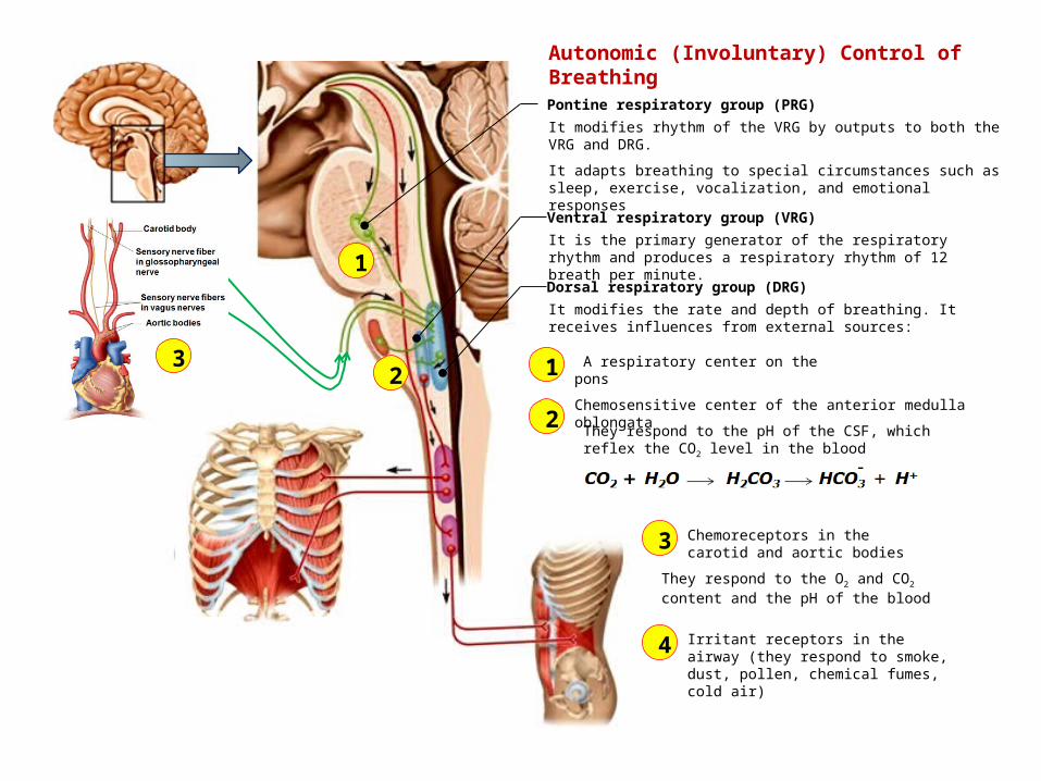
It is the primary generator of the respiratory rhythm and produces a respiratory rhythm of 12 breath per minute.
It modifies the rate and depth of breathing. It receives influences from external sources:
Dorsal respiratory group (DRG)
2
1
3 A respiratory center on the pons
1
Chemosensitive center of the anterior medulla oblongata
2
Chemoreceptors in the carotid and aortic bodies
3
Irritant receptors in the airway (they respond to smoke, dust, pollen, chemical fumes, cold air)
4
Pontine respiratory group (PRG)
Ventral respiratory group (VRG)
It modifies rhythm of the VRG by outputs to both the VRG and DRG.
It adapts breathing to special circumstances such as sleep, exercise, vocalization, and emotional responses
They respond to the pH of the CSF, which reflex the CO2 level in the blood
They respond to the O2 and CO2 content and the pH of the blood
Autonomic (Involuntary) Control of Breathing
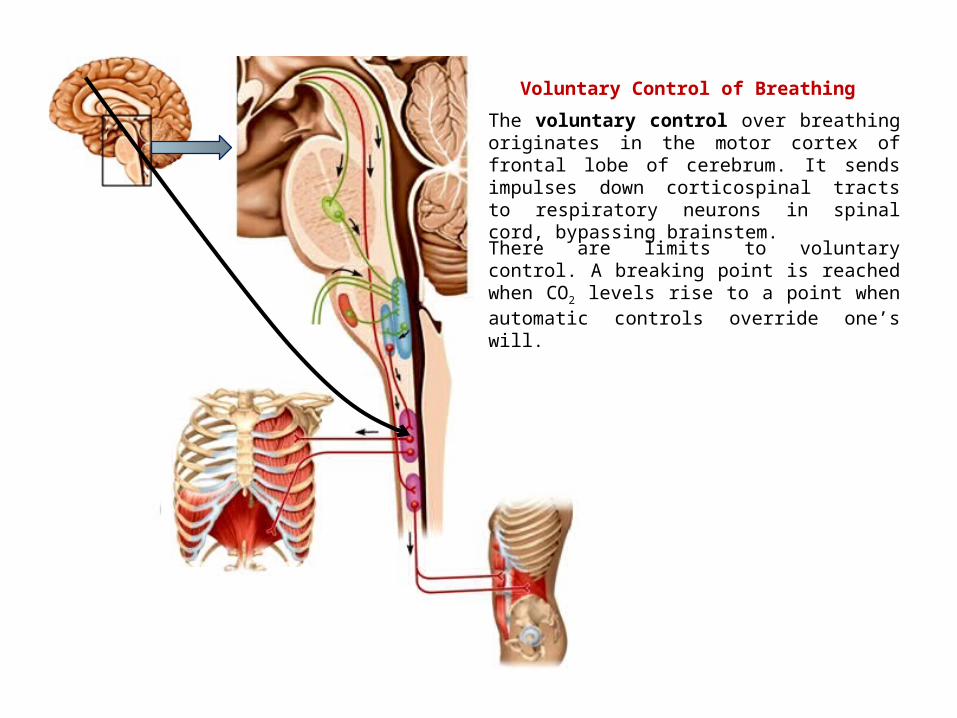
The voluntary control over breathing originates in the motor cortex of frontal lobe of cerebrum. It sends impulses down corticospinal tracts to respiratory neurons in spinal cord, bypassing brainstem.
Voluntary Control of Breathing
There are limits to voluntary control. A breaking point is reached when CO2 levels rise to a point when automatic controls override one’s will.
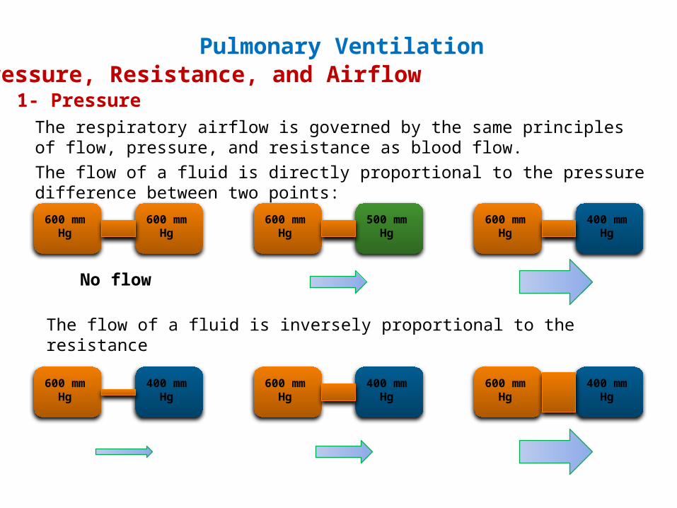
Pressure, Resistance, and Airflow
The respiratory airflow is governed by the same principles of flow, pressure, and resistance as blood flow.The flow of a fluid is directly proportional to the pressure difference between two points:
600 mm Hg
600 mm Hg
600 mm Hg
500 mm Hg
600 mm Hg
400 mm Hg
No flow
The flow of a fluid is inversely proportional to the resistance
600 mm Hg
400 mm Hg
600 mm Hg
400 mm Hg
600 mm Hg
400 mm Hg
1- Pressure
Pulmonary Ventilation
Expiration
Inspiration
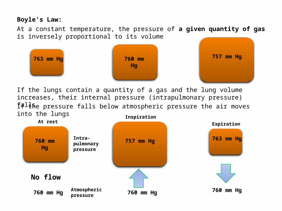
Boyle’s Law:
At a constant temperature, the pressure of a given quantity of gas is inversely proportional to its volume
763 mm Hg
760 mm Hg
757 mm Hg
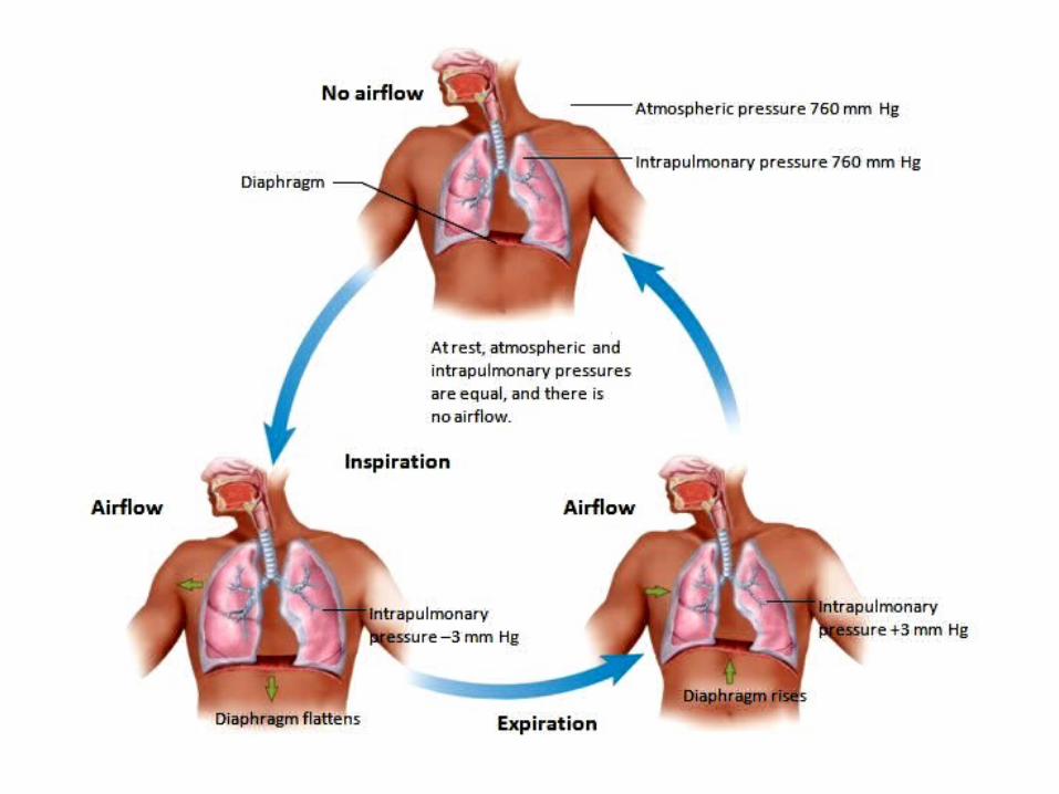
If the lungs contain a quantity of a gas and the lung volume increases, their internal pressure (intrapulmonary pressure) fallsIf the pressure falls below atmospheric pressure the air moves into the lungs
760 mm Hg Atmospheric pressure
Intra-pulmonary pressure
No flow
At rest
760 mm Hg
757 mm Hg
760 mm Hg
763 mm Hg
760 mm Hg
Intercostal muscles elevates the rib cage and diaphragm
is contracted
Rib cage in normal position and diaphragm is relaxed
Volume Volume
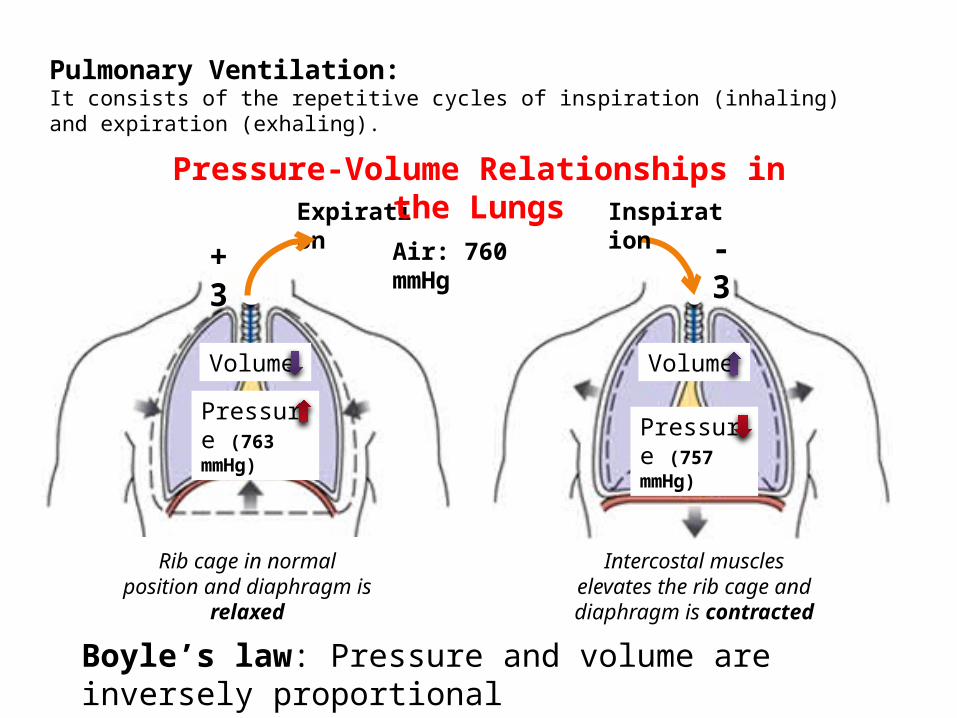
Boyle’s law: Pressure and volume are inversely proportional
Pressure (757 mmHg)
Pressure (763 mmHg)
Air: 760 mmHg
Expiration Inspiration
Pressure-Volume Relationships in the Lungs
+3 -3
Pulmonary Ventilation:It consists of the repetitive cycles of inspiration (inhaling) and expiration (exhaling).
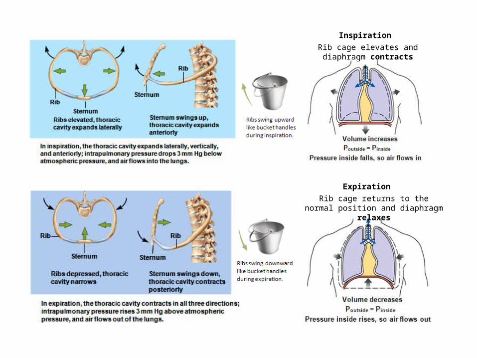
Inspiration
Expiration
Rib cage elevates and diaphragm contracts
Rib cage returns to the normal position and diaphragm relaxes
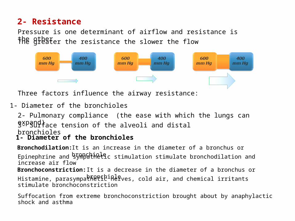
2- ResistancePressure is one determinant of airflow and resistance is the other
The greater the resistance the slower the flow
3- Surface tension of the alveoli and distal bronchioles
1- Diameter of the bronchioles
2- Pulmonary compliance (the ease with which the lungs can expand)
Three factors influence the airway resistance:
1- Diameter of the bronchioles
Bronchodilation:
Bronchoconstriction:
It is an increase in the diameter of a bronchus or bronchiole
It is a decrease in the diameter of a bronchus or bronchiole
Histamine, parasympathetic nerves, cold air, and chemical irritants stimulate bronchoconstriction
Epinephrine and sympathetic stimulation stimulate bronchodilation and increase air flow
Suffocation from extreme bronchoconstriction brought about by anaphylactic shock and asthma
It determines the change in lung volume relative to a given pressure change. The thoracic cage expands normally but the lungs expand relatively little.
2- Pulmonary compliance (the ease with which the lungs can expand)
Pulmonary compliance reduced by degenerative lung diseases in which the lungs are stiffened by scar tissue (tuberculosis, black lung disease *).
* Black lung disease is a chronic occupational lung disease contracted by the prolonged breathing of coal mine dust. Black lung disease is also called anthracosis, black lung, black spittle, coal worker's pneumoconiosis, miner's asthma, pneumoconiosis, and silicosis.
3- Surface tension of the alveoli and distal bronchioles
Water molecules in the alveolar epithelium are attracted to each other by hydrogen bonds, creating a surface tension.
Surface tension draws the walls of the alveoli inward toward the lumen and resisting the reinflation.
A surfactant is a substance produced by the great alveolar cells (type II cells) that disrupts the hydrogen bonds and allows the lungs to expand.
Premature infants often have a deficiency of pulmonary surfactant and experience great difficulty breathing.
The result is the “infant respiratory distress syndrome (IRDS), which is treated with artificial surfactant.
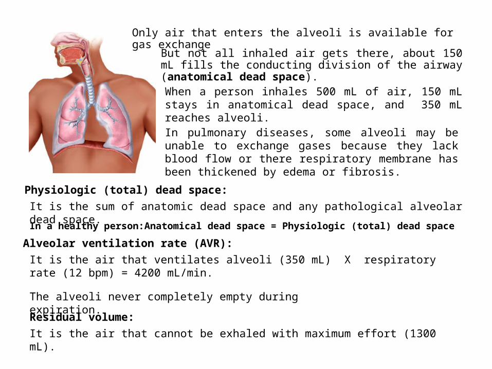
Only air that enters the alveoli is available for gas exchange
But not all inhaled air gets there, about 150 mL fills the conducting division of the airway (anatomical dead space).
In pulmonary diseases, some alveoli may be unable to exchange gases because they lack blood flow or there respiratory membrane has been thickened by edema or fibrosis.
Physiologic (total) dead space:
When a person inhales 500 mL of air, 150 mL stays in anatomical dead space, and 350 mL reaches alveoli.
It is the sum of anatomic dead space and any pathological alveolar dead space.
In a healthy person: Anatomical dead space = Physiologic (total) dead space
Alveolar ventilation rate (AVR):
It is the air that ventilates alveoli (350 mL) X respiratory rate (12 bpm) = 4200 mL/min.
The alveoli never completely empty during expiration.
Residual volume:
It is the air that cannot be exhaled with maximum effort (1300 mL).
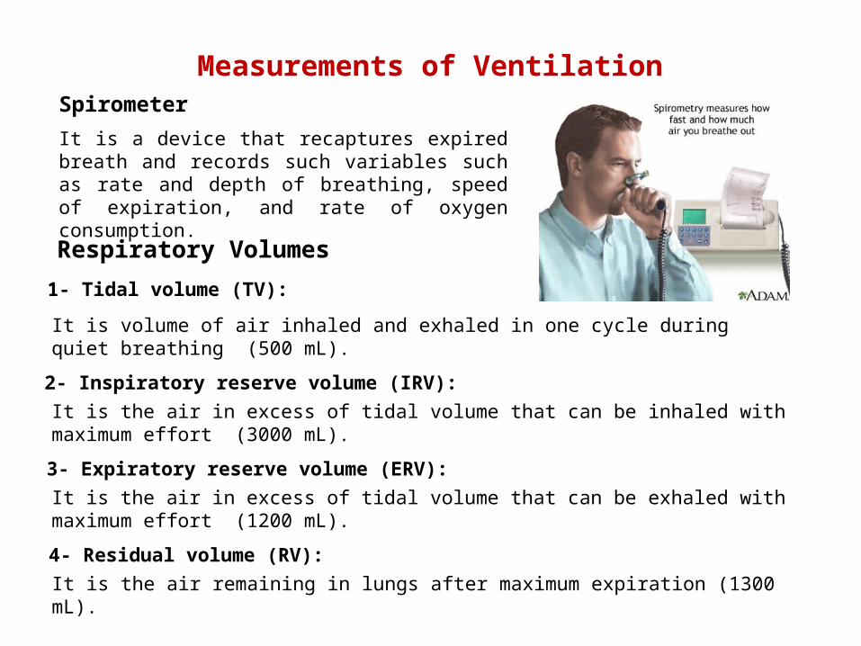
Measurements of Ventilation
It is a device that recaptures expired breath and records such variables such as rate and depth of breathing, speed of expiration, and rate of oxygen consumption.
Spirometer
Respiratory Volumes
1- Tidal volume (TV):
2- Inspiratory reserve volume (IRV):
3- Expiratory reserve volume (ERV):
4- Residual volume (RV):
It is volume of air inhaled and exhaled in one cycle during quiet breathing (500 mL).
It is the air in excess of tidal volume that can be inhaled with maximum effort (3000 mL).
It is the air in excess of tidal volume that can be exhaled with maximum effort (1200 mL).
It is the air remaining in lungs after maximum expiration (1300 mL).
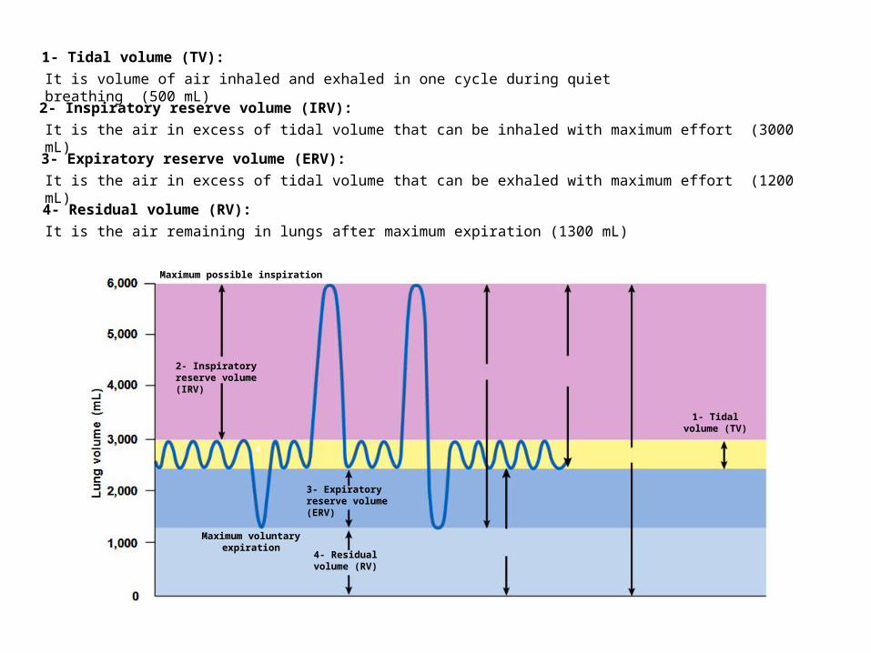
1- Tidal volume (TV):
2- Inspiratory reserve volume (IRV):
3- Expiratory reserve volume (ERV):
4- Residual volume (RV):
It is volume of air inhaled and exhaled in one cycle during quiet breathing (500 mL)
It is the air in excess of tidal volume that can be inhaled with maximum effort (3000 mL)
It is the air in excess of tidal volume that can be exhaled with maximum effort (1200 mL)
It is the air remaining in lungs after maximum expiration (1300 mL)
1- Tidal volume (TV)
Maximum possible inspiration
2- Inspiratory reserve volume (IRV)
Maximum voluntaryexpiration
3- Expiratory reserve volume (ERV)
4- Residual volume (RV)
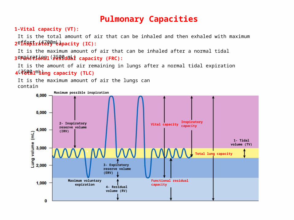
1-Vital capacity (VT):
2-Inspiratory capacity (IC):
3-Functional residual capacity (FRC):
4-Total lung capacity (TLC)
It is the total amount of air that can be inhaled and then exhaled with maximum effort (4700mL)
It is the maximum amount of air that can be inhaled after a normal tidal expiration (3500 mL)
It is the amount of air remaining in lungs after a normal tidal expiration (2500 mL)
It is the maximum amount of air the lungs can contain
1- Tidal volume (TV)
Maximum possible inspiration
2- Inspiratory reserve volume (IRV)
Maximum voluntaryexpiration
3- Expiratory reserve volume (ERV)
4- Residual volume (RV)
Total lung capacity
Vital capacityInspiratorycapacity
Functional residualcapacity
Pulmonary Capacities


![Respiratory System Ppt[1]Gaga](https://img.pdfslide.us/doc/110x75/546d66bfb4af9f86038b4d16/respiratory-system-ppt1gaga.jpg)


![Respiratory system[1]](https://img.pdfslide.us/doc/110x75/55643c7ed8b42ace308b51ab/respiratory-system1.jpg)