Embed Size (px)
Citation preview
The Respiratory System
PROGRAM STUDI S1 KEPERAWATAN
FAKULTAS ILMU KESEHATAN UNIVERSITAS MUHAMMADIYAH MALANG
2014
5 Functions of the Respiratory System
1. Provides extensive gas exchange surface area between air and circulating blood
2. Moves air to and from exchange surfaces of lungs
3. Protects respiratory surfaces from outside environment
4. Produces sounds
5. Participates in olfactory sense

Components of the Respiratory System
Organization of the Respiratory System
• The respiratory system is divided into the upper respiratory system, above the larynx, and the lower respiratory system, from the larynx down

The Respiratory Tract
• Consists of a conducting portion:• from nasal cavity to terminal bronchioles
• Consists of a respiratory portion:• the respiratory bronchioles and alveoli
Alveoli• Are air-filled pockets within the lungs
• where all gas exchange takes place
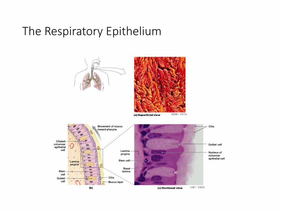
The Respiratory Epithelium

The Respiratory Epithelium
• For gases to exchange efficiently:• alveoli walls must be very thin (< 1 µm)
• surface area must be very great (about 35 times the surface area of the body)

The Respiratory Mucosa
• Consists of:• an epithelial layer
• an areolar layer
• Lines conducting portion of respiratory system

The Lamina Propria
• Underlies areolar tissue
• In the upper respiratory system, trachea, and bronchi:• contains mucous glands that secrete onto epithelial surface
• In the conducting portion of lower respiratory system:• contains smooth muscle cells that encircle lumen of bronchioles

Structure of Respiratory Epithelium
• Changes along respiratory tract
• Alveolar Epithelium
• Is a very delicate, simple squamous epithelium
• Contains scattered and specialized cells
• Lines exchange surfaces of alveoli

How are delicate respiratory exchange surfaces protected from pathogens, debris, and other hazards?

The Respiratory Defense System • Consists of a series of filtration mechanisms
• Removes particles and pathogens
* Components of the Respiratory Defense System
• Goblet cells and mucous glands: produce mucus that bathes exposed surfaces
• Cilia: sweep debris trapped in mucus toward the pharynx (mucus escalator)
• Filtration in nasal cavity removes large particles
• Alveolar macrophages engulf small particles that reach lungs

The Upper Respiratory System

The Nose
• Air enters the respiratory system:• through nostrils or external nares
• into nasal vestibule
• Nasal hairs:• are in nasal vestibule
• are the first particle filtration system

The Nasal Cavity
• The nasal septum:• divides nasal cavity into left and right
• Mucous secretions from paranasal sinus and tears:• clean and moisten the nasal cavity
• Superior portion of nasal cavity is the olfactory region:• provides sense of smell

Air Flow
• From vestibule to internal nares:• through superior, middle, and inferior meatuses
Meatuses• Constricted passageways that produce air turbulence:
• warm and humidify incoming air
• trap particles

The Palates
• Hard palate:• forms floor of nasal cavity
• separates nasal and oral cavities
• Soft palate:• extends posterior to hard palate
• divides superior nasopharynx from lower pharynx
Air Flow
• Nasal cavity opens into nasopharynx through internal nares
The Nasal Mucosa• Warm and humidify inhaled air for arrival at lower respiratory
organs
• Breathing through mouth bypasses this important step

The Pharynx and Divisions
• A chamber shared by digestive and respiratory systems
• Extends from internal nares to entrances to larynx and esophagus
• Nasopharynx
• Oropharynx
• Laryngopharynx

The Nasopharynx• Superior portion of the pharynx
• Contains pharyngeal tonsils and openings to left and right auditory tubes
The Oropharynx
• Middle portion of the pharynx
• Communicates with oral cavity
The Laryngopharynx
• Inferior portion of the pharynx
• Extends from hyoid bone to entrance to larynx and esophagus

What is the structure of the larynx and its role in normal breathing ?

Anatomy of the Larynx
Air Flow-From the pharynx enters the larynx:
a cartilaginous structure that surrounds the
glottis

Cartilages of the Larynx
• 3 large, unpaired cartilages form the larynx:• the thyroid cartilage
• the cricoid cartilage
• the epiglottis

The Thyroid Cartilage
• Also called the Adam’s apple
• Is a hyaline cartilage
• Forms anterior and lateral walls of larynx
• Ligaments attach to hyoid bone, epiglottis, and laryngeal cartilages

The Cricoid Cartilage• Is a hyaline cartilage
• Form posterior portion of larynx
• Ligaments attach to first tracheal cartilage
• Articulates with arytenoid cartilages
The Epiglottis• Composed of elastic cartilage
• Ligaments attach to thyroid cartilage and hyoid bone

Cartilage Functions
• Thyroid and cricoid cartilages support and protect:• the glottis
• the entrance to trachea
• During swallowing:• the larynx is elevated
• the epiglottis folds back over glottis
• Prevents entry of food and liquids into respiratory tract

The Glottis
3 pairs of Small Hyaline Cartilages of the Larynx
arytenoid cartilages, corniculate cartilages cuneiform
cartilages

Cartilage Functions
• Corniculate and arytenoid cartilages function in:• opening and closing of glottis
• production of sound

Ligaments of the Larynx
• Vestibular ligaments and vocal ligaments:• extend between thyroid cartilage and arytenoid cartilages
• are covered by folds of laryngeal epithelium that project into glottis
1) The Vestibular Ligaments• Lie within vestibular folds:
• which protect delicate vocal folds

The Laryngeal Musculature
• The larynx is associated with:• muscles of neck and pharynx
• intrinsic muscles that:• control vocal folds
• open and close glottis
•Coughing reflex: food or liquids went “down the wrong pipe”

Anatomy of the Trachea
Figure 23–6
What is the structure of airways outside the lungs?

The Trachea
• Also called the windpipe
• Extends from the cricoid cartilage into mediastinum• where it branches into right and left pulmonary bronchi – karina
The Submucosa• Beneath mucosa of trachea
• Contains mucous glands

The Tracheal Cartilages
• 15–20 tracheal cartilages:• strengthen and protect airway
• discontinuous where trachea contacts esophagus
• Ends of each tracheal cartilage are connected by:• an elastic ligament and trachealis muscle

The Primary Bronchi
• Right and left primary bronchi:• separated by an internal ridge (the carina)
1) The Right Primary Bronchus• Is larger in diameter than the left
• Descends at a steeper angle

Structure of Primary Bronchi
• Each primary bronchus:• travels to a groove (hilus) along medial surface of the lung
Hilus-• Where pulmonary nerves, blood vessels, and lymphatics enter lung
• Anchored in meshwork of connective tissue

The Root of the Lung
• Complex of connective tissues, nerves, and vessels in hilus:• anchored to the mediastinum

Figure 23–7
Gross Anatomy of the Lungs
Left and right lungs:
are in left and right
pleural cavities
The base:
inferior portion of
each lung rests on
superior surface of
diaphragm
Lobes of the Lungs
• Lungs have lobes separated by deep fissures
1) The Right Lung- Has 3 lobes: • superior, middle, and inferior
• separated by horizontal and oblique fissures
2) The Left Lung- Has 2 lobes:
• superior and inferior
• are separated by an oblique fissure

Relationship between Lungs and Heart
Figure 23–8

Lung Shape
• Right lung:• is wider
• is displaced upward by liver
• Left lung:• is longer
• is displaced leftward by the heart forming the cardiac notch

The Bronchial Tree
• Is formed by the primary bronchi and their branches
• Extrapulmonary Bronchi
• The left and right bronchi branches outside the lungs
• Intrapulmonary Bronchi
• Branches within the lungs

Bronchi and Lobules
A Primary Bronchus
Branches to form
secondary bronchi (lobar
bronchi)
1 secondary bronchus
goes to each lobe

Secondary Bronchi
• Branch to form tertiary bronchi, also called the segmental bronchi
• Each segmental bronchus:
• supplies air to a single bronchopulmonary segment-• The right lung has 10
• The left lung has 8 or 9

Bronchial Structure
• The walls of primary, secondary, and tertiary bronchi:• contain progressively less cartilage and more smooth muscle
• increasing muscular effects on airway constriction and resistance

The Bronchioles
Bronchitis: Inflammation of bronchial
walls: causes constriction
and breathing difficulty

The Bronchioles
• Each tertiary bronchus branches into multiple bronchioles
• Bronchioles branch into terminal bronchioles:• 1 tertiary bronchus forms about 6500 terminal bronchioles

Bronchiole Structure
• Bronchioles:• have no cartilage
• are dominated by smooth muscle
Autonomic Control• Regulates smooth muscle:
• controls diameter of bronchioles
• controls airflow and resistance in lungs

Bronchodilation
• Dilation of bronchial airways
• Caused by sympathetic ANS activation
• Reduces resistance
Bronchoconstriction• Constricts bronchi
• Caused by: • parasympathetic ANS activation
• histamine release (allergic reactions)

Asthma
• Excessive stimulation and bronchoconstriction
• Stimulation severely restricts airflow

Pulmonary Lobules
• Are the smallest compartments of the lung
• Are divided by the smallest trabecular partitions (interlobular septa)
• Each terminal bronchiole delivers air to a single pulmonary lobule
• Each pulmonary lobule is supplied by pulmonary arteries and veins

Exchange Surfaces
• Within the lobule:• each terminal bronchiole branches to form several respiratory bronchioles,
where gas exchange takes place

Alveolar OrganizationRespiratory bronchioles
are connected to alveoli
along alveolar ducts
Alveolar ducts end at
alveolar sacs:
common chambers
connected to many
individual alveoli

An Alveolus
• Has an extensive network of capillaries
• Is surrounded by elastic fibers
Alveolar Epithelium
• Consists of simple squamous epithelium
• Consists of thin, delicate Type I cells
• Patrolled by alveolar macrophages, also called dust cells
• Contains septal cells (Type II cells) that produce Surfactant- an oily secretion which
• 1) Contains phospholipids and proteins
• 2) Coats alveolar surfaces and reduces surface tension

Respiratory Distress
• Difficult respiration:• due to alveolar collapse
• caused when septal cells do not produce enough surfactant

Respiratory Membrane - The thin membraneof alveoli where gas exchange takes place
3 Parts of the Respiratory Membrane
• Squamous epithelial lining of alveolus
• Endothelial cells lining an adjacent capillary
• Fused basal laminae between alveolar and endothelial cells
• Diffusion- Across respiratory membrane is very rapid:• because distance is small
• gases (O2 and CO2) are lipid soluble

Inflammation of Lobules
• Also called pneumonia:• causes fluid to leak into alveoli
• compromises function of respiratory membrane

Blood Supply to Respiratory Surfaces
• Each lobule receives an arteriole and a venule1. respiratory exchange surfaces receive blood:
• from arteries of pulmonary circuit
2. a capillary network surrounds each alveolus:• as part of the respiratory membrane
3. blood from alveolar capillaries:• passes through pulmonary venules and veins
• returns to left atrium

Blood Supply to the Lungs
• Capillaries supplied by bronchial arteries:• provide oxygen and nutrients to tissues of conducting passageways of lung
• Venous blood bypasses the systemic circuit and flows into pulmonary veins

Blood Pressure
• In pulmonary circuit is low (30 mm Hg)
• Pulmonary vessels are easily blocked by blood clots, fat, or air bubbles, causing pulmonary embolism

Figure 23–8
Pleural Cavities and Pleural Membranes

Pleural Cavities and Pleural Membranes
• 2 pleural cavities:• are separated by the mediastinum
• Each pleural cavity:• holds a lung
• is lined with a serous membrane (the pleura)
• Pleura consist of 2 layers: • parietal pleura -- rib
• visceral pleura -- lung
• Pleural fluid:• lubricates space between 2 layers

Respiration
• Refers to 2 integrated processes:
•External respiration-Includes all processes involved in exchanging O2 and CO2 with the environment
•Internal respiration- Also called cellular respiration
• Involves the uptake of O2 and production of CO2 within individual cells

3 Processes of External Respiration
1. Pulmonary ventilation (breathing)
2. Gas diffusion:• across membranes and capillaries
3. Transport of O2 and CO2:• between alveolar capillaries
• between capillary beds in other tissues

Intrapulmonary Pressure
• Also called intra-alveolar pressure
• Is relative to Patm
• In relaxed breathing, the difference between Patm and intrapulmonary pressure is small:• about —1 mm Hg on inhalation or +1 mm Hg on expiration

The Respiratory Pump
• Cyclical changes in intrapleural pressure operate the respiratory pump:• which aids in venous return to heart
Tidal Volume• Amount of air moved in and out of lungs in a single respiratory cycle

Injury to the Chest Wall
• Pneumothorax:
•allows air into pleural cavity• Atelectasis:
•also called a collapsed lung•result of pneumothorax

What are the origins and actions of the respiratory muscles responsible for respiratory movements?
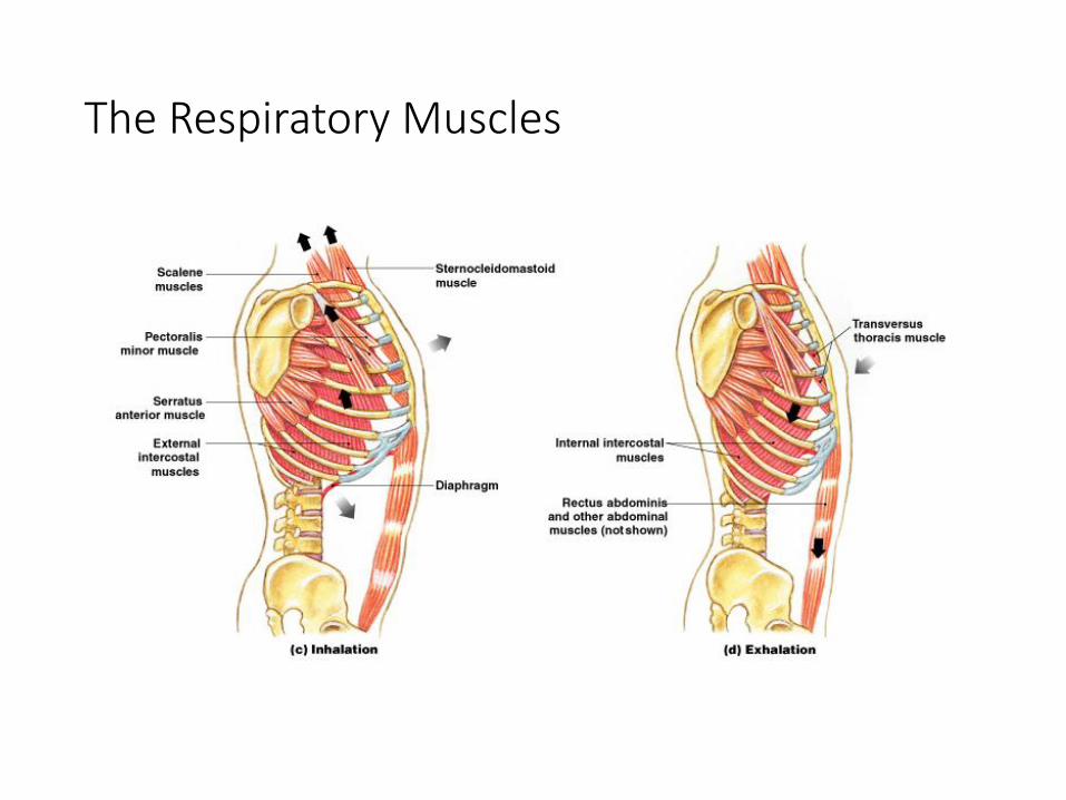
The Respiratory Muscles
The Mechanics of Breathing
• Inhalation:• always active
• Exhalation:• active or passive

3 Muscle Groups of Inhalation
1. Diaphragm:• contraction draws air into lungs
• 75% of normal air movement
2. External intracostal muscles:• assist inhalation
• 25% of normal air movement
3. Accessory muscles assist in elevating ribs:• sternocleidomastoid
• serratus anterior
• pectoralis minor
• scalene muscles

Muscles of Active Exhalation
1. Internal intercostal and transversus thoracis muscles:• depress the ribs
2. Abdominal muscles:• compress the abdomen
• force diaphragm upward

Modes of Breathing
• Respiratory movements are classified:• by pattern of muscle activity
• into quiet breathing and forced breathing

Quiet Breathing (Eupnea) • Involves active inhalation and passive exhalation
• Diaphragmatic breathing or deep breathing:• is dominated by diaphragm
• Costal breathing or shallow breathing:• is dominated by ribcage movements
Elastic Rebound• When inhalation muscles relax:
• elastic components of muscles and lungs recoil
• returning lungs and alveoli to original position

Forced Breathing
• Also called hyperpnea
• Involves active inhalation and exhalation
• Assisted by accessory muscles
• Maximum levels occur in exhaustion

Respiratory Rates and Volumes
• Respiratory system adapts to changing oxygen demands by varying:• the number of breaths per minute (respiratory rate)
• the volume of air moved per breath (tidal volume)
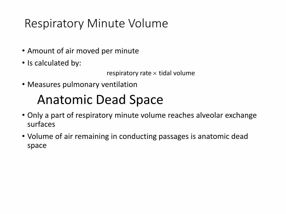
Respiratory Minute Volume
• Amount of air moved per minute
• Is calculated by:respiratory rate tidal volume
• Measures pulmonary ventilation
Anatomic Dead Space• Only a part of respiratory minute volume reaches alveolar exchange
surfaces
• Volume of air remaining in conducting passages is anatomic dead space

Alveolar Ventilation
• Amount of air reaching alveoli each minute
• Calculated as:(tidal volume — anatomic dead space) respiratory rate
• Alveoli contain less O2, more CO2 than atmospheric air:• because air mixes with exhaled air

Alveolar Ventilation Rate
• Determined by respiratory rate and tidal volume:• for a given respiratory rate:
• increasing tidal volume increases alveolar ventilation rate
• for a given tidal volume:• increasing respiratory rate increases alveolar ventilation

Respiratory Volumes and Capacities
Gas Exchange
• Occurs between blood and alveolar air
• Across the respiratory membrane
• Depends on:• partial pressures of the gases
• diffusion of molecules between gas and liquid

Gas Content & Solubility in body fluids
• The actual amount of a gas in solution (at given partial pressure and temperature) depends on the solubility of that gas in that particular liquid
• CO2 is very soluble
• O2 is less soluble
• N2 has very low solubility

How is oxygen picked up, transported, and released in the blood?What is the structure and function of hemoglobin?

Gas Pickup and Delivery
• Blood plasma can’t transport enough O2 or CO2 to meet physiological needs
Red Blood Cells (RBCs)• Transport O2 to, and CO2 from, peripheral tissues
• Remove O2 and CO2 from plasma, allowing gases to diffuse into blood

Oxygen Transport
• O2 binds to iron ions in hemoglobin (Hb) molecules:• in a reversible reaction
• Each RBC has about 280 million Hb molecules:• each binds 4 oxygen molecules -saturated
• The percentage of heme units in a hemoglobin molecule:• that contain bound oxygen

Oxyhemoglobin Saturation Curve
Figure 23–20 (Navigator)
Environmental Factors Affecting Hemoglobin
PO2 of blood, Blood pH, Temperature
Metabolic activity within RBCs

Oxyhemoglobin Saturation Curve
• Is a graph relating the saturation of hemoglobin to partial pressure of oxygen:• higher PO
2results in greater Hb saturation
• Is a curve rather than a straight line:• because Hb changes shape each time a molecule of O2 is bound
• each O2 bound makes next O2 binding easier
• allows Hb to bind O2 when O2 levels are low

Oxygen Reserves• O2 diffuses:
• from peripheral capillaries (high PO2)
• into interstitial fluid (low PO2)
• Amount of O2 released depends on interstitial PO2
• Up to 3/4 may be reserved by RBCs
Carbon Monoxide
• CO from burning fuels:• binds strongly to hemoglobin
• takes the place of O2
• can result in carbon monoxide poisoning

pH, Temperature, and Hemoglobin Saturation

The Oxyhemoglobin Saturation Curve
• Is standardized for normal blood (pH 7.4, 37°C)
• When pH drops or temperature rises:• more oxygen is released
• curve shift to right
• When pH rises or temperature drops:• less oxygen is released
• curve shifts to left

Fetal and Adult Hemoglobin

Fetal and Adult Hemoglobin
• The structure of fetal hemoglobin:• differs from that of adult Hb
• At the same PO2:
• fetal Hb binds more O2 than adult Hb
• which allows fetus to take O2 from maternal blood

KEY CONCEPT• Hemoglobin in RBCs:
• carries most blood oxygen
• releases it in response to low O2 partial pressure in surrounding plasma
• If PO2
increases, hemoglobin binds oxygen
• If PO2
decreases, hemoglobin releases oxygen
• At a given PO2:
• hemoglobin will release additional oxygen
• if pH decreases or temperature increases

How is carbon dioxide transported in the blood?Carbon Dioxide Transport

Carbon Dioxide (CO2)• Is generated as a byproduct of aerobic metabolism (cellular respiration)
CO2 in the Blood Stream
• May be:• converted to carbonic acid
• bound to protein portion of hemoglobin
• dissolved in plasma
Bicarbonate Ions• Move into plasma by an exchange mechanism (the chloride shift) that takes
in Cl— ions without using ATP

CO2 in the Blood Stream
• 70% is transported as carbonic acid (H2CO3):• which dissociates into H+ and bicarbonate (HCO3
—)
• 23% is bound to amino groups of globular proteins in Hb molecule:• forming carbaminohemoglobin
• 7% is transported as CO2 dissolved in plasma

KEY CONCEPT
• CO2 travels in the bloodstream primarily as bicarbonate ions, which form through dissociation of carbonic acid produced by carbonic anhydrase in RBCs
• Lesser amounts of CO2 are bound to Hb or dissolved in plasma
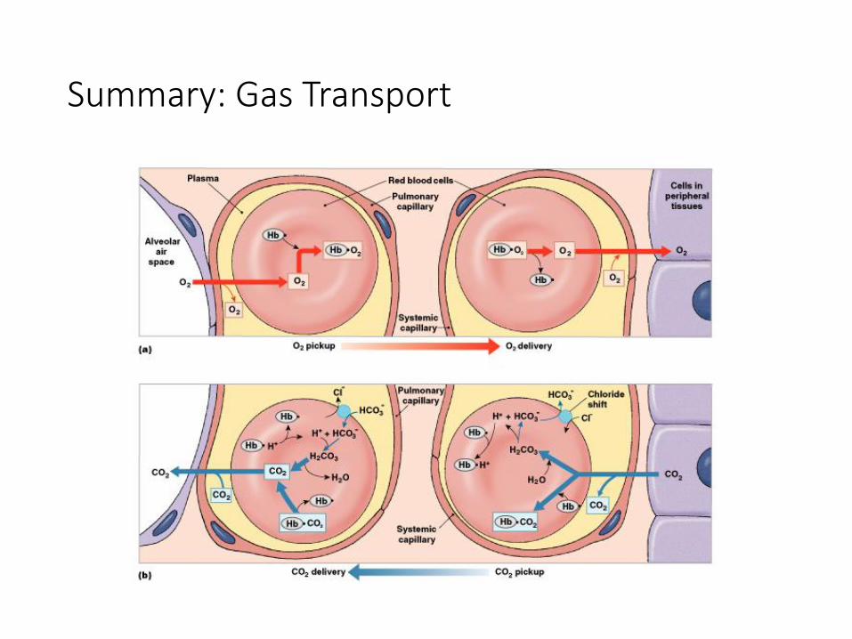
Summary: Gas Transport
Figure 23–24

Control of Respiration
• Gas diffusion at peripheral and alveolar capillaries maintain balance by:• changes in blood flow and oxygen delivery
• changes in depth and rate of respiration
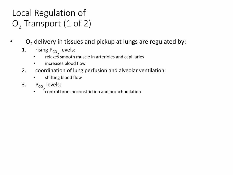
Local Regulation of O2 Transport (1 of 2)
• O2 delivery in tissues and pickup at lungs are regulated by:1. rising PCO
2levels:
• relaxes smooth muscle in arterioles and capillaries
• increases blood flow
2. coordination of lung perfusion and alveolar ventilation:• shifting blood flow
3. PCO2
levels:• control bronchoconstriction and bronchodilation

Respiratory Centers of the Brain
• When oxygen demand rises:• cardiac output and respiratory rates increase under neural control
• Have both voluntary and involuntary components
Involuntary Centers• Regulate respiratory muscles
• In response to sensory information
Voluntary Centers
• In cerebral cortex affect:• respiratory centers of pons and medulla oblongata
• motor neurons that control respiratory muscles
•The Respiratory Centers• 3 pairs of nuclei in the reticular formation of medulla oblongata and
pons
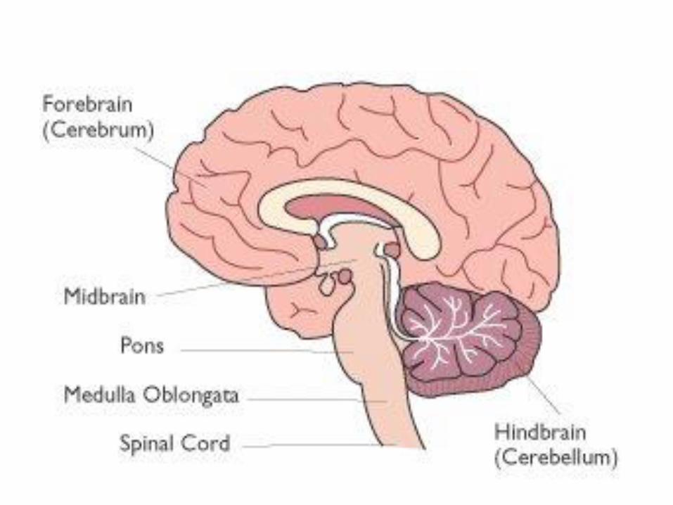
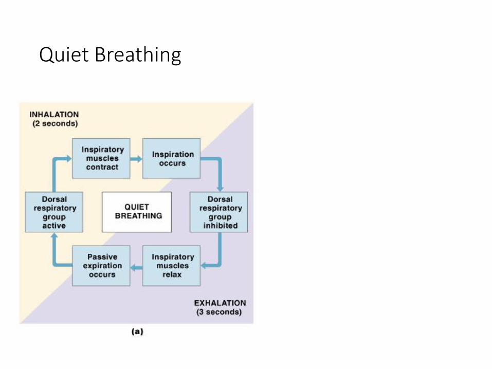
Figure 23–25a
Quiet Breathing
Brief activity in
the DRG:
stimulates
inspiratory
muscles
DRG neurons
become
inactive:
allowing
passive
exhalation

Figure 23–25b
Forced Breathing
Increased activity in
DRG:
stimulates VRG
which activates
accessory inspiratory
muscles
After inhalation:
expiratory center
neurons stimulate
active exhalation
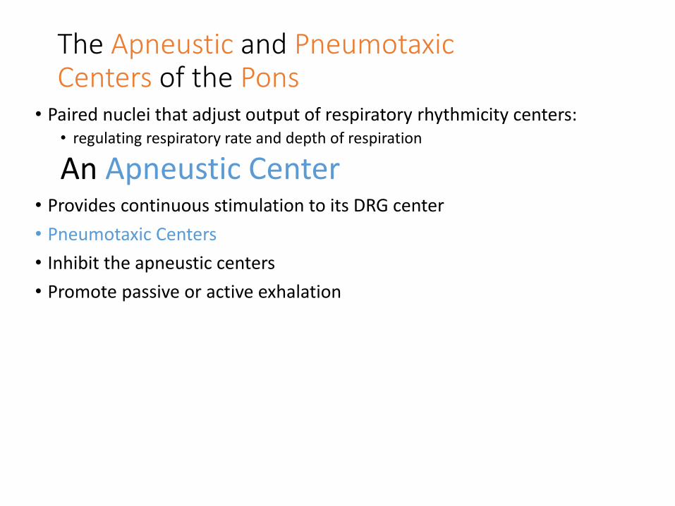
The Apneustic and PneumotaxicCenters of the Pons
• Paired nuclei that adjust output of respiratory rhythmicity centers:• regulating respiratory rate and depth of respiration
An Apneustic Center• Provides continuous stimulation to its DRG center
• Pneumotaxic Centers
• Inhibit the apneustic centers
• Promote passive or active exhalation

Respiratory Centers and Reflex Controls
Figure 23–26
Interactions between VRG
and DRG:
establish basic pace and
depth of respiration
The pneumotaxic center:
modifies the pace

5 Sensory Modifiers of Respiratory Center Activities
• Chemoreceptors are sensitive to:
• PCO2, PO
2, or pH
• of blood or cerebrospinal fluid
• Baroreceptors in aortic or carotic sinuses:
• sensitive to changes in blood pressure

5 Sensory Modifiers of Respiratory Center Activities
• Stretch receptors:• respond to changes in lung volume
• Irritating physical or chemical stimuli:• in nasal cavity, larynx, or bronchial tree
• Other sensations including:• pain
• changes in body temperature
• abnormal visceral sensations

Chemoreceptor Reflexes• Respiratory centers are strongly influenced by chemoreceptor input from:
* cranial nerve IX -The glossopharyngeal nerve:
• from carotid bodies
• stimulated by changes in blood pH or PO2
* cranial nerve X -The vagus nerve:
• from aortic bodies
• stimulated by changes in blood pH or PO2
* receptors that monitor cerebrospinal fluid-
• Are on ventrolateral surface of medulla oblongata
• Respond to PCO2
and pH of CSF

Chemoreceptor Responses to PCO2
Figure 23–27

Hypercapnia- An increase in arterial PCO2
• Stimulates chemoreceptors in the medulla oblongata:• to restore homeostasis
Hypoventilation- A common cause of hypercapnia
• Abnormally low respiration rate:• allows CO2 build-up in blood
Hyperventilation-Excessive ventilation
• Results in abnormally low PCO2
(hypocapnia)
• Stimulates chemoreceptors to decrease respiratory rate

Baroreceptor Reflexes
• Carotid and aortic baroreceptor stimulation:• affects blood pressure and respiratory centers
• When blood pressure falls:• respiration increases
• When blood pressure increases:• respiration decreases

Protective Reflexes
• Triggered by receptors in epithelium of respiratory tract when lungs are exposed to:• toxic vapors
• chemicals irritants
• mechanical stimulation
• Cause sneezing, coughing, and laryngeal spasm

Apnea
• A period of suspended respiration
• Normally followed by explosive exhalation to clear airways:• sneezing and coughing
Laryngeal Spasm• Temporarily closes airway:
• to prevent foreign substances from entering

The Cerebral Cortex and Respiratory Centers
1. Strong emotions:• can stimulate respiratory centers in hypothalamus
2. Temporarily closes airway:• to prevent foreign substances from entering
3. Anticipation of strenuous exercise:• can increase respiratory rate and cardiac output
• by sympathetic stimulation
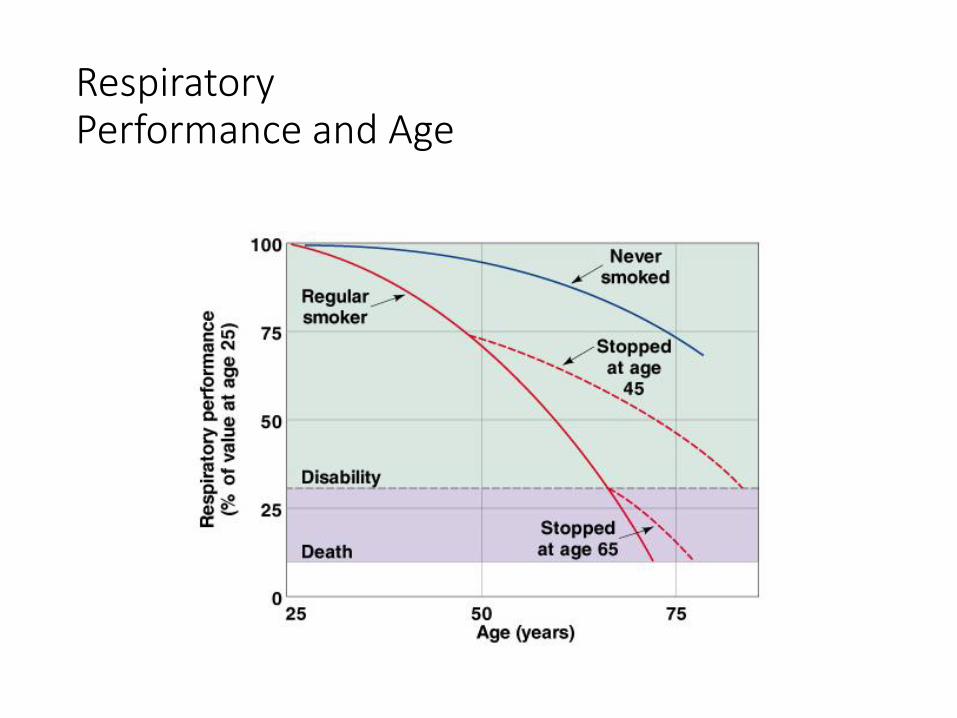
Respiratory Performance and Age
Figure 23–28

3 Effects of Aging on the Respiratory System
1. Elastic tissues deteriorate:• reducing lung compliance
• lowering vital capacity
2. Arthritic changes:• restrict chest movements
• limit respiratory minute volume
3. Emphysema:• affects individuals over age 50
• depending on exposure to respiratory irritants (e.g., cigarette smoke)

Integration with Other Systems
• Maintaining homeostatic O2 and CO2 levels in peripheral tissues requires coordination between several systems: • particularly the respiratory and cardiovascular systems

Coordination of Respiratory and Cardiovascular Systems
1. Improves efficiency of gas exchange:• by controlling lung perfusion
2. Increases respiratory drive:• through chemoreceptor stimulation
3. Raises cardiac output and blood flow:• through baroreceptor stimulation

Figure 23–29
The Respiratory System and Other Systems


![Respiratory system roadmap.pptx [Repaired] - Loginanatomical-sciences.health.wits.ac.za/roadmaps/Respiratory system... · DIVISION OF THE RESPIRATORY SYSTEM CONDUCTING PORTION Nasal](https://img.pdfslide.us/doc/110x75/5a78c3d87f8b9ae6228c9db0/respiratory-system-repaired-loginanatomical-scienceshealthwitsaczaroadmapsrespiratory.jpg)


![Respiratory System [โหมดความเข้ากันได้] · PATHOLOGY OF RESPIRATORY SYSTEM นพ. อรรณพ นาคะป ท Respiratory system U it](https://img.pdfslide.us/doc/110x75/5fa578efd4e80f055f6b3401/respiratory-system-aaaaaaaaaaaaaaaaaa-pathology.jpg)