Embed Size (px)
Citation preview

The Ramachandran diagram.

Allowed phi and psi torsion angles in proteins.

The Ramachandran diagram of Gly residues in a polypeptide chain.
CαN
CαO Cα
N
Cα
O
Cis/Trans Isomerization: Proline
trans cis
Energy difference between these forms is small.
Nearly all Xaa-Pro linkages are biosynthesized in the trans form.
~10% of these peptide bonds are in the cis form in globular proteins.
Interconversion catalyzed by peptidyl prolyl cis-trans isomerases
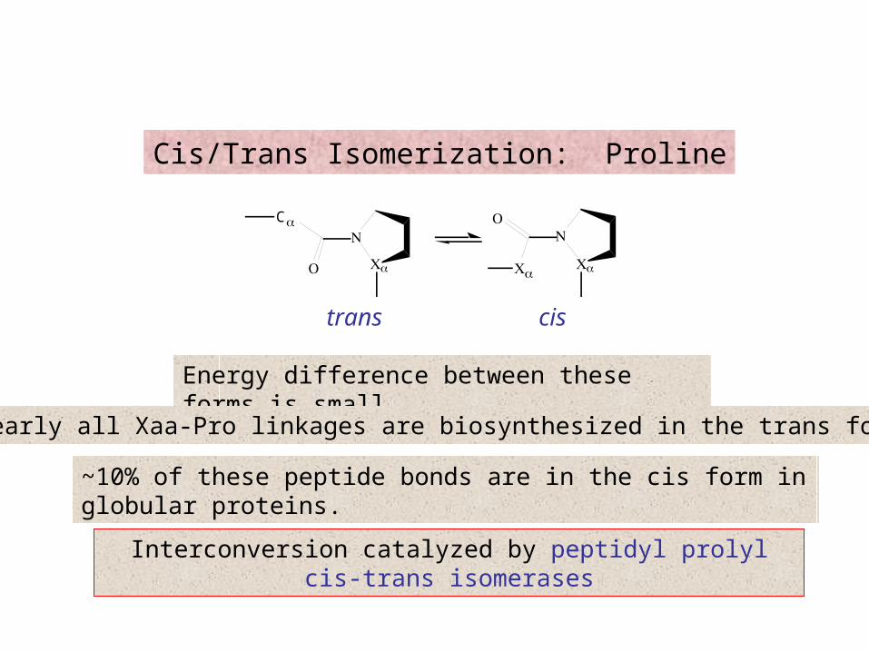
Stereo space-filling representation of an α helical segment of sperm whale myoglobin (its E-helix) as determined by X-ray crystal
structure analysis.
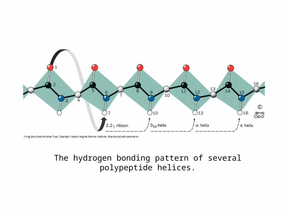
The hydrogen bonding pattern of several polypeptide helices.

Comparison of the two polypeptide helices that occasionally occur in proteins with the commonly occurring α helix.

pleated sheet: antiparallel orientation

pleated sheets: parallel orientation

A two-stranded antiparallel pleated sheet drawn to emphasize its pleated appearance.
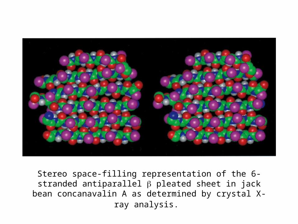
Stereo space-filling representation of the 6-stranded antiparallel pleated sheet in jack bean concanavalin A as determined by crystal
X-ray analysis.
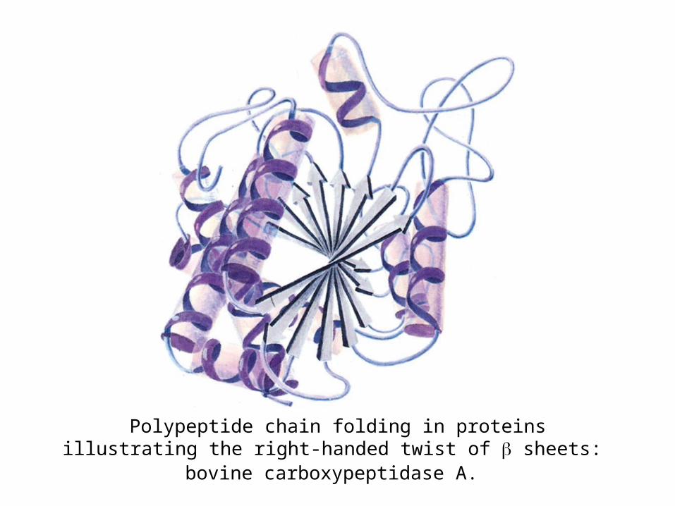
Polypeptide chain folding in proteins illustrating the right-handed twist of sheets: bovine carboxypeptidase A.

Polypeptide chain folding in proteins illustrating the right-handed twist of sheets: chicken muscle triose phosphate isomerase.
( barrel)

Connections between adjacent polypeptide strands in pleated sheets.
hairpin
out-of-planecrossovers
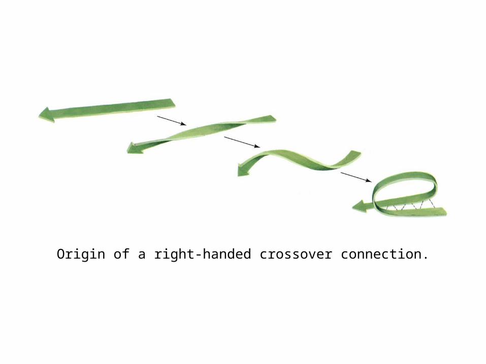
Origin of a right-handed crossover connection.

Reverse turns in polypeptide chains.

Space-filling representation of an Ω loop comprising residues 40 to 54 of cytochrome c.

The structure of α keratin.
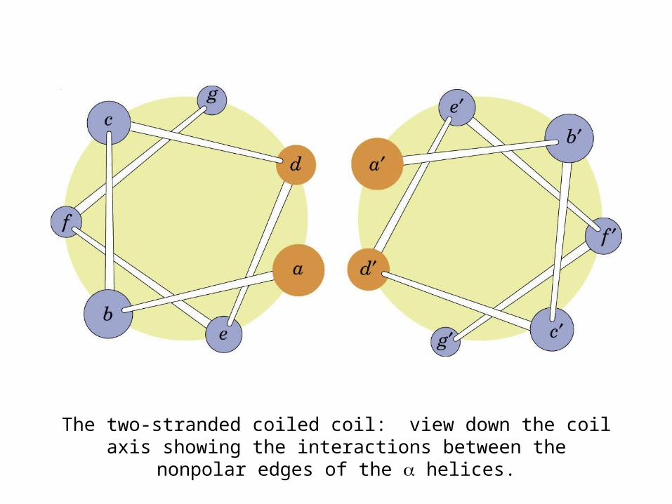
The two-stranded coiled coil: view down the coil axis showing the interactions between the nonpolar edges of the α helices.
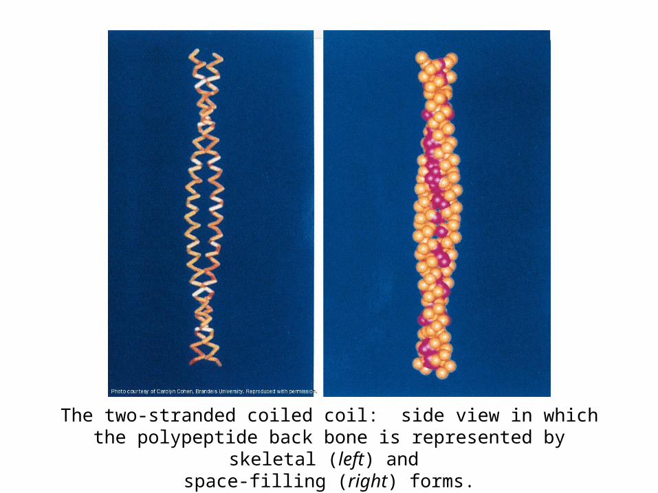
The two-stranded coiled coil: side view in which the polypeptide back bone is represented by skeletal (left) and
space-filling (right) forms.
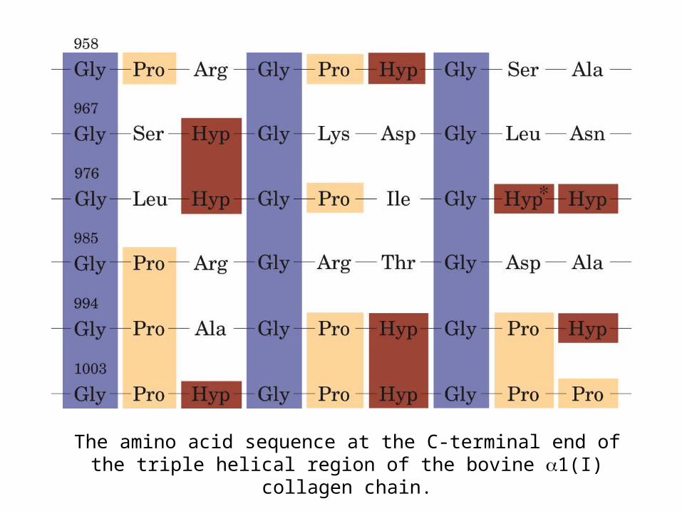
The amino acid sequence at the C-terminal end of the triple helical region of the bovine α1(I) collagen chain.
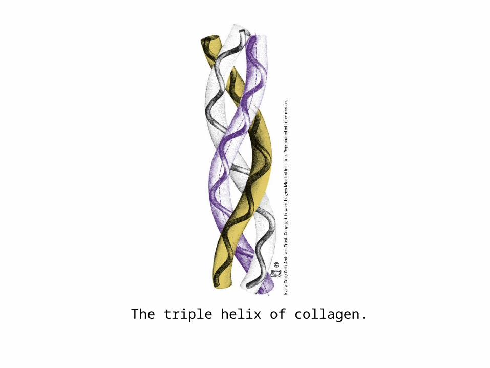
The triple helix of collagen.

X-Ray structure of the triple helical collagen model peptide (Pro-Hyp-Gly)10 in which the fifth Gly is replaced by Ala. (a) Ball and stick
representation.
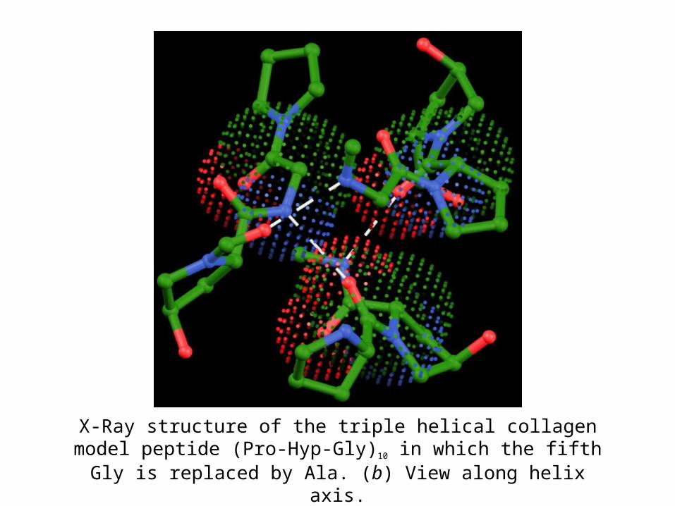
X-Ray structure of the triple helical collagen model peptide (Pro-Hyp-Gly)10 in which the fifth Gly is replaced by Ala. (b) View along
helix axis.

X-Ray structure of the triple helical collagen model peptide (Pro-Hyp-Gly)10 in which the fifth Gly is replaced by Ala. (c) A schematic
diagram.

A biosynthetic pathway for cross-linking Lys, Hyl, and His side chains in collagen.

X-Ray diffraction photograph of a single crystal of sperm whale myoglobin.
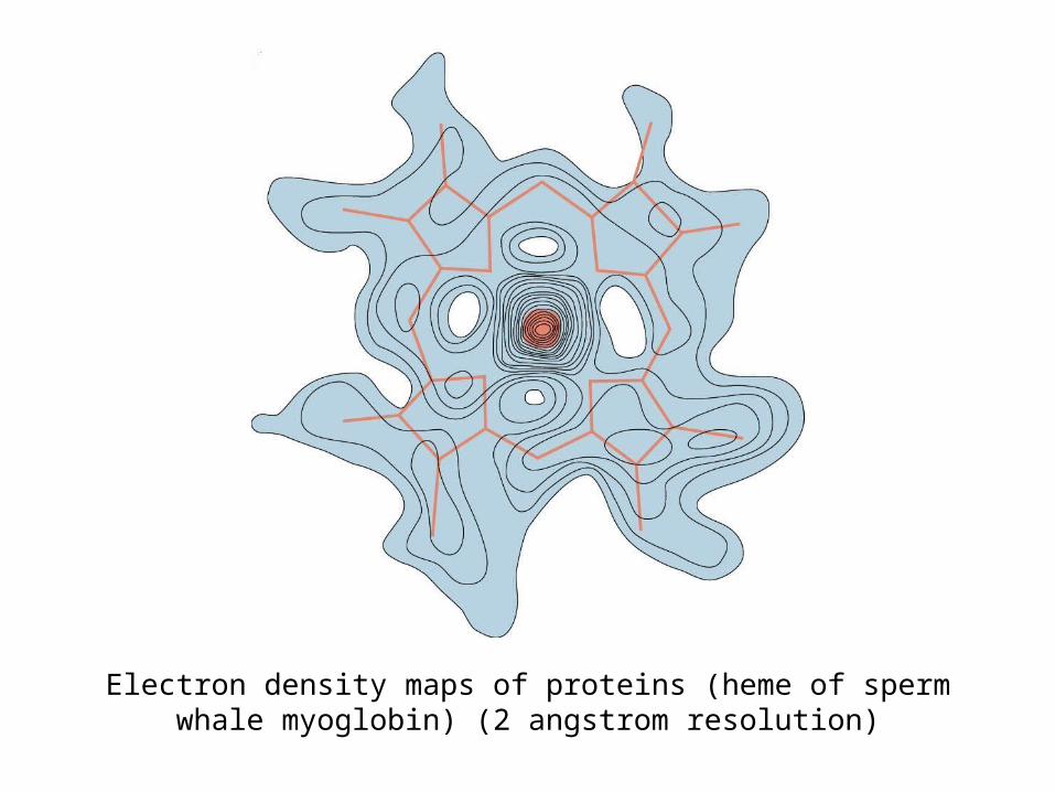
Electron density maps of proteins (heme of sperm whale myoglobin) (2 angstrom resolution)

Electron density maps of proteins (sperm whale myoglobin)(2.4 angstrom resolution)

Sections through the electron density map of diketopiperazine calculated at the indicated resolution.
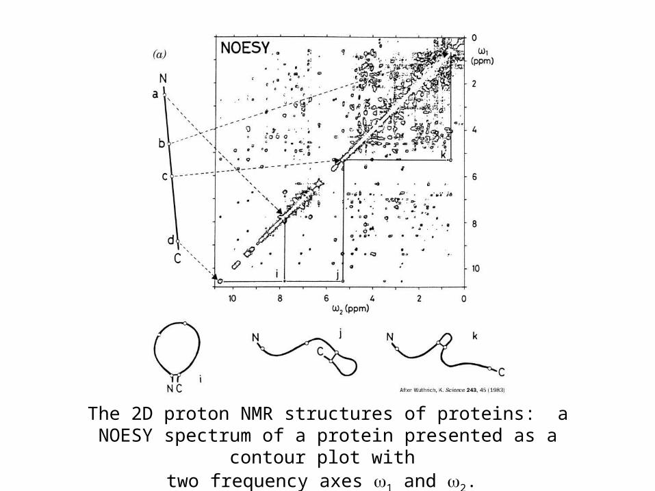
The 2D proton NMR structures of proteins: a NOESY spectrum of a protein presented as a contour plot with
two frequency axes 1 and 2.
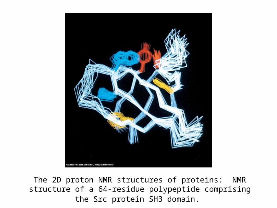
The 2D proton NMR structures of proteins: NMR structure of a 64-residue polypeptide comprising the Src protein SH3 domain.
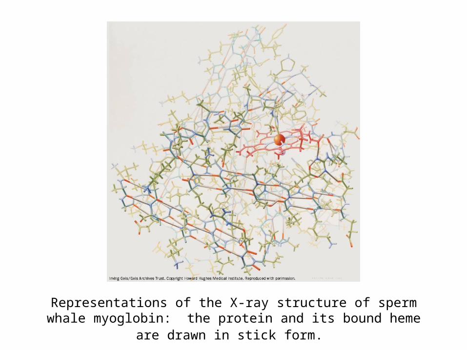
Representations of the X-ray structure of sperm whale myoglobin: the protein and its bound heme are drawn in stick form.

Representations of the X-ray structure of sperm whale myoglobin: a diagram in which the protein is represented by its computer-
generated Cα backbone.
8 helices

Representations of the X-ray structure of sperm whale myoglobin: a computer-generated cartoon drawing.
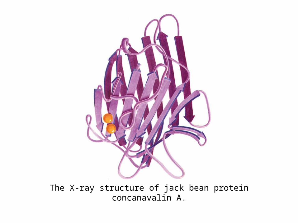
The X-ray structure of jack bean protein concanavalin A.
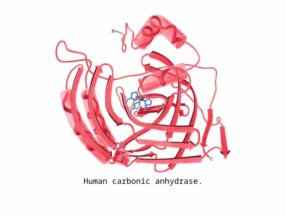
Human carbonic anhydrase.
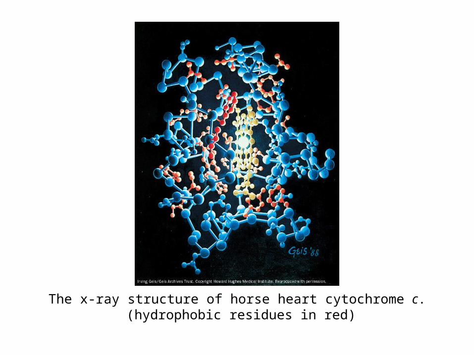
The x-ray structure of horse heart cytochrome c. (hydrophobic residues in red)

The x-ray structure of horse heart cytochrome c. (hydrophilic residues in green)

Representations of the x-ray structure of sperm whale myoglobin: a diagram in which the protein is represented by its computer-
generated Cα backbone.
H-helix

The H helix of sperm whale myoglobin. (a) A helical wheel representation in which the side chain positions about the α helix are
projected down the helix axis onto a plane.
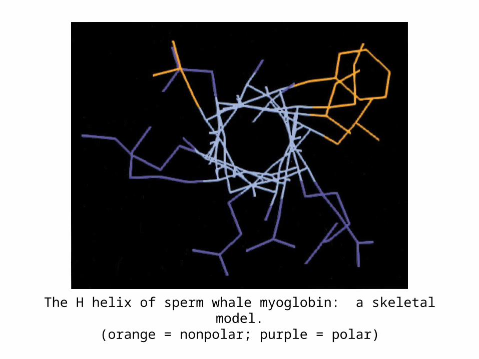
The H helix of sperm whale myoglobin: a skeletal model.(orange = nonpolar; purple = polar)
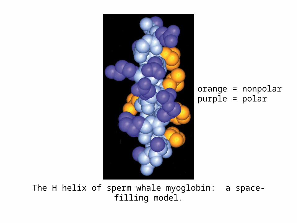
The H helix of sperm whale myoglobin: a space-filling model.
orange = nonpolarpurple = polar

A space-filling model of an antiparallel sheet from concanavalin A.
red = nonpolarpurple = polar
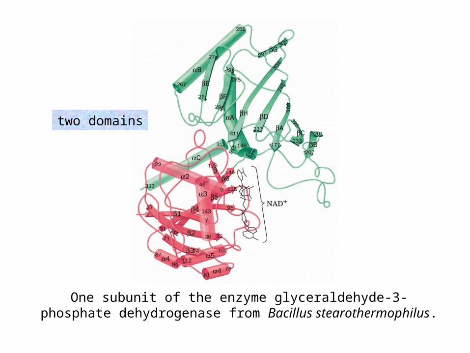
One subunit of the enzyme glyceraldehyde-3-phosphate dehydrogenase from Bacillus stearothermophilus.
two domains
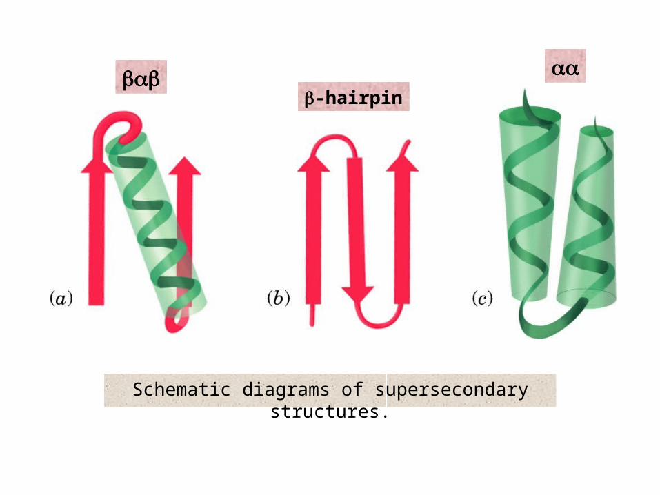
Schematic diagrams of supersecondary structures.
α-hairpin
αα

Schematic diagrams of supersecondary structures.
Greek key motif

X-ray structures of 4-helix bundle proteins: E. coli cytochrome b562.
directionality of helices
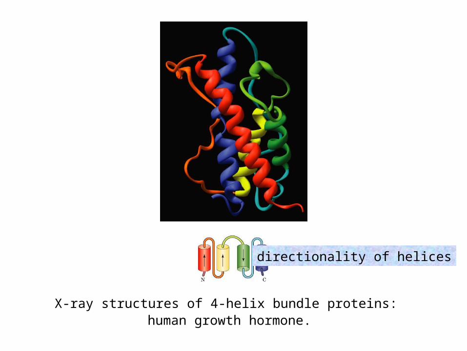
X-ray structures of 4-helix bundle proteins: human growth hormone.
directionality of helices
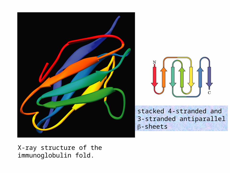
X-ray structure of the immunoglobulin fold.
stacked 4-stranded and3-stranded antiparallel-sheets

X-ray structure of retinol binding protein.
Up-down -barrel
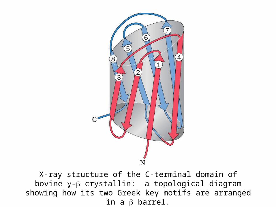
X-ray structure of the C-terminal domain of bovine - crystallin: a topological diagram showing how its two Greek key motifs are
arranged in a barrel.
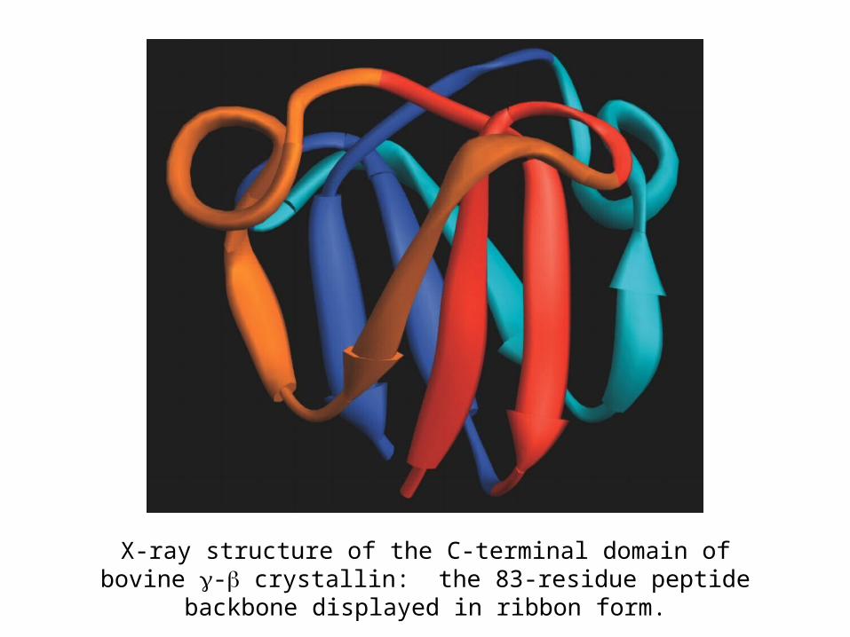
X-ray structure of the C-terminal domain of bovine - crystallin: the 83-residue peptide backbone displayed in ribbon form.

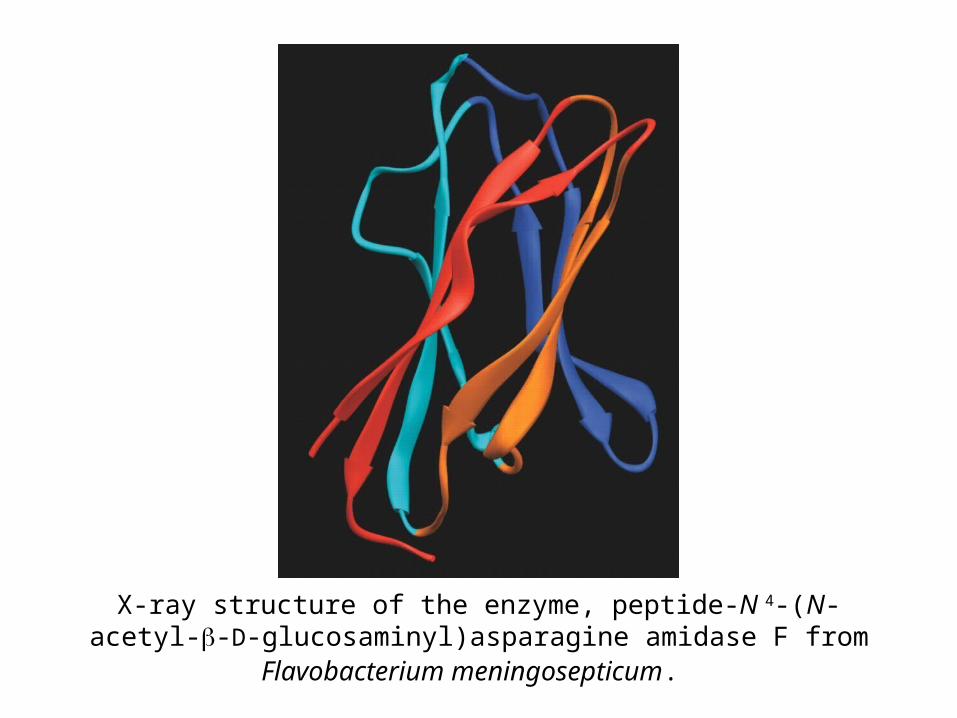
X-ray structure of the enzyme, peptide-N4-(N-acetyl--D-glucosaminyl)asparagine amidase F from Flavobacterium
meningosepticum.
X-ray structure of the enzyme, peptide-N 4-(N-acetyl--D-glucosaminyl)asparagine amidase F from Flavobacterium
meningosepticum.

The X-ray structure of the 247-residue enzyme triose phosphate isomerase (TIM) from chicken muscle.
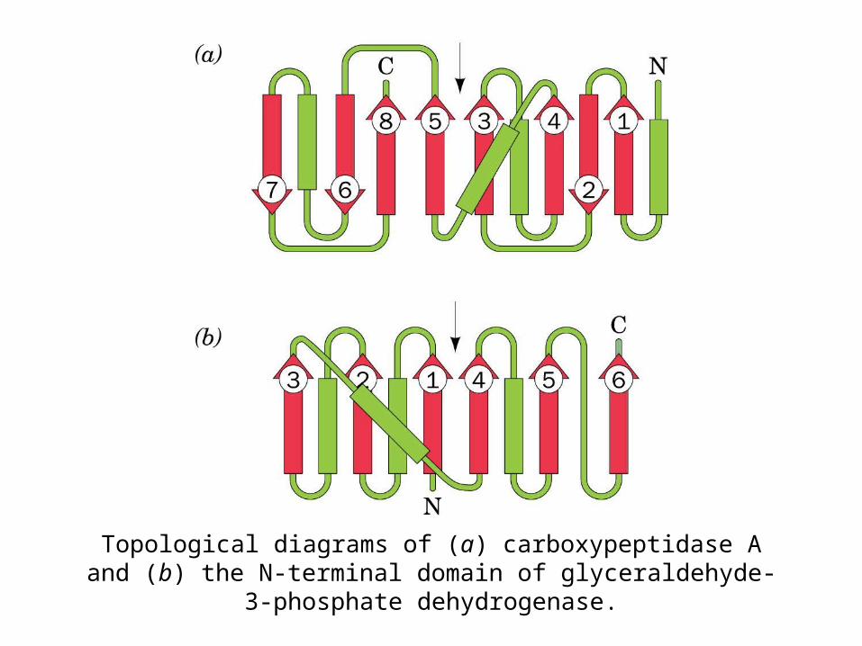
Topological diagrams of (a) carboxypeptidase A and (b) the N-terminal domain of glyceraldehyde-3-phosphate dehydrogenase.

X-ray structures of open sheet-containing enzymes: dogfish lactate dehydrogenase, N-terminal domain (residues 20-163 of this
330-residue protein).

X-ray structures of open sheet-containing enzymes: porcine adenylate kinase (195 residues).
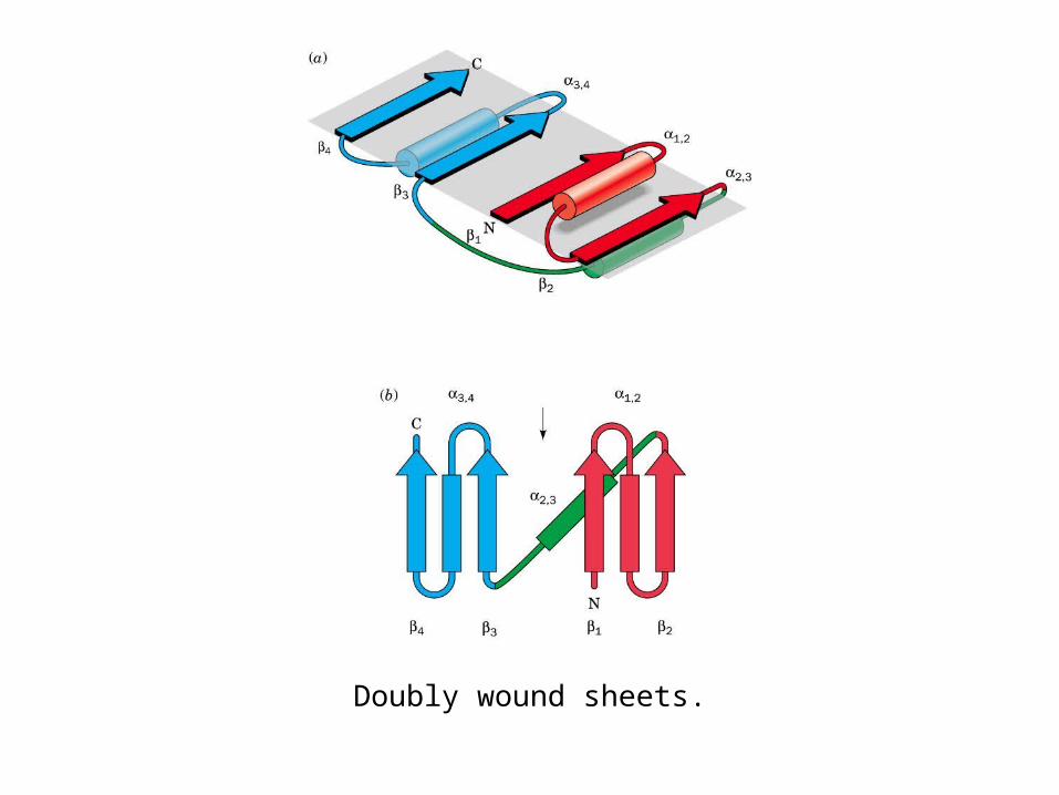
Doubly wound sheets.

A GRASP diagram of human growth hormone (helps predict protein interactions with charged molecules)
Graphical Representationand Analysis of
SurfaceProperties

Thermodynamic changes for transferring hydrocarbons from water to nonpolar solvents at 25°C.

Hydropathy Scale for Amino Acid Side Chains

Hydropathic index plot for bovine chymotrypsinogen.

Protein denaturation
curve
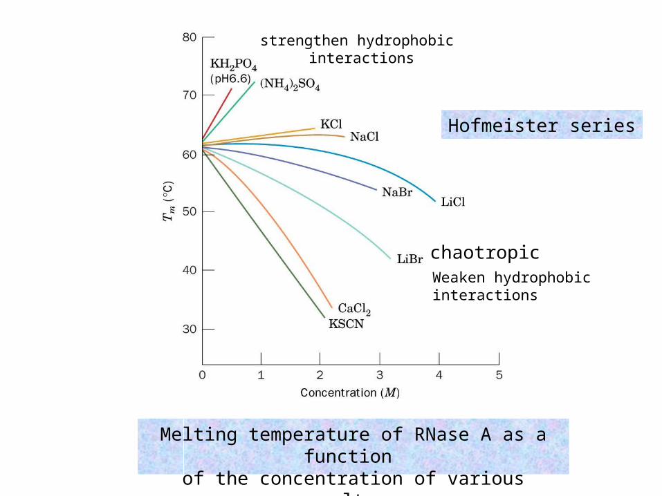
Melting temperature of RNase A as a function of the concentration of various salts.
chaotropic
strengthen hydrophobic interactions
Weaken hydrophobicinteractions
Hofmeister series
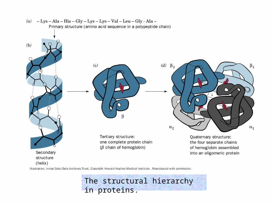
The structural hierarchy in proteins.

The quaternary structure of hemoglobin
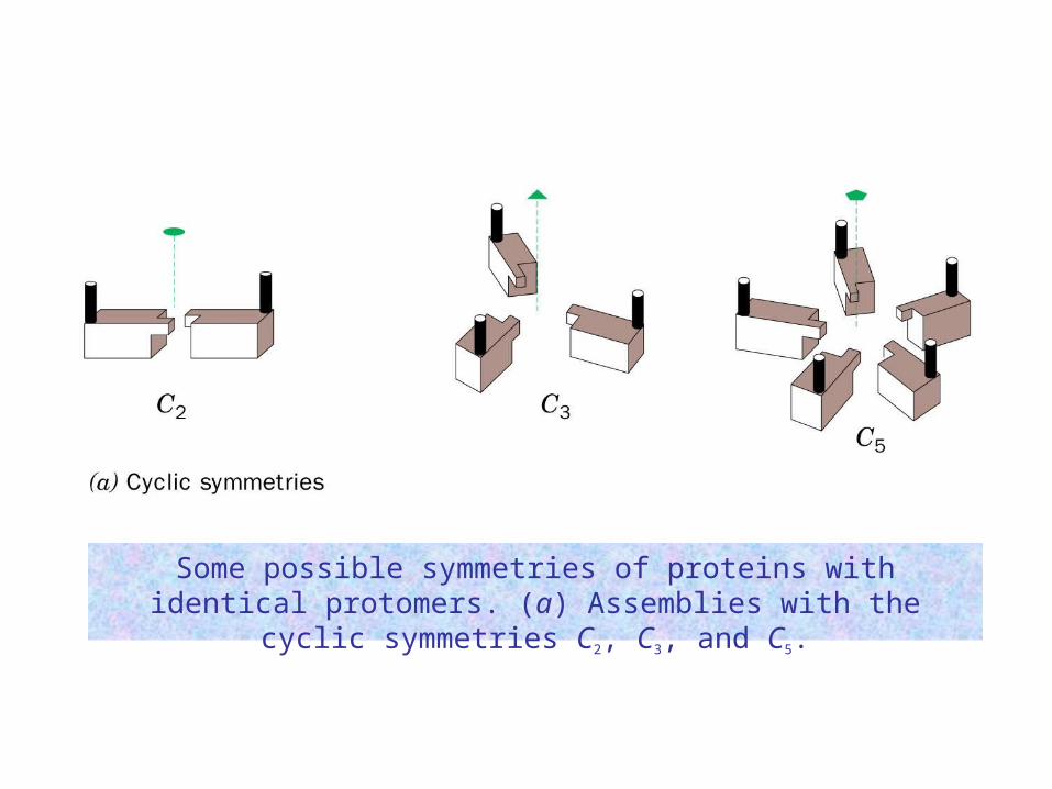
Some possible symmetries of proteins with identical protomers. (a) Assemblies with the cyclic symmetries C2, C3, and C5.

Some possible symmetries of proteins with identical protomers. (b) Assemblies with the dihedral symmetries D2, D4, and D3.
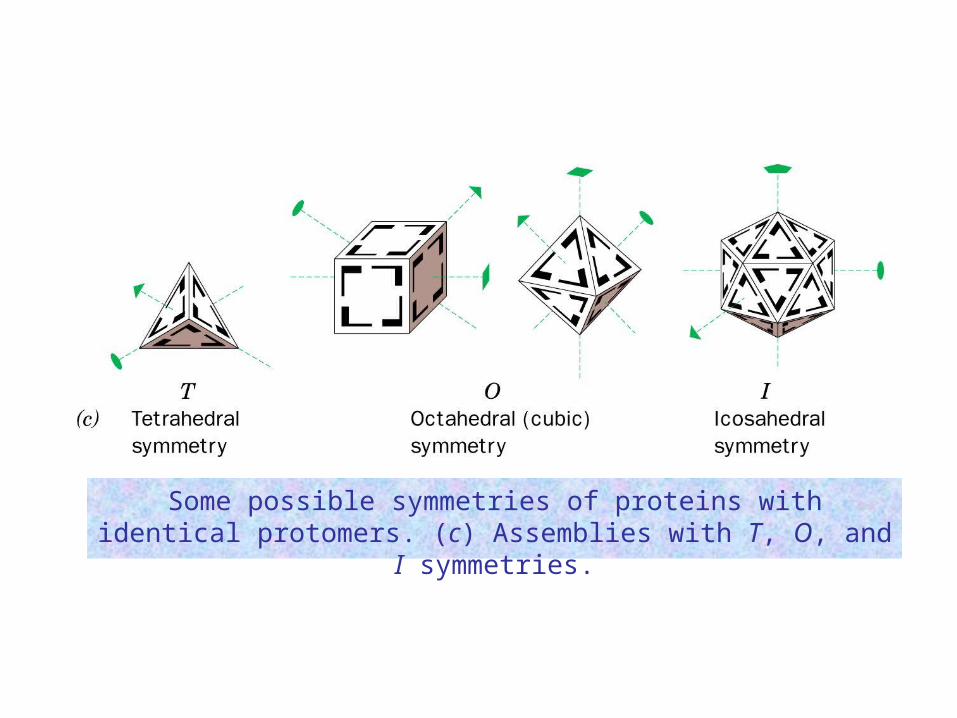
Some possible symmetries of proteins with identical protomers. (c) Assemblies with T, O, and I symmetries.
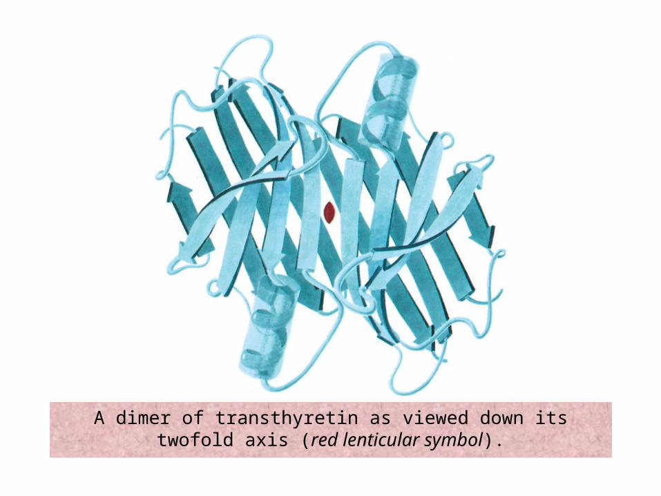
A dimer of transthyretin as viewed down its twofold axis (red lenticular symbol).

X-ray structure of glutamine synthetase from Salmonella typhimurium - view down 6-fold symmetry axis
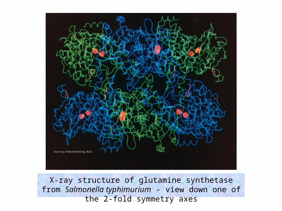
X-ray structure of glutamine synthetase from Salmonella typhimurium - view down one of the 2-fold symmetry axes

A helical structure composed of a single kind of subunit.
actin, tubulin
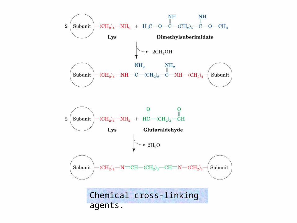
Chemical cross-linking agents.

Structural Bioinformatics Websites (URLs)

Structural Bioinformatics Websites (URLs)

Structural Bioinformatics Websites (URLs)