Embed Size (px)
Citation preview
The radial tunnel syndrome
Twenty patients with symptoms present for an average of 21.1 months and who in most cases had been treated by several conservative techniques for chronic tennis elbow were diagnosed as having radial tunnel syndrome, as originally described by Roles and Maudsley. Release of the tunnel was followed by eventual relief in 19 instances (95%).
G. D. Lister, F.R.C.S., R. B. Belsole, M.D., and H. E. Kleinert, M.D., Louisville, Ky.
T he radial tunnel syndrome was first described by Roles and Maudsley! in 1972, their paper having the subtitle "Resistant tennis elbow as a nerve entrapment." Having been frustrated previously by cases of intractable tennis elbow, in the past 4 years we have turned our attention increasingly to the radial tunnel.
Anatomy
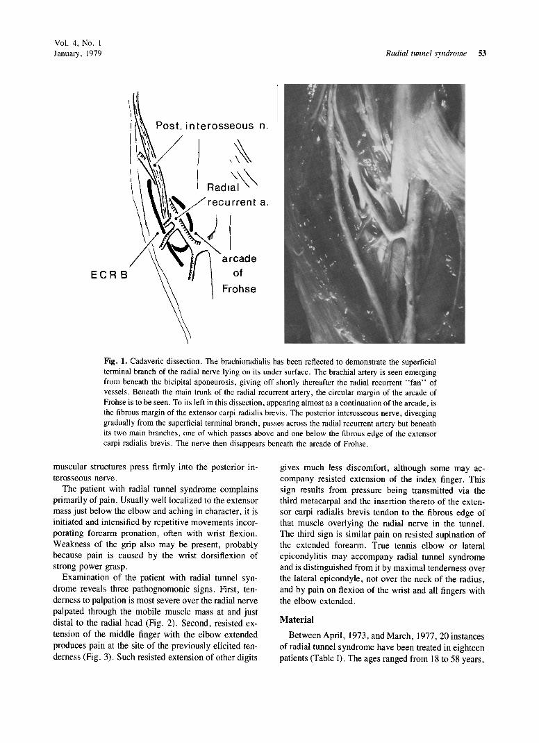
Before entering the tunnel, the radial nerve gains the anterior aspect of the forearm by piercing the lateral intermuscular septum 10 cm proximal to the lateral epicondyle (Fig. 1). Then it lies between the brachialis and biceps tendon and the muscles arising from the lateral supracondylar ridge, namely the brachioradialis and the extensors carpi radialis longus and brevis. At some point between 3 cm above and below the elbow joint or, more specifically, the humeroradial joint, the nerve divides into its posterior interosseous and superficial terminal branches.
The radial tunnel commences at the point in the course of the radial nerve where it lies anterior to the humeroradial joint. The tunnel is barely 5 cm or three fingersbreadth long and remains on the anterior aspect of the radius, with the nerve passing laterally around the bone only as it exits from the tunnel beneath the superficial part of the supinator muscle. In the tunnel the nerve lies initially on the anterior capsule of the humeroradial joint I cm lateral to the biceps tendon, in which position it can be palpated, if done firmly, with discomfort.
It then lies on the supinator, with the superficial terminal branch remaining on the surface of that muscle and passing out of the tunnel unscathed. The posterior
Received for publication Feb. 21, 1978.
Revised for publication July I, 1978.
Reprint requests: G. D. Lister, F.R.C.S., 1001 Doctor's Office Building, 250 E. Liberty SI., Louisville, KY 40202.
interosseous nerve diverges laterally and posteriorly to pass beneath the arcade of Frohse which is the proximal edge of the superficial part of the supinator. The arcade has been shown in 30% of cases to be tendinous and hence unyielding. 2
The lateral wall of the tunnel is formed by the muscles of the mobile wad, the extensors carpi radialis longus and brevis, and the brachioradialis, which also spirals around and over the nerve from lateral to anterior to form the roof of the tunnel. The extensor carpi radialis brevis, like the supinator, has a tendinous origin from the lateral epicondyle. Frequently this fibrous origin gives a flat, rigid medial edge to the extensor carpi radialis brevis, which also may be densely adherent to the underlying fibrous origin of the superficial part of the supinator.
Anterior to the head of the radius, the radial nerve is crossed superficially by transverse fibrous bands, which frequently are tenuous and few in number, but which may be of greater substance. 3
The posterior interosseous nerve is crossed in the tunnel at the level of the neck of the radius by the fan of radial recurrent vessels, of which Henry4 has pointed out that only one is truly recurrent, the others tying the mobile wad to the radial artery and its venae comitantes.
Etiology and clinical findings
All of the reported cases of radial tunnel syndrome, that is, the 36 in the paper of Roles and Maudsley and the 20 here, have been due to anatomical causes, of which there are potentially four: the fibrous bands in front of the radial head, the "radial recurrent fan" of vessels, and the sharp, tendinous margins of extensor carpi radialis brevis and of the arcade of Frohse.
In the latter two instances the compression can be demonstrated at operation by full pronation of the forearm and flexion of the wrist when either or both
52 THE JOURNAL OF HAND SURGERY 0363-5023/79/010052+08$00.80/0 © 1979 American Society for Surgery of the Hand
Vol. 4, No. I January, 1979
ECR Frohse
Radial tunnel syndrome 53
n.
Fig. 1. Cadaveric dissection . The brachioradialis has been reRected to demonstrate the superficial terminal branch of the radial nerve lying on its under surface. The brachial artery is seen emerging from beneath the bicipital aponeurosis, giving off shortly thereafter the radial recurrent " fan" of vessels. Beneath the main trunk of the radial recurrent artery, the circular margin of the arcade of Frohse is to be seen. To its left in this dissection, appearing almost as a continuation of the arcade, is the fibrous margin of the extensor carpi radialis brevis . The posterior interosseous nerve, diverging gradually from the superficial terminal branch, passes across the radial recurrent artery but beneath its two main branches, one of which passes above and one below the fibrou s edge of the extensor carpi radialis brevis. The nerve then disappears beneath the arcade of Frohse.
muscular structures press firmly into the posterior interosseous nerve.
The patient with radial tunnel syndrome complains primarily of pain. Usually well localized to the extensor mass just below the elbow and aching in character. it is initiated and intensified by repetitive movements incorporating forearm pronation , often with wrist flexion. Weakness of the grip also may be present, probably because pain is caused by the wrist dorsiflexion of strong power grasp.
Examination of the patient with radial tunnel syndrome reveals three pathognomonic signs . First, tenderness to palpation is most severe over the radial nerve palpated through the mobile muscle mass at and just distal to the radial head (Fig. 2). Second, resisted extension of the middle finger with the elbow extended produces pain at the site of the previously elicited tenderness (Fig. 3). Such resisted extension of other digits
gives much less discomfort, although some may accompany resisted extension of the index finger. This sign results from pressure being transmitted via the third metacarpal and the insertion thereto of the extensor carpi radialis brevis tendon to the fibrous edge of that muscle overlying the radial nerve in the tunnel. The third sign is similar pain on resisted supination of the extended forearm. True tennis elbow or lateral epicondylitis may accompany radial tunnel syndrome and is distinguished from it by maximal tenderness over the lateral epicondyle, not over the neck of the radius, and by pain on flexion of the wrist and al\ fingers with the elbow extended.
Material
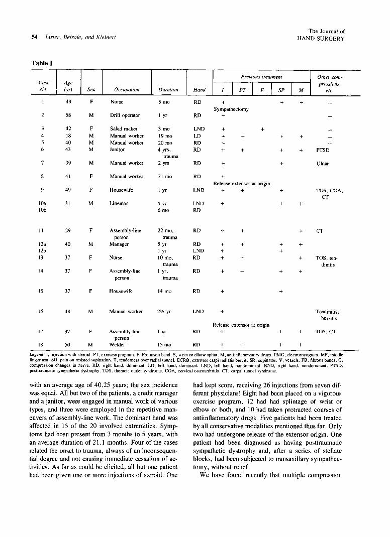
Between April, 1973, and March, 1977,20 instances of radial tunnel syndrome have been treated in eighteen patients (Table I). The ages ranged from 18 to 58 years,
54 Lister, Belsole, and Kleinert
Table I
Case No.
2
3 4 5 6
7
8
9
lOa lOb
II
12a 12b
13
14
15
16
17
18
Age (yr)
49
58
42 18 40 43
39
41
49
31
29
40
37
37
37
48
37
50
Sex
F
M
F M M M
M
F
F
M
F
M
F
F
F
M
F
M
Occupation
Nurse
Drill operator
Salad maker
Manual worker Manual worker Janitor
Manual worker
Manual worker
Housewife
Lineman
Assembly-line person
Manager
Nurse
Assembly-line
person
Housewife
Manual worker
Assembly-line person
Welder
Duration
5 mo
I yr
3 mo
19 mo 20 mo 4 yrs,
trauma 2 yrs
21 mo
I yr
4 yr 6 mo
22 mo, trauma
5 yr I yr 10 mo,
trauma I yr,
trauma
14 mo
2lh yr
I yr
15 mo
Previous treatment
Hand
RD
RD
LND
LD RD RD
RD
RD
I I PT
+ Sympathectomy
+
+ + + +
+
+
+
+
I F
+
Release extensor at origin LND + +
LND + RD
RD
RD LND RD
RD
RD
LND
RD
RD
+
+ + +
+
+
+
+
+
+
+
Release extensor at origin
+
+ +
I SP
+
+
+
+
+
+
+ +
+
+
+
+
I
The Journal of
HAND SURGERY
M
+
+
+
+
+
+
+
+
+
+
Other com-pressions,
etc.
PTSD
Ulnar
TOS, COA, CT
CT
TOS, tendinitis
Tendinitis, bursitis
TOS,CT
Legend: I, injection with steroid. PT, exercise program. F, Froimson band. S, wrist or elbow splint. M, antiinflammatory drugs. EMG, electromyogram. MF, middle finger test. SU, pain on resisted supination. T, tenderness over radial tunnel. ECRB, extensor carpi radialis brevis. SR, supinator. V, vessels. FB, fibrous bands. C, compression changes in nerve. RD, right hand, dominant. LD, left hand, dominant. LND, left hand, nondominant. RND, right hand, nondominant. PTSD, posttraumatic sympathetic dystrophy. TOS, thoracic outlet syndrome. COA, cervical osteoarthritis. CT, carpal tunnel syndrome.
with an average age of 40.25 years; the sex incidence was equal. All but two ofthe patients, a credit manager and a janitor, were engaged in manual work of various types, and three were employed in the repetitive maneuvers of assembly-line work. The dominant hand was affected in 15 of the 20 involved extremities. Symptoms had been present from 3 months to 5 years, with an average duration of 21.1 months. Four of the cases related the onset to trauma, always of an inconsequential degree and not causing immediate cessation of activities. As far as could be elicited, all but one patient had been given one or more injections of steroid. One
had kept score, receiving 26 injections from seven different physicians! Eight had been placed on a vigorous exercise program, 12 had had splintage of wrist or elbow or both, and lO had taken protracted courses of antiinflammatory drugs. Five patients had been treated by all conservative modalities mentioned thus far. Only two had undergone release of the extensor origin. One patient had been diagnosed as having posttraumatic sympathetic dystrophy and, after a series of stellate blocks, had been subjected to transaxillary sympathectomy, without relief.
We have found recently that multiple compression
Vol. 4, No. I January, 1979
EMG
Interference pattern
Normal
Reduced voltage
Compression
Normal
Irritability , ra-dial nerve
Signs
MF I SU
+ +
+ +
+ + + + + + + +
+
+ +
+ +
+ + + +
+ +
+ + + + + +
+ +
+ +
+ + + +
+ +
I T Date ECRB I + 3173
+ 9173
+ 10173 + 11173 + + 7174 + + 8174 +
+ 9174 +
+ 12174 +
+ 3175 +
+ 3175 + 1176 +
+ 4175 +
+ 5175 + + 7176 + + 10175
+ 11175 +
+ 6176 +
11176 + 3177 +
+ 3/77 +
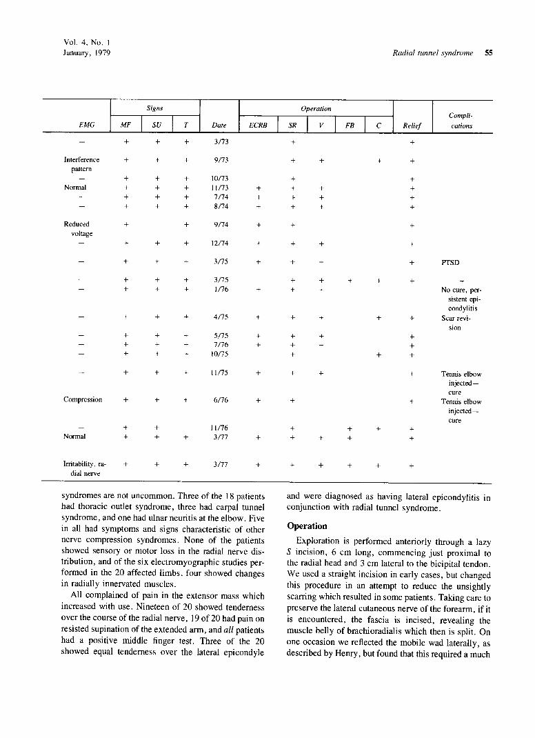
syndromes are not uncommon. Three of the 18 patients had thoracic outlet syndrome, three had carpal tunnel syndrome, and one had ulnar neuritis at the elbow. Five in all had symptoms and signs characteristic of other nerve compression syndromes. None of the patients showed sensory or motor loss in the radial nerve dis-tribution, and of the six electromyographic studies per-formed in the 20 affected limbs, four showed changes in radially innervated muscles.
All complained of pain in the extensor mass which increased with use. Nineteen of 20 showed tenderness over the course of the radial nerve, 19 of 20 had pain on resisted supination of the extended arm, and all patients had a positive middle finger test. Three of the 20 showed equal tenderness over the lateral epicondyle
Radial tunnel syndrome 55
Operation Compli-
SR I V I FB I C Relief cations
+ +
+ + + +
+ + + + + + + + + + +
+ +
+ + +
+ + + PTSD
+ + + + + + + + No cure, per-
sistent epi-condylitis
+ + + + Scar revi-sion
+ + + + + + + + +
+ + + Tennis elbow injected-cure
+ + Tennis elbow injected-cure
+ + + + + + + +
+ + + + +
and were diagnosed as having lateral epicondylitis in conjunction with radial tunnel syndrome.
Operation
Exploration is performed anteriorly through a lazy S incision, 6 cm long, commencing just proximal to the radial head and 3 cm lateral to the bicipital tendon. We used a straight incision in early cases , but changed this procedure in an attempt to reduce the unsightly scarring which resulted in some patients. Taking care to preserve the lateral cutaneous nerve of the forearm , if it is encountered, the fascia is incised, revealing the muscle belly of brachioradialis which then is split. On one occasion we reflected the mobile wad laterally , as described by Henry, but found that this required a much

56 Lister, Be/sole, and Kleinert The Journal of
HAND SURGERY
Fig. 2. Maximal tenderness is located just distal to the radial head palpated through the mobile muscle wad and is located beneath the examiner's thumb. All pain on indirect testing. as shown in Fig . 3 and in resisted supination of the extended forearm, is referred to this point. (From Lister GD: The hand: Diagnosis and indications. by permission of the publishers. Churchill-Livingstone, Ltd ., of Edinburgh , London , New York.)
Fig. 3. The middle finger test. With the elbow and wrist extended, resisted extension of the middle finger elicits pain over the site indicated in Fig . 2. Similar pressure over other fingers may cause some pain but not as severe. (From Lister GD: The hand: Diagnosis and indications, by permission of the publishers , Churchill-Livingstone , Ltd ., of Edinburgh , London , and New York.)
more extensive incision and offered inferior exposure, requiring considerable retraction . In all other 19 cases, we split the muscle belly, as recommended by Roles and Maudsley, aiming always for the radial head . The belly is thick and requires progressively deeper retraction and confidence that one is proceeding correctly. It is one of those very rare situations in which one sees no landmarks and should not. In all cases the radial nerve
was located without difficulty . The split in the brachioradialis then should be lengthened to equal the skin incision and to expose the entire tunnel. Some intramuscular neurovascular bundles will be encountered. When possible, these should be preserved, but those which limit exposure can be coagulated and divided without fear. No patient has shown any detectable weakness of brachioradialis following operation.
Vol. 4, No. I January, 1979 RadiaL tunnel syndrome 57
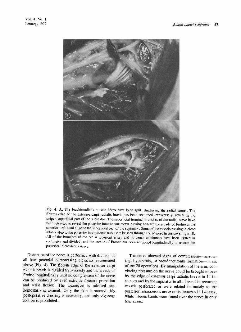
Fig. 4. A, The brachioradialis muscle fibers have been split, displaying the radial tunnel. The fibrous edge of the extensor carpi radialis brevis has been sectioned transversely, revealing the striped superficial part of the supinator. The superficial terminal branches of the radial nerve have been retracted to reveal the posterior interosseous nerve passing beneath the arcade of Frohse at the superior, left-hand edge of the superficial part of the supinator. Some of the vessels passing in close relationship to the posterior interosseous nerve can be seen through the adipose tissue covering it. B, All of the branches of the radial recurrent artery and its venae comitantes have been ligated in continuity and divided, and the arcade of Frohse has been sectioned longitudinally to release the posterior interosseous nerve.
Dissection of the nerve is performed with division of all four potential compressing elements enumerated above (Fig . 4). The fibrous edge of the extensor carpi radialis brevis is divided transversely and the arcade of Frohse longitudinally until no compression of the nerve can be produced by even extreme forearm pronation and wrist flexion. The tourniquet is released and hemostasis is assured. Only the skin is sutured. No postoperative dressing is necessary, and only vigorous motion is prohibited.
The nerve showed signs of compression-narrowing, hyperemia, or pseudoneuroma formation-in six of the 20 operations. By manipulation of the arm, convincing pressure on the nerve could be brought to bear by the edge of extensor carpi radialis brevis in 14 instances and by the supinator in all. The radial recurrent vessels perforated or were related intimately to the posterior interosseous nerve or its branches in 14 cases, while fibrous bands were found over the nerve in only four cases.
58 Lister, Belsole, and Kleinert
Results
None of the patients has been lost to follow-up, which was continued from 9 months to 4 years and 6 months, with an average of 29.8 months (Table I) . Nineteen of the 20 procedures have given relief from symptoms. In the majority of the patients, the relief has been almost immediate, being reported by the patient during the first I or 2 weeks . The recovery was delayed in the three patients diagnosed as having concomitant epicondylitis. All were injected with steroid and two are free of symptoms 9 and 16 months after operation. The third patient is the only one of the group of 20 in whom the procedure has not given complete relief. The only other delayed recovery occurred in a patient who developed posttraumatic sympathetic dystrophy which required multiple stellate blocks, but who now has been free of symptoms for over 2 years.
The single source of dissatisfaction with the procedure has been the rather obvious scar which has become hypertrophic in three cases, with two responding to steroid injection and the third requiring subsequent revision of the scar.
Discussion
Many causes have been described to account for chronic pain on the lateral aspect of the elbow, often being grouped together as tennis elbow. These causes include tendinitis, epicondylitis, stenosis of the orbicular ligament,6 tears in the common tendon of origin,7 fibrillation of the radial head ,8 cervical spine disorders,9 and nerve compression. The documented success of treatment directed more or less specifically at each of these causes indicates that all may produce symptoms so alike as to be indistinguishable to the patient. The fact that two or more may coexist further complicates diagnostic endeavors. However, we believe that it is possible to distinguish radial tunnel syndrome by the following four observations: (1) the point of maximum pain indicated by the patient from history and during indirect testing is over the radial tunnel, not the lateral epicondyle; (2) the point of maximum tenderness likewise is over the radial tunnel; (3) the middle finger test gives more pain than does passive stretching of the entire extensor mass; (4) pain can be elicited by resisted supination in extension. It is possible that thermography may prove of benefit, as findings suggest that epicondylitis shows as a "hot spot," whereas radial tunnel compression is "cold ." 10
One of the difficulties for some in accepting the existence of the radial tunnel syndrome is the fact that the posterior interosseous nerve is considered as a purely motor nerve and therefore is incapable of causing pain .
The Journal of HAND SURGERY
However, the posterior interosseous nerve carries sensory afferent fibers from the radiocarpal, intercarpal, and carpometacarpal joints and also carries afferent fibers from the muscles it serves , for example from the "flower-spray" endings which have afferents of group IIA fibers. Any who deny the possibility of pain mediated by motor nerves need only recall their last attack of cramping or squeeze any muscle belly. The discomfort produced by such a maneuvre is considerable and, incidentall y, is recognized immediatel y by the patient with radial tunnel syndrome .
It could be argued with some justification that the procedure described above incorporates transverse division of the extensor carpi radialis brevis fascial origin and therefore is similar in effect to the operation performed by Garden,l1 especially in his earlier cases. This cannot be refuted, since we have not felt justified in leaving the other structures unattended in certain cases to act as controls. However, the persistence after operation of significant tenderness over the lateral epicondyle which had been present prior to operation in three of the 20 patients and the fact that two of the patients who experienced relief as a result of radial tunnel exploration previously had undergone release of the common extensor origin without abatement of pain suggests that the described procedure does not relieve pure lateral epicondylitis and that there is more value to it than mere tenotomy of the extensor carpi radialis brevis.
We believe that any or all of the four structures listed above may be the compressing factor in each case . In particular, the vessels encountered have been impressive both in number and in the manner in which they frequently are woven into the structure of the nerve, a relationship which has been shown to cause nerve compression in other sites. 12 [t can be theorized that these vessels increase in caliber with exercise and may account for the large number of manual workers presenting with this condition. Another effect of sustained exercise may be evidenced by the appearance of the arcade of Frohse. The arcade was thickened and fibrous in all 20 patients operated on in this series. Spinnerl2 recorded that the arcade of Frohse was not tendinous in any of his fetal dissections, but was fibrous in 30% of the adults whom he examined . These facts suggest that the thickening and fibrosis of the arch occurs during a patient's lifetime, possibly as a result of a particular occupation or recreation.
The results of radial tunnel release have been sufficiently good thus far as to warrant some attention. Roles and Maudsley had excellent or good results in 35 of 38 cases, Hagert l3 in 45 of 50 cases, and we in

Vol. 4, No. I January, 1979
19 of 20, giving an overall significant improvement in 99 of 108 cases (91.7%).
Cadaveric dissections were performed by Myles J. Cohen, M.D., and Moulton K. Johnson, M.D., Los Angeles, Calif.
REFERENCES
I. Roles NC, Maudsley RH: Radial tunnel syndrome; resistant tennis elbow as a nerve entrapment. 1 Bone Joint Surg [Br] 54:499-508, 1972
2. Spinner M: The arcade of Frohse and its relationship to posterior interosseous nerve paralysis . J Bone Joint Surg [Br] 50:809-12, 1968
3. Sharrard, WJW: Posterior interosseous neuritis. J Bone Joint Surg [Br] 48:777-80, 1966
4. Henry AK : Extensile exposure, ed 2, Edinburgh, London, New York , 1973, Churchill-Livingstone , Ltd
5. Froimson A: Treatment of tennis elbow with forearm support band. J Bone Joint Surg [Am] 53: 183-4, 1971
6. Bosworth OM: Surgical treatment of tennis elbow . J Bone Joint Surg [Am] 47: 1533-6, 1965
Radial tunnel syndrome 59
7. Coonrad RW, Hooper WR: Tennis elbow: Its course, natural history, conservative and surgical management. J Bone Joint Surg [Am] 55: 1177-87, 1973
8. Newman JH, Goodfellow JW: Fibrillation of head of radius as one cause of tennis elbow . Br Med J 1 :328-30, 1975
9. Gunn C, Milbrandt WE: Tennis elbow and the cervical spine . J Can Med Assoc 114:803-9, 1976
10. Shilo VR, Engel l, Farin I, Horochowski H: Thermography as a diagnostic aid in tennis elbow. Handchirurgie 8: 101-3, 1976
II. Garden RS: Tennis elbow. J Bone Joint Surg [Br] 43:100-6, 1961
12. Spinner M: Cryptogenic infraclavicular brachial plexus neuritis. Bull Hosp Joint Dis 37:98-104, 1976
13 . Hagert CG: Entrapment of the posterior interosseous nerve causing forearm pain, in Carstam N: Proceedings: The Scandinavian Society for Surgery of the Hand . J HAND SURG 2:486-8, 1977