Embed Size (px)
Citation preview
Page 1 of 27
Carpal Tunnel Syndrome, an overview Jim Lewis, R.NCS.T.
Learning Objectives:
Median nerve compression at the wrist is the most common entrapment seen in the
electrodiagnostic laboratory. Although it is not the only entrapment of the median nerve it
is, by far, the most common. Carpal Tunnel Syndrome, the term that includes the various
collections of symptoms that result from median nerve entrapment at the wrist, is much
more common in women than men, by at least a 3:1 ratio.
In this paper will review median nerve anatomy, clinical features of CTS, differential
diagnosis, underlying/predisposing conditions, nerve conduction recording parameters,
techniques and case studies. The reader will gain insight to this common entrapment as
well as the importance of the nerve conduction studies used to confirm the diagnosis of
CTS.
Anatomy of the Median Nerve:
Understanding median nerve anatomy is important to sort out the various conditions in
that make up the differential diagnosis, whether it is a cervical radiculopathy or a
proximal median nerve lesion.
The median nerve, a mixed nerve, arises from cervical roots C6-T1, thus median nerve
fibers transverse all three trunks (upper, middle and lower). The nerve continues along
via the lateral and medial cords before forming the median nerve proper. There are no
median nerve innervations in the upper arm. The nerve continues between or beneath the
two heads of the pronator teres muscle in the forearm. The median nerve innervates the
pronator teres, flexor digitorum superficialis and flexor carpi radialis before the purely
motor nerve branch called the anterior interosseous nerve is given off. The main median
nerve branch continues through the forearm and just distal to the wrist it gives off the
palmar sensory branch which supplies sensation over the thenar area. Nine flexor tendons
and the median nerve go through
the tunnel created by the carpal
bones on three sides and the
transverse carpal ligament on the
top. This is called the carpal
tunnel. After exiting the carpal
tunnel the recurrent thenar motor
branch turns around and innervates
the muscles of the thenar eminence
(abductor pollicis brevis, opponens
pollicis and the lateral head of the
flexor pollicis brevis). The first
and second lumbrical muscles are
innervated be the remaining motor
Carpal Tunnel Syndrome, an Overview
Page 2 of 27
branch of the median nerve while the sensory branch of the median nerve innervates the
medial thumb, forefinger, middle finger and the lateral half of the ring finger.
Clinical features of CTS:
There may be a variety of signs and symptoms that most commonly includes pain and
paresthesias. Women are affected more than men by a wide margin and although CTS
can be seen in various ages it is most common in the 5th
and 6th
decades of life. Carpal
tunnel syndrome tends to affect the dominant hand more though bilateral findings are
common.
Short lasting pain localized to the wrist and fingers can be the earliest sign, but radiating
pain to the forearm, elbow or even shoulder is common. The numbness and burning
reported by many patients is maximal in the lateral 3 fingers, but subjectively can include
the entire hand. It is important to note that the sensation over the thenar eminence is
spared as that area is supplied by the palmar sensory branch given off before the carpal
tunnel. Patients often report an increase of pain and paresthesias with movements that
flex or extend the wrist such as driving or holding a book. Many patients report the
symptoms worsen during the night leading to arousals that require them to shake the
feelings back into their hands. This is thought to be secondary to the habit of persistent
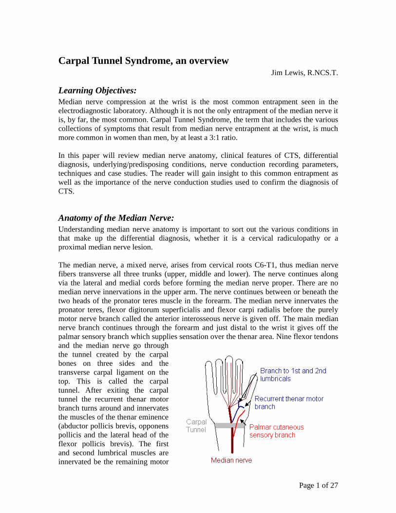
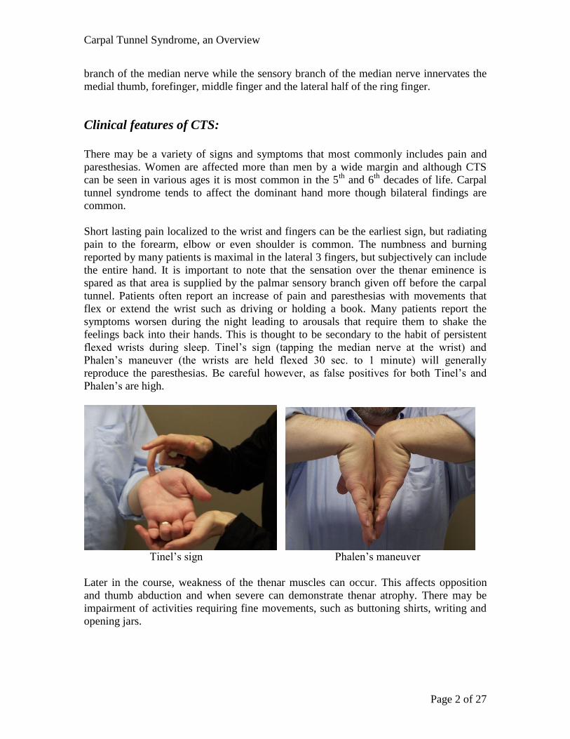
flexed wrists during sleep. Tinel’s sign (tapping the median nerve at the wrist) and
Phalen’s maneuver (the wrists are held flexed 30 sec. to 1 minute) will generally
reproduce the paresthesias. Be careful however, as false positives for both Tinel’s and
Phalen’s are high.
Tinel’s sign Phalen’s maneuver
Later in the course, weakness of the thenar muscles can occur. This affects opposition
and thumb abduction and when severe can demonstrate thenar atrophy. There may be
impairment of activities requiring fine movements, such as buttoning shirts, writing and
opening jars.
Carpal Tunnel Syndrome, an Overview
Page 3 of 27
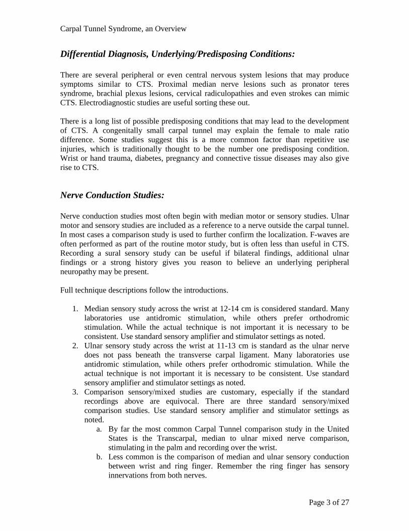
Differential Diagnosis, Underlying/Predisposing Conditions:
There are several peripheral or even central nervous system lesions that may produce
symptoms similar to CTS. Proximal median nerve lesions such as pronator teres
syndrome, brachial plexus lesions, cervical radiculopathies and even strokes can mimic
CTS. Electrodiagnostic studies are useful sorting these out.
There is a long list of possible predisposing conditions that may lead to the development
of CTS. A congenitally small carpal tunnel may explain the female to male ratio
difference. Some studies suggest this is a more common factor than repetitive use
injuries, which is traditionally thought to be the number one predisposing condition.
Wrist or hand trauma, diabetes, pregnancy and connective tissue diseases may also give
rise to CTS.
Nerve Conduction Studies:
Nerve conduction studies most often begin with median motor or sensory studies. Ulnar
motor and sensory studies are included as a reference to a nerve outside the carpal tunnel.
In most cases a comparison study is used to further confirm the localization. F-waves are
often performed as part of the routine motor study, but is often less than useful in CTS.
Recording a sural sensory study can be useful if bilateral findings, additional ulnar
findings or a strong history gives you reason to believe an underlying peripheral
neuropathy may be present.
Full technique descriptions follow the introductions.
1. Median sensory study across the wrist at 12-14 cm is considered standard. Many
laboratories use antidromic stimulation, while others prefer orthodromic
stimulation. While the actual technique is not important it is necessary to be
consistent. Use standard sensory amplifier and stimulator settings as noted.
2. Ulnar sensory study across the wrist at 11-13 cm is standard as the ulnar nerve
does not pass beneath the transverse carpal ligament. Many laboratories use
antidromic stimulation, while others prefer orthodromic stimulation. While the
actual technique is not important it is necessary to be consistent. Use standard
sensory amplifier and stimulator settings as noted.
3. Comparison sensory/mixed studies are customary, especially if the standard
recordings above are equivocal. There are three standard sensory/mixed
comparison studies. Use standard sensory amplifier and stimulator settings as
noted.
a. By far the most common Carpal Tunnel comparison study in the United
States is the Transcarpal, median to ulnar mixed nerve comparison,
stimulating in the palm and recording over the wrist.
b. Less common is the comparison of median and ulnar sensory conduction
between wrist and ring finger. Remember the ring finger has sensory
innervations from both nerves.
Carpal Tunnel Syndrome, an Overview
Page 4 of 27
c. Additionally, comparison of median and radial sensory conduction
between wrist and thumb is useful when the patient has an ulnar
neuropathy and an unaffected nerve that does not go under the transverse
carpal ligament is needed.
4. Median motor study to the thenar muscles is a standard. The distal motor latency
is important to the evaluation of CTS, but the forearm conduction velocity also
verifies the health of the median nerve more proximally. Use standard motor
amplifier and stimulator settings as noted.
5. Ulnar motor study to the hypothenar muscle is a standard study as the ulnar nerve
does no go under the transverse carpal ligament and would be spared in CTS. Use
standard motor amplifier and stimulator settings as noted.
6. F-waves can be performed, however they are not often useful to diagnosis CTS.
7. In cases where no response of the median sensory or motor nerve across the wrist
can be obtained the technique for median to 2nd
lumbrical to ulnar 2nd
volar
interosseous can be used. Fascicles to the 2nd
lumbrical tend to be spared in even
the most severe CTS cases. Use standard motor amplifier and stimulator settings
as noted.
8. Needle EMG as performed by the physician is useful to rule out cervical
radiculopathy or to gauge severity when the clinical signs indicate.
Full technique notes:
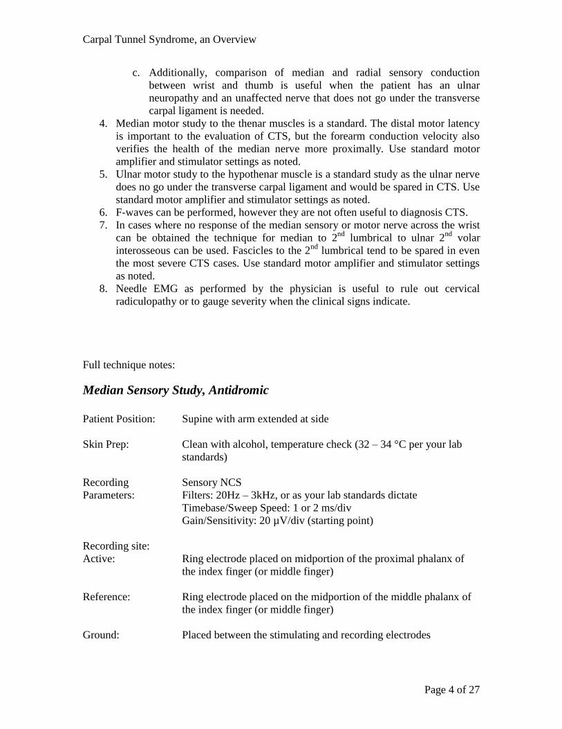
Median Sensory Study, Antidromic
Patient Position: Supine with arm extended at side
Skin Prep: Clean with alcohol, temperature check (32 – 34 °C per your lab
standards)
Recording Sensory NCS
Parameters: Filters: 20Hz – 3kHz, or as your lab standards dictate
Timebase/Sweep Speed: 1 or 2 ms/div
Gain/Sensitivity: 20 µV/div (starting point)
Recording site:
Active: Ring electrode placed on midportion of the proximal phalanx of
the index finger (or middle finger)
Reference: Ring electrode placed on the midportion of the middle phalanx of
the index finger (or middle finger)
Ground: Placed between the stimulating and recording electrodes
Carpal Tunnel Syndrome, an Overview
Page 5 of 27
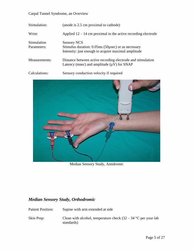
Stimulation: (anode is 2.5 cm proximal to cathode)
Wrist: Applied 12 – 14 cm proximal to the active recording electrode
Stimulation Sensory NCS
Parameters: Stimulus duration: 0.05ms (50µsec) or as necessary
Intensity: just enough to acquire maximal amplitude
Measurements: Distance between active recording electrode and stimulation
Latency (msec) and amplitude (µV) for SNAP
Calculations: Sensory conduction velocity if required
Median Sensory Study, Antidromic
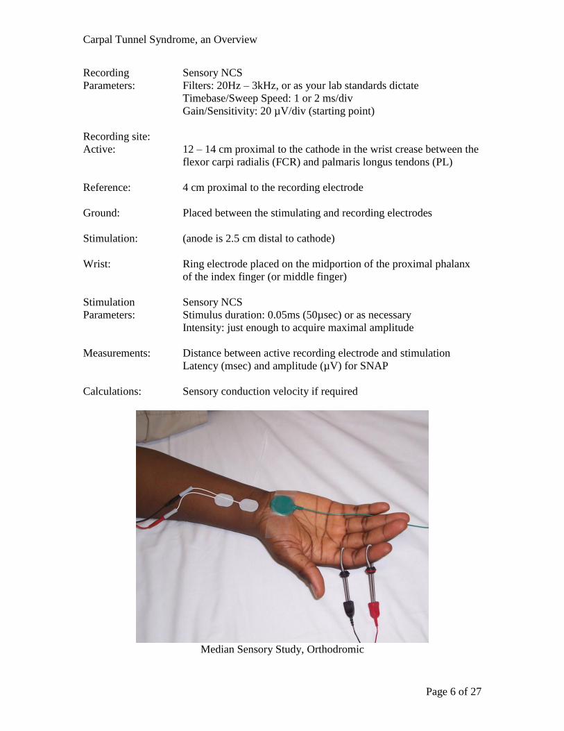
Median Sensory Study, Orthodromic
Patient Position: Supine with arm extended at side
Skin Prep: Clean with alcohol, temperature check (32 – 34 °C per your lab
standards)
Carpal Tunnel Syndrome, an Overview
Page 6 of 27
Recording Sensory NCS
Parameters: Filters: 20Hz – 3kHz, or as your lab standards dictate
Timebase/Sweep Speed: 1 or 2 ms/div
Gain/Sensitivity: 20 µV/div (starting point)
Recording site:
Active: 12 – 14 cm proximal to the cathode in the wrist crease between the
flexor carpi radialis (FCR) and palmaris longus tendons (PL)
Reference: 4 cm proximal to the recording electrode
Ground: Placed between the stimulating and recording electrodes
Stimulation: (anode is 2.5 cm distal to cathode)
Wrist: Ring electrode placed on the midportion of the proximal phalanx
of the index finger (or middle finger)
Stimulation Sensory NCS
Parameters: Stimulus duration: 0.05ms (50µsec) or as necessary
Intensity: just enough to acquire maximal amplitude
Measurements: Distance between active recording electrode and stimulation
Latency (msec) and amplitude (µV) for SNAP
Calculations: Sensory conduction velocity if required
Median Sensory Study, Orthodromic
Carpal Tunnel Syndrome, an Overview
Page 7 of 27
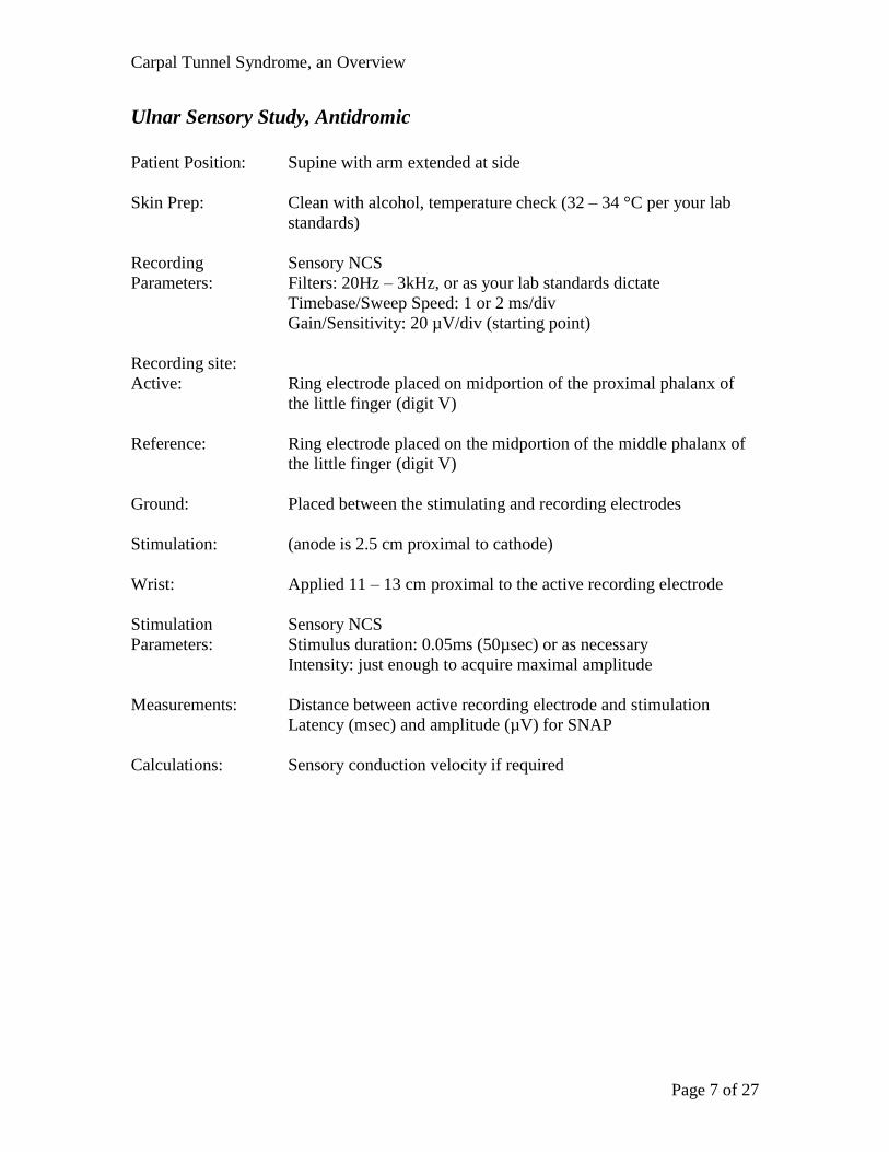
Ulnar Sensory Study, Antidromic
Patient Position: Supine with arm extended at side
Skin Prep: Clean with alcohol, temperature check (32 – 34 °C per your lab
standards)
Recording Sensory NCS
Parameters: Filters: 20Hz – 3kHz, or as your lab standards dictate
Timebase/Sweep Speed: 1 or 2 ms/div
Gain/Sensitivity: 20 µV/div (starting point)
Recording site:
Active: Ring electrode placed on midportion of the proximal phalanx of
the little finger (digit V)
Reference: Ring electrode placed on the midportion of the middle phalanx of
the little finger (digit V)
Ground: Placed between the stimulating and recording electrodes
Stimulation: (anode is 2.5 cm proximal to cathode)
Wrist: Applied 11 – 13 cm proximal to the active recording electrode
Stimulation Sensory NCS
Parameters: Stimulus duration: 0.05ms (50µsec) or as necessary
Intensity: just enough to acquire maximal amplitude
Measurements: Distance between active recording electrode and stimulation
Latency (msec) and amplitude (µV) for SNAP
Calculations: Sensory conduction velocity if required
Carpal Tunnel Syndrome, an Overview
Page 8 of 27
Ulnar Sensory Study, Antidromic
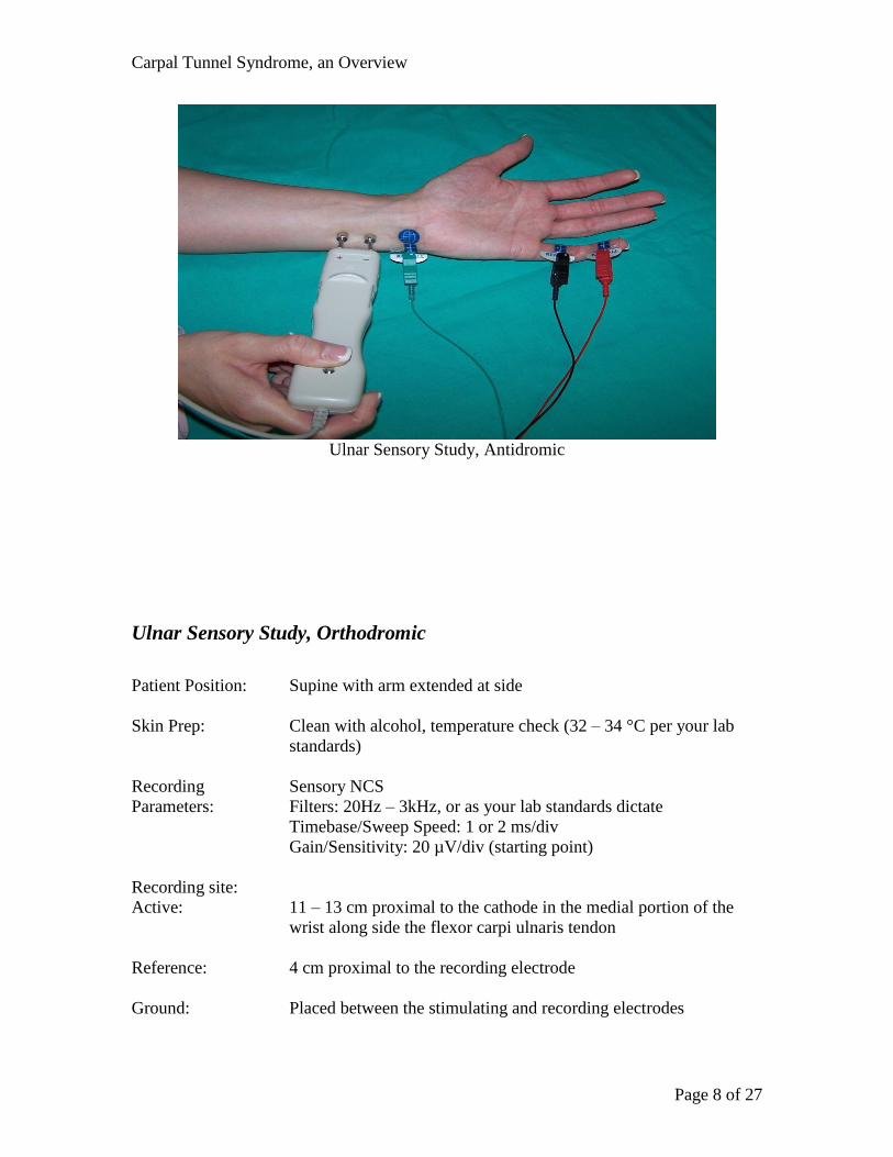
Ulnar Sensory Study, Orthodromic
Patient Position: Supine with arm extended at side
Skin Prep: Clean with alcohol, temperature check (32 – 34 °C per your lab
standards)
Recording Sensory NCS
Parameters: Filters: 20Hz – 3kHz, or as your lab standards dictate
Timebase/Sweep Speed: 1 or 2 ms/div
Gain/Sensitivity: 20 µV/div (starting point)
Recording site:
Active: 11 – 13 cm proximal to the cathode in the medial portion of the
wrist along side the flexor carpi ulnaris tendon
Reference: 4 cm proximal to the recording electrode
Ground: Placed between the stimulating and recording electrodes
Carpal Tunnel Syndrome, an Overview
Page 9 of 27
Stimulation: (anode is 2.5 cm distal to cathode)
Wrist: Ring electrode placed on the midportion of the proximal phalanx
of the little finger (digit V)
Stimulation Sensory NCS
Parameters: Stimulus duration: 0.05ms (50µsec) or as necessary
Intensity: just enough to acquire maximal amplitude
Measurements: Distance between active recording electrode and stimulation
Latency (msec) and amplitude (µV) for SNAP
Calculations: Sensory conduction velocity if required
Ulnar Sensory Study, Orthodromic
Comparison studies:
Transcarpal Median to Ulnar Palmar Sensory Study
Patient Position: Supine with arm supinated and extended at side
Carpal Tunnel Syndrome, an Overview
Page 10 of 27
Skin Prep: Clean with alcohol, temperature check (32 – 34 °C per your lab
standards)
Recording
Parameters: Sensory NCS
Median –
Recording site:
Active: Placed 2 cm proximal to the distal wrist crease between the flexor
carpi radialis (FCR) and pollicis longus (PL) tendons
Reference: Placed 4 cm proximal to the active recording electrode along the
median nerve
Ground: Placed between the recording and stimulating electrodes
Stimulation:
Cathode/Anode Applied 8 cm distal to the active recording electrode between the
index and third digit
Anode is 2.5 cm distal to the cathode
Ulnar –
Recording site:
Active: Placed 2 cm proximal to the distal wrist crease, just anterior to the
flexor carpi ulnaris tendon
Reference: Placed 4 cm proximal to the active recording electrode along the
ulnar nerve
Ground: Placed between the stimulating and recording electrodes
Stimulation:
Cathode/Anode Applied 8 cm distal to the active recording electrode between the
fourth and fifth digit
Anode is 2.5 cm distal to the cathode
Stimulation
Parameters: Sensory NCS
Measurements: Distance between active recording electrode and cathode
Latency and amplitude of SNAP
Calculations: Latency (msec) difference between the two nerves
Carpal Tunnel Syndrome, an Overview
Page 11 of 27
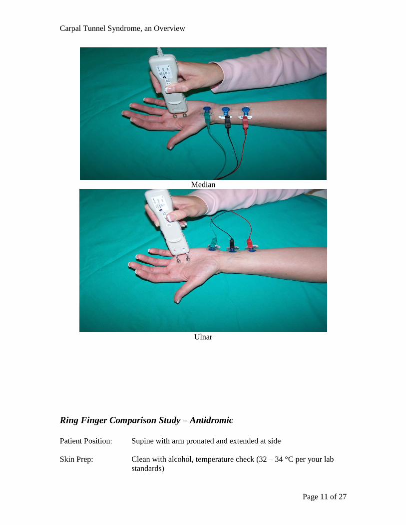
Median
Ulnar
Ring Finger Comparison Study – Antidromic
Patient Position: Supine with arm pronated and extended at side
Skin Prep: Clean with alcohol, temperature check (32 – 34 °C per your lab
standards)
Carpal Tunnel Syndrome, an Overview
Page 12 of 27
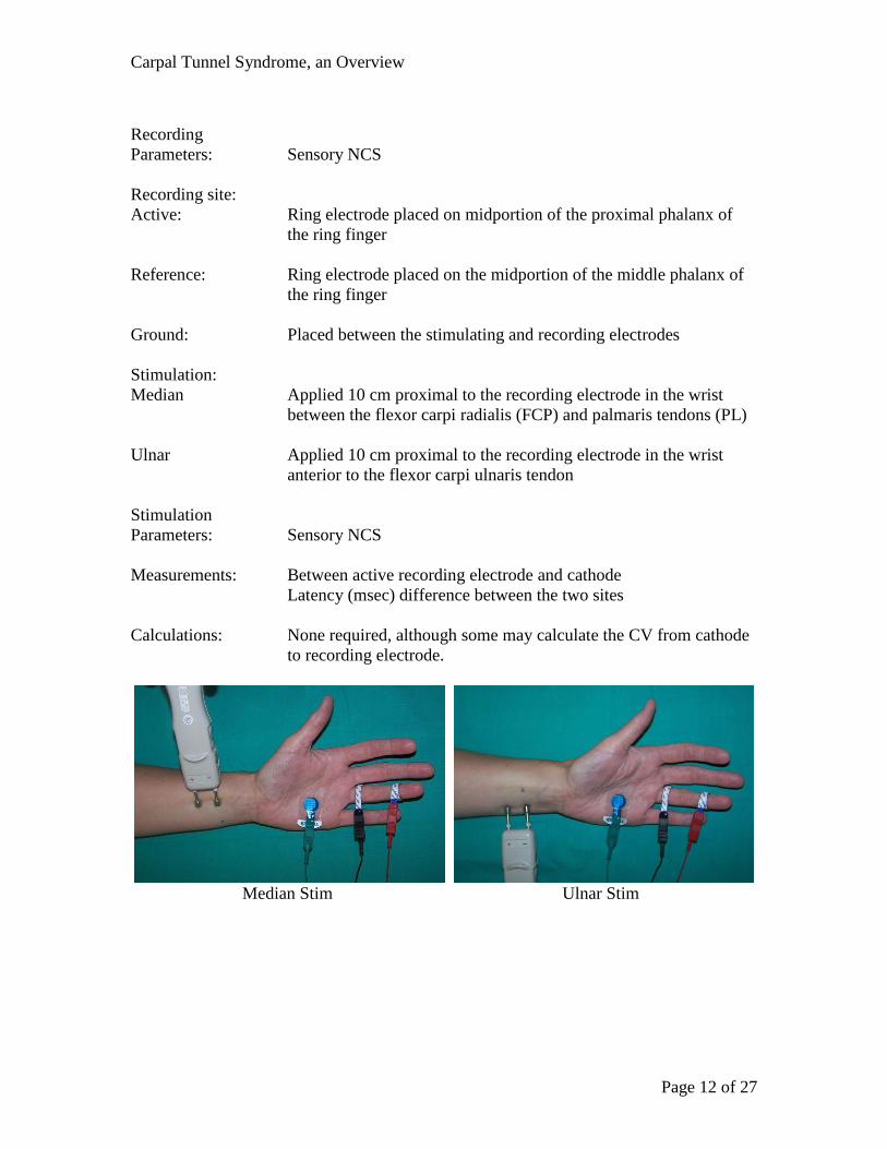
Recording
Parameters: Sensory NCS
Recording site:
Active: Ring electrode placed on midportion of the proximal phalanx of
the ring finger
Reference: Ring electrode placed on the midportion of the middle phalanx of
the ring finger
Ground: Placed between the stimulating and recording electrodes
Stimulation:
Median Applied 10 cm proximal to the recording electrode in the wrist
between the flexor carpi radialis (FCP) and palmaris tendons (PL)
Ulnar Applied 10 cm proximal to the recording electrode in the wrist
anterior to the flexor carpi ulnaris tendon
Stimulation
Parameters: Sensory NCS
Measurements: Between active recording electrode and cathode
Latency (msec) difference between the two sites
Calculations: None required, although some may calculate the CV from cathode
to recording electrode.
Median Stim Ulnar Stim
Carpal Tunnel Syndrome, an Overview
Page 13 of 27
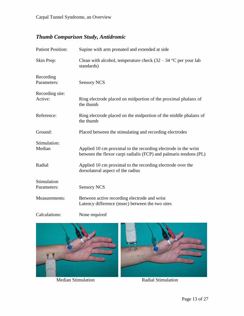
Thumb Comparison Study, Antidromic
Patient Position: Supine with arm pronated and extended at side
Skin Prep: Clean with alcohol, temperature check (32 – 34 °C per your lab
standards)
Recording
Parameters: Sensory NCS
Recording site:
Active: Ring electrode placed on midportion of the proximal phalanx of
the thumb
Reference: Ring electrode placed on the midportion of the middle phalanx of
the thumb
Ground: Placed between the stimulating and recording electrodes
Stimulation:
Median Applied 10 cm proximal to the recording electrode in the wrist
between the flexor carpi radialis (FCP) and palmaris tendons (PL)
Radial Applied 10 cm proximal to the recording electrode over the
dorsolateral aspect of the radius
Stimulation
Parameters: Sensory NCS
Measurements: Between active recording electrode and wrist
Latency difference (msec) between the two sites
Calculations: None required
Median Stimulation Radial Stimulation
Carpal Tunnel Syndrome, an Overview
Page 14 of 27
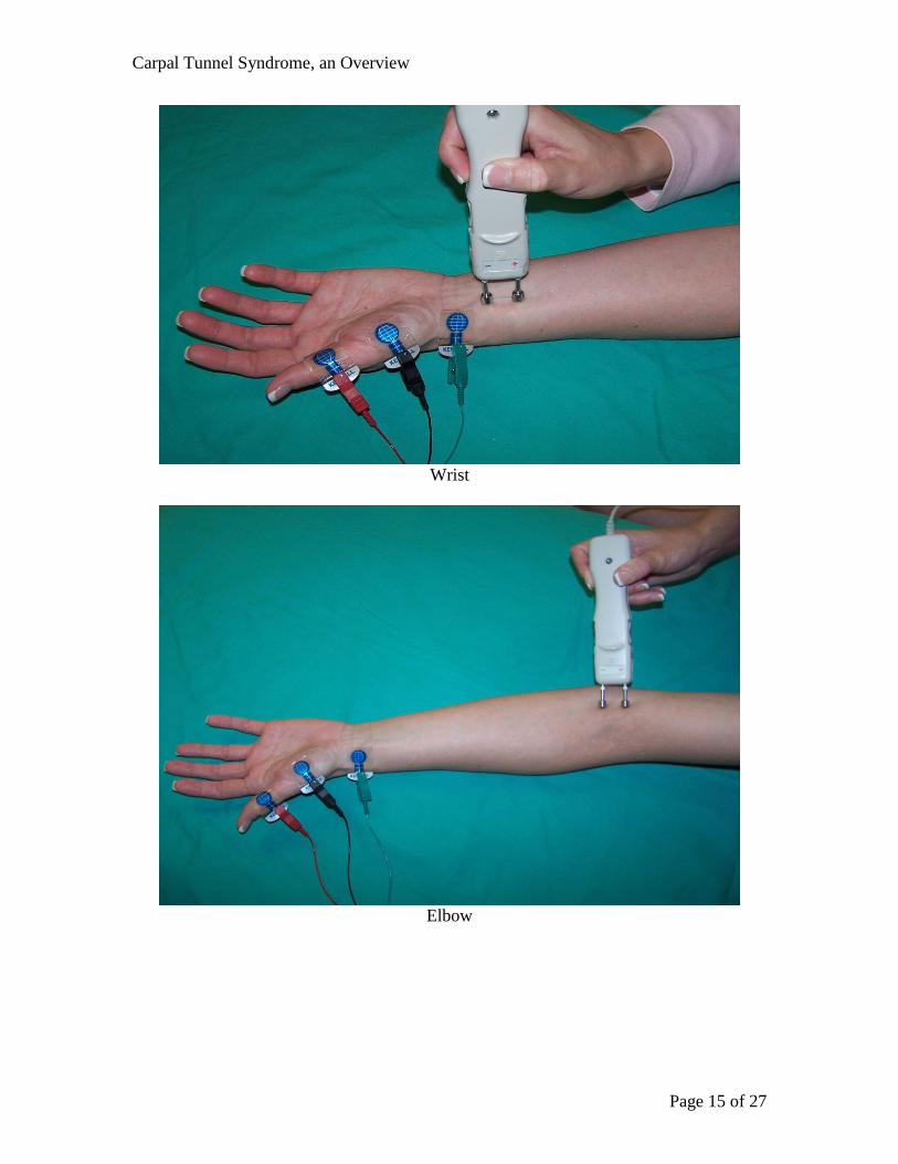
Median Motor Study
Patient Position: Supine with arm supinated and extended at side
Skin Prep: Clean with alcohol, temperature check (32 – 34 °C per your lab
standards)
Recording Motor NCS
Parameters: Filters: 2Hz – 10kHz, or as your lab standards dictate
Timebase/Sweep Speed: 2 or 5 ms/div
Gain/Sensitivity: 5 mV/div (starting point)
Recording site:
Active: Placed over the belly of the Abductor Pollicis Brevis (APB), ½
distance between metacarpophalangeal (MCP) joint of thumb and
midpoint of the distal wrist crease
Reference: Placed on the distal phalanx of the thumb
Ground: Placed between the stimulating and recording electrodes
Stimulation: (cathode distal)
Wrist: Applied 2 cm proximal to the distal wrist crease between the flexor
carpi radialis (FCR) and the palmaris longus (PL) tendons
Elbow: Applied at the elbow crease, just medial to biceps tendon
Stimulation Motor NCS
Parameters: Stimulus duration: 0.05ms (50µsec) or as necessary
Intensity: enough to acquire supramaximal amplitude
Measurements: Between active recording electrode and wrist
Between wrist and elbow
Latency (ms) and amplitude (mV) for CMAP recordings
Calculations: Conduction velocity wrist to elbow
Carpal Tunnel Syndrome, an Overview
Page 15 of 27
Wrist
Elbow
Carpal Tunnel Syndrome, an Overview
Page 16 of 27
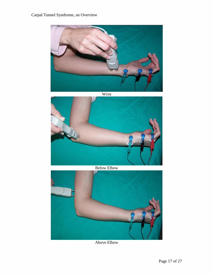
Ulnar Motor Study
Patient Position: Supine, or on their side with arm supinated and abducted 70-90
degrees
Skin Prep: Clean with alcohol, temperature check (32 – 34 °C per your lab
standards)
Recording Motor NCS
Parameters: Filters: 2Hz – 10kHz, or as your lab standards dictate
Timebase/Sweep Speed: 2 or 5 ms/div
Gain/Sensitivity: 5 mV/div (starting point)
Recording site:
Active: Placed on the belly of the Abductor Digiti Minimi
(ADM) ½ distance between the distal wrist crease and the base of
the fifth digit
Reference: Placed on the proximal phalanx of the fifth digit
Ground: Placed between the stimulating and recording electrodes
Stimulation: (cathode distal)
Wrist: Applied 2 cm proximal to the distal wrist crease, anterior to the
flexor carpi ulnaris tendon
Below elbow: Applied 2-4 cm distal to the ulnar groove on the
(BE) medial side of the forearm
Above elbow: Applied at least 10 cm proximal to the below elbow
(AE) site on the medial aspect of the arm
Stimulation Motor NCS
Parameters: Stimulus duration: 0.05ms (50µsec) or as necessary
Intensity: enough to acquire supramaximal amplitude
Measurements: Between the active recording electrode and wrist following a
straight line
Wrist to BE following contour of the medial aspect of the arm
Between BE and AE through the ulnar groove following contour of
the medial aspect of the arm
Latency (msec) and amplitude (mV) for CMAP recordings
Calculations: Motor conduction velocity wrist to BE and wrist to AE
Carpal Tunnel Syndrome, an Overview
Page 17 of 27
Wrist
Below Elbow
Above Elbow
Carpal Tunnel Syndrome, an Overview
Page 18 of 27
F Wave Study (Median Nerve)
Patient Position: Supine with arm supinated and extended at side
Skin Prep: Clean with alcohol, temperature check (32 – 34 °C per your lab
standards)
Recording Motor NCS
Parameters: Filters: 2Hz – 10kHz, or as your lab standards dictate
Timebase/Sweep Speed: 5 ms/div
Gain/Sensitivity: 5 mV/div (starting point) and 500µV in split
screen
Recording site:
Active: Placed over the belly of the Abductor Pollicis Brevis (APB) ½
distance between the metacarpophalangeal (MCP) joint of thumb
and midpoint of distal wrist crease
Reference: Placed on the distal phalanx of the thumb
Ground: Placed between the stimulating and recording electrodes
Stimulation: (cathode proximal)
Stimulation Motor NCS
Parameters: Stimulus duration: 0.05ms (50µsec) or as necessary
Intensity: enough to acquire supramaximal amplitude
Wrist: Applied 2 cm proximal to the distal wrist crease between the flexor
carpi radialis (FCR) and palmaris longus (PL) tendons
Measurements: Shortest reproducible F wave latency (msec) out of a series of at
least 10 responses
Carpal Tunnel Syndrome, an Overview
Page 19 of 27
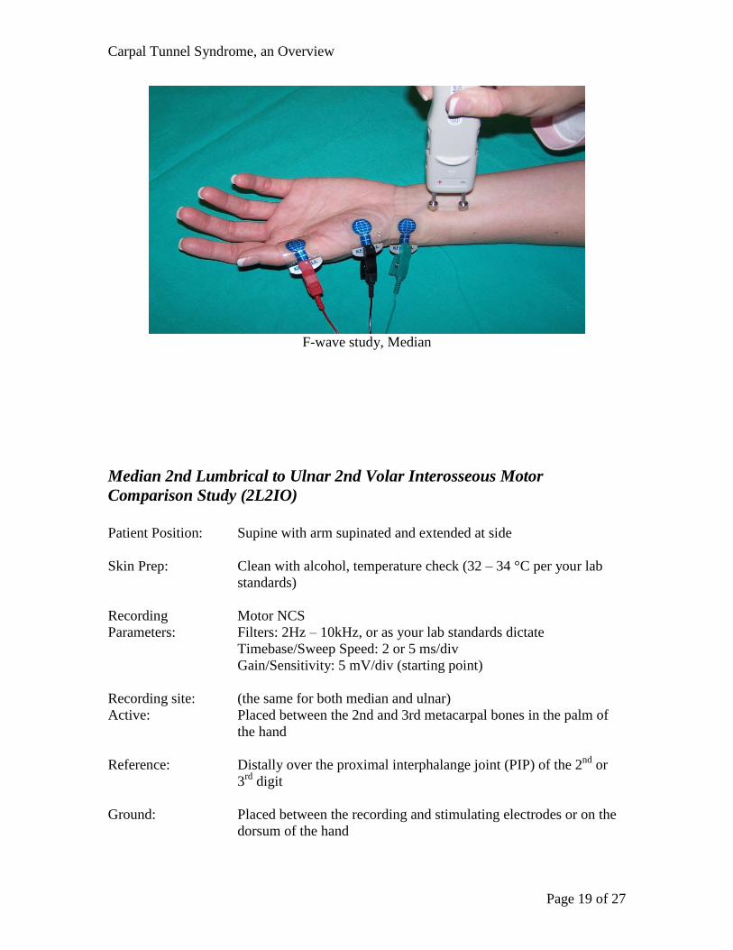
F-wave study, Median
Median 2nd Lumbrical to Ulnar 2nd Volar Interosseous Motor
Comparison Study (2L2IO)
Patient Position: Supine with arm supinated and extended at side
Skin Prep: Clean with alcohol, temperature check (32 – 34 °C per your lab
standards)
Recording Motor NCS
Parameters: Filters: 2Hz – 10kHz, or as your lab standards dictate
Timebase/Sweep Speed: 2 or 5 ms/div
Gain/Sensitivity: 5 mV/div (starting point)
Recording site: (the same for both median and ulnar)
Active: Placed between the 2nd and 3rd metacarpal bones in the palm of
the hand
Reference: Distally over the proximal interphalange joint (PIP) of the 2nd
or
3rd
digit
Ground: Placed between the recording and stimulating electrodes or on the
dorsum of the hand
Carpal Tunnel Syndrome, an Overview
Page 20 of 27
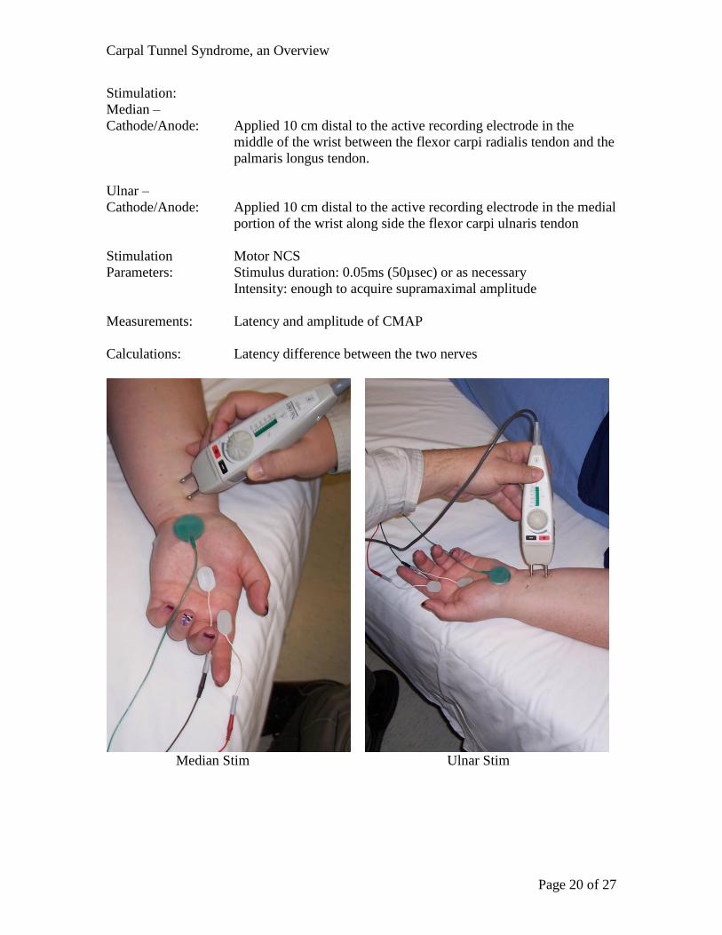
Stimulation:
Median –
Cathode/Anode: Applied 10 cm distal to the active recording electrode in the
middle of the wrist between the flexor carpi radialis tendon and the
palmaris longus tendon.
Ulnar –
Cathode/Anode: Applied 10 cm distal to the active recording electrode in the medial
portion of the wrist along side the flexor carpi ulnaris tendon
Stimulation Motor NCS
Parameters: Stimulus duration: 0.05ms (50µsec) or as necessary
Intensity: enough to acquire supramaximal amplitude
Measurements: Latency and amplitude of CMAP
Calculations: Latency difference between the two nerves
Median Stim Ulnar Stim
Carpal Tunnel Syndrome, an Overview
Page 21 of 27
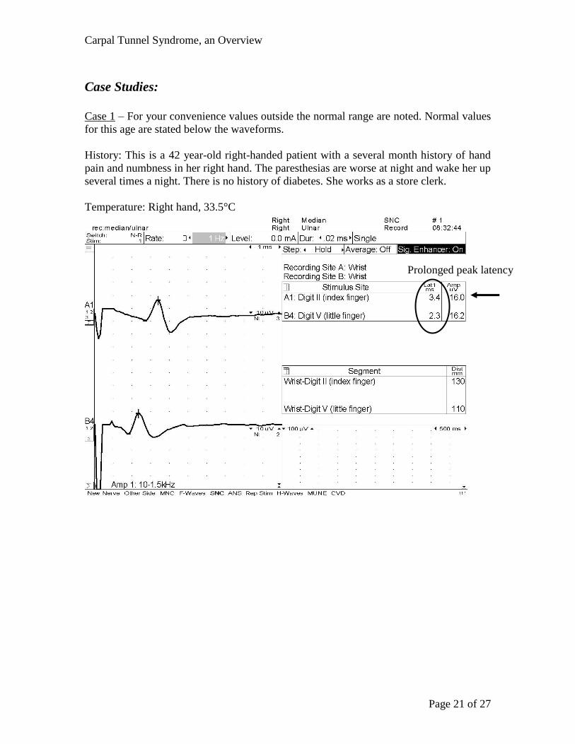
Case Studies:
Case 1 – For your convenience values outside the normal range are noted. Normal values
for this age are stated below the waveforms.
History: This is a 42 year-old right-handed patient with a several month history of hand
pain and numbness in her right hand. The paresthesias are worse at night and wake her up
several times a night. There is no history of diabetes. She works as a store clerk.
Temperature: Right hand, 33.5°C
Prolonged peak latency
Carpal Tunnel Syndrome, an Overview
Page 22 of 27
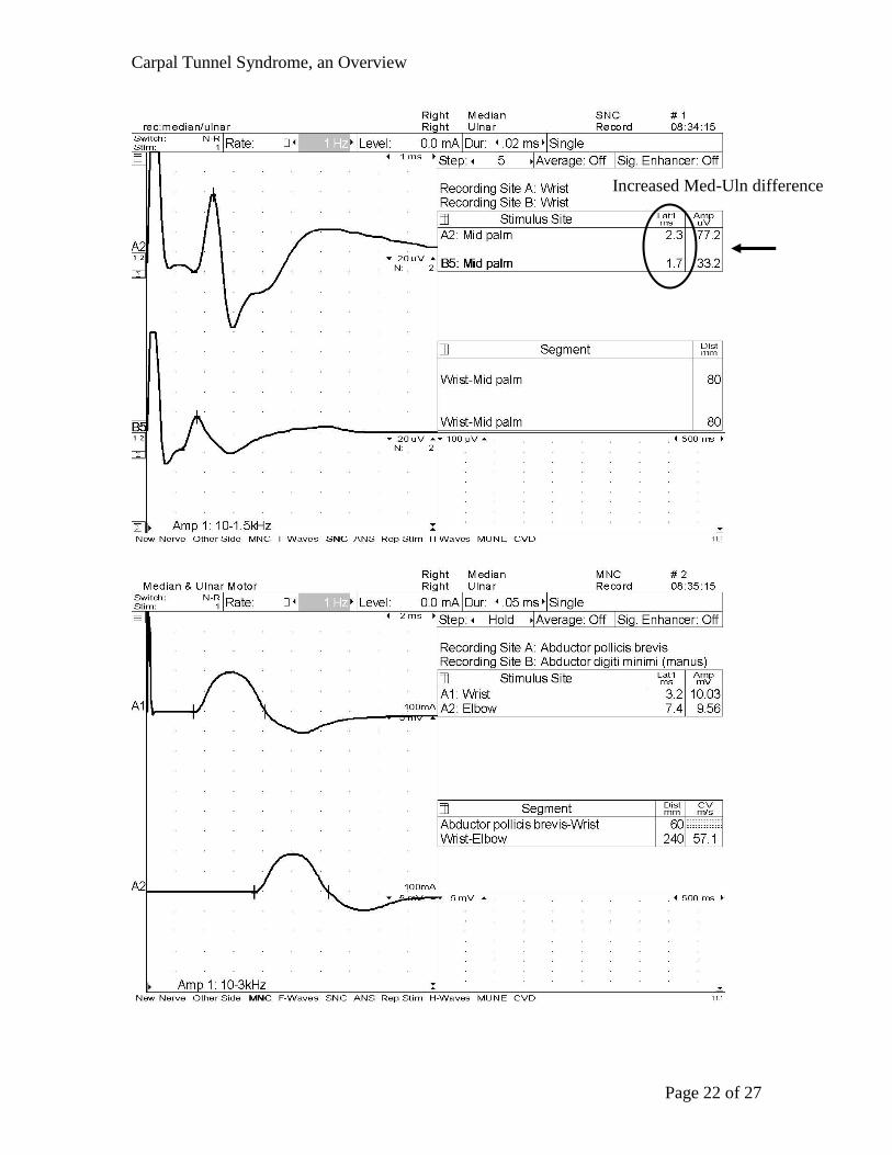
Increased Med-Uln difference
Carpal Tunnel Syndrome, an Overview
Page 23 of 27
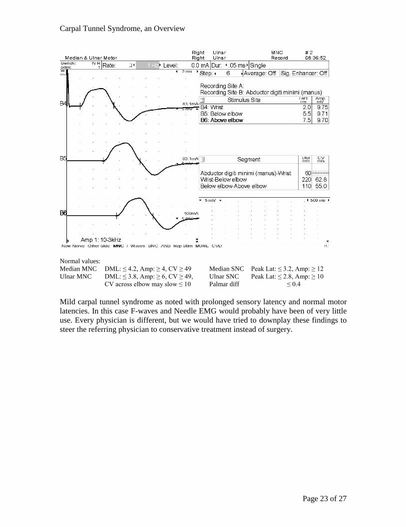
Normal values:
Median MNC DML: ≤ 4.2, Amp: ≥ 4, CV ≥ 49 Median SNC Peak Lat: ≤ 3.2, Amp: ≥ 12
Ulnar MNC DML: ≤ 3.8, Amp: ≥ 6, CV ≥ 49, Ulnar SNC Peak Lat: ≤ 2.8, Amp: ≥ 10
CV across elbow may slow ≤ 10 Palmar diff ≤ 0.4
Mild carpal tunnel syndrome as noted with prolonged sensory latency and normal motor
latencies. In this case F-waves and Needle EMG would probably have been of very little
use. Every physician is different, but we would have tried to downplay these findings to
steer the referring physician to conservative treatment instead of surgery.
Carpal Tunnel Syndrome, an Overview
Page 24 of 27
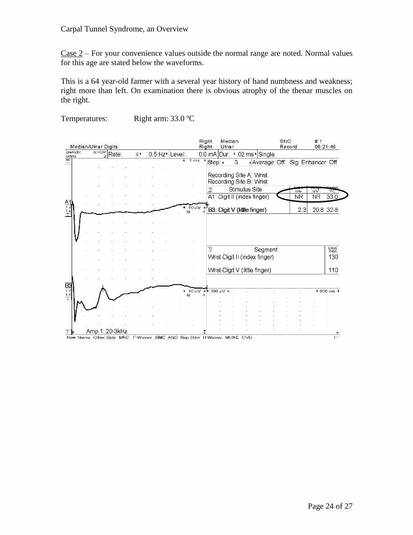
Case 2 – For your convenience values outside the normal range are noted. Normal values
for this age are stated below the waveforms.
This is a 64 year-old farmer with a several year history of hand numbness and weakness;
right more than left. On examination there is obvious atrophy of the thenar muscles on
the right.
Temperatures: Right arm: 33.0 ºC
Carpal Tunnel Syndrome, an Overview
Page 25 of 27
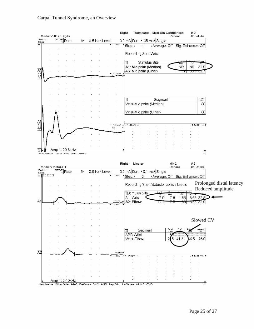
Prolonged distal latency
Reduced amplitude
Slowed CV
Carpal Tunnel Syndrome, an Overview
Page 26 of 27
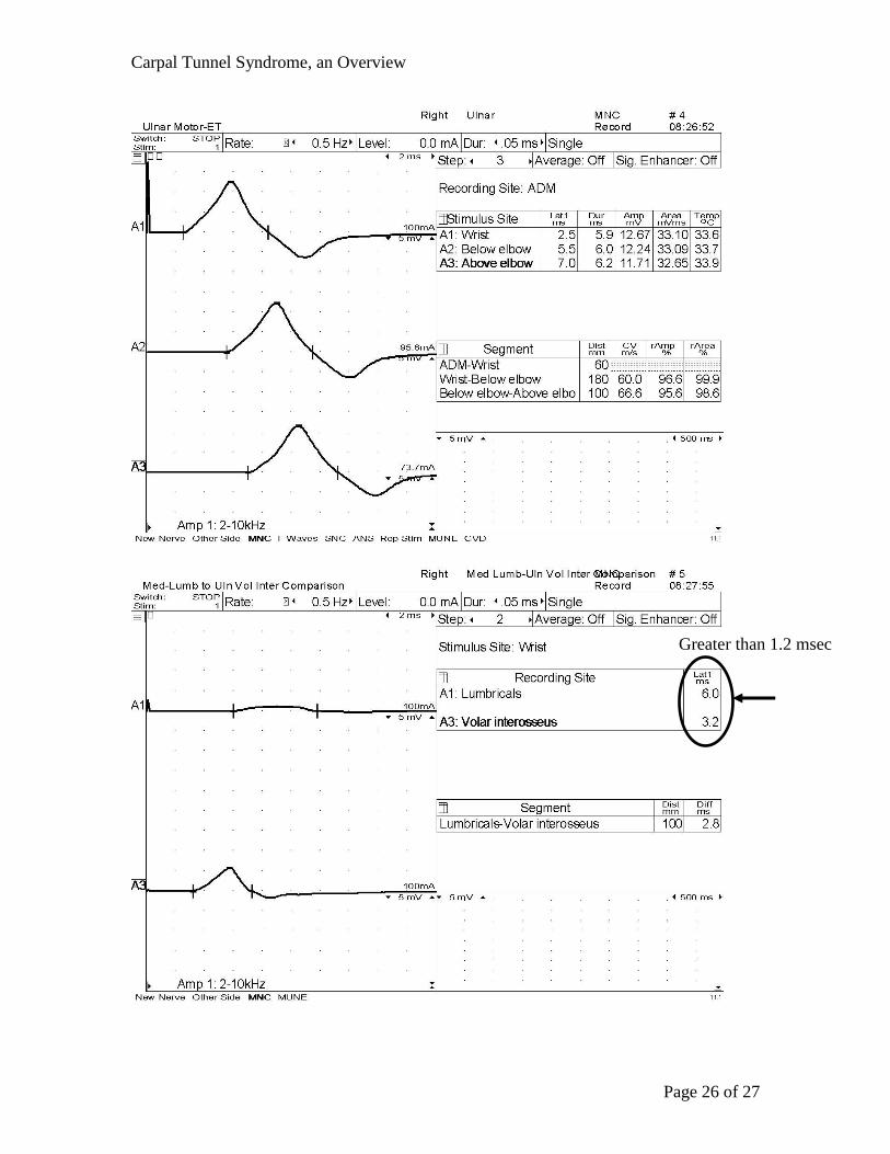
Greater than 1.2 msec
Carpal Tunnel Syndrome, an Overview
Page 27 of 27
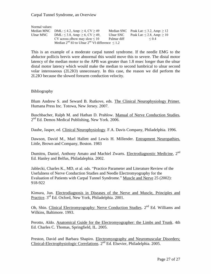
Normal values:
Median MNC DML: ≤ 4.2, Amp: ≥ 4, CV ≥ 49 Median SNC Peak Lat: ≤ 3.2, Amp: ≥ 12
Ulnar MNC DML: ≤ 3.8, Amp: ≥ 6, CV ≥ 49, Ulnar SNC Peak Lat: ≤ 2.8, Amp: ≥ 10
CV across elbow may slow ≤ 10 Palmar diff ≤ 0.4
Median 2nd
IO to Ulnar 2nd
VI difference ≤ 1.2
This is an example of a moderate carpal tunnel syndrome. If the needle EMG to the
abductor pollicis brevis were abnormal this would move this to severe. The distal motor
latency of the median motor to the APB was greater than 1.8 msec longer than the ulnar
distal motor latency which would make the median to second lumbrical to ulnar second
volar interosseous (2L2IO) unnecessary. In this case, the reason we did perform the
2L2IO because the slowed forearm conduction velocity.
Bibliography
Blum Andrew S. and Seward B. Rutkove, eds. The Clinical Neurophysiology Primer.
Humana Press Inc. Totowa, New Jersey. 2007.
Buschbacher, Ralph M. and Hathan D. Prahlow. Manual of Nerve Conduction Studies,
2nd
Ed. Demos Medical Publishing, New York. 2006.
Daube, Jasper, ed. Clinical Neurophysiology. F.A. Davis Company, Philadelphia. 1996.
Dawson, David M., Marl Hallett and Lewis H. Millender. Entrapment Neuropathies.
Little, Brown and Company, Boston. 1983
Dumitru, Daniel, Anthony Amato and Machiel Zwarts. Electrodiagnostic Medicine, 2nd
Ed. Hanley and Belfus, Philadalephia. 2002.
Jablecki, Charles K., MD, et al. eds. “Practice Parameter and Literature Review of the
Usefulness of Nerve Conduction Studies and Needle Electromyography for the
Evaluation of Patients with Carpal Tunnel Syndrome.” Muscle and Nerve 25 (2002):
918-922
Kimura, Jun. Electrodiagnosis in Diseases of the Nerve and Muscle, Principles and
Practice. 3rd
Ed. Oxford, New York, Philadelphia. 2001.
Oh, Shin. Clinical Electromyography: Nerve Conduction Studies. 2nd
Ed. Williams and
Wilkins, Baltimore. 1993.
Perotto, Aldo. Anatomical Guide for the Electromyographer: the Limbs and Trunk. 4th
Ed. Charles C. Thomas, Springfield, IL. 2005.
Preston, David and Barbara Shapiro. Electromyography and Neuromuscular Disorders;
Clinical-Electrophysiologic Correlations. 2nd
Ed. Elsevier, Philadelphia. 2005.