Embed Size (px)
Citation preview
© 20
16 b
y Ac
ta N
euro
biol
ogia
e Exp
erim
enta
lis
The physiology of blood platelets and changes of their biological activities in multiple sclerosis
Barbara Wachowicz1, Agnieszka Morel1, Elżbieta Miller2, 3, and Joanna Saluk1*
1 Faculty of Biology and Environmental Protection, Department of General Biochemistry University of Lodz, Lodz, Poland, 2 Department of Physical Medicine, Medical University of Lodz, Lodz, Poland, 3 Neurorehabilitation Ward, III General Hospital in Lodz, Lodz, Poland,
* Email: [email protected]
Increasing evidence indicates that blood platelets contribute to diverse processes that extend beyond hemostasis. Many of the same mechanisms that play a role in hemostasis and thrombosis facilitate platelets the participation in other physiological and pathological processes, particularly in the inflammation, the immune response and central nervous system disorders. Platelets are involved in pathophysiology of central nervous system diseases, especially in the pathogenesis of multiple sclerosis, but their role appears to be neglected. Platelets contribute to the inflammation and cooperate with immune cells in inflammatory and immune responses. These blood cells were identified in inflamed spinal cord and in the brain in chronic active lesions of multiple sclerosis and in the related animal models referred as Experimental Autoimmune Encephalomyelitis. This review summarizes recent insights in the platelet activation accompanied by the exocytosis of bioactive compounds stored in granules, formation of platelet microparticles, expression of specific membrane receptors, synthesis of numerous biomediators, generation of free radicals, and introduces the mechanisms by which activated platelets may be involved in the pathophysiology of multiple sclerosis. Understanding the role of platelets in multiple sclerosis may be essential for improved therapies.
Key words: platelets, multiple sclerosis, inflammation, oxidative stress, neurodegeneration
INTRODUCTION
Platelets have been recognized as the smallest blood cells that fulfill a complex role in hemostasis and thrombosis. Knowledge regarding the role of platelets in the development and severity of various disorders of central nervous system beyond thrombosis continues to emerge, but the role of platelets in multiple sclerosis appears to be neglected. There is a link between platelets and pathophysiology of multiple sclerosis (MS), and platelets may be key players in this disease (Starossom et al. 2015, Morel et al. 2015, 2016, Marcos‑Ramiro et al. 2014, Sáenz‑Cuesta et al. 2014). Understanding the role of platelets in MS seems to be essential for improved therapy.
MS is a chronic, demyelinating immune‑mediated disease of the central nervous system with axonal degeneration and astrogliosis (Kawahi et al. 2016). It is considered as a complex neurological disease with a variable clinical course and several pathophysiological mechanisms, such as axonal/neuronal damage, inflammation, demyelination, gliosis, remyelination and repair mechanisms (Miller 2012). In pathomechanisms of MS, the alterations of the immune system together with
biochemical disturbances and disruption of blood‑brain barrier (BBB), are involved. Such processes are not uniformly represented in patients’ population, but may selectively predominate in individuals (Lehmann et al. 2015). Activation and infiltration of mononuclear cells, mainly Ag‑specific CD4 and CD8 T cells in central nervous system and their reactivation by resident Ag‑presenting brain‑spinal cord glial cells are characteristic for the pathogenesis of MS. Activated T cells and brain glial cells secrete proinflammatory cytokines/chemokines along with generation of inflammatory mediators – highly reactive free radicals that leads to axonal loss (Jones et al. 2016). This autoimmune disease has a unknown precise etiology, however epidemiological data indicate multifactorial interactions between genetic susceptibility and environmental factors (Goodin 2016). MS affecting about 2.3 million people of the population in high‑prevalence areas, and it is one of the most common causes of neurological disability of young adults (Brola et al. 2016). There are three main types of MS, defined as: relapsing‑remitting (RR), secondary progressive (SP), primary progressive (PP) with a progressive‑relapsing (PR) subtype (Ayache et al. 2016). The neuroaxonal degeneration is dominated in
Correspondence should be addressed to J. Saluk Email: [email protected]
Received 26 February 2016, accepted 14 December 2016
Review
Acta Neurobiol Exp 2016, 76: 269–281
2_785_Wachowicz_v4.indd 269 05/01/17 21:15
270 B. Wachowicz et al. Acta Neurobiol Exp 2016, 76: 269–281
both progressive forms – PPMS and SPMS, and correlate with brain disability and spinal cord atrophy (Seizer and May 2013). In SPMS, the central nervous system (CNS) atrophy is characteristic. It is generally very difficult to predict the clinical course of MS. Progression of disability seems to be increased in patients with higher number of relapses during the first and second year of the disease (Seizer and May 2013).
BLOOD PLATELETS
Blood platelets are anucleate cells that serve a critical function in hemostasis. In human body about 1×10¹¹ platelets are formed every day as the result of complex processes of differentiation, maturation and fragmentation of megakaryocytes (Xu et al. 2016b). Individual platelets vary in terms of volume, density and reactivity (Jones 2016). The normal platelet counts is in the range of 150–400×109/L. Under normal conditions platelets circulate in blood stream for 8–10 days and upon vascular injury, platelets instantly adhere to the exposed extracellular matrix resulting in platelet activation to form hemostatic plug. When platelet number is low, the risk of bleeding is high.
Hemostatic activity of blood platelets
The primary biological function of platelets is to form hemostatic thrombi that prevents blood loss and maintains vascular integrity (Xu et al. 2016a). Platelets in the circulation are in resting state and become activated at sites of vascular injury by adhesion to adhesive proteins such as von Willebrand factor (vWf) and collagen, or by soluble platelets agonists such as thrombin, ADP, or thromboxaneA2 formed from arachidonic acid in stimulated platelets. The expression of multiple membrane receptors, both constitutive and activation dependent mediates platelet adhesion and aggregation at sites of vascular injury. Numerous platelet agonists induce signal transduction via their respective receptors and intermediate signaling events that stimulate platelet shape, secretory process, activation of integrin αIIbβ3. Binding of fibrinogen or other ligands to integrin αIIbβ3 mediates platelet aggregation. Platelet activation is a dynamic process and involves multiple feedback loops and cross talk between different pathways in the cell. The most abundant integrin in platelets αIIbβ3 is normally in resting state, but is transformed into activated state after platelet activation. Binding of fibrinogen, vWf and some matrix proteins containing RGD sequences mediates stable platelet adhesion, aggregation and thrombus formation (Xu et al. 2016a). During activation of platelets
microparticles (MPs) are released and are regard as a markers of platelet activation. Platelet MPs are the most abundant MPs found in the circulation (Badimon et al. 2016, Ponomareva et al. 2016, Papapanagiotou et al. 2016, Boilard et al. 2015, Varon et al. 2015), and are potential mediators of blood coagulation by serving as circulating sources of tissue factor (TF) that is a transmembrane protein involved in thrombin generation. On the surface of MPs some quantity of phospatidylserine – a negatively charged aminophospholipid is exposed. Mechanisms of interaction of MPs with effector cells may involve a membrane fusion, endocytosis, or interaction of MPs with membrane receptor to stimulate cellular signaling events (Varon et al. 2015).
The most important adhesion molecules on platelets are integrins, heterodimeric transmembrane proteins that mediate interactions with extracellular matrix molecules and other adhesion molecules on the blood cells. They also play an important role in platelet signaling. Besides integrin αIIbβ3 (GPIIb/IIIa) platelets express beta 1 and beta 3 integrins such as α5β1 (VLA‑5), α6β1 (VLA‑6), α2β1 (GPIa/IIa, VLA‑2) (Bender et al. 2016). Platelets possess a number of Ig superfamily of cellular adhesion molecules: ICAM‑2 (intercellular adhesion molecule‑2), JAM‑A, JAM‑C (junctional adhesion molecule) and PECAM‑1 (platelet endothelial cell adhesion molecule‑1). These molecules may interact with other cells. On the platelet membranes glycoprotein complex (GPIb‑V‑IX) is involved in the interaction of platelets with exposed subendothelium through binding to vWf. GPVI mediates platelet binding to collagen. Platelets express complement receptors, receptors for immunoglobins (FcR) and Toll‑like receptors (Lam et al. 2015).
Platelet secretory process
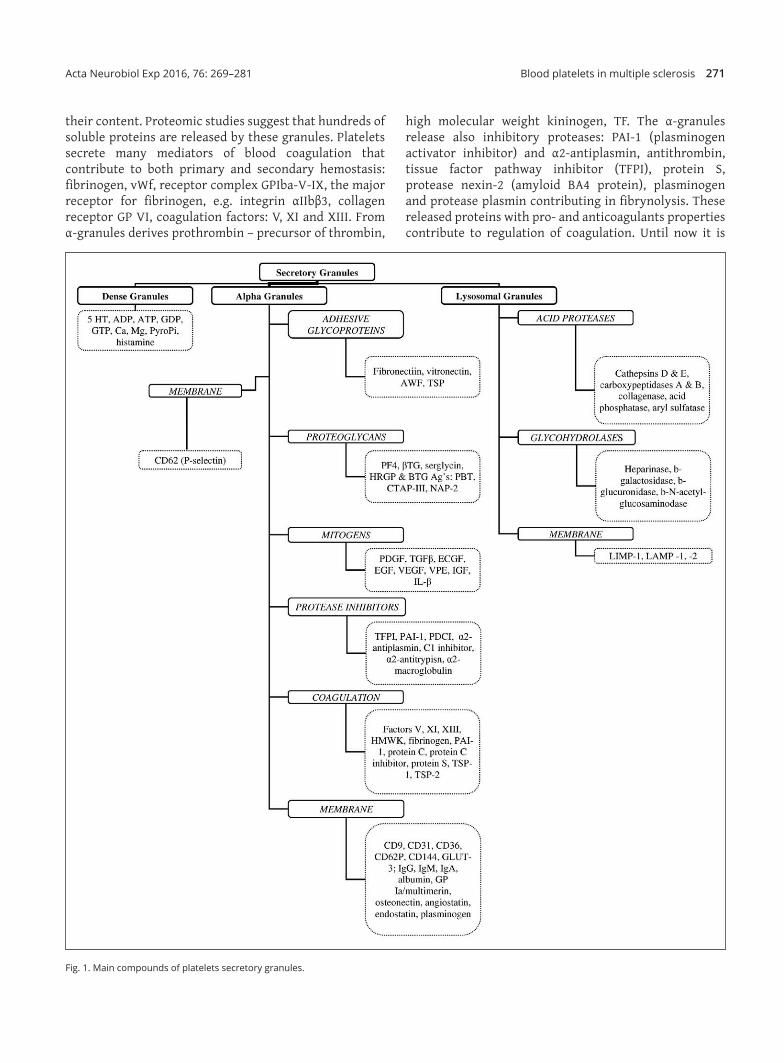
Platelet activation leads to exocytosis of granule constituents, release of stored and newly synthesized mediators. The discharge of membrane‑bound transcellular signaling molecules are also observed. Platelets are replete with secretory granules, which are critical to normal platelet function (Lam et al. 2015). There are three types of platelet secretory granules; dense granules, lysosomes and the most abundant α‑granules (Fig. 1). There are approximately 50–80 α‑granules per platelet. The α‑granules are essential to normal platelet activity, are formed in megakaryocytes and distributed to platelets during megakaryopoiesis. Their contents are released from platelets in order to achieve their physiological function. Granule membranes fuse with surface‑connected membranes of open canalicular system or the plasma membrane with expression of P‑selectin on the platelet surface. The α‑granules function depends on
2_785_Wachowicz_v4.indd 270 05/01/17 21:15
Blood platelets in multiple sclerosis 271Acta Neurobiol Exp 2016, 76: 269–281
their content. Proteomic studies suggest that hundreds of soluble proteins are released by these granules. Platelets secrete many mediators of blood coagulation that contribute to both primary and secondary hemostasis: fibrinogen, vWf, receptor complex GPIba‑V‑IX, the major receptor for fibrinogen, e.g. integrin αIIbβ3, collagen receptor GP VI, coagulation factors: V, XI and XIII. From α‑granules derives prothrombin – precursor of thrombin,
high molecular weight kininogen, TF. The α‑granules release also inhibitory proteases: PAI‑1 (plasminogen activator inhibitor) and α2‑antiplasmin, antithrombin, tissue factor pathway inhibitor (TFPI), protein S, protease nexin‑2 (amyloid BA4 protein), plasminogen and protease plasmin contributing in fibrynolysis. These released proteins with pro‑ and anticoagulants properties contribute to regulation of coagulation. Until now it is
Fig. 1. Main compounds of platelets secretory granules.
2_785_Wachowicz_v4.indd 271 05/01/17 21:15
272 B. Wachowicz et al. Acta Neurobiol Exp 2016, 76: 269–281
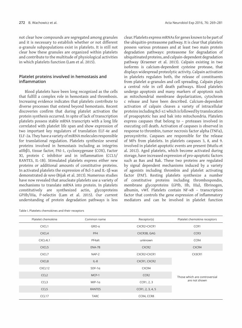
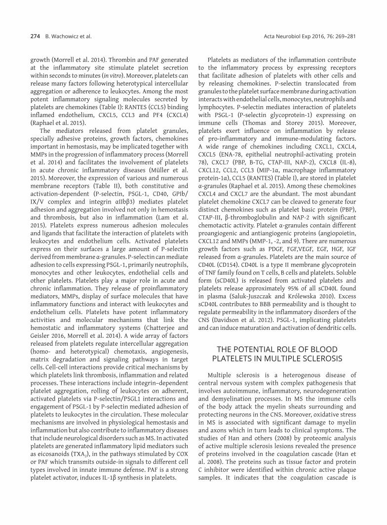
not clear how compounds are segregated among granules and it is necessary to establish whether or not different α‑granule subpopulations exist in platelets. It is still not clear how these granules are organized within platelets and contribute to the multitude of physiological activities in which platelets function (Lam et al. 2015).
Platelet proteins involved in hemostasis and inflammation
Blood platelets have been long recognized as the cells that fulfill a complex role in hemostasis and thrombosis. Increasing evidence indicates that platelets contribute to diverse processes that extend beyond hemostasis. Recent discoveries confirm that during platelet activation the protein synthesis occurred. In spite of lack of transcription platelets possess stable mRNA transcripts with a long life correlated with platelet life span and show expression of two important key regulators of translation ELF‑4e and ELF‑2a. They have a variety of miRNA molecules responsible for translational regulation. Platelets synthesize several proteins involved in hemostasis including as integrins αIIbβ3, tissue factor, PAI‑1, cyclooxygenase (COX), Factor XI, protein C inhibitor and in inflammation (CCL5/RANTES, IL‑1B). Stimulated platelets express either new proteins or additional amounts of constitutive proteins. In activated platelets the expression of Bcl‑3 and IL‑1β was demonstrated de novo (Bijak et al. 2013). Numerous studies have now revealed that anucleate platelets use a variety of mechanisms to translate mRNA into protein. In platelets constitutively are synthesized actin, glycoproteins GPIIb/IIIa, P‑selectin (Lam et al. 2015). Our current understanding of protein degradation pathways is less
clear. Platelets express mRNAs for genes known to be part of the ubiquitin‑proteasome pathway. It is clear that platelets possess various proteases and at least two main protein degradation pathways: proteasome for degradation of ubiquitinated proteins, and calpain‑dependent degradation pathway (Kraemer et al. 2013). Calpain existing in two isoforms is calcium‑dependent cysteine protease, that displays widespread proteolytic activity. Calpain activation in platelets regulates both, the release of constituents from platelet α‑granules and cell spreading. Calpain plays a central role in cell death pathways. Blood platelets undergo apoptosis and many markers of apoptosis such as mitochondrial membrane depolarization, cytochrom c release and have been described. Calcium‑dependent activation of calpain cleaves a variety of intracellular proteins including Bcl‑x1 which is followed by translocation of proapoptotic bax and bak into mitochondria. Platelets express caspases that belong to – proteases involved in executing cell death. Activation of caspases is observed in response to thrombin, tumor necrosis factor alpha (TNFα), peroxynitrite. Caspases are responsible for the release of MPs from platelets. In platelets caspases 3, 8, and 9. involved in platelet apoptotic events are present (Mutlu et al. 2012). Aged platelets, which become activated during storage, have increased expression of pro‑apoptotic factors such as Bax and Bak. These two proteins are regulated by signal dependent mechanisms induced by a variety of agonists including thrombin and platelet activating factor (PAF). Resting platelets synthesize a number of constitutive proteins including thrombospondin, membrane glycoproteins (GPIb, IIb, IIIa), fibrinogen, albumin, vWf. Platelets contain NF‑κB – transcription factor that controls the gene expression of inflammatory mediators and can be involved in platelet function
Table I. Platelets chemokines and their receptors
Platelet chemokine Common name Receptor(s) Platelet chemokine receptors
CXCL1 GRO‑α CXCR2>CXCR1 CCR1
CXCL4 PF4 CXCR3B, GAG CCR3
CXCL4L1 PF4alt unknown CCR4
CXCL5 ENA‑78 CXCR2 CXCR4
CXCL7 NAP‑2 CXCR2>CXCR1 CX3CR1
CXCL8 IL‑8 CXCR1, CXCR2
Those which are controversial are not shown
CXCL12 SDF‑1α CXCR4
CCL2 MCP‑1 CCR2
CCL3 MIP‑1α CCR1, 2, 3
CCL5 RANTES CCR1, 2, 3, 4, 5
CCL17 TARC CCR4, CCR8
2_785_Wachowicz_v4.indd 272 05/01/17 21:15
Blood platelets in multiple sclerosis 273Acta Neurobiol Exp 2016, 76: 269–281
(Fuentes et al. 2016). Platelets synthesize and secrete a variety of matrix metalloproteinases (MMPs) including MMP‑1, MMP‑2, MMP‑3 and MMP‑14 (MT1‑MMP), and potentially MMP‑9 as well as the tissue inhibitors of metalloproteinases (TIMPs). MMPs are zinc‑dependent endopeptidases and together with the ADAM family (A Disintegrin And Metalloproteinase) belong to the family of metalloproteinases. They are synthesized and secreted as inactive proenzymes (Mastenbroek et al. 2015). Four endogenous tissue inhibitors of MMPs (TIMP‑1, TIMP‑2, TIMP‑3 and TIMP‑4) have been described in nucleated cells. In platelets and megakaryocytes, mRNA for TIMP‑1, TIMP‑2 and TIMP‑3 have been identified (Seizer and May 2013).
MMP‑1 in platelets is more abundant than other MMPs. Platelets exposure to collagen converts surfrace bound pro‑MMP‑1 to active MMP‑1 that in turn activates protease activated receptor 1 (PAR‑1) by cleaving the receptor and promotes platelet aggregation by PAR‑1. MMP‑2 after binding to GPIIb/GPIIIa on platelets, becomes converted to active MMP‑2 and can promote integrin‑mediated platelet aggregation. Moreover, CD40Ls shed from the platelet surface by active MMP‑2 may be a potent inducer of inflammatory processes in various CD40 expressing cells. Platelets can induce several MMPs in various cell types. Recently the so called Extracellular Matrix Metalloproteinase Inducer (EMMPRIN, CD147) has been identified on platelets (Zhu et al. 2014, Seizer and May 2013). It is an immunoglobin‑like receptor stored in α‑granules and released after thrombin or ADP stimulation. Interaction of platelets with monocytes induces EMMPRIN‑dependent monocyte production of NF‑κB‑mediated MMP‑9 and inflammatory cytokines such as IL‑6 and TNFα (Kral et al. 2016). The direct interaction of neutrophils and platelets seems to be mediated by neutrophilic PSGL‑1 and P‑selectin on activated platelets (Krishnamurthy et al. 2015).
Platelets contribution in inflammatory processes
Platelets by virtue of their immense number in the circulation, their numerous receptors, immunomodulatory mediators, cell adhesion molecules and platelet MPs (Lam et al. 2015) are able to participate in the inflammatory response (Koenen 2016). Bacterial endotoxins induce thrombocytopenia, hypotension, and sepsis. Blood platelet activation plays a critical role in LPS‑induced thrombocytopenia and tissue damage. Activated platelets produce ROS that cause damage of the vascular endothelium and promote multiple organ dysfunctions in sepsis (Casarin et al. 2011, Tyml 2011). Some studies showed that LPS stimulates platelet secretion of dense and alpha granules, and release of biological active inflammatory mediators (Zhang et al. 2009). Adhesion and aggregation of blood platelets is involved in pathogenesis of septic shock after damage of endothelial cells by LPS or inflammatory cytokines (Schrottmaier et al. 2016).
Circulating platelet‑leukocyte aggregates (PLAs) are the hallmark feature of sepsis and indicate that the close interaction between platelets and the immune system during inflammatory responses takes place (Thomas and Storey 2015). Adherent platelets rapidly express on their surfaces large quantities of P‑selectin and neutrophils can tether and roll via their PSGL‑1. This rolling is followed by firm adhesion of the neutrophil to adherent platelets via Mac‑1 binding to fibrinogen bound to platelet integrin GPIIb/IIIa or to GPIba (Wu et al. 2016). Recently, the ability of platelets to mediate adhesion of neutrophils has been presented by Langer and others (2012) in a mouse model of autoimmune encephalomyelitis (EAE).
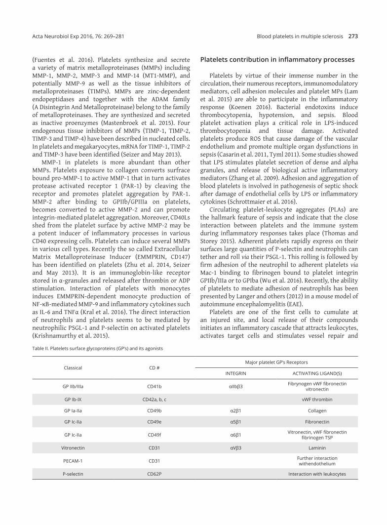
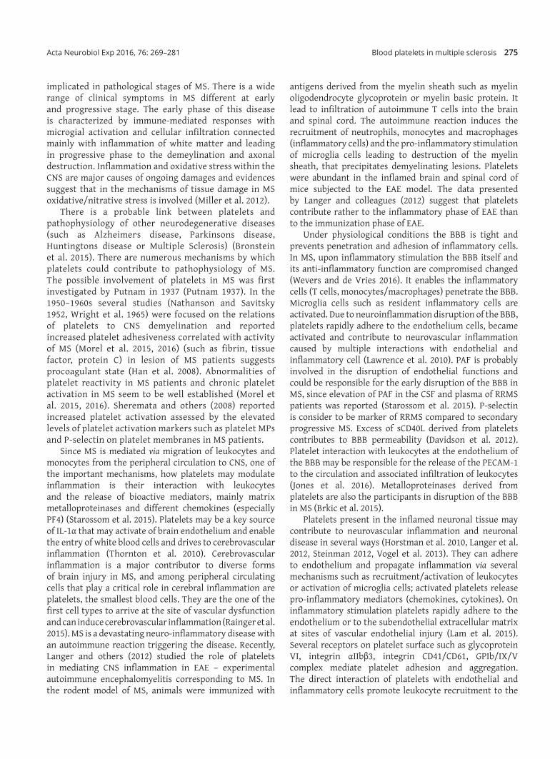
Platelets are one of the first cells to cumulate at an injured site, and local release of their compounds initiates an inflammatory cascade that attracts leukocytes, activates target cells and stimulates vessel repair and
Table II. Platelets surface glycoproteins (GP’s) and its agonists
Classical CD #Major platelet GP’s Receptors
INTEGRIN ACTIVATING LIGAND(S)
GP IIb/IIIa CD41b αIIbβ3 Fibrynogen vWF fibronectin vitronectin
GP Ib‑IX CD42a, b, c vWF thrombin
GP Ia‑IIa CD49b α2β1 Collagen
GP Ic‑IIa CD49e α5β1 Fibronectin
GP Ic‑IIa CD49f α6β1 Vitronectin, vWF fibronectin fibrinogen TSP
Vitronectin CD31 αVβ3 Laminin
PECAM‑1 CD31 Further interaction withendothelium
P‑selectin CD62P Interaction with leukocytes
2_785_Wachowicz_v4.indd 273 05/01/17 21:15
274 B. Wachowicz et al. Acta Neurobiol Exp 2016, 76: 269–281
growth (Morrell et al. 2014). Thrombin and PAF generated at the inflammatory site stimulate platelet secretion within seconds to minutes (in vitro). Moreover, platelets can release many factors following heterotypical intercellular aggregation or adherence to leukocytes. Among the most potent inflammatory signaling molecules secreted by platelets are chemokines (Table I): RANTES (CCL5) binding inflamed endothelium, CXCL5, CCL3 and PF4 (CXCL4) (Raphael et al. 2015).
The mediators released from platelet granules, specially adhesive proteins, growth factors, chemokines important in hemostasis, may be implicated together with MMPs in the progression of inflammatory process (Morrell et al. 2014) and facilitates the involvement of platelets in acute chronic inflammatory diseases (Müller et al. 2015). Moreover, the expression of various and numerous membrane receptors (Table II), both constitutive and activation‑dependent (P‑selectin, PSGL‑1, CD40, GPIb/IX/V complex and integrin αIIbβ3) mediates platelet adhesion and aggregation involved not only in hemostasis and thrombosis, but also in inflammation (Lam et al. 2015). Platelets express numerous adhesion molecules and ligands that facilitate the interaction of platelets with leukocytes and endothelium cells. Activated platelets express on their surfaces a large amount of P‑selectin derived from membrane α‑granules. P‑selectin can mediate adhesion to cells expressing PSGL‑1, primarily neutrophils, monocytes and other leukocytes, endothelial cells and other platelets. Platelets play a major role in acute and chronic inflammation. They release of proinflammatory mediators, MMPs, display of surface molecules that have inflammatory functions and interact with leukocytes and endothelium cells. Platelets have potent inflammatory activities and molecular mechanisms that link the hemostatic and inflammatory systems (Chatterjee and Geisler 2016, Morrell et al. 2014). A wide array of factors released from platelets regulate intercellular aggregation (homo‑ and heterotypical) chemotaxis, angiogenesis, matrix degradation and signaling pathways in target cells. Cell‑cell interactions provide critical mechanisms by which platelets link thrombosis, inflammation and related processes. These interactions include integrin–dependent platelet aggregation, rolling of leukocytes on adherent, activated platelets via P‑selectin/PSGL1 interactions and engagement of PSGL‑1 by P‑selectin mediated adhesion of platelets to leukocytes in the circulation. These molecular mechanisms are involved in physiological hemostasis and inflammation but also contribute to inflammatory diseases that include neurological disorders such as MS. In activated platelets are generated inflammatory lipid mediators such as eicosanoids (TXA2), in the pathways stimulated by COX or PAF which transmits outside‑in signals to different cell types involved in innate immune defense. PAF is a strong platelet activator, induces IL‑1β synthesis in platelets.
Platelets as mediators of the inflammation contribute to the inflammatory process by expressing receptors that facilitate adhesion of platelets with other cells and by releasing chemokines. P‑selectin translocated from granules to the platelet surface membrane during activation interacts with endothelial cells, monocytes, neutrophils and lymphocytes. P‑selectin mediates interaction of platelets with PSGL‑1 (P‑selectin glycoprotein‑1) expressing on immune cells (Thomas and Storey 2015). Moreover, platelets exert influence on inflammation by release of pro‑inflammatory and immune‑modulating factors. A wide range of chemokines including CXCL1, CXCL4, CXCL5 (ENA‑78, epithelial neutrophil‑activating protein 78), CXCL7 (PBP, B‑TG, CTAP‑III, NAP‑2), CXCL8 (IL‑8), CXCL12, CCL2, CCL3 (MIP‑1α, macrophage inflammatory protein‑1α), CCL5 (RANTES) (Table I), are stored in platelet α‑granules (Raphael et al. 2015). Among these chemokines CXCL4 and CXCL7 are the abundant. The most abundant platelet chemokine CXCL7 can be cleaved to generate four distinct chemokines such as platelet basic protein (PBP), CTAP‑III, β‑thromboglobulin and NAP‑2 with significant chemotactic activity. Platelet α‑granules contain different proangiogenic and antiangiogenic proteins (angiopoietin, CXCL12 and MMPs (MMP‑1, ‑2, and 9). There are numerous growth factors such as PDGF, FGF,VEGF, EGF, HGF, IGF released from α‑granules. Platelets are the main source of CD40L (CD154). CD40L is a type II membrane glycoprotein of TNF family found on T cells, B cells and platelets. Soluble form (sCD40L) is released from activated platelets and platelets release approximately 95% of all sCD40L found in plasma (Saluk‑Juszczak and Królewska 2010). Excess sCD40L contributes to BBB permeability and is thought to regulate permeability in the inflammatory disorders of the CNS (Davidson et al. 2012). PSGL‑1, implicating platelets and can induce maturation and activation of dendritic cells.
THE POTENTIAL ROLE OF BLOOD PLATELETS IN MULTIPLE SCLEROSIS
Multiple sclerosis is a heterogenous disease of central nervous system with complex pathogenesis that involves autoimmune, inflammatory, neurodegeneration and demyelination processes. In MS the immune cells of the body attack the myelin sheats surrounding and protecting neurons in the CNS. Moreover, oxidative stress in MS is associated with significant damage to myelin and axons which in turn leads to clinical symptoms. The studies of Han and others (2008) by proteomic analysis of active multiple sclerosis lesions revealed the presence of proteins involved in the coagulation cascade (Han et al. 2008). The proteins such as tissue factor and protein C inhibitor were identified within chronic active plaque samples. It indicates that the coagulation cascade is
2_785_Wachowicz_v4.indd 274 05/01/17 21:15
Blood platelets in multiple sclerosis 275Acta Neurobiol Exp 2016, 76: 269–281
implicated in pathological stages of MS. There is a wide range of clinical symptoms in MS different at early and progressive stage. The early phase of this disease is characterized by immune‑mediated responses with microgial activation and cellular infiltration connected mainly with inflammation of white matter and leading in progressive phase to the demeylination and axonal destruction. Inflammation and oxidative stress within the CNS are major causes of ongoing damages and evidences suggest that in the mechanisms of tissue damage in MS oxidative/nitrative stress is involved (Miller et al. 2012).
There is a probable link between platelets and pathophysiology of other neurodegenerative diseases (such as Alzheimer,s disease, Parkinsons disease, Huntingtons disease or Multiple Sclerosis) (Bronstein et al. 2015). There are numerous mechanisms by which platelets could contribute to pathophysiology of MS. The possible involvement of platelets in MS was first investigated by Putnam in 1937 (Putnam 1937). In the 1950–1960s several studies (Nathanson and Savitsky 1952, Wright et al. 1965) were focused on the relations of platelets to CNS demyelination and reported increased platelet adhesiveness correlated with activity of MS (Morel et al. 2015, 2016) (such as fibrin, tissue factor, protein C) in lesion of MS patients suggests procoagulant state (Han et al. 2008). Abnormalities of platelet reactivity in MS patients and chronic platelet activation in MS seem to be well established (Morel et al. 2015, 2016). Sheremata and others (2008) reported increased platelet activation assessed by the elevated levels of platelet activation markers such as platelet MPs and P‑selectin on platelet membranes in MS patients.
Since MS is mediated via migration of leukocytes and monocytes from the peripheral circulation to CNS, one of the important mechanisms, how platelets may modulate inflammation is their interaction with leukocytes and the release of bioactive mediators, mainly matrix metalloproteinases and different chemokines (especially PF4) (Starossom et al. 2015). Platelets may be a key source of IL‑1α that may activate of brain endothelium and enable the entry of white blood cells and drives to cerebrovascular inflammation (Thornton et al. 2010). Cerebrovascular inflammation is a major contributor to diverse forms of brain injury in MS, and among peripheral circulating cells that play a critical role in cerebral inflammation are platelets, the smallest blood cells. They are the one of the first cell types to arrive at the site of vascular dysfunction and can induce cerebrovascular inflammation (Rainger et al. 2015). MS is a devastating neuro‑inflammatory disease with an autoimmune reaction triggering the disease. Recently, Langer and others (2012) studied the role of platelets in mediating CNS inflammation in EAE – experimental autoimmune encephalomyelitis corresponding to MS. In the rodent model of MS, animals were immunized with
antigens derived from the myelin sheath such as myelin oligodendrocyte glycoprotein or myelin basic protein. It lead to infiltration of autoimmune T cells into the brain and spinal cord. The autoimmune reaction induces the recruitment of neutrophils, monocytes and macrophages (inflammatory cells) and the pro‑inflammatory stimulation of microglia cells leading to destruction of the myelin sheath, that precipitates demyelinating lesions. Platelets were abundant in the inflamed brain and spinal cord of mice subjected to the EAE model. The data presented by Langer and colleagues (2012) suggest that platelets contribute rather to the inflammatory phase of EAE than to the immunization phase of EAE.
Under physiological conditions the BBB is tight and prevents penetration and adhesion of inflammatory cells. In MS, upon inflammatory stimulation the BBB itself and its anti‑inflammatory function are compromised changed (Wevers and de Vries 2016). It enables the inflammatory cells (T cells, monocytes/macrophages) penetrate the BBB. Microglia cells such as resident inflammatory cells are activated. Due to neuroinflammation disruption of the BBB, platelets rapidly adhere to the endothelium cells, became activated and contribute to neurovascular inflammation caused by multiple interactions with endothelial and inflammatory cell (Lawrence et al. 2010). PAF is probably involved in the disruption of endothelial functions and could be responsible for the early disruption of the BBB in MS, since elevation of PAF in the CSF and plasma of RRMS patients was reported (Starossom et al. 2015). P‑selectin is consider to be marker of RRMS compared to secondary progressive MS. Excess of sCD40L derived from platelets contributes to BBB permeability (Davidson et al. 2012). Platelet interaction with leukocytes at the endothelium of the BBB may be responsible for the release of the PECAM‑1 to the circulation and associated infiltration of leukocytes (Jones et al. 2016). Metalloproteinases derived from platelets are also the participants in disruption of the BBB in MS (Brkic et al. 2015).
Platelets present in the inflamed neuronal tissue may contribute to neurovascular inflammation and neuronal disease in several ways (Horstman et al. 2010, Langer et al. 2012, Steinman 2012, Vogel et al. 2013). They can adhere to endothelium and propagate inflammation via several mechanisms such as recruitment/activation of leukocytes or activation of microglia cells; activated platelets release pro‑inflammatory mediators (chemokines, cytokines). On inflammatory stimulation platelets rapidly adhere to the endothelium or to the subendothelial extracellular matrix at sites of vascular endothelial injury (Lam et al. 2015). Several receptors on platelet surface such as glycoprotein VI, integrin αIIbβ3, integrin CD41/CD61, GPIb/IX/V complex mediate platelet adhesion and aggregation. The direct interaction of platelets with endothelial and inflammatory cells promote leukocyte recruitment to the
2_785_Wachowicz_v4.indd 275 05/01/17 21:15
276 B. Wachowicz et al. Acta Neurobiol Exp 2016, 76: 269–281
inflamed tissue and various platelet receptors are involved in these processes: P‑selectin of activated platelets and GPIb (component of the GPIb/GPIX/GPV complex) that is responsible for binding of von Willebrand factor. Receptor GPIb also interacts with the leukocyte integrin Mac‑1 promoting the interactions of platelets and leukocytes. On activated platelets interactions of leukocytes with platelets include the binding of P‑selectin glycoprotein ligand‑1 (PSGL‑1) on leukocytes to platelet P‑selectin (Rainger et al. 2015). Recently, Sotnikov and others (2013) showed a new role of platelets which directly recognize a neuronal damage and communicate with the cells of the immune system in the pathogenesis of MS. Platelets recognize specific glycolipid structure in brain, respond to neurovascular damage and promote neuroinflammation. The study of Sotnikov and others (2013) revealed that platelets recognize the sialated gangliosides in the lipid rafts on the surface of astrogial and neuronal cells that are important for the development of the EAE in an animal model for MS. Gangliosides are present in many tissues, but the most abundant source is the brain. Among numerous gangliosides only gangliosides GT1b and GQ1b were specifically recognized by the platelets (Sotnikov et al. 2013). The recognition of these sialated glycosphingolipids by platelets involved multiple receptors with P‑selectin playing the central role. Platelets penetrate the BBB, may recognize sialated gangliosides within the lipid rafts and accumulate in the lipid rafts‑rich areas in the model of neuroinflammation. Sotnikov and others (2013) examined the platelet reaction to the brain lipid rafts in the EAE model and observed the platelet secretory process in the CNS during EAE by measuring the levels of released of CD41+ and PF4/CXCL4. Moreover, they observed that mice with EAE had 25‑fold higher level of PF4 in serum compared to the healthy mice, and similar to the brain lipid rafts treated mice. It indicates that during neuroinflammation platelets interact with brain lipid rafts and are highly sensitive to the activation and degranulation in the periphery. The obtained data suggest that platelet‑lipid rafts interaction could play an important role in EAE, and could be involved in the activation of cells of immune system both in the periphery and in the CNS. The study reveals the lipid rafts (stable dense areas of the membrane) of astrocytes and neurons as new ligands within the CNS that are recognized by platelets and could play an important role in a neuronal damage in the induction and perpetuation of inflammation in the CNS. The role of various platelet‑derived bioactive lipids in MS is not yet clear. In inflammatory process are involved not only platelet prostaglandins but other lipids such as resolvins generated via lipoxygenase pathway (Moro et al. 2016), PAF (Edwards and Constantinescu 2009), and endocannabinoids released from activated platelets (anandamid and 2‑arachidonoylglycerol) (Hind et al. 2015). Moreover, platelets express endocannabinoid receptors CB1
and CB2 (Brantl et al. 2013). Increased platelet activation could lead even to the changes of endocannabinoid level in brain (Brantl et al. 2013).
Platelets produce approximately 95% of all sCD40L found in plasma, which is released upon activation and excess soluble CD40L contributes to BBB permeability when increased infiltration of the CNS by leucocytes and platelets occurs (Davidson et al. 2012). Moreover, CD40Ls shed from the platelet surface by active platelet MMP‑2 may be a potent inducer of inflammatory processes in various CD40 expressing cells (Saluk‑Juszczak and Królewska 2010).
MS patients displayed higher levels of platelet activation markers in their peripheral blood and plasma. The levels of platelet MPs, platelet aggregates and P‑selectin, markers of platelet activation measured by flow cytometry method were increased (Sheremata et al. 2008). It indicates that platelets are chronically activated in MS patients. Data described by Habets and colleagues (2013) confirmed that adhesion of platelets from MS patients are augmented and aggregation is also increased. In platelets from patients with relapsing‑remitting form of MS the increased aggregation was not observed, but activities of the enzymes that hydrolyze adenine nucleotides in platelets were decreased (Spanevello et al. 2010). It may suggest that enzymes ectonucleotidases contribute to the alteration of platelet function in MS (Spanevello et al. 2010).
REACTIVE OXYGEN SPECIES AND REACTIVE NITROGEN SPECIES
IN MULTIPLE SCLEROSIS
Reactive Oxygen Species and Reactive Nitrogen Species (ROS, RNS) are generated in brain in large amounts as part of cellular physiology. A failure of antioxidant mechanisms or overproduction of ROS may cause damage to proteins, lipids and nucleic acids, leading even to cell death. There are defense mechanisms against the oxidative/nitrative stress. CNS is particularly vulnerable to oxidative damage due to cellular features of CNS and predisposing to oxidative damage within the oligodendrocyte population (Kim et al. 2015). CNS is characterized by low levels of antioxidant defenses, membrane rich in polyunsaturated fatty acids, high iron content and a high demand for oxygen. Moreover, the composition of myelin seems to be a preferential target of ROS (Alizadeh et al. 2015) since it contains high protein: lipid ratio. Reduction in oxidative damage seems to be an important therapeutic strategy to slow or stop disease processes in MS. Many drugs in clinical practice or currently in trial target this mechanism. Stem cells, specifically mesenchymal stem cells (MSCs) therapies could offer an alternative source of antioxidant capability (DeSantiago et al. 2013).
2_785_Wachowicz_v4.indd 276 05/01/17 21:15
Blood platelets in multiple sclerosis 277Acta Neurobiol Exp 2016, 76: 269–281
In the CNS nitric oxide (NO) produced in response to inflammation mainly through the induced NO synthase (iNOS) (Förstermann and Sessa 2012) plays a role in the immunopathogenesis of MS (Miljković and Spasojević 2013). In the CNS of animals with EAE the increase of NO production was observed (D’Souza et al. 2016).
Evidence suggests that inflammatory process and oxidative stress within CNS are major causes of ongoing tissue damage and demyelination (Desai et al. 2016, Förstermann and Sessa 2012) in the MS patients. Demyelination and axonal destruction, that are the pathological hallmarks of MS are caused by ROS and RNS generated by invading inflammatory cells as well as resident CNS cells. ROS initiate lesion formation by inducing the BBB disruption, enhance leukocyte migration and myelin phagocytosis. ROS by inducing cellular damage to essential biological structures of vulnerable CNS cells contribute to lesions persistence (Beckhauser et al. 2016). The presence of extensive damage to lipids, proteins and nucleotides occurring in the active demyelinating MS lesions, predominantly in reactive astrocytes and myelin laden macrophages was presented by van Horssen and colleagues (2008). The authors showed that in active demyelinating MS lesions compared to normal‑appearing white matter and white matter tissue antioxidant enzymes (superoxide dismutase (SOD), catalase and heme oxygenase‑1) are markedly upregulated, especially in hypertrophic astrocytes and myelin‑laden macrophages. They suggest that this increase of antioxidant enzyme production in inflammatory MS lesions might reflect an adaptive defense mechanism against ROS‑induced damage (van Horssen et al. 2008). Blood platelets by the production of ROS (Morel et al. 2016) may be involved in the damage of CNS in MS patients. The levels of ROS and RNS can increase dramatically under condition such as inflammation. This increase can overwhelm the inherent antioxidant defense within lesions and lead to oxidative/nitrative stress and damage of different cellular structures and potentially cell death. Oligodendrocytes are more sensitive to oxidative/nitrative stress in vitro than astrocytes and microglia, probably due to a decreased antioxidant defense. And therefore it might lead in vivo to selective oligodendrocyte death, and thereby demyelination. The reactive species may also damage of the myelin sheath, promoting the attack by macrophages. Evidence for the existence of oxidative stress within inflammatory demyelinating lesions includes the presence of oxidative/nitrative stress markers such as lipid and protein peroxides and nitrotyrosine. ROS and RNS are produced as part of the inflammatory response and have a potential role in tissue damage in MS. Activated platelets as an additional source of ROS/RNS could be involved in this process. High levels of superoxide anion, NO and peroxynitrite have been demonstrated in spinal fluid from patients with
MS (Calabrese et al. 2002) and NO in spinal cord of mice with EAE (Dasgupta et al. 2013). Redox modulation by S‑nitrosylation contributes to neuronal synaptic damage in neurodegenerative diseases (Nakamura and Lipton 2011). Pathological changes noted in post‑mortem or biopsy tissue from MS patients revealed some of the mechanisms of tissue damage, some of them specifically relating to oxidative damage (Witherick et al. 2011).
Activation of platelets is important for their function in both, physiological and pathological processes. It is well known that stimulated platelets produce ROS and RNS that can be involved in regulation of platelet activation (Chen et al. 2013). ROS generated by activated platelets induce the changes in intracellular Ca ions and behaves as second messengers in thrombin and collagen activated platelets. After platelet stimulation the formation of platelet aggregates is associated with the burst of hydrogen peroxide, which in turn induces the platelet aggregation mediated by binding of fibrinogen to integrin receptors (GPIIb/IIa) independent on ADP secreted from dense granules and enhances aggregation induced by arachidonic acid. Intracellular source of ROS is arachidonic pathways via COX or 12‑lipoxygenase (12‑LOX), metabolism of phosphoinositides, the glutathione cycle. ROS are generated mostly by activation of NADPH oxidase and xantine oxidase. ROS may modulate the signal transduction, or regulate platelet function by reducing NO bioavailability, since ROS scavenge platelet or endothelium derived NO. Rapid reaction of NO and superoxide anion produces toxic peroxynitrite which is a potent nitrating and oxidizing agent and can modify structure and function of different biomolecules. In stimulated platelets iNOS expression is increased. On the other hand, platelets represent a relevant target for the action of exogenous ROS derived from vascular wall and under inflammatory conditions are also exposed to phagocyte dependent production of high quantities of ROS (Chen et al. 2013). Activated platelets in CNS may be an additional source of reactive oxygen and nitrogen species leading to oxidative stress that is also responsible for tissue damage and demyelination observed in MS. Platelets seem to be important perpetrators of MS pathology and targeting these cells might be a novel therapeutic approach to consider (Langer et al. 2012, Steinman 2012).
CONCLUSIONS
Platelets are one of the most important elements of human blood, derived from the megakaryocytes in the bone marrow. Blood platelets are multifunctional and what is more, they are two‑faced. They play a central role in hemostasis and thrombosis, clot retraction, vessel constriction and repair. Thanks to their shape
2_785_Wachowicz_v4.indd 277 05/01/17 21:15
278 B. Wachowicz et al. Acta Neurobiol Exp 2016, 76: 269–281
and small size, platelets take part in initial stages of the blood coagulation processes resulting in the arrest of bleeding sites of vascular injury. In this way they stop blood loss.
Apart from their well‑recognized role in hemostasis, there is increasing recognition of the importance of platelets during inflammatory processes and platelets are a major source of pro‑innflamatory molecules (P‑selectin, tissue factor, CD40L, metalloproteinases).
Platelets are important for the innate and adaptive immune response and combat infection: viruses, bacteria and microorganisms.
Activated platelets and MPs are crucial in propagation of major diseases like CNS diseases (MS, Alzheimer), atherosclerosis, rheumatoid arthritis, cancer and tumor growth. Upon activation, platelets change their shape from a smooth, rippled surface to spherical with the extrusion of the pseudopods and release the contents of their α‑granules, secreting a variety of cytokines, chemokines and growth factors. Membrane budding gives rise to MPs, another biologically active participant within the blood stream. They are also capable of post‑transcriptional modification of mRNA, which was packaged during platelet formation from megakaryocytes. Such process shows that platelets are capable of producing over 1100 proteins as identified by proteomics.
What is more, blood platelets have been regarded as biomarkers of neurological diseases, including MS. There is a coexisting between platelets and this demyelinating, inflammatory, autoimmune disease. There are numerous mechanisms by which platelets might be involve in the pathophysiology of MS. The presence of platelet specific glycoproteins – GPIIb/IIIa and also several coagulation proteins – fibrin, tissue factor, protein C, in lesion of MS patients suggests procoagulant state. In patients with MS, abnormalities of platelet reactivity and chronic platelet activation, are observed. Sheremata and others (2008) have shown that platelets are significantly activated in MS patients assessed by the elevated levels of MPs and P‑selectine on platelet membranes – an platelet activation markers. Moreover, platelet activation might be an epiphenomenon consequent to the disease process in MS, probably secondary to endothelial injury. Sheremata and others (2008) have reported elevated plasma endothelial microparticles (EMP) in MS, positive for platelet endothelial cell adhesion molecule‑1 (CD31/PECAM‑1). The authors suggested that this increase in EMP can reflect the interaction of activated T‑cells with endothelium. The interaction of released PMP with underlying endothelium contributes to the endothelial abnormalities involved in the pathophysiology of MS has been also possible. It is known that variety of bioactive agents including PAF, amyloid precursor protein, complement factors and other molecules could be transported by the PMP, all of which
might contribute to the disease process in some manner. Although researches of the role of cell‑derived MPs in disease processes are still in its infancy, it is reasonable to postulate that PMP may play an active role in the pathogenesis of MS.
In others studies, Nathanson and Savitsky (1952) have proved the association between increased platelet adhesiveness with MS, what correlates with disease activity. Lindberg and colleagues (2004) based on microarray analysis of chronic human MS lesions, they revealed upregulation of the platelet‑specific GPIIb receptor. In their studies, platelets were indentified in inflamed spinal cord and also in brain of experimental autoimmune EAE mice. The authors demonstrated that by targeting the platelets‑specific receptor GPIbα, the disease severity is reduced. Platelets in lesions of human MS patients were identified in the same manner. Furthermore, they described the correlation between EAE symptoms and levels of PAF. Their discovery is consistent with the observation of increasing PAF levels in CSF of relapsing MS patients. Kim and others (2015) have shown that the brain inflamation has been modulated by the platelet‑derived serotonin through promoting neutrophil recruitment. Likewise, SERT knockout mice displayed increased levels of circulating 5‑hydroxytryptamine (5‑HT) and attenuated symptoms of EAE. What is more, the number of newly formed lesions in patients with relapsing MS had been reduced by the selective serotonin reuptake inhibitors, which were served patients with relapsing MS.
Langer and others (2012) have described a key role for platelets in the pathogenesis of demeylinating disease. They observed that platelets are present in chronic active lesions of MS. An early of MS lesions using gene microarrays indicated that glycoprotein IIb/IIIa was highly expressed in MS lesions and particularly in the more chronic stages of the disease. There is evidence that platelets themselves are highly activated in some patients suffering for MS, again emphasizing that modulating platelet activity may be beneficial.
It is well known that blood platelets contain at least 300 proteins, some of them take part in regulating inflammation, and after platelet activation the numerous immunomodulatory mediators have been released, such as: RANTES, IL‑1β, PF4, PAF (Li et al. 2011). It is believed that blood platelets participate in one of the most common pathological process of MS – initiation of immune‑mediated cascade in the peripheral immune system and targeting CNS myelin (Palmer 2013). The therapeutic approaches in MS are mainly aimed at down regulation of the multifarious elements of the immune system, which are involved in this immunological cascade. Since 1993 when interferon therapy has been introduced, a major step has been taken in the field of
2_785_Wachowicz_v4.indd 278 05/01/17 21:15
Blood platelets in multiple sclerosis 279Acta Neurobiol Exp 2016, 76: 269–281
MS immunotheraphy. Over the years, the more effective and specific immunoactive drugs have been discovered and approved for the treatment of MS (i.e. alemtuzumab, azathioprine, dimethyl fumarate, fingolimod, glatiramer acetate, mitoxantrone, natalizumab and teriflunomide). During the last five years the orally administered medications have been approved, such as: fingolimod, teriflunomide and dimethyl fumarate, also monoclonal antibody (alemtuzumab) and glatiramer acetate and a pegylated formulation of interferon beta 1a (Radick and Mehr 2015). It seems that the increased specificity of these new treatments in the course of MS is paralleled by greater efficacy. Unluckily, this new therapy on one side increases efficacy but on other side is associated with an increased risk of drug intolerance. Interferon beta 1a as well as glatiramer acetate belong to the injectable modifying drugs and are used as a first‑line treatment in the course of MS. Glatiramer acetate is a synthetic polymer of amino acids that contains mixture of L‑glutamic acid, L‑lysine, L‑alanine and L‑tyrosine which is commonly used as a safe and effective drug. It is characterized by long‑term clinical efficacy with approximately 30% decrease in annual number of relapses (McGraw and Lublin 2013). Ponomareva and colleagues (2016), using flow cytometry method, have been proved that glatiramer acetate modulates platelet functioning by decreasing the level of their activation that leads to the increased bleeding time in vivo. Thus, authors suggested that blood platelets cannot be exposed to high concentration of glatiramer acetate (500 µg/ml) in order to decrease side effects of glatiramer acetate injections. It was also proved that interfern beta 1a reduces the level of blood platelets, which results in greater susceptibility of MS patients to bleeding and brusining (Ponomareva et al. 2016). Similarly fingolimod that is approved as a second‑line treatment in MS rapidly decreases the number of blood platelets. It is very promising oral drug modifying course of the MS which efficacy in reduction of the diseases relapses is about ~60% (Gasperini and Ruggieri 2012). Farrokhi and others (2015) suggested that fingolimod’s effectiveness may depend on its ability to reduce a level of blood platelets in MS patients. This supposition confirms that blood platelets may be involved in inflammation and neurodegeneration in MS.
The broadened treatment options enable a much more differentiated and also individualized therapy of MS. Also, implementation of the pharmacogenetics, which is related with individual response of the organism to the drugs, may constitute a promising search direction supporting MS treatment and can help to strengthen the efficacy of the drug before its use. It should be bear in mind that an evidence‑based data for therapeutic decision‑making which are relevant in clinical practise are not always available.
Generally available methods of MS therapy potentially contribute only to delay or prevent relapses from occurring, relieve their effects and limit their incidence. As yet, there is no effective, scientifically proven treatment method, which would slow down the progress of MS. Therefore, understanding platelet physiology in the pathobiological processes associated with venues in the inflamed brain might be a key for providing new targets for therapy in MS.
REFERENCES
Alizadehn A, Dyck SM, Karimi‑Abdolrezaee S (2015) Myelin damage and repair in pathologic CNS: challenges and prospects. Front Mol Neurosci 8: 35.
Ayache SS, Chalah MA (2016) Stem cells therapy in multiple sclerosis – a new hope for progressive forms. J Stem Cells Regen Med 1: 49–51.
Badimon L, Suades R, Fuentes E, Palomo I, Padró T (2016) Role of platelet‑derived microvesicles as crosstalk mediators in atherothrombosis and future pharmacology targets: a link between inflammation, atherosclerosis, and thrombosis. Front Pharmacol 7: 293.
Beckhauser TF, Francis‑Oliveira J, De Pasquale R (2016) Reactive oxygen species: physiological and physiopathological effects on synaptic plasticity. J Exp Neurosci 10(Suppl 1): 23–48.
Bender M, Stegner D, Nieswandt B (2016) Model systems for platelet receptor shedding. Platelets. doi: 10.1080/09537104.2016.1195491.
Bijak B, Saluk J, Ponczek BM, Nowak P, Wachowicz B (2013) Synteza białek w bezjądrzastych płytkach krwi (The synthesis of proteins in unnucleated blood platelets) (in Polish). Postepy Hig Med Dosw 67: 672–679.
Boilard E, Duchez AC, Brisson A (2015) The diversity of platelet microparticles. Curr Opin Hematol 22(5): 437–444.
Brantl SA, Khandoga AL, Siess W (2013) Mechanism of platelet activation induced by endocannabinoids in blood and plasma. Platelets 25(3): 151–161.
Brkic M, Balusu S, Libert C, Vandenbroucke RE (2015) Friends or foes: matrix metalloproteinases and their multifaceted roles in neurodegenerative diseases. Mediators Inflamm 2015: 620581.
Brola W, Sobolewski P, Flaga S, Fudala M, Szczuchniak W, Stoiński J, Rosołowska A, Wójcik J, Kapica‑Topczewska K, Ryglewicz D (2016) Prevalence and incidence of multiple sclerosis in central Poland, 2010–2014. BMC Neurol 16(1): 134.
Bronstein JM, Paul K, Yang L, Haas RH, Shults CW, Le T, Ritz B (2015) Platelet mitochondrial activity and pesticide exposure in early Parkinson’s disease. Mov Disord 30(6): 862–866.
Calabrese V, Scapagnini G, Spacagnini G (2002) Nitric oxide synthase is present in the cerebrospinal fluid of patients with active multiple sclerosis and is associated with increases in cerebrospinal fluid protein nitrotyrosine and S‑nitrosothols and with changes in glutathione levels. J Neurosci Res 70: 580–587.
Casarin AL, Lopes‑Pires ME, Morganti RP, Antunes E, Marcondes S (2011) Reactive oxygen and nitrogen species modulate the ex‑vivo effects of LPS on platelet adhesion to fibrinogen. Life Sci 89(21–22): 773–778.
Chatterjee M, Geisler T (2016) Inflammatory contribution of platelets revisited: new players in the arena of inflammation. Semin Thromb Hemost 42(3): 205–214.
Chen S, Su J, Wang J (2013) ROS‑mediated platelet generation: a microenvironment‑dependent manner for megakaryocyte proliferation, differentiation, and maturation. Cell Death Dis 4: e722.
Dasgupta A, Zheng J, Perrone‑Bizzozero NI, Bizzozero OA (2013) Increased carbonylation, protein aggregation and apoptosis in the spinal cord of mice with experimental autoimmune encephalomyelitis. ASN Neuro 5(1): e00111.
2_785_Wachowicz_v4.indd 279 05/01/17 21:15
280 B. Wachowicz et al. Acta Neurobiol Exp 2016, 76: 269–281
Davidson DC, Hirschman MP, Sun A, Singh MV, Kasischke K, Maggirwar SB (2012) Excess soluble CD40L contributes to blood brain barrier permeability in vivo: implications for HIV‑associated neurocognitive disorders. PLoS One 7(12): e51793.
Desai RA, Davies AL, Tachrount M, Kasti M, Laulund F, Golay X, Smith KJ (2016) Cause and prevention of demyelination in a model multiple sclerosis lesion. Ann Neurol 79(4): 591–604.
DeSantiago J, Bare DJ, Banach K (2013) Ischemia/reperfusion injury protection by mesenchymal stem cell derived antioxidant capacity. Stem Cells Dev 22(18): 2497–2507.
D’Souza CA, Zhao FL, Li X, Xu Y, Dunn SE, Zhang Li (2016) OGR1/GPR68 modulates the severity of experimental autoimmune encephalomyelitis and regulates nitric oxide production by macrophages. PLoS One 11(2): e0148439.
Edwards LJ, Constantinescu CS (2009) Platelet activating factor/platelet activating factor receptor pathway as a potential therapeutic target in autoimmune diseases. Inflamm Allergy Drug Targets 8(3): 182–190.
Farrokhi M, Beni AA, Etemadifar M, Rezaei A, Rivard L, Zadeh AR, Sedaghat N, Ghadimi M (2015) Effect of fingolimod on platelet count among multiple sclerosis patients. Int J Prev Med 6: 125. doi: 10.4103/2008‑7802.172539.
Förstermann U, Sessa WC (2012) Nitric oxide synthases: regulation and function. Eur Heart J 33(7): 829–837.
Fuentes E, Rojas A, Palomo I (2016) NF‑κB signaling pathway as target for antiplatelet activity. Blood Rev 30(4): 309–315.
Gasperini C, Ruggieri S (2012) Development of oral agent in the treatment of multiple sclerosis: how the first available oral therapy, fingolimod will change therapeutic paradigm approach. Drug Des Devel Ther 6: 175–186.
Goodin DS (2016) The epidemiology of multiple sclerosis: insights to a causal cascade. Handb Clin Neurol 138: 173–206.
Habets KL, Huizinga TW, Toes RE (2013) Platelets and autoimmunity. Eur J Clin Invest 43(7): 746–757.
Han MH, Hwang SI, Roy DB, Lundgren DH, Price JV, Ousman SS, Fernald GH, Gerlitz B, Robinson WH, Baranzini SE, Grinnell BW, Raine CS, Sobel RA, Han DK, Steinman L (2008) Proteomic analysis of active multiple sclerosis lesions reveals therapeutic targets. Nature 451(7182): 1076–1081.
Hind WH, Tufarelli C, Neophytou M, Anderson SI, England TJ, O’Sullivan SE (2015) Endocannabinoids modulate human blood‑brain barrier permeability in vitro. Br J Pharmacol 172(12): 3015–3027.
Horstman LL, Jy W, Ahn YS, Zivadinov R, Maghzi AH, Etemadifar M, Steven AJ, Minagar A (2010) Role of platelets in neuroinflammation: a wide‑angle perspective. J Neuroinflammation 7: 10.
Jones AP, Kermode AG, Lucas RM, Carroll WM, Nolan D, Hart PH (2016) Circulating immune cells in multiple sclerosis. Clin Exp Immunol. doi: 10.1111/cei.12878.
Jones CI (2016) Platelet function and ageing. Mamm Genome 27: 358–366.Kawachi I, Lassmann H (2016) Neurodegeneration in multiple sclerosis and
neuromyelitis optica. J Neurol Neurosurg Psychiatry 2016: 313300.Kim GH, Kim JE, Rhie SJ, Yoon S (2015) The role of oxidative stress in
neurodegenerative diseases. Exp Neurobiol 24: 325–340.Koenen RR (2016) The prowess of platelets in immunity and inflammation.
Thromb Haemost 116: 605–612.Koseoglu S, Flaumenhaft R (2013) Advances in platelet granule biology.
Curr Opin Hematol 20: 464–471.Kraemer BF, Weyrich AS, Lindemann S (2013) Protein degradation systems
in platelets. Thromb Haemost 110: 920–924.Kral JB, Schrottmaier WC, Salzmann M, Assinger A (2016) Platelet interaction
with innate immune cells. Transfus Med Hemother 43: 78–88.Krishnamurthy VR, Sardar MY, Ying Y, Song X, Haller C, Dai E, Wang X,
Hanjaya‑Putra D, Sun L, Morikis V, Simon SI, Woods RJ, Cummings RD, Chaikof EL (2015) Glycopeptide analogues of PSGL‑1 inhibit P‑selectin in vitro and in vivo. Nat Commun 6: 6387.
Lam FW, Vijayan KV, Rumbaut RE (2015) Platelets and their interactions with other immune cells. Compr Physiol 5: 1265–1280.
Langer HF, Choi EY, Zhou H, Schleicher R, Chung KJ, Tang Z, Göbel K, Bdeir K, Chatzigeorgiou A, Wong C, Bhatia S, Kruhlak MJ, Rose JW, Burns JB, Hill KE, Qu H, Zhang Y, Lehrmann E, Becker KG, Wang Y, Simon DI, Nieswandt B, Lambris JD, Li X, Meuth SG, Kubes P, Chavakis T (2012) Platelets contribute to the pathogenesis of experimental autoimmune encephalomyelitis. Circ Res 110: 1202–1210.
Lawrence L Horstman, Wenche Jy, Yeon S Ahn, Robert Zivadinov, Amir H Maghzi, Masoud Etemadifar, J Steven Alexander, Alireza Minagar (2010) Role of platelets in neuroinflammation: a wide‑angle perspective. J Neuroinflammation 7: 10.
Lehmann PV, Rottlaender A, Kuerten S (2015) The autoimmune pathogenesis of multiple sclerosis. Pharmazie 70: 5–11.
Li Z, Yang F, Dunn S, Gross AK, Smyth SS (2011) Platelets as immune mediators: their role in host defense responses and sepsis. Thromb Res 3: 184–188.
Lindberg RL, De Groot CJ, Certa U, Ravid R, Hoffmann F, Kappos L, Leppert D (2004) Multiple sclerosis as a generalized CNS disease – comparative microarray analysis of normal appearing white matter and lesions in secondary progressive MS. J Neuroimmunol 152(1–2): 154–167.
Marcos‑Ramiro B, Oliva Nacarino P, Serrano‑Pertierra E, Blanco‑Gelaz MA, Weksler BB, Romero IA, Couraud PO, Tuñón A, López‑Larrea C, Millán J, Cernuda‑Morollón E (2014) Microparticles in multiple sclerosis and clinically isolated syndrome: effect on endothelial barrier function. BMC Neurosci 15: 110.
Mastenbroek TG, Feijge MA, Kremers RM, van den Bosch MT, Swieringa F, De Groef L, Moons L, Bennett C, Ghevaert C, Johnson JL, van der Meijden PE, Cosemans JM (2015) Platelet‑associated matrix metalloproteinases regulate thrombus formation and exert local collagenolytic activity. Arterioscler Thromb Vasc Biol 35: 2554–2561.
McGraw CA, Lublin FD (2013) Interferon Beta and Glatiramer Acetate therapy. Neurotherapeutics 10: 2–18.
Miljković D, Spasojević I (2013) Multiple sclerosis: molecular mechanisms and therapeutic opportunities. Antioxid Redox Signal 19(18): 2286–2334.
Miller E (2012) Multiple sclerosis. Adv Exp Med Biol 724: 222–238.Morel A, Bijak M, Miller E, Rywaniak J, Miller S, Saluk J (2015) Relationship
between the increased haemostatic properties of blood platelets and oxidative stress level in multiple sclerosis patients with the secondary progressive stage. Oxid Med Cell Longev 2015: 240918.
Morel A, Miller E, Bijak M, Saluk J (2016) The increased level of COX‑dependent arachidonic acid metabolism in blood platelets from secondary progressive multiple sclerosis patients. Mol Cell Biochem 420(1–2): 85–94.
Moro K, Nagahashi M, Ramanathan R, Takabe K, Wakai T (2016) Resolvins and omega three polyunsaturated fatty acids: clinical implications in inflammatory diseases and cancer. World J Clin Cases 4(7): 155–164.
Morrell CN, Aggrey AA, Chapman LM, Modjeski KL (2014) Emerging roles for platelets as immune and inflammatory cells. Blood 123: 2759–2767.
Müller KA, Chatterjee M, Rath D, Geisler T (2015) Platelets, inflammation and anti‑inflammatory effects of antiplatelet drugs in ACS and CAD. Thromb Haemost 114: 498–518.
Mutlu A, Gyulkhandanyan AV, Freedman J, Leytin V (2012) Activation of caspases‑9, ‑3 and ‑8 in human platelets triggered by BH3‑only mimetic ABT‑737 and calcium ionophore A23187: caspase‑8 is activated via bypass of the death receptors. Br J Haematol 159: 565–571.
Nakamura T, Lipton SA (2011) Redox modulation by S‑nitrosylation contributes to protein misfolding, mitochondrial dynamics, and neuronal synaptic damage in neurodegenerative disease. Cell Death Differ 18: 1478–1486.
Nathanson M, Savitsky JP (1952) Platelet adhesiveness index studies in multiple sclerosis and other neurologic disorders. Bull N Y Acad Med 7: 462–468.
Palmer AM (2013) Multiple sclerosis and the blood‑central nervous system barrier. Cardiovasc Psychiatry Neurol 2013: 530356.
2_785_Wachowicz_v4.indd 280 05/01/17 21:15
Blood platelets in multiple sclerosis 281Acta Neurobiol Exp 2016, 76: 269–281
Papapanagiotou A, Daskalakis G, Siasos G, Gargalionis A, Papavassiliou AG (2016) The role of platelets in cardiovascular disease: molecular mechanisms. Curr Pharm Des. epub ahead of print.
Ponomareva AA, Nevzorova TA, Mordakhanova ER, Andrianova IA, Litvinov RI (2016) Structural characterization of platelets and platelet‑derived microvesicles. Tsitologiia 58: 105–114.
Putnam TJ (1937) The cerebral circulation: Some new points in its anatomy, physiology and pathology. J Neurol Psychopathol 17(67): 193–212.
Radick L, Mehr SR (2015) The latelest innovations in the drug pipeline for multiple sclerosis. Am Health Drug Benefits 8: 448–453.
Rainger GE, Chimen M, Harrison MJ, Yates CM, Harrison P, Watson SP, Lordkipanidzé M, Nash GB (2015) The role of platelets in the recruitment of leukocytes during vascular disease. Platelets 26: 507–520.
Raphael I, Webb J, Stuve O, Haskins WE, Forsthuber TG (2015) Body fluid biomarkers in multiple sclerosis: how far we have come and how they could affect the clinic now and in the future. Expert Rev Clin Immunol 11: 69–91.
Sáenz‑Cuesta M, Irizar H, Castillo‑Triviño T, Muñoz‑Culla M, Osorio‑Querejeta I, Prada A, Sepúlveda L, López‑Mato MP, López de Munain A, Comabella M, Villar LM, Olascoaga J, Otaegui D (2014) Circulating microparticles reflect treatment effects and clinical status in multiple sclerosis. Biomark Med 8: 653–661.
Saluk‑Juszczak J, Królewska K (2010) The role of CD40/CD40L pathway in biological activity of blood platelets: part I (in Polish). Przegl Menopauz 5: 305–308.
Schrottmaier WC, Kral JB, Zeitlinger M, Salzmann M, Jilma B, Assinger A (2016) Platelet activation at the onset of human endotoxemia is undetectable in vivo. Platelets 27: 479–483.
Seizer P, May AE (2013) Platelets and matrix metalloproteinases. Thromb Haemost 110: 903–909.
Sheremata WA, Jy W, Horstman LL, Ahn YS, Alexander JS, Minagar A (2008) Evidence of platelet activation in multiple sclerosis. J Neuroinlammation 5: 27.
Sotnikov I, Veremeyko T, Starossom SC, Barteneva N, Weiner HL, Ponomarev ED (2013) Platelets recognize brain‑specific glycolipid structures, respond to neurovascular damage and promote neuroinflammation. PLoS One 8: e58979.
Spanevello RM, Mazzanti CM, Bagatini M, Correa M, Schmatz R, Stefanello N, Thomé G, Morsch VM, Becker L, Bellé L, de Oliveira L, Schetinger MR (2010) Activities of the enzymes that hydrolyze adeninę nucleotides in platelets from multiple sclerosis patients. J Neurol 257: 24–30.
Starossom SC, Veremeyko T, Yung AW, Dukhinova M, Au C, Lau AY, Weiner HL, Ponomarev ED (2015) Platelets play differential role during
the initiation and progression of autoimmune neuroinflammation. Circ Res 117: 779–792.
Steinman L (2012) Platelets provide a bounty of potential targets for therapy in multiple sclerosis. Circ Res 110: 1157–1158.
Thomas MR, Storey RF (2015) The role of platelets in inflammation. Thromb Haemost 114: 449–458.
Thornton P, McColl BW, Greenhalgh A, Denes A, Allan SM, Rothwell NJ (2010) Platelet interleukin 1‑alfa drives cerebrovascular inflammation. Blood 115: 3632–639.
Tyml K (2011) Critical role for oxidative stress, platelets, and coagulation in capillary blood flow impairment in sepsis. Microcirculation 18: 152–162.
van Horssen J, Schreibelt G, Drexhage J, Hazes T, Dijkstra CD, van der Valk P, de Vries HE (2008) Oxidative damage in multiple sclerosis lesions coincides with enhanced antioxidant enzyme expression. Free Radic Biol Med 45: 1729–1737.
Varon D, Shai E (2015) Platelets and their microparticles as key players in pathophysiological responses. J Thromb Haemost 1: S40–S46.
Vogel DYS, Vereyken EJF, Glim JE, Heijnen PDAM, Moeton M, van der Valk, Amor S, Teunissen CE, van Horssen J, Dijkstra CD (2013) Macrophages in inflammatory multiple sclerosis lesions have an intermediate activation status. J Neuroinflammation 10: 35.
Wevers NR, de Vries HE (2016) Morphogens and blood‑brain barrier function in health and disease. Tissue Barriers 4: e1090524.
Witherick J, Wilkins A, Scolding N, Kemp K (2011) Mechanisms of oxidative damage in multiple sclerosis and a cell therapy approach to treatment. Autoimmune Dis 2011: 164608.
Wright HP, Thompson RHS, Zilkha KJ (1965) Platelet adhesiveness in multiple sclerosis. Lancet 65: 1109–1110.
Wu Q, Ren J, Hu D, Wu X, Li G, Wang G, Gu G, Chen J, Li R, Li Y, Hong Z, Ren H, Zhao Y, Li J (2016) Monocyte subsets and monocyte‑platelet aggregates: implications in predicting septic mortality among surgical critical illness patients. Biomarkers 21: 509–516.
Xu XR, Gallant RC, Ni H (2016a) Platelets, immune‑mediated thrombocytopenias, and fetal hemorrhage. Thromb Res 141(Suppl 2): S76–S79.
Xu XR, Zhang D, Oswald BE, Carrim N, Wang X, Hou Y, Zhang Q, Lavalle C, McKeown T, Marshall AH, Ni H (2016b) Platelets are versatile cells: New discoveries in hemostasis, thrombosis, immune responses, tumor metastasis and beyond. Crit Rev Clin Lab Sci 53(6): 409–430.
Zhang G, Han J, Welch EJ, Ye RD, Voyno‑Yasenetskaya TA, Malik AB, Du X, Li Z (2009) Lipopolysaccharide stimulates platelet secretion and potentiates platelet aggregation via TLR4/MyD88 and the cGMP‑dependent protein kinase pathway. J Immunol 182(12): 7997–8004.
Zhu X, Song Z, Zhang S, Nanda A, Li G (2014) CD147: a novel modulator of inflammatory and immune disorders. Curr Med Chem 21(19): 2138–2145.
2_785_Wachowicz_v4.indd 281 05/01/17 21:15