Embed Size (px)
Citation preview

From the Department of Medical Nutrition Karolinska Institutet, Stockholm, Sweden
The pathophysiology of respiratory chain dysfunction
José Pablo Silva
Stockholm 2005

2
Handledare: Professor Nils-Göran Larsson
Avdelningen för Medicinsk Näringslära, Karolinska Institutet
Opponent: Professor Robert N. Lightowlers
School of Neurology, Neurobiology and Psychiatry, Medical School, University of Newcastle upon Tyne, Newcastle, United Kingdom
Betygsnämnden: Professor Elzbieta Glaser
Institutionen för biokemi och biophysik, Stockholms Universitet
Professor Anna Wedell
Institutionen för Molekylär Medicin, Karolinska Institutet
Professor Rune Toftgård
Institutionen för biovetenskaper, Karolinska Institutet
José Pablo Silva, 2005 ISBN 91-7140-234-9 Printed by Repro Print AB, Stockholm, Sweden

3
To my parents

4
5
Abstract
Mutations of mitochondrial DNA cause a variety of clinical syndromes. It is unclear
whether impaired oxidative phosphorylation on its own is the main cause of pathology or whether other factors such as secondary metabolic alterations, enhanced formation
of reactive oxygen species (ROS) and induction of apoptosis also may contribute to the clinical phenotype. This thesis focuses on several topics relevant to the
pathophysiology of mitochondrial disease, i.e. a mouse model for mitochondrial
diabetes, analyses of ROS formation and cell death in mouse strains with tissue-specific respiratory chain (RC) deficiency and regulation of uncoupling protein (UCP)
activity and RC function by superoxide. We studied the pathogenesis of mitochondrial diabetes by tissue-specific (Cre-
loxP mediated) inactivation of the gene encoding the mitochondrial transcription
factor A (Tfam) in insulin producing pancreatic β-cells. Inactivation of Tfam resulted
in mtDNA depletion, severe RC deficiency and abnormally appearing mitochondria in mutant islets. The β-cell specific Tfam knockout (KO) mice were diabetic with
lowered pancreatic insulin release. Studies of isolated islets showed the lowered
insulin release was caused by altered Ca2+ signaling. The RC deficient pancreatic β-
cells eventually died as concluded from histological studies. This study thus
demonstrates 2 phases in the pathogenesis of mitochondrial diabetes. Impaired glucose-stimulated insulin release is observed at an early stage and is later followed
by β-cell loss. This study provides the first genetic in vivo evidence for a critical role
of the RC in glucose-stimulated insulin release (Paper I).
We next examined whether RC deficiency caused ROS formation and cell death. We found increased cell death in homozygous germline Tfam KO embryos and
in mice with tissue-specific inactivation of Tfam in cardiomyocytes and pyramidal forebrain neurons. Tfam KO embryos showed massive induction of apoptosis at
embryonic day 9.5, Tfam KO cardiomyocytes showed a moderate increase in
apoptosis and Tfam KO in forebrain neurons caused massive cell death. The majority of the neurons affected by the knockout died by necrosis as reflected by absence of
apoptosis markers and presence of an inflammatory reaction in brain sections. We found only a moderate induction of antioxidant defenses in the Tfam KO
cardiomyocytes and almost no induction in Tfam KO neurons. The activities of
several iron-sulphur-cluster enzymes sensitive to ROS damage were essentially
6
normal in mtDNA-depleted heart and brain cortex. This result suggests that
mitochondrial ROS production is normal or only moderately increased in tissues with RC deficiency and that any increase in ROS formation is fully compensated for by
induction of antioxidant defenses (Paper II and III). Superoxide has been reported to activate mitochondrial UCPs thus providing a
feedback control to lower generation of superoxide by uncoupling respiration. The
superoxide-effect is expected to significantly affect energy metabolism due to widespread tissue distribution of UCPs. We generated transgenic mice harboring P1
artificial chromosomes with the human SOD2 gene to investigate whether superoxide regulates UCP activity and energy expenditure in vivo. The human SOD2 protein was
ubiquitously expressed and SOD2 enzyme activities showed an overall increase in
transgenic mice. There was a linear correlation between SOD2 enzyme activity and mitochondrial oxidative capacity. Mitochondria with increased SOD2 activity were
also resistant to induction of mitochondrial permeability. However, despite these
obvious effects on mitochondria, SOD2 overexpressing mice exhibited normal UCP activities and adapted normally to cold. They also displayed normal metabolic rates
and no change in mitochondrial mass or mitochondrial gene expression. These results suggest that superoxide does not regulate UCP activity and energy expenditure in vivo
(Paper IV).
Keywords: mitochondrial disease, mitochondrial DNA (mtDNA), mitochondrial transcription factor A (Tfam), Cre-loxP, conditional knockout, reactive oxygen species (ROS), superoxide, uncoupling protein (UCP), mitochondrial bioenergetics, energy expenditure ISBN: 91-7140-234-9
7
List of publications
This thesis is based on the following publications, which will be referred to in the text
by their roman numerals.
I Impaired insulin secretion and β-cell loss in tissue-specific knockout mice
with mitochondrial diabetes Silva JP, Koehler M, Graff C, Oldfors A, Magnuson MA, Berggren PO,
Larsson NG.
Nature Genetics 2000, 26 (3): 336-340
II Increased in vivo apoptosis in cells lacking mitochondrial DNA gene expression Wang J, Silva JP, Gustafsson CM, Rustin P, Larsson NG.
Proceedings of the National Academy of Sciences USA 2001, 98 (7): 4038-4043
III Late-onset corticohippocampal neurodepletion attributable to catastrophic failure of oxidative phosphorylation in MILON mice Sörensen L, Ekstrand M, Silva JP, Lindqvist E, Xu B, Rustin P, Olson L,
Larsson NG. The Journal of Neuroscience 2001, 21 (20): 8082-8090
IV Superoxide dismutase 2 overexpression: enhanced mitochondrial tolerance but absence of effect on uncoupling protein activity Silva JP, Shabalina IG, Dufour E, Petrovic N, Backlund EC, Hultenby K, Wibom R, Nedergaard J, Cannon B, Larsson NG.
Manuscript
The original publications were reproduced with permission from the publishers.

8
Table of contents Abstract ................................................................................................................... 5
List of publications.................................................................................................. 7
Abbreviations .........................................................................................................10
Background ............................................................................................................11
Mitochondrial Genetics .....................................................................................11
Mitochondria .......................................................................................................11
Evolution of Mitochondria...................................................................................13
Regulation of mitochondrial fusion and fission ....................................................14
Structure, gene content and organization of mammalian mtDNA .........................15
Transcription of mammalian mtDNA...................................................................18
Replication of mammalian mtDNA......................................................................20
Mitochondrial transciption factor A (TFAM).......................................................22
Transmission and segregation of mammalian mtDNA .........................................23
Regulation of mitochondrial gene expression and biogenesis ...............................24
Mitochondrial bioenergetics and ROS formation ............................................26
The respiratory chain ...........................................................................................26
Generation of ROS by the respiratory chain.........................................................32
Antioxidant defense mechanisms .........................................................................34
Mitochondrial uncoupling proteins and their role in energy expenditure
and control of ROS homeostasis ..........................................................................37
Mitochondrial regulation of apoptosis..................................................................39
Free radical theory of aging .................................................................................44
Respiratory chain diseases ................................................................................46
Respiratory chain diseases ...................................................................................46
Mitochondrial diabetes ........................................................................................50
ROS formation in respiratory chain deficient cells ...............................................51
Metabolic alterations and cell death in respiratory chain deficient cells................53
Mouse models for respiratory chain disease .........................................................55
Aims of this study...................................................................................................59

9
Comments on methods ...........................................................................................60
Tfam knockout strategy........................................................................................60
Enzyme histochemistry........................................................................................60
Results and Discussion ...........................................................................................62
Pathogenesis studies in a mouse model of mitochondrial diabetes (Paper I) .........62
Apoptosis studies of cells and tissues lacking mtDNA gene expression
(Paper II) .............................................................................................................64
Analysis of the relationship between prolonged respiratory chain deficiency,
ROS formation and cell death in late-onset neurodegeneration mice (Paper III) ...66
Regulation of uncoupling protein activity and mitochondrial bioenergetics
by superoxide (Paper IV) .....................................................................................68
Concluding remarks...............................................................................................74
Acknowledgements.................................................................................................77
References...............................................................................................................79

10
Abbreviations AIF Apoptosis-inducing factor ANT Adenine nucleotide translocase bp, Kbp, Mbp base pair, kilo base pair, mega base pair CaMK Calcium/calmodulin-dependent protein kinase COX Cytochrome c oxidase CSB conserved sequence block Da, kDa Dalton, kDa DNA pol γ DNA polymerase γ DFF DNA fragmentation factor ds double-stranded EndoG Endonuclease G FAD flavine-adenine dinucleotide GPx glutathione peroxidase GSH glutathione H2O2 hydrogen peroxide HSP Heavy strand promoter IMS mitochondrial intermembrane space LHON Leber’s hereditary optic neuropathy LSP Light strand promoter MELAS Mitochondrial encephalopathy, lactic acidosis and stroke-like episodes MERFF myoclonic epilepsy and ragged-red fibres mtDNA mitochondrial DNA mtSSB mitochondrial single stranded protein MIM mitochondrial inner membrane MOM mitochondrial outer membrane MM mitochondrial matrix NRF-1/NRF-2 nuclear respiratory factor-1/nuclear respiratory factor-2 NO Nitric Oxide O2
-· superoxide OH· hydroxyl radical PAC P1 artificial chromosome PGC-1α PPARγ coactivator-1α POLRMT human mtRNA polymerase PT permeability transition PTP permeability transition pore Q ubiquinone Q· ubisemiquinone QH2 ubiquinol RC respiratory chain ROS reactive oxygen species SDH Succinate dehydrogenase SOD superoxide dismutase STP Staurosporine ss single stranded TAS termination associated sequence TFAM mitochondrial transcription factor A TCA cycle tricarboxylic acid cycle TNFα Tumour necrosis factor α TUNEL terminal deoxynucleotidyl transferase mediated dUTP nick end labelling UCP uncoupling protein VDAC voltage dependent anion channel

11
Background
Mitochondrial Genetics
Mitochondria
Mitochondria exist in most eukaryotic cells and form a dynamic inter-connected,
tubular network in which they constantly divide and fuse. Mitochondria consist of two membranes, the outer and inner membrane separated by the intermembrane space
(IMS). The interior part of the mitochondria is called the mitochondrial matrix (MM).
The mitochondrial inner membrane (MIM) forms invaginations called tubular cristae that extensively increase the surface area of the MIM. The MIM is impermeable to
most molecules. Transport of small molecules across the MIM depends on specifc carriers. The MIM harbors the respiratory chain (RC) and the adenine nucleotide
translocase (ANT) whose main function is to exchange mitochondrial ATP for
cytosolic ADP. The mitochondrial outer membrane (MOM) is permeable to solutes up to 1500Da through the voltage dependent anion channel (VDAC). Most of the
mitochondrial proteins are nucleus-encoded and imported into mitochondria by a mitochondrial protein import machinery. This import machinery sorts the protein to
the corresponding mitochondrial compartment. Mitochondria also have their own
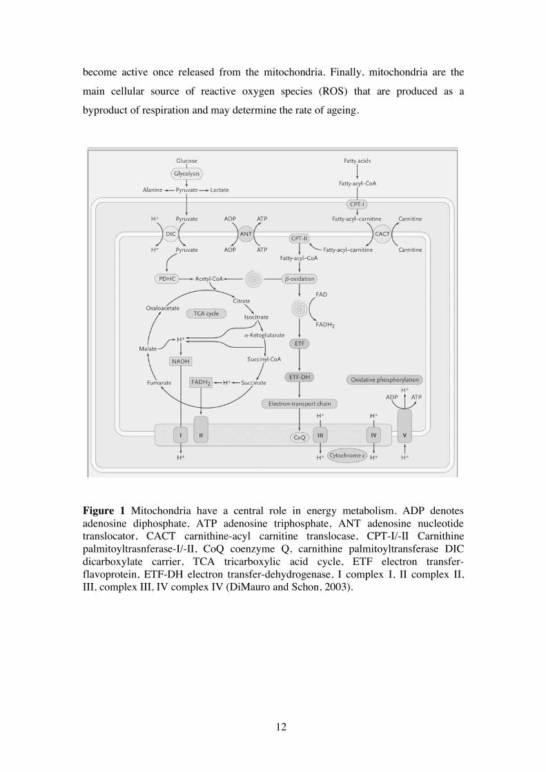
genome (mtDNA) encoding several essential subunits of the respiratory chain (RC). Mitochondria have a central role in energy metabolism (Figure 1). They are the main
cellular site of ATP generation through the process of oxidative phosphorylation whereby electron transport along the RC is coupled to ADP phosphorylation.
Mitochondria harbor the tricarboxylic acid cycle (TCA), a pathway that generates
reducing equivalents, NADH and FADH2 to feed electrons to the RC. The TCA cycle is connected to the cytosolic glycolytic pathway and to the mitochondrial fatty acid β-
oxidation cycle. The TCA cycle is also a source of amino acid precursors and
porphyrin/heme biosyntheses. In specific cell-types, mitochondria are also a site of
steroid biosynthesis and carry out some steps of the gluconeogenesis and urea synthesis pathways. Mitochondria also participate in regulation of cellular Ca2+
homoestasis. The high Ca2+ buffering capacity of mitochondria protects the cell from high cytosolic Ca2+ concentrations. Transient cytosolic Ca2+ peaks can act as a
metabolic signal and activate Ca2+ sensitive enzymes of the TCA cycle. The
mitochondrial intermembrane space harbors several apoptosis-inducing factors that
12
become active once released from the mitochondria. Finally, mitochondria are the
main cellular source of reactive oxygen species (ROS) that are produced as a byproduct of respiration and may determine the rate of ageing.
Figure 1 Mitochondria have a central role in energy metabolism. ADP denotes adenosine diphosphate, ATP adenosine triphosphate, ANT adenosine nucleotide translocator, CACT carnithine-acyl carnitine translocase, CPT-I/-II Carnithine palmitoyltrasnferase-I/-II, CoQ coenzyme Q, carnithine palmitoyltransferase DIC dicarboxylate carrier, TCA tricarboxylic acid cycle, ETF electron transfer-flavoprotein, ETF-DH electron transfer-dehydrogenase, I complex I, II complex II, III, complex III, IV complex IV (DiMauro and Schon, 2003).

13
Evolution of Mitochondria
Mitochondria probably originate from a single ancient invasion of an Archea-type
host by a α-proteobacterium-like ancestor over 2 billion years ago. The symbiosis
between the host and the proto-mitochondrial ancestor was probably driven by metabolic requirements. One theory assumes the host was a methanogenic archaean
that associated with a methanotropic proteobacterium to obtain essential compounds,
for example hydrogen (Martin and Muller, 1998), about 2.7 billion years ago under anaerobic conditions well before the rise in atmospheric oxygen tension. A second
theory proposes that symbiosis was driven by the rise in atmospheric oxygen levels caused by photosynthetic cyanobacteria 2.2 billion years ago. Anaerobic life forms
were exposed to the toxicity of oxygen and adopted α-proteobacteria to detoxify
oxygen by respiration. The genome sequence of the α-proteobacterium Rickettsia
prowazeckii is the most mitochondria-like genome. The genome of Rickettsia
prowazeckii has a size of 1.1Mbp and encodes 834 proteins. It contains a complete set of genes for aerobic respiration, ATP production and ATP transport functions
(Andersson et al., 1998). The mitochondrial genome (mtDNA) has the same
fundamental role in all eukaryotes. It encodes a limited number of components of the RC, several ribosomal RNAs and a full or partial complement of tRNAs. MtDNA
exhibits remarkable variation in conformation, size and actual gene content. MtDNA molecules are circular but linear mtDNAs exist as well. Mitochondrial genome sizes
range from less than 6kbp (Plasmodium falciparum) to over 200kbp in land plants.
Contemporary mitochondrial genomes contain 3 to 67 protein-coding genes which is much less than the 834 proteins encoded by the genome of Rickettsia prowazeckii.
The evolution of the mitochondrial proteome displays both, reductive and expansive processes: (i) Many ancestral mitochondrial genes have been lost. The function of
these genes are no longer required because these genes were needed by the α-
proteobacterium as a free-living organism, for example genes for bacterial cell wall
synthesis; (ii) the coding sequences of a small number of mitochondrial proteins have been transferred to the nucleus; (iii) the largest number of mitochondrial proteins have
no bacterial or archeal orthologues and have been adopted by the mitochondrion after
the endosymbiotic event. They are encoded by the nuclear genome and imported into mitochondria (Andersson et al., 2003). Gene transfer from the mitochondrion to the
nucleus is an ongoing process in eukaryotes, for example in angiosperms (Adams and

14
Palmer, 2003) and yeast (Thorsness and Fox, 1990). Numerous examples of
mitochondrial pseudogenes have been documented in the nucleus of humans (Woischnik and Moraes, 2002). Furthermore, some nuclear genes appear to derive
from edited mitochondrial transcripts (Nugent and Palmer, 1991) suggesting both, RNA- and DNA–mediated mechanisms that drive genome transfer and reduction.
Nucleic acids could escape from the mitochondrion during mitochondrial
fusion/fission or following damage to mitochondrial membranes (Adams and Palmer, 2003).
Several hypotheses have been proposed why mitochondria retain genes (Adams and Palmer, 2003). Firstly, some highly hydrophobic proteins may be
difficult to import across the mitochondrial membranes and to sort to the correct
location (Popot and de Vitry, 1990). The only two protein genes contained in every completely sequenced mitochondrial genome – cox1 and cob – encode the most
hydrophobic proteins present in mitochondria (Claros et al., 1995). Secondly, mtDNA
encoded protein genes may be toxic in the cytosol (Martin and Schnarrenberger, 1997). However this would only account for the cox1 and cob proteins that have been
retained in all mitochondrial genomes. Thirdly, expression of genes in the mitochondrion may allow for quick and direct regulation by the redox state of the
mitochondrion (Allen, 1993). However, there is no experimental evidence for this
assumption. Fourthly, the use of the non-standard genetic code in the mitochondria prevents further gene transfer to the nucleus. In this context it is noteworthy that
animal mitochondrial protein-coding gene content is almost completely constant at 13 genes (Boore, 1999).
Regulation of mitochondrial fusion and fission
Mitochondria divide, grow and segregate independently from the cell cycle through mitochondrial fission and fusion mechanisms (Osteryoung and Nunnari, 2003). A
group of proteins referred to as dynamin related proteins (DRPs) regulate
mitochondrial fission. DRPs form a group of self-assembling GTPases. The mitochondrial fission DRPs in yeast, Dnm1, and its homolog in higher eukaryotes,
Drp1, self assemble to form punctate structures that associate with the cytosolic face of the MOM at sites of mitochondrial constriction and fission. At least two more
proteins interact with self-assembled Dnm1 structures, the integral MOM protein Fis1

15
and Mdv1, a peripheral MOM protein. Fis1 targets assembled Dnm1 structures to the
MOM and an interaction between Fis1 and Mdv1 transmits a signal for mitochondrial membrane fission by Dnm1. Fis1 homologs have been identified in animals and
structural data suggest mouse Fis1 acts as an adaptor molecule similar to yeast Fis1. Mdv1 homologs have not been identified yet in mammals. All components required
for mitochondrial fission to date associate with the outer membrane and it is at present
unclear how fission of the outer and inner membrane is coordinated and what components of the inner membrane are involved in this process (Shaw and Nunnari,
2002). The coordinated fusion of the mitochondrial outer and inner membranes is
regulated by a group of proteins called the fuzzy onions (Fzo)/mitofusin (Mfn) family
of mitochondrial outer membrane GTPases (Shaw and Nunnari, 2002). Yeast cells possess one Fzo-related gene, Fzo-1, that plays a direct role in mitochondrial fusion.
In mouse and humans, two Mfn genes, Mfn1 and Mfn2, have been identified that are
ubiquitously expressed and required for mouse embryonic development (Chen et al., 2003). In yeast, Fzo-1 interacts physically with the integral MOM protein Ugo-1 and
the IMS protein Mgm1 but the exact nature of their interactions and their specific roles in mitochondrial fusion are largely unknown. Mutations of the human
homologue of Mgm1, Opa1, result in autosomal dominant optic atrophy and cause
defects in mitochondrial morphology (Alexander et al., 2000; Delettre et al., 2000). Mitochondrial fission may allow segregation of mitochondria by transmission
of their membranes and genomes without need for de novo synthesis and the exchange of mtDNA molecules between mitochondria. Mitochondrial fission may
also participate in organelle remodelling during apoptosis as suggested by the
colocalization of the pro-apoptotic BAX protein with assembled Drp1 structures on the MOM (Newmeyer and Ferguson-Miller, 2003).
Structure, gene content and organization of mammalian mtDNA
The structure, gene content and organization of mammalian mtDNA is strongly conserved (Fernandez-Silva et al., 2003). Human mtDNA is a double-stranded (ds)
closed-circular molecule of about 16.6kb length (Figure 2). In most cells it represents about 0.5-1% of the total cellular DNA content. MtDNA is thought to be normally

16
present as a supercoiled molecule. In yeast, mtDNA is organized as supramolecular
structures called nucleoids. Yeast nucleoids contain several (3-4) mtDNA molecules, about 10-20 different polypeptides (Kaufman et al., 2000; Miyakawa et al., 1987) and
are associated with mitochondrial membranes (Hobbs et al., 2001). Very little is known about the organization of mtDNA molecules in mammalian cells. In most cells
it forms monomeric closed double-stranded circles. In quiescent cultured cells,
catenated structures have been observed. Studies indicate an association between mtDNA and the mitochondrial inner membrane in vertebrate cells (Albring et al.,
1977).
Figure 2 The gene organization in human mitochondrial DNA showing the 13 protein coding genes (ND1-ND6, COXI-III, ATP6 and 8), the 22 tRNA genes (three letter amino acid symbols) and 2 rRNA genes (12S and 16S). The D-loop region controls replication and transcription of mtDNA. OH denotes the origin of H-strand replication and OL the origin of light strand replication (DiMauro and Schon, 2003).

17
The two strands of mtDNA are called the heavy (H-) and the light (L-) strand
because they can be separated in density gradients due to their different Guanosine plus Thymidine content. A high proportion of mtDNA molecules in a metabolically
active cell contain a triple-stranded structure called the displacement loop (D-loop) (Figure 3). In the D-loop, a nascent H-strand of about 700-800nt length, called 7S
DNA, is annealed to the parental L-strand. The D-loop is about 1kb long in humans. It
is the major non-coding segment comprising the origin of replication of the H-strand (OH) and the promoters for H- and L-strand transcription. The D-loop also contains
conserved sequences called CSB (conserved sequence blocks) and TAS (termination associated sequences). The D-loop sequence is also the most variable sequence
between different species. The second non-coding region is around 30nt long and
about two-thirds of the entire mtDNA molecule away from the OH. It contains the origin of replication for the L-strand (OL) and is located inside a tRNA cluster.
Figure 3 The D-loop region controls replication and transcription of mtDNA. L denotes the light strand promoter, H1 the Heavy strand promoter 1, H2 the Heavy strand promoter 2, Phe the tRNAPhe gene, and Leu the tRNALeu gene, mtTFA the mitochondrial transciption factor A, TFB1M/TFB2M the mitochondrial transcription factor B1 and B2, mTERF the transcription termination factor, mtRNA pol the mitochondrial RNA polymerase (Fernandez-Silva et al., 2003).
Mammalian mtDNA encodes 37 genes, of these 22 are tRNA genes, 2 are rRNA genes (16S rRNA and 12S rRNA), and 13 are mRNAs encoding critical
subunits of the RC (Figure 2). MtDNA encodes seven subunits of complex I (ND1-
ND6 and ND4L), one subunit of complex III (cytochrome b), three subunits of

18
complex IV (COXI, COXII, and COXIII) and two subunits of complex V (ATP 6 and
ATP 8). Of note, complex II is completely nucleus-encoded. The genes are asymmetrically distributed in mtDNA: The H-strand encodes 2 rRNA, 14 tRNAs, 12
mRNAs. The L-strand encodes the remaining 8 tRNAs and 1 mRNA (ND6). Mammalian mtDNA has a very compact gene organization consisting of
closely packed, intron-less genes. All the coding sequences are contiguous to each
other or separated by a few bases. Some of the protein genes overlap (ATP 6 and ATP 8 by 46 nucleotides in humans; ND4 and ND4L by 7 nucleotides in humans). In
several cases part of the termination codons are not encoded in mtDNA but are generated by the posttranscriptional polyadenylation of the corresponding mRNA
(Ojala et al., 1981). The genetic code used for translation of genes encoded by
mtDNA shows differences to the universal genetic code, for example in mammalian mitochondria, UGA specifies tryptophan instead of a termination codon.
Transcription of mammalian mtDNA
Transcription of human mtDNA starts at 3 different promoters in the D-loop region (Figure 3), one in the L-strand (Light strand promoter; LSP) and two in the H-strand
(Heavy strand promoter 1 and 2; HSP1 and HSP2) (Bogenhagen et al., 1984; Montoya et al., 1982). Transcription from HSP1 starts 19nt upstream of the tRNAPhe gene and
yields the two rRNAs (12S and 16S rRNA), tRNAPhe and tRNAVal (Figure 2 and 3). Transcription from HSP1 is subject to transcription termination immediately downstream of the 16S rRNA gene inside the gene for tRNALeu (Kruse et al., 1989;
Montoya et al., 1983). Transciption from HSP2 starts close to the 12S rRNA 5’-end
and originates an almost full-genomic length polycistronic transcript of the H-strand that is further processed to release individual mRNAs and tRNAs. Transcription from
HSP2 operates 20 times less frequent compared with transcription from HSP1. The presence of two transcription units, originating at HSP1 and HSP2, suggests a
differential regulation of rRNA versus mRNA transcription (Montoya et al., 1983).
Transcription from LSP results in a single polycistronic full-length genomic transcript from which the eight tRNAs and the ND6 mRNA derive.
HSP1 and LSP have bipartite structure. Both consist of a 15bp consensus sequence surrounding the transcription start site and of an element immediately
upstream of the initiation site (-15 to -39bp) containing a binding site for the

19
mitochondrial transcription factor A (TFAM). The third promoter, HSP2, has only
limited similarity to the 15bp consensus sequence and lacks the upstream TFAM binding site. Transcription termination at the 3’end of the 16S rRNA gene (following
initiation at HSP1) is dependent on a specific tridecamer sequence in the tRNALeu gene and a specific protein factor, mTERF (Christianson and Clayton, 1988; Hess et al.,
1991). The termination promoting activity of mTERF is bidirectional (Shang and
Clayton, 1994) and therefore it could also act to terminate L-strand transcription. The primary polycistronic transcripts originating from LSP and HSP2 are
endonucleolytically cleaved (Doersen et al., 1985; Montoya et al., 1983; Ojala et al., 1981). tRNA sequences located between each rRNA and mRNA (Figure 2) probably
act as signal for the processing enzymes after acquiring a cloverleaf structure on the
nascent RNA chain. The 5’ endonucleolytic cleavage is probably mediated by mitochondrial RNAse P (Doersen et al., 1985) or other unidentified RNases.
Polyadenylation of the rRNAs and mRNAs occurs during or immediately after
cleavage of the primary transcript by a mitochondrial poly (A) polymerase (Amalric et al., 1978).
Transcription of human mtDNA requires an organelle-specific RNA polymerase, human mtRNA polymerase (POLRMT). POLRMT is homologous to
phage T7 RNA polymerase (Tiranti et al., 1997). Recombinant POLRMT together
with recombinant TFAM and either recombinant human mitochondrial transcription factor B1 (TFB1M) or B2 (TFB2M) initiate transcription in vitro from an mtDNA
template containing the mitochondrial promoters HSP1 or LSP. TFB1M and TFB2M are not needed for transcription elongation (Falkenberg et al., 2002). TFB2M has at
least two orders of magnitude more activity than TFB1M. TFB1M and TFB2M
interact with POLRMT in a 1:1 ratio suggesting heterodimerization between POLRMT and TFB1M or TFB2M. TFB1M and TFB2M were identified through
database homology searches for the yeast transcription factor Sc-mtTFB. Sc-mtTFB binds to the catalytic subunit of yeast mtRNA polymerase, Rpo41p, to confer specific
promoter binding.
The rRNAs are methylated and contain a short poly(A) tail of 1-10 residues (Dubin et al., 1982). Mitochondrial tRNAs are smaller, have some structural
differences and additional functions in replication and transcription regulation compared to nucleus-encoded tRNAs. Mitochondrial mRNAs start directly at the

20
initiation codon or have an extremely short untranslated 5’end (of 1-3nt) and have a
poly(A) tail of about 55 residues immediately after the stop codon. Thus, they lack the typical features of cytosolic mRNAs, for example 5’ and 3’ untranslated regions, 3’
polyadenylation signal or the 5’end cap structure (Montoya et al., 1981).
Replication of mammalian mtDNA
MtDNA replicates independently from the cell cycle phase and from nuclear DNA
replication (Bogenhagen and Clayton, 1977). Two models have been proposed for mtDNA replication: (i) the strand-displacement and (ii) the strand-coupled replication
model. The strand displacement model involves two unidirectional, independent
origins. Replication starts at OH located downstream of the LSP in the D-loop region (Figure 3) and proceeds along the parental L-strand producing a daughter H-strand.
When H-strand replication reaches the OL (Figure 2), the replication site of the L-
strand is exposed and replication of the light strand in the opposite direction starts. H-strand replication is dependent on the generation of an RNA primer by transcription
from the LSP. Consequently, replication of mtDNA is likely coupled to the mitochondrial transcription machinery, consisting of POLRMT, TFAM and the
TFBM factors. The triplicate structure consisting of the newly synthesized RNA
strand upstream of OH, the parental H- and L-strand is called an R-loop. RNA processing activities, probably mediated by RNAse MRP or Endonuclease G (Cote
and Ruiz-Carrillo, 1993), cut the RNA strand generating the mature RNA primer. The mtDNA polymerase (DNA pol γ) starts H-strand replication through extension of the
RNA primer (Xu and Clayton, 1996). The R-loop is processed at the CSB I-III. The
CSB are D-loop sequences of extreme sequence conservation between human, rat and
mouse and are located downstream of the LSP and upstream of OH (Figure 3). A major H-strand replication initiation point is almost always found near CSB I
(Walberg and Clayton, 1983). After replication of the H-strand has started, most of the nascent DNA strand is arrested around the TAS (termination associated sequence)
creating the 7S DNA strand and the characteristic triple stranded structure of the D-
loop. At a much lower frequency however, replication proceeds over the entire length of the genome. The function of the 7S DNA strand and the mechanisms that
21
determine whether elongation proceeds beyond TAS are unknown. The initiation for
replication of the L-strand at OL occurs in a small non-coding region that is thought to assume a stable stem-loop structure once it is exposed as a single strand. Initiation of
L-strand replication requires the activity of a specific primase to generate a short RNA primer with 5’ends mapping onto a T-rich portion of the loop in the stem-loop
structure. Once initiation has started, elongation proceeds until completion of L-strand
duplication (Wong and Clayton, 1985). The whole process of replication has been estimated to last 1 h implying a polymerization rate of 270nt/min (Graves et al.,
1998). After completion of the synthesis of both strands, RNA primers must be removed and the gaps filled and ligated. The closed circular mtDNA adopts its tertiary
structure following introduction of supercoils and interaction with DNA binding
proteins. The second model, the strand-coupled replication model, has been proposed to
exist along with the strand-displacement model and corresponds to coupled leading
and lagging strand synthesis of mtDNA (Holt et al., 2000). MtDNA polymerase, DNA pol γ, is composed of a catalytic α-subunit and a
smaller β-subunit. The β-subunit is needed for processivity and primer recognition.
DNA pol γ has a 3’-5’ exonuclease activity in the α-subunit (Longley et al., 1998).
DNA pol γ requires an ATP-dependent mitochondrial helicase for unwinding mtDNA
called Twinkle. Twinkle has a helicase domain that shares homology to the helicase domain of phage T7 primase/helicase. Replication of mtDNA requires mitochondrial
single stranded (ss) binding protein (mtSSB) to stabilize single stranded DNA during
replication and to increase the activity and fidelity of DNA pol γ (Farr et al., 1999;
Hoke et al., 1990). A minimal mammalian mtDNA replisome has been reconstituted consisting of DNA pol γ, mtSSB and Twinkle in recombinant form. In combination,
recombinant DNA pol γ and Twinkle form a processive replication machinery that
can use dsDNA as a template to synthesize ssDNA of about 2 kb. Addition of
recombinant mtSSB stimulates the reaction further generating DNA products of about
16kb. The observed DNA synthesis rate in this in vitro system is 180bp/min corresponding closely to the previously reported in vivo value of 270bp/min. The
functional interaction between Twinkle and DNA pol γ explains why mutations in
these two proteins cause an identical human disease, autosomal dominant external ophtalmoplegia (Korhonen et al., 2004)

22
Mitochondrial transciption factor A (TFAM)
TFAM is a nucleus encoded transcription factor of 24kDa. TFAM belongs to a group of small nuclear DNA binding proteins, the nuclear high mobility group box proteins
(HMG box proteins). TFAM contains two high mobility group domains (HMG box)
that confer DNA binding. TFAM has the ability to wrap, unbend and unwind the DNA (Fisher et al., 1992). The C-terminal tail is essential for the activation of
mitochondrial transcription (Dairaghi et al., 1995). TFAM is required for mitochondrial transcription initiation (Falkenberg et al., 2002) and likely for mtDNA
replication because an RNA primer must be synthesized to initiate H-strand
replication. TFAM has a leader peptide sequence for protein import into the mitochondria.
TFAM appears to have a second, structural role besides its function as a transcription initiation factor. It appears that TFAM is regularly spaced bound to the
D-loop probably allowing other transcription factors to interact with their target
sequences (Ghivizzani et al., 1994). TFAM levels exceed the levels that would be expected for a transcription factor with regulatory functions. It has been proposed to
have a histone-like function covering the entire mtDNA molecule (Fisher et al., 1992; Ghivizzani et al., 1994). In this sense, TFAM homologues of yeast, ABF2, and of
Xenopus, wrap entirely the mtDNA molecule (Shen and Bogenhagen, 2001). In
Xenopus, the TFAM homologue has been found to bind to mtDNA as a tetramer (Antoshechkin et al., 1997).
The yeast homologue of TFAM is ABF2p (sc-mtTFA). ABF2p also bends and
introduces negative supercoils in DNA (Fisher et al., 1992) and functions as a histone-like protein with DNA packaging properties (Megraw and Chae, 1993). However,
ABF2p lacks the C-terminal domain of TFAM and has only a minor effect on transcription (Xu and Clayton, 1992). Yeast strains lacking ABF2p lose mtDNA when
grown on glucose indicating that ABF2p has a role in mtDNA maintenance. TFAM
when expressed in an ABF2p deficient yeast strain can complement ABF2p function by partially rescuing the growth and respiratory defects of this strain (Parisi et al.,
1993). The level of TFAM is lower in cells depleted of mtDNA after treatment with
Ethidium bromide and higher in tissues from patients with increased levels of mtDNA

23
(Davis et al., 1996; Larsson et al., 1994). TFAM levels may follow the mtDNA levels
by a feedback regulation or may be more stable in the presence of DNA targets. Heterozygous Tfam KO mice have reduced mtDNA copy number and homozygous
Tfam KO embryos completely lack mtDNA and die at embryonic day 8.5-10.5 (Larsson et al., 1998) demonstrating that Tfam is required for mtDNA maintenance in
vivo. Overexpression of the human TFAM protein upregulates mtDNA copy number
in mice (Ekstrand et al., 2004). TFAM likely controls mtDNA copy number by activating L-strand transcription and primer generation for H-strand replication
(Ekstrand et al., 2004). TFAM may also enhance mtDNA stability by acting as a histone-like protein.
Transmission and segregation of mammalian mtDNA
The mtDNA copy number spans usually a range of 200-5000 in different cell types (Ekstrand et al., 2004)(Bogenhagen and Clayton, 1974; Robin and Wong, 1988; Veltri
et al., 1990). Each mitochondrion contains several copies of mtDNA. In a given cell,
all mtDNA copies are presumably identical, a condition called homoplasmy. Mutations of mtDNA can coexist with wild-type mtDNA and this condition is called
heteroplasmy. As a consequence of this, the fraction of mutated mtDNA molecules has to reach a certain percentage, in order to cause disease. This usually happens
when the fraction of mutated mtDNA is higher than 60-80% (Lightowlers et al.,
1997). The mitochondrial genome is maternally inherited. The few mitochondria
from the sperm cell that enter the oocyte during fertilization are actively eliminated by
a ubiquitin-dependent mechanism (Sutovsky and Schatten, 2000). In some rare cases, paternal mtDNA can escape the mechanism of active elimination and be transmitted
to the muscle tissues of affected patients (Schwartz and Vissing, 2002). The mature oocyte contains 105 mtDNA molecules. The sperm cell has about 102 copies and there
is some evidence that downregulation of TFAM contributes to the observed
downregulation of mtDNA copy number during spermatogenesis (Larsson et al., 1997b; Rantanen et al., 2001). MtDNA undergoes a bottleneck phenomenon during
oogenesis by which a small subset of mtDNA molecules are amplified and transmitted to the offspring (Marchington et al., 1998). There is little mitochondrial proliferation
despite massive cell proliferation between the primordial germ cell in the 3 week-old

24
fetus and the diplotene primary oocyte in the 20 week-old fetus resulting in a
mitochondrial bottleneck (Poulton and Marchington, 2002). The reduction in mtDNA copy number is followed by amplification and selection of mtDNA later during
oogenesis. This phenomenon probably explains the shift towards homoplasmy within one or a few generations. It appears that the number of transmitted mtDNA molecules
(transmission units) is between 1-200 in the female germ line (Howell et al., 2000;
Jenuth et al., 1997). Studies of heteroplasmic mice demonstrate random genetic drift in some
tissues but in others strong, tissue-specific and age-related, directional selection for different mtDNA genotypes (Jenuth et al., 1996; Jenuth et al., 1997). This trait
showed linkage to loci on several chromosomes providing evidence for nuclear
control of mtDNA segregation (Battersby et al., 2003).
Regulation of mitochondrial gene expression and biogenesis
Different cell-types have a rather constant mtDNA copy number (Moraes, 2001) and
the ratio between mtDNA copy number and total DNA is cell-type specific (Shay et al., 1990). The mtDNA copy number in mammalian cells harboring wild-type
mtDNA, mtDNA with partial deletions or mtDNA with partial duplications is inversely proportional to the size of the respective mtDNA suggesting that cells
maintain a constant mass of mtDNA rather than a constant copy number of
mitochondrial genomes (Tang et al., 2000). The above study also reported a correlation between mtDNA copy number and the levels of mtDNA-encoded RNA
and polypeptides indicating that mtDNA copy number affects mtDNA expression
(Tang et al., 2000). Genetic studies have addressed the role of transcription and replication factors in the regulation of mtDNA copy number. Homozygous Tfam KO
mice display severe mtDNA depletion demonstrating the importance of Tfam for mtDNA maintenance (Larsson et al., 1998). Overexpression of human TFAM in
transgenic mice upregulates mtDNA copy number without affecting RC function and
mitochondrial mass, suggesting that regulation of mtDNA copy number can be dissociated from regulation of mtDNA expression and mitochondrial biogenesis
(Ekstrand et al., 2004). Studies regarding the regulation of mtDNA transcription have been carried out in isolated mitochondria and have revealed some degree of
autonomous regulation. External signals, for example a change in ATP concentration

25
or treatment with thyroid hormone activate mtDNA transcription. This may be
relevant in the local (subcellular) tuning of mitochondria (Enriquez et al., 1999a; Enriquez et al., 1999b; Enriquez et al., 1996). However, no evidence has been
provided yet that thyroid hormone receptors can use the mitochondrial transcriptional machinery to direct mitochondrial gene expression.
The vast majority of proteins required for mitochondrial biogenesis are
nucleus-encoded including: (i) most of the about 100 subunits of the RC; (ii) the metabolic enzymes needed for fatty acid β-oxidation, the TCA cycle, the biosyntheses
of certain amino acids and of heme; (iii) mitochondrial protein import and assembly
factors; (iv) factors of the mitochondrial replication, transcription and translation
machinery. These genes are under transcriptional control of the nuclear respiratory factors-1 (NRF-1) and -2 (NRF-2), thyroide hormone T3, stimulation protein 1 (Sp1),
and others (Kelly and Scarpulla, 2004). NRF-1 and -2 regulate the expression of most nucleus-encoded subunits of all respiratory complexes, mtDNA transcription and
replication factors, mitochondrial enzymes of the heme biosynthetic pathway,
mitochondrial protein import and assembly factors, and ion channels of the MOM. The expression of the human TFAM and TFB1M/TFB2M genes was found to rely on
functional NRF-1 and NRF-2 consensus sites in their promoters suggesting a regulatory link between nuclear and mitochondrial gene expression in mitochondrial
biogenesis (Kelly and Scarpulla, 2004; Virbasius and Scarpulla, 1994). However, this
contrasts with the presence of NRF-2 but absence of obvious NRF-1 sites in the human TFB1M/TFB2M promoters and rodent Tfam and Tfb1m/Tfb2m promoters
(Larsson et al., 1997a; Rantanen et al., 2001) (Rantanen et al., 2003). The transcriptional coactivator PPARγ coactivator-1α (PGC-1α) was cloned
in a yeast two hybrid screen for brown adipose-specific factors that interacted with the
adipogenic nuclear receptor PPARγ (Puigserver et al., 1998) and has proven to be a
key regulator of mitochondrial biogenesis in various tissues. Forced expression of
PGC-1α in adipogenic and myogenic mammalian cell lines induces the expression of
NRF-1, NRF-2 and Tfam. PGC-1α can interact directly with NRF-1 to activate the
mouse Tfam gene promoter (Wu et al., 1999). Overexpression of PGC-1α in primary
cardiac myocytes and in hearts of transgenic mice upregulates the expression of genes
involved in mitochondrial fatty acid β-oxidation (Lehman et al., 2000). Forced
expression of PGC-1α in skeletal muscle of transgenic mice drives mitochondrial

26
proliferation and the formation of mitochondrial-rich type I, oxidative (slow-twitch)
muscle fibres (Lin et al., 2002). PGC-1α interacts with a variety of nuclear-receptors
and non-nuclear receptors (Kelly and Scarpulla, 2004). Several upstream signaling events activate PGC-1α and mitochondrial biogenesis in a tissue-specific manner.
Upon cold exposure, the β-adrenergic/c-AMP pathway activates PGC1α- and UCP1-
expression in brown adipose tissue (Puigserver et al., 1998). Exercise stimulates PGC-
1α gene expression in skeletal muscle. Forced overexpression of
Calcium/calmodulin-dependent protein kinase (CaMK) or of PGC-1α in skeletal
muscle activates mitochondrial biogenesis (Lin et al., 2002; Wu et al., 2002).
Overexpression of CaMK and calcineurin A induces PGC-1α expression (Handschin
et al., 2003; Wu et al., 2002) suggesting that CaMK and calcineurin A are upstream of PGC-1α. Nitric oxide (NO) can activate PGC-1α and mitochondrial biogenesis in
adipocytes and HeLa cells. The NO effect is dependent on cGMP activation and does
not result from inhibition of oxidative phosphorylation by NO (Nisoli et al., 2003).
Mitochondrial bioenergetics and ROS formation
The respiratory chain
The RC consists of five distinct complexes (Complex I-V) that are bound to the mitochondrial inner membrane. Electrons are fed to Complex I or II and transferred to
Complex III and IV. During the passage of electrons a proton gradient across the inner membrane is built up by proton translocation at Complex I, III and IV. The
proton gradient is then used by Complex V to synthesize ATP from ADP and
phosphate (Figure 4). This general mechanistic principle of oxidative phosphorylation explaining the coupling between respiration and ATP synthesis in mitochondria is
referred to as the chemiosmotic theory and was proposed by Peter Mitchell in 1961
(Mitchell, 1961). Recent observations suggest that the different respiratory complexes are organized in supercomplexes (Schagger and Pfeiffer, 2000). The crystal structures
of Complex II-V have been elucidated to some extent and important mechanistic conclusions have been drawn (Saraste, 1999).

27
Figure 4 The respiratory chain consists of 5 complexes, designated Complex I-V. Complex II does not translocate protons and is completely nucleus-encoded. Reprinted with permission from (Saraste, 1999). © AAAS. Complex I (NADH:ubiquinone oxidoreductase) is the largest of all complexes, consisting of at least 46 subunits, one flavin mononucleotide (FMN), seven or eight
different iron-sulfur centers, covalently bound lipids and at least 3 bound ubiquinol molecules. Seven subunits (ND1-ND6, ND4L) are encoded by mtDNA. Two
electrons from NADH enter Complex I. The electrons are transferred along an
electron transport chain consisting of FMN, iron-sulfur centers and bound ubiquinones to a mobile ubiquinone (Q) that takes up electrons to yield ubiquinol
(QH2) (Stryer, 1995). The flow of 2 electrons from NADH to ubiquinone leads to the pumping of 4 protons from the mitochondrial matrix side to the IMS. Electron
microscopy (EM) studies demonstrated that Complex I has an L-shaped structure with
two major domains separated by a thin collar (Grigorieff, 1998; Guenebaut et al., 1998).
Complex II, succinate:ubiquinone reductase (SQR) or succinate dehydrogenase (SDH), is completely nucleus-encoded and a component of the TCA
cycle. It catalyzes the conversion of succinate to fumarate using FAD (flavine-adenine
dinucleotide) as a cofactor. This reaction yields FADH2. FADH2 donates 2 electrons that are transferred to Q along several iron-sulfur centers. Mammalian mitochondrial
and many bacterial SQR are composed of two hydrophilic subunits, a flavoprotein
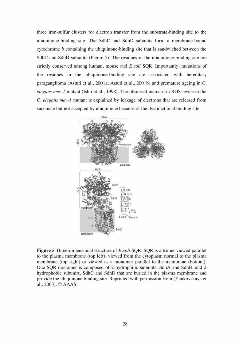
(SdhA) and iron-sulfur protein (SdhB), and two hydrophobic membrane anchor subunits, SdhC and SdhD (Figure 6). The crystal structure of E.coli SQR has recently
been determined (Yankovskaya et al., 2003). SQR is composed of a trimer. Each monomer consists of one SdhA, SdhB, SdhC and SdhD subunit. The SdhA subunit
contains the FAD cofactor and the substrate-binding site. The SdhB subunit contains
28
three iron-sulfur clusters for electron transfer from the substrate-binding site to the
ubiquinone-binding site. The SdhC and SdhD subunits form a membrane-bound cytochrome b containing the ubiquinone-binding site that is sandwiched between the
SdhC and SdhD subunits (Figure 5). The residues in the ubiquinone-binding site are strictly conserved among human, mouse and E.coli SQR. Importantly, mutations of
the residues in the ubiquinone-binding site are associated with hereditary
paraganglioma (Astuti et al., 2001a; Astuti et al., 2001b) and premature ageing in C.
elegans mev-1 mutant (Ishii et al., 1998). The observed increase in ROS levels in the
C. elegans mev-1 mutant is explained by leakage of electrons that are released from succinate but not accepted by ubiquinone because of the dysfunctional binding site.
Figure 5 Three-dimensional structure of E.coli SQR. SQR is a trimer viewed parallel to the plasma membrane (top left), viewed from the cytoplasm normal to the plasma membrane (top right) or viewed as a monomer parallel to the membrane (bottom). One SQR monomer is composed of 2 hydrophilic subunits, SdhA and SdhB, and 2 hydrophobic subunits, SdhC and SdhD that are buried in the plasma membrane and provide the ubiquinone binding site. Reprinted with permission from (Yankovskaya et al., 2003). © AAAS.
29
Similarly, it has been proposed that mutations of the SdhB, SdhC and SdhD subunits
associated with hereditary paraganglioma cause ROS formation and thereby predispose to cancer (Rustin et al., 2002).
Complex III (cytochrome bc1) delivers electrons from QH2 to cytochrome c. The transfer of electrons is coupled to the transfer of protons across the inner
membrane by the so-called Q cycle (Figure 6). The mammalian Complex III is a
dimer and each monomer contains eleven subunits but only three of them carry the redox centers for electron transfer. These key subunits are: (i) cytochrome b, the only
mtDNA encoded subunit of Complex III; (ii) a membrane-anchored iron-sulphur protein (ISP) carrying a Rieske-type center (Fe2S2); and (iii) a membrane anchored
cytochrome c1. There are two active sites in Complex III (Iwata et al., 1998; Xia et al.,
1997; Zhang et al., 1998b): one for oxidation of QH2 and release of protons on the outer surface of the membrane, Qo; and one for the reduction of Q coupled to the
uptake of protons from the inner side of the membrane, Qi. In the first step of the Q
cycle, one ubiquinol donates one electron to the to the Rieske-type iron-sulphur center and one electron to the bL heme group of the cytochrome b subunit. The donation of
both electrons is coupled to the release of two protons to the intermembrane space at the Qo site. The first electron passes from the Rieske-type iron-sulphur center to the
heme c group of cytochrome c1 and from there to cytochrome c. The second electron
passes from the bL heme to the bH heme group of cytochrome b and is then transferred back to a new incoming ubiquinone molecule at the Qi site of cytochrome b. A
subsequent Q cycle provides a second electron at the Qi site to reduce ubisemiquinone (Q·) to QH2 that can again enter the Q cycle.
Complex IV (cytochrome c oxidase) contains 13 subunits (Figure 7). The three
major subunits are encoded for by mtDNA (COXI-III) and form the functional core of complex IV. This core is surrounded by 10 smaller nucleus-encoded subunits. Subunit
II receives electrons from cytochrome c. These electrons are first transferred to cytochrome a in subunit I and then to the bimetallic cytochrome a3/CuB active site in
subunit I. Two hydrophilic channels, called D and K, connect the active site to the
aequeous phase of the mitochondrial matrix. The reduction of oxygen at the active site in subunit I is linked to the translocation of four protons, two for the reduction of

30
Figure 6 Structure of the bovine mitochondrial cytochrome bc1 and the Q cycle. Cytochrome bc1 is a dimer and each monomer consists of 11 subunits (left). The functional core of the enzyme consists of three subunits: cytochrome b, Rieske ISP and cytochrome c1 (middle). In the Q cycle, bifurcation of electron transfer occurs at the Qo site. One electron is transferred to the Fe2S2 center of Rieske ISP and from there to cytochrome c1. The second electron is transferred to the heme bL and from there to the heme bH of cytochrome b and delivered back to a new incoming ubiquinone at the Qi site. Reprinted with permission from (Saraste, 1999). © AAAS.
Figure 7 Structure of the bovine cytochrome c oxidase. The functional core of the enzyme consists of subunits I-III (COXI-III) that are mtDNA encoded. Subunits I and II contain the metal centers and the active site (cytochrome a3/CuB) resides in subunit I. Protons that are used for reducing oxygen to water or pumped to the IMS are transferred through two channels, D and K, from the matrix side. Reprinted with permission from (Saraste, 1999). © AAAS.
31
oxygen into water, and two for release into the intermembrane space (Tsukihara et al.,
1995; Tsukihara et al., 1996; Yoshikawa et al., 1998). Complex V (F1F0 ATPase) synthesizes ATP using a proton-motive force
across the inner mitochondrial membrane, but it can also hydrolyze ATP to pump protons against an electrochemical gradient. The bovine enzyme appears to contain 16
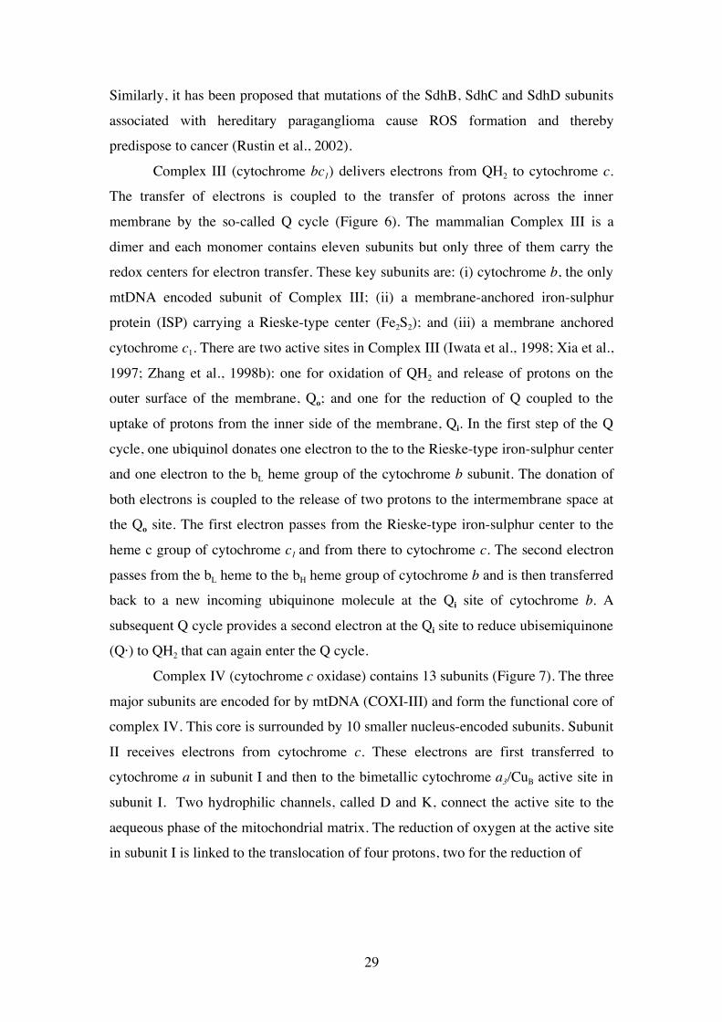
subunits (Lutter et al., 1993). It consists of a membrane bound part containing a
proton channel, called Fo, and of a catalytic part located in the matrix, called F1, which contains an ATP synthesizing or hydrolyzing activity (Figure 8). MtDNA encodes 2
subunits of the Fo part. The Fo and F1 parts are connected by two parallel structures, referred to as the “rotor” and the “stator” (Elston et al., 1998). The F1 part can be
detached from the F0 part and acts as a soluble ATPase. The F1 part consists of 5
subunits (α, β, γ, δ, ε). The β subunit contains the catalytic site and there are three
active sites per one F1F0 ATPase because each F1 part contains three β subunits. Each
catalytic site passes through a cycle of open (unbound) state, loose (bound ADP and phosphate) state and tight (tightly bound ATP) state. The formation of ATP requires
energy for substrate binding and ATP release but not for the phosphorylation reaction
itself. The crystal structure of the F1 part of the bovine F1F0 ATPase indicates that it operates as a rotational catalyst (Abrahams et al., 1994). A central structure inside the
Figure 8 Structure of ATP synthase. The F1 part corresponds to the crystallized bovine ATP synthase (Abrahams et al., 1994) and the remaining parts to the crystal structure of the bacterial ATP synthase (Lutter et al., 1993). A dodecamer of subunit c, the ε and γ subunits form the rotor, while subunits b, d and δ form the stator. The active sites are present in the β subunits. Reprinted with permission from (Saraste, 1999). © AAAS.

32
F1 ATPase rotates in 120o steps during catalysis (Noji et al., 1997; Sabbert et al.,
1996; Yasuda et al., 1998). The membrane bound Fo part consists of three subunits called a, b and c. The subunit c forms a dodecamer and is thought to constitute the
rotor together with the γ and ε subunits of the F1 part (Figure 8). The subunit a and b
form the stator together with the δ subunit of the F1 part. The mechanism how ATP
synthesis is coupled to proton transfer across the inner membrane has not been
clarified yet. The current model proposes that proton movement through the interface between a-subunit and the subunit c dodecamer (Figure 8) causes the rotor and stator
to move in opposite directions thereby causing a torque. The subsequent release of free energy by the torque may allow substrate binding and release of ATP,
respectively (Elston et al., 1998; Wang and Oster, 1998). Complex V has recently
been ascribed a role in the biogenesis of the mitochondrial inner membrane (Paumard et al., 2002).
Generation of ROS by the respiratory chain
O2-· is constantly produced in respiring mitochondria at a rate of about 1-3% of all
oxygen consumed (Boveris and Chance, 1973). O2-· dismutates spontaneously to H2O2
and this reaction is accelerated by superoxide dismutase (SOD). H2O2 can react with reduced transition metals, for example with Fe2+ in the Fenton reaction (Fe2+ + H2O2
-> Fe3+ + OH + OH·), and give rise to the highly reactive hydroxyl radical (OH·). O2-·,
H2O2 and OH· are collectively referred to as reactive oxygen species (ROS). It is difficult to determine the exact site of O2
-· generation because O2-· has a
short lifetime due to its rapid conversion to H2O2 by SOD or because it immediately reacts with lipids of the mitochondrial inner membrane. H2O2 is more stable than O2
-·
and can diffuse through the membrane lipid bilayer out of the mitochondrion. Most of
the O2-· is probably produced at the matrix side of the inner mitochondrial membrane
because O2-· generation is only found in submitochondrial particles, which are inside
out with respect to mitochondria while in intact mitochondria only H2O2 is detectable extramitochondrially. However, a recent study using spin traps revealed O2
-·
formation in mitoplasts (mitochondria devoid of the outer membrane) (Han et al.,
2001). O2
-· generation depends on whether mitochondria are actively respiring (low
O2-· production) or the RC is highly reduced (high O2
-· production). The rate of O2-·

33
generation increases when electron flow slows down or the concentration of oxygen
increases. The proton gradient built up during respiration is dissipated through complex V to synthesize ATP. In the absence of complex V activity, for example
following inhibition by oligomycin, the proton gradient builds up massively increasing the mitochondrial membrane potential and causing electron flow to slow
down and the RC to become more reduced. This results in increased steady-state
concentration of O2-· (Boveris et al., 1972). Inhibitors of the RC result in O2
-· generation due to increased reduction of the carriers upstream of the site of inhibition
and have revealed specific sites of O2-· formation within complex I, II, and III. In
complex III, O2-· is formed when the electron flow between the bL and bH heme of
cytochrome b is blocked, for example by antimycin A (Figure 9). Reoxidation of
ubisemiquinone (Q·) to ubiquinone (Q) at the outer side of the membrane and reduction of Q· to ubiquinol (QH2) at the inner side of the membrane are inhibited,
leading to increased steady-state concentrations of Q· (Figure 9). O2-· formation by
Figure 9 The Q cycle in complex III. Ubiquinone (Q) is reduced to ubiquinol (QH2) either by electron transfer from Complex I or Complex II or by electrons transferred from cytochrome b of complex III. QH2 releases one electron to the Rieske ISP, cytochrome c1 (c1) and cytochrome c (c). The second electron is released at Qo from a ubisemiquinone (Q·) and transfers to the heme groups of cytochrome b to reduce a new Q at the Qi site. Superoxide is probably produced at the two sites, Qo and Qi, but the contribution of each site has not been clearly determined yet, Antimycin A blocks electron transfer between bL and bH heme of cytochrome b.
34
antimycin A is blocked when electron flow between the Rieske protein and
cytochrome c1 or oxygen is inhibited, for example by myxathiazol or cyanide, indicating that O2
-· formation depends on generation of Q· by the Q-cycle (Figure 9)
(Trumpower, 1990; Turrens, 2003; Turrens et al., 1985). In Complex I, O2-·
production likely occurs in one of the iron-sulphur clusters, either N1 or N2 upstream of the binding site for ubiquinone and rotenone (Genova et al., 2001; Kushnareva et
al., 2002). In Complex II, mutations affecting the ubiquinone binding site result in
electron leakage and increased O2-· formation (Yankovskaya et al., 2003). Reduced
FAD was also identified as an electron donor to oxygen in reconstituted complex II
(Zhang et al., 1998a).
Antioxidant defense mechanisms
Antioxidant defenses are classified into non-enzymatic and enzymatic defenses. The
non-enzymatic defenses are compounds that reduce oxidizing agents, for example the lipid soluble vitamin E and ubiquinone, or the water-soluble vitamin C, glutathione,
uric acid and ceruloplasmin (Halliwell and Gutteridge, 1989).
The superoxide dismutases (SOD) are the first line of enzymatic antioxidant defense. SOD catalyze the reaction O2
-· + O2-· —> H2O2 + O2. All members of the
SOD familiy utilize different transition metals at their active sites. There are three
different SOD isoforms (Fridovich, 1995). SOD1 (Cu/ZnSOD) is expressed in the cytosol and in the mitochondrial intermembrane space (Okado-Matsumoto and
Fridovich, 2001). SOD2 (MnSOD) is expressed in the mitochondrial matrix and is closely related to the bacterial MnSOD. The expression of SOD2 is induced by agents
that cause oxidative stress, for example radiation and hyperoxia through oxidative
activation of the transcription factor NFkB and by various cytokines, for example TNFα , IL-1 and IFN-γ (Li and Karin, 1999). SOD2 is transported into the
mitochondrial matrix where it assembles into an active homo-tetramer (Fridovich, 1995). Hydrogen peroxide, the product of the dismutation reaction is reduced to water
and oxygen mainly by glutathione peroxidases and to a lesser extent by catalases. Glutathione peroxidases utilize glutathione (GSH) as a reductant according to the
following reaction: H2O2 + 2 GSH —> 2H2O + GSSG. There are four GPx isoforms.
The major GPx isoform, GPx1, is expressed in mitochondria and cytosol of most tissues (Esposito et al., 2000; Frampton et al., 1987). GPx2 is found in plasma
35
(Maddipati and Marnett, 1987), GPx3 in gastrointestinal tract, i.e. liver, intestine and
colon (Chu et al., 1993; Chu and Esworthy, 1995), and GPx4 (phospholipid GPx) in the mitochondria of testis and brain (Arai et al., 1999; Esposito et al., 2000).
Glutathione is a tri-peptide consisting of L-γ-glutamyl-L-cysteinyl-glycine. Each GSH
molecule provides one reducing equivalent to the conversion of H2O2 into water and oxygen by the sulfhydril moiety of the cysteine residue leading to the formation of
oxidized GSH in form of the disulfide–bonded compound GS-SG. The enzyme
glutathione reductase utilizes NADPH to re-reduce one molecule of GS-SG into two molecules of GSH: GS-SG + 2NADPH —> 2GSH + 2 NADP+.
Catalases are almost exclusively found in peroxisomes to remove H2O2 formed
during β-oxidation of long chain fatty acids. Catalases have only been detected in
heart mitochondria but not in other mitochondria (Radi et al., 1991). Mitochondrial uncoupling may provide another line of anti-oxidant defense
(Skulachev, 1996). Uncoupling decreases the mitochondrial membrane potential, leading to increased respiration, oxidation of the RC and lowered O2
-· formation. A
threshold membrane potential exists below which no ROS is formed (Korshunov et
al., 1997). In rat hepatocytes, the futile cycle of proton pumping and proton leak may account for 20-25% of respiration. In perfused rat muscle, this value is increased to
35% in contracting and 50% in resting muscle. Mitochondrial uncoupling proteins (UCP) have been proposed to regulate O2
-· formation and there is evidence that O2-·
directly activates UCP (Brand, 2000).
Damage repair and removal systems have been documented for oxidized proteins (Davies, 2000). Cysteine residues can form a disulfide bond on the same
protein or a disulfide crosslink between two different proteins upon oxidation. The re-reduction is performed by disulfide reductases. Oxidized proteins can be also be
recognized by proteases and completely degraded into amino acids. In the cytoplasm
and the nucleus of eukaryotic cells the oxidized proteins can be recognized and degraded by the proteasome complex. In mammalian mitochondria, there is a separate
set of proteases that conduct the degradation of oxidized proteins, for example LON
protease (Bota and Davies, 2002). Membrane phospholipids undergo lipid peroxidation upon oxidative stress
(Halliwell and Gutteridge, 1989). Lipid peroxidation is the result of a chain reaction: It is initiated by extraction of a hydrogen atom from an unsaturated fatty acyl, for

36
example by OH· (O2-· is not reactive enough to induce lipid peroxidation by itself)
giving rise to a lipid radical (L·). L· then reacts with oxygen producing a lipid peroxy radical (LOO·). LOO· can further propagate the peroxidation chain reaction by
extracting a hydrogen atom from another unsaturated fatty acid. The lipid hydroperoxides (LOOH) can easily decompose into other reactive species.
Peroxidized membranes become rigid, loose selective permeability, and can even
loose their integrity. Water-soluble lipid peroxidation products, such as dialdehydes, can diffuse from membranes into other subcellular compartments and cause protein
aggregation leading to formation of the age pigment lipofuscin. Lipid peroxidation products can also impair enzyme function or may form DNA adducts and induce
mutations. Oxidized lipid bilayers become more susceptible to the action of
phospholipases, which hydrolyze the phospholipid glycerol backbone to release a free fatty acid hydroperoxide. Fatty acid hydroperoxides are detoxified by glutathione
peroxidases reducing them to their corresponding hydroxy fatty acid (Davies, 2000).
Oxidative damage of DNA interferes with DNA replication and transcription. The extent of oxidative DNA damage has been estimated to be as high as 1 base
modification per 130.000 bases in nuclear DNA. Damage to mtDNA is estimated to be even higher at 1 per 8.000 bases. Oxidants can give rise to strand breaks (single
and double) by damaging the phosphodiester backbone, sister chromatid exchange,
DNA-DNA and DNA-protein crosslinks, and base modifications. The base modifications can result in dissociation of the base from the DNA double strand,
creating so called apurinic/apyrimidinic sites. Several DNA repair enzyme systems exist that can repair oxidatively damaged DNA (Davies, 2000).
Adaptive responses of cells to oxidative stress involves transient growth-arrest
to enable DNA damage repair and expression of factors involved in damage removal/repair and antioxidant defenses, for example catalase, GPx and SOD2,
mediated by several transcription factors (Shull et al., 1991) (Davies, 2000). Cells exposed to oxidative stress may ultimately undergo apoptosis or
necrosis. The mechanism involves activation of the mitochondrial permeability
transition pore and cytochrome c release (see below).

37
Mitochondrial uncoupling proteins and their role in energy expenditure and control of ROS homeostasis
The mitochondrial uncoupling proteins (UCP) belong to the large family of anion carriers of the mitochondrial inner membrane containing six transmembrane domains
(Ricquier and Bouillaud, 2000). The first identified uncoupling protein, UCP1, is
specifically expressed in brown adipocytes. Based on cDNA sequence similarity, two homologues of UCP1 were identified and named UCP2 (Fleury et al., 1997) and
UCP3 (Boss et al., 1997; Gong et al., 1997; Vidal-Puig et al., 1997). UCP2 and UCP3 share, respectively, 72 and 57% amino acid identity with UCP1. Of note, UCP2 and
UCP3 are adjacent genes on human chromosome 11 and mouse chromosome 7. UCP2
mRNA is found almost ubiquitously, but the protein has been identified only in some organs and cell types including spleen, lung, stomach, brain, kidney, thymocytes and
pancreatic β-cells (Pecqueur et al., 2001; Zhang et al., 2001). UCP3 is mainly
expressed in skeletal muscle. UCP1 KO mice are cold sensitive but not obese and an extension of these studies demonstrated that UCP1 is the only effector of adaptive
non-shivering thermogenesis to cold exposure (Enerback et al., 1997; Golozoubova et
al., 2001). UCP1 creates a proton leak across the inner membrane of brown fat mitochondria to dissipate the electrochemical proton gradient into heat. Brown
adipose tissue (BAT) is a particular form of adipose tissue required for thermogenesis
in infants at birth or rodents exposed to cold. Brown adipocytes differ from white adipocytes by direct sympathetic innervation, a central nucleus, and numerous
mitochondria. Sympathetic innervation of brown adipocytes induces lipolysis, respiration and thermogenesis, accompanied by a marked increase in blood flow and
heat conductance to other organs by the blood stream. UCP1 is abundantly expressed
in brown adipocytes accounting for up to 4% of the total and 8% of the mitochondrial protein content upon cold acclimation. UCP1 expression is induced by the
Norepinephrine/cAMP-pathway leading to the activation of cAMP response elements
(CRE) in the UCP1 promoter. Various transcription factors, i.e. PPARα, PPARγ, and
thyroid hormone T3 bind to a complex enhancer region upstream of the UCP1 promoter and can stimulate UCP1 expression by co-activation with PGC-1α, but the
precise roles of these factors in the regulation of UCP1 expression have not been clarified yet (Cannon and Nedergaard, 2004). Reconstituted UCP1 exhibits fatty-acid
dependent proton translocating activity (Klingenberg et al., 2001). The proton

38
translocation activity of native UCP1 is stimulated by fatty acids and inhibited by
purine nucleotides, such as GDP, in a competitive manner (Shabalina et al., 2004). Of note, fatty acids are obligatory for uncoupling. There are currently two competing
models explaining the proton location mechanism of UCP. In the first model, UCP1 is a pure proton carrier (Klingenberg and Echtay, 2001). In the second model, UCP1 is
an anionic fatty acid exporter: protonated fatty acids move from the external to the
internal lipid layer of the mitochondrial inner membrane by a flip-flop mechanism, release a proton into the matrix and then return as an anionic fatty acid to the cytosol
by UCP-mediated export (Garlid et al., 1998). UCP2 and UCP3 were proposed to work as uncouplers similar to UCP1.
Polymorphic markers encompassing the UCP2-UCP3 locus show genetic linkage to
the resting metabolic rate (Bouchard et al., 1997). Similarly, polymorphisms in the coding region of the UCP2 gene are linked to the metabolic rate at sleep (Walder et
al., 1998). However, homozygous UCP2 and UCP3 KO mice have normal metabolic
rates (Arsenijevic et al., 2000; Vidal-Puig et al., 2000) but compound homozygous UCP2/UCP3 KO mice have not been analyzed yet due to linkage of the UCP2 and
UCP3 genes. UCP2 and UCP3 KO mice show signs indicative of increased ROS production (Arsenijevic et al., 2000; Vidal-Puig et al., 2000) supporting the
hypothesis that UCP2 and UCP3 function as uncouplers to lower mitochondrial ROS
formation. Consistent with this hypothesis, skeletal muscle mitochondria from UCP3 KO mice show improved respiratory control ratios (Vidal-Puig et al., 2000) and
enhanced in vivo ATP production rates (Cline et al., 2001) indicative of improved coupling. UCP2 and UCP3 have been reconstituted into liposomes and both exhibit
proton translocation activity that requires the presence of fatty acids and is inhibited
by GDP (Jaburek et al., 1999; Jezek et al., 2004). A recent report demonstrated superoxide–mediated activation of UCP1, UCP2 and UCP3. This mechanism was
proposed as a feedback control to lower excessive O2-· production by uncoupling
(Echtay et al., 2002b). Superoxide–mediated activation of UCP is fatty-acid
dependent and inhibited by purine nucleotides (Echtay et al., 2002b). The superoxide-
activation of UCP2 occurs from the matrix side (Echtay et al., 2002a). This finding has potentially important consequences for energy metabolism due to the widespread
tissue distribution of UCPs and their suggested role as uncouplers. The hypothesis that O2
-· activates UCPs however is not universally accepted (Couplan et al., 2002).
39
The experiments leading to the conclusion that superoxide activates UCP were
conducted on isolated mitochondria exposed to supraphysiological levels of exogenous superoxide (Echtay et al., 2002a; Echtay et al., 2002b) and only one study
that used forced, adenovirus-mediated SOD2 overexpression in islet cells has indicated that superoxide-stimulated UCP-mediated uncoupling may also occur in
vivo (Krauss et al., 2003).
High fat diet of human subjects does not change mitochondrial coupling despite clear upregulation of UCP3 protein content in muscle (Hesselink et al.,
2003a). It has been proposed that UCP3 functions as an anionic fatty acid exporter to prevent accumulation of free fatty acids inside the mitochondria that cannot enter the
mitochondrial fatty acid β-oxidation cycle. Accumulation of anionic free fatty acids
causes mitochondrial dysfunction, a phenomenon called lipotoxicity. This role of
UCP3 is consistent with: (i) upregulation of UCP3 expression by conditions increasing circulating FFA (fasting, high fat diet, diabetes, and obesity); (ii) an
inverse relationship between mitochondrial fat oxidation capacity and UCP3 protein
levels (low fat oxidation capacity in untrained individuals correlates with high UCP3 protein content; high fat oxidation capacity in trained individuals correlates with low
UCP3 protein levels) (Hesselink et al., 2003b).
Mitochondrial regulation of apoptosis
Apoptosis is a distinct genetic and biochemical pathway required for normal
embryonic development and maintenance of normal tissue homeostasis. Apoptosis was initially defined as a morphological entity. The morphological hallmarks of
apoptosis are cell shrinkage, condensation and fragmentation of the cell at later stages
followed by phagocytosis of the cell remnants by neighbouring cells. Apoptosis is an active process that does not cause inflammation. In contrast to apoptosis, necrosis is a
passive process caused by massive cellular injury. The cell swells because it is unable to maintain ion homeostasis, leading to rupture and release of intracellular contents
causing inflammation (Kerr et al., 1972). Mitochondria are key regulators of apoptosis
because many factors activating apoptosis are kept in the mitochondrial intermembrane space (IMS) and become active when released into the cytosol. These
pro-apoptotic factors include cytochrome c, apoptosis-inducing factor (AIF), SMAC/DIABLO, and Endonuclease G (Endo G) among others. Release of pro-

40
apoptotic factors from the mitochondrial IMS is regulated to prevent accidental
activation of apoptosis. The pro-apoptotic factors are impermeable to the mitochondrial outer membrane. At least three distinct release mechanisms have been
proposed: (i) The existence of a proteinaceous permeability transition pore (PTP) has
been postulated at contact sites between the inner and the outer mitochondrial
membrane (Figure 10) (Susin et al., 1998). The PTP is proposingly formed by the adenine nucleotide translocator (ANT) and cyclophyllin D at the inner membrane
associated with VDAC and the peripheral benzodiazepine receptor in the MOM. Sustained opening of the PTP results in osmotic equilibration of ions between matrix
and cytosol, dissipation of the mitochondrial inner membrane potential and
mitochondrial swelling, a biophysical state called permeability transition (PT). The MOM ruptures because the MOM cannot expand as much as the highly invaginated
MIM. Several triggers of PT have been described, notably lowered mitochondrial
membrane potential, Ca2+, fatty acids, ROS hyperproduction, NAD(P)H depletion, Nitric oxide (NO) and ATP depletion. Cyclosporin blocks opening of the PTP, while
Figure 10 A putative model of the mitochondrial permeability transition pore (Green and Reed, 1998).
atractyloside activates PTP opening. Interactions between BCL-2 protein family members, BAX and BCL-2 (see below), modulate the opening of the postulated PTP
pore. However, gene knockout studies do not support a requirement for ANT, one of
the postulated components of the PTP, for cells to undergo PT (Kokoszka et al., 2004). Furthermore, the importance of the PTP has been questionend in the majority

41
of cell deaths because protein release can often occur in the absence of any of the
hallmarks of PT (Bossy-Wetzel et al., 1998; Newmeyer and Ferguson-Miller, 2003). (ii) Members of the BCL2-family form channels in the MOM that allow
passage of large macromolecules (Danial and Korsmeyer, 2004). The proteins of the BCL-2 family are structurally classified according to the presence of four conserved
regions called the BCL-2 homology (BH) 1 - 4 domains. Anti-apoptotic BCL-2
family proteins (BCL-2, BCL-xL, A1 and BCL-W) contain all BH1-4 domains. Pro-apoptotic BCL-2 family proteins can be subdivided into members containing the BH1
- 3 domains (BAX, BAK) or the BH3 domain only (BID, BAD, BIM, Noxa, Puma). Cell line studies showed that the pro-apoptotic BCL-2 family members, BAX and
BAK, present as inactive monomers in unstimulated cells homo-oligomerize to form
channels that allow passage of cytochrome c (Desagher et al., 1999; Wei et al., 2000). Homo-oligomerization of recombinant monomeric BAX requires recombinant
protease-activated BID (tBID) in the absence of any other protein to permeabilize
membrane vesicles, allowing passage of extremely large macromolecules (Kuwana et al., 2002). This model accounts for the fact that most of the cytochrome c release
clearly occurs prior to swelling of the mitochondria, and that BAX/BAK compound homozygous KO cells are resistant to most stimuli that activate mitochondrial
induction of apoptosis (Lindsten et al., 2000; Wei et al., 2001a). Anti-apoptotic BCL-
2 family members, BCL-2 and BCL-xL, bind and sequester pro-apoptotic BCL-2 family members by interaction via their BH3 domain (Cheng et al., 2001; Oltvai et al.,
1993). Accordingly, the ratio of anti- to pro-apoptotic molecules is one determinant of susceptibility to apoptosis induction (Oltvai et al., 1993).
(iii) BCL-2 family proteins interact with VDAC in the mitochondrial outer
membrane to release pro-apoptotic factors from the IMS. VDAC reconstituted into liposomes mediates passage of cytochrome c. In this model, BAX and BAK stimulate
opening of the VDAC while BCL-xL stimulates VDAC closure (Shimizu et al., 1999). However, genetic proof for this model is lacking and genetic studies indicate
that one of the three VDAC isoforms, VDAC-2, binds BAK and inhibits BAK
oligomerization and BAK-mediated apoptosis (Cheng et al., 2003). The release of cytochrome c is rapid and the extent almost complete
(Goldstein et al., 2000). The minor fraction of the total cytochrome c, about 20%, is located in the very narrow IMS adjunct to the MOM, whereas most of the cytochrome
42
c (about 80%) is stored within involutions of the MIM, so called tubular cristae which
is a highly sequestered compartment separated from the more narrow IMS through narrow cristae junction. In one EM study, tBID induced striking remodeling of the
tubular cristae, mobilizing the stores of cytochrome c (Scorrano et al., 2002). The redistribution was accompanied by a transient opening of the PTP but not by swelling
of the mitochondria. In the opening state of the PTP solutes of up to 1500Da are
permeable (Bernardi et al., 1999). Transient PTP opening thus remodels tubular cristae and redistributes the cytochrome c pool.
Upon release from the IMS, cytochrome c binds to Apaf-1 and this complex recruits pro-caspase 9 in the presence of ATP to form the apoptosome (Li et al., 1997;
Liu et al., 1996). The binding of cytocrome c and ATP to Apaf-1 results in a
conformational change of Apaf-1 that enables Apaf-1 to bind six pro-caspase 9 molecules (Acehan et al., 2002). Caspases are specific proteases that activate effectors
of the apoptotic programme (Shi, 2002). Caspases are usually present as inactive
zymogens (pro-caspase) possessing a large and small subunit. They cleave specific motifs possessing an aspartate. The cleavage depends on a cysteine and occurs at the
aspartate (Asp) site, therefore the name “caspase”. Cleavage of internal specific Asp residues releases one large and one small subunit that dimerize to form the active site
of the enzyme. Caspases can be divided into two groups, initiator and effector
caspases. Initiator caspases are usually upstream caspases that possess the ability of autocatalytic activation. Downstream effector caspases need initiator caspases for
their activation. The formation of the apoptosome enables autolytic activation of pro-caspase 9 into active caspase 9. The binding of six pro-caspase 9 molecules to one
Apaf-1 molecule allows for an amplification of the initial apoptotic signal. Activated
caspase 9 in turn activates the effector caspases 3 and 7. Effector caspases cleave structural components, for example actin and nuclear lamin, and DNA fragmentation
factor 45 (DFF45). Cleavage of DFF45 by caspase 3 releases the DFF45-DFF40 dimer. DFF40 subsequently degrades chromosomes into nucleosomal fragments
(Enari et al., 1998; Liu et al., 1997). Mitochondria also release AIF (Susin et al.,
1999) and EndoG (Li et al., 2001). AIF and Endo G translocate to the nucleus and induce caspase-independent large-scale DNA fragmentation. Mitochondria also
release SMAC/DIABLO, an inhibitor of the inhibitor of apoptosis proteins (IAP) that block effector caspases (Chai et al., 2000; Verhagen et al., 2000).

43
Apoptosis can be triggered by the activation of cell death receptors on the
plasma membrane and can involve mitochondrial release of cytochrome c. The binding of Fas ligand to Fas (receptor for Fas ligand) or of TNFα to the TNFα
receptor, results in homo-oligomerization of the receptor, binding of adaptor proteins
to the oligomerized receptors and recruitment of pro-caspase 8, an intiator caspase, to the receptor complex (Baud and Karin, 2001) leading to autocatalytic activation of
caspase 8. Activated caspase 8 can directly cleave caspase 3 in so-called type-I cells,
for example in immune cells. In so called type-II cells, for example in hepatocytes, a mitochondrial amplification step is required for efficient activation of caspase 3 by
cell death receptors (Scaffidi et al., 1998). In type- II cells, activated caspase 8 cleaves
BID yielding tBID that induces cytochrome c release (Li et al., 1998; Luo et al., 1998) by mediating oligomerization of BAK (Wei et al., 2001a) or BAX (Desagher et al.,
1999). Ca2+ can trigger induction of mitochondrial PT and promote apoptosis or
necrosis. IP3-mediated Ca2+ release from the endoplasmic reticulum (ER) can cause
PT and apoptosis (Szalai et al., 1999). The Ca2+ flow between the ER and mitochondria is facilitated by close contact sites (Rizzuto et al., 1998). BCL-2 reduces
the amount of Ca2+ releasable from the ER and thus prevents mitochondrial Ca2+ overload (Vanden Abeele et al., 2002). Massive cytosolic Ca2+ overflow can cause
necrosis following a wide variety of cellular injuries, for example following
excitotoxic stimulation of neurons. It has been demonstrated that glutamate-induced neuronal cell death by stimulation of NMDA receptors requires mitochondrial Ca2+
uptake (Stout et al., 1998). Glutamate induces necrosis or apoptosis of neurons in culture dependent on mitochondrial function as reflected by the mitochondrial
membrane potential and cellular ATP content (Ankarcrona et al., 1995). The
intracellular ATP level determines the cell death fate by apoptosis or necrosis (Eguchi et al., 1997; Leist et al., 1997). Inhibition of oxidative phosphorylation with
oligomycin reduces staurosporine (STP) and FAS-stimulated apoptosis of T-lymphocytes and leads to enhanced necrosis (Eguchi et al., 1997; Leist et al., 1997).
An ATP depeletion of more than 50-70% is required to shift the cell death modus
towards necrosis (Leist et al., 1997). Addition of glucose to cells pretreated with oligomycin changes the mode of cell death from necrosis to apoptosis indicating that

44
glycolytic ATP production may allow execution of apoptosis in cells devoid of
oxidative phosphorylation (Leist et al., 1997).
Free radical theory of aging
The free radical theory of ageing proposes that mitochondrial ROS generation
determines the rate of ageing (Sohal and Weindruch, 1996). This is supposed to result from an imbalance between prooxidants and antioxidants leading to an age-dependent
accumulation of oxidative cell-damage that exceeds the capacity of cellular repair systems. This theory is based on the following observations: (i) Variations in
longevity among different species inversely correlate with the rates of mitochondrial
generation of superoxide and hydrogen peroxide; (ii) Restriction of caloric intake lowers steady-state levels of oxidative stress and damage, retards age-associated
changes, and extends the maximum life-span in mammals (Sohal and Weindruch, 1996). ROS may damage subunits of the RC, membrane lipids causing proton leak,
and enzymes of the TCA cycle leading to lowered energetic efficiency, cell
dysfunction and cell death through activation of the PTP (Figure 11) (Wallace, 1992; Wallace, 2001). It is assumed that chronic ROS formation increases the mutant
mtDNA load over time leading to synthesis of defective RC subunits, which in turn increase ROS formation and the mutant mtDNA load further (Figure 11). Correlative
Figure 11 Free radical theory of ageing. Chronic mitochondrial ROS formation damages the RC and increases the mtDNA mutant load leading to progressive decline of oxidative phosphorylation, and further increases in ROS formation and mtDNA mutations with age. The final outcome is cell dysfunction and cell death.

45
evidence suggests a causative link between ROS and accumulation of mtDNA
mutations over time. Prolonged exposure to oxidative stress causes more extensive and persistent damage to mtDNA than to nuclear DNA (Yakes and Van Houten,
1997). The levels of an oxidative DNA modification marker were 10-fold increased in mtDNA compared to nuclear DNA (Bowling et al., 1993). Several reasons have been
proposed to contribute to this selective vulnerability: (i) the lack of histones that
protect from oxidative DNA damage; (ii) the lack of an efficient repair system in mitochondria; (iii) the compact gene organization of mtDNA increasing the likelihood
that a gene is mutated by ROS; (iv) the nearby location of mtDNA to the mitochondrial inner membrane, the major site of ROS production (Shigenaga et al.,
1994). MtDNA point mutations and mtDNA deletions have been reported to increase
with age (Larsson et al., 1990; Michikawa et al., 1999; Wang et al., 2001b). Complex I- IV deficiency, isolated or in combination was reported along with oxidative damage
to mtDNA in the brains of patients with age-associated neurodegeneration, i.e.
Parkinson disease, Alzheimer disease, amyotrophic lateral sclerosis, and Huntington disease (Kirkinezos and Moraes, 2001). However, a causative relationship between
ROS and the accumulation of mtDNA mutations remains to be demonstrated. A proof reading deficient mtDNA polymerase γ knockin mouse that accumulates somatic
mtDNA mutations over time develops a premature ageing phenotype (Trifunovic et
al., 2004). In C. elegans, mutations have been documented that confer increased
resistance against ROS and significant prolongation of lifespan: (i) clock-1 mutants that cannot synthesize ubiquinone and accumulate the biosynthetic precursor
demethoxyubiquinone, a more potent anti-oxidant, that can replace Q functionally, at least in part (Lakowski and Hekimi, 1996); (ii) Isp-1 mutants with a mutation in the
Rieske iron sulphur protein of Complex III leading to low oxygen consumption (Feng
et al., 2001). EUK-8 and EUK-134, antioxidant compounds that act simultaneously as SOD and Catalase mimetic increased lifespan in C. elegans (Melov et al., 2000).
Simultaneous overexpression of SOD1 and Catalase prolonged lifespan in Drosophila
melanogaster (Orr and Sohal, 1994).

46
Respiratory chain diseases
Respiratory chain diseases
Diseases of the respiratory chain (RC) have an incidence of at least 1 in 10’000 live births (Chinnery and Turnbull, 2001). RC diseases are caused by mutations in the
nuclear or the mitochondrial genome. MtDNA mutations include point mutations in
protein coding genes, tRNA genes, rRNA genes or deletions that usually encompass at least one tRNA gene (Smeitink et al., 2001). Nuclear mutations involve protein
subunits of the RC, factors required for the assembly of respiratory complexes and for maintenance of mtDNA. Furthermore there are several nuclear mutations affecting
proteins that are important to preserve mitochondrial homoestasis and indirectly affect
the RC, for example components of the mitochondrial protein import machinery. The largest group of patients with a respiratory chain disease display only an
enzymatically verified deficiency but no identified mutation (Graff et al., 2002). The
same clinical features can be caused by various mutations in mtDNA or nuclear DNA. Conversely, the same genetic defect in mtDNA can lead to different clinical
manifestations. The segregation of mutant mtDNA is one key determinant of this variability. It has been demonstrated the percentage of heteroplasmy varies between
different individuals, tissues and cells within a patient (Chinnery et al., 2000;
Chinnery et al., 1999). However, heteroplasmy is not always sufficient to explain this clinical variability: Firstly, in numerous cases the percentage of mutant mtDNA does
not correlate with the clinical phenotype (Zhou et al., 1997). Secondly, symptoms may develop only in specific tissues even when the mtDNA mutation is homoplasmic
in all tissues. Thirdly, nuclear mutations affecting one oxidative phosphorylation
complex can also cause variable phenotypes or involve selective tissues (DiMauro and Schon, 2003) (Rossignol et al., 2003).
The level of heteroplasmy that causes disease is called the phenotypic threshold (Rossignol et al., 2003). The phenotypic threshold depends on the type of
the mutation and can be approximated to >90% for most mutations in protein coding
genes, 65%-90% for tRNA gene mutations and 60% for deletions (Hayashi et al., 1991). The phenotypic threshold effect is based on an excess of wild-type mtDNA,
mRNA, tRNA, and active RC complexes compared to what is needed for normal respiration. This reserve provides a safety margin against deleterious mutations. The

47
phenotypic threshold is the sum total of thresholds at the level of transcription,
translation, enzyme activity and respiration (Rossignol et al., 2003). The steady-state transcript level of mtDNA encoded subunits appears to
correlate with the mtDNA copy number (D'Aurelio et al., 2001; Tang et al., 2000). Heteroplasmic point mutations of protein coding genes give rise to proportionate
amounts of mutated mRNAs (Bai et al., 2000; D'Aurelio et al., 2001), while
heteroplasmic mtDNA deletions result in proportionate reductions in mRNA levels of the deleted genes (Hayashi et al., 1991). The mutant tRNAs do not necessarily rise
proportionately to the level of heteroplasmy (Chomyn et al., 2000), for example the A3243G tRNALeu MELAS mutation may affect transcription or be subject to
degradation (Jacobs, 2003). A selective upregulation of transcription of wild-type
mtDNA over mutant mtDNA has not been observed in heteroplasmic cells (Bai et al., 2000; D'Aurelio et al., 2001) (Hayashi et al., 1991). This implies that the
transcriptional threshold is determined by (i) the ratio of mutant to wild-type mtDNA
or (ii) by the number of wild-type mtDNA copies. The relationship between mtDNA copy number and steady state-transcript levels may however not be straightforward as
there are indications for posttranscriptional regulation in mitochondria (Gagliardi et al., 2004). Pathogenic mtDNA mutations or deletions impair translation of wild-type
subunits, when the fraction of mutated mRNA (Bai et al., 2000), tRNA (Boulet et al.,
1992) or mtDNA deletions (Hayashi et al., 1991) exceeds 40-50%. Again the translational threshold may be influenced by posttranscriptional regulation (Gagliardi
et al., 2004). Pathogenic mutations in both mtDNA and nuclear DNA can impair the activity of the respective respiratory complex by (i) decreasing the total amount of
assembled active enzyme complexes and/or (ii) by changing the intrinsic kinetic
properties of the complex. Significant reductions in the protein levels of different subunits may not result in lowered enzyme activity (Spelbrink et al., 1994; Triepels et
al., 2001). In cell lines, a decrease in respiration has been reported following an 85% reduction in Complex I activity (Barrientos and Moraes, 1999), or a 40-80% reduction
in Complex IV activity (Davey et al., 1998; Villani and Attardi, 1997), dependent on
the cell lines and experimental conditions used. The respiratory threshold can reflect an excess of active respiratory chain complexes, activation of inactive complexes
(Grivennikova et al., 2001), recruitment and integration of inactive complexes into respiratory super-complexes (Schagger and Pfeiffer, 2000), or biochemical regulation

48
of the kinetic properties of the complex, for example cAMP-mediated
phosphorylation of Complex I (Rossignol et al., 2003). The respiratory threshold for a given complex may also relate to inhibition of the metabolic flux upstream of the
enzymatic deficiency leading to variations in the concentrations of intermediary metabolites (Rossignol et al., 1999). The transcriptional, translational and respiratory
thresholds are clearly tissue-specific (Rossignol et al., 1999) and may explain some of
the phenotypic differences caused by mtDNA mutations. Treatment of mitochondrial disease may therefore aim at modulating these threshold (Taylor et al., 2001).
MtDNA mutations in protein coding genes and tRNA genes cause multi-systemic disorders, for example MELAS (mitochondrial encephalomyopathy, lactic
acidosis and stroke like episodes) and MERFF (myoclonic epilepsy and ragged red
fibres), or present as organ-specific disease, for example mitochondrial diabetes, mitochondrial cardiomyopathy, mitochondrial myopathy, mitochondrial deafness,
Leigh syndrome and LHON (Lebers hereditary optic neuropathy).
LHON is usually caused by mutations of the ND1, ND4 and ND6 subunits of Complex I. The mutations are usually homoplasmic. The penetrance is low and an
additional X-linked factor has been suggested. Affected young adult patients display a bilateral optic neuropathy with degenerating retinal ganglion cells.
Leigh syndrome is a histopathological entity characterized by bilateral
necrosis of the striatum that usually affects infants. Leigh syndrome is caused by mitochondrial or nuclear mutations. The most common mitochondrial mutation
involves the ATPase 6 subunit (T8993C). Another mutation at the same site of the ATPase 6 subunit (T8993G) causes a milder phenotype known as the NARP
syndrome (neurogenic weakness, ataxia and retinitis pigmentosa) with adult onset.
The threshold level to develop symptoms has been found at 80-90% in blood for the T8993C mutation and 60-70% in blood for the T8993G mutation (White et al., 1999).
The most documented mutation leading to MELAS is the A3243G mutation of the tRNALeu gene, while MERFF most commonly associates with the A8344G
tRNALys gene mutation. The MELAS and MERFF mutations are usually
heteroplasmic. In MERFF, symptoms develop above a mutant load of 90% in muscle (Chinnery et al., 1997). Mutations of tRNA genes usually produce a variety of
structural and functional defects that result in impaired aminoacylation, impaired pre-tRNA processing, loss of modifications in the anticodon leading to mistranslations

49
and lowered translational activity (Yasukawa et al., 2001; Yasukawa et al., 2000)
(Jacobs, 2003). MtDNA deletions are most commonly associated with multi-systemic
disorders. MtDNA deletions arise spontaneously, probably somewhere between oogenesis and early embryogenesis, and are rarely transmitted. They are always
heteroplasmic and include at least one tRNA gene. The most common deletion (called
“common deletion”) is found in one third of all patients carrying mtDNA deletions. Large-scale mtDNA deletions encompassing several tRNA genes cause Pearsson or
Kearns-Sayre syndrome. Pearson syndrome has an onset early in childhood and is characterized by severe bone marrow deficiency and exocrine pancreatic dysfunction.
Children can recover from Pearsson syndrome but patients are at risk to develop
Kearns-Sayre Syndrome later in life, characterized by ophthalmoplegia, ptosis, retinal degeneration, ataxia, and heart block. An increased level of mutated mtDNA has been
found over time in muscle of patients with Kearns-Sayre Syndrome probably
representing a mechanism for disease progression (Larsson et al., 1990). Nuclear mutations cause at most 10% of all known respiratory chain disorders,
but they probably represent the majority of all cases with deficient respiratory chain function (Graff et al., 2002). Nuclear gene mutations affect: (i) subunits of the RC, so
far identified only in Complex I and II, that can result in Leigh-syndrome or
leukodystrophy. Complex II mutations are also associated with hereditary paragangliomas and pheochromocytomas (DiMauro and Schon, 2003; Smeitink et al.,
2001); (ii) assembly factors of respiratory chain complexes, so far identified for complex III and IV, and associated with Leigh syndrome (de Lonlay et al., 2001)
(Tiranti et al., 1998; Zhu et al., 1998); (iii) proteins regulating the stability of mtDNA
leading to mtDNA depletion or mtDNA deletions, for example mitochondrial thymidine kinase 2 (Saada et al., 2001), thymidine phosphorylase (Nishino et al.,
1999), adenine nucleotide translocator 1 (Kaukonen et al., 2000), DNA pol γ
(Lamantea et al., 2002) and Twinkle (Spelbrink et al., 2001). The affected proteins are required to maintain mitochondrial nucleotide pools or to replicate mtDNA. The
clinical hallmark of most of these diseases is opthalmoplegia.
50
Mitochondrial diabetes
MtDNA mutations cause approximately 0.5-1% of all types of diabetes mellitus. At
least 42 different mtDNA mutations have been associated with diabetes (Mathews and Berdanier, 1998), for example mtDNA rearrangements such as deletions associated
with partial mtDNA duplications (Ballinger et al., 1992; Rotig et al., 1992), or
mtDNA triplications (Dunbar et al., 1993). The most common mutation reported in patients with mitochondrial diabetes is the mitochondrial A3243G tRNALeu gene
mutation (Maassen et al., 2004). The majority of these patients become clinically manifest at the age of 35-40 years. Hearing impairment precedes in most cases the
onset of clinically manifest diabetes by several years. The A3243G tRNALeu mutation
is generally heteroplasmic and present in all the tissues but the heteroplasmy levels seem to be highest in tissues with low mitogenic activity, for example in muscle.
However, additional studies did not identify insulin resistance as a common pathogenic factor in most carriers of the A3243G tRNALeu mutation, although insulin
resistance has been reported in some cases. Increased hepatic glucose production may
represent another mechanism leading to hyperglycemia but no data are at present available regarding hepatic glucose production in carriers of the A3243G tRNALeu
mutation. Diabetic patients with the A3243G tRNALeu mutation exhibit impaired pancreatic insulin secretion in response to glucose stimulation (Velho et al., 1996).
Pancreatic mtDNA deficient β-cell lines show lowered glucose-stimulated insulin
release (Kennedy et al., 1998; Soejima et al., 1996; Tsuruzoe et al., 1998). Loss of
mtDNA gene expression by the A3243G tRNALeu mutation is expected to impair oxidative phosphorylation. Lowered ATP levels are thought to block the stimulus-
secretion coupling, whereby blood glucose stimulates insulin secretion by the
pancreatic β-cell (Maechler and Wollheim, 2001) . The stimulus-secretion coupling is
initiated by glucose uptake through the Glucose-2 transporter. Glucose enters the glycolytic pathway and is metabolized to pyruvate yielding ATP. Pyruvate enters the
mitochondria and stimulates oxidative phosphorylation leading to further ATP synthesis. The increase in cytosolic ATP originating from both sources, glycolysis
and oxidative phosphorylation, results in closure of ATP dependent K+ channels,
depolarization of the plasma membrane, opening of voltage dependent L-type Ca2+ channels and Ca2+ influx. Cytosolic Ca2+ triggers exocytosis of insulin containing
51
granules located near the plasma membrane. Impaired oxidative phosphorylation may
thus inhibit closure of ATP dependent K+ channels and pancreatic insulin-secretion. Some studies indicate that other molecular processes contribute to
mitochondrial dysfunction. Firstly, it has been reported that high levels of the A3243G tRNALeu mutation can cause severe mitochondrial dysfunction without
significant reduction in mitochondrial protein synthesis (Janssen et al., 1999).
Secondly, studies of transmitochondrial cell lines (cells repopulated with donor-derived wild-type or mutant mitochondria) containing the A3243G tRNALeu mutation
indicate a high threshold level for a reduction in oxygen consumption and oxidative phosphorylation at 80-90% mutant mtDNA versus wild type mtDNA (Chomyn et al.,
1992). These high levels of heteroplasmy are not frequently found in diabetic patients
with the A3243G tRNALeu mutation. In situ characterization of islets of diabetic patients with the A3243G tRNALeu
mutation revealed severely deficient cytochrome c oxidase (COX) activity and a
decreased number of pancreatic β–cells. The heteroplasmy levels of micropunched
pancreatic islets were quantified by laser densitometry and last cycle hot PCR labeling and revealed that they were 63 ± 5% (Kobayashi et al., 1997). Similarly,
immunohistochemical studies of pancreas from patients with MELAS syndrome
revealed a reduction in the number of pancreatic β- and α-cells and in total islet mass.
The level of heteroplasmy for the A3243G tRNALeu mutation found in this study was
43% (Otabe et al., 1999). Interestingly, none of these studies revealed apoptosis markers in pancreatic islets.
ROS formation in respiratory chain deficient cells
Fibroblasts of patients with Complex I deficiency and different mitochondriopathies
showed a significant increment in ROS formation (Luo et al., 1997; Pitkanen and
Robinson, 1996) along with upregulation of SOD2 (Pitkanen and Robinson, 1996). Correlations between the levels of Complex I deficiency, ROS production, lipid
peroxidation and apoptosis were observed in studies of human xenomitochondrial cybrids harboring a 40% Complex I deficiency. Cell death was in this case
quantitatively associated with ROS production rather than with Complex I deficiency
(Barrientos and Moraes, 1999). Complex II mutations causing hereditary paraganglioma likely result in increased O2
-· formation as a consequence of impaired

52
ubiquinone binding (Yankovskaya et al., 2003). In transmitochondrial cybrids
containing a mitochondrial cytochrome b gene mutation of a patient with MELAS and parkinsonism, high levels of the mutation were associated with Complex III
deficiency and increased intracellular hydrogen peroxide levels (Rana et al., 2000), Fibroblasts of patients with the NARP mutation of the mitochondrial ATP synthase 6
gene show massive superoxide generation along with upregulated SOD activity
(Geromel et al., 2001). Fibroblasts of patients with MELAS or MERFF display increased intracellular H2O2 levels and oxidative damage to lipids and DNA (Wei et
al., 2001b). In whole blood cells of patients with MELAS-related mitochondriopathy, ROS-associated telomere shortening was observed (Oexle and Zwirner, 1997). In a
mouse model of sarcopenia, the age-related decline of muscle mass and function,
mtDNA deletions were found to colocalize with sites of oxidative damage to nucleic acids (Wanagat et al., 2001). Finally, mtDNA depleted (rho-0) cells showed an
induction of SOD2 and other antioxidant defenses conferring enhanced resistance
against oxidative stress-induced cell death (Park et al., 2004). The molecular mechanisms whereby mtDNA mutations induce ROS formation are not well defined.
Mutations of respiratory subunits may block electron transfer within or between the respiratory complexes leading to electron leakage and superoxide formation. The
mutations can also impair complex assembly as demonstrated for complex III (Rana
et al., 2000), IV (D'Aurelio et al., 2001; Hanson et al., 2001), and V (Nijtmans et al., 2001), leading to loss of the respective complex or to a misassembled dysfunctional
complex (Nijtmans et al., 2001), block in electron transfer and/or electron leakage and superoxide formation (Rana et al., 2000). Finally the mutations may inhibit
ubiquinone binding to the complex causing electron leakage (Yankovskaya et al.,
2003). Impaired mtDNA expression, caused by tRNA gene mutations, mtDNA deletions or mtDNA depletion may result in misassembled complexes and ROS
formation. However, there are 2 tendencies that potentially counteract ROS formation in this situation: (i) the lack of mtDNA-encoded subunits could block assembly of
several complexes and only deplete the number of fully active complexes without
causing any block or leak in electron transfer; (ii) the combined deficiency of several respiratory complexes lowers oxygen consumption dramatically.

53
Metabolic alterations and cell death in respiratory chain deficient cells
Mitochondria have a central role in energy metabolism. Firstly they provide the vast
amount of cellular ATP though oxidative phosphorylation. Secondly, glycolysis and mitochondrial fatty acid β-oxidation are linked to respiration (Stryer, 1995). Severe
RC deficiency increases the [NADH]/[NAD+] ratio and slows down the activities of
the TCA cycle and fatty acid β-oxidation that are both primarily regulated by the
[NADH]/[NAD+] ratio (Erecinska and Wilson, 1982). Deficient oxidative
phosphorylation lowers the [ATP]/[ADP] ratio that serves as the primary cytosolic sensor for altered energy metabolism and controls glycolytic rates (Erecinska and
Wilson, 1982). Lowered mitochondrial ATP production is compensated for by up-regulation of glycolysis (Hansson et al., 2004; Wang et al., 2001a) and increased
mitochondrial biogenesis (Hansson et al., 2004; Heddi et al., 1999; Wredenberg et al.,
2002). This is also reflected by the formation of ragged red fibres in muscle (DiMauro and Schon, 2003). The increased mitochondrial biogenesis increases the overall
mitochondrial ATP production rate in respiratory chain-deficient skeletal muscle
(Wredenberg et al., 2002) but not in respiratory chain-deficient heart muscle (Hansson et al., 2004). Respiratory chain-deficient heart muscle displays a switch from fatty
acid metabolism to glycolysis early in the progression of cardiac mitochondrial dysfunction prior to the increase in mitochondrial mass (Hansson et al., 2004). The
observed switch from fatty acid metabolism to glycolysis is unlikely to benefit energy
homeostasis in the respiratory chain-deficient hearts because fatty acid oxidation yields more ATP than glycolysis and is the chief myocardial energy source (Barger
and Kelly, 1999). In most cancer lines with deficient oxidative phosphorylation, the energy for growth is almost exclusively derived from glycolysis. One reason for this
is that glycolysis provides ATP at a low yield but high rate whereas oxidative
phosphorylation produces ATP at a lower rate but higher yield (Pfeiffer et al., 2001). Upregulation of glycolysis results in lactic acidosis due to lowered pyruvate
utilisation by the TCA cycle. Severe RC deficiency is thus expected to result in lowered mitochondrial ATP production, lowered TCA cycle activity, impaired amino
acid and heme biosyntheses, impaired fatty acid degradation, up-regulation of
glycolysis and increased mitochondrial biogenesis. MtDNA depleted (rho-0) cells can grow in glucose medium supplemented with uridine and pyruvate. The growth rate is
lowered but there is no apparent increased cell death rate under these conditions (Dey

54
and Moraes, 2000; Jiang et al., 1999; Wang et al., 2001a). Oxygen consumption of
rho-0 cells is almost abolished but rho-0 cells still preserve the mitochondrial membrane potential to some extent (Jiang et al., 1999). This occurs through the
reverse action of the F1-ATP synthase that acts as a soluble ATP hydrolase in the absence of the mtDNA-encoded subunits of the Fo part. The electrogenic exchange of
mitochondrial ADP3- for cytosolic ATP4- maintains the membrane potential in rho-0
cells and upregulation of glycolysis provides ATP4-. Preservation of the mitochondrial membrane potential is likely required for protein import and mitochondrial biogenesis
in order to maintain some important metabolic functions, for example biosynthetic pathways (Stryer, 1995) or buffering of apoptotic factors. Pharmacological inihibition
of the reverse action of the soluble F1 ATPase in rho-0 cells has detrimental effects
(Buchet and Godinot, 1998). It is therefore not clear to what extent RC function is indeed required for cell maintenance in vivo. RC deficiency lowers the mitochondrial
membrane potential and may increase ROS formation rendering the cell more
susceptible to induction of PT (Susin et al., 1998). RC deficiency may significantly impair Ca2+ homeostasis by decreasing the mitochondrial Ca2+ buffering capacity, the
activity and expression of ATP dependent endoplasmic and plasma membrane-bound Ca2+ pumps (Arai et al., 1994; Carafoli, 2004; Wang et al., 2001a; Wankerl and
Schwartz, 1995) leading to cytosolic Ca2+ overflow, mitochondrial Ca2+ uptake and
either necrosis or apoptosis. Apoptosis is an energy dependent process, for example the formation of the apoptosome requires ATP (Liu et al., 1996). Furthermore, the
intracellular ATP level determine the cell death fate, either by necrosis or apoptosis (Ankarcrona et al., 1995; Eguchi et al., 1997; Leist et al., 1997). Enhanced glycolytic
ATP production in the absence of oxidative phosphorylation can allow execution of
apoptosis (Leist et al., 1997). Nevertheless, mtDNA depleted cells are more resistant against induction of apoptosis in culture (Dey and Moraes, 2000; Park et al., 2004).
Reports regarding apoptosis/cell death in respiratory chain disease are very scarce. It is very difficult to monitor cell death in vivo and the availability of patient material is
rather limited. Cell death is a well-documented pathological hallmark of Leigh
syndrome and LHON. Cell loss has also been documented in diabetic carriers of the A3243G tRNALeu mutation exhibiting a reduction in the number of pancreatic insulin-
producing β-cells in post-mortem pancreas sections (Kobayashi et al., 1997; Otabe et
al., 1999). Apoptosis has also been observed in COX-negative muscle fibres of

55
patients carrying > 40% mtDNA deletions and > 70% tRNA point mutations
(Mirabella et al., 2000). Cultured fibroblasts of patients with NARP harboring the 8993 mutation of the mitochondrial ATP synthase 6 gene display massive superoxide-
induced apoptosis (Geromel et al., 2001). Thus, it is possible that increased cell death is a general pathogenic mechanism of RC disorders.
Mouse models for respiratory chain disease
It is often assumed that ATP deficiency is the main cause of RC disease but there is evidence that additional processes influence the phenotype (Hansson et al., 2004;
Wredenberg et al., 2002). The limited availability of human tissue makes it necessary
to perform studies of molecular pathogenesis in model organisms. Mice and humans display quite similar gene content, comparable types of internal organs and
physiological processes. The generation of transgenic mice with mutant mtDNA molecules is a significant technical problem because attempts to transfect
mitochondria with whole mtDNA have been unsuccesful. This problem has been
solved by two alternative approaches: (i) cybrid-mediated transfer of mutant mtDNA into ES cells or pro-nucleus stage embryos followed by propagation of the mutation
through the mouse female germline; (ii) inactivation of mtDNA gene expression through disruption of the gene encoding Tfam. In addition to mouse models that
directly affect the expression of subunits of the RC, mouse models have been created
that alter the homeostasis of the mitochondria and thereby indirectly affect RC function, for example gene ablation of SOD2, Gpx1, ANT1.
Cybrid-mediated transfer of mtDNA has been used to create: (i)
Heteroplasmic mice harboring mtDNA haplotypes from two different mouse strains by fusion of enucleated zygotes from one mouse strain to pronucleus-stage embryos
from another mouse strain (Jenuth et al., 1996; Jenuth et al., 1997); (ii) Mice carrying deleted mtDNA (ΔmtDNA) (Inoue et al., 2000). Mouse cell lines heteroplasmic for
mtDNA with a 4969bp-deletion were propagated in a mouse rho-0 cell line. These
cybrids were enucleated and fused to pro-nucleus-stage embros. Heteroplasmic mice
transmitted the ΔmtDNA to several generations, displayed high levels of rearranged
mtDNA in most tissues and RC deficiency in heart, skeletal muscle and kidney leading to myopathy and kidney failure. Surprisingly, a partially duplicated mtDNA
56
molecule was identified in postmitotic tissues despite being undetectable in the
cybrids used to create these mice and it is therefore unclear wether ΔmtDNA was
transmitted intact through the female germline or generated later by intramolecular rearrangements. ΔmtDNA syndromes in humans have in most instances an adult-
onset, are not transmitted, exhibit high levels of ΔmtDNA in postmitotic tissues, such
as brain, heart and skeletal muscle and cause no kidney failure. Thus the limitiation of
this mouse model is that it does not reproduce well the pathology found in adult
patients with ΔmtDNA (Larsson and Rustin, 2001). (iii) Mice carrying a mutation in
the 16S rRNA gene (chloramphenicol resistance, CAPR mtDNA) (Sligh et al., 2000). CAPR mtDNA was propagated in a rho-0 cell line. The enucleated cybrids were fused
to a female ES cell line. The cybrid ES cells were injected into C57/Bl6 blastocysts and high degree chimeras were obtained. Mice heteroplasmic for CAPR mtDNA
transferred the mutant mtDNA through the female germline for several generations
and exhibited a severe phenotype including myopathy and cardiomyopathy but the respiratory defects in these mice have not been demonstrated yet.
Disruption of the Tfam gene causes depletion of mtDNA, mitochondrial
transcripts, loss of mtDNA encoded polypeptides and severe RC deficiency (Larsson et al., 1998; Wang et al., 1999). Tfam KO mice therefore mimic human disease caused
by impaired mtDNA gene expression, for example ΔmtDNA syndromes, mtDNA
mutations of tRNA genes, and mtDNA depletion syndromes. Homozygous Tfam KO mice show severe developmental delay and die between embryonic day 8.5 and 10.5.
They exhibit complete absence of Tfam protein, complete absence of mtDNA and
abolished oxidative phophorylation. Heterozygous Tfam KO mice have a 50% reduction in Tfam transcripts and proteins, a 34% reduction in mtDNA copy number,
a 22% reduction in mitochondrial transcripts and a mild respiratory chain deficiency in heart (Larsson et al., 1998). Tissue-specific inactivation of Tfam in heart results in
postnatal death due to dilated cardiomyopathy. These animals develop conduction
defects with prolongation of the PQ interval and intermittent atrioventricular blocks under anesthesia. Heart-specific Tfam KO mice exhibit a reduction in Tfam protein,
mtDNA levels and mtRNA levels in heart and muscle, and reduced activities of Complex I and IV. Histochemical analyses of the Tfam KO hearts revealed a mosaic-
staining pattern with COX-negative and SDH-hypereactive cardiomyocytes. The

57
heart–specific Tfam KO mice thus reproduce typical morphological and physiological
features of human mitochondrial cardiomyopathy (Wang et al., 1999). ANT1-/- mice develop a myopathy with ragged red fibres and a hypertrophic
cardiomyopathy associated with a marked proliferation of mitochondria (Graham et al., 1997). The inhibition of ADP/ATP exchange deprives the ATP synthase of
substrate leading to hyperpolarization of the mitochondrial membrane potential,
inhibition of electron transport and increased superoxide production. Consistently, ANT1-/- mice exhibit six- to eight fold increased H2O2 production in skeletal muscle
and heart mitochondria and upregulated SOD2 and GPx1 enzyme activities (Esposito et al., 1999). MtDNA rearrangements were observed in hearts and skeletal muscle of
aged ANT1 -/- mice and may be explained by the enhanced ROS formation acting as a
mutagen or alternatively by alterations of the mitochondrial nucleotide pool impairing mtDNA replication (Esposito et al., 1999).
SOD2 and GPx1 KO mice were created to assess the consequences of
enhanced superoxide and hydrogen peroxide formation, respectively, on mitochondrial function. On the CD1 background, SOD2-/- mice die of dilated
cardiomyopathy at the age of 8 days (Li et al., 1995). On the B6 background, SOD2-/- mice die at the age of 18 days due to neurodegeneration (Lebovitz et al., 1996). While
inactivation of SOD2 proves to be lethal early in life, the inactivation of either the
cytosolic Cu/Zn-SOD (SOD1) (Reaume et al., 1996) or the extracellular SOD (SOD3) (Carlsson et al., 1995) have little effect on the viability of the mice. This suggests that
mitochondrial production and toxicity of O2-· is far more deleterious than the cytosolic
and extracellular formation of O2-·. SOD2-/- mice develop massive lipid droplets in the
liver, deficient activities of Complex I and II, citrate synthase and aconitase in heart.
Increased O2-· concentrations thus inactivate enzymes containing mitochondrial iron-
sulphur centers blocking the TCA cycle and electron transport (Melov et al., 1999).
Respiratory measurements demonstrated signs indicative of increased mitochondrial basal proton leak. Mitochondria from SOD2-/- mice are more susceptible to activation
of the PTP indicating that O2-· sensitizes the PTP (Kokoszka et al., 2001). Similar
effects on respiration and accumulation of ROS-induced protein and lipid damage are observed in middle-aged SOD2+/- mice and in wild-type mice at a later age.
Furthermore SOD2+/- mice display a three-fourfold increased apoptosis-rate in liver (Kokoszka et al., 2001).

58
Prior to generating GPx1 KO mice, a reporter cassette (β-galactosidase) was
inserted into the GPx1 mouse gene and revealed that the GPx1 gene locus is mainly
active in liver, brain, kidney and weakly in heart and skeletal muscle. GPx1 protein is found in the cytosol and the mitochondria of liver and kidney. GPx1-/- mice are viable
but display a 20% weight reduction. H2O2 levels are increased fourfold in liver mitochondria. Respiratory measurements indicated an increased mitochondrial
uncoupling in liver. However augmented mitochondrial H2O2 production in liver,
brain and kidney appears to cause a rather mild phenotype in the living animal compared to inactivation of SOD2 (Esposito et al., 2000).

59
Aims of this study
RC deficiency causes specific mitochondrial disorders. In addition, various types of
evidence links RC deficiency with age-associated common disorders and ageing. RC deficiency is expected to lower mitochondrial ATP generation. However, there are
potentially other factors contributing to the pathophysisology of RC dysfunction. The specific aims of this proposal are to investigate whether:
1) RC deficiency in pancreatic β-cells impairs glucose-stimulated insulin release and
β-cell survival.
2) RC deficiency affects cell survival. 3) RC deficiency causes ROS formation.
4) Mitochondrial superoxide regulates uncoupling protein activity, RC function and
energy expenditure.
60
Comments on methods
All methods used in this study have been described in the original publications that
are included in the end of the text. Central methods that have not been described or are only referred to in the original publications will be described in this section.
Tfam knockout strategy
The mouse Tfam gene consists of 7 exons. LoxP sites were introduced into the mouse
Tfam gene by homologous recombination of ES cells (Larsson et al., 1998). The loxP sites flank exon 6 and 7 of the mouse Tfam gene including the polyadenylation signal.
Mice homozygous for the floxed Tfam allele (TfamloxP/TfamloxP) were mated to
different transgenic strains expressing cre-recombinase from cell-type specific promoters: (i) mice expressing a β-actin-cre transgene (Lewandoski et al., 1997)
ubiquitously in the preimplantation embryo to generate germline Tfam KO mice
(paper II); (ii) mice expressing cre-recombinase from the rat insulin 2 promoter (RIP2-cre) (Postic and Magnuson, 1999) to generate pancreatic β-cell specific Tfam
KO mice (paper I); (iii) mice expressing cre-recombinase from the muscle creatine
kinase promoter (Ckmm-cre) (Bruning et al., 1998) to disrupt Tfam in cardiomyocytes
(paper II); (iv) mice expressing cre-recombinase from the calcium dependent calmodulin kinase II promoter (CaMKII-cre) (Xu et al., 2000) to disrupt Tfam
specifically in forebrain neurons (Paper III). Cre-mediated excision of exon 6 and 7 of
the Tfam gene results in loss of the polyadenylation signal and Tfam transcript instability.
Enzyme histochemistry
Cryostat sections were prepared and stored at -20o C. To detect COX enzyme
activities, cryostat sections were incubated 30-60 min at room temperature with a reaction mix containing 1mg/ml cytochrome c (Sigma-Aldrich Sweden AB), 2mg/ml
catalase (Sigma-Aldrich Sweden AB), 75mg/ml sucrose (BDH AnalaR, England),
0.5mg/ml 3,3’- diaminobenzidine tetrahydrochloride dihydrate (DAB, Fluka Chemicals, Switzerland) in 0.05M phosphate buffer (0.01M NaH2PO4, 0.04M
Na2HPO4, pH 7.4). To stain for succinate dehydrogenase (SDH) enzyme activity,
61
cryostat sections were incubated 30-90 min at 37 o C with a reaction mix containing
54mg/ml succinate acid Na-salt (Sigma-Aldrich Sweden AB), and 1mg/ml 4-nitro blue tetrazolium chloride (NBT) (Boehringer Mannheim Scandinavia, Sweden) in
0.2M phosphate buffer (0.16M Na2HPO4, pH 7.2-7.6). COX/SDH double staining was performed by first staining for COX and then for SDH.

62
Results and Discussion
Pathogenesis studies in a mouse model of mitochondrial diabetes (Paper I) Mutations of mtDNA cause approximately 0.5-1% of all types of diabetes mellitus
and studies of mtDNA depleted pancreatic β-cell lines suggest that RC function is
critical for insulin release. To study the pathogenesis of mitochondrial diabetes, we inactivated the Tfam gene specifically in pancreatic β-cells by cre-mediated excision
of a loxP-flanked Tfam allele. To this end, transgenic mice expressing cre-
recombinase from the rat insulin-2 promoter (RIP2-cre) (Postic and Magnuson, 1999)
were bred to knockin mice harboring a loxP flanked Tfam allele (Larsson et al., 1998). The RIP2-cre strain expresses cre-recombinase specifically in pancreatic β-cells and
in certain hypothalamic regions as evidenced by crosses between the RIP2-cre strain
and a mouse strain harboring a conditional lacZ gene (Soriano, 1999) (Gannon et al., 2000) and immunohistological analyses of cre-recombinase expression (Gannon et
al., 2000). As expected, PCR analyses demonstrated the presence of the Tfam KO
allele only in pancreas and brain of young (7-week old) homozygous Tfam KO mice (RIP2-cre/+, TfamloxP/TfamloxP).
At the age of 7 weeks there was a highly efficient inactivation of mtDNA gene
expression in homozygous Tfam KO mice as revealed by: (i) in situ hybridization studies of pancreas sections showing severe mtDNA depletion in islets of mutant
mice; (ii) enzyme histochemical double staining for cytochrome c oxidase (COX) and succinate dehydrogenase (SDH) activities showing a severe COX deficiency and
normal SDH activities in islets of mutant mice. This result was expected because
COX contains mtDNA encoded catalytic subunits whereas SDH is completely nucleus-encoded; (iii) EM pictures of insulin producing β-cells demonstrating
abundant, abnormally appearing mitochondria in mutant islets.
The homozygous Tfam KO mice developed diabetes with decreased blood insulin concentrations from the age of about 5 weeks onwards. Insulin tolerance tests
showed normal insulin sensitivity in younger (12-week old) mutant mice and
enhanced insulin sensitivity in older (27-33 week) mutant mice. Histochemical analyses of islet cell composition showed a normal cell-type composition and point
counting morphometry demonstrated normal β-cell mass in young (7-week old)

63
mutant mice. Older (33-39 week old) mutant mice showed a marked change in the
cell-type composition of the islets and a reduction in β-cell mass. There was a patchy
loss of β-cells and a relative increase in glucagon- and somatostatin-producing α- and
δ-cells, respectively. These data conclusively demonstrate that mutant β-cells had
been lost over time. However, we found no evidence for increased apoptosis (<1% of TUNEL reactive islet cells) or islet inflammation as a consequence of necrosis in
young 7-week old mutant mice.
Next, we analyzed the β-cell stimulus-secretion coupling at the age of 7
weeks, when β-cell mass was still normal in mutant mice. The β-cell stimulus-
secretion coupling is the signal transduction pathway whereby glucose stimulation of the β-cell results in insulin secretion. We found a decreased hyperpolarization of the
mitochondrial membrane potential in isolated mutant islets in response to glucose
stimulation consistent with a RC deficiency. We then monitored intracellular Ca2+
after glucose stimulation of isolated islets. We found a decreased rise in intracellular Ca2+ and an almost complete absence of the fast Ca2+ oscillations in mutant islets.
When both, mutant and control islets, were exposed to extracellular K+ which depolarizes the plasma membrane potential, both had normal intracellular Ca2+ rise
indicating that the stimulus-secretion pathway downstream of ATP-dependent K+
channels worked normally in mutant islets but also demonstrating that the lack of a Ca2+ rise in response to glucose stimulation was not due to cytosolic Ca2+ overflow.
Next, we studied the kinetics of glucose-induced insulin release by perfusing islets isolated at the age of 7 weeks. Insulin release was significantly reduced in mutant
islets. We also determined insulin content and found no significant differences
between mutant and control islets. The homozygous Tfam KO mice thus displayed impaired stimulus-secretion coupling at the age of 7 weeks.
Blood glucose levels dropped in older mutant mice but remained elevated. Older (14- and 27-39-week old) mutant mice showed presence of the Tfam allele only
in brain but not in pancreas. Consistently, islets of older mutant mice showed normal
COX activity and normally appearing mitochondria on EM pictures. Immunofluorescence analyses showed that cre-recombinase was abundantly
expressed in islets of 7-week old mutant mice while cre-recombinase was not
expressed at all or only in very few islet cells in older (22-25 week old) mutant mice. Thus, the recombination event inactivating the Tfam allele and the RC deficiency

64
ceased when the mutant mice became older. The diabetic phenotype in older mutant
mice is thus caused by the loss in β-cell mass and the improvement in glucose
homeostasis observed in the older mutant mice is likely attributable to expansion of β-
cell mass over time and enhanced insulin sensitivity. This study provides the first genetic in vivo evidence that loss of mitochondrial
DNA gene expression in pancreatic β-cells causes diabetes. This study identified two
phases in the progression of mitochondrial diabetes: mutant β-cells first display
impaired stimulus-secretion coupling. This is later followed by β-cell loss. This
mouse model reproduces the clinical features of patients with mitochondrial diabetes,
i.e. impaired pancreatic insulin secretion and β-cell loss.
Apoptosis studies of cells and tissues lacking mtDNA gene expression (Paper II) Apoptosis may contribute to the pathology of RC disease. Mitochondria are key
regulators of apoptosis and a RC deficiency may directly activate cytochrome c
release because of lowered mitochondrial membrane potential and ROS formation (Susin et al., 1998). MtDNA depleted (rho-0) cells do not exhibit increased cell death
in culture and are reportedly more resistant against apoptosis induction suggesting that intact RC function is required for apoptosis execution (Dey and Moraes, 2000).
In order to clarify wether respiratory chain-deficient cells are able to undergo
apoptosis and are more apoptosis-prone in vivo we investigated heart-specific Tfam KO mice, Tfam KO embryos and an mtDNA depleted osteosarcoma rho-0 cell line.
Mice with heart-specific inactivation of the Tfam gene exhibit postnatal onset of
severe mitochondrial cardiomyopathy (Wang et al., 1999). We found increased apoptosis in homozygous heart-specific Tfam KO hearts as reflected by an increase in
TUNEL reactive cells, activation of caspase 3 and caspase 7, and apoptotic DNA fragmentation. Trichrome staining of heart sections did not reveal fibrosis in mutant
hearts as a consequence of necrosis. Northern blot analysis revealed increased
expression of genes regulating apoptosis, namely BAX and BCL-xL, and massive up-regulation of glycerladehyde-3-phosphate dehydrogenase (Gapdh) indicative of
increased glycolysis. We next analyzed homozygous Tfam KO embryos. Homozygous Tfam KO embryos die between E8.5 and 10.5 and have abolished
oxidative phsophorylation (Larsson et al., 1998). We found massive induction of

65
apoptosis in homozygous Tfam KO embryos at E9.5 as reflected by massive TUNEL
reactive cells and activation of caspase 3, followed by resorption of the embryo at E10.5. We next reexamined wether mtDNA depleted (rho-0) human osteosarcoma
cells were able to undergo apoptosis. We stimulated apoptosis using Staurosporine (STP), TNFα and anti-Fas antibodies (anti-Fas) that stimulate Fas, the receptor of Fas
ligand. STP stimulates mitochondrial cytochrome c release while TNFα and anti-Fas
antibodies (Anti-Fas) activate caspase 8 which activates the effector caspase 3 or the pro-apoptotic BID protein (tBID) that in turn induces mitochondrial cytochrome c
release. Human osteosarcoma cells were scored for apoptosis by simultaneous labeling of the cells with Annexin V and Propidium iodide followed by flow
cytometry to determine the percentage of apoptotic cells (Annexin positive and
Propidium iodide negative). Rho-0 cells were able to undergo apoptosis in response to all apoptotic stimuli and they were less sensitive to induction of apoptosis by STP
and more sensitive to induction of apoptosis by TNFα and Anti-Fas. Apoptosis was
further confirmed by detection of DNA fragmentation. Thus, the human rho-0 osteosarcoma cells exhibited an increased threshold for
induction of the cytochrome c release pathway as previously reported (Dey and
Moraes, 2000) and were more sensitive to induction of the death receptor pathway as previously observed in cytochrome c KO cells (Li et al., 2000). The significance of
these differences however remainns unclear because cancer-cell lines carry
chromosomal rearrangements, and thus likely consist of a mixed genetic background. We explored whether ROS formation was associated with the observed in vivo
apoptosis of respiratory chain-deficient cardiomyocytes. We found significantly increased transcript levels of SOD2 and GPx. Next we determined total SOD and GPx
activities and found a significant increment in GPx activity, only. Furthermore, the
activities of Complex II and of aconitase that are readily impaired by ROS due to inactivation of iron-sulphur clusters proved normal.
This study shows that RC deficient cells can undergo apoptosis and that RC deficient cardiomyocytes and embryos are more prone to undergo apoptosis in vivo.
Apoptosis may thus contribute to RC disease. There is evidence for increased ROS
formation in RC deficient hearts but the normal activities of enzymes readily impaired by ROS suggest that the enhanced ROS formation is compensated for by upregulation
of antioxidant defenses.

66
Analysis of the relationship between prolonged respiratory chain deficiency, ROS formation and cell death in late-onset neurodegeneration mice (Paper III) Correlative data suggest a connection between deficient RC function, ROS production
and apoptosis induction in age-associated neurodegeneration (Wallace, 1992). We performed a genetic experiment to test whether these processes are linked. To this
end, we inactivated the Tfam gene in neurons of the frontal neocortex and the
hippocampus. Mice expressing cre-recombinase from the calcium dependent calmodulin kinase II promoter were crossed to mice with a loxP flanked Tfam allele to
obtain homozygous tissue-specific Tfam KO mice (TfamloxP/TfamloxP, +/CaMKII-cre) called MILON mice (Mitochondrial late onset neurodegeneration mice). In the
CaMKII-cre transgenic line, cre-recombinase is expressed from postnatal day 14
(P14) and maximal recombination of loxP flanked alleles is observed at P29 causing recombination in about 50% of the neurons in neocortex and hippocampus (Xu et al.,
2000). Consistent with these earlier observations we found identical levels of the mutant Tfam allele at 1 month (relative level, 24 ± 3%) and 5 months of age (relative
level, 25 ± 2%) in pre-symptomatic MILON mice confirming that maximal
recombination was obtained by 1month of age. Tfam protein levels in neocortex of MILON mice were reduced from the age of 2 months onwards. MtDNA copy number
and mtRNA levels were reduced by 40% from the age of 2 and 4 months, respectively. In situ hybridization showed a dramatic reduction of mtRNA in necortex
and hippocampus. Biochemical measurements demonstrated reduced activities of
Complex I and IV in 4- and 5-month old MILON mice. Enzyme histochemical stainings revealed severe COX deficiency in individual neurons of hippocampus and
neocortex of MILON mice at the age of 4 months. The reduction in RC function in tissue-extracts of neocortex is thus the result of profound COX deficiency of
individual Tfam KO neurons.
MILON mice displayed onset of rapidly progressing, severe neuro-degeneration in hippocampus and neocortex at 5-5.5 months and died at the age of
5.5-6 months. Cresyl violet staining showed massive loss of neocortical neurons and of hippocampal neurons of the CA1 and to a lesser extent of the CA3 region of the
hippocampus accompanied by a cellular infiltration with macrophages and gitter cells.
The S-phase marker PCNA demonstrated numerous dividing glia or inflammatory

67
response cells in neocortex and hippocampus as judged from their morphological
appearance. Immunostainings for neurofilament (NF-10) showed axonal degeneration and immunostainings for GFAP to detect astrocytes showed reactive astrocytosis
further confirming the presence of neurodegeneration and an inflammatory response in end-stage MILON mice. End-stage MILON mice, displayed massive induction of
TUNEL positive cells in hippocampus and neocortex. In the hippocampus, the first
areas displaying TUNEL positive cells were the CA1 and to some extent the CA3 areas, followed by appearance of TUNEL positive cells in the dentate gyrus at a later
stage. In order to distinguish between apoptosis and necrosis, we performed additional analyses. Gel electrophoresis of DNA from neocortex and hippocampus revealed no
apoptotic DNA fragmentation pattern. However, a more sensitive PCR assay detected
DNA fragmentation in endstage MILON mice. Immunostainings did not show presence of activated caspase 3 or 7 in brain sections of end-stage MILON mice.
Northern blot analysis revealed no changes in the transcript levels of the proapoptotic
BAX and anti-apoptotic BCL-xL. Thus apoptosis markers were mainly negative despite massive neuronal cell death suggesting that a major fraction of dying neurons
underwent necrosis rather than apoptosis. Northern blot analyses showed normal levels of the transcripts for glycerladehyde-3-phosphate dehydrogenase (Gapdh)
suggesting that glycolysis is not upregulated in MILON mice.
Transcript levels and enzyme activities of SOD2 and GPx were determined at 2-, 4- and 5-months of age and revealed only an increase in GPx transcripts in 5-
month old endstage MILON mice. Western Blot analyses and immunohistochemical stainings showed no enhanced protein nitrosylation, another marker for oxidative
stress induced by the conversion of superoxide to peroxynitrite, in 4-6 month old
mutant animals. The activity of the nucleus encoded Complex II whose activity is readily impaired by ROS was not changed in mutant mice determined at 2-, 4- and 5-
months of age. These results suggest unexpectedly low levels of ROS production in MILON mice at all investigated ages, i.e. at all stages of development of severe RC
deficiency. Next we explored whether RC deficient neurons are more susceptible to
an excitotoxic challenge with Kainic acid (KA). Excitotoxic insults result in apoptosis or necrosis dependent on the level of ATP present in a neuron (Ankarcrona et al.,
1995). We challenged MILON mice with KA at the age of 4 months when MILON mice displayed severe RC deficiency but no signs of neurodegeneration. MILON
68
mice with level 3-5 seizures (level 5 is the most severe grade) exhibited a much larger
number of TUNEL positive cells in the hippocampus (CA3 region) 24 hours after injection of KA. Thus, RC deficient neurons are more susceptible to an excitotoxic
challenge. We conclude that neurons with severe RC deficiency are viable for a
prolonged period before they undergo cell death. Surprisingly, there is no apparent
upregulation of glycolysis suggesting that glial cells may sustain neuronal energy metabolism. RC deficient neurons likely die by necrosis rather than by apoptosis and
are more sensitive to excitotoxicity. Most surprisingly, RC deficient neurons did not exhibit significant induction of ROS defenses or signs of oxidative protein damage
during the development of severe RC deficiency.
Regulation of uncoupling protein activity and mitochondrial bioenergetics by superoxide (Paper IV) Mitochondrial generation of superoxide (O2
-·) and other ROS species has been
implicated in ageing. However, it is at present not clear whether mitochondrial O2-·
also regulates RC function and energetic efficiency. A recent study reported
superoxide-mediated activation of mitochondrial uncoupling proteins (UCP) (Echtay et al., 2002b). Activation of UCPs by O2
-· is expected to increase the proton leak
across the mitochondrial inner membrane, to increase respiration and reduce the
generation of O2-· (Korshunov et al., 1997). The mechanism of superoxide-mediated
activation of UCPs would thus provide a feedback control to lower further O2-·
production by the RC (Echtay et al., 2002b). The activation of UCP occurs from the mitochondrial matrix side (Echtay et al., 2002a). Superoxide-mediated activation of
UCPs is expected to have a significant regulatory effect on whole body metabolism
due to the widespread tissue expression of UCPs. For example in BAT, O2-· may
enhance UCP1 function and facilitate nonshivering thermogenesis. In pancreatic
islets, O2-· may activate UCP2 function and lower insulin release. In muscle, O2
-· may
activate UCP3 and influence energetic efficiency. The hypothesis that O2-· activates
UCPs is not universally accepted (Couplan et al., 2002). The study reporting
superoxide-mediated activation of UCPs was performed using isolated mitochondria exposed to artificial systems generating very high concentration of exogenous O2
-·

69
(Echtay et al., 2002b) and there is only one study using adenovirus-mediated SOD2
overexpression in islet cells indicating that superoxide-stimulated UCP-mediated uncoupling may also occur in vivo (Krauss et al., 2003).
The aim of this study was to perform a genetic experiment to investigate wether superoxide-activation of UCPs is a regulatory mechanism in vivo. To this end,
we generated a genetically modified mouse strain with a regulated increase of SOD2
enzyme activity. We used P1 artificial chromosomes (PAC) harboring the entire human SOD2 gene to create transgenic mice overexpressing SOD2. PAC mediated
overexpression confers physiological regulation of gene expression due to: (i) the presence of sequence elements needed for physiological regulation of gene expression
within the large genomic fragment; (ii) minimization of positional effects due to the
presence of large amount of flanking sequences (Heintz, 2000); (iii) moderate overexpression of the introduced transgene within a physiologically relevant interval
(Ekstrand et al., 2004). The use of the human SOD2 protein will make it easy to
establish that the transgene is present and expressed in the transgenic mice by using species-specific DNA and antibody probes for human SOD2 (Ekstrand et al., 2004).
PAC clones from a human genomic library were screened for the presence of the SOD2 gene and ordered in a contig by using Southern blotting with end probes
(generated by vectorette PCR) and by size determination with pulse field gel
electrophoresis as previously described (Ekstrand et al., 2004). Three PAC clones with overlapping 5’ and 3’ sequences named PAC662, PAC817 and PAC737 were
used to generate transgenic lines by pronuclear injections of embryos of the inbred FVB/N mouse strain. Southern blots of tail DNA using a human SOD2 cDNA probe
showed integration of the human SOD2 gene into the mouse genome without signs of
rearrangements in all founders. We obtained germline transmission from most founder mice. Two independent lines, PAC662D1 (from the founder D1 of clone
PAC662) and PAC737D2 (from the founder D2 of clone PAC737), were characterized further. The transgenes of these two lines were constantly transmitted at
less than the expected Mendelian ratio of 50% (PAC662D1 34% and PAC737D2
38%; p<0.001 obtained by chi-square test for uneven genotype distribution). Increased expression of SOD2 thus had some effects on fertility, consistent with a
previous report (Raineri et al., 2001). Western blot analyses showed expression of the human SOD2 protein in muscle and kidney of the PAC737D2 line and in most tissues

70
of the PAC662D1 line. SOD enzyme activities were determined in isolated
mitochondria of liver, skeletal muscle, brain and BAT and were increased in skeletal muscle of the PAC737D2 line and in all investigated tissues in the PAC662D1 line.
The observed SOD enzyme activity pattern was thus consistent with the observed human SOD2 protein expression pattern.
SOD2 overexpression did not affect mitochondrial morphology and
mitochondrial biogenesis. EM showed normally appearing mitochondria in heart, skeletal muscle and liver. Mitochondrial mass markers, i.e. mitochondrial volume
density by transmission electron microscopy, mtDNA copy number, mtDNA transcript levels and Tfam, cytochrome c and COXII protein levels, revealed no
differences in mitochondrial mass between PAC662D1 and wild-type mice in skeletal
muscle, BAT and liver. In BAT, following cold acclimation, UCP1 protein and PGC-1α protein levels increased 2-3 fold in both wild-type and PAC662D1mice, but again
no differences were found between the wild-type and the SOD2-overexpressing mice.
Thus, while markers of recruitment and mitochondrial biogenesis in BAT responded
as expected to cold stimulus, the overexpression of SOD2 was without influence on the process.
Oxidative stress damages cellular membranes (Davies, 1995) and renders mitochondria more prone to induction of mitochondrial permeability transition
(Kokoszka et al., 2001). We examined induction of mitochondrial permeability by
Ca2+ and oleate in liver and muscle mitochondria and found as expected that PAC662D1 mitochondria were more resistant to induction of high amplitude swelling
than wild-type mitochondria. The response to alamethicin, a pore forming peptide inducing maximal mitochondrial swelling, was greater in PAC662D1 mitochondria
showing that mutant mitochondria were significantly less swollen than wild-type
mitochondria prior to the addition of alamethicin. SOD2 overexpression thus conferred resistance to induction of mitochondrial permeability.
We performed respiration measurements of isolated mitochondria to assess UCP activity. If matrix O2
-· is a significant activator of UCPs, mitochondria
overexpressing SOD2 would be expected to show a markedly lower basal respiration
because of reduced proton leak. Basal respiration (i.e. state 4, after oligomycin addition) was not changed in liver mitochondria that lack all three UCPs, in brain
mitochondria, that have been reported to express UCP2, and in skeletal muscle
71
mitochondria, that express UCP3 (see Table I of Paper IV). In brown fat
mitochondria, which express UCP1, the equivalent respiration (after GDP addition) was also unchanged (see Table II of Paper IV). Thus in the basal state, mitochondrial
superoxide did not have an observable activating effect on proton conductance mediated by UCPs. Similarly, ADP-stimulated state 3 respiration in liver, brain and
muscle was unaffected by the SOD2 overexpression thus indicating no change in
proton leak (see Table I of the manuscript). However, unexpectedly, in all tissues, SOD2 overexpression increased maximal mitochondrial oxygen consumption
(oxidative) capacity upon addition of a chemical unoupler, FCCP (see Tables I and II of Paper IV). The increase in mitochondrial oxidative capacity upon addition of FCCP
correlated with the increase in SOD enzyme activity. These observations thus indicate
a hitherto unrecognized regulatory effect of endogenous superoxide on mitochondrial oxidative capacity.
It is well-accepted that respiration inhibition by GDP in BAT mitochondria is
mediated by effects on UCP1. SOD2 overexpression did not affect the sensitivity for GDP-mediated inhibition of UCP1 in PAC662D1 mitochondria compared to wild-
type mitochondria (Km of wild-type = 162 ± 25 µM, n = 3 and of PAC662D1 = 168 ±
28 µM, n = 3). Superoxide-mediated activation of UCPs is fatty-acid dependent
(Echtay et al., 2002). SOD2 overexpression did not influence the sensitivity of UCP1 to fatty acids (Km of wild-type = 24 ± 5 nM, n = 5 and of PAC662D1 = 26 ± 8 nM, n
= 5). We repeated these measurements with glycerol-3-phosphate which gives rise to
reverse electron flow and increased superoxide generation at complex I (Turrens,
2003). We found similar oxygen consumption rates in SOD2-overexpressing and control mitochondria when using glycerol-3-phosphate as a substrate, and GDP titration experiments showed no significant difference between wild-type
(Km=169±30, n=5) and PAC662D1 (Km= 146±28, n=5) BAT mitochondria.
Next, we explored UCP3 activities. Purified UCP3 incorporated into liposomes displays a proton translocation capacity that can be stimulated by fatty
acids and inhibited by GDP (Jaburek and Garlid, 2003; Jaburek et al., 1999). As UCP3 is found in skeletal muscle mitochondria, we used fatty acid activation and GDP inhibition to estimate UCP3 activity in isolated skeletal muscle mitochondria
assuming that fatty acid effects in intact skeletal muscle mitochondria are mediated by
UCP3. When respiration was stimulated with pyruvate plus malate, skeletal muscle
72
mitochondria did not exhibit innate GDP-responsive uncoupling, in accordance with a
previous report (Cadenas et al., 2002). However, GDP can inhibit basal proton leak upon induction of reverse electron flow generating high levels of O2
-· (Talbot et al.,
2004). We induced reverse electron flow by using succinate in the absence of rotenone and detected a very small GDP-mediated inhibitory effect on basal
respiration. However, the degree of inhibition was the same in wild-type and
PAC662D1 skeletal muscle mitochondria. Furthermore, we found no significant difference in the sensitivity for fatty-acid mediated activation of UCP3 between wild-
type (Km = 2.6 ± 0.6 µM, n=4) and PAC662D1 skeletal muscle mitochondria (Km = 2.5 ± 0.8 µM, n=4). Also, the maximal response to fatty acid was decreased to the
same extent by GDP treatment of wild-type and PAC662D1 mitochondria. Thus
SOD2 overexpression did not influence the effects of fatty acids or GDP on skeletal muscle mitochondria and, provided that these effects are UCP3-mediated, this
demonstrates that UCP3 activity is not influenced by SOD2 overexpression. It has
previously been indicated that loss of UCP3 activity through gene ablation enhances ATP production of the skeletal muscle (Cline et al., 2001). We determined
mitochondrial ATP production rates (MAPR) in skeletal muscle and found no difference between wild-type and PAC662D1 mice further supporting the conclusion
that SOD2 overexpression did not inhibit UCP3 activities.
We also investigated whether signs of a regulatory role for O2-· could be
observed in the intact animal since the above experiments were performed on isolated
systems and potentially important regulatory factors may have been absent. No difference in body weight was found when wild-type and PAC662D1 mice of both
sexes were compared. The resting metabolic rates determined in mice acclimated to
25°C or 4°C for 4 weeks showed no differences between wild-type and PAC662D1
mice. Thus, SOD2 overexpression does not affect energy expenditure, i.e. the innate UCP activation was apparently not altered. To estimate the importance of superoxide
for UCP1 activity in vivo, we induced nonshivering thermogenesis by norepinephrine (NE) injection of mice acclimated to 25°C or 4°C for 4 weeks and measured oxygen
consumption. The respiratory response to NE was similar in wild-type and PAC662D1 mice. Thus, SOD2 overexpression did not affect UCP1 activity in vivo.
Lowered activation of UCP1 by superoxide could be compensated for by increased BAT organ mass, mitochondrial mass per BAT cell or UCP1 protein levels

73
per cell. We excluded these possibilities by measuring the wet weight and protein
concentration of the left lobe of the interscapular BAT, transcript and protein levels of mitochondrial biogenesis markers in BAT (see above) and Western Blot analysis of
UCP1 expression in BAT following cold exposure of the mice to 4o C for > 3 weeks. Thus there was no indication that a putative decrease in UCP1 activity was
compensated for by augmented recruitment of BAT.
We conclude that SOD2 overexpression did not affect the regulation of UCP1 and 3 activities despite clear effects on other mitochondrial parameters, i.e. oxidative
capacity and resistance to inducers of mitochondrial permeability. Also, in a global measure, that of basal metabolic rate in the intact animal, we found no evidence for
effects of SOD2 overexpression, i.e. no significant part of basal metabolism appears
to derive from superoxide-regulated UCP activity. The absence of a regulatory effect of endogenously generated superoxide on UCP1 and UCP3 activities thus indicates a
basic difference between effects of exogenously generated and endogenously
occurring superoxide.

74
Concluding remarks
We have generated a mouse model for mitochondrial diabetes by inactivating the Tfam gene in pancreatic β-cells. We found that severe RC deficiency of pancreatic β-
cells causes diabetes. Two phases were identified in the progression of mitochondrial
diabetes: first pancreatic β-cells display impaired stimulus-secretion coupling; this is
later followed by β-cell loss.
We next explored whether respiratory chain-deficient cells are able to undergo apoptosis and are more apoptosis-prone in vivo. We used the previously characterized
germline Tfam KO embryos and heart-specific Tfam KO mice and also reexamined the capacity of mtDNA depleted human osteosarcoma cells to undergo apoptosis.
Germline homozygous Tfam KO embryos displayed massive induction of apoptosis at
E9.5 while homozygous heart-specific Tfam KO mice showed a moderate induction of apoptosis. MtDNA depleted (rho-0) human osteosarcoma cells were able to
undergo apoptosis in response to various stimuli. Respiratory chain-deficient cells are thus able to undergo apoptosis and are more apoptosis-prone in vivo suggesting that
apoptosis may contribute to RC disease. We also explored whether apoptosis was
associated with increased ROS generation. Respiratory chain-deficient cardiomyocytes showed slight induction of antioxidant defenses but normal activities
of iron-sulphur containing enzymes that are readily impaired by ROS. These findings indicate that increased ROS generation is not a major disease mechanism in mtDNA-
depleted tissues.
The relationship between RC deficiency, ROS generation and cell death was studied
in more detail in a mouse model of mitochondrial neurodegeneration (MILON mice).
MILON mice were obtained by inactivation of the Tfam gene in neurons of the neocortex and hippocampus. MILON mice developed late-onset neurodegeneration of
neocortex and hippocampus at the age of 5-5.5 months. There was a long lag time between onset of severe RC deficiency at the age of 4 months and onset of
neurodegeneration approximately one month later. We observed massive induction of
neuronal cell death at the age of 5-5.5 months. Respiratory chain-deficient neurons died rather by necrosis than apoptosis as concluded from absence of apoptosis

75
markers and presence of an inflammatory reaction. Most surprisingly, MILON mice
displayed almost no upregulation of antioxidant defenses and no signs of oxidative protein damage at different ages, i.e. during the development of severe RC deficiency.
Interestingly, RC deficient neurons were more susceptible to apoptosis induction by an excitotoxic challenge with kainic acid.
It can be concluded that cell death is a common feature of respiratory chain-deficient tissues. Embryonal cells, cardiomyocytes, and neurons likely utilize different cell
death pathways as indicated by the lag time between onset of severe RC deficiency and cell death, presence of apoptosis markers and inflammatory changes. Glycolysis
was significantly upregulated in cardiomyocytes but not in neurons. Upregulation of
glycolysis may allow execution of apoptosis (Leist et al., 1997) in cardiomyocytes and its absence may explain why respiratory chain-deficient neurons rather undergo
necrosis. ROS formation is not a prominent feature of cardiomyocytes and neurons
with a severe RC deficiency induced by mtDNA depletion.
We adressed whether mitochondrial O2-· regulates UCP activity as indicated by
studies of isolated mitochondria that used artificial systems to generate high levels of
superoxide. We created PAC transgenic mice with a regulated increase in SOD2
enzyme activity to investigate whether superoxide-mediated activation of UCP is a regulatory mechanism in vivo. SOD2 overexpression improved mitochondrial
parameters, i.e. oxidative capacity and resistance against induction of mitochondrial permeability, but did not alter UCP1 or UCP3 activities. The resting metabolic rate
and the respiratory response to induction of nonshivering thermogenesis were normal
in SOD2 overexpressing mice further confirming that SOD2 overexpression did not affect UCP activities. BAT organ weight, mitochondrial mass and UCP1 protein
levels were not upregulated excluding the possibility that the failure to observe alterations in UCP activities were compensated for by increased recruitment of the
tissue. We conclude that superoxide does not regulate UCP activities under in vivo
physiological conditions, however we observed a hitherto unrecognized effect of superoxide on mitochondrial oxidative capacity. The absence of a regulatory effect of
endogenously generated superoxide on UCP1 and UCP3 activities thus indicates a

76
basic difference between effects of exogenously generated and endogenously
occurring superoxide.

77
Acknowledgements
I wish to express my gratitude to all those who have made this thesis possible,
especially:
Nils-Göran Larsson, my supervisor. It is a very high privilege to be the student of such a distinguished scientist. You taught me to see the critical issues in science.
Thank you for sharing your experience and knowledge with me, for your trust,
generosity and support throughout my entire PhD period, for your friendship and for every advice I got from you to take important decisions.
Claes Gustafsson for your enthusiasm and optimism, for sharing your wealth of ideas
with me, for your help and for all valuable advice I got from you.
I would like to thank Jan-Åke Gustafsson for providing excellent scientific resources
at the Department of Medical Nutrition.
I would like to thank all the collaborators who have contributed to these studies: Per-Olof Berggren, Martin Köhler, Pierre Rustin, Lars Olson, Eva Lindqvist, Anders Oldfors, Barbara Cannon, Jan Nedergaard, Irina Shabalina, Natasa Petrovic, Emma Backlund, Rolf Wibom and Kjell Hultenby. You have all provided
important expertise in your fields!
I would like to thank present and former members of the Nils-Göran Larsson’s Group: Caroline Graff and Jianming Wang for fruitful and pleasant collaborations, Eric Dufour for showing me the real altruism, for your constant interest and willingness to
help, for valuable inputs and fruitful collaboration, Mats Ekstrand for introducing me into new protocols of Molecular Biology and for support whenever I requested it,
Aleksandra Trifunovic for carefully managing the laboratory and providing a platform to perform scientific work, Anna Hansson, Nicole Hance, Mina Pellegrini, Muegen Terzioglu for pleasant company in the laboratory, Anna Wredenberg for
interesting discussions extending beyond the world of science, Christoph Freyer for helping to organize laboratory issues and for organizing the journal club, Martina Gaspari for cheerful company, Chan Bae Park for support, wisdom and fine sense

78
of humour, Ivan Khvorostov for cheerful company and Russian annecdotes at late
and lonely laboratory hours, Zekye Cansu for support in practical laboratory matters, Lene Sörensen for sharing with me her IT expertise, Anja Rantanen and Hans Wilhelmsson for teaching me the Finish and Swedish Culture.
Furthermore I would like to thank Maria Falkenberg and all her group members for
sharing our scientific activities and for having a great time outside the laboratory.
I wish to acknowledge colleagues and friends in/outside the institute, Maria Eriksson for your friendship and your scientific inputs, Annika Wallberg for extremely
valuable advice regarding post-doctoral studies, Margaret Warner and Magnus Nord for critical inputs into my thesis, Gil-Shin Shim for providing me with protocols and technical help and Charlotta Lindvall for your company in my early
years in Sweden.
Finally I would like to thank my mother and my father, to whom I dedicate my
thesis, for your eternal and unconditional support.

79
References
Abrahams, J.P., Leslie, A.G., Lutter, R. and Walker, J.E. (1994) Structure at 2.8 A resolution of F1-ATPase from bovine heart mitochondria. Nature, 370, 621-628.
Acehan, D., Jiang, X., Morgan, D.G., Heuser, J.E., Wang, X. and Akey, C.W. (2002) Three-dimensional structure of the apoptosome: implications for assembly, procaspase-9 binding, and activation. Mol Cell, 9, 423-432.
Adams, K.L. and Palmer, J.D. (2003) Evolution of mitochondrial gene content: gene loss and transfer to the nucleus. Mol Phylogenet Evol, 29, 380-395.
Albring, M., Griffith, J. and Attardi, G. (1977) Association of a protein structure of probable membrane derivation with HeLa cell mitochondrial DNA near its origin of replication. Proc Natl Acad Sci U S A, 74, 1348-1352.
Alexander, C., Votruba, M., Pesch, U.E., Thiselton, D.L., Mayer, S., Moore, A., Rodriguez, M., Kellner, U., Leo-Kottler, B., Auburger, G., Bhattacharya, S.S. and Wissinger, B. (2000) OPA1, encoding a dynamin-related GTPase, is mutated in autosomal dominant optic atrophy linked to chromosome 3q28. Nat Genet, 26, 211-215.
Allen, J.F. (1993) Control of gene expression by redox potential and the requirement for chloroplast and mitochondrial genomes. J Theor Biol, 165, 609-631.
Amalric, F., Merkel, C., Gelfand, R. and Attardi, G. (1978) Fractionation of mitochondrial RNA from HeLa cells by high-resolution electrophoresis under strongly denaturing conditions. J Mol Biol, 118, 1-25.
Andersson, S.G., Karlberg, O., Canback, B. and Kurland, C.G. (2003) On the origin of mitochondria: a genomics perspective. Philos Trans R Soc Lond B Biol Sci, 358, 165-177; discussion 177-169.
Andersson, S.G., Zomorodipour, A., Andersson, J.O., Sicheritz-Ponten, T., Alsmark, U.C., Podowski, R.M., Naslund, A.K., Eriksson, A.S., Winkler, H.H. and Kurland, C.G. (1998) The genome sequence of Rickettsia prowazekii and the origin of mitochondria [see comments]. Nature, 396, 133-140.
Ankarcrona, M., Dypbukt, J.M., Bonfoco, E., Zhivotovsky, B., Orrenius, S., Lipton, S.A. and Nicotera, P. (1995) Glutamate-induced neuronal death: a succession of necrosis or apoptosis depending on mitochondrial function. Neuron, 15, 961-973.
Antoshechkin, I., Bogenhagen, D.F. and Mastrangelo, I.A. (1997) The HMG-box mitochondrial transcription factor xl-mtTFA binds DNA as a tetramer to activate bidirectional transcription. Embo J, 16, 3198-3206.
Arai, M., Imai, H., Koumura, T., Yoshida, M., Emoto, K., Umeda, M., Chiba, N. and Nakagawa, Y. (1999) Mitochondrial phospholipid hydroperoxide glutathione peroxidase plays a major role in preventing oxidative injury to cells. J Biol Chem, 274, 4924-4933.
Arai, M., Matsui, H. and Periasamy, M. (1994) Sarcoplasmic reticulum gene expression in cardiac hypertrophy and heart failure. Circ Res, 74, 555-564.
Arsenijevic, D., Onuma, H., Pecqueur, C., Raimbault, S., Manning, B.S., Miroux, B., Couplan, E., Alves-Guerra, M.C., Goubern, M., Surwit, R., Bouillaud, F., Richard, D., Collins, S. and Ricquier, D. (2000) Disruption of the uncoupling protein-2 gene in mice reveals a role in immunity and reactive oxygen species production. Nat Genet, 26, 435-439.
Astuti, D., Douglas, F., Lennard, T.W., Aligianis, I.A., Woodward, E.R., Evans, D.G., Eng, C., Latif, F. and Maher, E.R. (2001a) Germline SDHD mutation in familial phaeochromocytoma. Lancet, 357, 1181-1182.
Astuti, D., Latif, F., Dallol, A., Dahia, P.L., Douglas, F., George, E., Skoldberg, F., Husebye, E.S., Eng, C. and Maher, E.R. (2001b) Gene mutations in the succinate dehydrogenase subunit SDHB cause susceptibility to familial pheochromocytoma and to familial paraganglioma. Am J Hum Genet, 69, 49-54.
Bai, Y., Shakeley, R.M. and Attardi, G. (2000) Tight control of respiration by NADH dehydrogenase ND5 subunit gene expression in mouse mitochondria. Mol Cell Biol, 20, 805-815.

80
Ballinger, S.W., Shoffner, J.M., Hedaya, E.V., Trounce, I., Polak, M.A., Koontz, D.A. and Wallace, D.C. (1992) Maternally transmitted diabetes and deafness associated with a 10.4 kb mitochondrial DNA deletion. Nat Genet, 1, 11-15.
Barger, P.M. and Kelly, D.P. (1999) Fatty acid utilization in the hypertrophied and failing heart: molecular regulatory mechanisms. Am J Med Sci, 318, 36-42.
Barrientos, A. and Moraes, C.T. (1999) Titrating the effects of mitochondrial complex I impairment in the cell physiology. J Biol Chem, 274, 16188-16197.
Battersby, B.J., Loredo-Osti, J.C. and Shoubridge, E.A. (2003) Nuclear genetic control of mitochondrial DNA segregation. Nat Genet, 33, 183-186.
Baud, V. and Karin, M. (2001) Signal transduction by tumor necrosis factor and its relatives. Trends Cell Biol, 11, 372-377.
Bernardi, P., Scorrano, L., Colonna, R., Petronilli, V. and Di Lisa, F. (1999) Mitochondria and cell death. Mechanistic aspects and methodological issues. Eur J Biochem, 264, 687-701.
Bogenhagen, D. and Clayton, D.A. (1974) The number of mitochondrial deoxyribonucleic acid genomes in mouse L and human HeLa cells. Quantitative isolation of mitochondrial deoxyribonucleic acid. J Biol Chem, 249, 7991-7995.
Bogenhagen, D. and Clayton, D.A. (1977) Mouse L cell mitochondrial DNA molecules are selected randomly for replication throughout the cell cycle. Cell, 11, 719-727.
Bogenhagen, D.F., Applegate, E.F. and Yoza, B.K. (1984) Identification of a promoter for transcription of the heavy strand of human mtDNA: in vitro transcription and deletion mutagenesis. Cell, 36, 1105-1113.
Boore, J.L. (1999) Animal mitochondrial genomes. Nucleic Acids Res, 27, 1767-1780. Boss, O., Samec, S., Paoloni-Giacobino, A., Rossier, C., Dulloo, A., Seydoux, J., Muzzin, P.
and Giacobino, J.P. (1997) Uncoupling protein-3: a new member of the mitochondrial carrier family with tissue-specific expression. FEBS Lett, 408, 39-42.
Bossy-Wetzel, E., Newmeyer, D.D. and Green, D.R. (1998) Mitochondrial cytochrome c release in apoptosis occurs upstream of DEVD-specific caspase activation and independently of mitochondrial transmembrane depolarization. Embo J, 17, 37-49.
Bota, D.A. and Davies, K.J. (2002) Lon protease preferentially degrades oxidized mitochondrial aconitase by an ATP-stimulated mechanism. Nat Cell Biol, 4, 674-680.
Bouchard, C., Perusse, L., Chagnon, Y.C., Warden, C. and Ricquier, D. (1997) Linkage between markers in the vicinity of the uncoupling protein 2 gene and resting metabolic rate in humans. Hum Mol Genet, 6, 1887-1889.
Boulet, L., Karpati, G. and Shoubridge, E.A. (1992) Distribution and threshold expression of the tRNA(Lys) mutation in skeletal muscle of patients with myoclonic epilepsy and ragged-red fibers (MERRF). Am J Hum Genet, 51, 1187-1200.
Boveris, A. and Chance, B. (1973) The mitochondrial generation of hydrogen peroxide. General properties and effect of hyperbaric oxygen. Biochem J, 134, 707-716.
Boveris, A., Oshino, N. and Chance, B. (1972) The cellular production of hydrogen peroxide. Biochem J, 128, 617-630.
Bowling, A.C., Mutisya, E.M., Walker, L.C., Price, D.L., Cork, L.C. and Beal, M.F. (1993) Age-dependent impairment of mitochondrial function in primate brain. J Neurochem, 60, 1964-1967.
Brand, M.D. (2000) Uncoupling to survive? The role of mitochondrial inefficiency in ageing. Exp Gerontol, 35, 811-820.
Bruning, J.C., Michael, M.D., Winnay, J.N., Hayashi, T., Horsch, D., Accili, D., Goodyear, L.J. and Kahn, C.R. (1998) A muscle-specific insulin receptor knockout exhibits features of the metabolic syndrome of NIDDM without altering glucose tolerance. Mol Cell, 2, 559-569.
Buchet, K. and Godinot, C. (1998) Functional F1-ATPase essential in maintaining growth and membrane potential of human mitochondrial DNA-depleted rho degrees cells. J Biol Chem, 273, 22983-22989.

81
Cadenas, S., Echtay, K.S., Harper, J.A., Jekabsons, M.B., Buckingham, J.A., Grau, E., Abuin, A., Chapman, H., Clapham, J.C. and Brand, M.D. (2002) The basal proton conductance of skeletal muscle mitochondria from transgenic mice overexpressing or lacking uncoupling protein-3. J Biol Chem, 277, 2773-2778.
Cannon, B. and Nedergaard, J. (2004) Brown adipose tissue: function and physiological significance. Physiol Rev, 84, 277-359.
Carafoli, E. (2004) The ambivalent nature of the calcium signal. J Endocrinol Invest, 27 Suppl, 134-136.
Carlsson, L.M., Jonsson, J., Edlund, T. and Marklund, S.L. (1995) Mice lacking extracellular superoxide dismutase are more sensitive to hyperoxia. Proc Natl Acad Sci U S A, 92, 6264-6268.
Chai, J., Du, C., Wu, J.W., Kyin, S., Wang, X. and Shi, Y. (2000) Structural and biochemical basis of apoptotic activation by Smac/DIABLO. Nature, 406, 855-862.
Chen, H., Detmer, S.A., Ewald, A.J., Griffin, E.E., Fraser, S.E. and Chan, D.C. (2003) Mitofusins Mfn1 and Mfn2 coordinately regulate mitochondrial fusion and are essential for embryonic development. J Cell Biol, 160, 189-200.
Cheng, E.H., Sheiko, T.V., Fisher, J.K., Craigen, W.J. and Korsmeyer, S.J. (2003) VDAC2 inhibits BAK activation and mitochondrial apoptosis. Science, 301, 513-517.
Cheng, E.H., Wei, M.C., Weiler, S., Flavell, R.A., Mak, T.W., Lindsten, T. and Korsmeyer, S.J. (2001) BCL-2, BCL-X(L) sequester BH3 domain-only molecules preventing BAX- and BAK-mediated mitochondrial apoptosis. Mol Cell, 8, 705-711.
Chinnery, P.F., Howell, N., Lightowlers, R.N. and Turnbull, D.M. (1997) Molecular pathology of MELAS and MERRF. The relationship between mutation load and clinical phenotypes. Brain, 120 (Pt 10), 1713-1721.
Chinnery, P.F., Thorburn, D.R., Samuels, D.C., White, S.L., Dahl, H.M., Turnbull, D.M., Lightowlers, R.N. and Howell, N. (2000) The inheritance of mitochondrial DNA heteroplasmy: random drift, selection or both? Trends Genet, 16, 500-505.
Chinnery, P.F. and Turnbull, D.M. (2001) Epidemiology and treatment of mitochondrial disorders. Am J Med Genet, 106, 94-101.
Chinnery, P.F., Zwijnenburg, P.J., Walker, M., Howell, N., Taylor, R.W., Lightowlers, R.N., Bindoff, L. and Turnbull, D.M. (1999) Nonrandom tissue distribution of mutant mtDNA. Am J Med Genet, 85, 498-501.
Chomyn, A., Enriquez, J.A., Micol, V., Fernandez-Silva, P. and Attardi, G. (2000) The mitochondrial myopathy, encephalopathy, lactic acidosis, and stroke-like episode syndrome-associated human mitochondrial tRNALeu(UUR) mutation causes aminoacylation deficiency and concomitant reduced association of mRNA with ribosomes. J Biol Chem, 275, 19198-19209.
Chomyn, A., Martinuzzi, A., Yoneda, M., Daga, A., Hurko, O., Johns, D., Lai, S.T., Nonaka, I., Angelini, C. and Attardi, G. (1992) MELAS mutation in mtDNA binding site for transcription termination factor causes defects in protein synthesis and in respiration but no change in levels of upstream and downstream mature transcripts. Proc Natl Acad Sci U S A, 89, 4221-4225.
Christianson, T.W. and Clayton, D.A. (1988) A tridecamer DNA sequence supports human mitochondrial RNA 3'-end formation in vitro. Mol Cell Biol, 8, 4502-4509.
Chu, F.F., Doroshow, J.H. and Esworthy, R.S. (1993) Expression, characterization, and tissue distribution of a new cellular selenium-dependent glutathione peroxidase, GSHPx-GI. J Biol Chem, 268, 2571-2576.
Chu, F.F. and Esworthy, R.S. (1995) The expression of an intestinal form of glutathione peroxidase (GSHPx-GI) in rat intestinal epithelium. Arch Biochem Biophys, 323, 288-294.
Claros, M.G., Perea, J., Shu, Y., Samatey, F.A., Popot, J.L. and Jacq, C. (1995) Limitations to in vivo import of hydrophobic proteins into yeast mitochondria. The case of a cytoplasmically synthesized apocytochrome b. Eur J Biochem, 228, 762-771.

82
Cline, G.W., Vidal-Puig, A.J., Dufour, S., Cadman, K.S., Lowell, B.B. and Shulman, G.I. (2001) In vivo effects of uncoupling protein-3 gene disruption on mitochondrial energy metabolism. J Biol Chem, 276, 20240-20244.
Cote, J. and Ruiz-Carrillo, A. (1993) Primers for mitochondrial DNA replication generated by endonuclease G. Science, 261, 765-769.
Couplan, E., del Mar Gonzalez-Barroso, M., Alves-Guerra, M.C., Ricquier, D., Goubern, M. and Bouillaud, F. (2002) No evidence for a basal, retinoic, or superoxide-induced uncoupling activity of the uncoupling protein 2 present in spleen or lung mitochondria. J Biol Chem, 277, 26268-26275.
D'Aurelio, M., Pallotti, F., Barrientos, A., Gajewski, C.D., Kwong, J.Q., Bruno, C., Beal, M.F. and Manfredi, G. (2001) In vivo regulation of oxidative phosphorylation in cells harboring a stop-codon mutation in mitochondrial DNA-encoded cytochrome c oxidase subunit I. J Biol Chem, 276, 46925-46932.
Dairaghi, D.J., Shadel, G.S. and Clayton, D.A. (1995) Addition of a 29 residue carboxyl-terminal tail converts a simple HMG box-containing protein into a transcriptional activator. J Mol Biol, 249, 11-28.
Danial, N.N. and Korsmeyer, S.J. (2004) Cell death: critical control points. Cell, 116, 205-219.
Davey, G.P., Peuchen, S. and Clark, J.B. (1998) Energy thresholds in brain mitochondria. Potential involvement in neurodegeneration. J Biol Chem, 273, 12753-12757.
Davies, K.J. (1995) Oxidative stress: the paradox of aerobic life. Biochem Soc Symp, 61, 1-31. Davies, K.J. (2000) Oxidative stress, antioxidant defenses, and damage removal, repair, and
replacement systems. IUBMB Life, 50, 279-289. Davis, A.F., Ropp, P.A., Clayton, D.A. and Copeland, W.C. (1996) Mitochondrial DNA
polymerase gamma is expressed and translated in the absence of mitochondrial DNA maintenance and replication. Nucleic Acids Res, 24, 2753-2759.
de Lonlay, P., Valnot, I., Barrientos, A., Gorbatyuk, M., Tzagoloff, A., Taanman, J.W., Benayoun, E., Chretien, D., Kadhom, N., Lombes, A., de Baulny, H.O., Niaudet, P., Munnich, A., Rustin, P. and Rotig, A. (2001) A mutant mitochondrial respiratory chain assembly protein causes complex III deficiency in patients with tubulopathy, encephalopathy and liver failure. Nat Genet, 29, 57-60.
Delettre, C., Lenaers, G., Griffoin, J.M., Gigarel, N., Lorenzo, C., Belenguer, P., Pelloquin, L., Grosgeorge, J., Turc-Carel, C., Perret, E., Astarie-Dequeker, C., Lasquellec, L., Arnaud, B., Ducommun, B., Kaplan, J. and Hamel, C.P. (2000) Nuclear gene OPA1, encoding a mitochondrial dynamin-related protein, is mutated in dominant optic atrophy. Nat Genet, 26, 207-210.
Desagher, S., Osen-Sand, A., Nichols, A., Eskes, R., Montessuit, S., Lauper, S., Maundrell, K., Antonsson, B. and Martinou, J.C. (1999) Bid-induced conformational change of Bax is responsible for mitochondrial cytochrome c release during apoptosis. J Cell Biol, 144, 891-901.
Dey, R. and Moraes, C.T. (2000) Lack of oxidative phosphorylation and low mitochondrial membrane potential decrease susceptibility to apoptosis and do not modulate the protective effect of Bcl-x(L) in osteosarcoma cells. J Biol Chem, 275, 7087-7094.
DiMauro, S. and Schon, E.A. (2003) Mitochondrial respiratory-chain diseases. N Engl J Med, 348, 2656-2668.
Doersen, C.J., Guerrier-Takada, C., Altman, S. and Attardi, G. (1985) Characterization of an RNase P activity from HeLa cell mitochondria. Comparison with the cytosol RNase P activity. J Biol Chem, 260, 5942-5949.
Dubin, D.T., Montoya, J., Timko, K.D. and Attardi, G. (1982) Sequence analysis and precise mapping of the 3' ends of HeLa cell mitochondrial ribosomal RNAs. J Mol Biol, 157, 1-19.
Dunbar, D.R., Moonie, P.A., Swingler, R.J., Davidson, D., Roberts, R. and Holt, I.J. (1993) Maternally transmitted partial direct tandem duplication of mitochondrial DNA associated with diabetes mellitus. Hum Mol Genet, 2, 1619-1624.

83
Echtay, K.S., Murphy, M.P., Smith, R.A., Talbot, D.A. and Brand, M.D. (2002a) Superoxide activates mitochondrial uncoupling protein 2 from the matrix side. Studies using targeted antioxidants. J Biol Chem, 277, 47129-47135.
Echtay, K.S., Roussel, D., St-Pierre, J., Jekabsons, M.B., Cadenas, S., Stuart, J.A., Harper, J.A., Roebuck, S.J., Morrison, A., Pickering, S., Clapham, J.C. and Brand, M.D. (2002b) Superoxide activates mitochondrial uncoupling proteins. Nature, 415, 96-99.
Eguchi, Y., Shimizu, S. and Tsujimoto, Y. (1997) Intracellular ATP levels determine cell death fate by apoptosis or necrosis. Cancer Res, 57, 1835-1840.
Ekstrand, M.I., Falkenberg, M., Rantanen, A., Park, C.B., Gaspari, M., Hultenby, K., Rustin, P., Gustafsson, C.M. and Larsson, N.G. (2004) Mitochondrial transcription factor A regulates mtDNA copy number in mammals. Hum Mol Genet, 13, 935-944.
Elston, T., Wang, H. and Oster, G. (1998) Energy transduction in ATP synthase. Nature, 391, 510-513.
Enari, M., Sakahira, H., Yokoyama, H., Okawa, K., Iwamatsu, A. and Nagata, S. (1998) A caspase-activated DNase that degrades DNA during apoptosis, and its inhibitor ICAD. Nature, 391, 43-50.
Enerback, S., Jacobsson, A., Simpson, E.M., Guerra, C., Yamashita, H., Harper, M.E. and Kozak, L.P. (1997) Mice lacking mitochondrial uncoupling protein are cold-sensitive but not obese. Nature, 387, 90-94.
Enriquez, J.A., Fernandez-Silva, P., Garrido-Perez, N., Lopez-Perez, M.J., Perez-Martos, A. and Montoya, J. (1999a) Direct regulation of mitochondrial RNA synthesis by thyroid hormone. Mol Cell Biol, 19, 657-670.
Enriquez, J.A., Fernandez-Silva, P. and Montoya, J. (1999b) Autonomous regulation in mammalian mitochondrial DNA transcription. Biol Chem, 380, 737-747.
Enriquez, J.A., Fernandez-Silva, P., Perez-Martos, A., Lopez-Perez, M.J. and Montoya, J. (1996) The synthesis of mRNA in isolated mitochondria can be maintained for several hours and is inhibited by high levels of ATP. Eur J Biochem, 237, 601-610.
Erecinska, M. and Wilson, D.F. (1982) Regulation of cellular energy metabolism. J Membr Biol, 70, 1-14.
Esposito, L.A., Kokoszka, J.E., Waymire, K.G., Cottrell, B., MacGregor, G.R. and Wallace, D.C. (2000) Mitochondrial oxidative stress in mice lacking the glutathione peroxidase-1 gene. Free Radic Biol Med, 28, 754-766.
Esposito, L.A., Melov, S., Panov, A., Cottrell, B.A. and Wallace, D.C. (1999) Mitochondrial disease in mouse results in increased oxidative stress. Proc Natl Acad Sci U S A, 96, 4820-4825.
Falkenberg, M., Gaspari, M., Rantanen, A., Trifunovic, A., Larsson, N.G. and Gustafsson, C.M. (2002) Mitochondrial transcription factors B1 and B2 activate transcription of human mtDNA. Nat Genet, 31, 289-294.
Farr, C.L., Wang, Y. and Kaguni, L.S. (1999) Functional interactions of mitochondrial DNA polymerase and single-stranded DNA-binding protein. Template-primer DNA binding and initiation and elongation of DNA strand synthesis. J Biol Chem, 274, 14779-14785.
Feng, J., Bussiere, F. and Hekimi, S. (2001) Mitochondrial electron transport is a key determinant of life span in Caenorhabditis elegans. Dev Cell, 1, 633-644.
Fernandez-Silva, P., Enriquez, J.A. and Montoya, J. (2003) Replication and transcription of mammalian mitochondrial DNA. Exp Physiol, 88, 41-56.
Fisher, R.P., Lisowsky, T., Parisi, M.A. and Clayton, D.A. (1992) DNA wrapping and bending by a mitochondrial high mobility group-like transcriptional activator protein. J Biol Chem, 267, 3358-3367.
Fleury, C., Neverova, M., Collins, S., Raimbault, S., Champigny, O., Levi-Meyrueis, C., Bouillaud, F., Seldin, M.F., Surwit, R.S., Ricquier, D. and Warden, C.H. (1997) Uncoupling protein-2: a novel gene linked to obesity and hyperinsulinemia. Nat Genet, 15, 269-272.

84
Frampton, J., Conkie, D., Chambers, I., McBain, W., Dexter, M. and Harrison, P. (1987) Changes in minor transcripts from the alpha 1 and beta maj globin and glutathione peroxidase genes during erythropoiesis. Nucleic Acids Res, 15, 3671-3688.
Fridovich, I. (1995) Superoxide radical and superoxide dismutases. Annu Rev Biochem, 64, 97-112.
Gagliardi, D., Stepien, P.P., Temperley, R.J., Lightowlers, R.N. and Chrzanowska-Lightowlers, Z.M. (2004) Messenger RNA stability in mitochondria: different means to an end. Trends Genet, 20, 260-267.
Gannon, M., Shiota, C., Postic, C., Wright, C.V. and Magnuson, M. (2000) Analysis of the Cre-mediated recombination driven by rat insulin promoter in embryonic and adult mouse pancreas. Genesis, 26, 139-142.
Garlid, K.D., Jaburek, M. and Jezek, P. (1998) The mechanism of proton transport mediated by mitochondrial uncoupling proteins. FEBS Lett, 438, 10-14.
Genova, M.L., Ventura, B., Giuliano, G., Bovina, C., Formiggini, G., Parenti Castelli, G. and Lenaz, G. (2001) The site of production of superoxide radical in mitochondrial Complex I is not a bound ubisemiquinone but presumably iron-sulfur cluster N2. FEBS Lett, 505, 364-368.
Geromel, V., Kadhom, N., Cebalos-Picot, I., Ouari, O., Polidori, A., Munnich, A., Rotig, A. and Rustin, P. (2001) Superoxide-induced massive apoptosis in cultured skin fibroblasts harboring the neurogenic ataxia retinitis pigmentosa (NARP) mutation in the ATPase-6 gene of the mitochondrial DNA. Hum Mol Genet, 10, 1221-1228.
Ghivizzani, S.C., Madsen, C.S., Nelen, M.R., Ammini, C.V. and Hauswirth, W.W. (1994) In organello footprint analysis of human mitochondrial DNA: human mitochondrial transcription factor A interactions at the origin of replication. Mol Cell Biol, 14, 7717-7730.
Goldstein, J.C., Waterhouse, N.J., Juin, P., Evan, G.I. and Green, D.R. (2000) The coordinate release of cytochrome c during apoptosis is rapid, complete and kinetically invariant. Nat Cell Biol, 2, 156-162.
Golozoubova, V., Hohtola, E., Matthias, A., Jacobsson, A., Cannon, B. and Nedergaard, J. (2001) Only UCP1 can mediate adaptive nonshivering thermogenesis in the cold. Faseb J, 15, 2048-2050.
Gong, D.W., He, Y., Karas, M. and Reitman, M. (1997) Uncoupling protein-3 is a mediator of thermogenesis regulated by thyroid hormone, beta3-adrenergic agonists, and leptin. J Biol Chem, 272, 24129-24132.
Graff, C., Bui, T.H. and Larsson, N.G. (2002) Mitochondrial diseases. Best Pract Res Clin Obstet Gynaecol, 16, 715-728.
Graham, B.H., Waymire, K.G., Cottrell, B., Trounce, I.A., MacGregor, G.R. and Wallace, D.C. (1997) A mouse model for mitochondrial myopathy and cardiomyopathy resulting from a deficiency in the heart/muscle isoform of the adenine nucleotide translocator. Nat Genet, 16, 226-234.
Graves, S.W., Johnson, A.A. and Johnson, K.A. (1998) Expression, purification, and initial kinetic characterization of the large subunit of the human mitochondrial DNA polymerase. Biochemistry, 37, 6050-6058.
Green, D.R. and Reed, J.C. (1998) Mitochondria and apoptosis. Science, 281, 1309-1312. Grigorieff, N. (1998) Three-dimensional structure of bovine NADH:ubiquinone
oxidoreductase (complex I) at 22 A in ice. J Mol Biol, 277, 1033-1046. Grivennikova, V.G., Kapustin, A.N. and Vinogradov, A.D. (2001) Catalytic activity of
NADH-ubiquinone oxidoreductase (complex I) in intact mitochondria. evidence for the slow active/inactive transition. J Biol Chem, 276, 9038-9044.
Guenebaut, V., Schlitt, A., Weiss, H., Leonard, K. and Friedrich, T. (1998) Consistent structure between bacterial and mitochondrial NADH:ubiquinone oxidoreductase (complex I). J Mol Biol, 276, 105-112.
Halliwell, B. and Gutteridge, J. (1989) Free Radicals in Biology and Medicine. Oxford University Press, New York.
85
Han, D., Williams, E. and Cadenas, E. (2001) Mitochondrial respiratory chain-dependent generation of superoxide anion and its release into the intermembrane space. Biochem J, 353, 411-416.
Handschin, C., Rhee, J., Lin, J., Tarr, P.T. and Spiegelman, B.M. (2003) An autoregulatory loop controls peroxisome proliferator-activated receptor gamma coactivator 1alpha expression in muscle. Proc Natl Acad Sci U S A, 100, 7111-7116.
Hanson, B.J., Carrozzo, R., Piemonte, F., Tessa, A., Robinson, B.H. and Capaldi, R.A. (2001) Cytochrome c oxidase-deficient patients have distinct subunit assembly profiles. J Biol Chem, 276, 16296-16301.
Hansson, A., Hance, N., Dufour, E., Rantanen, A., Hultenby, K., Clayton, D.A., Wibom, R. and Larsson, N.G. (2004) A switch in metabolism precedes increased mitochondrial biogenesis in respiratory chain-deficient mouse hearts. Proc Natl Acad Sci U S A, 101, 3136-3141.
Hayashi, J., Ohta, S., Kikuchi, A., Takemitsu, M., Goto, Y. and Nonaka, I. (1991) Introduction of disease-related mitochondrial DNA deletions into HeLa cells lacking mitochondrial DNA results in mitochondrial dysfunction. Proc Natl Acad Sci U S A, 88, 10614-10618.
Heddi, A., Stepien, G., Benke, P.J. and Wallace, D.C. (1999) Coordinate induction of energy gene expression in tissues of mitochondrial disease patients. J Biol Chem, 274, 22968-22976.
Heintz, N. (2000) Analysis of mammalian central nervous system gene expression and function using bacterial artificial chromosome-mediated transgenesis. Hum Mol Genet, 9, 937-943.
Hess, J.F., Parisi, M.A., Bennett, J.L. and Clayton, D.A. (1991) Impairment of mitochondrial transcription termination by a point mutation associated with the MELAS subgroup of mitochondrial encephalomyopathies. Nature, 351, 236-239.
Hesselink, M.K., Greenhaff, P.L., Constantin-Teodosiu, D., Hultman, E., Saris, W.H., Nieuwlaat, R., Schaart, G., Kornips, E. and Schrauwen, P. (2003a) Increased uncoupling protein 3 content does not affect mitochondrial function in human skeletal muscle in vivo. J Clin Invest, 111, 479-486.
Hesselink, M.K., Mensink, M. and Schrauwen, P. (2003b) Human uncoupling protein-3 and obesity: an update. Obes Res, 11, 1429-1443.
Hobbs, A.E., Srinivasan, M., McCaffery, J.M. and Jensen, R.E. (2001) Mmm1p, a mitochondrial outer membrane protein, is connected to mitochondrial DNA (mtDNA) nucleoids and required for mtDNA stability. J Cell Biol, 152, 401-410.
Hoke, G.D., Pavco, P.A., Ledwith, B.J. and Van Tuyle, G.C. (1990) Structural and functional studies of the rat mitochondrial single strand DNA binding protein P16. Arch Biochem Biophys, 282, 116-124.
Holt, I.J., Lorimer, H.E. and Jacobs, H.T. (2000) Coupled leading- and lagging-strand synthesis of mammalian mitochondrial DNA. Cell, 100, 515-524.
Howell, N., Chinnery, P.F., Ghosh, S.S., Fahy, E. and Turnbull, D.M. (2000) Transmission of the human mitochondrial genome. Hum Reprod, 15 Suppl 2, 235-245.
Inoue, K., Nakada, K., Ogura, A., Isobe, K., Goto, Y., Nonaka, I. and Hayashi, J.I. (2000) Generation of mice with mitochondrial dysfunction by introducing mouse mtDNA carrying a deletion into zygotes [In Process Citation]. Nat Genet, 26, 176-181.
Ishii, N., Fujii, M., Hartman, P.S., Tsuda, M., Yasuda, K., Senoo-Matsuda, N., Yanase, S., Ayusawa, D. and Suzuki, K. (1998) A mutation in succinate dehydrogenase cytochrome b causes oxidative stress and ageing in nematodes [see comments]. Nature, 394, 694-697.
Iwata, S., Lee, J.W., Okada, K., Lee, J.K., Iwata, M., Rasmussen, B., Link, T.A., Ramaswamy, S. and Jap, B.K. (1998) Complete structure of the 11-subunit bovine mitochondrial cytochrome bc1 complex. Science, 281, 64-71.
Jaburek, M. and Garlid, K. (2003) Reconstitution of recombinant uncoupling proteins: UCP1, -2, and -3 have similar affinities for ATP and are unaffected by coenzyme Q10. J Biol Chem, 278, 25825-25831.

86
Jaburek, M., Varecha, M., Gimeno, R.E., Dembski, M., Jezek, P., Zhang, M., Burn, P., Tartaglia, L.A. and Garlid, K.D. (1999) Transport function and regulation of mitochondrial uncoupling proteins 2 and 3. J Biol Chem, 274, 26003-26007.
Jacobs, H.T. (2003) Disorders of mitochondrial protein synthesis. Hum Mol Genet, 12 Spec No 2, R293-301.
Janssen, G.M., Maassen, J.A. and van Den Ouweland, J.M. (1999) The diabetes-associated 3243 mutation in the mitochondrial tRNA(Leu(UUR)) gene causes severe mitochondrial dysfunction without a strong decrease in protein synthesis rate. J Biol Chem, 274, 29744-29748.
Jenuth, J.P., Peterson, A.C., Fu, K. and Shoubridge, E.A. (1996) Random genetic drift in the female germline explains the rapid segregation of mammalian mitochondrial DNA [see comments]. Nat Genet, 14, 146-151.
Jenuth, J.P., Peterson, A.C. and Shoubridge, E.A. (1997) Tissue-specific selection for different mtDNA genotypes in heteroplasmic mice. Nat Genet, 16, 93-95.
Jezek, P., Zackova, M., Ruzicka, M., Skobisova, E. and Jaburek, M. (2004) Mitochondrial uncoupling proteins--facts and fantasies. Physiol Res, 53 Suppl 1, S199-211.
Jiang, S., Cai, J., Wallace, D.C. and Jones, D.P. (1999) Cytochrome c-mediated apoptosis in cells lacking mitochondrial DNA. Signaling pathway involving release and caspase 3 activation is conserved. J Biol Chem, 274, 29905-29911.
Kaufman, B.A., Newman, S.M., Hallberg, R.L., Slaughter, C.A., Perlman, P.S. and Butow, R.A. (2000) In organello formaldehyde crosslinking of proteins to mtDNA: identification of bifunctional proteins. Proc Natl Acad Sci U S A, 97, 7772-7777.
Kaukonen, J., Juselius, J.K., Tiranti, V., Kyttala, A., Zeviani, M., Comi, G.P., Keranen, S., Peltonen, L. and Suomalainen, A. (2000) Role of adenine nucleotide translocator 1 in mtDNA maintenance. Science, 289, 782-785.
Kelly, D.P. and Scarpulla, R.C. (2004) Transcriptional regulatory circuits controlling mitochondrial biogenesis and function. Genes Dev, 18, 357-368.
Kennedy, E.D., Maechler, P. and Wollheim, C.B. (1998) Effects of depletion of mitochondrial DNA in metabolism secretion coupling in INS-1 cells. Diabetes, 47, 374-380.
Kerr, J.F., Wyllie, A.H. and Currie, A.R. (1972) Apoptosis: a basic biological phenomenon with wide-ranging implications in tissue kinetics. Br J Cancer, 26, 239-257.
Kirkinezos, I.G. and Moraes, C.T. (2001) Reactive oxygen species and mitochondrial diseases. Semin Cell Dev Biol, 12, 449-457.
Klingenberg, M. and Echtay, K.S. (2001) Uncoupling proteins: the issues from a biochemist point of view. Biochim Biophys Acta, 1504, 128-143.
Klingenberg, M., Winkler, E. and Echtay, K. (2001) Uncoupling protein, H+ transport and regulation. Biochem Soc Trans, 29, 806-811.
Kobayashi, T., Nakanishi, K., Nakase, H., Kajio, H., Okubo, M., Murase, T. and Kosaka, K. (1997) In situ characterization of islets in diabetes with a mitochondrial DNA mutation at nucleotide position 3243. Diabetes, 46, 1567-1571.
Kokoszka, J.E., Coskun, P., Esposito, L.A. and Wallace, D.C. (2001) Increased mitochondrial oxidative stress in the Sod2 (+/-) mouse results in the age-related decline of mitochondrial function culminating in increased apoptosis. Proc Natl Acad Sci U S A, 98, 2278-2283.
Kokoszka, J.E., Waymire, K.G., Levy, S.E., Sligh, J.E., Cai, J., Jones, D.P., MacGregor, G.R. and Wallace, D.C. (2004) The ADP/ATP translocator is not essential for the mitochondrial permeability transition pore. Nature, 427, 461-465.
Korhonen, J.A., Pham, X.H., Pellegrini, M. and Falkenberg, M. (2004) Reconstitution of a minimal mtDNA replisome in vitro. Embo J, 23, 2423-2429.
Korshunov, S.S., Skulachev, V.P. and Starkov, A.A. (1997) High protonic potential actuates a mechanism of production of reactive oxygen species in mitochondria. FEBS Lett, 416, 15-18.

87
Krauss, S., Zhang, C.Y., Scorrano, L., Dalgaard, L.T., St-Pierre, J., Grey, S.T. and Lowell, B.B. (2003) Superoxide-mediated activation of uncoupling protein 2 causes pancreatic beta cell dysfunction. J Clin Invest, 112, 1831-1842.
Kruse, B., Narasimhan, N. and Attardi, G. (1989) Termination of transcription in human mitochondria: identification and purification of a DNA binding protein factor that promotes termination. Cell, 58, 391-397.
Kushnareva, Y., Murphy, A.N. and Andreyev, A. (2002) Complex I-mediated reactive oxygen species generation: modulation by cytochrome c and NAD(P)+ oxidation-reduction state. Biochem J, 368, 545-553.
Kuwana, T., Mackey, M.R., Perkins, G., Ellisman, M.H., Latterich, M., Schneiter, R., Green, D.R. and Newmeyer, D.D. (2002) Bid, Bax, and lipids cooperate to form supramolecular openings in the outer mitochondrial membrane. Cell, 111, 331-342.
Lakowski, B. and Hekimi, S. (1996) Determination of life-span in Caenorhabditis elegans by four clock genes. Science, 272, 1010-1013.
Lamantea, E., Tiranti, V., Bordoni, A., Toscano, A., Bono, F., Servidei, S., Papadimitriou, A., Spelbrink, H., Silvestri, L., Casari, G., Comi, G.P. and Zeviani, M. (2002) Mutations of mitochondrial DNA polymerase gammaA are a frequent cause of autosomal dominant or recessive progressive external ophthalmoplegia. Ann Neurol, 52, 211-219.
Larsson, N.G., Barsh, G.S. and Clayton, D.A. (1997a) Structure and chromosomal localization of the mouse mitochondrial transcription factor A gene (Tfam). Mamm Genome, 8, 139-140.
Larsson, N.G., Holme, E., Kristiansson, B., Oldfors, A. and Tulinius, M. (1990) Progressive increase of the mutated mitochondrial DNA fraction in Kearns-Sayre syndrome. Pediatr Res, 28, 131-136.
Larsson, N.G., Oldfors, A., Garman, J.D., Barsh, G.S. and Clayton, D.A. (1997b) Down-regulation of mitochondrial transcription factor A during spermatogenesis in humans. Hum Mol Genet, 6, 185-191.
Larsson, N.G., Oldfors, A., Holme, E. and Clayton, D.A. (1994) Low levels of mitochondrial transcription factor A in mitochondrial DNA depletion. Biochem Biophys Res Commun, 200, 1374-1381.
Larsson, N.G. and Rustin, P. (2001) Animal models for respiratory chain disease. Trends Mol Med, 7, 578-581.
Larsson, N.G., Wang, J., Wilhelmsson, H., Oldfors, A., Rustin, P., Lewandoski, M., Barsh, G.S. and Clayton, D.A. (1998) Mitochondrial transcription factor A is necessary for mtDNA maintenance and embryogenesis in mice [see comments]. Nat Genet, 18, 231-236.
Lebovitz, R.M., Zhang, H., Vogel, H., Cartwright, J., Jr., Dionne, L., Lu, N., Huang, S. and Matzuk, M.M. (1996) Neurodegeneration, myocardial injury, and perinatal death in mitochondrial superoxide dismutase-deficient mice. Proc Natl Acad Sci U S A, 93, 9782-9787.
Lehman, J.J., Barger, P.M., Kovacs, A., Saffitz, J.E., Medeiros, D.M. and Kelly, D.P. (2000) Peroxisome proliferator-activated receptor gamma coactivator-1 promotes cardiac mitochondrial biogenesis. J Clin Invest, 106, 847-856.
Leist, M., Single, B., Castoldi, A.F., Kuhnle, S. and Nicotera, P. (1997) Intracellular adenosine triphosphate (ATP) concentration: a switch in the decision between apoptosis and necrosis. J Exp Med, 185, 1481-1486.
Lenaz, G. (2001) The mitochondrial production of reactive oxygen species: mechanisms and implications in human pathology. IUBMB Life, 52, 159-164.
Lewandoski, M., Meyers, E.N. and Martin, G.R. (1997) Analysis of Fgf8 gene function in vertebrate development. Cold Spring Harb Symp Quant Biol, 62, 159-168.
Li, H., Zhu, H., Xu, C.J. and Yuan, J. (1998) Cleavage of BID by caspase 8 mediates the mitochondrial damage in the Fas pathway of apoptosis. Cell, 94, 491-501.
Li, K., Li, Y., Shelton, J.M., Richardson, J.A., Spencer, E., Chen, Z.J., Wang, X. and Williams, R.S. (2000) Cytochrome c deficiency causes embryonic lethality and attenuates stress-induced apoptosis. Cell, 101, 389-399.
88
Li, L.Y., Luo, X. and Wang, X. (2001) Endonuclease G is an apoptotic DNase when released from mitochondria. Nature, 412, 95-99.
Li, N. and Karin, M. (1999) Is NF-kappaB the sensor of oxidative stress? Faseb J, 13, 1137-1143.
Li, P., Nijhawan, D., Budihardjo, I., Srinivasula, S.M., Ahmad, M., Alnemri, E.S. and Wang, X. (1997) Cytochrome c and dATP-dependent formation of Apaf-1/caspase-9 complex initiates an apoptotic protease cascade. Cell, 91, 479-489.
Li, Y., Huang, T.T., Carlson, E.J., Melov, S., Ursell, P.C., Olson, J.L., Noble, L.J., Yoshimura, M.P., Berger, C., Chan, P.H. and et al. (1995) Dilated cardiomyopathy and neonatal lethality in mutant mice lacking manganese superoxide dismutase. Nat Genet, 11, 376-381.
Lightowlers, R.N., Chinnery, P.F., Turnbull, D.M. and Howell, N. (1997) Mammalian mitochondrial genetics: heredity, heteroplasmy and disease. Trends Genet, 13, 450-455.
Lin, J., Wu, H., Tarr, P.T., Zhang, C.Y., Wu, Z., Boss, O., Michael, L.F., Puigserver, P., Isotani, E., Olson, E.N., Lowell, B.B., Bassel-Duby, R. and Spiegelman, B.M. (2002) Transcriptional co-activator PGC-1 alpha drives the formation of slow-twitch muscle fibres. Nature, 418, 797-801.
Lindsten, T., Ross, A.J., King, A., Zong, W.X., Rathmell, J.C., Shiels, H.A., Ulrich, E., Waymire, K.G., Mahar, P., Frauwirth, K., Chen, Y., Wei, M., Eng, V.M., Adelman, D.M., Simon, M.C., Ma, A., Golden, J.A., Evan, G., Korsmeyer, S.J., MacGregor, G.R. and Thompson, C.B. (2000) The combined functions of proapoptotic Bcl-2 family members bak and bax are essential for normal development of multiple tissues. Mol Cell, 6, 1389-1399.
Liu, X., Kim, C.N., Yang, J., Jemmerson, R. and Wang, X. (1996) Induction of apoptotic program in cell-free extracts: requirement for dATP and cytochrome c. Cell, 86, 147-157.
Liu, X., Zou, H., Slaughter, C. and Wang, X. (1997) DFF, a heterodimeric protein that functions downstream of caspase-3 to trigger DNA fragmentation during apoptosis. Cell, 89, 175-184.
Longley, M.J., Prasad, R., Srivastava, D.K., Wilson, S.H. and Copeland, W.C. (1998) Identification of 5'-deoxyribose phosphate lyase activity in human DNA polymerase gamma and its role in mitochondrial base excision repair in vitro. Proc Natl Acad Sci U S A, 95, 12244-12248.
Luo, X., Budihardjo, I., Zou, H., Slaughter, C. and Wang, X. (1998) Bid, a Bcl2 interacting protein, mediates cytochrome c release from mitochondria in response to activation of cell surface death receptors. Cell, 94, 481-490.
Luo, X., Pitkanen, S., Kassovska-Bratinova, S., Robinson, B.H. and Lehotay, D.C. (1997) Excessive formation of hydroxyl radicals and aldehydic lipid peroxidation products in cultured skin fibroblasts from patients with complex I deficiency. J Clin Invest, 99, 2877-2882.
Lutter, R., Saraste, M., van Walraven, H.S., Runswick, M.J., Finel, M., Deatherage, J.F. and Walker, J.E. (1993) F1F0-ATP synthase from bovine heart mitochondria: development of the purification of a monodisperse oligomycin-sensitive ATPase. Biochem J, 295 (Pt 3), 799-806.
Maassen, J.A., LM, T.H., Van Essen, E., Heine, R.J., Nijpels, G., Jahangir Tafrechi, R.S., Raap, A.K., Janssen, G.M. and Lemkes, H.H. (2004) Mitochondrial diabetes: molecular mechanisms and clinical presentation. Diabetes, 53 Suppl 1, S103-109.
Maddipati, K.R. and Marnett, L.J. (1987) Characterization of the major hydroperoxide-reducing activity of human plasma. Purification and properties of a selenium-dependent glutathione peroxidase. J Biol Chem, 262, 17398-17403.
Maechler, P. and Wollheim, C.B. (2001) Mitochondrial function in normal and diabetic beta-cells. Nature, 414, 807-812.
Marchington, D.R., Macaulay, V., Hartshorne, G.M., Barlow, D. and Poulton, J. (1998) Evidence from human oocytes for a genetic bottleneck in an mtDNA disease. Am J Hum Genet, 63, 769-775.

89
Martin, W. and Muller, M. (1998) The hydrogen hypothesis for the first eukaryote. Nature, 392, 37-41.
Martin, W. and Schnarrenberger, C. (1997) The evolution of the Calvin cycle from prokaryotic to eukaryotic chromosomes: a case study of functional redundancy in ancient pathways through endosymbiosis. Curr Genet, 32, 1-18.
Mathews, C.E. and Berdanier, C.D. (1998) Noninsulin-dependent diabetes mellitus as a mitochondrial genomic disease. Proc Soc Exp Biol Med, 219, 97-108.
Megraw, T.L. and Chae, C.B. (1993) Functional complementarity between the HMG1-like yeast mitochondrial histone HM and the bacterial histone-like protein HU. J Biol Chem, 268, 12758-12763.
Melov, S., Coskun, P., Patel, M., Tuinstra, R., Cottrell, B., Jun, A.S., Zastawny, T.H., Dizdaroglu, M., Goodman, S.I., Huang, T.T., Miziorko, H., Epstein, C.J. and Wallace, D.C. (1999) Mitochondrial disease in superoxide dismutase 2 mutant mice. Proc Natl Acad Sci U S A, 96, 846-851.
Melov, S., Ravenscroft, J., Malik, S., Gill, M.S., Walker, D.W., Clayton, P.E., Wallace, D.C., Malfroy, B., Doctrow, S.R. and Lithgow, G.J. (2000) Extension of life-span with superoxide dismutase/catalase mimetics. Science, 289, 1567-1569.
Michikawa, Y., Mazzucchelli, F., Bresolin, N., Scarlato, G. and Attardi, G. (1999) Aging-dependent large accumulation of point mutations in the human mtDNA control region for replication. Science, 286, 774-779.
Mirabella, M., Di Giovanni, S., Silvestri, G., Tonali, P. and Servidei, S. (2000) Apoptosis in mitochondrial encephalomyopathies with mitochondrial DNA mutations: a potential pathogenic mechanism. Brain, 123 (Pt 1), 93-104.
Mitchell, P. (1961) Coupling of phosphorylation to electron and hydrogen transfer by a chemi-osmotic type of mechanism. Nature, 191, 144-148.
Miyakawa, I., Sando, N., Kawano, S., Nakamura, S. and Kuroiwa, T. (1987) Isolation of morphologically intact mitochondrial nucleoids from the yeast, Saccharomyces cerevisiae. J Cell Sci, 88 (Pt 4), 431-439.
Montoya, J., Christianson, T., Levens, D., Rabinowitz, M. and Attardi, G. (1982) Identification of initiation sites for heavy-strand and light-strand transcription in human mitochondrial DNA. Proc Natl Acad Sci U S A, 79, 7195-7199.
Montoya, J., Gaines, G.L. and Attardi, G. (1983) The pattern of transcription of the human mitochondrial rRNA genes reveals two overlapping transcription units. Cell, 34, 151-159.
Montoya, J., Ojala, D. and Attardi, G. (1981) Distinctive features of the 5'-terminal sequences of the human mitochondrial mRNAs. Nature, 290, 465-470.
Moraes, C.T. (2001) What regulates mitochondrial DNA copy number in animal cells? Trends Genet, 17, 199-205.
Newmeyer, D.D. and Ferguson-Miller, S. (2003) Mitochondria: releasing power for life and unleashing the machineries of death. Cell, 112, 481-490.
Nijtmans, L.G., Henderson, N.S., Attardi, G. and Holt, I.J. (2001) Impaired ATP synthase assembly associated with a mutation in the human ATP synthase subunit 6 gene. J Biol Chem, 276, 6755-6762.
Nishino, I., Spinazzola, A. and Hirano, M. (1999) Thymidine phosphorylase gene mutations in MNGIE, a human mitochondrial disorder. Science, 283, 689-692.
Nisoli, E., Clementi, E., Paolucci, C., Cozzi, V., Tonello, C., Sciorati, C., Bracale, R., Valerio, A., Francolini, M., Moncada, S. and Carruba, M.O. (2003) Mitochondrial biogenesis in mammals: the role of endogenous nitric oxide. Science, 299, 896-899.
Noji, H., Yasuda, R., Yoshida, M. and Kinosita, K., Jr. (1997) Direct observation of the rotation of F1-ATPase. Nature, 386, 299-302.
Nugent, J.M. and Palmer, J.D. (1991) RNA-mediated transfer of the gene coxII from the mitochondrion to the nucleus during flowering plant evolution. Cell, 66, 473-481.
Oexle, K. and Zwirner, A. (1997) Advanced telomere shortening in respiratory chain disorders. Hum Mol Genet, 6, 905-908.
90
Ojala, D., Montoya, J. and Attardi, G. (1981) tRNA punctuation model of RNA processing in human mitochondria. Nature, 290, 470-474.
Okado-Matsumoto, A. and Fridovich, I. (2001) Subcellular distribution of superoxide dismutases (SOD) in rat liver: Cu,Zn-SOD in mitochondria. J Biol Chem, 276, 38388-38393.
Oltvai, Z.N., Milliman, C.L. and Korsmeyer, S.J. (1993) Bcl-2 heterodimerizes in vivo with a conserved homolog, Bax, that accelerates programmed cell death. Cell, 74, 609-619.
Orr, W.C. and Sohal, R.S. (1994) Extension of life-span by overexpression of superoxide dismutase and catalase in Drosophila melanogaster. Science, 263, 1128-1130.
Osteryoung, K.W. and Nunnari, J. (2003) The division of endosymbiotic organelles. Science, 302, 1698-1704.
Otabe, S., Yasuda, K., Mori, Y., Shimokawa, K., Kadowaki, H., Jimi, A., Nonaka, K., Akanuma, Y., Yazaki, Y. and Kadowaki, T. (1999) Molecular and histological evaluation of pancreata from patients with a mitochondrial gene mutation associated with impaired insulin secretion. Biochem Biophys Res Commun, 259, 149-156.
Parisi, M.A., Xu, B. and Clayton, D.A. (1993) A human mitochondrial transcriptional activator can functionally replace a yeast mitochondrial HMG-box protein both in vivo and in vitro. Mol Cell Biol, 13, 1951-1961.
Park, S.Y., Chang, I., Kim, J.Y., Kang, S.W., Park, S.H., Singh, K. and Lee, M.S. (2004) Resistance of mitochondrial DNA-depleted cells against cell death: role of mitochondrial superoxide dismutase. J Biol Chem, 279, 7512-7520.
Paumard, P., Vaillier, J., Coulary, B., Schaeffer, J., Soubannier, V., Mueller, D.M., Brethes, D., di Rago, J.P. and Velours, J. (2002) The ATP synthase is involved in generating mitochondrial cristae morphology. Embo J, 21, 221-230.
Pecqueur, C., Alves-Guerra, M.C., Gelly, C., Levi-Meyrueis, C., Couplan, E., Collins, S., Ricquier, D., Bouillaud, F. and Miroux, B. (2001) Uncoupling protein 2, in vivo distribution, induction upon oxidative stress, and evidence for translational regulation. J Biol Chem, 276, 8705-8712.
Pfeiffer, T., Schuster, S. and Bonhoeffer, S. (2001) Cooperation and competition in the evolution of ATP-producing pathways. Science, 292, 504-507.
Pitkanen, S. and Robinson, B.H. (1996) Mitochondrial complex I deficiency leads to increased production of superoxide radicals and induction of superoxide dismutase. J Clin Invest, 98, 345-351.
Popot, J.L. and de Vitry, C. (1990) On the microassembly of integral membrane proteins. Annu Rev Biophys Biophys Chem, 19, 369-403.
Postic, C. and Magnuson, M.A. (1999) [Role of glucokinase (GK) in the maintenance of glucose homeostasis. Specific disruption of the gene by the Cre-loxP technique]. Journ Annu Diabetol Hotel Dieu, 115-124.
Poulton, J. and Marchington, D.R. (2002) Segregation of mitochondrial DNA (mtDNA) in human oocytes and in animal models of mtDNA disease: clinical implications. Reproduction, 123, 751-755.
Puigserver, P., Wu, Z., Park, C.W., Graves, R., Wright, M. and Spiegelman, B.M. (1998) A cold-inducible coactivator of nuclear receptors linked to adaptive thermogenesis. Cell, 92, 829-839.
Radi, R., Turrens, J.F., Chang, L.Y., Bush, K.M., Crapo, J.D. and Freeman, B.A. (1991) Detection of catalase in rat heart mitochondria. J Biol Chem, 266, 22028-22034.
Raineri, I., Carlson, E.J., Gacayan, R., Carra, S., Oberley, T.D., Huang, T.T. and Epstein, C.J. (2001) Strain-dependent high-level expression of a transgene for manganese superoxide dismutase is associated with growth retardation and decreased fertility. Free Radic Biol Med, 31, 1018-1030.
Rana, M., de Coo, I., Diaz, F., Smeets, H. and Moraes, C.T. (2000) An out-of-frame cytochrome b gene deletion from a patient with parkinsonism is associated with impaired complex III assembly and an increase in free radical production. Ann Neurol, 48, 774-781.

91
Rantanen, A., Gaspari, M., Falkenberg, M., Gustafsson, C.M. and Larsson, N.G. (2003) Characterization of the mouse genes for mitochondrial transcription factors B1 and B2. Mamm Genome, 14, 1-6.
Rantanen, A., Jansson, M., Oldfors, A. and Larsson, N.G. (2001) Downregulation of Tfam and mtDNA copy number during mammalian spermatogenesis. Mamm Genome, 12, 787-792.
Reaume, A.G., Elliott, J.L., Hoffman, E.K., Kowall, N.W., Ferrante, R.J., Siwek, D.F., Wilcox, H.M., Flood, D.G., Beal, M.F., Brown, R.H., Jr., Scott, R.W. and Snider, W.D. (1996) Motor neurons in Cu/Zn superoxide dismutase-deficient mice develop normally but exhibit enhanced cell death after axonal injury. Nat Genet, 13, 43-47.
Ricquier, D. and Bouillaud, F. (2000) The uncoupling protein homologues: UCP1, UCP2, UCP3, StUCP and AtUCP. Biochem J, 345 Pt 2, 161-179.
Rizzuto, R., Pinton, P., Carrington, W., Fay, F.S., Fogarty, K.E., Lifshitz, L.M., Tuft, R.A. and Pozzan, T. (1998) Close contacts with the endoplasmic reticulum as determinants of mitochondrial Ca2+ responses. Science, 280, 1763-1766.
Robin, E.D. and Wong, R. (1988) Mitochondrial DNA molecules and virtual number of mitochondria per cell in mammalian cells. J Cell Physiol, 136, 507-513.
Rossignol, R., Faustin, B., Rocher, C., Malgat, M., Mazat, J.P. and Letellier, T. (2003) Mitochondrial threshold effects. Biochem J, 370, 751-762.
Rossignol, R., Malgat, M., Mazat, J.P. and Letellier, T. (1999) Threshold effect and tissue specificity. Implication for mitochondrial cytopathies. J Biol Chem, 274, 33426-33432.
Rotig, A., Bessis, J.L., Romero, N., Cormier, V., Saudubray, J.M., Narcy, P., Lenoir, G., Rustin, P. and Munnich, A. (1992) Maternally inherited duplication of the mitochondrial genome in a syndrome of proximal tubulopathy, diabetes mellitus, and cerebellar ataxia. Am J Hum Genet, 50, 364-370.
Rustin, P., Munnich, A. and Rotig, A. (2002) Succinate dehydrogenase and human diseases: new insights into a well-known enzyme. Eur J Hum Genet, 10, 289-291.
Saada, A., Shaag, A., Mandel, H., Nevo, Y., Eriksson, S. and Elpeleg, O. (2001) Mutant mitochondrial thymidine kinase in mitochondrial DNA depletion myopathy. Nat Genet, 29, 342-344.
Sabbert, D., Engelbrecht, S. and Junge, W. (1996) Intersubunit rotation in active F-ATPase. Nature, 381, 623-625.
Saraste, M. (1999) Oxidative phosphorylation at the fin de siecle. Science, 283, 1488-1493. Scaffidi, C., Fulda, S., Srinivasan, A., Friesen, C., Li, F., Tomaselli, K.J., Debatin, K.M.,
Krammer, P.H. and Peter, M.E. (1998) Two CD95 (APO-1/Fas) signaling pathways. Embo J, 17, 1675-1687.
Schagger, H. and Pfeiffer, K. (2000) Supercomplexes in the respiratory chains of yeast and mammalian mitochondria. Embo J, 19, 1777-1783.
Schwartz, M. and Vissing, J. (2002) Paternal inheritance of mitochondrial DNA. N Engl J Med, 347, 576-580.
Scorrano, L., Ashiya, M., Buttle, K., Weiler, S., Oakes, S.A., Mannella, C.A. and Korsmeyer, S.J. (2002) A distinct pathway remodels mitochondrial cristae and mobilizes cytochrome c during apoptosis. Dev Cell, 2, 55-67.
Shang, J. and Clayton, D.A. (1994) Human mitochondrial transcription termination exhibits RNA polymerase independence and biased bipolarity in vitro. J Biol Chem, 269, 29112-29120.
Shaw, J.M. and Nunnari, J. (2002) Mitochondrial dynamics and division in budding yeast. Trends Cell Biol, 12, 178-184.
Shay, J.W., Pierce, D.J. and Werbin, H. (1990) Mitochondrial DNA copy number is proportional to total cell DNA under a variety of growth conditions. J Biol Chem, 265, 14802-14807.
Shen, E.L. and Bogenhagen, D.F. (2001) Developmentally-regulated packaging of mitochondrial DNA by the HMG-box protein mtTFA during Xenopus oogenesis. Nucleic Acids Res, 29, 2822-2828.

92
Shi, Y. (2002) Mechanisms of caspase activation and inhibition during apoptosis. Mol Cell, 9, 459-470.
Shigenaga, M.K., Hagen, T.M. and Ames, B.N. (1994) Oxidative damage and mitochondrial decay in aging. Proc Natl Acad Sci U S A, 91, 10771-10778.
Shimizu, S., Narita, M. and Tsujimoto, Y. (1999) Bcl-2 family proteins regulate the release of apoptogenic cytochrome c by the mitochondrial channel VDAC. Nature, 399, 483-487.
Shull, S., Heintz, N.H., Periasamy, M., Manohar, M., Janssen, Y.M., Marsh, J.P. and Mossman, B.T. (1991) Differential regulation of antioxidant enzymes in response to oxidants. J Biol Chem, 266, 24398-24403.
Skulachev, V.P. (1996) Role of uncoupled and non-coupled oxidations in maintenance of safely low levels of oxygen and its one-electron reductants. Q Rev Biophys, 29, 169-202.
Sligh, J.E., Levy, S.E., Waymire, K.G., Allard, P., Dillehay, D.L., Nusinowitz, S., Heckenlively, J.R., MacGregor, G.R. and Wallace, D.C. (2000) Maternal germ-line transmission of mutant mtDNAs from embryonic stem cell-derived chimeric mice. Proc Natl Acad Sci U S A, 97, 14461-14466.
Smeitink, J., van den Heuvel, L. and DiMauro, S. (2001) The genetics and pathology of oxidative phosphorylation. Nat Rev Genet, 2, 342-352.
Soejima, A., Inoue, K., Takai, D., Kaneko, M., Ishihara, H., Oka, Y. and Hayashi, J.I. (1996) Mitochondrial DNA is required for regulation of glucose-stimulated insulin secretion in a mouse pancreatic beta cell line, MIN6. J Biol Chem, 271, 26194-26199.
Sohal, R.S. and Weindruch, R. (1996) Oxidative stress, caloric restriction, and aging. Science, 273, 59-63.
Soriano, P. (1999) Generalized lacZ expression with the ROSA26 Cre reporter strain. Nat Genet, 21, 70-71.
Spelbrink, J.N., Li, F.Y., Tiranti, V., Nikali, K., Yuan, Q.P., Tariq, M., Wanrooij, S., Garrido, N., Comi, G., Morandi, L., Santoro, L., Toscano, A., Fabrizi, G.M., Somer, H., Croxen, R., Beeson, D., Poulton, J., Suomalainen, A., Jacobs, H.T., Zeviani, M. and Larsson, C. (2001) Human mitochondrial DNA deletions associated with mutations in the gene encoding Twinkle, a phage T7 gene 4-like protein localized in mitochondria. Nat Genet, 28, 223-231.
Spelbrink, J.N., Van Oost, B.A. and Van den Bogert, C. (1994) The relationship between mitochondrial genotype and mitochondrial phenotype in lymphoblasts with a heteroplasmic mtDNA deletion. Hum Mol Genet, 3, 1989-1997.
Stout, A.K., Raphael, H.M., Kanterewicz, B.I., Klann, E. and Reynolds, I.J. (1998) Glutamate-induced neuron death requires mitochondrial calcium uptake. Nat Neurosci, 1, 366-373.
Stryer, L. (1995) Biochemistry. W.H. Freeman and Company, New York. Susin, S.A., Lorenzo, H.K., Zamzami, N., Marzo, I., Snow, B.E., Brothers, G.M., Mangion,
J., Jacotot, E., Costantini, P., Loeffler, M., Larochette, N., Goodlett, D.R., Aebersold, R., Siderovski, D.P., Penninger, J.M. and Kroemer, G. (1999) Molecular characterization of mitochondrial apoptosis-inducing factor. Nature, 397, 441-446.
Susin, S.A., Zamzami, N. and Kroemer, G. (1998) Mitochondria as regulators of apoptosis: doubt no more. Biochim Biophys Acta, 1366, 151-165.
Sutovsky, P. and Schatten, G. (2000) Paternal contributions to the mammalian zygote: fertilization after sperm-egg fusion. Int Rev Cytol, 195, 1-65.
Szalai, G., Krishnamurthy, R. and Hajnoczky, G. (1999) Apoptosis driven by IP(3)-linked mitochondrial calcium signals. Embo J, 18, 6349-6361.
Talbot, D.A., Lambert, A.J. and Brand, M.D. (2004) Production of endogenous matrix superoxide from mitochondrial complex I leads to activation of uncoupling protein 3. FEBS Lett, 556, 111-115.
Tang, Y., Schon, E.A., Wilichowski, E., Vazquez-Memije, M.E., Davidson, E. and King, M.P. (2000) Rearrangements of human mitochondrial DNA (mtDNA): new insights into the regulation of mtDNA copy number and gene expression. Mol Biol Cell, 11, 1471-1485.

93
Taylor, R.W., Wardell, T.M., Smith, P.M., Muratovska, A., Murphy, M.P., Turnbull, D.M. and Lightowlers, R.N. (2001) An antigenomic strategy for treating heteroplasmic mtDNA disorders. Adv Drug Deliv Rev, 49, 121-125.
Thorsness, P.E. and Fox, T.D. (1990) Escape of DNA from mitochondria to the nucleus in Saccharomyces cerevisiae. Nature, 346, 376-379.
Tiranti, V., Hoertnagel, K., Carrozzo, R., Galimberti, C., Munaro, M., Granatiero, M., Zelante, L., Gasparini, P., Marzella, R., Rocchi, M., Bayona-Bafaluy, M.P., Enriquez, J.A., Uziel, G., Bertini, E., Dionisi-Vici, C., Franco, B., Meitinger, T. and Zeviani, M. (1998) Mutations of SURF-1 in Leigh disease associated with cytochrome c oxidase deficiency. Am J Hum Genet, 63, 1609-1621.
Tiranti, V., Savoia, A., Forti, F., D'Apolito, M.F., Centra, M., Rocchi, M. and Zeviani, M. (1997) Identification of the gene encoding the human mitochondrial RNA polymerase (h-mtRPOL) by cyberscreening of the Expressed Sequence Tags database. Hum Mol Genet, 6, 615-625.
Triepels, R.H., Hanson, B.J., van den Heuvel, L.P., Sundell, L., Marusich, M.F., Smeitink, J.A. and Capaldi, R.A. (2001) Human complex I defects can be resolved by monoclonal antibody analysis into distinct subunit assembly patterns. J Biol Chem, 276, 8892-8897.
Trifunovic, A., Wredenberg, A., Falkenberg, M., Spelbrink, J.N., Rovio, A.T., Bruder, C.E., Bohlooly, Y.M., Gidlof, S., Oldfors, A., Wibom, R., Tornell, J., Jacobs, H.T. and Larsson, N.G. (2004) Premature ageing in mice expressing defective mitochondrial DNA polymerase. Nature, 429, 417-423.
Trumpower, B.L. (1990) The protonmotive Q cycle. Energy transduction by coupling of proton translocation to electron transfer by the cytochrome bc1 complex. J Biol Chem, 265, 11409-11412.
Tsukihara, T., Aoyama, H., Yamashita, E., Tomizaki, T., Yamaguchi, H., Shinzawa-Itoh, K., Nakashima, R., Yaono, R. and Yoshikawa, S. (1995) Structures of metal sites of oxidized bovine heart cytochrome c oxidase at 2.8 A. Science, 269, 1069-1074.
Tsukihara, T., Aoyama, H., Yamashita, E., Tomizaki, T., Yamaguchi, H., Shinzawa-Itoh, K., Nakashima, R., Yaono, R. and Yoshikawa, S. (1996) The whole structure of the 13-subunit oxidized cytochrome c oxidase at 2.8 A. Science, 272, 1136-1144.
Tsuruzoe, K., Araki, E., Furukawa, N., Shirotani, T., Matsumoto, K., Kaneko, K., Motoshima, H., Yoshizato, K., Shirakami, A., Kishikawa, H., Miyazaki, J. and Shichiri, M. (1998) Creation and characterization of a mitochondrial DNA-depleted pancreatic beta-cell line: impaired insulin secretion induced by glucose, leucine, and sulfonylureas. Diabetes, 47, 621-631.
Turrens, J. (2003) Mitochondrial formation of reactive oxygen species. J Physiol, 15, 335-344.
Turrens, J.F., Alexandre, A. and Lehninger, A.L. (1985) Ubisemiquinone is the electron donor for superoxide formation by complex III of heart mitochondria. Arch Biochem Biophys, 237, 408-414.
Vanden Abeele, F., Skryma, R., Shuba, Y., Van Coppenolle, F., Slomianny, C., Roudbaraki, M., Mauroy, B., Wuytack, F. and Prevarskaya, N. (2002) Bcl-2-dependent modulation of Ca(2+) homeostasis and store-operated channels in prostate cancer cells. Cancer Cell, 1, 169-179.
Velho, G., Byrne, M.M., Clement, K., Sturis, J., Pueyo, M.E., Blanche, H., Vionnet, N., Fiet, J., Passa, P., Robert, J.J., Polonsky, K.S. and Froguel, P. (1996) Clinical phenotypes, insulin secretion, and insulin sensitivity in kindreds with maternally inherited diabetes and deafness due to mitochondrial tRNALeu(UUR) gene mutation. Diabetes, 45, 478-487.
Veltri, K.L., Espiritu, M. and Singh, G. (1990) Distinct genomic copy number in mitochondria of different mammalian organs. J Cell Physiol, 143, 160-164.
Verhagen, A.M., Ekert, P.G., Pakusch, M., Silke, J., Connolly, L.M., Reid, G.E., Moritz, R.L., Simpson, R.J. and Vaux, D.L. (2000) Identification of DIABLO, a mammalian protein that promotes apoptosis by binding to and antagonizing IAP proteins. Cell, 102, 43-53.

94
Vidal-Puig, A., Solanes, G., Grujic, D., Flier, J.S. and Lowell, B.B. (1997) UCP3: an uncoupling protein homologue expressed preferentially and abundantly in skeletal muscle and brown adipose tissue. Biochem Biophys Res Commun, 235, 79-82.
Vidal-Puig, A.J., Grujic, D., Zhang, C.Y., Hagen, T., Boss, O., Ido, Y., Szczepanik, A., Wade, J., Mootha, V., Cortright, R., Muoio, D.M. and Lowell, B.B. (2000) Energy metabolism in uncoupling protein 3 gene knockout mice. J Biol Chem, 275, 16258-16266.
Villani, G. and Attardi, G. (1997) In vivo control of respiration by cytochrome c oxidase in wild-type and mitochondrial DNA mutation-carrying human cells. Proc Natl Acad Sci U S A, 94, 1166-1171.
Virbasius, J.V. and Scarpulla, R.C. (1994) Activation of the human mitochondrial transcription factor A gene by nuclear respiratory factors: a potential regulatory link between nuclear and mitochondrial gene expression in organelle biogenesis. Proc Natl Acad Sci U S A, 91, 1309-1313.
Walberg, M.W. and Clayton, D.A. (1983) In vitro transcription of human mitochondrial DNA. Identification of specific light strand transcripts from the displacement loop region. J Biol Chem, 258, 1268-1275.
Walder, K., Norman, R.A., Hanson, R.L., Schrauwen, P., Neverova, M., Jenkinson, C.P., Easlick, J., Warden, C.H., Pecqueur, C., Raimbault, S., Ricquier, D., Silver, M.H., Shuldiner, A.R., Solanes, G., Lowell, B.B., Chung, W.K., Leibel, R.L., Pratley, R. and Ravussin, E. (1998) Association between uncoupling protein polymorphisms (UCP2-UCP3) and energy metabolism/obesity in Pima indians. Hum Mol Genet, 7, 1431-1435.
Wallace, D.C. (1992) Mitochondrial genetics: a paradigm for aging and degenerative diseases? Science, 256, 628-632.
Wallace, D.C. (2001) Mouse models for mitochondrial disease. Am J Med Genet, 106, 71-93. Wanagat, J., Cao, Z., Pathare, P. and Aiken, J.M. (2001) Mitochondrial DNA deletion
mutations colocalize with segmental electron transport system abnormalities, muscle fiber atrophy, fiber splitting, and oxidative damage in sarcopenia. Faseb J, 15, 322-332.
Wang, H. and Oster, G. (1998) Energy transduction in the F1 motor of ATP synthase. Nature, 396, 279-282.
Wang, J., Silva, J.P., Gustafsson, C.M., Rustin, P. and Larsson, N.G. (2001a) Increased in vivo apoptosis in cells lacking mitochondrial DNA gene expression. Proc Natl Acad Sci U S A, 98, 4038-4043.
Wang, J., Wilhelmsson, H., Graff, C., Li, H., Oldfors, A., Rustin, P., Bruning, J.C., Kahn, C.R., Clayton, D.A., Barsh, G.S., Thoren, P. and Larsson, N.G. (1999) Dilated cardiomyopathy and atrioventricular conduction blocks induced by heart-specific inactivation of mitochondrial DNA gene expression. Nat Genet, 21, 133-137.
Wang, Y., Michikawa, Y., Mallidis, C., Bai, Y., Woodhouse, L., Yarasheski, K.E., Miller, C.A., Askanas, V., Engel, W.K., Bhasin, S. and Attardi, G. (2001b) Muscle-specific mutations accumulate with aging in critical human mtDNA control sites for replication. Proc Natl Acad Sci U S A, 98, 4022-4027.
Wankerl, M. and Schwartz, K. (1995) Calcium transport proteins in the nonfailing and failing heart: gene expression and function. J Mol Med, 73, 487-496.
Wei, M.C., Lindsten, T., Mootha, V.K., Weiler, S., Gross, A., Ashiya, M., Thompson, C.B. and Korsmeyer, S.J. (2000) tBID, a membrane-targeted death ligand, oligomerizes BAK to release cytochrome c. Genes Dev, 14, 2060-2071.
Wei, M.C., Zong, W.X., Cheng, E.H., Lindsten, T., Panoutsakopoulou, V., Ross, A.J., Roth, K.A., MacGregor, G.R., Thompson, C.B. and Korsmeyer, S.J. (2001a) Proapoptotic BAX and BAK: a requisite gateway to mitochondrial dysfunction and death. Science, 292, 727-730.
Wei, Y.H., Lu, C.Y., Wei, C.Y., Ma, Y.S. and Lee, H.C. (2001b) Oxidative stress in human aging and mitochondrial disease-consequences of defective mitochondrial respiration and impaired antioxidant enzyme system. Chin J Physiol, 44, 1-11.

95
White, S.L., Collins, V.R., Wolfe, R., Cleary, M.A., Shanske, S., DiMauro, S., Dahl, H.H. and Thorburn, D.R. (1999) Genetic counseling and prenatal diagnosis for the mitochondrial DNA mutations at nucleotide 8993. Am J Hum Genet, 65, 474-482.
Woischnik, M. and Moraes, C.T. (2002) Pattern of organization of human mitochondrial pseudogenes in the nuclear genome. Genome Res, 12, 885-893.
Wong, T.W. and Clayton, D.A. (1985) In vitro replication of human mitochondrial DNA: accurate initiation at the origin of light-strand synthesis. Cell, 42, 951-958.
Wredenberg, A., Wibom, R., Wilhelmsson, H., Graff, C., Wiener, H.H., Burden, S.J., Oldfors, A., Westerblad, H. and Larsson, N.G. (2002) Increased mitochondrial mass in mitochondrial myopathy mice. Proc Natl Acad Sci U S A, 99, 15066-15071.
Wu, H., Kanatous, S.B., Thurmond, F.A., Gallardo, T., Isotani, E., Bassel-Duby, R. and Williams, R.S. (2002) Regulation of mitochondrial biogenesis in skeletal muscle by CaMK. Science, 296, 349-352.
Wu, Z., Puigserver, P., Andersson, U., Zhang, C., Adelmant, G., Mootha, V., Troy, A., Cinti, S., Lowell, B., Scarpulla, R.C. and Spiegelman, B.M. (1999) Mechanisms controlling mitochondrial biogenesis and respiration through the thermogenic coactivator PGC-1. Cell, 98, 115-124.
Xia, D., Yu, C.A., Kim, H., Xia, J.Z., Kachurin, A.M., Zhang, L., Yu, L. and Deisenhofer, J. (1997) Crystal structure of the cytochrome bc1 complex from bovine heart mitochondria. Science, 277, 60-66.
Xu, B. and Clayton, D.A. (1992) Assignment of a yeast protein necessary for mitochondrial transcription initiation. Nucleic Acids Res, 20, 1053-1059.
Xu, B. and Clayton, D.A. (1996) RNA-DNA hybrid formation at the human mitochondrial heavy-strand origin ceases at replication start sites: an implication for RNA-DNA hybrids serving as primers. Embo J, 15, 3135-3143.
Xu, B., Zang, K., Ruff, N.L., Zhang, Y.A., McConnell, S.K., Stryker, M.P. and Reichardt, L.F. (2000) Cortical degeneration in the absence of neurotrophin signaling: dendritic retraction and neuronal loss after removal of the receptor TrkB. Neuron, 26, 233-245.
Yakes, F.M. and Van Houten, B. (1997) Mitochondrial DNA damage is more extensive and persists longer than nuclear DNA damage in human cells following oxidative stress. Proc Natl Acad Sci U S A, 94, 514-519.
Yankovskaya, V., Horsefield, R., Tornroth, S., Luna-Chavez, C., Miyoshi, H., Leger, C., Byrne, B., Cecchini, G. and Iwata, S. (2003) Architecture of succinate dehydrogenase and reactive oxygen species generation. Science, 299, 700-704.
Yasuda, R., Noji, H., Kinosita, K., Jr. and Yoshida, M. (1998) F1-ATPase is a highly efficient molecular motor that rotates with discrete 120 degree steps. Cell, 93, 1117-1124.
Yasukawa, T., Suzuki, T., Ishii, N., Ohta, S. and Watanabe, K. (2001) Wobble modification defect in tRNA disturbs codon-anticodon interaction in a mitochondrial disease. Embo J, 20, 4794-4802.
Yasukawa, T., Suzuki, T., Ueda, T., Ohta, S. and Watanabe, K. (2000) Modification defect at anticodon wobble nucleotide of mitochondrial tRNAs(Leu)(UUR) with pathogenic mutations of mitochondrial myopathy, encephalopathy, lactic acidosis, and stroke-like episodes. J Biol Chem, 275, 4251-4257.
Yoshikawa, S., Shinzawa-Itoh, K., Nakashima, R., Yaono, R., Yamashita, E., Inoue, N., Yao, M., Fei, M.J., Libeu, C.P., Mizushima, T., Yamaguchi, H., Tomizaki, T. and Tsukihara, T. (1998) Redox-coupled crystal structural changes in bovine heart cytochrome c oxidase. Science, 280, 1723-1729.
Zhang, C.Y., Baffy, G., Perret, P., Krauss, S., Peroni, O., Grujic, D., Hagen, T., Vidal-Puig, A.J., Boss, O., Kim, Y.B., Zheng, X.X., Wheeler, M.B., Shulman, G.I., Chan, C.B. and Lowell, B.B. (2001) Uncoupling protein-2 negatively regulates insulin secretion and is a major link between obesity, beta cell dysfunction, and type 2 diabetes. Cell, 105, 745-755.
Zhang, L., Yu, L. and Yu, C.A. (1998a) Generation of superoxide anion by succinate-cytochrome c reductase from bovine heart mitochondria. J Biol Chem, 273, 33972-33976.

96
Zhang, Z., Huang, L., Shulmeister, V.M., Chi, Y.I., Kim, K.K., Hung, L.W., Crofts, A.R., Berry, E.A. and Kim, S.H. (1998b) Electron transfer by domain movement in cytochrome bc1. Nature, 392, 677-684.
Zhou, L., Chomyn, A., Attardi, G. and Miller, C.A. (1997) Myoclonic epilepsy and ragged red fibers (MERRF) syndrome: selective vulnerability of CNS neurons does not correlate with the level of mitochondrial tRNAlys mutation in individual neuronal isolates. J Neurosci, 17, 7746-7753.
Zhu, Z., Yao, J., Johns, T., Fu, K., De Bie, I., Macmillan, C., Cuthbert, A.P., Newbold, R.F., Wang, J., Chevrette, M., Brown, G.K., Brown, R.M. and Shoubridge, E.A. (1998) SURF1, encoding a factor involved in the biogenesis of cytochrome c oxidase, is mutated in Leigh syndrome. Nat Genet, 20, 337-343.