Embed Size (px)
Citation preview
ARTICLES
The origin of the electrostatic perturbationin acetoacetate decarboxylaseMeng-Chiao Ho1, Jean-Francois Menetret1, Hiro Tsuruta2 & Karen N. Allen1{
Acetoacetate decarboxylase (AADase) has long been cited as the prototypical example of the marked shifts in the pKa valuesof ionizable groups that can occur in an enzyme active site. In 1966, it was hypothesized that in AADase the origin of the largepKa perturbation (24.5 log units) observed in the nucleophilic Lys 115 results from the proximity of Lys 116, marking the firstproposal of microenvironment effects in enzymology. The electrostatic perturbation hypothesis has been demonstrated in anumber of enzymes, but never for the enzyme that inspired its conception, owing to the lack of a three-dimensional structure.Here we present the X-ray crystal structures of AADase and of the enamine adduct with the substrate analogue2,4-pentanedione. Surprisingly, the shift of the pKa of Lys 115 is not due to the proximity of Lys 116, the side chain of which isoriented away from the active site. Instead, Lys 116 participates in the structural anchoring of Lys 115 in a long, hydrophobicfunnel provided by the novel fold of the enzyme. Thus, AADase perturbs the pKa of the nucleophile by means of a desolvationeffect by placement of the side chain into the protein core while enforcing the proximity of polar residues, which facilitatedecarboxylation through electrostatic and steric effects.
AADase, a 365 kDa homododecameric enzyme that catalyses theconversion of acetoacetate to acetone, is a key component in theanaerobic metabolism of carbohydrate in solventogenic bacteria. Inthe early 1960s, Westheimer used AADase to pioneer the applicationof methods in physical organic chemistry to the study of the chemicaland catalytic mechanism of enzymes1. These studies revealed that themechanism of AADase proceeds through a Schiff-base intermediateformed by reaction of Lys 115 with substrate2–5. A reporter groupused to measure directly the pKa of Lys 115 revealed it to be 5.96, avalue 4.5 orders of magnitude below that expected6. Westheimerhypothesized that the pKa of Lys 115 was electrostatically perturbedby charge–charge repulsion due to the proximity of the protonatede-amino group of an adjacent Lys 116 (ref. 4). This marked the firstappearance of the proposal of microenvironment effects in enzymo-logy, a hypothesis that has since been demonstrated in a number ofenzymes7 but never for the enzyme that inspired its conception,AADase.
Here we present the X-ray crystal structures of AADase fromClostridium acetobutylicum (CaAAD) and Chromobacterium violaceum(CvAAD, 50% sequence identity) at 2.4 A and 2.1 A resolution,respectively. CaAAD (kcat 5 165 s21, Km 5 4.1 mM) and CvAAD(kcat 5 349 s21, Km 5 5.7 mM) have the same catalytic efficiency,subunit structures (1.2 A root mean squared deviation, Supple-mentary Fig. 1) and overall oligomerization properties. The AADasestructure exhibits a previously unknown fold (Fig. 1a andSupplementary Fig. 2) consisting predominately of b-strands (15b-strands and 5 short a-helices with a b-strand and a-helical contentof 44.3% and 15.6%, respectively). The tertiary structure is formed bythree antiparallel b-sheets, dominated by a central seven-strandedcone-shaped b-barrel (b-cone) and flanked by four-stranded andthree-stranded b-sheet structures (Fig. 1b). The strands of the b-coneare twisted such that the first and last strands are perpendicular to oneanother, similar to a single blade of ab-propeller. The order of the threesheets is discontinuous, with the carboxy-terminal sequence of the
protein forming the final strand of all three sheets. Each protomerincludes a complete active site, wherein the b-cone encompasses theactive site in its hollow core, which extends to a depth of 26.8 A, withthe catalytic Lys 115 positioned at the bottom (CaAAD residuenumbering is used throughout). Examination of the solvent-accessiblesurface shows a single narrow channel (Fig. 1c) leading from bulksolvent at the rim of the cone to the active-site Lys e-amino moiety.We suggest that the AADase fold be called the Westheimer fold inhonour of the many contributions of Frank H. Westheimer to the fieldof mechanistic enzymology8.
A hydrophobic active site
The originally proposed pKa perturbation of Lys 115 by means ofCoulombic destabilization by a like-charged residue is not supportedby this structure, because the e-amino groups of Lys 115 and Lys 116are separated by 14.8 A (Fig. 2a). Instead, it is the hydrophobic environ-ment of the active site, comprising Phe 26, Leu 71, Tyr 74, Met 96,Leu 98, Tyr 113 and Leu 233 (Fig. 2b), that destabilizes the protonatedamine. Furthermore, the side chain of Lys 115 does not form anyhydrogen-bonding interactions. Notably, Lys 115 is positioned inparallel with, and in close proximity (,4.7 A) to, the aromatic ringof Tyr 113, but it is oriented such that there is little potential forstabilization of the lysine ammonium group by cation–p interactions9.Indeed, the constrained phi/psi dihedral angles of Pro 114 maintain therelative orientation of Tyr 113 and Lys 115. Sequence alignmentdemonstrates that this (Y/F)PKK motif and neighbouring hydro-phobic residues are conserved in all 25 analogues of AADase(Supplementary Table 2). The structure supports a physical role forLys 116 in the precise positioning of the nucleophilic Lys 115. This isconsistent with previous studies including site-directed mutagenesisand chemical rescue10 that established the essentiality of the e-aminogroup of Lys 116 in catalysis and the maintenance of the depressed pKa
of the nucleophilic Lys. Lys 116 lies at the subunit interface, pinning theb-strand on which it and Lys 115 reside into position by means of
1Department of Physiology and Biophysics, Boston University School of Medicine, Boston, Massachusetts 02118-2394, USA. 2Stanford Synchrotron Radiation Lightsource, SLACNational Accelerator Laboratory, MS69, Menlo Park, California 94025-7015, USA. {Present address: Department of Chemistry, Boston University, Boston, Massachusetts 02215-2521, USA.
Vol 459 | 21 May 2009 | doi:10.1038/nature07938
393 Macmillan Publishers Limited. All rights reserved©2009

hydrogen-bonding interactions with Ser 16 and the carbonyl oxygen ofMet 210 (Fig. 2a).
There are only two charged residues, Arg 29 and Glu 76, near theactive-site cavity (Fig. 2b); these are strictly conserved in AADase.Arg 29 and Glu 76 are too distant (6.6 A and 4.3 A, respectively) fromthe Lys 115 e-amino group to form hydrogen bonds or salt bridges,and thus the local environment is non-polar. Therefore, perturbationof the pKa of Lys 115 results from the energetically unfavourableprocess of transferring a charged group from a polar, aqueous solvent(in this case water, with dielectric constant e5 78.54) to the non-polar interior of a protein (with e5 4)11–13. AADase is not the onlyexample of charge destabilization by means of desolvation in anenzyme; indeed, a similar DpKa of 24.9 was determined for a mutantof staphylococcal nuclease in which a Val buried in the hydrophobiccore of the protein was replaced by Lys14. Likewise, the reactivity ofthe Schiff base forming Lys in a catalytic antibody with decarboxylaseand aldolase activity at neutral pH was demonstrated to be due to thehydrophobicity of the binding site by means of X-ray crystallographyand the analysis of the linear free energy relationship between sub-strate partitioning into n-octanol and kcat/Km
15. These cases closelymimic the case of AADase in which the neutral form of the Lys sidechain is favoured by placement in a hydrophobic active site.Moreover, binding of a carboxylate group in a site less polar thanwater has been shown to be an important strategy for catalysis indecarboxylation reactions16.
The catalytic mechanism
The structure of AADase bound to a ligand was also determined toinvestigate potential conformational changes upon substrate bindingby enzyme and to provide insight into the interactions of the sub-strate with the active-site residues. The intermediate analogue 2,4-pentanedione acts as a potent inhibitor of AADase (Ki 5 7 3 1027 M
for CaAAD)17 by forming an enamine adduct with the enzyme-basednucleophile, mimicking the substrate Schiff base. The crystal structureof CvAAD complexed with 2,4-pentanedione (2.1 A resolution)shows no major conformational differences when compared to theunliganded enzyme (0.5 A root mean squared deviation). The posi-tion of the ligand with respect to the solvent-accessible surface (Fig. 1c)shows that the acyl group of the inhibitor sits at the bottom of theactive site, with the b-carbonyl group corresponding to the carbox-ylate of acetoacetate pointing towards the solvent channel. This find-ing indicates that the funnel shape of theb-cone guides the substrate tothe active site. There are only two polar residues in proximity to thebound inhibitor, Arg 29 and Glu 76 (Fig. 2c). The position of Arg 29 inthe analogous substrate complex would allow a weak ionic interaction(3.8 A) with the substrate b-carboxylate, favouring a productive bind-ing mode wherein the carbonyl group of the substrate points towardsthe catalytic lysine and carboxylate group towards solvent, but with-out stabilizing the carboxylate ground state. A role in promotingproductive substrate binding for Arg 29 is consistent with the resultsof kinetic analysis of CaAAD bearing an Arg29Gln mutation(Supplementary Table 3). The activity of the mutant was too low toaccurately determine the kinetic constants; however, the upper limitfor activity shows a diminution of .2,000-fold in maximal rate, whichcould only be measured at high substrate concentrations (.10 timesKm of wild type). The fact that the catalytic activity of Arg29GlnCaAAD did not increase at pH values above the wild-type optimumfor CvAAD of ,5.4 (pH range tested 4.2–6.6, Supplementary Fig. 3)indicates that Arg 29 does not cause the pKa perturbation of Lys 115.
In the decarboxylation ofb-keto acids, the bond undergoing cleavagemust be out of the plane of the iminep bond18. In the AADase enaminecomplex, the observed electron density for the 2,4-pentanedione showsthat the proximity of Glu 76 forces the group corresponding to thecarboxylate group of substrate out of the plane of the enolic oxygen
c
ba
Figure 1 | The AADase tertiary structure depicted as a ribbon diagram.a, CaAAD ramped from blue to red (N terminus to C terminus) and,b, coloured to differentiate the central cone shaped seven-stranded b-sheet(orange), the four-stranded sheet that comprises the trimer interface (green)and the three-stranded sheet and helix that is a major component of thedimer interface (red). The lysine nucleophile (blue sticks) lies at the bottom
of the active site. c, Stereo-view of the active site funnel highlighted by meansof a Connolly surface (blue transparent surface, showing accessibility to a1.4 A radius solvent probe) overlayed on a cutaway of the ribbon diagramcoloured as in a. For reference, Lys 115 and Arg 29 (cpk coloured ball andstick) from the structure of CvAAD liganded to 2,4-pentanedione (magenta)are shown.
ARTICLES NATURE | Vol 459 | 21 May 2009
394 Macmillan Publishers Limited. All rights reserved©2009
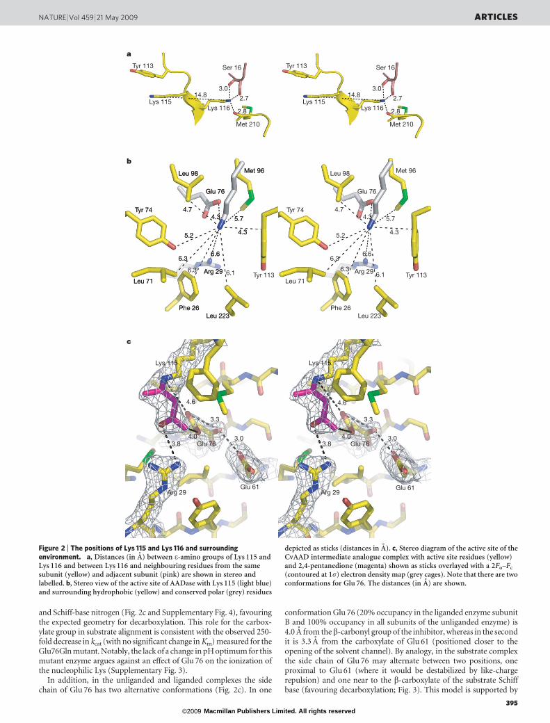
and Schiff-base nitrogen (Fig. 2c and Supplementary Fig. 4), favouringthe expected geometry for decarboxylation. This role for the carbox-ylate group in substrate alignment is consistent with the observed 250-fold decrease in kcat (with no significant change in Km) measured for theGlu76Gln mutant. Notably, the lack of a change in pH optimum for thismutant enzyme argues against an effect of Glu 76 on the ionization ofthe nucleophilic Lys (Supplementary Fig. 3).
In addition, in the unliganded and liganded complexes the sidechain of Glu 76 has two alternative conformations (Fig. 2c). In one
conformation Glu 76 (20% occupancy in the liganded enzyme subunitB and 100% occupancy in all subunits of the unliganded enzyme) is4.0 A from theb-carbonyl group of the inhibitor, whereas in the secondit is 3.3 A from the carboxylate of Glu 61 (positioned closer to theopening of the solvent channel). By analogy, in the substrate complexthe side chain of Glu 76 may alternate between two positions, oneproximal to Glu 61 (where it would be destabilized by like-chargerepulsion) and one near to the b-carboxylate of the substrate Schiffbase (favouring decarboxylation; Fig. 3). This model is supported by
Tyr 113 Ser 16
Lys 115Lys 116
Met 210
2.8
2.7
3.014.8
Tyr 113 Ser 16
Lys 115Lys 116
Met 210
2.8
2.7
3.014.8
Leu 98 Met 96
Glu 76
Tyr 74
Leu 71
Arg 29
Phe 26Leu 223
4.7
4.3
5.74.3
5.2
6.36.6
Leu 98 Met 96
Glu 76
Tyr 74
Leu 71
Arg 29
Phe 26Leu 223
4.7
4.3
5.74.3
5.2
6.36.6
Leu 98 Met 96
Glu 76
Tyr 74
Leu 71
Arg 29
Phe 26Leu 223
4.7
4.3
5.74.3
5.2
6.36.6
6.1 6.16.3 6.3
Lys 115
4.6
3.3
3.8
Arg 29
Glu 763.0
Glu 61
4.0
Lys 115
4.6
3.3
3.8
Arg 29
Glu 763.0
Glu 61
4.0
a
b
c
Tyr 113 Tyr 113
Figure 2 | The positions of Lys 115 and Lys 116 and surroundingenvironment. a, Distances (in A) between e-amino groups of Lys 115 andLys 116 and between Lys 116 and neighbouring residues from the samesubunit (yellow) and adjacent subunit (pink) are shown in stereo andlabelled. b, Stereo view of the active site of AADase with Lys 115 (light blue)and surrounding hydrophobic (yellow) and conserved polar (grey) residues
depicted as sticks (distances in A). c, Stereo diagram of the active site of theCvAAD intermediate analogue complex with active site residues (yellow)and 2,4-pentanedione (magenta) shown as sticks overlayed with a 2Fo–Fc
(contoured at 1s) electron density map (grey cages). Note that there are twoconformations for Glu 76. The distances (in A) are shown.
NATURE | Vol 459 | 21 May 2009 ARTICLES
395 Macmillan Publishers Limited. All rights reserved©2009

the catalytic activity of the Glu61Gln mutant, which shows a decreasein kcat (,20-fold with no change in Km) similar in magnitude to thatcaused by the Glu76Gln mutation. The slight downward shift of the pHoptimum of the Glu61Gln mutant (Supplementary Fig. 3) is not in thedirection expected if this residue had an effect on the pKa of Lys 115.
Oligomeric structure
The AADase homododecamer known to exist in solution19 was gen-erated by crystallographic symmetry from the protomers in the
asymmetric unit; the resultant assembly did not differ between thestructures from the two clostridial species. The 12 monomers form atetrahedron, with axial lengths of 78 A and longest dimension of118 A, enclosing a central tetrahedral cavity with axial lengths of31 A (Fig. 4a). The crystallographic dodecamer is consistent withdimensions determined for CaAAD by means of small angle X-rayscattering of 117 6 3 A in maximal diameter (Supplementary Figs 5and 6). Additionally, two-dimensional cryo-electron microscopyprojections of single CaAAD particles yielded a three-dimensional
H-ALys 115 NH2 H-A
–O
OH
O
NLys 115
–O
O
O–O
OH
O
HNLys 115
Arg 29
H2NNH2
Glu 76
O–
O Arg 29
H2NNH2
H-A
NLys 115 NH
O–O
Lys 115O
C
O
Glu 61O
–OGlu 61
O
O–
Glu 76
O–
O
O
O–
O
H2O
H2
Arg 29
H2NNH2
Glu 76
O–
O Glu 76
O–
O
A
Arg 29
H2NNH2
Lys 115 NH2
A
AH-A
H2O
Glu 76
O–
O Glu 76
O–
O Glu 76
O–
O
NLys 115H
+
+ +
++
+
+
+
+
+
+
Figure 3 | The proposed mechanism of AADase. The two alternative conformations of the side chain of Glu 76 are shown in blue and red. A, acid.
a
b c
Figure 4 | The biological dodecamer of AADase built from thecrystallographic asymmetric unit. a, Shown is the view parallel to the three-fold of the tetrahedral macromolecular assembly, with each trimer coloureddifferently (left) and cutaway to show the hollow interior (right). b, View ofthe dimer, with each subunit coloured separately and the helix and
three-stranded sheet comprising the interface from a single subunit depictedin red. c, The structure is shown as a trimer, with each subunit colouredseparately and the four-stranded sheet comprising the interface depicted ingreen (also shown in green in Fig. 1b).
ARTICLES NATURE | Vol 459 | 21 May 2009
396 Macmillan Publishers Limited. All rights reserved©2009

volume with inner cavity dimensions (,30 A) that did not differfrom those determined from the crystallographic dodecamer(Supplementary Fig. 7). Thus, the crystallographic model appearsto be an accurate representation of the state of the enzyme in solu-tion. Macromolecular assemblies with tetrahedral geometry areextremely rare. The basic symmetry of a tetrahedron is that of fourtrimers, with each three-fold symmetry axis at a vertex, or that of sixdimers with each two-fold at the centre of the axes connecting thevertices. The dimer interface comprising one three-stranded b-sheetand the amino-terminal a-helix of each monomer lies at the bottomof the b-cone and interdigitates with the identical interface of theneighbouring subunit (Fig. 4b). The trimer interface is formed fromthe four-stranded b-sheets, which form a platform with intersubunitinteractions at the three-fold symmetry axis (Fig. 4c) by means ofhydrophobic (CaAAD) or ionic (CvAAD) interactions.
The dimer interface forms extensive hydrophobic, hydrogen-bonding and ionic interactions involving more than 10% of theamino-acid residues of AADase (Supplementary Table 4 andSupplementary Fig. 8). It is these interactions that establish thedimeric form of CaAAD even in the presence of 4 M urea19. Theconversion of acetoacetate to acetone is a key process in the anaerobicmetabolism of carbohydrate in solventogenic bacteria such asC. acetobutylicum. The oligomeric state of AADase may be used tostabilize the enzyme at the very low pH and high solvent environmentfound in the cell during solventogenesis, or to colocalize the enzymesin the solventogenic pathway to facilitate the diffusion of product tothe next enzyme in the pathway. The high Km values for acetyl CoAacetyl transferase and AADase in the solventogenesis pathway (0.27and 8 mM, respectively)10,20 support this concept.
METHODS SUMMARYThe structure of AADase was determined by X-ray crystallography using phases
determined experimentally by means of single wavelength anomalous diffraction
on the selenomethionine-substituted protein. The dodecameric state of AADase
greatly increased the complexity of de novo structure determination of the enzyme
from C. acetobutylicum owing to the presence of near perfect merohedral twinning
in many crystals and the presence of two dodecamers in the asymmetric unit (216
Se sites) in other crystal forms. The final successful structure analysis depended on
determination of the structure of AADase from another bacterial source, CvAAD
(Supplementary Table 2), which crystallizes with a smaller number of protomers
in the asymmetric unit, and the use of the resulting model to determine the phases
by means of molecular replacement of the native CaAAD and liganded CvAAD
structures (see Supplementary Table 1 and Methods). Steady-state-kinetic ana-
lyses were performed on wild-type and mutant enzymes using a simple spectro-
photometric assay. The dimensions of the oligomer of CaAAD in solution were
determined by means of small angle X-ray scattering and further verified usingthree-dimensional particle reconstruction of cryo-electron microscopy images.
Full Methods and any associated references are available in the online version ofthe paper at www.nature.com/nature.
Received 19 September 2008; accepted 25 February 2009.
1. Westheimer, F. H. Coincidences, decarboxylation and electrostatic effects.Tetrahedron 51, 3–20 (1995).
2. Hamilton, G. A. & Westheimer, F. H. On the mechanism of the enzymaticdecarboxylation of acetoacetate. J. Am. Chem. Soc. 81, 6332–6333 (1959).
3. Fridovich, I. & Westheimer, F. H. On the mechanism of the enzymaticdecarboxylation of acetoacetate. II. J. Am. Chem. Soc. 84, 3208–3209 (1962).
4. Laursen, R. A. & Westheimer, F. H. The active site of acetoacetate decarboxylase.J. Am. Chem. Soc. 88, 3426–3430 (1966).
5. Warren, S., Zerner, B. & Westheimer, F. H. Acetoacetate decarboxylase.Identification of lysine at the active site. Biochemistry 5, 817–823 (1966).
6. Kokesh, F. C. & Westheimer, F. H. A reporter group at the active site ofacetoacetate decarboxylase. II. Ionization constant of the amino group. J. Am.Chem. Soc. 93, 7270–7274 (1971).
7. Harris, T. K. & Turner, G. J. Structural basis of perturbed pKa values of catalyticgroups in enzyme active sites. IUBMB Life 53, 85–98 (2002).
8. Gerlt, J. A. Obituary, Frank H. Westheimer. Nature 447, 543 (2007).9. Gallivan, J. P. & Dougherty, D. A. Cation–p interactions in structural biology. Proc.
Natl Acad. Sci. USA 96, 9459–9464 (1999).10. Highbarger, L. A., Gerlt, J. A. & Kenyon, G. L. Mechanism of the reaction catalyzed
by acetoacetate decarboxylase. Importance of lysine 116 in determining the pKa ofactive-site lysine 115. Biochemistry 35, 41–46 (1996).
11. Garcıa-Moreno, E. B. et al. Experimental measurement of the effective dielectric inthe hydrophobic core of a protein. Biophys. Chem. 64, 211–224 (1997).
12. Lee, J. K. & Houk, K. N. A proficient enzyme revisited: the predicted decarboxylasemechanism for orotidine monophosphate. Science 276, 942–945 (1997).
13. Rashin, V. & Honig, B. Reevaluation of the Born model of ion hydration. J. Phys.Chem. 89, 5588–5593 (1985).
14. Stites, W. E., Gittis, A. G., Lattman, E. E. & Shortle, D. In a staphylococcal nucleasemutant the side-chain of a lysine replacing valine66 is fully buried in thehydrophobic core. J. Mol. Biol. 221, 7–14 (1991).
15. Barbas, C. F. III et al. Immune versus natural selection: antibody aldolases withenzymic rates but broader scope. Science 278, 2085–2092 (1997).
16. Crosby, J., Stone, R. & Liehard, G. E. Mechanisms of thiamine-catalyzed reactions.Decarboxylation of 2-(1-carboxy-1-hydroxyethyl)-3,4-dimethylthiazoliumchloride. J. Am. Chem. Soc. 92, 2891–2900 (1970).
17. Fridovich, I. A study of the interaction of acetoacetic decarboxylase with severalinhibitors. J. Biol. Chem. 243, 1043–1051 (1968).
18. O’Leary, M. H. in Mechanisms of Catalysis (ed. Sigman, D. S.) 239 (Academic,1992).
19. Tagaki, W. & Westheimer, F. H. Acetoacetate decarboxylase. Reassociation ofsubunits. Biochemistry 7, 891–894 (1968).
20. Wiesenborn, D. P., Rudolph, F. B. & Papoutsakis, E. T. Thiolase from Clostridiumacetobutylicum ATCC 824 and its role in the synthesis of acids and solvents. Appl.Environ. Microbiol. 54, 2717–2722 (1988).
Supplementary Information is linked to the online version of the paper atwww.nature.com/nature.
Acknowledgements We thank A. Murzin for provisional classification of theAADase fold and C. Akey for valuable advice on the execution and interpretation ofthe electron microscopy work. We also thank H. Robinson and A. Soares for helpwith data collection. This work was supported by a grant to K.N.A. from theNational Science Foundation. Data for this study were measured at BeamlinesX12B, X25 and X29A of the National Synchrotron Light Source. Financial supportcomes principally from the Offices of Biological and Environmental Research (BER)and of Basic Energy Sciences (BES) of the US Department of Energy (DOE), andfrom the National Center for Research Resources (NCRR) of the National Institutesof Health (NIH). Small-angle X-ray scattering analyses were carried out at theStanford Synchrotron Radiation Lightsource, funded by DOE, BES. The SSRLStructural Molecular Biology Program is supported by DOE, BER, and by NIH,NCRR. The contents of this work are solely the responsibility of the authors and donot necessarily represent the official view of NCRR or NIH.
Author Contributions M.-C.H. cloned, expressed, purified, crystallized, collecteddata and performed crystal structure determination, refinement and modelanalysis. H.T. designed, executed, analysed and wrote the description of thesmall-angle X-ray scattering analysis. J.F.M. designed, executed, analysed andwrote the description of the electron microscopy experiments. K.N.A. conceived ofand designed the project. M.-C.H. and K.N.A. wrote the manuscript; all authorsdiscussed the results and commented on the manuscript.
Author Information The coordinates and structure factors have been deposited inthe Protein Data Bank with accession codes 3BH2, 3BGT and 3BH3 corresponding,respectively, to C. acetobutylicum acetoacetate decarboxylase and C. violaceumacetoacetate decarboxylase in the unliganded form and complexed with2,4-pentanedione. Reprints and permissions information is available atwww.nature.com/reprints. Correspondence and requests for materials should beaddressed to K.N.A. ([email protected]).
NATURE | Vol 459 | 21 May 2009 ARTICLES
397 Macmillan Publishers Limited. All rights reserved©2009

METHODSMaterials. Except where indicated, all chemicals were purchased from Sigma-
Aldrich or American Bioanalytical. DNase I was from Worthington Biochemical
Corp. All enzymes and primers for PCR, T4 DNA ligase and restriction enzymes
were from Invitrogen. The DNA cleanup kit and miniprep kit were from Qiagen.
Host cells and pET vectors were purchased from Novagen.
Cloning, expression and purification. The plasmid encoding the AADase gene
of C. acetobutylicum (CaAAD) was purified from host cells10 provided by J. Gerlt.
Primers (59-GCGCCCATGGTAAAGGATGAAGTAAT-39 and 59-CGCGGAT
CCTTACTTAAGATAATCATATAT-39) containing NCOI and BamHI endonu-
clease cleavage sites (underlined) were used to amplify the gene by PCR. The
genome of C. violaceum was purchased from American Type Culture Collection
(ATCC 12472D) and treated with the endonuclease BamHI before PCR. The
AADase from C. violaceum (CvAAD) gene was amplified using the primers
(59-TGAAGGGAGTTACATATGAAGCAACAGGAAGTCCAG-39 and 59-GGG
GCCGTTTTTCTCGGATCC-GCCGTTATTGCCGCAGG-39) containing NdeI
and BamHI restriction sites (underlined). The CaAAD and CvAAD were cloned
into pET3d and pET15b vectors, respectively. The ligation products were trans-
formed into Escherichia coli DH5a and sequenced by the Tufts University Core
Facility.
CaAAD was expressed and purified from E. coli BL21 (DE3) cells using a
modification of the procedure described previously10. The transformed cells were
grown in Luria Bertani medium at 37 uC and induced by 1 mM isopropyl-b-D-
thiogalactopyranoside for 20 h at room temperature (25 uC). The cells were col-
lected by centrifugation. The cell pellet was suspended in 40 ml ice-cold lysis
buffer (50 mM phosphate buffer, pH 7.4, 2 mM DTT, 1 mM PMSF and 1 mg
DNase I) and lysed by sonication (5 3 6 min, 80% power and 60% duty cycle,
Branson Sonifier 250). The cell lysate were centrifuged at 4 uC for 1 h at 147,000g.
The protein was precipitated by dropping the pH to ,3.8–3.9, and the precipi-
tated material was dissolved in 150 ml 200 mM phosphate buffer, pH 5.95, at 4 uCovernight (12 h). The remaining impurities were removed by precipitating with
55% (NH4)2SO4 followed by centrifugation at 3,700g for 30 min. CaAAD was
precipitated with 75% (NH4)2SO4, dissolved in 3 ml of 50 mM phosphate buffer,
pH 5.95, and loaded directly onto a S-200 gel filtration column (GE Healthcare)
pre-equilibrated with buffer A (50 mM phosphate buffer, pH 5.95). The fractions
containing CaAAD, detected using a kinetic assay (see below) and 12% SDS–
PAGE, were collected. If the CaAAD fractions from S-200 gel filtration were
.90% pure, the next purification step, using DEAE-Sepharose, was omitted. If
not, the collected fractions were loaded onto a DEAE-Sepharose column (GE
Healthcare) equilibrated with buffer A. The column was washed with buffer A
and eluted with a 0.5 l linear gradient of 0–0.5 M NaCl in buffer A. All fractions
containing CaAAD, detected by activity assays and 12% SDS–PAGE, were col-
lected and dialysed against 5 mM phosphate buffer, pH 5.95, and 2 mM DTT at
4 uC overnight, followed by concentration to ,10 mg ml21 using an Amicon
ultracentrifugal filter device (Millipore).
The expression and purification of CvAAD was similar to CaAAD with some
modification. Forty minutes before induction, 13 Augmedium (AthenaES) was
added to reduce the percentage of protein expressed as insoluble inclusion
bodies. The cells were collected by centrifugation and lysed by sonication in
50 ml ice-cold lysis buffer (50 mM HEPES, pH 7.4, 30 mM NaCl, 1 mM PMSF
and 1 mg DNase I). The cell lysate was centrifuged at 4 uC for 1 h at 147,000g and
the supernatant loaded onto a column packed with 15 ml TALON cobalt resin
(Clontech). The column was washed with 50 ml wash buffer (50 mM HEPES,
pH 7.4, 30 mM NaCl and 2 mM b-mercaptoethanol) and 50 ml wash buffer plus
30 mM imidazole, pH 7.6, and eluted with 50 ml elution buffer (50 mM HEPES
pH 7.4, 30 mM NaCl, 150 mM imidazole pH 7.6 and 2 mM b-mercaptoethanol).
The protein was dialysed against 5 mM imidazole buffer, pH 7.6, and 2 mM DTT
at 4 uC overnight and concentrated to ,20 mg ml21 using an Amicon Ultra
centrifugal filter device.
Selenomethionine-labelled CvAAD (SeMet CvAAD) was expressed in E. coli
(B834) cells. The cells were grown in SelenoMet medium base (AthenaES). A
custom Augmedium formulation (0.5 g l21 methionine was replaced by
0.15 g l21 selenomethionine) was added 2 h before induction. The cells were
induced with 1 mM isopropyl-b-D-thiogalactopyranoside at 18 uC for 48 h and
collected by centrifugation. The purification of selenomethionine-labelled
CvAAD was the same as that of native CvAAD.
Mutageneses of CvAAD was accomplished by means of quick-change site-
directed mutagenesis (Invitrogen) using CvAAD/pET3A as the template. The
R29Q mutant was created using the primers (59-GCCTTATCGTTTCGTCA
ACCAGGAATACATGATCATCACCTA-39 and 59-TAGGTGATGATCATGTA
TTCCTGGTTGACGAAACGATAAGGC-39). The E61Q mutant was created
using the primers (59-GGATTTGGCGATTACTCGCAAAGCGGGCAGG-39 and
59-CCTAAACCGCTAATGAGCGTTTCGCCCGTCC-39). The E76Q mutant
was created using the primers (59-GCCTTATCGTTTCGTCAACCAGGAATA
CATGATCATCACCTA-39, and 59-TAGGTGATGATCATGTATTCCTGGTTGA
CGAAACGATAAGGC-39). The mutated sites are underlined. The resulting PCR
products were digested with NdeI and BamHI endonuclease, and cloned into
pET15b vectors that were cut by the same enzymes. The ligation products were
transformed into E. coli DH5a, followed by DNA sequencing by Agencourt
Bioscience. The plasmid, encoding His6-tagged CvAAD point mutants, were
purified from E. coli DH5a and transformed into E. coli BL21(DE3) for protein
expression. The expression and purification of CvAAD mutants was identical to
that of wild-type CvAAD.
Crystallization and data collection. The initial crystallization conditions of
CaAAD and CvAAD were identified by high-throughput screening (Hauptman
Woodward Medical Research Institute) and refined by the hanging-drop vapour-
diffusion method. The final crystallization condition of CaAAD was 18–20%
glycerol, 40 mM phosphate buffer, pH 5.95, 100 mM sarcosine and 14–15%
PEG 3350 (Hampton Research). Drops were formed by mixing 1–2ml of protein
solution (10 mg ml21) with 1ml of well solution. Crystals of CaAAD were flash-
frozen in a gaseous N2 stream at 100 K. X-ray diffraction data were collected at the
National Synchrotron Light Source (Brookhaven National Laboratory, New
York), Beamline X25. CaAAD crystallized in space group R32 with unit-cell
dimensions a 5 b 5 104.07 A, c 5 578.09 A and diffracted to 2.4 A resolution.
Crystals of CvAAD and SeMet CvAAD were grown by hanging-drop vapour
diffusion over a well solution containing 0.4–0.5 M K2HPO4/NaH2PO4 at pH 7.5–
7.9. Drops consisted of 1–2ml of protein solution (8–12 mg ml21) and 1ml of well
solution. Crystals were transferred to well solution with 30% glucose as a cryo-
protectant and crystals were frozen in liquid N2. The automounter at BNL,
Beamline X12B, was used to screen crystals for diffraction and data were collected
at Beamline X29A. SeMet CvAAD crystallized in space group R3
(a 5 b 5 105.45 A, c 5 252.38 A). A 2.1 A multiple wavelength anomalous diffrac-
tion (MAD) data set was collected (0.9791 A, 0.9793 A and 0.9754 A), but only the
data collected at 0.9791 A were used to solve the phases. For the 2,4-pentanedione
complex structure with CvAAD, the enzyme (10 mg ml21) was co-crystallized
with 2 mM potassium acetyl acetonate under the same condition as unliganded
CvAAD. The crystal was additionally soaked in mother liquid plus 120 mM
potassium acetyl acetonate for 1 h at room temperature and transferred to
well solution with 30% glucose plus 4 mM potassium acetyl acetonate as cyro-
protectant and frozen in liquid N2. The inhibitor-soaked crystals diffracted to
2.1 A and were isomorphous to the SeMet CvAAD. A 2.1 A data set was collected
at 0.900 A at Brookhaven National Laboratories, beamline X12B. All data sets
were indexed and scaled using HKL2000 (ref. 21). Data collection statistics are
summarized in Supplementary Table 1.
Structure determination and model refinement. The structure of CvAAD was
phased by the single wavelength anomalous diffraction method, using the 2.1 A
SeMet CvAAD data set collected at the peak wavelength 0.9791 A. The Se sub-
structure was solved using HKL2MAP/SHELXD22,23, which identified 24 of 32
selenomethionine sites. The 24 sites were used to phase the protein structure in
SOLVE24 and generated a high-quality electron density map. The map was
improved using RESOLVE25, by means of NCS averaging (4 monomers per
asymmetric unit). An initial model including 44% of the structure was built
automatically by RESOLVE. The experimentally phased NCS averaged electron
density map permitted further manual rebuilding of one of the four monomers
using COOT26. This monomer was then used to perform molecular replacement
using MOLREP27 to find the remaining three monomers in the asymmetric unit.
Iterative rounds of model building and refinement were performed using
COOT, CNS and REFMAC526–28.
The structure of CaAAD was phased by molecular replacement with the pro-
gram PHASER using SeMet CvAAD as the search model29. The process of refine-
ment and rebuilding was the same as that used for SeMet CvAAD. The structure
of CaAAD and 2,4 pentanedione liganded CvAAD was phased by molecular
replacement with the program PHASER29 using the structure of SeMet
CvAAD as the search model. The process of refinement and rebuilding was
the same as that used for SeMet CvAAD. The model of the inhibitor was not
added to the refinement until the value of Rwork dropped below 28% to avoid
phase bias. The topology restraints for 2,4-pentanedione adduct of Lys 115 for
use in REFMAC5 were generated using SKETCHER in the CCP4 program
suite30. The Ramachandran plot, as assessed by Procheck31, had 99.5% of the
residues of unliganded CaAAD and CvAAD and 99.2% of the residues of
liganded CvAAD in the allowed regions with 1 residue (Ser 126 in CaAAD/
Gln 127 in CvAAD) in the disallowed regions for all three structures.
Small-angle X-ray scattering measurements. Solution X-ray scattering
measurements were performed at the Stanford Synchrotron Radiation
Lightsource Beamline 4-2 (ref. 32). Each protein sample solution containing
50 mM phosphate (pH 5.95) and 2 mM DTT was held in a sample cuvette,
maintained at 20 uC and located at 2.5 m from a MarCCD165 detector
doi:10.1038/nature07938
Macmillan Publishers Limited. All rights reserved©2009

(MarUSA). The detector pixel numbers were converted to the momentumtransfer Q 5 4psin(h)/l, where h is one half of the scattering angle and l the
X-ray wavelength 1.381 A, using the (100) reflection and related reflections
recorded from a cholesterol myristate powder sample. Twenty-four 10-s expo-
sures were acquired in series at 2, 5, 7.5, 10 and 15 mg ml21 (Supplementary
Fig. 5). No time-dependent radiation-induced protein aggregation was observed
and all 24 images were averaged after intensity scaling, with the exception of 10
and 15 mg ml21 samples, for which the last few exposures were excluded in the
averaging process due to the onset of radiation-induced aggregation. The radial
integration, intensity-scaling, statistical analysis, frame-averaging and back-
ground subtraction were done by MarParse32. Radii of gyration (Rg) and forward
scattered intensities (I(Q 5 0)) were obtained by Primus33 in the Q range 0.0117–
0.0297 A21 (Supplementary Fig. 5, inset). The concentration-scaled forward
scattered intensity remained within 3% of the mean value between 2 and
10 mg ml21, indicating no evidence of concentration-dependent aggregation.
Slightly lower Rg values at higher protein concentrations indicated mildly
repulsive inter-particle interaction. The composite scattering curve with satis-
factorily high statistics covering Q values 0.0117 to 0.25 A21 was thus obtained
by scaling and merging the scattering curve recorded at 2 mg ml21
(0.0117 , Q , 0.0611 A21) with the intermediary angle data recorded at
15 mg ml21 (0.0448 , Q , 0.25 A21) by Primus33 to minimize weak effects of
the particle structure factor due to the repulsive interaction (Supplementary
Fig. 6). The overlapping Q range contained 30 common data points to assure
accuracy of the scaling. The X-ray scattering profile and the electron pair distance
distribution function P(r) were computed for the crystallographic structure
model using ORNL_SAS34. The electron density and thickness of the hydration
layer were altered in the range 3–15% higher electron density of water and 2.5–
3.5 A. The best fit was obtained with 8% excess density and 2.5 A layer thickness,
although similarly satisfactory fits could be obtained with lower hydration layer
density and a slightly thicker hydration layer. Q values in the experimental data
were also adjusted by 1–3% to account for the small unit cell parameter variations
observed in the crystallographic study. The lowest chi square value we have
obtained with 1% Q value adjustment was 2.445, which confirms the presence
of the crystallographic dodecamer in solution but reflects the clearly recognizable
deviation in the Q range 0.06–0.10 A21. This deviation may suggest minor struc-
tural differences between solution and crystal structures at the tertiary and/or
quaternary structural level because experimental conditions can not be identical.Similar differences between experimental and computed X-ray scattering curves
have been observed for other oligomeric enzyme systems35. The indirect Fourier
transform analysis of the composite scattering curve by GNOM36 yielded the
electron pair distance distribution function, giving the maximum particle dimen-
sion of 117 (6 3 (6 s.d.)) A and Rg of 42.9 A, both of which are highly consistent
with the corresponding values of 118 and 43.3 A obtained for the crystal structure
with the hydration layer (Supplementary Fig. 6). The nearly perfect match of the
experimental and computed P(r) indicates that the solution and crystal structures
are essentially identical at the quaternary structure level (Supplementary Fig. 6,
inset).
Cryo-electron microscopy and image processing. The cryo-electron micro-
scopy and image processing of CaAAD were performed at Boston University
School of Medicine. CaAAD (10 mg ml21, 1 mM phosphate buffer, pH 5.95) was
loaded onto perforated carbon film on 400 mesh copper grids. The specimens
were plunge-frozen in liquid ethane cooled by liquid nitrogen37 and data col-
lected on a Tecnai TF20 with Kodak SO163 film at Boston University School of
Medicine. Electron micrographs were recorded at 200 kV at 362,000 with a
defocus range of 21.0 to 22.5mm. Negatives were digitized on a CreoScitexEVERSMART scanner using a 4.54 mm raster and binned 4 3 4 to 2.93 A per
pixel. Particles were picked with boxer in EMAN38 and classified using refine2d
in EMAN to avoid bias from a three-dimensional model39.
Additionally, the three-dimensional structure was refined in EMAN using
,8,700 particles and by imposing the tetrahedral symmetry found in the crystal
structure39. The resolution was estimated by splitting the data set into two halves
(according to even and odd numbered particles), generating an even and an odd
volume and then calculating their cross-correlation coefficient. Plotting the
correlation coefficient versus resolution gave the Fourier shell correlation
(FSC) curve. Using the FSC 0.5 criteria, it was concluded that the resolution
of the three-dimensional volume was 12 A.
Steady-state kinetics. The decarboxylation of acetoacetate by CvAAD and
CaAAD was assayed by monitoring the disappearance of the enolate form of
acetoacetate spectrophotometrically at 270 nm (De 5 26.7 M21 cm21) at 25 uCin 50 mM phosphate buffer, pH 5.95, on a Beckman Coulter DU-800 spectro-
photometer10. The same assay was used for CvAAD mutants in 50 mM phos-
phate/citrate buffer at various pH values on a Varian Carey-300
spectrophotometer. Data were fitted to the Michaelis–Mention equation
V 5 Vmax[S]/(Km 1 [S]), where V is the initial rate at substrate concentration
S, and Km is the Michaelis constant. kcat is obtained from Vmax and total enzyme
concentration in the reaction. The kapp for wild-type and mutant CvAAD pro-
teins versus pH was obtained from the initial rate and enzyme concentration at
the saturating substrate concentration of 20 mM, with the exception of the
CvAAD R29Q mutant for which activity could only be measured using 40 mM
substrate.
21. Otwinowski, Z. & Minor, W. Processing of X-ray diffraction data collected inoscillation mode. Methods Enzymol. 276, 307–326 (1997).
22. Pape, T. & Schneider, T. R. HKL2MAP: a graphical user interface for phasing withSHELX programs. J. Appl. Crystallogr. 37, 843–844 (2004).
23. Uson, I. & Sheldrick, G. M. Advances in direct methods for protein crystallography.Curr. Opin. Struct. Biol. 9, 643–648 (1999).
24. Terwilliger T.C. & Berendzen J.. Automated MAD and MIR structure solution. ActaCrystallogr. D 55, 849–861 (1999).
25. Terwilliger, T. C. Maximum likelihood density modification. Acta Crystallogr. D 56,965–972 (2000).
26. Emsley, P. & Cowtan, K. Coot: model-building tools for molecular graphic. ActaCrystallogr. D 60, 2126–2132 (2004).
27. Collaborative computation project number 4. The CCP4 suite: programs forprotein crystallography. Acta Crystallogr. D 50, 760–763 (1994).
28. Brunger, A. T. Crystallography & NMR system: a new software suite formacromolecular structure determination. Acta Crystallogr. D 54, 905–921 (1998).
29. Storoni, L. C., McCoy, A. J. & Read, R. J. Likelihood-enhanced fast rotationfunction. Acta Crystallogr. D 60, 432–438 (2004).
30. Potterton, E., Briggs, P., Turkenburg, M. & Dodson, E. A graphical user interface tothe CCP4 program suite. Acta Crystallogr. D 59, 1131–1137 (2003).
31. Laskowski, R. A., MacArthur, M. W., Moss, D. S. & Thornton, J. M. Procheck: aprogram to check the sterochemical quality of protein structures. J. Appl.Crystallogr. 26, 283–291 (1993).
32. Smolsky, I. L. et al. Biological small-angle x-ray scattering facility at the StanfordSynchrotron Radiation Laboratory. J. Appl. Crystallogr. 40, S453 (2007).
33. Konarev, P. V. et al. PRIMUS: a Windows PC-based system for small-anglescattering data analysis. J. Appl. Crystallogr. 36, 1277–1282 (2003).
34. Tjioe, E. & Heller, W. T. ORNL_SAS: software for calculation of small-anglescattering intensities of proteins and protein complexes. J. Appl. Crystallogr. 40,782–785 (2007).
35. Svergun, D. I. et al. Large differences are observed between the crystal andsolution quaternary structures of allosteric aspartate transcarbamylase in the Rstate. Proteins 27, 110–117 (1997).
36. Svergun, D. I. Determination of the regularization parameter in indirect-transformmethods using perceptual criteria. J. Appl. Crystallogr. 25, 495–503 (1992).
37. Menetret, J. F. et al. The structure of ribosome–channel complexes engaged inprotein translocation. Mol. Cell 6, 1219–1232 (2000).
38. Ludtke, S. J., Baldwin, P. R. & Chiu, W. EMAN: semiautomated software for high-resolution single-particle reconstructions. J. Struct. Biol. 128, 82–97 (1999).
39. Ludtke, S. J., Chen, D. H., Song, J. L., Chuang, D. T. & Chiu, W. Seeing GroEL at 6 Aresolution by single particle electron cryomicroscopy. Structure 12, 1129–1136(2004).
doi:10.1038/nature07938
Macmillan Publishers Limited. All rights reserved©2009