Embed Size (px)
Citation preview
THE OPERATIVE REPAIR OF MASSIVE RECTAL PROLAPSE*ROSCOE R. GRAHAM, M.D.
TORONTO, CANADA
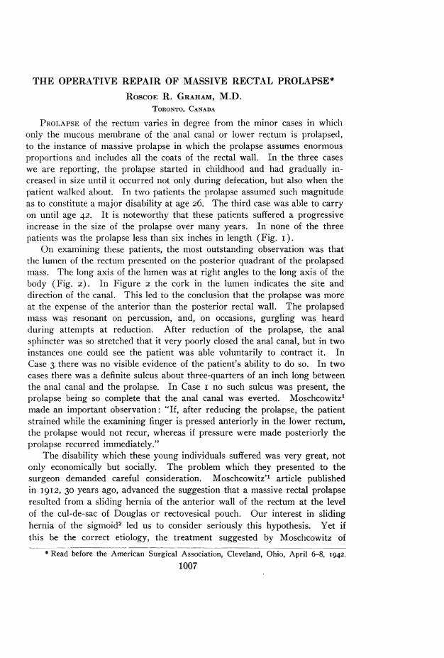
PROLAPSE of the rectum varies in degree from the minor cases in whichonly the mucous membrane of the anal canal or lower rectum is prolapsed,to the instance of massive prolapse in which the prolapse assumes enormousproportions and includes all the coats of the rectal wall. In the three caseswe are reporting, the prolapse started in childhood and had gradually in-creased in size until it occurred not only during defecation, but also when thepatient walked about. In two patients the prolapse assumed such magnitudeas to constitute a major disability at age 26. The third case was able to carryon until age 42. It is noteworthy that these patients suffered a progressiveincrease in the size of the prolapse over many years. In none of the threepatients was the prolapse less than six inches in length (Fig. i).
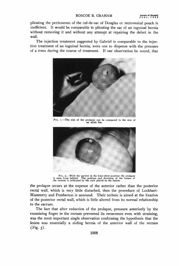
On examining these patients, the most outstanding observation was thatthe lumen of the rectum presented on the posterior quadrant of the prolapsedmass. The long axis of the lumen was at right angles to the long axis of thebody (Fig. 2). In Figure 2 the cork in the lumen indicates the site anddirection of the canal. This led to the conclusion that the prolapse was moreat the expense of the anterior than the posterior rectal wall. The prolapsedmass was resonant on percussion, and, on occasions, gurgling was heardduring attempts at reduction. After reduction of the prolapse, the analsphincter was so stretched that it very poorly closed the anal canal, but in twoinstances one could see the patient was able voluntarily to contract it. InCase 3 there was no visible evidence of the patient's ability to do so. In twocases there was a definite sulcus about three-quarters of an inch long betweenthe anal canal and the prolapse. In Case i no such sulcus was present, theprolapse being so complete that the anal canal was everted. Moschcowitz'made an important observation: "If, after reducing the prolapse, the patientstrained while the examining finger is pressed anteriorly in the lower rectum,the prolapse would not recur, whereas if pressure were made posteriorly theprolapse recurred immediately."
The disability which these young individuals suffered was very great, notonly economically but socially. The problem which they presented to thesurgeon demanded careful consideration. Moschcowitz" article publishedin 1912, 30 years ago, advanced the suggestion that a massive rectal prolapseresulted from a sliding hernia of the anterior wall of the rectum at the levelof the cul-de-sac of Douglas or rectovesical pouch. Our interest in slidinghernia of the sigmoid2 led us to consider seriously this hypothesis. Yet ifthis be the correct etiology, the treatment suggested by Moschcowitz of
*Read before the American Surgical Association, Cleveland, Ohio, April 6-8, 1942.1007
ROSCOE R. GRAHAM Annals of SurgeryJune, 1942
plicating the peritoneum of the cul-de-sac of Douglas or rectovesical pouch isinefficient. It would be comparable to plicating the sac of an inguinal herniawithout removing it and without any attempt at repairing the defect in thewall.
The injection treatment suggested by Gabriel is comparable to the injec-tion treatment of an inguinal hernia, were one to dispense with the pressureof a truss during the course of treatment. If our observation be sound, that
....
!!.................... ..
FIG. I.-The size of the prolapse can be compared to the size ofan adult fist.
FIG. 2.-With the patient in the knee-chest position the prolapseis seen from behind. The position and direction of the lumen ofthe rectum is indicated by the cork placed in the lumen.
the prolapse occurs at the expense of the anterior rather than the posteriorrectal wall, which is very little disturbed, then the procedure of Lockhart-Mummery and Pemberton is unsound. Their technic is aimed at the fixationof the posterior rectal wall, which is little altered from its normal relationshipto the sacrum.
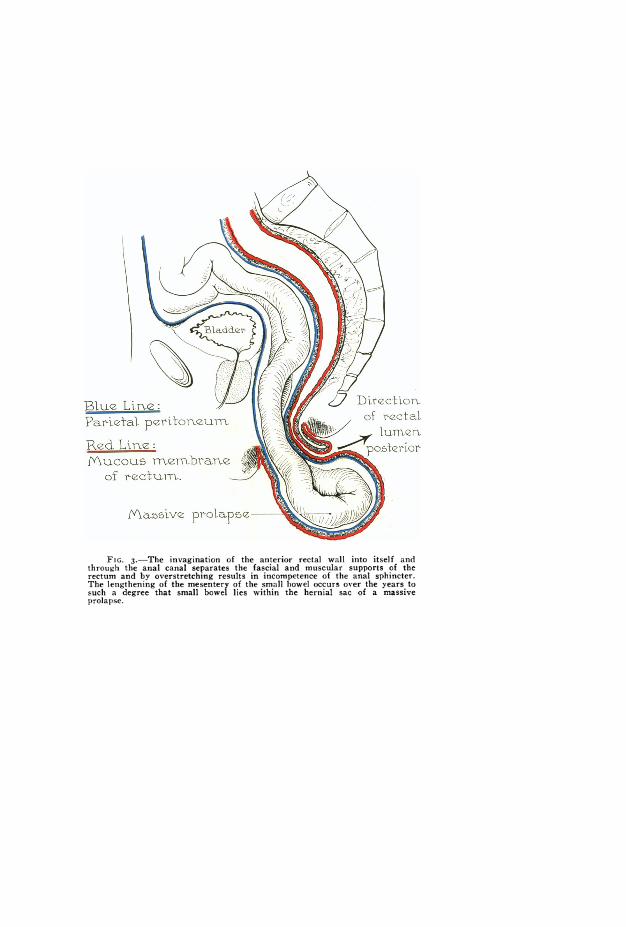
The fact that after reduction of the prolapse, pressure anteriorly by theexamining finger in the rectum prevented its recurrence even with straining,was the most important single observation confirming the hypothesis that thelesion was essentially a sliding hernia of the anterior wall of the rectum(Fig. 3).
1008
lumer.
of rectum.
M&65ive prolapse- 1
FIG. 3.-The invagination of the anterior rectal wall into itself andthrough the anal canal separates the fascial and muscular supports of therectum and by overstretching results in incompetence of the anal sphincter.The lengthening of the mesentery of the small bowel occurs over the years tosuch a degree that small bowel lies within the hernial sac of a massiveprolapse.
Volume 115 MASSIVE RECTAL PROLAPSENumber 6
In this mechanisiim the natural defect in the pelvic fascia which permitsthe passage of the rectum through the pelvic diaphragm is enlarged by thecontents of the cul-de-sac of Douglas or rectovesical pouch pressing downwardinto the anterior rectal wall. This increased bulk of rectum further separatesthe levator ani by stretching the pelvic fascia, which normally unites themmedially. This likewise decreases the normal fixation of the rectum at thislevel. The separation of the levators permits sufficient anterior wall of therectum to be invaginated into the lumeni of the rectum that the latter p)ro-
'IS*:~~~~~~~~~~~~~~~~~~~~~~~~~~~~~~~~~~. ..2.. ..........
*.~ ~ ~ ~ ~~~~~~~~~~~~~~~~~~~~~~~~~~~~~~~~~~~~~~~~ ..: l...........
,#^:8.,x,x,................................~~~~~~~~~~~~~~~~~~~~~.......
:aI C: PI.... i. .;;......
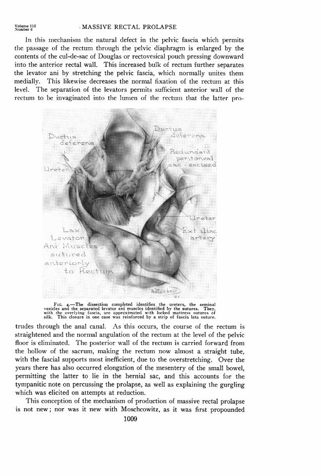
FIG. 4.-The dissection completed identifies the ureters, the seminalvesicles and the separated levator ani muscles identified by the sutures. They,with the overlying fascia, are approximated with locked mattress sutures ofsilk. This closure in one case was reinforced hy a strip of fascia lata suture.
trudes through the anal canal. As 'this occurs, the course of the rectum isstraightened and the normal angulation of the rectum at the level of the pelvicfloor is eliminated. The posterior wall of the rectum is carried forward fromthe hollow of the sacrum, making the rectum now almost a straight tube,with the fascial supports most inefficient, due to the overstretching. Over theyears there has also occurred elongation of the mesentery of the small bowel,permitting the latter to lie in the hernial sac, and this accounts for thetympanitic note on percussing the prolapse, as well as explaining the gurglingwhich was elicited on attempts at reduction.
This conception of the mechanism of production of massive rectal prolapseis not new; nor was it new with Moschcowitz, as it was first propounded
1009
ROSCOE R. GRAHAM Annals of SurgeryJune. 1 942
by Jeannell, in I89o. This conception, however, appealed to us so stronglythat we felt it pointed a way to safe and adequate treatment, using the sameprinciples as are applied in the operative repair of all herniae, particularlysliding herniae.2
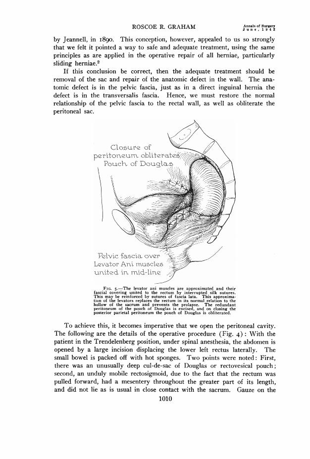
If this conclusion be correct, then the adequate treatment should beremoval of the sac and repair of the anatomic defect in the wall. The ana-tomic defect is in the pelvic fascia, just as in a direct inguinal hernia thedefect is in the transversalis fascia. Hence, we must restore the normalrelationship of the pelvic fascia to the rectal wall, as well as obliterate theperitoneal sac.
Closu5re of N
peritoneumr obliteratelPoxch of Douglab
Pelvic fa5cia. over '
L'va,tor Ani mnuc1e5urVited irn mid-lire
FIG. 5.-The levator ani muscles are approximated and theirfascial coveritng united to the rectum by interrupted silk sutures.This may be reinforced by sutures of fascia lata. This approxima-tion of the levators replaces the rectum in its normal relation to thehollow of the sacrum and prevents the prolapse. The redundantperitoneum of the pouch of Douglas is excised, and on closing theposterior parietal peritoneum the pouch of Douglas is obliterated.
To achieve this, it becomes imperative that we open the peritoneal cavity.The following are the details of the operative procedure (Fig. 4): With thepatient in the Trendelenberg position, under spinal anesthesia, the abdomen isopened by a large incision displacing the lower left rectus laterally. Thesmall bowel is packed off with hot sponges. Two points were noted: First,there was an unusually deep cul-de-sac of Douglas or rectovesical pouch;second, an unduly mobile rectosigmoid, due to the fact that the rectum waspulled forward, had a mesentery throughout the greater part of its length,and did not lie as is usual in close contact with the sacrum. Gauze on the
1010
Volume 115Number 6 MASSIVE RECTAL PROLAPSE
end of a sponge forcep placed on the bottom of the cul-de-sac of Douglas orrectovesical pouch, with downward pressure readily invaginated the anteriorrectal wall into the rectal lumen. The defect in the muscular pelvic floorcould readily be palpated. This invagination was easily carried through theanal canal, and reproduced the massive rectal prolapse, and convinced usthat it really was due to a sliding hernia of the anterior wall of the rectum.Furthermore, this prolapse was readily reduced by upward traction on therectosigmoid junction. Having convinced ourselves of these facts, the peri-toneum of the cul-de-sac of Douglas or rectovesical pouch was opened anddissected free from the extraperitoneal fat and areolar tissue. The ureterswere then identified and surrounded with tape, in order to retract them later-ally. A further dissection of the perirectal fat made possible the visualizationof the seminal vesicles and the widely separated fascial-covered medial bor-ders of the levator ani muscles. With the rectum pulled well up into theabdomen, and starting just behind the prostate, interrupted locked mattresssutures of silk were placed in the fascia covering the levator ani muscles.These sutures united the levators until their resultant approximation forcedthe rectum back into the hollow of the sacrum (Fig. 5). In Case 3 thisrepair was reenforced by a single suture of fascia lata. This maneuver restoredthe normal angulation of the rectum. It was then no longer possible toinvaginate the anterior wall of the rectum through the anal canal. Pressureexerted at the level of the rectum and new pelvic floor forced the rectum intothe hollow of the sacrum, not toward the anal canal. Interrupted silk stitchesthen united the lateral rectal wall to the fascia over the right and left levators(Fig. 5). This appeared to give a very adequate support to the rectum,particularly to its anterior wall, which is so important in preventing a re-currence of the massive prolapse. Excision of the redundant hernial sacand suture of the pelvic peritoneum obliterates the cul-de-sac of Douglas orrectovesical pouch entirely. The abdomen is then closed without drainage.
On return to the ward, the patient remains recumbent, with the foot ofthe bed elevated ten inches, for one week. A low-residue diet is given. Everyeffort is made to prevent a stool for a week to ten days, at the end of whichtime oil enemata are usually effective in producing a stool. The patient isencouraged to practice contraction of the overstretched anal sphincter manytimes a day, in order that it will regain its tonicity. The following are thedetails of the three cases:
CASE REPORTS
Case i.-Hospital No. A6482I: W. J. P., male, age 26. Admitted April 12, I939.Mass first present at age six, gradually increased in size, and recently appeared withslight straining when patient was erect. The mass had to be replaced by manipulation.There was marked urgency of defecation.
Examination.-The mass was as large as a man's fist, and could be prolapsed volun-tarily. In the knee-chest position it could be replaced by violent movements of theabdominal muscles. No ulceration was present. There was no sulcus at the analsphincter, as the anal canal was also prolapsed. The mass was tympanitic on percussion,
1011
ROSCOE R. GRAHAM Annals of SurgeryROSCOER. GRAHAM ~~~~~~Jun e, 1.9 4 2
and the lumen of the rectum pointed backward. With a finger in the rectum, pressureanteriorly controlled the prolapse.
Operation.-April 24, 1939: Primary healing. Discharged May i6, I939. Has hadno further trouble, and is doing hard physical work in a tannery at the present time.
Case 2.-Hospital No. A82662: W. B., male, age 42. Admitted April 4, 1940. Pro-lapse first noticed at age eight. Now comes down with stool and has to be replaced. Thesize has increased markedly in the previous five weeks. Now has a mass 6x8 inchesprotruding from anus, and reduction becoming difficult (Fig. i).
Examination.-Well nourished; sphincter contraction visible and surprisingly goodtone. The mass is difficult to replace. There is no ulceration and no hemorrhage. Thelumen points posteriorly. The mass is resonant on percussion (Fig. 2). With a fingerin the rectum, pressure anteriorly controlled the prolapse.
Operation.-April 12, I940: Had slight superficial wound separation due to coughingfrom a respiratory infection. Good recovery. Complete relief, with a very slight prolapseof one area of anal mucous membrane on straining. At present is working in munitionsplant as a laborer.
Case 3.-Hospital No. A99554: G. T., age 26. Admitted April i, 1941 to theNeurologic Service, with headaches, fainiting attacks, and a multiplicity of complaints.Only organic finding was rectal prolapse present since childhood. Had to be replacedafter each stool. Marked increase in size of prolapse during past five years, becomingdifficult to replace, and on admission prolapse occurs on walking or any exertion.
Examination.-Sphincter lax. No evidence of contraction could be demonstrated bythe patient. On straining, the rectum prolapsed at least six inches; a sulcus is presentbetween the anal canal and prolapse about one-half inch deep. By introducing two fingersinto the rectum and pressing anteriorly, the prolapse could be prevented as the patientstrained. Pressing posteriorly into the hollow of the sacrum while the patient strained,permitted recurrence of the prolapse.
Operation.-May I0, I94I: Bowels moved on tenth day. Primary union. Discharged.Progress.-No further prolapse of the bowel. Slight prolapse of mucous membrane
treated by injections of 2 CC. phenol and almond oil. Some difficulty in controlling stoolfor four months. At present has perfect conltrol of bowel movements. On examination,the tone of the sphincter ani is remarkable, but not as strong as normal. Her neurologicsymptoms have nearly all disappeared. She is working in our hospital as a ward aid atthe present time. We are indebted to Dr. Keith Welsh for the privilege of seeing andreporting this third case.
CONCLUSIONS
(i) Massive rectal prolapse is a sliding hernia of the anterior rectalwall through the anal canal.
(2) The lumen of the rectal canal points posteriorly as the prolapsed massis formed largely at the expense of the anterior rectal wall.
(3) This results in an overstretching of the pelvic fascial supports of therectum.
(4) With the examining finger in the rectum, the prolapse can be con-trolled as the patient strains if pressure be exerted anteriorly, whereas ifpressure be exerted posteriorly, the prolapse will recur.
(5) The treatment of this condition should be planned to apply the basicprinciples underlying the treatment of all herniae-first, remove the sac; sec-ond, restore the defect in the wall.
(6) A procedure is presented which fulfills these requirements.1012
Volume 11rNumber 6 MASSIVE RECTAL PROLAPSE
(7) It has been successfully carried out on three patients. Two havereturned to hard labor; the third is working as ward aid in our hospital.
(8) The return of tone in the anal sphincter is most remarkable.
REFERENCES
Moschcowitz, Alexis V.: The Rational Treatment of Sliding Hernia. Surg., Gynec.,and Obstet., 8i, 330, January, 1925.
2 Graham, R. R.: The Operative Repair of Sliding Hernia of the Sigmoid. ANNALS OFSURGERY, I02, No. 4, 784, October, I935.
DIscussION.-DR. VERNON C. DAVID (Chicago): Doctor Graham has limitedhis discussion to the type of prolapse of the rectum in which the anus, including thepatulous sphincter muscles, prolapses with the four to six inch tube of bowel carryingwith it the cul-de-sac of Douglas. In principle, the treatment he advocates requires anabdominal approach, opening of the cul-de-sac, and the approximation of the levatormuscles by suture in front of the rectum. He is to be congratulated on the results in thethree cases he reports.
To present a slightly different viewpoint, I should like to point out that the levatormuscles, which have a very delicate fascial covering, completely encircle the rectum andtheir highest or superior surface is at about the level of the sacrococcygeal junction. Totighten these muscles by suture via the abdominal approach in the depth of the pelvis isa difficult procedure, and I should like to ask Doctor Graham if he finds it necessary tocut the triangular ligaments of the rectum, which lie below the culdesac, before he reachesthe levator muscles. When the levator muscles are divided, as in removal of the rectumposteriorly, the rectum is still firmly anchored in place by the fascia propria, which is adense fascia about one millimeter thick, firmly attached to the sacrococcygeal junctionposteriorly and anteriorly to the prostate which it completely envelops. Before the rectumcan be mobilized this fascia must be cut.
It is my belief that it is not only the atrophy and weakness of the levator muscles anddepth of the cul-de-sac which favor this type of prolapse but more particularly a weaknessand stretching of the fascia which allows the rectum to completely prolapse carrying thecul-de-sac with it. In repair of this type of prolapse we, therefore, believe that the structuresin most need of support are in the prolapsed segment of bowel and on its outer surface,namely, the fascia propria and levator muscles. We also believe that the atonic sphinctermuscles, which have been greatly dilated by the prolapse of the bowel and levator musclesthrough them, are a factor which favors early recurrence of the prolapse.
With these anatomic facts in mind, I should like to call your attention to the opera-tion for the repair of this type of prolapse originally proposed by Delorme, in I890, andfirst carried out by Rehn, in I 896. This operation is easily carried out under novocainanesthesia and consists in the removal of the mucosa of the prolapsed segment from themucocutaneous line to the apex of the prolapse. The muscularis of the bowel, levatormuscles, and fascia propria in the outer layer of the prolapse are collapsed like a closedaccordion by longitudinal puckering-stitches, which reduces the prolapse and makes tighterthe supporting levator muscles and fascia propria and places the puckered mass of muscleand fascia above the sphincter muscles, which are narrowed below it by angulatingstitches. The excess of freed mucosa is then cut off and the cut surface sutured to theskin. This operation accomplishes everything but obliteration of the cul-de-sac. I firstsaw Doctor Bevan perform this operation, and I have carried it out in i 2 cases, withgood results, and no massive recurrences. In women, where the perineal body is gone andthe levator muscles are widely separated, it is advisable to perform a later perineorrhaphy.
This type of prolapse demands, essentially, strengthening of the pelvic fascia andlevator muscles. In my opinion this may be best accomplished by the abdominal opera-tion described by Bardenheuer, Moschcowitz, and Graham, or by the Rehn-Delorme opera-tion from below.
DR. CHARLES G. MIXTER (Boston): Massive rectal prolapse, particularly of therecurrent variety, is frequently a discouraging lesion from the surgeon's viewpoint. DoctorGraham has presented to us a well-conceived operation that has been successful in the
1013
ROSCOE R. GRAHAM Annals of Surgery
three patients he has subjected to this procedure, and in skilled hands it should yieldgood results. It is, however, a procedure of considerable magnitude. The lesion occursmany times in the aged and, perhaps, enfeebled group. It might not be amiss to bringbefore this Association briefly a simple procedure that has yielded satisfactory results inthe two cases upon whom I have had the opportunity to try it.
The abdomen is opened through a low left rectus muscle-splitting incision. A smallopening is made in the pelvic floor on either side of the rectosigmoid. The rectum ismobilized by blunt dissection and cigarette wicks are inserted to stimulate fixation of thisbowel segment by resultant fibrosis. The wicks are brought out through stab woundsabove the inguinal ligament on either side. Further experience may prove this step tobe unnecessary. The sigmoid which is usually redundant is brought out of the abdominalwound in a manner similar to a subcutaneous loop-colostomy or precolostomy. Thedistal limb of sigmoid should run tautly downward from the lower angle of the woundto the rectosigmoid. The proximal sigmoid reenters the abdomen at the upper end ofthe incision. All layers of the abdominal wall except the skin and superficial fascia areclosed in a routine manner beneath the exteriorized sigmoid through an opening estab-lished in the mesentery. The fat is separated from the anterior rectus sheath and allowedto gape to accommodate the loop and the skin is closed over the bowel. Care must betaken not to constrict the lumen where the bowel enters and leaves the abdomen.
Two cases, both having had three previous procedures, have been treated by opera-tion based on the principle of fixation of the sigmoid in the abdominal wound. In thefirst case, a woman of about 40, a double-barrel colostomy was done, the bowel lateropened, the spur crushed and the colostomy closed. This patient has remained free ofrecurrence and with satisfactory bowel function for two and one-half years. The secondcase was a rather feeble woman in the late sixties, who was operated upon by the methodoutlined above. She had no difficulty in moving her bowel postoperatively. It is nowabout nine months since operation, and I understand she has had no recurrence, thoughI have not had an opportunity to examine her personally.
The results in two cases are insufficient to draw conclusions from, but suggest thatthis simple procedure may be worthy of further trial, particularly where relief must begiven in the poor-risk group.
DR. JOHN PEMBERTON (Rochester, Minn.): Since Doctor Graham mentioned theoperation that I described four years ago, I would like to run over, very briefly, the prin-ciples of it, which are similar to what Doctor Mixter has described.
I think the fundamental principles of rectal prolapse are about the same that yousee in the colostomy. If the distal segment or the segment just distal or just proximalto the colostomy is fixed, you will not get a prolapse of the colostomy, if you get upclose to the descending colon, but if you take it in the middle of the sigmoid, then youare very likely to get a prolapse.
You cannot make a complete fixation of the rectum unless you divide the pelvicperitoneum, so this is done either on one side or both sides. Then the rectum is freedup from the segment going down there. This permits fibrosis to take place here, whichyou can readily determine by examining the rectum digitally after incision. This is freedup, and we get a space between the rectal wall and the sigmoid, until it heals. The prob-lem is, of course, to suspend the sigmoid afterward. The suspension will not hold, butif it holds temporarily, for a couple of weeks, I believe the fixation will hold up.
Four years ago I reported six cases that we had operated upon. I think the longestcase was two or three years. Since then we have done others, but of those six cases Iknow that one has had some recurrence of the prolapse.
DR. ROSCOE R. GRAHAM (closing): I have just one thought, and that is to stateour amazement at what happened at the anal sphincter. In the patient whom you sawin the moving picture there was no visible evidence, whatsoever, that the patient couldmake the slightest contraction of the anal sphincter. That was a year ago. At the presenttime, by encouraging her to voluntarily attempt contraction, she has an anal sphincterwhich is not as good as normal but is amazingly good in its grip of the examiningfinger. The other two men had visible evidence at the time of operation, and they havecome back with exercise in a way that is remarkable. While one is conscious of the factthat this is a major procedure, it also is undertaken to correct a very major disability.
1014


![Chassin's Operative Strategy in Colon and Rectal Surgery,(2006) [UnitedVRG]](https://img.pdfslide.us/doc/110x75/56d6c06f1a28ab30169a63d7/chassins-operative-strategy-in-colon-and-rectal-surgery2006-unitedvrg.jpg)