Embed Size (px)
Citation preview

The nucleosome: from structure to function throughphysicsAlexey V Onufriev1,2 and Helmut Schiessel3
Available online at www.sciencedirect.com
ScienceDirect
Eukaryotic cells must fit meters of DNA into micron-sized cell
nuclei and, at the same time, control and modulate the access
to the genetic material. The necessary amount of DNA
compaction is achieved via multiple levels of structural
organization, the first being the nucleosome — a unique
complex of histone proteins with �150 base pairs of DNA. Here
we use specific examples to demonstrate that many aspects of
the structure and function of nucleosomes can be understood
using principles of basic physics, physics-based tools and
models. For instance, the stability of a single nucleosome and
the accessibility to its DNA depend sensitively on the charges in
the histone core, which can be changed by post-translational
modifications. The positions of nucleosomes along DNA
molecules depend on the sequence-dependent shape and
elasticity of the DNA double helix that has to be wrapped into
the nucleosome complex. Larger-scale structures composed
of multiple nucleosomes, that is nucleosome arrays, depend in
turn on the interactions between its constituents that result
from delicately tuned electrostatics.
Addresses1Department of Computer Science, Virginia Tech, Blacksburg, VA,
United States2Department of Physics, Center for Soft Matter and Biological Physics,
Virginia Tech, Blacksburg, VA, United States3 Institute Lorentz for Theoretical Physics, Leiden University, Leiden, The
Netherlands
Corresponding authors: Onufriev, Alexey V ([email protected]),
Schiessel, Helmut ([email protected])
Current Opinion in Structural Biology 2019, 56:119–130
This review comes from a themed issue on Sequences and topology
Edited by Anna Panchenko and Monika Fuxreiter
https://doi.org/10.1016/j.sbi.2018.11.003
0959-440X/ ã 2018 Published by Elsevier Ltd.
IntroductionThe important role of chromatin structure in key cellular
processes such as cell differentiation, DNA replication,
repair, transcription, and epigenetic inheritance, that is,
inheritance that is not coded by the DNA sequence, is
now well recognized [1], Figure 1.
www.sciencedirect.com
Uncovering relationships between molecular structure
and biological function is never easy. While sometimes
the biological function can be related to structure in a
relatively direct way, as in the case of some enzymes with
well defined active sites and mechanism of action, the
relationship can also be very complex, involving, for
example, subtle dynamics of the macromolecule. How-
ever, compared to traditional structural biology, which
studies relationships between macromolecules, such as
proteins and nucleic acids, and their biological function,
making connections between chromatin structure and its
function is expected to be much harder. The reasons for
the difficulty are many. Compared to proteins, the degree
of compaction that the DNA undergoes as it ‘folds’ into
the cell nucleus is enormous [2]: depending on the
organism, about one meter of the DNA must fit within
the space of only several microns across. Eukaryotic cells
achieve the necessary amount of DNA compaction via
multiple levels of structural organization, many of which
are still poorly understood. Structures and functions of
these chromatin components can be modulated by a
myriad of factors in vitro and in vivo. And while the
structure of, for example, myoglobin is the same in all
cell types of the same organism, that may not be true of
chromatin structure [3].
The good news is that, despite the inherent complexity,
certain basic principles and physics-based methods still
operate at all levels of biological complexity — these
principles and methods help guide reasoning, explain
experiments, and generate testable hypotheses. For
example, classical electrostatics, thermodynamics, and
physics-based simulations proved extremely fruitful in
traditional structural biology. Here we use several exam-
ples to demonstrate that many of the same basic physical
principles, physics-based techniques and reasoning can
be just as useful in deciphering structure–function con-
nections in the nucleosome.
It is the opinion of the authors that despite the seemingly
daunting complexity of the relevant structures and
structure–function connections, physics-based
approaches can be very useful in the field of epigenetics
and chromatin. The review is aimed to support this
opinion with examples, rather than to provide a compre-
hensive account of the field.
The nucleosomeThe primary level of the DNA packaging in eukaryotic
organisms is the nucleosome [4–6], Figure 1. The
Current Opinion in Structural Biology 2019, 56:119–130

120 Sequences and topology
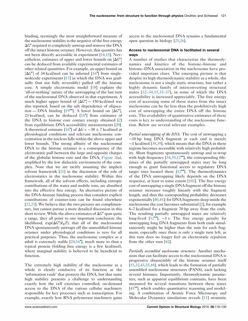
Figure 1
(a) Compaction of the DNA (chromatin) in eukaryotic cells is a complex hierarchy of various structures controlled by multiple modulating factors.
(b) The structure of the primary level of the DNA compaction — the nucleosome — is relatively well-defined. Various post-translational
modifications (PTM), such as acetylation of lysine residues, modulate the state of the nucleosome, including accessibility of its DNA. Shown are
4 examples of lysine acetylation sites, 1-4: H3K56, H4K91, H2BK5, H3K4. Positively charged N-terminal histone tails facilitate the condensation of
the net negatively charged nucleosomes into arrays. (c) Nucleosome arrays are likely represented by a variety of structural forms, depending on
the subtle interplay between several modulation factors. The arrays might switch between structures with different levels of compaction (top) or
the nucleosomes might occupy different sets of positions (bottom). (d) The state of chromatin affects vital processes such as gene expression and
cell differentiation; cell types (e.g. eye versus nose) can be different even though their DNA is identical. Deciphering this structure–function
connection in chromatin remains a fundamental problem in modern biology.
structure [7] of the nucleosome core particle, to which we
refer to as the nucleosome for simplicity, consists of
147 base pairs of DNA tightly wrapped �1.75 superheli-
cal turns around a roughly cylindrical protein core. The
core is an octamer made of two copies of each of the four
histone proteins H2A, H2B, H3, and H4. Chromatin
compaction at the nucleosome level (and also the next
level of nucleosome arrays, discussed further in this
review), is believed to be the most relevant to gene access
and recognition [8].
Connection to function through DNA accessibility
Increases in nucleosomal DNA accessibility as small as
1.5-fold can have significant biological consequences, for
example up to an order of magnitude increase in steady-
state transcript levels [9] and promoter activity [10];
importantly, these biological consequences of increased
DNA accessibility are not sequence-specific, that is the
effects appear to be the function of the increased DNA
accessibility per se. Thus, studying the DNA accessibility
in the nucleosome, and how it can be controlled, is of
critical importance for establishing structure–function
connections at this primary level of chromatin compac-
tion. Note that the very term ‘accessibility’ may have
different meanings depending on the context, for exam-
ple ‘solvent accessibility’ of a DNA base means that it can
make a steric contact with a nearby solvent (water)
Current Opinion in Structural Biology 2019, 56:119–130
molecule. For chromatin compaction at the nucleosome
level, one possible functionally relevant definition of
DNA accessibility is that the DNA fragment is accessible
if it is far enough from the histones so that a typical
nuclear factor such as PCNA can fit onto the DNA; in
quantitative terms that means at least �15 A distance
from the nearest histone atom [11]. By this definition, all
of the DNA in the X-ray structure of the nucleosome [7] is
inaccessible to protein complexes that perform, or initi-
ate, transcription, recombination, replication, and DNA
repair. However, structural fluctuations can make frag-
ments of the DNA spontaneously accessible. A strong
argument can be made [12��] in favor of the important role
of spontaneous DNA accessibility in gene regulation,
despite the ubiquitous activity of ATP-dependent remo-
deling enzymes that can use energy to expose DNA target
sites.
How stable is the nucleosome?
Spontaneous accessibility of nucleosomal DNA is directly
related to the strength of its association with the histone
core [13], so the first question one asks is how strong that
association is at physiological conditions, which is related
to the question of how stable is the nucleosome? As it
turns out, the question itself, and available answers to it,
are not as simple and unique as one may wish them to be.
By analogy with protein folding or protein–ligand
www.sciencedirect.com
The nucleosome: from structure to function through physics Onufriev and Schiessel 121
binding, seemingly the most straightforward measure of
the nucleosome stability is the negative of the free energy
DGu required to completely unwrap and remove the DNA
off the intact histone octamer. However, that quantity has
not been directly accessible in experiment [14,15]. Nev-
ertheless, estimates of upper and lower bounds on |DGu|
can be deduced from available experimental estimates of
other related quantities. For example, an upper bound on
|DGu| of 34 kcal/mol can be inferred [16�] from single-
molecule experiments [17] in which the DNA was grad-
ually (but not fully reversibly) pulled off the histone
core. A simple electrostatic model [18] explains the
‘all-or-nothing’ nature of the unwrapping of the last turn
of the nucleosomal DNA observed in that experiment. A
much higher upper bound of |DGu| � 150 kcal/mol was
also reported, based on the salt dependence of oligoca-
tion — DNA binding [19��]. A lower bound on |DGu|,
23 kcal/mol, can be deduced [16�] from estimates of
the DNA to histone core contact energy obtained [2]
from equilibrium DNA accessibility measurements [20].
A theoretical estimate [16�] of DG = �38 � 7 kcal/mol at
physiological conditions and relevant nucleosome con-
centration in the nucleus falls within the above upper and
lower bounds. The strong affinity of the nucleosomal
DNA to the histone octamer is a consequence of the
electrostatic pull between the large and opposite charges
of the globular histone core and the DNA, Figure 2(a),
amplified by the low dielectric environment of the com-
plex. Note that we are tacitly assuming the implicit
solvent framework [21] in the discussion of the role of
electrostatics in the nucleosome stability. Within this
framework, all of the solvent effects, including entropic
contributions of the water and mobile ions, are absorbed
into the effective free energy. An alternative picture of
the DNA–histone binding process that considers explicit
contributions of counter-ions can be found elsewhere
[22,23]. We believe that the two pictures are complimen-
tary, but cannot pursue a more detailed discussion in this
short review. While the above estimates of DGu span quite
a range, they all point to one important conclusion: the
likelihood, exp(DGu/kBT), that all of the nucleosomal
DNA spontaneously unwraps off the unmodified histone
octamer under physiological conditions is zero for all
practical purposes. Thus, the nucleosome complex as awhole is extremely stable [24,16�], much more so than a
typical protein (folding free energy is a few kcal/mol),
where marginal stability is believed to be beneficial to
function.
The extremely high stability of the nucleosome as a
whole is clearly conducive of its function as the
‘information vault’ that protects the DNA, but that same
high stability presents a challenge to understanding
exactly how the cell exercises controlled, on-demand
access to the DNA of the various cellular machinery
responsible for key processes such as transcription. For
example, exactly how RNA polymerase machinery gains
www.sciencedirect.com
access to the nucleosomal DNA remains a fundamental
open question in biology [25,26].
Access to nucleosomal DNA is facilitated in several
ways
A number of studies that characterize the thermody-
namics and kinetics of the histone–histone and
histone–DNA association in the nucleosome have pro-
vided important clues. The emerging picture is that
despite its high thermodynamic stability as a whole, the
nucleosome is not a single static structure, but rather a
highly dynamic family of interconverting structural
states [32–34,15,35–37], in some of which the DNA
accessibility is increased appreciably. The free energy
cost of accessing some of these states from the intact
nucleosome can be far less than the prohibitively high
cost of unwrapping the entire DNA off the histone
core. The availability of quantitative estimates of these
costs is key to understanding of the nucleosome func-
tion. Below are several relevant examples.
Partial unwrapping of the DNA. The cost of unwrapping a
�10 bp long DNA fragment at each end is merely
�1 kcal/mol [38,39], which means that the DNA in these
regions becomes accessible with relatively high probabil-
ity. Short fragments spontaneously unwrap and re-wrap
with high frequency [34,39,12��], the corresponding life-
times of the partially unwrapped states may be long
enough to grant functional access to regulatory DNA
target sites located there [12��]. The thermodynamics
of the DNA unwrapping likely depends on the DNA
sequence, at least to some extent [102]. The free energy
cost of unwrapping a single DNA fragment off the histone
octamer increases roughly linearly with the fragment
length, and thus the corresponding probability decreases
exponentially [40,41]; for DNA fragments deep inside the
nucleosome the cost becomes substantial [2], for example
6–7 kcal/mol for a fragment 70 bp away from the ends.
The resulting partially unwrapped states are relatively
long-lived [12��], �1 s. The free energy penalty for
unwrapping long DNA fragments from both ends simul-
taneously might be higher than the sum for each frag-
ment, especially once there is only a single turn left, as
this turn does no longer feel an electrostatic repulsion
from the other turn [42].
Partially assembled nucleosome structures. Another mecha-
nism that can facilitate access to the nucleosomal DNA is
progressive disassembly of the histone octamer itself
[15,32,43,35,44], which leads to the formation of partially
assembled nucleosome structures (PANS), each lacking
several histones. Importantly, thermodynamic parame-
ters, such as apparent equilibrium constants, have been
measured for several transitions between these states
[45��], which enables quantitative reasoning and model-
ing. A combination of Atomic Force Microscopy and
Molecular Dynamics simulations reveals [11] atomistic
Current Opinion in Structural Biology 2019, 56:119–130

122 Sequences and topology
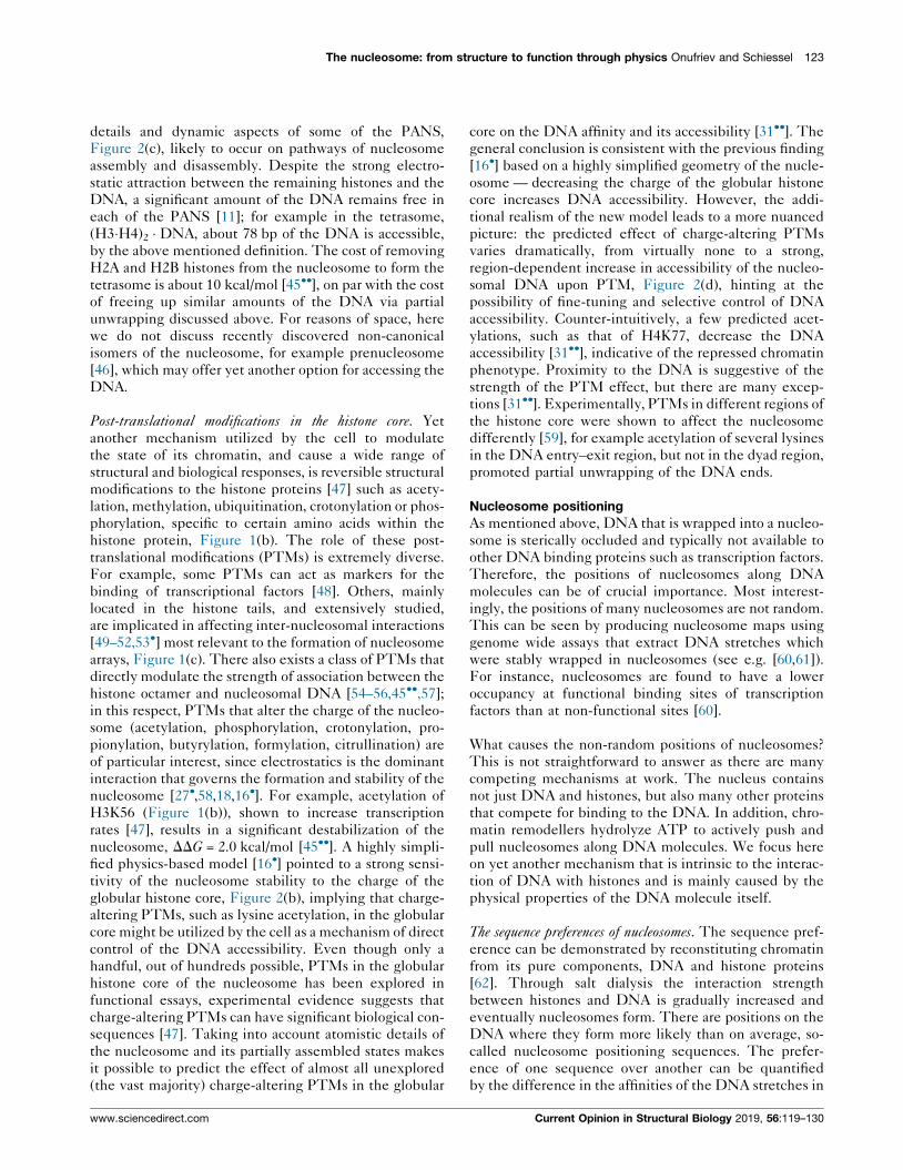
Figure 2
While the nucleosome as a whole is highly stable, access to its DNA can be facilitated in a number of ways. (a) The high stability of the
nucleosome stems mainly from the strong electrostatic attraction between the oppositely charged globular histone core (blue) and the DNA (red)
[27�,16�]. Contribution of the histone tails (green) to the over-all stability of the nucleosome is relatively small [28]; the tails affect partial
unwrapping of the DNA ends [29] and may have an effect on the nucleosome core structure [30]. (b) At physiological conditions, the state of the
nucleosome (red dot) is close to the phase boundary separating it from the ‘unwrapped’ states where the DNA is more accessible — a small drop
in the charge of the globular histone core can significantly lower nucleosome stability, and thus increase DNA accessibility [16�]. (c)
Conformational ensembles of partially assembled nucleosome structures (PANS) [11]: hexasome, (H2A�H2B) � (H3�H4)2 � DNA; tetrasome,
(H3�H4)2 � DNA; and disome, (H3�H4) � DNA. Significant portions of the DNA become accessible in PANS as a consequence of partial histone
removal from the nucleosome (2(H2A�H2B) � (H3�H4)2 � DNA). (d) Effect of all possible lysine acetylations in the globular histone core on the DNA
accessibility: while most acetylations are predicted to increase the accessibility, few (e.g. H4K77Ac) may have the opposite effect [31��].
Current Opinion in Structural Biology 2019, 56:119–130 www.sciencedirect.com
The nucleosome: from structure to function through physics Onufriev and Schiessel 123
details and dynamic aspects of some of the PANS,
Figure 2(c), likely to occur on pathways of nucleosome
assembly and disassembly. Despite the strong electro-
static attraction between the remaining histones and the
DNA, a significant amount of the DNA remains free in
each of the PANS [11]; for example in the tetrasome,
(H3�H4)2 � DNA, about 78 bp of the DNA is accessible,
by the above mentioned definition. The cost of removing
H2A and H2B histones from the nucleosome to form the
tetrasome is about 10 kcal/mol [45��], on par with the cost
of freeing up similar amounts of the DNA via partial
unwrapping discussed above. For reasons of space, here
we do not discuss recently discovered non-canonical
isomers of the nucleosome, for example prenucleosome
[46], which may offer yet another option for accessing the
DNA.
Post-translational modifications in the histone core. Yet
another mechanism utilized by the cell to modulate
the state of its chromatin, and cause a wide range of
structural and biological responses, is reversible structural
modifications to the histone proteins [47] such as acety-
lation, methylation, ubiquitination, crotonylation or phos-
phorylation, specific to certain amino acids within the
histone protein, Figure 1(b). The role of these post-
translational modifications (PTMs) is extremely diverse.
For example, some PTMs can act as markers for the
binding of transcriptional factors [48]. Others, mainly
located in the histone tails, and extensively studied,
are implicated in affecting inter-nucleosomal interactions
[49–52,53�] most relevant to the formation of nucleosome
arrays, Figure 1(c). There also exists a class of PTMs that
directly modulate the strength of association between the
histone octamer and nucleosomal DNA [54–56,45��,57];in this respect, PTMs that alter the charge of the nucleo-
some (acetylation, phosphorylation, crotonylation, pro-
pionylation, butyrylation, formylation, citrullination) are
of particular interest, since electrostatics is the dominant
interaction that governs the formation and stability of the
nucleosome [27�,58,18,16�]. For example, acetylation of
H3K56 (Figure 1(b)), shown to increase transcription
rates [47], results in a significant destabilization of the
nucleosome, DDG = 2.0 kcal/mol [45��]. A highly simpli-
fied physics-based model [16�] pointed to a strong sensi-
tivity of the nucleosome stability to the charge of the
globular histone core, Figure 2(b), implying that charge-
altering PTMs, such as lysine acetylation, in the globular
core might be utilized by the cell as a mechanism of direct
control of the DNA accessibility. Even though only a
handful, out of hundreds possible, PTMs in the globular
histone core of the nucleosome has been explored in
functional essays, experimental evidence suggests that
charge-altering PTMs can have significant biological con-
sequences [47]. Taking into account atomistic details of
the nucleosome and its partially assembled states makes
it possible to predict the effect of almost all unexplored
(the vast majority) charge-altering PTMs in the globular
www.sciencedirect.com
core on the DNA affinity and its accessibility [31��]. The
general conclusion is consistent with the previous finding
[16�] based on a highly simplified geometry of the nucle-
osome — decreasing the charge of the globular histone
core increases DNA accessibility. However, the addi-
tional realism of the new model leads to a more nuanced
picture: the predicted effect of charge-altering PTMs
varies dramatically, from virtually none to a strong,
region-dependent increase in accessibility of the nucleo-
somal DNA upon PTM, Figure 2(d), hinting at the
possibility of fine-tuning and selective control of DNA
accessibility. Counter-intuitively, a few predicted acet-
ylations, such as that of H4K77, decrease the DNA
accessibility [31��], indicative of the repressed chromatin
phenotype. Proximity to the DNA is suggestive of the
strength of the PTM effect, but there are many excep-
tions [31��]. Experimentally, PTMs in different regions of
the histone core were shown to affect the nucleosome
differently [59], for example acetylation of several lysines
in the DNA entry–exit region, but not in the dyad region,
promoted partial unwrapping of the DNA ends.
Nucleosome positioning
As mentioned above, DNA that is wrapped into a nucleo-
some is sterically occluded and typically not available to
other DNA binding proteins such as transcription factors.
Therefore, the positions of nucleosomes along DNA
molecules can be of crucial importance. Most interest-
ingly, the positions of many nucleosomes are not random.
This can be seen by producing nucleosome maps using
genome wide assays that extract DNA stretches which
were stably wrapped in nucleosomes (see e.g. [60,61]).
For instance, nucleosomes are found to have a lower
occupancy at functional binding sites of transcription
factors than at non-functional sites [60].
What causes the non-random positions of nucleosomes?
This is not straightforward to answer as there are many
competing mechanisms at work. The nucleus contains
not just DNA and histones, but also many other proteins
that compete for binding to the DNA. In addition, chro-
matin remodellers hydrolyze ATP to actively push and
pull nucleosomes along DNA molecules. We focus here
on yet another mechanism that is intrinsic to the interac-
tion of DNA with histones and is mainly caused by the
physical properties of the DNA molecule itself.
The sequence preferences of nucleosomes. The sequence pref-
erence can be demonstrated by reconstituting chromatin
from its pure components, DNA and histone proteins
[62]. Through salt dialysis the interaction strength
between histones and DNA is gradually increased and
eventually nucleosomes form. There are positions on the
DNA where they form more likely than on average, so-
called nucleosome positioning sequences. The prefer-
ence of one sequence over another can be quantified
by the difference in the affinities of the DNA stretches in
Current Opinion in Structural Biology 2019, 56:119–130

124 Sequences and topology
question to the histone octamer, allowing to determine
the relative free energies [14]. The sequence preference can
be substantial, and comparable to the effect of some
charge-altering PTMs: for example, the artificial ‘high
affinity’ sequence 601 (discussed in more detail further
below) has been reported to have a 2.89 kcal/mol lower
free energy than the strong natural positioning sequence
5S of the sea urchin [14]. It is, however, worthwhile to
mention that such affinity values have to be obtained
under identical experimental conditions. A more recent
study [45��] using a different approach reported a much
lower value of 0.7 kcal/mol.
When sequencing the stably wrapped DNA portions
(after digesting the rest with micrococcal nuclease) one
learns what types of base pair sequences cause higher-
than-average affinities to nucleosomes, namely sequences
where a larger than average number of particular base-pair
steps are at certain positions on the nucleosome, see
Figure 3 [60,63]. But what is precisely the mechanism
that causes these sequence preferences? Is it mainly
related to DNA mechanics and geometry or instead to
some specific interactions between nucleobases and his-
tones? A simple computational nucleosome model that
mainly accounts for the sequence dependent elasticity
and geometry of the DNA double helix does indeed
predict the sequence preferences of real nucleosomes
in vitro [64�], suggesting that the sequence dependent
nucleosome affinity mainly reflects the ease with which
DNA can be wrapped inside a nucleosome. We note,
however, that the first-order elasticity approach used in
this and many other studies to describe the strongly
distorted DNA states inside nucleosomes is under debate
as, for example discussed in Ref. [65].
The in vitro preferences carry over to some extent to
nucleosome positioning in vivo. For instance, the charac-
teristic dinucleotide preferences shown in Figure 3 were
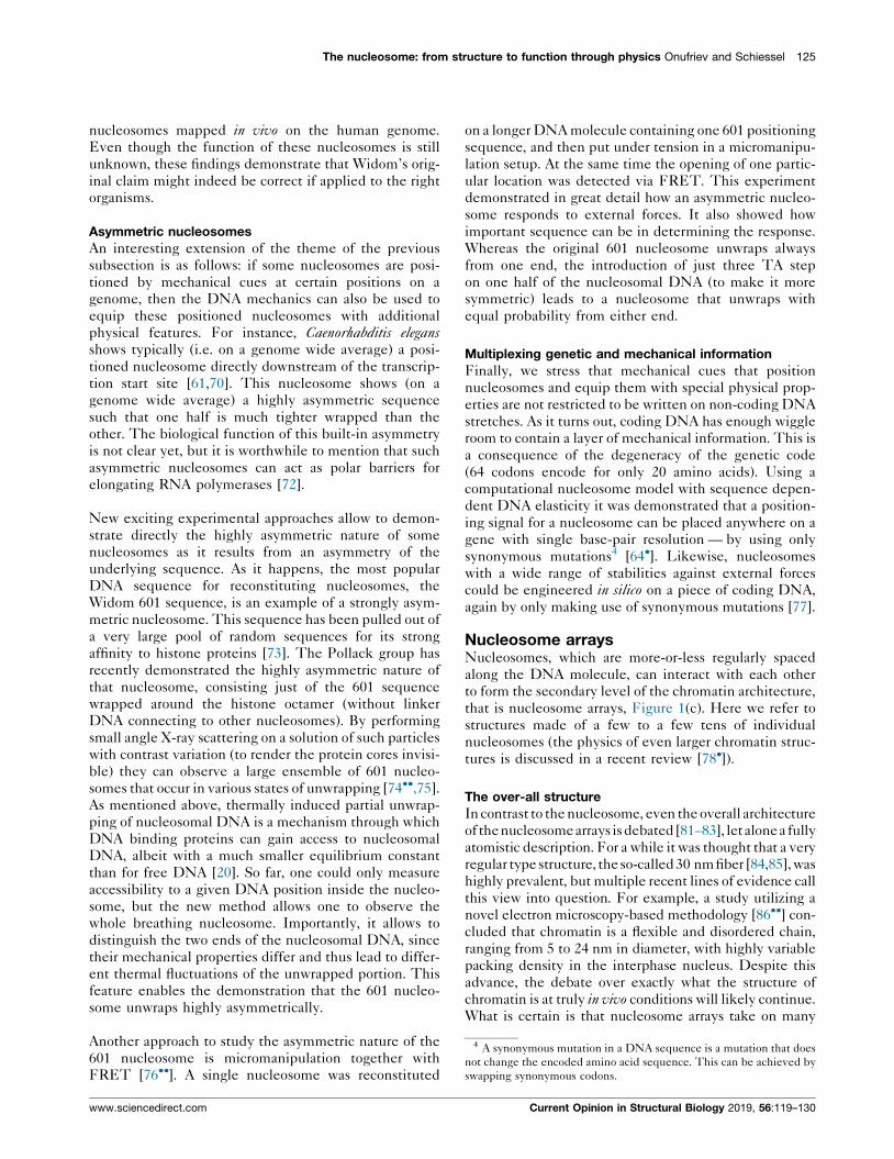
Figure 3
The nucleosome in vitro sequence preferences. GC steps (nucleotide
G followed by nucleotide C) are more likely to occur at positions
where the major groove faces the histone octamer (every 10th bp) and
TT, AA and TA steps at positions where the minor groove faces the
octamer [60,63].
Current Opinion in Structural Biology 2019, 56:119–130
already known to characterize stable nucleosomes
extracted from chicken [63]. Such observations led the
late Jonathan Widom and coworkers in 2006 [60] to
propose ‘a genomic code for nucleosome positioning’,
suggesting therefore that genomes have evolved to posi-
tion nucleosomes. Building a probabilistic model trained
on experimental nucleosome maps (of yeast or chicken)
they noticed that they could predict the positions of a
substantial (about 50%) fraction of nucleosomes in yeast.
However, these claims have led to a major debate that has
not subsided yet [66].
Yeast versus humans. It becomes increasingly clear that the
extent to which and the mechanisms by which sequence-
dependent DNA elasticity determines nucleosome posi-
tions in living organisms vary vastly between species. We
illustrate this by contrasting yeast [60,62,67] and recent
results from humans [68��] and other higher vertebrates
[69]. The nucleosome patterns around transcription start
sites in yeast suggest a non-random ordering of nucleo-
somes, especially when looking at the genome-wide
average. One can even count the nucleosomes that are
‘positioned’ as one moves into the gene as +1 nucleosome,
+2 nucleosome and so on [67]. But are these nucleosomes
really positioned by dedicated mechanical signals on the
DNA molecule?
As it turns out, yeast (and many other single-celled
organisms [70]) feature, just in front of transcription start
sites, regions characterized by a low content of G’s and
C’s and the presence of A-tracts. Such sequences have a
low affinity to nucleosomes and as a result act effectively
as barriers to nucleosomes. Nucleosomes nearby (e.g.
downstream of a transcription start site) are quite densely
crowded and form, on average, a statistical pattern as they
exclude each other. Such a statistical pattern close to a
boundary constraint (in the current context provided by a
stretch of stiff DNA repelling nucleosomes) has been
already suggested by Kornberg and Stryer [71] and this
mechanism might in fact be also largely responsible for
the nucleosome positioning in yeast, at least close to
transcription start sites. The claim in Ref. [60] that many
nucleosomes in yeast are positioned mainly by the DNA
sequence has therefore to be taken with a grain of salt, as
there is not much indication of dedicated local mechani-
cal signals to position individual nucleosomes.
In contrast, in humans and other higher vertebrates the
situation is rather different and much more in favor of the
idea of dedicated mechanical cues. Audit, Arneodo and
coworkers [68��,69] found well-positioned nucleosomes
located around so-called nucleosome inhibiting barriers
spread all over the genome of those organisms. The
nucleosomes around those barriers are not just statistically
ordered as in yeast, but instead they are positioned by
characteristic patterns of GC-rich and TA-rich regions.
These nucleosomes alone contain about 30% of the
www.sciencedirect.com
The nucleosome: from structure to function through physics Onufriev and Schiessel 125
4 A synonymous mutation in a DNA sequence is a mutation that does
not change the encoded amino acid sequence. This can be achieved by
swapping synonymous codons.
nucleosomes mapped in vivo on the human genome.
Even though the function of these nucleosomes is still
unknown, these findings demonstrate that Widom’s orig-
inal claim might indeed be correct if applied to the right
organisms.
Asymmetric nucleosomes
An interesting extension of the theme of the previous
subsection is as follows: if some nucleosomes are posi-
tioned by mechanical cues at certain positions on a
genome, then the DNA mechanics can also be used to
equip these positioned nucleosomes with additional
physical features. For instance, Caenorhabditis elegansshows typically (i.e. on a genome wide average) a posi-
tioned nucleosome directly downstream of the transcrip-
tion start site [61,70]. This nucleosome shows (on a
genome wide average) a highly asymmetric sequence
such that one half is much tighter wrapped than the
other. The biological function of this built-in asymmetry
is not clear yet, but it is worthwhile to mention that such
asymmetric nucleosomes can act as polar barriers for
elongating RNA polymerases [72].
New exciting experimental approaches allow to demon-
strate directly the highly asymmetric nature of some
nucleosomes as it results from an asymmetry of the
underlying sequence. As it happens, the most popular
DNA sequence for reconstituting nucleosomes, the
Widom 601 sequence, is an example of a strongly asym-
metric nucleosome. This sequence has been pulled out of
a very large pool of random sequences for its strong
affinity to histone proteins [73]. The Pollack group has
recently demonstrated the highly asymmetric nature of
that nucleosome, consisting just of the 601 sequence
wrapped around the histone octamer (without linker
DNA connecting to other nucleosomes). By performing
small angle X-ray scattering on a solution of such particles
with contrast variation (to render the protein cores invisi-
ble) they can observe a large ensemble of 601 nucleo-
somes that occur in various states of unwrapping [74��,75].As mentioned above, thermally induced partial unwrap-
ping of nucleosomal DNA is a mechanism through which
DNA binding proteins can gain access to nucleosomal
DNA, albeit with a much smaller equilibrium constant
than for free DNA [20]. So far, one could only measure
accessibility to a given DNA position inside the nucleo-
some, but the new method allows one to observe the
whole breathing nucleosome. Importantly, it allows to
distinguish the two ends of the nucleosomal DNA, since
their mechanical properties differ and thus lead to differ-
ent thermal fluctuations of the unwrapped portion. This
feature enables the demonstration that the 601 nucleo-
some unwraps highly asymmetrically.
Another approach to study the asymmetric nature of the
601 nucleosome is micromanipulation together with
FRET [76��]. A single nucleosome was reconstituted
www.sciencedirect.com
on a longer DNA molecule containing one 601 positioning
sequence, and then put under tension in a micromanipu-
lation setup. At the same time the opening of one partic-
ular location was detected via FRET. This experiment
demonstrated in great detail how an asymmetric nucleo-
some responds to external forces. It also showed how
important sequence can be in determining the response.
Whereas the original 601 nucleosome unwraps always
from one end, the introduction of just three TA step
on one half of the nucleosomal DNA (to make it more
symmetric) leads to a nucleosome that unwraps with
equal probability from either end.
Multiplexing genetic and mechanical information
Finally, we stress that mechanical cues that position
nucleosomes and equip them with special physical prop-
erties are not restricted to be written on non-coding DNA
stretches. As it turns out, coding DNA has enough wiggle
room to contain a layer of mechanical information. This is
a consequence of the degeneracy of the genetic code
(64 codons encode for only 20 amino acids). Using a
computational nucleosome model with sequence depen-
dent DNA elasticity it was demonstrated that a position-
ing signal for a nucleosome can be placed anywhere on a
gene with single base-pair resolution — by using only
synonymous mutations4 [64�]. Likewise, nucleosomes
with a wide range of stabilities against external forces
could be engineered in silico on a piece of coding DNA,
again by only making use of synonymous mutations [77].
Nucleosome arraysNucleosomes, which are more-or-less regularly spaced
along the DNA molecule, can interact with each other
to form the secondary level of the chromatin architecture,
that is nucleosome arrays, Figure 1(c). Here we refer to
structures made of a few to a few tens of individual
nucleosomes (the physics of even larger chromatin struc-
tures is discussed in a recent review [78�]).
The over-all structure
In contrast to the nucleosome, even the overall architecture
of thenucleosomearrays isdebated [81–83], letalonea fully
atomistic description. For a while it was thought that a very
regular type structure, theso-called 30 nm fiber [84,85], was
highly prevalent, but multiple recent lines of evidence call
this view into question. For example, a study utilizing a
novel electron microscopy-based methodology [86��] con-
cluded that chromatin is a flexible and disordered chain,
ranging from 5 to 24 nm in diameter, with highly variable
packing density in the interphase nucleus. Despite this
advance, the debate over exactly what the structure of
chromatin is at truly in vivo conditions will likely continue.
What is certain is that nucleosome arrays take on many
Current Opinion in Structural Biology 2019, 56:119–130

126 Sequences and topology
different, inter-converting structural forms [87,88], which
could be dependent on cell type and cell-cycle stage [89].
However, even in the absence of well-defined chromatin
structures, basic physical principles, physics-based simula-
tions and experiments contribute to the understanding of
which structures are likely to occur under certain condi-
tions, and how various biologically relevant modulating
factors [88] affect transitions between different states of
chromatin compaction, Figure 1. Several approaches exist
for making the structure-to-function connection at this
level [90], including a version [86��] of the DNA accessi-
bility argument.
Role of the tails
The positively charged terminal histone tails, Figure 2(a),
play a critical role in the formation of nucleosome array
structures [49,79,51]: the tails interact with the negatively
charged DNA, the neighboring nucleosomes, and linker
DNA. A long-standing unresolved question in the field is
whether a ‘histone code’ exists — that is whether each
specific combination of PTMs conveys a distinct func-
tional meaning, akin to the triplet genetic code of the
DNA. A recent computational work [80] suggests that, in
this respect, the effect of combined acetylations of H4 tail
may be more analogous to a rheostat rather than to a
‘binary code’: how many of the sites are acetylated maybe
more important than which specific ones. On the other
hand, certain acetylation sites, such as H4K16 discussed
below, are known to ‘code for’ strong and specific effects.
Thus, the true picture is likely more nuanced, possibly
including both cumulative non-specific and specific
features.
DNA condensation by oppositely charged particles
One fruitful physics-based approach to understanding
chromatin structure at the nucleosome array level is based
on the idea that the basic physics [91] that governs
condensation of the self-repelling DNA by oppositely
charged particles is universal, and therefore applies to
nucleosome arrays as well [19��,92]. The physics of
nucleic acid condensation by polyions is indeed relatively
well understood by now [91,19��,93–95]. In particular, the
majority of the DNA charge must be neutralized for the
remaining charge–charge repulsion to be weak enough for
the condensation to occur [91]. Since, in the case of the
nucleosome, the histones (including the tails) neutralize
only about 50% of the nucleosomal DNA, a significant
portion of the negative DNA charge must be neutralized
by other readily available positively charged entities
[19��], including Mg++, linker histones, protamines, basic
domains of the nuclear proteins, polyamines, etc. The
state of chromatin at physiological conditions appears to
be ‘nearly condensed’, close to the phase boundary sepa-
rating it from states of much looser compaction [19��].This ‘nearly condensed’ state of chromatin is maintained
by a tightly controlled balance between some of the
modulating factors: the amount of the core histones,
Current Opinion in Structural Biology 2019, 56:119–130
linker histones, and nucleosome repeat length [96,97].
Even minor alterations of the delicate charge balance,
such as acetylation of a single lysine (K16) on the H4
histone tail, may lead to chromatin de-compaction [98],
which, in turn, leads to transcription activation [99]. The
de-compacting effect on chromatin structure of reducing
the positive charge of the histone tails is consistent with
the general picture of DNA condensation governed by a
subtle interplay between charge–charge repulsion, ion-
ion correlations, and, in the case of the nucleosome arrays,
histone-tail bridging that facilitate formation of the
folded/aggregated structures [53�].
A nuanced picture. While the most general physical prin-
ciples behind chromatin condensation at the nucleosome
array level may be well understood, the detailed picture of
nucleosome array condensation/de-condensation is highly
nuanced. For example, the effect of charge-altering post-
translational modifications on the array compaction varies
widely, even within the same histone tail: the effect of
H4K16 acetylation on the array unfolding is much stron-
ger than that of H4K12, H4K8 or H4K5 [53�]. The specific
strong effect of H4K16 acetylation may be due to its role
of promoting tail-mediated nucleosome–nucleosome
stacking [53�]. Simulation reveals [49] that H4K16 is
the only acetylation site interacting with the acidic patch
on the neighboring nucleosome; its acetylation disrupts
the electrostatic interactions of K16 that favor array
compaction. And even that detailed picture may be more
nuanced still [100].
From the point of view of its function — providing on-
demand access to the genomic information — it makes
sense that condensed chromatin at physiological condi-
tions should be near the phase boundary separating the
condensed from the looser, less condensed states where
the DNA is easily accessible. Similar to the case of the
nucleosome reviewed above, Figure 2(b), the state of
chromatin condensation is then easy to control by small,
physiologically meaningful adjustments to relevant mod-
ulating factors.
ConclusionsIn this brief review we have offered an opinion that
physics-based methods, approaches and reasoning are
very useful tools in understanding the complexity of
chromatin structures, making structure–function connec-
tions, and generating experimentally verifiable predic-
tions. In this respect, of special interest are approaches
based on thermodynamics, classical electrostatics, and
physics-based simulations — well-established in the field
of traditional structural biology of proteins and DNA —
can also be quite useful in the emerging field of structure-
based epigenetics. For reasons of space, the examples we
chose to support our opinion are limited to the primary
(the nucleosome) and the secondary (nucleosome arrays)
level of the chromatin structural hierarchy.
www.sciencedirect.com
The nucleosome: from structure to function through physics Onufriev and Schiessel 127
A general picture that emerges is that the state of chro-
matin at physiologically relevant conditions is close to a
‘phase boundary’ separating compact, dense structures
where accessibility to genomic DNA is significantly
restricted, from looser structures with increased DNA
accessibility. Higher accessibility generally means
enhancement of processes that depend on it, such as
transcription. The closeness of chromatin to the
‘compact-loose’ phase boundary facilitates on-demand
fine-tuning of the DNA accessibility by the cell. In
modeling studies, bringing in more details, including
atomistic ones, allows for more detailed predictions, such
as the role of specific post-translation modifications of the
histone proteins or sequence effects of the wrapped DNA
on the stability of nucleosomes.
While evidence of success of physics-based approaches in
the field is growing, one also becomes aware of their
inherent limitations. Predictions of good models can be
expected to provide correct trends and guidance for
future experiments usefully above the Null model levels,
but one cannot expect in this field the spectacular level of
accuracy and reliability that physics delivers for the
hydrogen atom or planetary motion. Evolution, the Blind
Watchmaker, does not necessarily choose the most math-
ematically elegant or simple solutions so appealing to a
physicist — these can sometimes fail spectacularly when
checked against biological reality [101].
Conflict of interestNone.
Acknowledgements
AVO thanks Nikolay Korolev for many helpful suggestions, discussions, anda stimulating argument; and Andrew Fenley for commenting on themanuscript draft. Partial support from the NSF (MCB-1715207) to AVO isacknowledged.
References and recommended readingPapers of particular interest, published within the period of review,have been highlighted as:
� of special interest�� of outstanding interest
1. Henikoff S: Nucleosome destabilization in the epigeneticregulation of gene expression. Nat Rev Genet 2008, 9:15-26http://dx.doi.org/10.1038/nrg2206.
2. Garcia HG, Grayson P, Han L, Inamdar M, Kondev J, Nelson PCet al.: Biological consequences of tightly bent DNA: the otherlife of a macromolecular celebrity. Biopolymers 2007, 85:115-130 http://dx.doi.org/10.1002/bip.20627.
3. Tan L, Xing D, Chang CH, Li H, Xie XS: Three-dimensionalgenome structures of single diploid human cells. Science 2018,361:924-928 http://dx.doi.org/10.1126/science.aat5641.
4. Olins AL, Olins DE: Spheroid chromatin units (n bodies). Science1974, 183:330-332 http://dx.doi.org/10.1126/science.183.4122.330.
5. Woodcock C: Ultrastructure of inactive chromatin. J Cell Biol1973, 59:A368.
www.sciencedirect.com
6. Kornberg R: Chromatin structure: a repeating unit of histonesand DNA. Science 1974, 184:868-871 http://dx.doi.org/10.1126/science.184.4139.868.
7. Luger K, Mader AW, Richmond RK, Sargent DF, Richmond TJ:Crystal structure of the nucleosome core particle at 2.8 Aresolution. Nature 1997, 389:251-260.
8. Misteli T: Beyond the sequence: cellular organization ofgenome function. Cell 2007, 128:787-800 http://dx.doi.org/10.1016/j.cell.2007.01.028.
9. Zhu Z, Thiele DJ: A specialized nucleosome modulatestranscription factor access to a C. glabrata metal responsivepromoter. Cell 1996, 87:459-470 http://view.ncbi.nlm.nih.gov/pubmed/8898199.
10. Raveh-Sadka T, Levo M, Shabi U, Shany B, Keren L, Lotan-Pompan M et al.: Manipulating nucleosome disfavoringsequences allows fine-tune regulation of gene expression inyeast. Nat Genet 2012, 44:743-750 http://dx.doi.org/10.1038/ng.2305.
11. Rychkov GN, Ilatovskiy AV, Nazarov IB, Shvetsov AV, Lebedev DV,Konev AY et al.: Partially assembled nucleosome structures atatomic detail. Biophys J 2017, 112:460-472 http://view.ncbi.nlm.nih.gov/pubmed/28038734.
12.��
Tims H, Gurunathan K, Levitus M, Widom J: Dynamics ofnucleosome invasion by DNA binding proteins. J Mol Biol 2011,411:430-448 http://dx.doi.org/10.1016/j.jmb.2011.05.044.
Two independent complementary experimental approaches are used tomeasure the rates of nucleosome spontaneous unwrapping and re-wrapping for differing DNA sites from the end of the nucleosomal DNAinward toward the middle. This detailed study compliments and extendsearlier works from the same group. A compelling argument is made infavor of the important role of spontaneous DNA accessibility in generegulation.
13. Anderson JD, Widom J: Poly(dA–dT) promoter elementsincrease the equilibrium accessibility of nucleosomal DNAtarget sites. Mol Cell Biol 2001, 21:3830-3839 http://dx.doi.org/10.1128/mcb.21.11.3830-3839.2001.
14. Thastrom A, Gottesfeld JM, Luger K, Widom J: Histone–DNAbinding free energy cannot be measured in dilution-drivendissociation experiments. Biochemistry 2004, 43:736-741.
15. Andrews AJ, Luger K: Nucleosome structure(s) and stability:variations on a theme. Annu Rev Biophys 2011, 40:99-117.
16.�
Fenley AT, Adams DA, Onufriev AV: Charge state of the globularhistone core controls stability of the nucleosome. Biophys J2010, 99:1577-1585.
An electrostatics model of the nucleosome based on simplified geometrysuggests a resolution of the ‘high stability versus easy accessibility’challenge. The key finding is that the strength of the histone–DNAassociation is highly sensitive to the charge of the globular histone core,suggesting a possible role of charge-altering PTMs in the core for the DNAaccessibility control.
17. Brower-Toland BD, Smith CL, Yeh RC, Lis JT, Peterson CL,Wang MD: Mechanical disruption of individual nucleosomesreveals a reversible multistage release of DNA. Proc Natl AcadSci U S A 2002, 99:1960-1965.
18. Korolev N, Lyubartsev AP, Laaksonen A: Electrostaticbackground of chromatin fiber stretching. J Biomol Struct Dyn2004, 22:215-226 http://dx.doi.org/10.1080/07391102.2004.10506997.
19.��
Korolev N, Berezhnoy NV, Eom KD, Tam JP, Nordenskiold L: Auniversal description for the experimental behavior of salt-(in)dependent oligocation-induced DNA condensation. NucleicAcids Res 2009, 37:7137-7150 http://dx.doi.org/10.1093/nar/gkp683.
A systematic analysis of condensation of plasmid DNA by oligocationswith variation of the charge, from +3 to +31. On the basis of the analysis,the authors suggest that the conditions in the nucleus are such that thestate of chromatin is very close to the borderline separating the extendedand collapsed phases.
20. Polach KJ, Widom J: Mechanism of protein access to specificDNA sequences in chromatin: a dynamic equilibrium model forgene regulation. J Mol Biol 1995, 254:130-149.
Current Opinion in Structural Biology 2019, 56:119–130
128 Sequences and topology
21. Roux B, Simonson T: Implicit solvent models. Biophys Chem1999, 78:1-20.
22. Iwaki T, Saito T, Yoshikawa K: How are small ions involved in thecompaction of DNA molecules? Colloids Surf B: Biointerfaces2007, 56:126-133 http://view.ncbi.nlm.nih.gov/pubmed/17254757.
23. Korolev N, Lyubartsev AP, Nordenskiold L: Cation-inducedpolyelectrolyte–polyelectrolyte attraction in solutions of DNAand nucleosome core particles. Adv Colloid Interface Sci 2010,158:32-47.
24. Korolev N, Vorontsova OV, Nordenskiold L: Physicochemicalanalysis of electrostatic foundation for DNA–proteininteractions in chromatin transformations. Prog Biophys MolBiol 2007, 95:23-49 http://view.ncbi.nlm.nih.gov/pubmed/17291569.
25. Teves ST, Weber CM, Henikoff S: Transcribing through thenucleosome. Trends Biochem Sci 2014, 39:577-586 http://dx.doi.org/10.1016/j.tibs.2014.10.004.
26. Kulaeva O, Hsieh FK, Chang HW, Luse D, Studitsky VM:Mechanism of transcription through a nucleosome by RNApolymerase II. Biochim Biophys Acta 2013, 1829:76-83 http://dx.doi.org/10.1016/j.bbagrm.2012.08.015.
27.�
Kunze KK, Netz RR: Complexes of semiflexible polyelectrolytesand charged spheres as models for salt-modulatednucleosomal structures. Phys Rev E: Stat Nonlinear Soft MatterPhys 2002, 66:011918 http://view.ncbi.nlm.nih.gov/pubmed/12241395.
A highly simplified electrostatic model of the nucleosome is explored indetail, including the salt dependence of the resulting complex structure,the influence of externally applied forces, and DNA length variation.
28. Gottesfeld JM, Luger K: Energetics and affinity of the histoneoctamer for defined DNA sequences. Biochemistry 2001,40:10927-10933 http://view.ncbi.nlm.nih.gov/pubmed/11551187.
29. Andresen K, Jimenez-Useche I, Howell SC, Yuan C, Qiu X:Solution scattering and FRET studies on nucleosomes revealDNA unwrapping effects of H3 and H4 tail removal. PLOS ONE2013, 8:e78587 http://view.ncbi.nlm.nih.gov/pubmed/24265699.
30. Biswas M, Voltz K, Smith JC, Langowski J: Role of histone tails instructural stability of the nucleosome. PLoS Comput Biol 2011,7:e1002279 http://dx.doi.org/10.1371/journal.pcbi.1002279.
31.��
Fenley AT, Anandakrishnan R, Kidane YH, Onufriev AV:Modulation of nucleosomal DNA accessibility via charge-altering post-translational modifications in histone core.Epigenet Chromatin 2018, 11:11 http://dx.doi.org/10.1186/s13072-018-0181-5.
An atomically detailed electrostatic model predicts the effect of nearly allpossible charge-altering PTMs in the histone core on the DNA accessi-bility, making a connection to resulting biological phenotypes. The frame-work is validated against experimentally known nucleosome stabilitychanges due to the acetylation of specific lysines. The effects of individualPTMs are classified based on changes in the accessibility of variousregions throughout the nucleosomal DNA. The PTM’s resulting imprint onthe DNA accessibility, ‘PTMprint’, is used to predict effects of many yetunexplored PTMs.
32. Luger K, Dechassa ML, Tremethick DJ: New insights intonucleosome and chromatin structure: an ordered state or adisordered affair? Nat Rev Mol Cell Biol 2012, 13:436-447 http://dx.doi.org/10.1038/nrm3382.
33. Shaytan A, Armeev G, Goncearenco A, Zhurkin VB, Landsman D,Panchenko AR: Coupling between histone conformations andDNA geometry in nucleosomes on a microsecond timescale:atomistic insights into nucleosome functions. J Mol Biol 2016,428:221-237 http://dx.doi.org/10.1016/j.jmb.2015.12.004.
34. Gansen A, Valeri A, Hauger F, Felekyan S, Kalinin S, Toth K et al.:Nucleosome disassembly intermediates characterized bysingle-molecule FRET. Proc Natl Acad Sci U S A 2009,106:15308-15313 http://dx.doi.org/10.1073/pnas.0903005106.
35. Zlatanova J, Bishop TC, Victor JM, Jackson V, van Holde K: Thenucleosome family: dynamic and growing. Structure (London,England: 1993) 2009, 17:160-171 http://dx.doi.org/10.1016/j.str.2008.12.016.
Current Opinion in Structural Biology 2019, 56:119–130
36. Bohm V, Hieb AR, Andrews AJ, Gansen A, Rocker A, Toth K et al.:Nucleosome accessibility governed by the dimer/tetramerinterface. Nucleic Acids Res 2011, 39:3093-3102 http://dx.doi.org/10.1093/nar/gkq1279.
37. Chen Y, Tokuda J, Topping T, Sutton J, Meisburger S, Pabit Set al.: Revealing transient structures of nucleosomes as DNAunwinds. Nucleic Acids Res 2014, 42:8767-8776 http://dx.doi.org/10.1093/nar/gku562.
38. Wei S, Falk SJ, Black BE, Lee THH: A novel hybrid singlemolecule approach reveals spontaneous DNA motion in thenucleosome. Nucleic Acids Res 2015, 43:e111 http://view.ncbi.nlm.nih.gov/pubmed/26013809.
39. Koopmans WJA, Buning R, Schmidt T, van Noort J: spFRET usingalternating excitation and FCS reveals progressive DNAunwrapping in nucleosomes. Biophys J 2009, 97:195-204 http://dx.doi.org/10.1016/j.bpj.2009.04.030.
40. Blossey R, Schiessel H: The dynamics of the nucleosome:thermal effects, external forces and ATP. FEBS J 2011,278:3619-3632 http://view.ncbi.nlm.nih.gov/pubmed/21812931.
41. Culkin J, de Bruin L, Tompitak M, Phillips R, Schiessel H: The roleof DNA sequence in nucleosome breathing. Eur Phys J E 2017,40:106 http://dx.doi.org/10.1140/epje/i2017-11596-2.
42. Kuli�c IM, Schiessel H: DNA spools under tension. Phys Rev Lett2004, 92:228101 http://dx.doi.org/10.1103/PhysRevLett.92.228101.
43. Hutcheon T, Dixon G, Levy-Wilson B: Transcriptionally activemononucleosomes from trout testis are heterogeneous incomposition. J Biol Chem 1980, 255:681-685.
44. Kato D, Osakabe A, Arimura Y, Mizukami Y, Horikoshi N, Saikusa Ket al.: Crystal structure of the overlapping dinucleosomecomposed of hexasome and octasome. Science 2017, 356:205-208 http://dx.doi.org/10.1126/science.aak9867.
45.��
Andrews AJ, Chen X, Zevin A, Stargell LA, Luger K: The histonechaperone Nap1 promotes nucleosome assembly byeliminating nonnucleosomal histone DNA interactions. MolCell 2010, 37:834-842 http://dx.doi.org/10.1016/j.molcel.2010.01.037.
A thermodynamic assay is developed to probe transitions betweenvarious partially assembled nucleosome states in vitro. Together with apreviously published work from the same group, the paper presents afairly comprehensive picture of transitions between these states, andprovides equilibrium constants. The mechanism of action of the histonechaperone nucleosome assembly protein Nap1 is revealed.
46. Fei J, Torigoe SE, Brown CR, Khuong MT, Kassavetis GA,Boeger H et al.: The prenucleosome, a stable conformationalisomer of the nucleosome. Genes Dev 2015, 29:2563-2575http://view.ncbi.nlm.nih.gov/pubmed/26680301.
47. Tessarz P, Kouzarides T: Histone core modifications regulatingnucleosome structure and dynamics. Nat Rev Mol Cell Biol2014, 15:703-708 http://dx.doi.org/10.1038/nrm3890.
48. Berger SL: The complex language of chromatin regulationduring transcription. Nature 2007, 447:407-412 http://dx.doi.org/10.1038/nature05915.
49. Zhang R, Erler J, Langowski J: Histone acetylation regulateschromatin accessibility: role of H4K16 in inter-nucleosomeinteraction. Biophys J 2017, 112:450-459 http://dx.doi.org/10.1016/j.bpj.2016.11.015.
50. Arya G, Schlick T: Role of histone tails in chromatin foldingrevealed by a mesoscopic oligonucleosome model. Proc NatlAcad Sci U S A 2006, 103:16236-16241 http://dx.doi.org/10.1073/pnas.0604817103.
51. Collepardo-Guevara R, Portella G, Vendruscolo M, Frenkel D,Schlick T, Orozco M: Chromatin unfolding by epigeneticmodifications explained by dramatic impairment ofinternucleosome interactions: a multiscale computationalstudy. J Am Chem Soc 2015, 137:10205-10215 http://dx.doi.org/10.1021/jacs.5b04086.
52. Zhou J, Fan JY, Rangasamy D, Tremethick DJ: The nucleosomesurface regulates chromatin compaction and couples it with
www.sciencedirect.com
The nucleosome: from structure to function through physics Onufriev and Schiessel 129
transcriptional repression. Nat Struct Mol Biol 2007, 14:1070-1076 http://dx.doi.org/10.1038/nsmb1323.
53.�
Allahverdi A, Yang R, Korolev N, Fan Y, Davey CA, Liu CFF et al.:The effects of histone H4 tail acetylations on cation-inducedchromatin folding and self-association. Nucleic Acids Res2011, 39:1680-1691 http://dx.doi.org/10.1093/nar/gkq900.
A systematic experimental investigation of a 12-nucleosome arrayscontaining various combinations of completely acetylated lysines atpositions 5, 8, 12 and 16 of histone H4. The effect of acetylation of H4on the array compaction is strong, and is not mimicked by chargeneutralization via K ! Q mutation; a non-electrostatic mechanism forthe highly specific effect is proposed.
54. Manohar M, Mooney AM, North JA, Nakkula RJ, Picking JW,Edon A et al.: Acetylation of histone H3 at the nucleosome dyadalters DNA–histone binding. J Biol Chem 2009, 284:23312-23321 http://dx.doi.org/10.1074/jbc.m109.003202.
55. Bowman GD, Poirier MG: Post-translational modifications ofhistones that influence nucleosome dynamics. Chem Rev2015, 115:2274-2295 http://dx.doi.org/10.1021/cr500350x.
56. Brehove M, Wang T, North J, Luo Y, Dreher SJ, Shimko JC et al.:Histone core phosphorylation regulates DNA accessibility. JBiol Chem 2015, 290:22612-22621 http://dx.doi.org/10.1074/jbc.m115.661363.
57. Materese CK, Savelyev A, Papoian G: Counterion atmosphereand hydration patterns near a nucleosome core particle. J AmChem Soc 2009, 131:15005-15013 http://dx.doi.org/10.1021/ja905376q.
58. Manning GS: Is a small number of charge neutralizationssufficient to bend nucleosome core DNA onto its superhelicalramp? J Am Chem Soc 2003, 125:15087-15092 http://view.ncbi.nlm.nih.gov/pubmed/14653743.
59. Simon M, North JA, Shimko JC, Forties RA, Ferdinand MB,Manohar M et al.: Histone fold modifications controlnucleosome unwrapping and disassembly. Proc Natl Acad SciU S A 2011, 108:12711-12716 http://dx.doi.org/10.1073/pnas.1106264108.
60. Segal E, Fondufe-Mittendorf Y, Chen L, Thastrom A, Field Y,Moore IK et al.: A genomic code for nucleosome positioning.Nature 2006, 442:772-778 http://dx.doi.org/10.1038/nature04979.
61. Ercan S, Lubling Y, Segal E, Lieb JD: High nucleosomeoccupancy is encoded at x-linked gene promoters in C.elegans. Genome Res 2011, 21:237-244 http://dx.doi.org/10.1101/gr.115931.110.
62. Kaplan N, Moore IK, Fondufe-Mittendorf Y, Gossett AJ, Tillo D,Field Y et al.: The DNA-encoded nucleosome organization of aeukaryotic genome. Nature 2009, 458:362-366 http://dx.doi.org/10.1038/nature07667.
63. Satchwell SC, Drew HR, Travers AA: Sequence periodicities inchicken nucleosome core DNA. J Mol Biol 1986, 191:659-675.
64.�
Eslami-Mossallam B, Schram RD, Tompitak M, van Noort J,Schiessel H: Multiplexing genetic and nucleosome positioningcodes: a computational approach. PLOS ONE 2016, 11:e0156905 http://dx.doi.org/10.1371/journal.pone.0156905.
A computer simulation of a coarse grained nucleosome model withsequence dependent DNA elasticity. The model predicts the well-knownsequence preferences of nucleosomes and is used to demonstratemultiplexing of classical genetic and mechanical information.
65. Zhurkin VB, Olson WK: Can nucleosomal DNA be described byan elastic model? Phys Life Rev 2013, 10:70-72 http://dx.doi.org/10.1016/j.plrev.2013.01.009.
66. Zhang Y, Moqtaderi Z, Rattner BP, Euskirchen G, Snyder M,Kadonaga JT et al.: Evidence against a genomic code fornucleosome positioning. Nat Struct Mol Biol 2010, 17:920-923.
67. Brogaard K, Xi L, Wang JP, Widom J: A map of nucleosomepositions in yeast at base-pair resolution. Nature 2012,486:496-501 http://dx.doi.org/10.1038/nature11142.
68.��
Drillon G, Audit B, Argoul F, Arneodo A: Evidence of selection foran accessible nucleosomal array in human. BMC Genomics2016, 17:526 http://dx.doi.org/10.1186/s12864-016-2880-2.
www.sciencedirect.com
On the basis of a physical model for nucleosome formation the authorspredict 1.6 million nucleosome inhibiting barriers in the human genome.Around these barriers are nucleosomes positioned by mechanical signalsin the DNA molecules. It is speculated that these motifs are selected forimpairing the condensation of nucleosomal arrays.
69. Brunet FG, Audit B, Drillon G, Argoul F, Volff JN, Arneodo A:Evidence for DNA sequence encoding of an accessiblenucleosomal array across vertebrates. Biophys J 2018,114:2308-2316 http://dx.doi.org/10.1186/s12864-016-2880-2.
70. Tompitak M, Vaillant C, Schiessel H: Genomes of multicellularorganisms have evolved to attract nucleosomes to promoterregions. Biophys J 2017, 112:505-511 http://dx.doi.org/10.1016/j.bpj.2016.12.041.
71. Kornberg RD, Stryer L: Statistical distributions of nucleosomes:nonrandom locations by a stochastic mechanism. NucleicAcids Res 1988, 16:6677-6690.
72. Bondarenko VA, Steele LM, Ujvari A, Gaykalova DA, Kulaeva OI,Polikanov YS et al.: Nucleosomes can form a polar barrier totranscript elongation by RNA polymerase II. Mol Cell 2006,24:469-479 http://dx.doi.org/10.1016/j.molcel.2006.09.009.
73. Lowary PT, Widom J: New DNA sequence rules for high affinitybinding to histone octamer and sequence-directednucleosome positioning. J Mol Biol 1998, 276:19-42.
74.��
Mauney AW, Tokuda JM, Gloss LM, Gonzalez O, Pollack L: LocalDNA sequence controls asymmetry of DNA unwrapping fromnucleosome core particles. Biophys J 2018, 115:773-781 http://dx.doi.org/10.1016/j.bpj.2018.07.009.
Small angle X-ray scattering with contrast variation on a solution ofnucleosomes demonstrates the highly asymmetric nature of the601 nucleosome. Especially remarkable is the fact that the authorscan distinguish the two ends of the nucleosomal DNA based on theirthermal fluctuations as they partially unwrap from the nucleosomes.
75. Schiessel H: Telling left from right in breathing nucleosomes.Biophys J 2018, 115:749-750 http://dx.doi.org/10.1016/j.bpj.2018.07.026.
76.��
Ngo TTM, Zhang Q, Zhou R, Yodh JG, Ha T: Asymmetricunwrapping of nucleosomes under tension directed by DNAlocal flexibility. Cell 2015, 160:1135-1144 http://dx.doi.org/10.1016/j.cell.2015.02.001.
Combining micromanipulation and FRET measurements this paperreports on the force-induced unwrapping of the 601 nucleosome inunprecedented detail.
77. Tompitak M, de Bruin L, Eslami-Mossallam B, Schiessel H:Designing nucleosomal force sensors. Phys Rev E 2017,95:052402 http://dx.doi.org/10.1103/PhysRevE.95.052402.
78.�
Sazer S, Schiessel H: The biology and polymer physicsunderlying large-scale chromosome organization. Traffic 2018,19:87-104 http://dx.doi.org/10.1111/tra.12539.
A review on older and more recent experimental discoveries on the largescale chromatin structure and how they have been interpreted in terms ofpolymer physics.
79. Korolev N, Lyubartsev AP, Nordenskiold L: A systematic analysisof nucleosome core particle and nucleosome–nucleosomestacking structure. Sci Rep 2018, 8:1543 http://dx.doi.org/10.1038/s41598-018-19875-0.
80. Winogradoff D, Echeverria I, Potoyan DA, Papoian GA: Theacetylation landscape of the H4 histone tail: disentangling theinterplay between the specific and cumulative effects. J AmChem Soc 2015, 137:6245-6253 http://dx.doi.org/10.1021/jacs.5b00235.
81. Nishino Y, Eltsov M, Joti Y, Ito K, Takata H, Takahashi Y et al.:Human mitotic chromosomes consist predominantly ofirregularly folded nucleosome fibres without a 30-nmchromatin structure. EMBO J 2012, 31:1644-1653 http://dx.doi.org/10.1038/emboj.2012.35.
82. van Holde K, Zlatanova J: Chromatin fiber structure: where isthe problem now? Semin Cell Dev Biol 2007, 18:651-658 http://dx.doi.org/10.1016/j.semcdb.2007.08.005.
83. Li G, Reinberg D: Chromatin higher-order structures and generegulation. Curr Opin Genet Dev 2011, 21:175-186 http://dx.doi.org/10.1016/j.gde.2011.01.022.
Current Opinion in Structural Biology 2019, 56:119–130
130 Sequences and topology
84. Wong H, Victor JM, Mozziconacci J: An all-atom model of thechromatin fiber containing linker histones reveals a versatilestructure tuned by the nucleosomal repeat length. PLoS ONE2007, 2:e877.
85. Depken M, Schiessel H: Nucleosome shape dictates chromatinfiber structure. Biophys J 2009, 96:777-784 http://dx.doi.org/10.1016/j.bpj.2008.09.055.
86.��
Ou HD, Phan S, Deerinck TJ, Thor A, Ellisman MH, O’Shea CC:ChromEMT: visualizing 3D chromatin structure andcompaction in interphase and mitotic cells. Science (New York,NY) 2017, 357:eaag0025 http://dx.doi.org/10.1126/science.aag0025 eaag0025.
An experimental method (ChromEMT) is developed to visualize chroma-tin in situ. Chromatin is seen as a disordered 5-nm to 24-nm-diametercurvilinear chain that is packed together at different 3D concentrations ininterphase and mitosis. The authors suggest the possibility that the 3Dconcentration of chromatin in the nucleus might be a simple and universalself-organizing principle that determines the functional activity andaccessibility of genomic DNA.
87. Boule JB, Mozziconacci J, Lavelle C: The polymorphisms of thechromatin fiber. J Phys Condens Matter 2015, 27:033101 http://dx.doi.org/10.1088/0953-8984/27/3/033101.
88. Collepardo-Guevara R, Schlick T: Chromatin fiberpolymorphism triggered by variations of DNA linker lengths.Proc Natl Acad Sci U S A 2014, 111:8061-8066 http://dx.doi.org/10.1073/pnas.1315872111.
89. McGinty RK, Tan S: Nucleosome structure and function. ChemRev 2015, 115:2255-2273 http://dx.doi.org/10.1021/cr500373h.
90. Bascom G, Schlick T: Linking chromatin fibers to gene foldingby hierarchical looping. Biophys J 2017, 112:434-445 http://dx.doi.org/10.1016/j.bpj.2017.01.003.
91. Bloomfield VA: DNA condensation. Curr Opin Struct Biol 1996,6:334-341 http://dx.doi.org/10.1016/S0959-440X(96)80052-2.
92. Clark DJ, Kimura T: Electrostatic mechanism of chromatinfolding. J Mol Biol 1990, 211:883-896 http://view.ncbi.nlm.nih.gov/pubmed/2313700.
Current Opinion in Structural Biology 2019, 56:119–130
93. DeRouchey J, Parsegian VA, Rau DC: Cation chargedependence of the forces driving DNA assembly. Biophys J2010, 99:2608-2615 http://dx.doi.org/10.1016/j.bpj.2010.08.028.
94. Kornyshev A, Leikin S: Electrostatic interaction between helicalmacromolecules in dense aggregates: an impetus for DNApoly- and mesomorphism. Proc Natl Acad Sci U S A 1998,95:13579-13584 http://dx.doi.org/10.1073/pnas.95.23.13579.
95. Tolokh IS, Pabit SA, Katz AM, Chen Y, Drozdetski A, Baker N et al.:Why double-stranded RNA resists condensation. Nucleic AcidsRes 2014, 42:10823-10831 PMCID: PMC25123663.
96. Woodcock CL, Skoultchi AI, Fan Y: Role of linker histone inchromatin structure and function: H1 stoichiometry andnucleosome repeat length. Chromosome Res 2006, 14:17-25http://view.ncbi.nlm.nih.gov/pubmed/16506093.
97. Cherstvy AG, Teif VB: Electrostatic effect of H1-histone proteinbinding on nucleosome repeat length. Phys Biol 2014,11:044001 http://stacks.iop.org/1478-3975/11/i=4/a=044001.
98. Shogren-Knaak M, Ishii H, Sun JMM, Pazin MJ, Davie JR,Peterson CL: Histone H4-K16 acetylation controls chromatinstructure and protein interactions. Science (New York, NY)2006, 311:844-847 http://dx.doi.org/10.1126/science.1124000.
99. Shia WJ, Pattenden SG, Workman JL: Histone H4 lysine16 acetylation breaks the genome’s silence. Genome Biol 2006,7:217.
100. Chen Q, Yang R, Korolev N, Liu CFF, Nordenskiold L: Regulationof nucleosome stacking and chromatin compaction by thehistone H4 N-terminal tail-H2A acidic patch interaction. J MolBiol 2017, 429:2075-2092 http://view.ncbi.nlm.nih.gov/pubmed/28322915.
101. Crick FHC, Griffith JS, Orgel LE: Codes without commas. ProcNatl Acad Sci U S A 1957, 43:416-421 http://view.ncbi.nlm.nih.gov/pubmed/16590032.
102. Winogradoff D, Aksimentiev A: Molecular mechanism ofspontaneous nucleosome unraveling. J Mol Biol 2019, 431:323-335 http://dx.doi.org/10.1016/j.jmb.2018.11.013.
www.sciencedirect.com