Embed Size (px)
Citation preview

The Neuropsychopharmacology and Toxicologyof 3,4-methylenedioxy-N-ethyl-amphetamine (MDEA)
Roland W. Freudenmann and Manfred Spitzer
Department of Psychiatry, University of Ulm, Germany
Keywords: Amphetamine derivatives — Amphetamine toxicity — Ecstasy — Eve — MDE —
MDEA — MDMA — RSA — Street drugs.
ABSTRACT
This paper reviews the pharmacology and toxicology of 3,4-methylenedioxy-N-ethyl-
amphetamine (MDEA, “eve”). MDEA is a ring-substituted amphetamine (RSA) like
MDMA, its well known N-methyl analog. Both have become very popular substances of
abuse in the techno- and house-music scene. They can evoke psychomotor stimulation,
mild alterations of perception, sensations of closeness and a positive emotional state as
well as sympathomimetic physical effects. At present, the name “ecstasy” is no longer
used only for MDMA, but for the whole group of RSAs (MDA, MDMA, MDEA and
MBDB) as they are chemically and pharmacologically nearly identical; moreover, many
ecstasy pills contain mixtures of the RSAs. Hence, for a selective review on MDEA, it is
crucial to strictly differentiate between: 1) street and chemical names, and 2) studies with
or without chemically defined substances. In order to present MDEA-specific infor-
mation, the pharmacodynamics and kinetics are described on the basis of MDEA chal-
lenge studies in animals and humans. In the toxicology section, we present a collection of
case reports on fatalities where MDEA was toxicologically confirmed. On the question of
serotonergic neurotoxicity and possible long-term consequences, however, MDEA-spe-
cific information is available from animal studies only. The neurotoxic potential of MDEA
in humans is difficult to estimate, as ecstasy users do not consume pure substances. For
future research, challenge studies in animals using dosing regimens adapted to human
consumption patterns are needed. Such challenge studies should directly compare indi-
vidual RSAs. They will represent the most viable and fruitful approach to the resolution
of the highly controversial issues of serotonergic neurotoxicity and its functional
consequences.
89
CNS Drug ReviewsVol. 10, No. 2, pp. 89–116© 2004 Neva Press, Branford, Connecticut
Address correspondence and reprint requests to Dr. R. Freudenmann, MD, Department of Psychiatry, Uni-
versity of Ulm, Leimgrubenweg 12, 89075 Ulm, Germany.
Tel.: +49 (731) 500-21451; Fax: +49 (731) 500-26751; E-mail: [email protected]

INTRODUCTION
Ecstasy has become the party and club drug of the 1990s, particularly in the techno-
musical scene (78,122). In many users, ecstasy produces psychomotor stimulation, eu-
phoria and alterations of perception, obviously intensifying the “rave” experience. Ecstasy
received considerable medical and mass media attention because of its increasing popu-
larity (75), indications of neurotoxic effects in the brain (133,136) and intoxications with a
fatal outcome (78).
Usually, the term ecstasy refers to pills containing 3,4-methylenedioxy-methamphet-
amine (MDMA). However, based on a proposal by the World Health Organization (181),
the name ecstasy is currently used for the whole group of ring-substituted amphetamines
(RSAs), since the single substances: MDA (for 3,4-methylenedioxy-amphetamine,
“love”), MDMA (for 3,4-methylenedioxy-methamphetamine, “XTC,” “E,” “adam”),
MDEA (for 3,4-methylenedioxy-ethylamphetamine, “eve”) and MBDB (for N-methyl-1-
[1,3-benzodioxol-5-yl]-2-butanamine, “eden”)1 are pharmacologically very similar. “Ent-
actogens” is another name commonly used for these drugs, although it was originally
coined for MDMA and MBDB only (113); the name is based on the belief that the sub-
stances help individuals to experience a “touching within” (from Greek “en” = in(side),
Latin “tangere” = touch, Greek “gennan” = create).
Numerous reviews and handbooks have been published on MDA and MDMA, the two
RSAs that entered the market first (22,31,52,54,57,60,78,102,109,110,120,122,127,137).
The present paper reviews the pharmacology and toxicology of MDEA, a newer member
of the family. The “PubMed”-Medline was searched (search termes MDE(A), eve, but
also MDMA, ecstasy, MDA), and the retrieved papers were reviewed to identify MDEA-
specific information. However, it would be rather artificial to focus on MDEA without
mentioning MDA and MDMA, since 1) they have been far better investigated, 2) MDA is
the active metabolite of MDMA and MDEA (35,82,90), and most importantly, 3) ecstasy
tablets de facto often contain mixtures of different RSAs (163). Hence, studies based on
retrospective user reports cannot provide information on a single RSA. In the following,
we strictly distinguish chemical from street names, and differentiate between studies with
defined (i.e., challenge or in vitro studies, case reports with chemical or toxicological
workup) and undefined chemical compounds (i.e., epidemiological studies, retrospective
studies with self-reported use, case reports without toxicological workup).
CNS Drug Reviews, Vol. 10, No. 2, 2004
90 R. W. FREUDENMANN AND M. SPITZER
1 Abbreviations: I) neurotransmitters: 5-HT, serotonin (5-hydroxytryptamine); DA, dopamine; NA, norad-
renaline (norepinephrine); ii) ring-substituted amphetamines: MDA, (R,S)-3,4-methylenedioxy-amphetamine =
1-(3,4-methylenedioxyphenyl)-2-aminopropane; MDMA, (R,S)-3,4-methylenedioxy-methamphetamine = N-me-
thyl-1-(3,4-methylenedioxyphenyl)-2-aminopropane; MDE(A), (R,S)-3,4-methylenedioxy-(N)-eth(yl)amphetamine
= (R,S)-N-ethyl-3,4-methylenedioxy-amphetamine, N-ethyl-1-(3,4-methylenedioxyphenyl)-2-aminopropane;
MBDB, (R,S)-N-methyl-1-(1,3-benzodioxol-5-yl)-2-butanamine; iii) other substances: DOM, 2,5-dimethoxy-4-
methylamphetamine = 1-(2,5-dimethoxy-4-methylphenyl)-2-aminopropane = “STP” (for serenity, tranquility,
and peace); DOE, 2,5-dimethoxy-4-ethylamphetamine; DOB, 2,5-dimethoxy-4-bromo-amphetamine = 1-(2,5-
dimethoxy-4-bromophenyl)-2-aminopropane; DOI, 1-(2,5-dimethoxy-4- iodophenyl)-2-aminopropane; THC, te-
trahydrocannabinol; LSD, lysergic acid diethylamide.
CHEMISTRY, CLASSIFICATION, AND SYNTHESIS
Chemistry
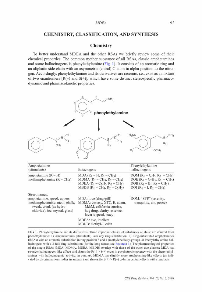
To better understand MDEA and the other RSAs we briefly review some of their
chemical properties. The common mother substance of all RSAs, classic amphetamines
and some hallucinogens is phenylethylamine (Fig. 1). It consists of an aromatic ring and
an aliphatic side chain with an asymmetric (chiral) C-atom in alpha-position to the nitro-
gen. Accordingly, phenylethylamine and its derivatives are racemic, i.e., exist as a mixture
of two enantiomers [R(–) and S(+)], which have some distinct stereospecific pharmaco-
dynamic and pharmacokinetic properties.
CNS Drug Reviews, Vol. 10, No. 2, 2004
MDEA 91
1
2
3
4
5
6NH2
phenylethylamine
áâ
3
4
NH
R1
O
O
R2
á
NH2
R2
R1 OCH 3
H CO3 áN
CH3
H
Rá
Amphetamines(stimulants) Entactogens
Phenylethylaminehallucinogens
amphetamine (R = H)methamphetamine (R = CH )3
MDA (R = H, R = CH )MDMA (R = CH , R = CH )MDEA (R = C H , R = CH )MBDB (R = CH , R = C H )
1 2 3
1 3 2 3
1 2 5 2 3
1 3 2 2 5
DOM (R = CH , R = CH )DOE (R = C H , R = CH )DOB (R = Br, R = CH )DOI (R = I, R = CH )
1 3 2 3
1 2 5 2 3
1 2 3
1 2 3
methamphetamine: meth, chalk,tweak, crank (as hydro-chloride), ice, crystal, glass)
MDA: love (drug pill)�MDMA: ecstasy, XTC, E, adam,
M&M, california sunrise,hug drug, clarity, essence,lover’s speed, stacy
MDEA: eve, intellectMBDB: methyl-J, eden
DOM: “STP” ( erenity,ranquility, and eace)
st p
Street names:amphetamine: speed, uppers
FIG. 1. Phenylethylamine and its derivatives. Three important classes of substances of abuse are derived from
phenethylamine: 1) Amphetamines (stimulants) lack any ring-substitution, 2) Ring-substituted amphetamines
(RSAs) with an aromatic substitution in ring position 3 and 4 (methylenedioxy-group), 3) Phenylethylamine hal-
lucinogens with a 3-fold ring-substitution (for the long names see Footnote 1). The pharmacological properties
of the single RSAs (MDA, MDMA, MDEA, MBDB) overlap with those of the other two classes: MDA has
stronger hallucinogen-like effects and shares the R(–) > S(+) order in psychotropic potency with the phenylethyl-
amines with hallucinogenic activity; in contrast, MDMA has slightly more amphetamine-like effects (as indi-
cated by discrimination studies in animals) and shares the S(+) > R(–) order in central effects with stimulants.

Classification
As illustrated in Fig. 1, RSAs take an intermediate position between amphetamines and
phenylethylamine hallucinogens (54), sharing many chemical and pharmacological prop-
erties with the other two classes (partly overlapping effects). Some authors call all RSAs
or even all amphetamines “designer drugs.” According to the original definition by
G. L. Henderson (65), however, this term is restricted to substances synthesized to bypass
legal sanctions by modifying an illicit drug. MDA and MDMA were first synthesized as
legally unrestricted byproducts; in contrast, MDEA entered the market to substitute for
MDMA after its federal ban in the late 1980s (33,63), followed by MBDB in the early
1990s (29). Hence, only these two newer RSAs are designer drugs (not MDMA and
MDA).
Nomenclature
Phenylethylamines have a babel of names (Fig. 1). For MDEA, the usual street name
(apart from ecstasy) is “eve,” while “intellect” is much less common. Apart from several
other names used in the literature (see Footnote 1), the proper chemical name according to
the International Union of Pure and Applied Chemistry is 1-(1,3-benzodioxol-5-yl)pro-
pan-2-yl(ethyl)azan (www.iupac.org).
Synthesis
MDEA and the other phenylethylamines can be easily synthesized in clandestine labo-
ratories without expensive or bulky equipment (mainly in Belgium and The Netherlands).
Instructions for the synthesis are readily available from books (158) or the internet. More
than 20 pathways are known for the synthesis of MDMA or MDEA, the easiest starts from
the illicit MDA (by simple N-alkylation). Popular precursors are safrole (available from
natural sources like sassafras, nutmeg, dill etc.) or MDP2P (= 3,4-methylenedioxyphenyl-
2-propanone, used by the fragrance and food industry). For further details see refs. 15,35,
97,153,158.
Usually, ecstasy and eve are marketed as pills (78) based on excipients like sorbitol,
cellulose or glucose (10). The manufacturers stamp the tablets with signs alluding to the
“rave” way of life (106). However, the symbols create a false sense of security as the psy-
choactive agents in the tablets vary greatly in terms of quality and quantity [(78,106,165),
cf. www.ecstasydata.org].
Qualitatively, an eve pill may contain pure MDEA, but in recent years mixtures of
MDA, MDMA, MDEA, and MBDB are increasingly common. Moreover, countless adul-
terants have been detected in ecstasy pills, including other abused drugs, e.g., amphet-
amine, methamphetamine, pseudoephedrine, cocaine, opiates, benzodiazepines, LSD,
caffeine, 4-bromo-2,5-dimethoxyphenylethylamine (2C-B, “nexus”), paramethoxyam-
phetamine (PMA), phencyclidine (PCP), ketamine, gamma-hydroxy-butyrate (GHB,
“liquid ecstasy”), and a variety of other drugs, e.g., chloroquine, vasodilators, dextrometh-
orphan (43,106), which is associated with the danger of fatal drug interactions. Hence, re-
sults from studies based on user reports and street names (ecstasy, eve) cannot be linked to
a defined substance (MDMA, MDEA).
The amounts of MDEA detected in pills varied from almost nothing to 75 mg (106).
In a recent report 64 to 175 mg of MDEA were present in various samples
CNS Drug Reviews, Vol. 10, No. 2, 2004
92 R. W. FREUDENMANN AND M. SPITZER

(www.ecstasy.org�testing�mde.html). The actual MDEA content in street pills should be
considered unknown, unless a specific toxicological analysis is carried out.
HISTORY AND LEGAL ISSUES
The role of MDEA within the RSA group is best understood in the context of their
history. Contrary to common beliefs, MDA and MDMA are rather old substances.
MDMA, for example, was first synthesized at the pharmaceutical company E. Merck in
Darmstadt, Germany, in 1912 as an intermediate product in a chemical pathway aiming at
a new styptic agent. MDEA, however, was introduced to the scientific community only in
the 1970s by the seminal work of the American experimental psychopharmacologist Alex-
ander T. Shulgin (15,158,159). In the European and North American club scene MDEA
gained importance only after MDMA, its famous N-methyl analog, became federally
banned in the late 1980s (33,63). But the MDEA heyday was short. In the US, it became a
controlled drug already on August 13th 1987 on the basis of a new law prohibiting analogs
of controlled substances [MDEA as an analog of the already controlled substances MDA
and MDMA; the law came into force in order to stop the “designer drug” problem
(9,63,117)]. In Germany, it remained legal until 1991, and in The Netherlands until 1993
(117). Today, it is listed along with MDA, MDMA, and MBDB in the most restrictive cat-
egory of abused substances in the US (Schedule I according to the Controlled Substances
Act), Canada (Schedule III of the Controlled Drugs and Substances Act), the UK (Class A
according to the Misuse of Drugs Act), Germany (appendix 1 of the Law on Hypnotics)
and other countries (26,54,78,86, 117). Table 1 summarizes the history of MDEA and the
other RSAs (for further details we recommend refs. 8,9,75,76,86,122,158,160).
CNS Drug Reviews, Vol. 10, No. 2, 2004
MDEA 93
TABLE 1. Short history of MDEA and other ring-substituted amphetamines
Date Event
1909�10 German chemists C. Mannich and W. Jacobsohn first synthesized MDA
1912�14 MDMA (not under this name) first synthesized in 1912 at pharmaceutical companyE. Merck, Darmstadt, Germany, as an intermediate byproduct in a chemical pathwayfor a styptic agent; patent assigned to Merck April 27th 1914
1978�1980 MDEA first mentioned in scientific publications by A. Shulgin and co-workers
1985–1988 MDMA listed as a Schedule I controlled substance in the US in 1985 �8; it is also fed-erally banned in other countries; MDEA surfaced as a legal substitute (“designerdrug”), but became Schedule I on August 13th 1987 as well in the US
1986ff Techno-music and ecstasy use become a mass phenomenon in Europe in the 1990s;mass production of ecstasy in Belgium, The Netherlands, Germany, Poland
1987 Dowling et al.: first report of fatalities after polydrug intoxication including MDEA
Jan 28th 1991 MDEA federally controlled in Germany
1996 Iwersen & Schmoldt: first report of a fatal MDEA mono-intoxication
2003 currently MDA, MDMA, MDEA and MBDB are listed in the most restrictive cat-egory of abused substances in the USA (Schedule I), UK (Class A), and Germany(appendix 1 of the BTM, 16th edition from Nov 28th 2001), as well as other countries
Abbreviations. See Footnote 1.

EPIDEMIOLOGY
Ecstasy, as commonly known, became a quantitatively important drug of abuse in the
1990s with the emergence of the “rave” culture (techno- and house- music). Belonging to
this youth culture has been shown to be the best predictor for ecstasy use (42), indicating
strong sociocultural influences. However, there is a paucity of valid epidemiological infor-
mation on illicit drugs in general. The best epidemiological study available in the US is
“Monitoring the Future“ [MTF, www.monitoringthefuture.org (75,76)]. MTF covers all
important classes of drug of abuse. According to MTF the use of MDMA in the USA for
the year 2002 was as follows: 1) life-time use in 10th grade students 6.6%, in college stu-
dents 12.7%, and in young adults 14.6%; 2) annual use in 10th graders 4.9%, in college
students 6.8%, in young adults 6.2%; 3) 30 day-prevalence in 10th grade students 1.4%, in
college students 0.7%, and in young adults 1.3%.
In Europe a similar annual use of ecstasy of 0.5–3.0% was estimated for adults in 1998
(19). In a Spanish sample a life-time prevalence of 4.5% for designer drugs has been
found, a point prevalence of 0.6% (29).
The content of MDEA in the ecstasy pills is lower than that of MDMA or MDA. In
Spain, for example, the National Institute of Toxicology found that ~48% of the seized ec-
stasy pills contained MDMA, ~41% MDA, ~7% para-methoxymethamphetamine, and
only 4% MDEA (2). A similar rank order was found in Denmark in 1995–1999 (163) and
in England (25).
The temporal consumption pattern of ecstasy is very typical. Usually, one to three pills
are taken during a rave weekend (124), while the rest of the week remains drug-free. The
subjective effects of this regimen have been eloquently termed “weekend high followed
by mid-week low“ (28). Often, there is no increase in either dosage or frequency of intake
(123,126). On the long run, however, 76% of the ecstasy users note less rewarding and
more unpleasant side and after-effects (126). This leads to two user groups: 1) those
quitting “E“ (self-limiting use, often interpreted as sign of a small addictive potential); and
2) those increasing dose and�or frequency of intake in order to overcome the loss of ef-
fects (“stacking“).
Some new epidemiological trends include 1) increased drug trafficking, more profes-
sional production and distribution that lead to: a higher availability and greater use of ec-
stasy worldwide (87); 2) change in consumption patterns: growing street market for home
use, no longer a mere party drug for weekends, simultaneous use of other substances in
order to modulate effects (“flipping”) (149); and 3) change in user profile: ecstasy con-
sumers tend to be older, more multidrug users (173,180). So-called “safe use recommen-
dations” (see www.dancesafe.org) contributed to the use of refreshing drinks and regular
“chill-outs“ during rave weekends. This positive development is, however, counteracted
by a growing number of substance mixtures in ecstasy pills and multidrug use, which
raises clinical concern due to possible drug interactions (see below).
PHARMACODYNAMICS
Because of their chemical similarity to norepinephrine (NE, noradrenaline), dopamine
(DA) and serotonin (5-hydroxytryptamine, 5-HT), all phenylethylamines, including
CNS Drug Reviews, Vol. 10, No. 2, 2004
94 R. W. FREUDENMANN AND M. SPITZER

MDEA, interact with the corresponding transmitter systems in the central nervous system
(CNS). While amphetamines elicit psychomotor stimulation predominantly by a net re-
lease of DA due to a “reverse transport” in the plasmalemmal DA transporter, i.e., an in-
direct agonism [S(+)-enantiomer more potent than R(–), in the body sympathomimetic ac-
tions (77,171)], phenylethylamine hallucinogens induce psychotomimetic effects by direct
interactions with neuronal 5-HT2 receptors (44,143), with an inverse order in stereospeci-
fic potency [R(–) > S(+)].
RSAs (e.g., MDEA) act mainly by indirect serotonergic mechanisms in the CNS
(6,27,54,78,94,101,113,160). Recent studies have shown, however, that RSAs also release
DA (157) and NE (141) from intracellular stores (rank order 5-HT > DA > NE). In the
5-HT system, RSAs trigger a 5-HT net release and inhibit its reuptake (54,78,101). These
findings are supported by studies in different laboratory settings (in vitro, cell culture,
drug discrimination studies in animals2), different species (rodents, non-human primates
and humans), and different experimental time schedules (acute and long term treatments)
(Table 2).
The primary site of action of MDMA has been identified at the molecular level as the
serotonin transporter (SERT), a membrane-bound protein in 5-HT vesicules and the
presynpatic plasmalemma (83,111,140), where it acts as a “substrate-type 5-HT-releaser”
(140). The plasmalemmal SERT is also the target site for antidepressants like selective
serotonin reuptake inhibitors (SSRIs) (111). Therefore, co-administration of a SSRI
attenuates MDA, MDMA and MDEA effects in vitro (73,101,179) and in humans in vivo
(citalopram with MDMA) (89).
In contrast to the selective mechanism of action of SSRIs, RSAs evoke their effects by
several mechanisms. Apart from their predominant indirect agonistic effects in serotoner-
gic neurotransmission, RSAs also act by inhibiting MonoAmine Oxidase (MAO) (88).
They have also comparatively weak intrinsic activity at several neurotransmitter receptors,
acting as direct agonists at 5-HT1A�D, and 5-HT2, á1�2, â, D1�2, M1�2, and H1�2 receptors
(6,7,46,94,128)). The affinity to postsynaptic 5-HT2-receptors is likely to mediate the mild
hallucinogenic effects of RSAs (7,44). Furthermore, several neuroendocrine effects ob-
served in animal (41,62) and human studies (40,50,51,55) are thought to contribute to the
central and peripheral effects of these drugs.
The S(+)-enantiomers of RSAs, like MDA and MDMA, have higher affinity for presy-
naptic 5-HT transporters, resulting in a greater 5-HT releasing (7,101) and psychotropic
potencies (94,113,114,118,119,146). By contrast, the R(–)-enantiomer is more potent at
postsynaptic 5-HT2-receptors which mediate the hallucinogenic effects of RSAs (7).
In addition, RSAs have substantial peripheral sympathomimetic, serotonergic, and
neuroendocrine effects (see below for clinical and toxicological details).
Animal and in vitro studies on the mechanism of MDEA action are summarized in
Table 2. In respect to its 5-HT releasing action MDEA is as potent as other RSAs (121,
153). Its overall neuropsychotropic potency is, however, controversial. In a rat study
MDEA has been reported to be as potent as MDMA, but weaker than MDA (45). In an-
other study MDEA was found to be less potent than MDMA in inducing acute hyper-
thermia (23). MDEA has a greater selectivity for 5-HT release than other RSAs (101,150).
CNS Drug Reviews, Vol. 10, No. 2, 2004
MDEA 95
2 Drug discrimination (DD) studies use animal models to compare behavioral effects of different drugs. First,
animals are trained to discriminate the stimulus properties of drugs from vehicle (saline), in a second step (chal-
lenge), other drugs are administered and the degree of stimulus generalization is analyzed (generalization sug-
gests similar stimulus cues in the animal).

It is weaker than MDA and MDMA in blocking the neuronal tryptophan hydroxylase
(TPH, the enzyme responsible for de novo 5-HT synthesis) (73,151,152,168,169). Conse-
quently, tissue levels of 5-HT and of its main metabolite 5-hydroxyindoleacetic acid
(5-HIAA) (23,168,169) are lowered to a lesser extent by MDEA than by MDA or MDMA.
Therefore, the risk of inducing long-term toxic effects to serotonergic neurons is pre-
sumably less with MDEA (23,101,121).
Pharmacodynamically, MDEA and the other RSAs are “dirty drugs.” Their predomi-
nant mechanism of action is an increased release of 5-HT (probably responsible for emo-
tional changes). Direct effects at 5-HT2 receptors may account for DOM-like alterations of
perception.
PHARMACOKINETICS
The pharmacokinetic propertiess of RSAs, like those of MDEA, are determined by
their chemistry, which includes stereospecific differences. Animal studies revealed several
major aspects of MDEA kinetics, fostering further research in humans.
Animals
The challenge study by Boja & Schechter in rats (12) reported substantially faster ki-
netics of MDEA as compared to MDMA. The onset of action of MDEA is 10 min, peak
effect at 10 to 20 min, the effects are starting to decrease after 60 min, the duration of
action does not exceed 120 min, and the calculated half life is about 60 min. The duration
of action of MDMA, by contrast, is ~240 min, and the calculated half life ~100 min.
Hegadoren et al. (64) showed stereospecific differences in brain levels of MDEA enan-
tiomers with a R�S-ratio < 1 after administration of racemic MDEA (10 mg�kg i.p.) to rats
(after MDA, however, the R�S-ratio was > 1). MDA was detected as an active metabolite
of both MDEA and MDMA; surprisingly, the enantiomer ratios in the brain were inverse
to the ratio of the respective parent compounds (i.e., R�S-ratio of MDA after MDEA > 1,
after MDMA < 1).
Further evidence of stereospecific kinetics in animals was yielded by a study of the
urinary excretion of MDA, MDMA, and MDEA in rats (96). After administration of
racemic MDA, MDMA, or MDEA, a preferential excretion of the R(–)-enantiomer was
demonstrated with MDA and MDMA. This finding was in contrast to a higher excretion
rate of the S(+)-enantiomer after MDEA (30 mg�kg) administation.
Another study directly compared the metabolism and distribution of racemic MDMA
and MDEA (20 mg�kg, s.c.) in rats (103). MDEA (the racemic form and each isomer) was
significantly less metabolized than MDMA, resulting in significantly less neurotoxic
metabolites (e.g., S(+)-MDA). In the plasma, a R�S-ratio > 1 was found for MDEA as
well as for its metabolite MDA.
Humans
The pharmacokinetics of MDEA and other RSAs in humans has been the subject of
extensive research during the last decade in Kovar’s laboratory in Tübingen, Germany
(16,17,34,35,50,54,68,82,84,97–99,166).
CNS Drug Reviews, Vol. 10, No. 2, 2004
96 R. W. FREUDENMANN AND M. SPITZER

CN
SD
rug
Review
s,V
ol.
10,N
o.2,2004
MD
EA
97
TABLE 2. Molecular mechanism of action of MDEA (animal and human in vitro studies, challenge studies only)
Study Main finding
(74) rats [3H]5-HT-release: MDA = MDMA, R = S; [3H]DA-release: S-MDA > S-MDMA; MDEA does not release DA
(72) rats single dose (MDEA (10 mg�kg): after 1 h: frontal cortical and hippocampal 5-HT concentration reduced (no effect on hypothalamic and neostriatal5-HT, 5-HIAA and neostriatal TPH); after 3 h: 5-HT functions (incl. TPH) disturbed in most brain areas; the DA system, however, is almost unaffected(only neostriatal DOPAC reduction after 3 h)repeated dose: stronger decrease of 5-HT concentration and TPH; after 18 h: partial recovery (TPH activity), no more effects in DA system (transient);hence, MDEA mainly affects the 5-HT, but not the DA system, effects are only transient
(134) rats MDEA selectively depletes 5-HT neurons, but not DA and NA; MDEA is factor 4 less toxic than MDMA
(150) rats 3 h after baseline: MDA = MDMA = MDEA deplete 5-HT in cortex neurons to <30% of baseline; MDA > MDMA > MDEA affect DA in striatum cells(longer side chain => less effects)1 week after baseline: cortical 5-HT & synaptosomal [3H]5-HT uptake normalized in MDEA, but still decreased in MDA & MDMA; hence, toxicity to5-HT nerve terminals: MDA = MDMA (S > R in both), MDEA: no such effects
(168) rats MDA and MDMA (10 mg�kg, repeated dose): selectively alter 5-HT neurotransmission (decreased TPH, 5-HT & 5-HIAA tissue concentration),MDEA: 5-HT & 5-HIAA is less reduced and TPH unaffected; hence, serotonergic neurotoxicity: MDA = MDMA >> MDEA
(12) rats, in vivo MDEA and MDMA have identical effects (drug discrimination task), but different potencies: MDMA > MDEA
(91) mice, rats first indication of species-specific long-term effects in MDMA and MDEA
(147) rats,
in vivo
acute effects: MDMA = MDEA (full generalization in drug discrimination task)
(128) human,
in vitro
first study in human cells investigates direct agonistic effects in 5-HT and NA systems; affinities at “DOB-binding site” are high (MDA = MDMA =MDEA); at 5-HT1A�D- and alpha2-receptors, however, MDA has moderate, MDMA and MDEA little or no affinity
(73) rats MDEA (10 mg�kg) transiently reduces hippocampal 5-HT concentration and TPH activity (after 1 h, return to baseline after 12 h) as well as 5-HIAA(decrease within 2 h, rebound to 22% above control after 12 h, returned to control after 24 h);SSRI co-administration (fluoxetine) protects tissue 5-HT concentration and TPH activity (except for neostriatal TPH), in contrast to MAOI; hence,MAO-related H2O2 does not explain TPH activity changes
(112) pigeons,
in vivo
as measured by response rates in fixed-interval, fixed-ratio schedule controlled behavior (key pecking under food presentation): MDA is stronger thanMDMA and MDEA;MDA effects can be blocked by metergoline (5-HT1�2-blocker) and ketanserine (5-HT2-blocker) but not prazosin (alpha1-blocker), MDMA: oppositeeffects, MDEA: no effect of the co-administered drugs; the differential effects indicate different mechanisms of action
(47) rats,
in vivo
rats learned to discriminate S-amphetamine and R�S-DOM (i.e., a classic stimulant and a phenylethylamine hallucinogen); DOM stimulus did notgeneralize to S-MDMA, R-MDMA, R-MDEA, S-MDEA, R�S-MDEA; amphetamine-stimulus generalized to S-MDMA, but not R-MDMA, or anyMDEA-form; hence, RSAs (except for S-MDMA) differ from stimulants and hallucinogens
(45) rats,
in vivo
MDMA-stimulus (0.75 mg�kg) generalized to MDEA (0.73 mg�kg) and MDA (0.47 mg�kg); hence, MDMA, MDA and MDEA have similar prop-erties, but different potencies (MDA > MDMA = MDEA); MDMA partially generalized to S-amphetamine, however MDEA and MDA neither gener-alized to amphetamine nor DOM; hence, MDA, MDMA and MDEA differ from amphetamine, S-MDMA is the most amphetamine-like substance

CN
SD
rug
Review
s,V
ol.
10,N
o.2,2004
98
R.W
.F
RE
UD
EN
MA
NN
AN
DM
.SP
ITZ
ER
Study Main finding
(95) rats, in vivo S-MDEA > R-MDEA in reducing prepulse inhibition in acoustic startle (stereoselective effects)
(101) rats acute effect: [3H]5HT-release: MDA = MDMA (both S > R), partially blocked by fluoxetine; [3H]DA-release: MDA = MDMA (both SR); R�S-MDEA:5-HT release > DA releaselong-term effect: only S-MDA produced a significant loss of [3H]paroxetine-labeled 5-HT-uptake sites, while MDMA and MDEA did not, indicatingdifferences in neurotoxicity
(148) rats block of 5-HT synthesis with TPH-antagonist p-chlorophenylalanine interferes with discrimination of serotonergic substances like MDMA, MDEA,and fenfluramine, but neither dopaminergic (amphetamine) nor adrenergic drugs (yohimbine)
(13) rats stimulus generalization to MDEAwas achieved by some 5-HT-receptor agonists, but not buspirone, norfenfluramine; partial generalization by ACh ag-onist arecoline and amphetamine; amphetamine plus fenfluramine fully generalized to MDEA; 5-HT-blockers (cinanserin, metergoline) and adopamine-blocker (haloperidol) did not fully inhibit discrimination, only multiple pretreatments with TPH-blocker p-chlorophenylalanine; hence,MDEA-stimuli are 5-HT and DA-dependent
(125) rats S-MDMA and R�S-MDEA change the quantity, but not the quality of motor behavior
(156) rats not single, only repeated high doses of MDEA (40 mg�kg, 8 times over 2 weeks) evoked a significant reduction of serotonin-immunoreactive axons,particularly in fine-type varicosities (posterior cortex, hippocampus: CA1)
(144) baboons,
in vivo
after stopping cocaine i.v. self-injection MDEA and MDA were consistently continued by baboons
(23) rats acute effects: to induce hyperthermia 35 mg�kg MDEA i.p. and 15 mg�kg MDMA i.p. were needed; hence, potency MDEA < MDMA (~ factor 2)long-term effects: after MDMA (7 days, 15 mg�kg) 50% decrease of 5-HT, 5HIAA (cortex, hippocampus, striatum) and [3H]paroxetine-binding sites(cortex), whereas after MDEA (15, 25, 35 mg�kg i.p.) — even in the highest dose — only a 20% reduction of 5-HT parameters in cortex and hippo-campus was found (only weak dose-dependent effects); in contrast, MDMA (15 mg�kg) and MDEA (35 mg�kg) had no effect in the DA system; hence,neurotoxic potency in 5-HT neurons: MDEA < MDMA (~ factor 4); however, as human users need higher MDEA doses for the desired effects than inMDMA, MDEA still is no safe substance
(5) rats single dose (MDEA 10, 20, or 40 mg�kg i.p.): induced dose-related hyperthermia and corticosterone increase; only the highest dose reduced 5-HT andtransporter density after 7 days (frontal cortex, hippocampus)repeated doses (MDEA, 40 mg�kg i.p., b.i.d., for 4 consecutive days): stronger serotonergic deficits as compared with single dose
(121) rats single dose: MDMA and MDEA (20 mg�kg i.p.): increased locomotion, decreased rearing, induced stereotypy, Straub tail and head weaving as wellas hyperthermia (MDBA: no effects); [3H]5-HT-release (cortex, hippocampus): MDA = MDMA = MDEA >> MDBA; [3H]DA-release (striatum):MDA > MDMA > MDEA = MDBArepeated dose: 20 mg�kg, twice daily for 4 days): loss of forebrain [3H]paroxetine binding, 5-HT and 5-HIAA tissue concentration after 14 days (no ef-fects on NA and DA systems), rank order: MDMA > MDEA >> MDBA > or = saline
(153) rats,
in vivo
haloperidol-induced catalepsy was used as a model for parkinsonian symptoms; MDMA (1.0–5.0 mg�kg) dose-dependently counteracted catalepsy(S > R) with a much weaker effect in MDEA(2.5–5.0 mg�kg) or its two enantiomers (5.0 mg�kg), putatively based on less striatal dopamine release
(21) rats, in vivo cardiac toxicity of MDA, MDMA, and MDEA depends on a noradrenergic mechanism
Abbreviations. S, S(+)-enantiomer; R, R(–)-enantiomer; TPH, tryptophan hydroxylase; DOPAC, dihydroxyphenylacetic acid; MAOI, monoamine oxidase in-
hibitor; MDBA, N-butyl-analog of MDMA and MDEA.
TABLE 2 (continued)

Routes of administration
MDEA, like the other RSAs, is usually taken orally, rarely nasally or as a suppository.
As amines, all RSAs are soluble in water and alcohol (22,78) and can, therefore, be in-
jected. Their inhalation as a vapor is impossible due to their high boiling points (160).
Dosage
A regular dose of ecstasy in “recreational use” ranges from 1 to 3 pills. For the defined
chemical substances in ecstasy pills the dose ranges are as follows: MDEA [100 to 200 mg
(158), or about 2 mg�kg (17)] > MDMA [80 to 150 mg (158), “best“ around 120 to
130 mg, or about 1.5 mg�kg (61)] > MDA [60–120 mg (63)]. These dose ranges appear to
correlate inversely with their psychotropic potencies (MDEA < MDMA < MDA).
Absorption and distribution
After consumption, MDEA is readily available and distributed in the body; data on oral
bioavailability and plasma protein binding in man are not available.
Course of action
According to human challenge studies the onset of action of pure MDEA, 140 mg, is
20–85 min (54), with 160 mg it is 40 min (158). The duration of action ranged from 2 to
3 h after intake of 140 mg MDEA (54) to 3 to 5 h after 160 mg (158), indicating shorter ef-
fects as compared with 4 to 6 h for MDMA and 8 to 12 h for MDA (158). Other major
pharmacokinetic parameters of MDEA obtained from challenge studies in healthy volun-
teers are summarized in Table 3 (17,166). These parameters include maximal plasma con-
centrations (Cmax) after usual “recreational“ doses (Study 1, 2) and the time of maximal
plasma levels (tmax). Note that the study by Spitzer et al. (Study 3) gave first indications
for stereospecific pharmacokinetics in humans, e.g., the longer elimination half life in
R(–)-MDEA and the higher total clearance in S(+)-MDEA.
Metabolism and elimination
The hepatic metabolism of MDEA and of other RSAs is rather complex; two partly
overlapping phase I metabolic pathways have been identified by Ensslin et al. (35)
(Fig. 2). The major pathway is responsible for splitting the aromatic ring by O-dealkyla-
tion; via 3,4-dihydroxy-metabolites. N-ethyl-4-hydroxy-3-methoxyamphetamine (HME)
is formed as the main metabolite (35,129). The second path breaks down the side chain of
the MDEA molecule by N-dealkylation. Quantitatively, this path is less important, but
N-deethylation leads to MDA as an active metabolite; after several further intermediates
the pathway leads to benzoic acids which are subsequently conjugated with glycine, re-
sulting in substituted hippurates. In urine (after 140 mg racemic MDEA p.o.) unchanged
MDEA and 3,4-dihydroxyethylamphetamine (DHE) can be detected for 33 to 62 h after
ingestion, MDA for 32 to 36 h, and HME for 7 days. Initially detectable are also traces of
3,4-dihydroxyamphetamine (DHA), 4-hydroxy-3-methoxyamphetamine (HMA), pipero-
nyl acetone, 3,4-dihydroxyphenyl acetone, and 4-hydroxy-3-methoxyphenyl acetone (34).
More profound insight into the metabolic breakdown of RSAs has been gained by the
identification of the cytochrome P450 isoenzymes (CYP) that catalyze the two aforemen-
tioned pathways in animals and humans (98) (Fig. 2). In humans, MDEA catabolism de-
pends on CYP 2D6 and 3A4 (2D6 mediates ring degradation only, 3A4 ring and side
chain degradation). This is of major clinical importance, since: 1) about 10% of the Cau-
CNS Drug Reviews, Vol. 10, No. 2, 2004
MDEA 99

casian population have a CYP 2D6 “poor metabolizer” phenotype (30,84,106); 2) CYP
2D6 and 3A4 can be saturated by high doses of MDEA (84,175); and 3) P450 isoenzymes
can be inhibited by numerous drugs metabolized by the same isoenzymes, causing dispro-
portionally high plasma levels of MDEA (non-linear kinetics) (30).
The effects of different activities of these “bottleneck enzymes” can be seen, for ex-
ample, in the 10-fold variation in the levels of HME, the major metabolite of MDEA after
CNS Drug Reviews, Vol. 10, No. 2, 2004
100 R. W. FREUDENMANN AND M. SPITZER
N
3
4
H
R1
O
O
R2
á
MDEA(R = C H , R = CH )1 2 5 2 3
ring degradation by0-demethylenation:
2D12D6
3A23A4
side chain degradationby N-dealkylation:
(1A2) (2D1) 3A23A4
rat:man:
FIG. 2. Hepatic metabolism of MDEA in rat and man. Two major pathways, ring or side chain break down, and
the respective cytochrome P 450 isoenzymes in rat and man (2D1 = CYP 2D1). Data are adapted from Maurer et
al. (2000) (ref. 98).
TABLE 3. Pharmacokinetics of MDEA in man (challenge studies, healthy human subjects)
Study 1Brunnenberget al. (1998)
N = 6, fixed dose:140 mg MDEA
Study 2Brunnenberg et al. (1998)
N = 8, dose adjusted
to 2 mg�kg body mass(max. 140 mg) MDEA
Study 3Spitzer et al. (2001)
N = 5, fixed dose: 70 mgR(–) and S(+)-MDEA
R(–) S(+)
Cmax
(ng�mL)
MDEA 260 (203–333) 332 (235–465) 127 ± 34 80 ± 30
MDA 21 (15–32) 23 (7–33)
HME 436 (285–615) 389 (67–673)
tmax (h) MDEA 2.2 (1.6–2.9) 2.9 (1.8–5.0) 2.8 ± 0.9 2.6 ± 0.6
MDA 3.4 (15.0–32.0) 5.5 (4.0–7.0)
HME 2.8 (1.6–3.9) 2.7 (1.5–5.0)
t1�2 (h) MDEA — (—) — (—) 7.5 ± 2.4 4.2 ± 1.4
MDA
HME
AUC
(ng�h� mL)
MDEA — (657–875) — (832–1935) 1707 ± 897 535 ± 263
MDA — (38–95) — (19–120)
HME — (665–1976) — (218–3028)
Total Cl
(mL�min)
MDEA — (—) — (—) 718 ± 3404 2258 ± 1032
MDA
HME
Numbers given as mean and range (studies 1 and 2) or mean ± S.D. (study 3).
Abbreviations. Cmax, maximum plasma concentration; tmax, time at maximum plasma concentration;
t1�2, elimination half life; AUC, area under curve; Total Cl, Total Clearance (cleared volume per time
unit).
administration of bodyweight-adjusted doses of MDEA to healthy subjects (Table 3, Study
2) (17). It can be concluded that interindividual differences in toxic MDEA doses can be
explained partially by genetically determined CYP activities and pharmacokinetic interac-
tions (for further details on RSA metabolism see refs. 82,84,98).
Drug interactions
The dependence of MDEA metabolism on CYP 2D6 and 3A4 implies that substances
acting as CYP 2D6- and 3A4-inhibitors may substantially block hepatic MDEA degra-
dation. CYP 2D6-blockers that have caused harm when used with ecstasy are fluoxetine,
paroxetine (132,175), moclobemide (79,164), and dextromethorphan (14,131,132,182),
but haloperidol, thioridazine and quinidine should be avoided as well. Among CYP
3A4-blockers, ritonavir has been repeatedly reported to interfere with ecstasy (1,3,32,66,
155), but also fluoxetine, nefazodone, cimetidine, grapefruit juice, verapamil, erythromy-
cin, ketoconazole and metronidazole inhibit CYP 3A4. The SSRIs, fluoxetine and paroxe-
tine, as well as the MAO-inhibitor moclobemide cause not only pharmacokinetic, but also
pharmacodynamic interactions, which may lead to the serotonin syndrome and arterial
hypertension.
Use in pregnancy
The use of ecstasy in pregnancy may lead to teratogenic effects including congenital
heart disease and malformations (24, 80,100). No specific information on the relative tera-
togenicity of MDA, MDMA, MDEA or MBDB is available.
LABORATORY AND TOXICOLOGICAL ANALYSIS
The laboratory and toxicological analytical procedures applied for classic amphet-
amines (108,139) and the different RSAs, including MDEA, are very similar. Reliable
methods are needed for the legal prosecution of the use of these illicit substances and the
clinical management of the growing number of intoxications (97,183). The analytical pro-
cedures can be separated into two groups: 1) different types of immunoassays (85,92,142,
183), and 2) chromatographic and spectroscopic methods like high-performance liquid
chromatography (HPLC) (105), liquid chromatography-mass spectrometry (LC-MS) (11),
and gas chromatography-mass spectrometry (GC-MS) (97,170).
For screening purposes and routine sample testing, simple, cost-effective, but rather
sensitive methods, like immunoassays, are used (85,139). In contrast, for confirmatory
testing, forensic issues and quantitative analyses, GC-MS is considered to be the reference
method (107,183). For further information see refs. 11,20,98,107,142,170,183.
HUMAN NEUROPSYCHOPHARMACOLOGY
AND CLINICAL TOXICOLOGY
Amphetamine derivatives are primarily known as substances of abuse. Their effects
can be classified in several ways (e.g., acute vs. long-term, neuropsychiatric vs. physical,
CNS Drug Reviews, Vol. 10, No. 2, 2004
MDEA 101

and desired vs. undesired). We emphasize below MDEA challenge studies as being the
most MDEA-specific.
Acute Effects
The subjective experience under ecstasy varies greatly and depends on several factors,
such as the dose, presence of additional substances in the pill, frequency of previous ex-
posure to ecstasy, baseline mood, concomitant use of other drugs, etc. In general, the expe-
rience consists of “amphetamine-like” stimulation, mild “DOM-like” alteration of per-
ception and “entactogenic” effects. The psychotropic effects of RSAs seem to lie in
between classic amphetamines and phenylethylamine hallucinogens which are derived
from the same parent compound (161,162) (Fig. 1).
Anecdotal Data
The differences between individual RSAs are subtle (54,78). Reports from some expe-
rienced users indicate, however, a lower psychotropic potency, a shorter duration of
action, but stronger suppression of appetite for MDEA as compared to MDMA (54,158).
Users describe that under the influence of ecstasy they develop peculiar feeling of
“closeness” to other people (126), the loss of boundaries, sensation of unification with the
inner self, others or the environment. Since this experience depends to a great extent on
the environment during drug consumption, i.e., “trance”-like mass dancing, psychosocial
factors seem to contribute to the overall effect.
A certain sequence of subjective effects is considered typical for ecstasy use. The first
phase is a short rush called “coming on”. It is followed by a “plateau phase” with a
pleasant emotional state and increased physical energy. The plateau phase adds to the
“rave experience” and seems to be associated with a central serotonin rush and sympatho-
mimetic effects. In more than half of the cases, however, untoward effects accompany the
plateau phase: jaw clenching (trism), tachycardia, tooth grinding (bruxism), and dry
mouth (126). The third phase of the response is “coming down” and return to baseline,
often with “negative” emotions. The fourth and the last phase is characterized by psychic
and physical “aftereffects” that include muscle aches and stiffness, headache, depressed
mood, anxiety, cognitive dysfunctions and fatigue (126). This “nadir” represents pre-
sumably neuronal serotonin depletion; the time of its onset is variable, the estimates range
from one (126) to 5 days (28) after ingestion.
MDEA Challenge
The best information on desired and unwanted psychotropic and somatic effects of
MDEA can be derived from challenge studies with pure MDEA in healthy subjects in a
controlled setting (49,50,54,68,166). According to Gouzoulis-Mayfrank et al. (54) MDEA
effects in humans as seen in several challenge studies can be summarized as follows: After
administration of 140 mg MDEA per os, there is a sudden onset of effects (after a few
minutes), but with a different delay (20 to 85 min). Initial signs of drug action are mainly
somatic, e.g., nausea, blurred vision and deep breathing, sometimes also anxiety; subse-
quently, most subjects report strong relaxation, pleasant peacefulness and loss of anxiety,
contrasting with objective stimulating effects that subjects were unaware of (psychomotor
activation, logorrhea, tachycardia, elevated blood pressure, tremor). In at least half of the
CNS Drug Reviews, Vol. 10, No. 2, 2004
102 R. W. FREUDENMANN AND M. SPITZER

subjects, MDEA caused psychomotor stimulation, a positive emotional state and mild al-
terations of perception (visual, acoustic and tactile), impaired cognitive functioning and
sympathomimetic effects. Surprisingly, only a minority of subjects reported “entactoge-
nic” experiences. Still, MDEA challenges demonstrate amphetamine-like and hallucinoge-
nic effects in a unique blend.
In 1992 Gouzoulis et al. reported the results of a double-blind, placebo-controlled sleep
EEG study involving six individuals (49). It was the first in a series of studies that used
140 mg racemic MDEA synthesized in the Kovar’s laboratory. In the sleep laboratory,
MDEA was given to the subjects at 11 p.m.; at 1 to 2 h later all subjects awakened, indi-
cating the onset of the stimulant effect. Total sleep time and rapid eye movement (REM)
sleep were significantly reduced. The study revealed alterations of sleep architecture at-
tributed to MDEA’s amphetamine-like properties.
Another study by Gouzoulis et al. (50) investigated on the effects of MDEA on neuro-
endocrine and cardiovascular parameters. The study was double-blind, placebo-con-
trolled, crossover and involved eight male volunteers. In addition to marked sympathomi-
metic effects on blood pressure and heart rate, MDEA produced significant elevation of
serum cortisol and prolactin, supporting serotonergic actions in the hypothalamus, al-
though the attenuated growth hormone secretion did not fit this interpretation.
CNS Drug Reviews, Vol. 10, No. 2, 2004
MDEA 103
TABLE 4. Psychotropic effects of MDEA in healthy humans (challenge studies)
Pooleddata(%)
Study 1N = 8 (males)
day-time, personalcontact to scientist,systematic ratings
Study 2N = 6 (3 female)
night, no personalcontact, no
systematic ratings
Hypervigilance, increased drive 100 8�8 6�6
Euphoria, relaxation, peaceful satisfaction 50 4�8 3�6
Dysphoria, irritation 7 1�8 0�6
Feeling of happiness 21 2�8 1�6
“Entactogenic effects”: introspection, empathy,
loss of anxiety, happy self acceptance, con-
trolled communicative openness
21 3�8 0�6
Altered perception of time 21 2�8 1�6
Altered visual, tactile, acoustic perception:
colors more intense, blurred contours, things
bigger�smaller, sounds louder, haptic impres-
sions “as if through cotton wool”
50 4�8 3�8
Depersonalization, derealization 7 0�8 1�6
Religious-mystique experiences 21 1�8 2�6
Psychotic state: acoustic�visual hallucinations,
delusions, loss of control, anxiety
7 0�8 1�6
No loss of control 93 8�8 5�6
Disturbed concentration (subjective) 43 4�8 2�6
Severe cognitive dysfunction NA 0�0 NA
Adapted from Gouzoulis-Mayfrank et al. (1996) (ref. 54).
Abbreviations. NA, not available.

In order to further characterize MDEA effects in humans, Gouzoulis et al. directly com-
pared MDEA with amphetamine (stimulant) and psilocybin (hallucinogenic substance).
One study systematically assessed the effects of the three substances on psychopathologi-
cal, cardiovascular and vegetative functions (56). In addition to significant increases in
serum cortisol and prolactin, as well as an increase in body temperature, MDEA challenge
evoked subjective “relaxation” in a surprising blend with the sympathomimetic cardiovas-
cular effects. This pattern clearly differed from that induced by either amphetamine or psi-
locybin. Another study compared the same substances using positive emission tomog-
raphy with 18fluorodexoglucose (FDG-PET) (55,154). MDEA challenge led to changes in
the regional cerebral blood flow (rCBF), most prominently a rCBF decrease in frontal
cortex and cerebellum, and an increase in the right anterior cingulum. This pattern of ef-
fects was different from that of the other two substances. The authors interpreted the re-
sults of the two studies as a support of the earlier drug discrimination studies in animals
that classified “entactogens” (here MDEA) as a separate entity between stimulants and
hallucinogens.
The most comprehensive study of the psychotropic effects of MDEA was carried out
by Spitzer et al. (166). The double blind, crossover study addressed stereospecific effects
by the administration of the two MDEA enantiomers (70 mg) in five male physicians and
involved systematic controls of plasma levels. In addition to standardized rating scales to
assess subjective effects (mood, well-being, somatic symptoms, and hallucinogenic ef-
fects) the authors used a battery of neuropsychological tests (a task on basic visual pro-
cessing, the Wisconsin Card Sorting Test), and functional magnetic resonance imaging
(fMRI) to measure drug action. Despite the rather small doses used substantial stereospe-
cific differences were found. Significantly higher scores for well-being (Bf-S) were noted
with the S(+)-enantiomer. A trend for more somatic symptoms was reported for R(–)-
MDEA. Higher maximal plasma levels of R(–)-MDEA (p < 0.05) and a trend for a longer
elimination half-time (p < 0.08) resulted in a greater area under the curve (p < 0.04). In
contrast, S(+)-MDEA was preferentially excreted in urine (p < 0.04) (Table 3). A signifi-
cantly shorter reaction time in pop-out search (low level visual processing) was shown for
R(–)-MDEA. Higher switch costs in the Wisconsin Card Sorting Test were found for
S(+)-MDEA. In fMRI, semantic judgments were associated with significant BOLD “acti-
vations“ of right visual and left frontal areas under R(–)-MDEA, in contrast to right
frontal and bilateral temporoparietal “activations“ under S(+)-MDEA. The study sug-
gested that S(+)-MDEA is responsible for pleasant subjective effects, while the higher
plasma levels of R(–)-MDEA contribute to unpleasant somatic sensations and may also
enhance bottom-up processes in the visual system, possibly being the basis for visual
alterations of perception.
Complications
The possible acute complications of MDEA use range from psychiatric problems to
severe medical conditions and eventually fatal intoxications.
Acute psychiatric complications
MDEA has the potential to trigger acute psychotic disorders and induce dysphoric
mood, anxiety as well as panic attacks (48,68,70,176). These are the known complications
CNS Drug Reviews, Vol. 10, No. 2, 2004
104 R. W. FREUDENMANN AND M. SPITZER

of amphetamines, hallucinogens and cocaine (63,78). Accidents and misadventures under
the influence of MDEA and the other RSAs are also frequent.
Physical complications
In general, the physical problems associated with MDMA (63,78) have also been de-
scribed for MDEA. Either of the two drugs can elicit a variety of symptoms, including
mild unwanted effects, like nausea or headache, as well as more serious conditions re-
quiring medical treatment:
— One of the serious conditions is the serotonin syndrome (167) that involves hyper-
thermia (often aggravated by environmental heat, overcrowding, and dancing), neuro-
muscular signs (hyperreflexia, tremor, trism), psychopathology (hyperactivity, agitation,
confusion), and gastrointestinal symptoms (nausea, vomiting, diarrhea).
— In severe cases this syndrome leads to rhabdomyolysis, disseminated intravascular
coagulation (DIC), acute renal and eventually multiorgan failure (36,37,67,172,174,178),
that require referral to an intensive care unit.
— MDEA use may cause hyponatremia and the Syndrome of Inappropriate
AntiDiuretic Hormone secretion (SIADH) (18,41).
— Cardiovascular problems due to sympathomimetic effects after MDEA intake in-
clude: arterial hypertension, tachycardia, arrhythmias, acute heart failure and myocardial
lesions (33,70,106,178); possibly also fenfluramine-like valvular heart disease mediated
by 5-HT2B-receptors as shown by recent in vitro studies (157). Other reported complica-
tions include
— respiratory failure (26,93,178),
— intracranial hemorrhage due to ruptured aneurysms (106), cerebral seizures and
convulsions (174), as well as
— subacute toxic hepatitis (67,70,106,174).
The diagnostic and therapeutic measures in MDEA intoxications are the same as for
amphetamines and MDMA (67,78,139,177).
MDEA-related fatalities
The most comprehensive collection of lethal intoxications associated with the use of
ecstasy has been presented by Kalant (78, appendix 1). Table 5 summarizes 20 fatalities in
which the presence of MDEA was toxicologically confirmed (2,4,26,33,36,37,39,70,93,
106,130,178).
These fatalities clearly highlight the pathophysiological patterns mentioned above.
Note the rather large number of accidents as compared to the direct toxic effects. Most
cases are polydrug intoxications. The relative contribution of MDEA to the fatal outcome
cannot be exactly determined. The dangers of MDEA were, however, documented by four
lethal intoxications with the drug alone (70,93,178). The blood levels of MDEA in these
individuals ranged between 12 and over 20 mg�L.
Chronic Effects
Definitive information on the chronic effects of pure MDEA is not available, since
users do not usually consume MDEA alone, but various mixtures of RSAs and other
neurotoxic substances, such as amphetamine and methamphetamine. We will, therefore,
CNS Drug Reviews, Vol. 10, No. 2, 2004
MDEA 105

briefly review the issues of substance dependence, neurotoxic effects, and long-term neu-
ropsychiatric consequences for RSAs in general, rather than for MDEA alone.
Substance dependence
In general, the addictive potential of ecstasy is considered to be low. This conclusion is
based on the lack of tolerance and withdrawal effects after discontinuation, as well as on
CNS Drug Reviews, Vol. 10, No. 2, 2004
106 R. W. FREUDENMANN AND M. SPITZER
TABLE 5. MDEA-related fatalities
CaseNo.
Subject, clinical details,
detected substances (mg�L = ìg�mL), comments Reference
1 male�25y, car accident probably due to cardiac arrest; blood: MDEA (0.95),butalbital (0.8), underlying coronary heart disease
(33)
2 male�21y, found unconscious, CPR unsuccessful; blood: MDEA (2.0), propo-xyphene (0.26), norpropoxyphene (1.0), underlying idiopathic cardiomyopathia
(33)
3 male�21y, found dead in bed; blood: MDEA (3.5), MDMA (2.1), MDA (8.5),amphetamine (0.256)
(39)
4 male�19y, cardiac arrest, CPR unsuccessful; blood: MDEA (20.2 or 22.2 on ad-mission, 45 post mortem), no other drugs, cerebrospinal fluid: MDEA (40.6)first report of a fatal MDEA monointoxication, previously healthy person
(70)
5 male�24y, suicide by stabbing under the influence of drugs; blood: amphet-amine (0.75), MDEA (0.22), alcohol (0.46)
(70)
6 male�22y, tachykardia, hyperthermia (41.8°C), metabolic acidosis, DIC, pete-chial and subendocardial hemorrhages, lung edema, pleural effusion; blood:MDEA (0.49 on admission, 0.30 post mortem), MDMA (0.55, 0.43), MDA(0.24, 0.25), LSD (neither THC nor alcohol), healthy person
(26)
7 male�24y, collapsed dead at disco, focal necrosis in heart and liver;blood (post mortem): amphetamine (0.453), MDEA (0.187)
(106)
8 male�21y, found dead in bed, focal necrosis of liver and brain, inhalation ofvomit; blood (post mortem): MDA (8.5), MDEA (3.5), MDMA (2.1), amphet-amine (0.256)
(106)
9 male�23y, MDEA and amphetamine, violence (93)
10 male�17y, MDEA (alone), respiratory arrest�asphyxia? (93)
11 male�39y, MDMA and MDEA, cardiovascular death (93)
12 male�21y, MDEA (alone), traffic accident (93)
13 male�19y, MDEA (alone), traffic accident (93)
14 male�19y, MDEA (alone), “adverse drug reaction” (93)
15 male�20y, found dead in bed, hyperthermia and DIC; blood: MDEA (1.596),MDMA (0.185), brain: MDEA (8.430), MDMA (12.794), urine: MDEA(183.737), MDMA (263.132), intoxication with MDEA >> MDMA
(37)
16 female�27y, psychiatric patient, cardiac arrest; blood: MDEA(1.2), benzodiaze-pines and caffeine positive, other common drugs and phenelzine (last treatment)negative
(2)
17 male�19y, profuse sweating, muscular spasms, aggression, hallucinations, lossof consciousness, CPR unsuccessful, severe vascular congestion of all internalorgans, multiple subserous petechial hemorrhages, lung emphysema;blood: MDEA (12), MDMA (0.016), MDA (0.32), brain: MDEA (28), noMDMA, MDA (0.65) urine: MDEA (201), MDMA (0.135), MDA (7.1), otherdrugs of abuse incl. alcohol negative, well documented lethal monointoxicationwith about 1.3 g MDEA(and traces of MDMA) in normal ambient temperature
(178)
18 ??; blood: MDMA (2.0), MDEA (0.7) (130)
19+20 two more cases are mentioned in the literature, no details available (4,36)
Abbreviations. THC, tetrahydrocannabinol; LSD, lysergic acid diethylamide.

its consumption pattern (use on weekends only, often self-limiting, strong influence of
sociocultural factors) (71,78,104,126). Criteria for substance dependence according to the
current edition of the Diagnostic and Statistical Manual of Mental Disorders (DSM-IV)
are not met in many users of ecstasy. However, some recent case reports indicate that ec-
stasy has indeed the potential to evoke substance dependence (71). As the experimental
and epidemiological data basis is still limited, it is currently impossible to resolve the
issue of the addictive potential of ecstasy (63,78,122). For clinical purposes ecstasy use,
including complications, can be classified according to DSM-IV.
Neurotoxic effects
An even more intricate topic is the existence of neurotoxic effects after repeated ec-
stasy use. Animal and in vitro studies (Table 2) showed selective alterations of serotoner-
gic neurotransmission after repeated exposure to RSAs in the form of reduced levels of
5-HT and 5-HIAA in brain and liquor, reduced tryptophan hydroxylase activity, loss of
paroxetine-labeled binding sites, immunological and histological signs of damage to 5-HT
nerve terminals, and delayed “neuronal pruning” or “sprouting” (38,121,134,135,168).
Some authors argue that this evidence suffices to conclude that ecstasy has serotonergic
neurotoxic effects in humans. Others, by contrast, question the applicability of findings
from animal studies to humans and the significance of the results from human studies be-
cause of methodological limitations (retrospective design, possible exposure to other
neurotoxic agents, lack of toxicological workup, etc.) (78,81,145).
Most in vitro and animal studies suggest a lower serotonergic neurotoxicity with
MDEA than with other RSAs (MDEA < MDMA < MDA) (72,73,91,101,115,116,121,
134,150,168) with only a few exceptions (5,156).
The mechanism responsible for the toxic effects of RSAs to 5-HT neurons is not well
understood (60, 69). The proposed mechanisms include: 5-HT depletion, an increased stri-
atal dopamine release, calcium-influx, inhibition of tryptophan hydroxylase, hyperther-
mia, formation of free radicals, effects of excitatory amino acids, increased turnover of
cytoskeletal protein tau, impaired axonal transport, prolonged translocation and activation
of protein kinase C, etc. (for reviews see refs. 52,53,60, 63,121,184). The paper by
Ricaurte et al. describing dopaminergic neurotoxicity of MDMA in primates was recently
retracted (138). Future research is needed to elucidate the neurophysiological changes in-
duced by RSAs.
Chronic psychiatric complications
The discussion about neurotoxic effects is not only of academic but also of clinical im-
portance. There is a substantial evidence that many former ecstasy users seek professional
help because of various neuropsychiatric symptoms. They suffer from cognitive problems
that affect verbal and visual memory, decision-making and problem-solving as well as af-
fective (atypical depression or anxiety, 68) and psychotic symptoms (paranoid ideas, al-
tered visual or acoustic perception, depersonalization and flashbacks). Their appetite and
sexual functions can be impaired as well. The combined presence of these symptoms, to-
gether with the experimental finding of a reduced 5-HIAA concentration in the liquor, is
very indicative of a chronic serotonergic depletion (123). Some research groups, however,
failed to demonstrate neurocognitive deficits in former ecstasy users (122). Little is
known about the treatment of the possible long-term complications of ecstasy use. To our
knowledge, no controlled clinical trials have been carried out to establish evidence-based
CNS Drug Reviews, Vol. 10, No. 2, 2004
MDEA 107

strategies. An important but open question is how many of the vast number of ecstasy con-
sumers of the 1990s will need treatment.
Therapeutic Use
MDEA and the other RSAs are not approved for medical use. However, between 1960
and the late 1980s MDA and MDMA have been used as pharmacological tools to an in-
sight-oriented psychotherapy. Some experimental psychotherapists tried to stop the
federal control of MDMA in the 1980s and celebrated its therapeutic potential (58,59,113,
116,119). In 2001, about 15 years after MDMA was banned in the USA, the FDA ap-
proved controlled clinical trials of MDMA in the psychotherapy of Posttraumatic Stress
Disorder (PTSD). These trials were proposed by the USA-based Multidisciplinary Associ-
ation for Psychedelic Studies (MAPS) and are currently in progress.
CONCLUSIONS
The aim of this review was to summarize the current knowledge of pharmacology and
toxicology of MDEA. MDEA is chemically and pharmacologically similar to the other
ring-substituted amphetamines (MDA, MDMA, and MBDB), and all these substances can
be present in ecstasy pills. A regular ecstasy user does not consume pure MDEA or any
other RSA in a pure form. Therefore, user reports cannot be used for definitive conclu-
sions on the effects of pure chemical substances. Hence, we used primarily drug challenge
studies in this review. Many properties of MDEA are well understood, even though the lit-
erature provides less information on MDEA than on MDMA or MDA. The best experi-
mental data comes from in vitro studies on MDEA pharmacodynamics and drug discrimi-
nation in animals. Knowledge on the effects of pure MDEA in humans is based mainly on
a series of challenge studies conducted in Germany which investigated its pharmacokine-
tics and several neurobehavioral effects. MDEA was selected for these studies, instead of
other RSAs, because of its suggested lower toxicity to serotonergic neurons in animals
(54). This research required special approval by the legal authorities, because MDEA is a
federally controlled substance.
A possible goal for future research on MDEA is to investigate on stereospecific aspects
in kinetics and central effects, which are not yet completely understood. This may be ad-
dressed in further enantioselective MDEA challenge studies.
Other important yet unanswered questions refer to the whole group of RSAs, not only
MDEA. They include the characterization of the neurotoxic effects, their selectivity for
the serotonergic neurons, and their prevention. The long-term neuropsychiatric complica-
tions after repeated ecstasy use and their treatment have to be studied clinically. The dif-
ferences between individual RSAs in relation to neurotoxicity and clinical complications
should also be elucidated.
From the methodological point of view, it is impossible to answer these questions
solely by studies in abstinent ecstasy users and to define the relative contribution of an in-
dividual RSA, when other possible neurotoxic agents are ingested in unknown doses.
Such studies will have to be carefully controlled to exclude possible simultaneous use of
amphetamine or cannabinoids (51). More promising are challenge studies in non-human
primates using the enantiomers and racemic forms of different RSAs at doses comparable
CNS Drug Reviews, Vol. 10, No. 2, 2004
108 R. W. FREUDENMANN AND M. SPITZER

to those used by ecstasy users. Challenges in humans should only be carried out in experi-
enced laboratories, approved by the local authorities. Further research on neurotoxicity
and long-term neuropsychiatric deficits after ecstasy is urgently needed because ecstasy is
still very popular, despite legal sanctions.
Acknowledgment. The authors would like to thank Georg Grön and Roberto Viviani for helpful
comments on earlier versions of the manuscript.
REFERENCES
1. Antoniou T, Tseng AL. Interactions between recreational drugs and antiretroviral agents. Ann Pharmacother
2002;36:1598–1613.
2. Arimany J, Medallo J, Pujol A, Vingut A, Borondo JC, Valverde JL. Intentional overdose and death with
3,4-methylenedioxyethamphetamine (MDEA; “Eve”): Case report. Am J Forensic Med Pathol 1998;19:
148–151.
3. Baker R, Bowers M. Ritonavir and ecstasy. Beta 1997;March: 5.
4. Balanzó X, Rafel J, de la Torre R, Camí J. Intoxicación aguda mortal por metilendioxianfetamina (cited after
Arimany et al. 1998). Med Clin (Barc) 1996;106:718.
5. Barrionuevo M, Aguirre N, Del Rio JD, Lasheras B. Serotonergic deficits and impaired passive-avoidance
learning in rats by MDEA: A comparison with MDMA. Pharmacol Biochem Behav 2000;65:233–240.
6. Battaglia G, Brooks BP, Kulsakdinun C, De Souza EB. Pharmacologic profile of MDMA (3,4-methylenedi-
oxymethamphetamine) at various brain recognition sites. Eur J Pharmacol 1988;149:159–163.
7. Battaglia G, De Souza EB. Pharmacologic profile of amphetamine derivatives at various brain recognition
sites: Selective effects on serotonergic systems. NIDA Res Monogr 1989;94:240–258.
8. Beck C. MDMA — Die frühen Jahre. In: Rätsch C, Baker JR, Eds. Jahrbuch für Ethnomedizin und
Bewußtseinsforschung 1997�98. Berlin: VWB-Verlag, 2000;95–125.
9. Beck J. The public health implications of MDMA use. In: Peroutka SJ, Ed. Ecstasy: the clinical, pharmaco-
logical and neurotoxicological effects of the drug MDMA. Boston: Kluwer, 1990;77–103.
10. Bell SE, Burns DT, Dennis AC, Matchett LJ, Speers JS. Composition profiling of seized ecstasy tablets by
Raman spectroscopy. Analyst 2000;125:1811–1815.
11. Bogusz MJ, Kala M, Maier RD. Determination of phenylisothiocyanate derivatives of amphetamine and its
analogues in biological fluids by HPLC-APCI-MS or DAD. J Anal Toxicol 1997;21:59–69.
12. Boja JW, Schechter MD. Behavioral effects of N-ethyl-3,4-methylenedioxyamphetamine (MDE; “EVE”).
Pharmacol Biochem Behav 1987;28:153–156.
13. Boja JW, Schechter MD. Possible serotonergic and dopaminergic mediation of the N-ethyl-3,4-methylenedi-
oxyamphetamine discriminative stimulus. Eur J Pharmacol 1991;202:347–353.
14. Boyer EW, Quang L, Woolf A, Shannon M, Magnani B. Dextromethorphan and ecstasy pills. JAMA
2001;285:409–410.
15. Braun U, Shulgin AT, Braun G. Centrally active N-substituted analogs of 3,4-methylenedioxyphenylisopro-
pylamine (3,4-methylenedioxyamphetamine). J Pharm Sci 1980;69:192–195.
16. Brunnenberg M, Kovar KA. Stereospecific analysis of ecstasy-like N-ethyl-3,4-methylenedioxyamphet-
amine and its metabolites in humans. J Chromatogr B Biomed Sci Appl 2001;751:9–18.
17. Brunnenberg M, Lindenblatt H, Gouzoulis-Mayfrank E, Kovar KA. Quantitation of N-ethyl-3,4-methylene-
dioxyamphetamine and its major metabolites in human plasma by high-performance liquid chromatography
and fluorescence detection. J Chromatogr B Biomed Sci Appl 1998;719:79–85.
18. Cherney DZ, Davids MR, Halperin ML. Acute hyponatraemia and ‘ecstasy’: Insights from a quantitative
and integrative analysis. QJM 2002;95:475–483.
19. Christophersen AS. Amphetamine designer drugs — an overview and epidemiology. Toxicol Lett 2000;
112–113:127–131.
20. Clauwaert KM, Van Bocxlaer JF, De Letter EA, Van Calenbergh S, Lambert WE, De Leenheer AP. Determi-
nation of the designer drugs 3,4-methylenedioxymethamphetamine, 3,4-methylenedioxyethylamphetamine,
and 3,4-methylenedioxyamphetamine with HPLC and fluorescence detection in whole blood, serum, vit-
reous humor, and urine. Clin Chem 2000;46:1968–1977.
CNS Drug Reviews, Vol. 10, No. 2, 2004
MDEA 109
21. Cleary L, Buber R, Docherty JR. Effects of amphetamine derivatives and cathinone on noradrenaline-
evoked contractions of rat right ventricle. Eur J Pharmacol 2002;451:303–308.
22. Climko RP, Roehrich H, Sweeney DR, Al-Razi J. Ecstasy: A review of MDMA and MDA. Int J Psychiatry
Med 1986;16:359–372.
23. Colado MI, Granados R, O’Shea E, Esteban B, Green AR. The acute effect in rats of 3,4-methylenedioxy-
ethamphetamine (MDEA, “eve”) on body temperature and long term degeneration of 5-HT neurones in
brain: A comparison with MDMA (“ecstasy”). Pharmacol Toxicol 1999;84:261–266.
24. Colado MI, O’Shea E, Granados R, Misra A, Murray TK, Green AR. A study of the neurotoxic effect of
MDMA (‘ecstasy’) on 5-HT neurones in the brains of mothers and neonates following administration of the
drug during pregnancy. Br J Pharmacol 1997;121:827–833.
25. Cole JC, Bailey M, Sumnall HR, Wagstaff GF, King LA. The content of ecstasy tablets: Implications for the
study of their long-term effects. Addiction 2002;97:1531–1536.
26. Cox DE, Williams KR. “ADAM” or “EVE”? — A toxicological conundrum. Forensic Sci Int 1996;77:
101–108.
27. Crespi D, Mennini T, Gobbi M. Carrier-dependent and Ca2+-dependent 5-HT and dopamine release induced
by (+)-amphetamine, 3,4-methylendioxymethamphetamine, p-chloroamphetamine and (+)-fenfluramine. Br
J Pharmacol 1997;121:1735–1743.
28. Curran HV, Travill RA. Mood and cognitive effects of +�–3,4-methylenedioxymethamphetamine (MDMA,
‘ecstasy’): Week-end ‘high’ followed by mid-week low. Addiction 1997;92:821–831.
29. de la Fuente de Hoz L, Rodriguez Arenas MA, Vicente Orta J, Sanchez Paya J, Barrio Anta G. [Epidemi-
ology of designer drug use in Spain]. Med Clin (Barc) 1997;108:54–61.
30. de la Torre R, Farre M, Ortuno J, et al. Non-linear pharmacokinetics of MDMA (‘ecstasy’) in humans. Br J
Clin Pharmacol 2000;49:104–109.
31. de la Torre R, Farre M, Roset PN, et al. Pharmacology of MDMA in humans. Ann NY Acad Sci 2000;914:
225–237.
32. de la Torre R, Ortuno J, Mas M, Farre M, Segura J. Fatal MDMA intoxication. Lancet 1999;353:593.
33. Dowling GP, McDonough ET, 3rd, Bost RO. ‘Eve’ and ‘Ecstasy’. A report of five deaths associated with the
use of MDEA and MDMA. JAMA 1987;257:1615–1617.
34. Ensslin HK, Kovar KA, Maurer HH. Toxicological detection of the designer drug 3,4-methylenedioxyethyl-
amphetamine (MDE, “Eve”) and its metabolites in urine by gas chromatography-mass spectrometry and flu-
orescence polarization immunoassay. J Chromatogr B Biomed Appl 1996;683:189–197.
35. Ensslin HK, Maurer HH, Gouzoulis E, Hermle L, Kovar KA. Metabolism of racemic 3,4-methylenedioxy-
ethylamphetamine in humans. Isolation, identification, quantification, and synthesis of urinary metabolites.
Drug Metab Dispos 1996;24:813–820.
36. Fineschi V, Centini F, Mazzeo E, Turillazzi E. Adam (MDMA) and Eve (MDEA) misuse: An immunohisto-
chemical study on three fatal cases. Forensic Sci Int 1999;104:65–74.
37. Fineschi V, Masti A. Fatal poisoning by MDMA (ecstasy) and MDEA: A case report. Int J Legal Med 1996;
108:272–275.
38. Fischer C, Hatzidimitriou G, Wlos J, Katz J, Ricaurte G. Reorganization of ascending 5-HT axon projections
in animals previously exposed to the recreational drug (+�–)3,4-methylenedioxymethamphetamine
(MDMA, “ecstasy”). J Neurosci 1995;15:5476–5485.
39. Forrest AR, Galloway JH, Marsh ID, Strachan GA, Clark JC. A fatal overdose with 3,4-methylenedioxyam-
phetamine derivatives. Forensic Sci Int 1994;64:57–59.
40. Forsling M, Fallon JK, Kicman AT, Hutt AJ, Cowan DA, Henry JA. Arginine vasopressin release in re-
sponse to the administration of 3,4-methylenedioxymethamphetamine (“ecstasy”): Is metabolism a contrib-
utory factor? J Pharm Pharmacol 2001;53:1357–1363.
41. Forsling ML, Fallon JK, Shah D, et al. The effect of 3,4-methylenedioxymethamphetamine (MDMA,
‘ecstasy’) and its metabolites on neurohypophysial hormone release from the isolated rat hypothalamus. Br J
Pharmacol 2002;135:649–656.
42. Forsyth AJ, Barnard M, McKeganey NP. Musical preference as an indicator of adolescent drug use.
Addiction 1997;92:1317–1325.
43. Giroud C, Augsburger M, Sadeghipour F, Varesio E, Veuthey JL, Rivier L. [Ecstasy — the status in French-
speaking Switzerland. Composition of seized drugs, analysis of biological specimens and short review of its
pharmacological action and toxicity]. Schweiz Rundsch Med Prax 1997;86:510–523.
44. Glennon RA. Phenylalkylamine stimulants, hallucinogens, and designer drugs. NIDA Res Monogr 1991;
105:154–160.
CNS Drug Reviews, Vol. 10, No. 2, 2004
110 R. W. FREUDENMANN AND M. SPITZER

45. Glennon RA, Misenheimer BR. Stimulus effects of N-monoethyl-1-(3,4-methylenedioxyphenyl)-2-amino-
propane (MDE) and N-hydroxy-1-(3,4-methylenedioxyphenyl)-2-aminopropane (N-OH MDA) in rats
trained to discriminate MDMA from saline. Pharmacol Biochem Behav 1989;33:909–912.
46. Glennon RA, Young R. MDMA stimulus generalization to the 5-HT(1A) serotonin agonist 8-hydroxy-2-
(di-n-propylamino)tetralin. Pharmacol Biochem Behav 2000;66:483–488.
47. Glennon RA, Yousif M, Patrick G. Stimulus properties of 1-(3,4-methylenedioxyphenyl)-2-aminopropane
(MDA) analogs. Pharmacol Biochem Behav 1988;29:443–449.
48. Gouzoulis E, Borchardt D, Hermle L. A case of toxic psychosis induced by ‘eve’ (3,4-methylene-dioxyethyl-
amphetamine). Arch Gen Psychiatry 1993;50:75.
49. Gouzoulis E, Steiger A, Ensslin M, Kovar A, Hermle L. Sleep EEG effects of 3,4-methylenedioxyetham-
phetamine (MDE; “eve”) in healthy volunteers. Biol Psychiatry 1992;32:1108–1117.
50. Gouzoulis E, von Bardeleben U, Rupp A, Kovar KA, Hermle L. Neuroendocrine and cardiovascular effects
of MDE in healthy volunteers. Neuropsychopharmacology 1993;8:187–193.
51. Gouzoulis-Mayfrank E, Becker S, Pelz S, Tuchtenhagen F, Daumann J. Neuroendocrine abnormalities in
recreational ecstasy (MDMA) users: Is it ecstasy or cannabis? Biol Psychiatry 2002;51:766–769.
52. Gouzoulis-Mayfrank E, Daumann J, Sass H. Neurotoxische Langzeitschäden bei Ecstasy (MDMA)-Konsu-
menten. Nervenarzt 2002;73:405–421.
53. Gouzoulis-Mayfrank E, Daumann J, Tuchtenhagen F, et al. Impaired cognitive performance in drug free
users of recreational ecstasy (MDMA). J Neurol Neurosurg Psychiatry 2000;68:719–725.
54. Gouzoulis-Mayfrank E, Hermle L, Kovar KA, Sass H. Die Entaktogene “Ecstasy” (MDMA), “Eve” (MDE)
und andere ringsubstituierte Methamphetaminderivate. Eine neue Stoffklasse unter den illegalen Designer-
drogen? Nervenarzt 1996;67:369–380.
55. Gouzoulis-Mayfrank E, Schreckenberger M, Sabri O, et al. Neurometabolic effects of psilocybin, 3,4-me-
thylenedioxyethylamphetamine (MDE) and d-methamphetamine in healthy volunteers. A double-blind, pla-
cebo-controlled PET study with [18F]FDG. Neuropsychopharmacology 1999;20:565–581.
56. Gouzoulis-Mayfrank E, Thelen B, Habermeyer E, et al. Psychopathological, neuroendocrine and autonomic
effects of 3,4-methylenedioxyethylamphetamine (MDE), psilocybin and d-methamphetamine in healthy vol-
unteers. Results of an experimental double-blind placebo-controlled study. Psychopharmacology (Berl)
1999;142:41–50.
57. Green AR, Mechan AO, Elliott JM, O’Shea E, Colado MI. The pharmacology and clinical pharmacology of
3,4-methylenedioxymethamphetamine (MDMA, “Ecstasy”). Pharmacol Rev 2003;55:463–508.
58. Greer G. Using MDMA in psychotherapy. Advances 1985;2:57–62.
59. Grinspoon L, Bakalar JB. Can drugs be used to enhance the psychotherapeutic process? Am J Psychother
1986;40:393–404.
60. Gudelsky GA, Yamamoto BK. Neuropharmacology and neurotoxicity of 3,4-methylenedioxymethamphet-
amine. Methods Mol Med 2003;79:55–73.
61. Harris DS, Baggott M, Mendelson JH, Mendelson JE, Jones RT. Subjective and hormonal effects of 3,4-
methylenedioxymethamphetamine (MDMA) in humans. Psychopharmacology (Berl) 2002;162:396–405.
62. Hatzidimitriou G, Tsai EH, McCann UD, Ricaurte GA. Altered prolactin response to M-chlorophenylpipera-
zine in monkeys previously treated with 3,4-methylenedioxymethamphetamine (MDMA) or fenfluramine.
Synapse 2002;44:51–57.
63. Hegadoren KM, Baker GB, Bourin M. 3,4-Methylenedioxy analogues of amphetamine: Defining the risks to
humans. Neurosci Biobehav Rev 1999;23:539–553.
64. Hegadoren KM, Baker GB, Coutts RT. Analysis of the enantiomers of 3,4-methylenedioxy-N-ethylamphet-
amine (MDE, “Eve”) and its metabolite 3,4-methylenedioxyamphetamine (MDA) in rat brain. J Pharmacol
Toxicol Methods 1995;34:117–123.
65. Henderson GL. Designer drugs: Past history and future prospects. J Forensic Sci 1988;33:569–575.
66. Henry JA, Hill IR. Fatal interaction between ritonavir and MDMA. Lancet 1998;352:1751–1752.
67. Henry JA, Jeffreys KJ, Dawling S. Toxicity and deaths from 3,4-methylenedioxymethamphetamine (“ec-
stasy”). Lancet 1992;340:384–387.
68. Hermle L, Spitzer M, Borchardt D, Kovar KA, Gouzoulis E. Psychological effects of MDE in normal sub-
jects. Are entactogens a new class of psychoactive agents? Neuropsychopharmacology 1993;8:171–176.
69. Huether G, Zhou D, Ruther E. Causes and consequences of the loss of serotonergic presynapses elicited by
the consumption of 3,4-methylenedioxymethamphetamine (MDMA, “ecstasy”) and its congeners. J Neural
Transm 1997;104:771–794.
70. Iwersen S, Schmoldt A. Two very different fatal cases associated with the use of methylenedioxyethylam-
phetamine (MDEA): Eve as deadly as Adam. J Toxicol Clin Toxicol 1996;34:241–244.
CNS Drug Reviews, Vol. 10, No. 2, 2004
MDEA 111

71. Jansen KL. Ecstasy (MDMA) dependence. Drug Alcohol Depend 1999;53:121–124.
72. Johnson M, Hanson GR, Gibb JW. Effects of N-ethyl-3,4-methylenedioxyamphetamine (MDE) on central
serotonergic and dopaminergic systems of the rat. Biochem Pharmacol 1987;36:4085–4093.
73. Johnson M, Hanson GR, Gibb JW. Characterization of acute N-ethyl-3,4-methylenedioxyamphetamine
(MDE) action on the central serotonergic system. Biochem Pharmacol 1989;38:4333–4338.
74. Johnson MP, Hoffman AJ, Nichols DE. Effects of the enantiomers of MDA, MDMA and related analogues
on [3H]serotonin and [3H]dopamine release from superfused rat brain slices. Eur J Pharmacol 1986;132:
269–276.
75. Johnston LD, O’Malley PM, Bachman JD. Monitoring the Future national survey results on drug use,
1975–2002: Volume I, Secondary school students (NIH Publication No. 03–5375). Bethesa, MD: National
Institute on Drug Abuse, 2003.
76. Johnston LD, O’Malley PM, Bachman JD. Monitoring the Future national survey results on drug use,
1975–2002: Volume II, College students and adults ages 19–40 (NIH Publication No. 03–5376). Bethesa,
MD: National Institute on Drug Abuse, 2003.
77. Jones S, R,, Gainetdinov RR, Wightman RM, Caron MG. Mechanisms of amphetamine action revealed in
mice lacking the dopamine transporter. J Neurosci 1998;18:1979–1986.
78. Kalant H. The pharmacology and toxicology of “ecstasy” (MDMA) and related drugs. Can Med Assoc J
2001;165:917–928.
79. Kaskey GB. Possible interaction between an MAOI and “ecstasy.” Am J Psychiatry 1992;149:411–412.
80. Kelly PA, Ritchie IM, Quate L, McBean DE, Olverman HJ. Functional consequences of perinatal exposure
to 3,4-methylenedioxymethamphetamine in rat brain. Br J Pharmacol 2002;137:963–970.
81. Kish SJ. How strong is the evidence that brain serotonin neurons are damaged in human users of ecstasy?
Pharmacol Biochem Behav 2002;71:845–855.
82. Kraemer T, Maurer HH. Toxicokinetics of amphetamines: Metabolism and toxicokinetic data of designer
drugs, amphetamine, methamphetamine, and their N-alkyl derivatives. Ther Drug Monit 2002;24:277–289.
83. Kramer HK, Poblete JC, Azmitia EC. Characterization of the translocation of protein kinase C (PKC) by
3,4-methylenedioxymethamphetamine (MDMA�ecstasy) in synaptosomes: Evidence for a presynaptic lo-
calization involving the serotonin transporter (SERT). Neuropsychopharmacology 1998;19:265–277.
84. Kreth K, Kovar K, Schwab M, Zanger UM. Identification of the human cytochromes P450 involved in the
oxidative metabolism of “Ecstasy”-related designer drugs. Biochem Pharmacol 2000;59:1563–1571.
85. Kunsman GW, Manno JE, Cockerham KR, Manno BR. Application of the Syva EMIT and Abbott TDx am-
phetamine immunoassays to the detection of 3,4-methylene-dioxymethamphetamine (MDMA) and 3,4-me-
thylene-dioxyethamphetamine (MDEA) in urine. J Anal Toxicol 1990;14:149–153.
86. Lawn JC. Schedules of Controlled Substances; Scheduling of 3,4-methylenedioxymethamphetamine
(MDMA) into Schedule I of the Controlled Substance Act. Federal Register 1986;51:36552–36560.
87. Lekskulchai V, Mokkhavesa C. Evaluation of Roche Abuscreen ONLINE amphetamine immunoassay for
screening of new amphetamine analogues. J Anal Toxicol 2001;25:471–475.
88. Leonardi ET, Azmitia EC. MDMA (ecstasy) inhibition of MAO type A and type B: Comparisons with fen-
fluramine and fluoxetine (Prozac). Neuropsychopharmacology 1994;10:231–238.
89. Liechti ME, Baumann C, Gamma A, Vollenweider FX. Acute psychological effects of 3,4-methylenedioxy-
methamphetamine (MDMA, “Ecstasy”) are attenuated by the serotonin uptake inhibitor citalopram. Neuro-
psychopharmacology 2000;22:513–521.
90. Lim HK, Foltz RL. In vivo and in vitro metabolism of 3,4-(methylenedioxy)methamphetamine in the rat:
Identification of metabolites using an ion trap detector. Chem Res Toxicol 1988;1:370–378.
91. Logan BJ, Laverty R, Sanderson WD, Yee YB. Differences between rats and mice in MDMA (methylenedi-
oxymethylamphetamine) neurotoxicity. Eur J Pharmacol 1988;152:227–234.
92. Loor R, Lingenfelter C, Wason PP, Tang K, Davoudzadeh D. Multiplex assay of amphetamine, methamphet-
amine, and ecstasy drug using CEDIA technology. J Anal Toxicol 2002;26:267–273.
93. Lora-Tamayo C, Tena T, Rodriguez A. Amphetamine derivative related deaths. Forensic Sci Int 1997;85:
149–157.
94. Lyon RA, Glennon RA, Titeler M. 3,4-Methylenedioxymethamphetamine (MDMA): Stereoselective inter-
actions at brain 5-HT1 and 5-HT2 receptors. Psychopharmacology 1986;88:525–526.
95. Mansbach RS, Braff DL, Geyer MA. Prepulse inhibition of the acoustic startle response is disrupted by
N-ethyl-3,4-methylenedioxyamphetamine (MDEA) in the rat. Eur J Pharmacol 1989;167:49–55.
96. Matsushima K, Nagai T, Kamiyama S. Optical isomer analysis of 3,4-methylene-dioxyamphetamine ana-
logues and their stereoselective disposition in rats. J Anal Toxicol 1998;22:33–39.
CNS Drug Reviews, Vol. 10, No. 2, 2004
112 R. W. FREUDENMANN AND M. SPITZER

97. Maurer HH. On the metabolism and the toxicological analysis of methylenedioxyphenylalkylamine de-
signer drugs by gas chromatography- mass spectrometry. Ther Drug Monit 1996;18:465–470.
98. Maurer HH, Bickeboeller-Friedrich J, Kraemer T, Peters FT. Toxicokinetics and analytical toxicology of
amphetamine-derived designer drugs (‘Ecstasy’). Toxicol Lett 2000;112–113:133–142.
99. Maurer HH, Ensslin H, Kovar KA. Zum Metabolismus von 3,4-Methylendioxyetilampfetamin (MDEA)
beim Menschen. Sucht 1992;38:109–110.
100. McElhatton PR, Bateman DN, Evans C, Pughe KR, Thomas SH. Congenital anomalies after prenatal ec-
stasy exposure. Lancet 1999;354:1441–1442.
101. McKenna DJ, Guan XM, Shulgin AT. 3,4-Methylenedioxyamphetamine (MDA) analogues exhibit differ-
ential effects on synaptosomal release of 3H-dopamine and 3H-5-hydroxytryptamine. Pharmacol Biochem
Behav 1991;38:505–512.
102. McKenna DJ, Peroutka SJ. Neurochemistry and neurotoxicity of 3,4-methylenedioxymethamphetamine
(MDMA, “ecstasy”). J Neurochem 1990;54:14–22.
103. Meyer A, Mayerhofer A, Kovar KA, Schmidt WJ. Enantioselective metabolism of the designer drugs 3,4-
methylenedioxymethamphetamine (‘ecstasy’) and 3,4-methylenedioxyethylamphetamine (‘eve’) isomers
in rat brain and blood. Neurosci Lett 2002;330:193–197.
104. Meyer A, Mayerhofer A, Kovar KA, Schmidt WJ. Rewarding effects of the optical isomers of 3,4-methyl-
enedioxy-methylamphetamine (‘Ecstasy’) and 3,4-methylenedioxy-ethylamphetamine (‘Eve’) measured
by conditioned place preference in rats. Neurosci Lett 2002;330:280–284.
105. Michel RE, Rege AB, George WJ. High-pressure liquid chromatography�electrochemical detection
method for monitoring MDA and MDMA in whole blood and other biological tissues. J Neurosci Methods
1993;50:61–66.
106. Milroy CM, Clark JC, Forrest AR. Pathology of deaths associated with “ecstasy” and “eve” misuse. J Clin
Pathol 1996;49:149–153.
107. Moeller MR, Kraemer T. Drugs of abuse monitoring in blood for control of driving under the influence of
drugs. Ther Drug Monit 2002;24:210–221.
108. Moeller MR, Steinmeyer S, Kraemer T. Determination of drugs of abuse in blood. J Chromatogr B Biomed
Sci Appl 1998;713:91–109.
109. Montoya AG, Sorrentino R, Lukas SE, Price BH. Long-term neuropsychiatric consequences of “ecstasy”
(MDMA): A review. Harv Rev Psychiatry 2002;10:212–220.
110. Morgan MJ. Ecstasy (MDMA): A review of its possible persistent psychological effects. Psychopharma-
cology (Berl) 2000;152:230–248.
111. Mortensen OV, Kristensen AS, Rudnick G, Wiborg O. Molecular cloning, expression and characterization
of a bovine serotonin transporter. Brain Res Mol Brain Res 1999;71:120–126.
112. Nader MA, Hoffmann SM, Barrett JE. Behavioral effects of (±)3,4-methylenedioxyamphetamine (MDA)
and (±)3,4-methylenedioxymethamphetamine (MDMA) in the pigeon: Interactions with noradrenergic and
serotonergic systems. Psychopharmacology 1989;98:183–188.
113. Nichols DE. Differences between the mechanism of action of MDMA, MBDB, and the classic halluci-
nogens. Identification of a new therapeutic class: Entactogens. J Psychoactive Drugs 1986;18:305–313.
114. Nichols DE, Lloyd DH, Hoffman AJ, Nichols MB, Yim GK. Effects of certain hallucinogenic amphet-
amine analogues on the release of [3H]serotonin from rat brain synaptosomes. J Med Chem
1982;25:530–535.
115. Nichols DE, Oberlender R. Structure-activity relationships of MDMA and related compounds: A new class
of psychoactive agents? In: Peroutka SJ, Ed. Ecstasy: the clinical, pharmacological amd neurotoxicologi-
cal effects of the drug MDMA. Boston: Kluwer, 1990;105–131.
116. Nichols DE, Oberlender R. Structure-activity relationships of MDMA and related compounds: A new class
of psychoactive drugs? Ann NY Acad Sci 1990;600:613–623.
117. Nowoczyn N. Designer-Drogen. In: Gölz J, Ed. Moderne Suchtmedizin. Stuttgart New York: Thieme,
1998;B 4.4 1–12.
118. Oberlender R, Nichols DE. Drug discrimination studies with MDMA and amphetamine. Psychopharmacol-
ogy 1988;95:71–76.
119. Oberlender R, Nichols DE. (+)-N-methyl-1-(1,3-benzodioxol-5-yl)-2-butanamine as a discriminative sti-
mulus in studies of 3,4-methylenedioxy-methamphetamine-like behavioral activity. J Pharmacol Exp Ther
1990;255:1098–1106.
120. O’Leary G, Nargiso J, Weiss RD. 3,4-methylenedioxymethamphetamine (MDMA): A Review. Curr Psy-
chiatry Rep 2001;3:477–483.
CNS Drug Reviews, Vol. 10, No. 2, 2004
MDEA 113

121. O’Loinsigh ED, Boland G, Kelly JP, O’Boyle KM. Behavioural, hyperthermic and neurotoxic effects of
3,4-methylenedioxymethamphetamine analogues in the Wistar rat. Prog Neuropsychopharmacol Biol Psy-
chiatry 2001;25:621–638.
122. Parrott AC. Human psychopharmacology of Ecstasy (MDMA): A review of 15 years of empirical research.
Hum Psychopharmacol 2001;16:557–577.
123. Parrott AC. Recreational Ecstasy�MDMA, the serotonin syndrome, and serotonergic neurotoxicity. Phar-
macol Biochem Behav 2002;71:837–844.
124. Parrott AC, Lasky J. Ecstasy (MDMA) effects upon mood and cognition: Before, during and after a Sat-
urday night dance. Psychopharmacology (Berl) 1998;139:261–268.
125. Paulus MP, Geyer MA. The effects of MDMA and other methylenedioxy-substituted phenylalkylamines on
the structure of rat locomotor activity. Neuropsychopharmacology 1992;7:15–31.
126. Peroutka SJ, Newman H, Harris H. Subjective effects of 3,4-methylenedioxymethamphetamine in recre-
ational users. Neuropsychopharmacology 1988;1:273–277.
127. Peroutka SJ. Ecstasy: The clinical, pharmacological and neurotoxicological effects of MDMA. Boston:
Kluwer, 1990.
128. Pierce PA, Peroutka SJ. Ring-substituted amphetamine interactions with neurotransmitter receptor binding
sites in human cortex. Neurosci Lett 1988;95:208–212.
129. Pisternick W, Kovar KA, Ensslin H. High-performance thin-layer chromatographic determination of N-
ethyl-3,4-methylenedioxyamphetamine and its major metabolites in urine and comparison with high-per-
formance liquid chromatography. J Chromatogr B Biomed Sci Appl 1997;688:63–69.
130. Raikos N, Tsoukali H, Psaroulis D, Vassiliadis N, Tsoungas M, Njau SN. Amphetamine derivative related
deaths in northern Greece. Forensic Sci Int 2002;128:31–34.
131. Ramamoorthy Y, Tyndale RF, Sellers EM. Cytochrome P450 2D6.1 and cytochrome P450 2D6.10 differ in
catalytic activity for multiple substrates. Pharmacogenetics 2001;11:477–487.
132. Ramamoorthy Y, Yu AM, Suh N, Haining RL, Tyndale RF, Sellers EM. Reduced (+�–)-3,4-methylenedi-
oxymethamphetamine (“Ecstasy”) metabolism with cytochrome P450 2D6 inhibitors and pharmacogenetic
variants in vitro. Biochem Pharmacol 2002;63:2111–2119.
133. Ricaurte G, Bryan G, Strauss I, Seiden I, Schuster C. Hallucinogenic amphetamine selectively destroys
brain serotonin nerve terminals. Science 1985;229:986–988.
134. Ricaurte GA, Finnegan KF, Nichols DE, DeLanney LE, Irwin I, Langston JW. 3,4-Methylenedioxyethyl-
amphetamine (MDE), a novel analogue of MDMA, produces long-lasting depletion of serotonin in the rat
brain. Eur J Pharmacol 1987;137:265–268.
135. Ricaurte GA, McCann UD. Neurotoxic amphetamine analogues: Effects in monkeys and implications for
humans. Ann NY Acad Sci 1992;648:371–382.
136. Ricaurte GA, McCann UD. Assessing long-term effects of MDMA (Ecstasy). Lancet 2001;358:
1831–1832.
137. Ricaurte GA, McCann UD, Szabo Z, Scheffel U. Toxicodynamics and long-term toxicity of the recreatio-
nal drug, 3,4-methylenedioxymethamphetamine (MDMA, ‘Ecstasy’). Toxicol Lett 2000;112–113:143–146.
138. Ricaurte GA, Yuan J, Hatzidimitriou G, Cord BJ, McCann UD. Severe dopaminergic neurotoxicity in pri-
mates after a common recreational dose regimen of MDMA (“ecstasy”). Science 2002;297:2260–2263
(retracted).
139. Ropero-Miller JD, Goldberger BA. Recreational drugs. Current trends in the 90s. Clin Lab Med 1998;18:
727–746.
140. Rothman RB, Baumann MH. Therapeutic and adverse actions of serotonin transporter substrates. Pharma-
col Ther 2002;95:73–88.
141. Rothman RB, Baumann MH, Dersch CM, et al. Amphetamine-type central nervous system stimulants re-
lease norepinephrine more potently than they release dopamine and serotonin. Synapse 2001;39:32–41.
142. Ruangyuttikarn W, Moody DE. Comparison of three commercial amphetamine immunoassays for de-
tection of methamphetamine, methylenedioxyamphetamine, methylenedioxymethamphetamine, and me-
thylenedioxyethylamphetamine. J Anal Toxicol 1988;12:229–233.
143. Sadzot B, Baraban JM, Glennon RA, et al. Hallucinogenic drug interactions at human brain 5-HT2 re-
ceptors: Implications for treating LSD-induced hallucinogenesis. Psychopharmacology 1989;98:495–499.
144. Sannerud CA, Kaminski BJ, Griffiths RR. Intravenous self-injection of four novel phenethylamines in ba-
boons. Behav Pharmacol 1996;7:315–323.
145. Saunders N. Ecstasy and neurodegeneration. No evidence of neurotoxicity exists. BMJ 1996;313:423.
146. Schechter MD. MDMA as a discriminative stimulus: Isomeric comparisons. Pharmacol Biochem Behav
1987;27:41–44.
CNS Drug Reviews, Vol. 10, No. 2, 2004
114 R. W. FREUDENMANN AND M. SPITZER

147. Schechter MD. Serotonergic-dopaminergic mediation of 3,4-methylenedioxymethamphetamine (MDMA,
“ecstasy”). Pharmacol Biochem Behav 1988;31:817–824.
148. Schechter MD. Effect of serotonin depletion by p-chlorophenylalanine upon discriminative behaviours.
Gen Pharmacol 1991;22:889–893.
149. Schechter MD. ‘Candyflipping’: Synergistic discriminative effect of LSD and MDMA. Eur J Pharmacol
1998;341:131–134.
150. Schmidt CJ. Acute administration of methylenedioxymethamphetamine: Comparison with the neurochemi-
cal effects of its N-desmethyl and N-ethyl analogs. Eur J Pharmacol 1987;136:81–88.
151. Schmidt CJ, Taylor VL. Direct central effects of acute methylenedioxymethamphetamine on serotonergic
neurons. Eur J Pharmacol 1988;156:121–131.
152. Schmidt CJ, Taylor VL. Reversal of the acute effects of 3,4-methylenedioxymethamphetamine by 5-HT
uptake inhibitors. Eur J Pharmacol 1990;181:133–136.
153. Schmidt WJ, Mayerhofer A, Meyer A, Kovar KA. Ecstasy counteracts catalepsy in rats, an anti-parkinson-
ian effect? Neurosci Lett 2002;330:251–254.
154. Schreckenberger M, Gouzoulis-Mayfrank E, Sabri O, et al. “Ecstasy”-induced changes of cerebral glucose
metabolism and their correlation to acute psychopathology. An 18-FDG PET study. Eur J Nucl Med 1999;
26:1572–1579.
155. Schwab M, Seyringer E, Brauer RB, Hellinger A, Griese EU. Fatal MDMA intoxication. Lancet 1999;353:
593–594.
156. Series HG, Molliver ME. Immunocytochemical evidence for serotonergic neurotoxicity of N-ethyl-methy-
lenedioxyamphetamine (MDE). Exp Neurol 1994;128:50–58.
157. Setola V, Hufeisen SJ, Grande-Allen KJ, et al. 3,4-methylenedioxymethamphetamine (MDMA, “Ecstasy”)
induces fenfluramine-like proliferative actions on human cardiac valvular interstitial cells in vitro. Mol
Pharmacol 2003;63:1223–1229.
158. Shulgin A, Shulgin A. PiKHAL. A chemical love story. Berkeley, CA: Transform Press, 1991.
159. Shulgin AT. Psychotomimetic drugs: Structure-activity relationships. In: Iversen LL, Iversen SD, Sny-
der SH, Eds. Handbook of psychopharmacology. New York: Plenum, 1978;243–333.
160. Shulgin AT. The background and chemistry of MDMA. J Psychoactive Drugs 1986;18:291–304.
161. Shulgin AT, Nichols DE. Characterization of three new psychotomimetics. In: Stillman RC, Willette RE,
Eds. The psychopharmacology of hallucinogenes. New York: Pergamon Press, 1978;74–83.
162. Shulgin AT, Sargent T, Naranjo C. Structure–activity relationships of one-ring psychotomimetics. Nature
1969;221:537–541.
163. Simonsen KW, Kaa E. [Designer drugs in Jutland]. Ugeskr Laeger 2001;163:2248–2252.
164. Smilkstein MJ, Smolinske SC, Rumack BH. A case of MAO inhibitor�MDMA interaction: Agony after
ecstasy. J Toxicol Clin Toxicol 1987;25:149–159.
165. Sondermann N, Kovar KA. Screening experiments of ecstasy street samples using near infrared spec-
troscopy. Forensic Sci Int 1999;106:147–156.
166. Spitzer M, Franke B, Walter H, et al. Enantio-selective cognitive and brain activation effects of N-ethyl-
3,4-methylenedioxyamphetamine in humans. Neuropharmacology 2001;41:263–271.
167. Sternbach H. The serotonin syndrome. Am J Psychiatry 1991;148:705–713.
168. Stone DM, Johnson M, Hanson GR, Gibb JW. A comparison of the neurotoxic potential of methylenedi-
oxyamphetamine (MDA) and its N-methylated and N-ethylated derivatives. Eur J Pharmacol 1987;134:
245–248.
169. Stone DM, Stahl DC, Hanson GR, Gibb JW. The effects of 3,4-methylenedioxymethamphetamine
(MDMA) and 3,4-methylenedioxyamphetamine (MDA) on monoaminergic systems in the rat brain. Eur J
Pharmacol 1986;128:41–48.
170. Stout PR, Horn CK, Klette KL. Rapid simultaneous determination of amphetamine, methamphetamine,
3,4-methylenedioxyamphetamine, 3,4-methylenedioxymethamphetamine, and 3,4-methylenedioxyethyl-
amphetamine in urine by solid-phase extraction and GC-MS: A method optimized for high-volume labora-
tories. J Anal Toxicol 2002;26:253–261.
171. Sulzer D, Maidment NT, Rayport S. Amphetamine and other weak bases act to promote reverse transport
of dopamine in ventral midbrain neurons. J Neurochem 1993;60:527–535.
172. Tehan B, Hardern R, Bodenham A. Hyperthermia associated with 3,4-methylenedioxyethamphetamine
(‘Eve’). Anaesthesia 1993;48:507–510.
173. Tossmann P, Boldt S, Tensil MD. The use of drugs within the techno party scene in European metropolitan
cities. Eur Addict Res 2001;7:2–23.
CNS Drug Reviews, Vol. 10, No. 2, 2004
MDEA 115
174. Tsatsakis AM, Michalodimitrakis MN, Patsalis AN. MDEA related death in Crete: A case report and liter-
ature review. Vet Hum Toxicol 1997;39:241–244.
175. Tucker GT, Lennard MS, Ellis SW, et al. The demethylenation of methylenedioxymethamphetamine (“ec-
stasy”) by debrisoquine hydroxylase (CYP2D6). Biochem Pharmacol 1994;47:1151–1156.
176. Vaiva G, Boss V, Bailly D, Thomas P, Lestavel P, Goudemand M. An “accidental” acute psychosis with ec-
stasy use. J Psychoactive Drugs 2001;33:95–98.
177. Vollenweider-Scherpenhuyzen MFI, Vollenweider FX. Der Drogennotfall. Anaesthesist 1998;47:946–955.
178. Weinmann W, Bohnert M. Lethal monointoxication by overdosage of MDEA. Forensic Sci Int 1998;91:
91–101.
179. Wichems CH, Hollingsworth CK, Bennett BA. Release of serotonin induced by 3,4-methylenedioxymeth-
amphetamine (MDMA) and other substituted amphetamines in cultured fetal raphe neurons: Further evi-
dence for calcium-independent mechanisms of release. Brain Res 1995;695:10–18.
180. Winstock AR, Griffiths P, Stewart D. Drugs and the dance music scene: A survey of current drug use pat-
terns among a sample of dance music enthusiasts in the UK. Drug Alcohol Depend 2001;64:9–17.
181. World Health Organization. Amphetamine like stimulants. Report from the WHO meeting on amphet-
amines, MDMA and other psychostimulants. November 1996. Geneva: WHO, 1996;6.
182. Wu D, Otton SV, Inaba T, Kalow W, Sellers EM. Interactions of amphetamine analogs with human liver
CYP2D6. Biochem Pharmacol 1997;53:1605–1612.
183. Zhao H, Brenneisen R, Scholer A, et al. Profiles of urine samples taken from Ecstasy users at Rave parties:
Analysis by immunoassays, HPLC, and GC-MS. J Anal Toxicol 2001;25:258–269.
184. Zhou JF, Chen P, Zhou YH, Zhang L, Chen HH. 3,4-Methylenedioxymethamphetamine (MDMA) abuse
may cause oxidative stress and potential free radical damage. Free Radic Res 2003;37:491–497.
CNS Drug Reviews, Vol. 10, No. 2, 2004
116 R. W. FREUDENMANN AND M. SPITZER


![[Toxicology] toxicology introduction](https://img.pdfslide.us/doc/110x75/55c46616bb61ebb3478b4643/toxicology-toxicology-introduction.jpg)