Embed Size (px)
Citation preview
The Neurobiology of Aggression and
ViolenceChristopher M. Filley, M.D.
Professor of Neurology and PsychiatryDirector, Behavioral Neurology Section University of Colorado Denver School of
MedicineNeurology Service Chief,
Denver Veterans Affairs Medical Center
Outline
Overview Violence and Traumatic Brain
Injury Violence and Dementia Neuroimaging in the Study of
Violence Conclusions
Aspen Neurobehavioral Conference Consensus
Statement (Filley et al. Neuropsychiatry Neuropsychol Behav Neurol
2001; 14: 1-14) Working group representing neurology, psychiatry, neuropsychology, trauma surgery, nursing, evolutionary psychology, ethics, and law
Aggression can be adaptive, but violence is a an aggressive act characterized by the unwarranted infliction of physical injury
Violence can result from brain dysfunction, although social and evolutionary factors also contribute
Study of neurobehavioral aspects – frontal lobe dysfunction, altered serotonin metabolism, and the influence of heredity – promises to lead to deeper understanding
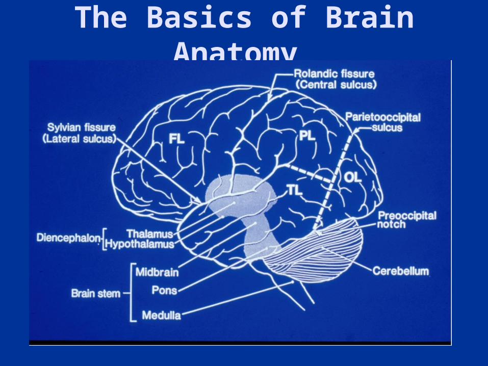
The Basics of Brain Anatomy
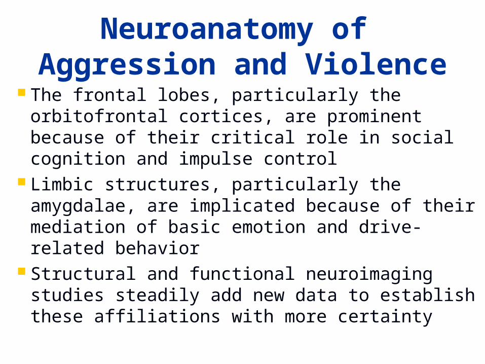
Neuroanatomy of Aggression and Violence
The frontal lobes, particularly the orbitofrontal cortices, are prominent because of their critical role in social cognition and impulse control
Limbic structures, particularly the amygdalae, are implicated because of their mediation of basic emotion and drive-related behavior
Structural and functional neuroimaging studies steadily add new data to establish these affiliations with more certainty
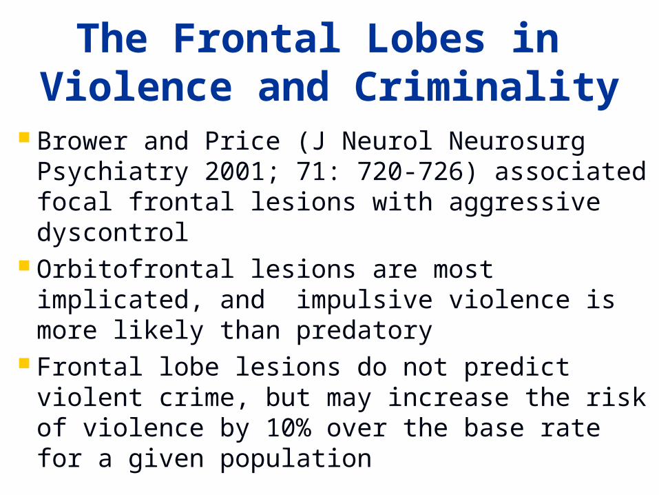
Brower and Price (J Neurol Neurosurg Psychiatry 2001; 71: 720-726) associated focal frontal lesions with aggressive dyscontrol
Orbitofrontal lesions are most implicated, and impulsive violence is more likely than predatory
Frontal lobe lesions do not predict violent crime, but may increase the risk of violence by 10% over the base rate for a given population
The Frontal Lobes in Violence and Criminality
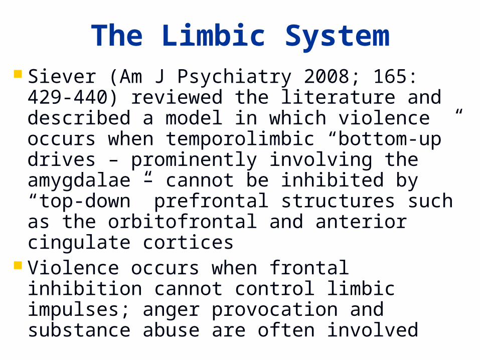
The Limbic System Siever (Am J Psychiatry 2008; 165: 429-
440) reviewed the literature and described a model in which violence occurs when temporolimbic “bottom-up” drives – prominently involving the amygdalae – cannot be inhibited by “top-down” prefrontal structures such as the orbitofrontal and anterior cingulate cortices
Violence occurs when frontal inhibition cannot control limbic impulses; anger provocation and substance abuse are often involved
White Matter Is Also Implicated Hoptman et al. (Biol Psychiatry 2002; 52:
9-14) used diffusion tensor imaging (DTI) in schizophrenics to associate impulsivity with lower fractional anisotropy (FA) in the inferior frontal white matter
Craig et al. (Mol Psychiatry 2009; 14: 946-953) used DTI in in psychopaths to correlate antisocial behavior with reduced FA in the uncinate fasciculus (UF)
An orbitofrontal cortex-UF-amygdala network is implicated in violent behavior
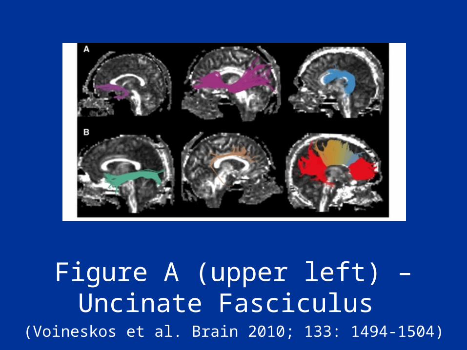
Figure A (upper left) – Uncinate Fasciculus
(Voineskos et al. Brain 2010; 133: 1494-1504)
Is the Right Side More Involved?
Recent frontotemporal dementia studies suggest loss of moral behavior with disease on right
Affiliative traits including warmth and empathy may rely on right ventromedial prefrontal and anteromedial temporal regions (Sollberger et al. Neuropsychologia 2009; 47: 2812-2827)
Sociopathic behavior can result from damage in these regions combined with right orbitofrontal damage causing disinhibition (Mendez MF. J Am Acad Psychiatry Law 2010; 38: 318-323)
Neuropharmacology of Violence
Serotonin is the major neurotransmitter implicated in the regulation of violence
Catecholamines (dopamine, norepinephrine) may potentiate violent behavior
Testosterone may influence aggressiveness, but is more associated with dominance than aggression (Glenn and Raine, Psychiatric Clin N Am 2008; 31: 463-475)
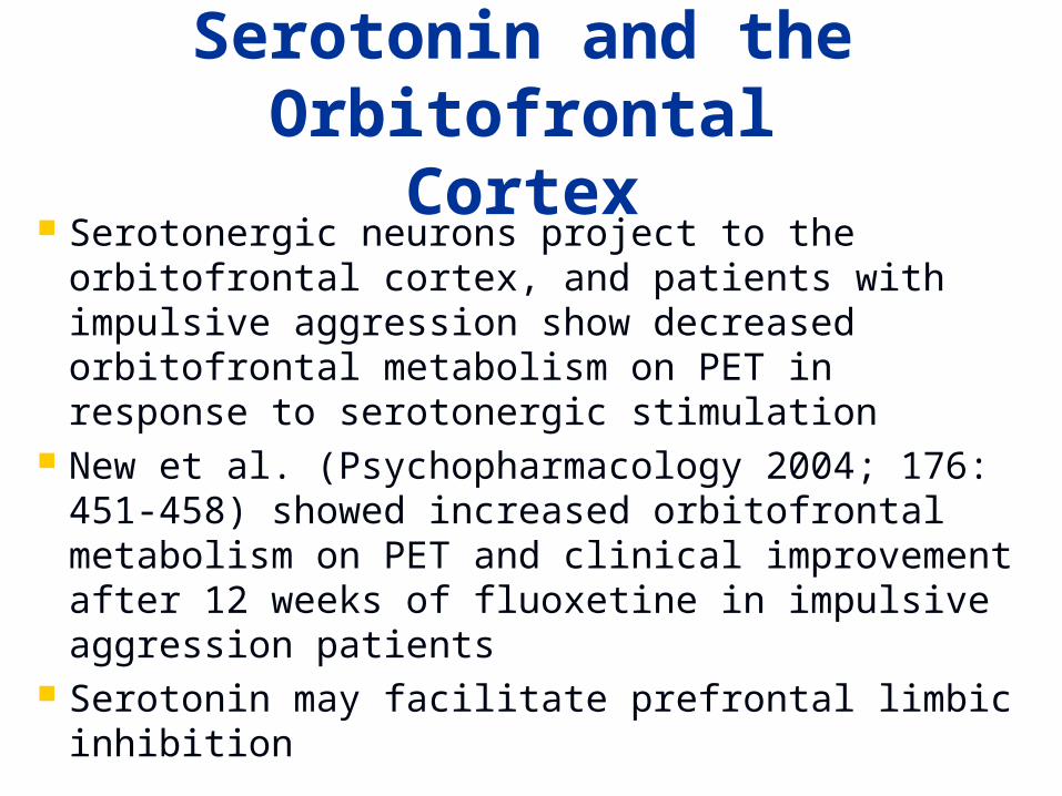
Serotonin and the Orbitofrontal Cortex
Serotonergic neurons project to the orbitofrontal cortex, and patients with impulsive aggression show decreased orbitofrontal metabolism on PET in response to serotonergic stimulation
New et al. (Psychopharmacology 2004; 176: 451-458) showed increased orbitofrontal metabolism on PET and clinical improvement after 12 weeks of fluoxetine in impulsive aggression patients
Serotonin may facilitate prefrontal limbic inhibition
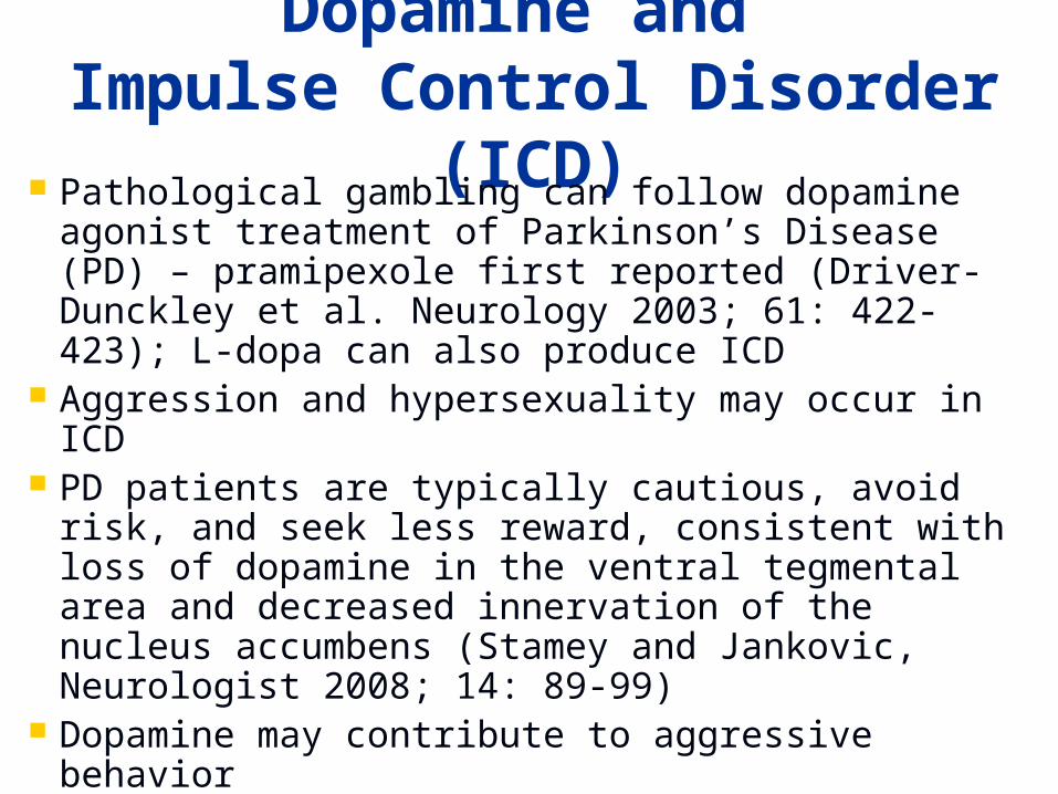
Dopamine and Impulse Control Disorder
(ICD) Pathological gambling can follow dopamine agonist treatment of Parkinson’s Disease (PD) – pramipexole first reported (Driver-Dunckley et al. Neurology 2003; 61: 422-423); L-dopa can also produce ICD
Aggression and hypersexuality may occur in ICD PD patients are typically cautious, avoid risk,
and seek less reward, consistent with loss of dopamine in the ventral tegmental area and decreased innervation of the nucleus accumbens (Stamey and Jankovic, Neurologist 2008; 14: 89-99)
Dopamine may contribute to aggressive behavior
Nature and Nurture Brain structure and function are determined by
both genetic and environmental factors Basic and clinical research supports some
heritability of violence, suggesting a genetic effect on brain systems engaged in aggression and its regulation
The brain is also subject to many environmental influences, including 1) prior brain pathology, 2) current brain dysfunction including drug use, and 3) sociocultural factors such as psychological stress, economic disadvantage, and low education
A Tentative Model of the Neurobiology of Violence
The frontal lobes, most notably orbitofrontal cortices, can fail to exert control over limbic structures, and violence may result
Limbic structures, most notably the amygdalae, can be excessively activated under certain circumstances to produce violence
Right cerebral dysfunction may be crucial Serotonin may inhibit and dopamine may
enhance violent behavior
Does This Model Apply to Suicide?
Jollant et al. (World J Biol Psychiatry; 2011 Mar 8) reviewed neuroimaging studies of suicide attempters and ideators published through 9/2010, and postulated dysfunction in ventrolateral, orbital, dorsomedial, and dorsolateral prefrontal cortices; anterior cingulate; amygdala; and white matter
Mann et al. (Arch Gen Psychiatry 2000; 57: 729-738) showed decreased serotonin transporter binding in the prefrontal cortex of autopsied suicide brains
These dysfunctions resemble those assoicated with aggression and violence (but right side not implicated)
Outline
Overview Violence and Traumatic Brain
Injury Violence and Dementia Neuroimaging in the Study of
Violence Conclusions
Traumatic Brain Injury (TBI)
Two major varieties of TBI: penetrating and nonpenetrating (including blast injury)
Nonpenetrating TBI is more prevalent in civilian populations, and blast injury is receiving much current attention; in wartime penetrating TBI becomes more common
Both types relevant to the neurobiology of aggression and violence
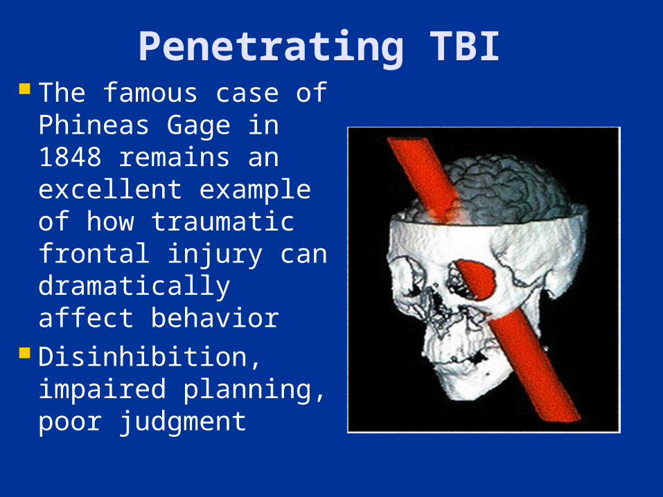
Penetrating TBI The famous case
of Phineas Gage in 1848 remains an excellent example of how traumatic frontal injury can dramatically affect behavior
Disinhibition, impaired planning, poor judgment
More Recent Investigation
The Vietnam Head Injury Study (Grafman et al. Neurology 1996; 46; 1231-1238) demonstrated that veterans with penetrating wounds of the ventromedial frontal lobes had a higher frequency of aggressive and violent behavior than control subjects, or veterans with lesions elsewhere in the brain
Nonpenetrating TBI Aggressive behavior is one of the major
limitations to successful recovery after nonpenetrating TBI
Kim et al. (J Neuropsychiatry Clin Neurosci 2007; 19; 106-127) found that 20-49% of children and ~ 33% of adults had agitation and aggression, usually beginning within the first year post-injury
Presence of frontal lobe lesions was associated with higher risk of post-TBI aggression
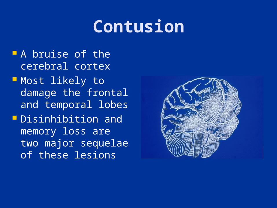
Contusion A bruise of the
cerebral cortex Most likely to
damage the frontal and temporal lobes
Disinhibition and memory loss are two major sequelae of these lesions
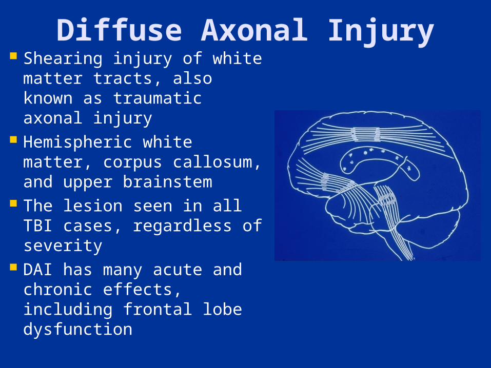
Diffuse Axonal Injury Shearing injury of white
matter tracts, also known as traumatic axonal injury
Hemispheric white matter, corpus callosum, and upper brainstem
The lesion seen in all TBI cases, regardless of severity
DAI has many acute and chronic effects, including frontal lobe dysfunction
Outline
Overview Violence and Traumatic Brain
Injury Violence and Dementia Neuroimaging in the Study of
Violence Conclusions
Violence in People with Dementia
Overall prevalence data are unavailable because of problems defining violent behavior, the uncertainty of dementia diagnoses, and the wide range of disorders that can cause dementia
Considering the two most common dementias, Alzheimer’s Disease (AD) and vascular dementia (VaD), Ballard et al. (Int Rev Psychiatry 2008; 20: 396-404) concluded that 40% display agitation
The risk of violent behavior in dementia likely depends on 1) specific diagnosis, 2) severity, 3) patient age, and 3) the localization of pathology
Alzheimer’s Disease Phenomena studied are typically
aggression, assaultiveness, and agitation Cummings and Victoroff (Neuropsychiatry
Neuropsychol Behav Neurol 1990; 3: 140-158) reported that 18-65% of AD patients display aggression or assaultiveness
Senanarong et al. (Dement Geriatr Cogn Disord 2008; 17: 14-20) correlated agitation in AD with markers of irritability, delusions, and disinhibition, and concluded that agitation is a manifestation of frontal lobe dysfunction
Frontotemporal Dementia
Miller et al. (Br J Psychiatry 1997; 170: 150-154) studied 22 patients with FTD and 22 with AD for evidence of antisocial behavior
10/22 patients with FTD had antisocial behavior as compared to 1/22 patients with AD
Behaviors included assault, indecent exposure, shoplifting, and hit-and-run driving, and did not occur before dementia developed
Disinhibition accounted for antisocial behavior
Outline
Overview Violence and Traumatic Brain
Injury Violence and Dementia Neuroimaging in the Study of
Violence Conclusions
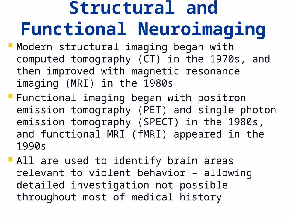
Structural and Functional
Neuroimaging Modern structural imaging began with computed tomography (CT) in the 1970s, and then improved with magnetic resonance imaging (MRI) in the 1980s
Functional imaging began with positron emission tomography (PET) and single photon emission tomography (SPECT) in the 1980s, and functional MRI (fMRI) appeared in the 1990s
All are used to identify brain areas relevant to violent behavior – allowing detailed investigation not possible throughout most of medical history
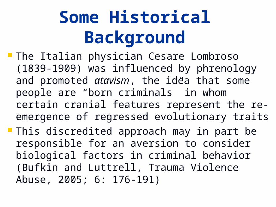
Some Historical Background
The Italian physician Cesare Lombroso (1839-1909) was influenced by phrenology and promoted atavism, the idea that some people are “born criminals” in whom certain cranial features represent the re-emergence of regressed evolutionary traits
This discredited approach may in part be responsible for an aversion to consider biological factors in criminal behavior (Bufkin and Luttrell, Trauma Violence Abuse, 2005; 6: 176-191)
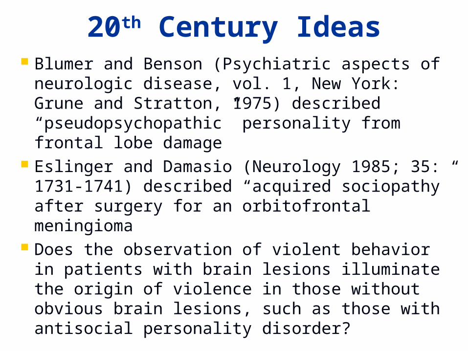
20th Century Ideas Blumer and Benson (Psychiatric aspects of
neurologic disease, vol. 1, New York: Grune and Stratton, 1975) described “pseudopsychopathic” personality from frontal lobe damage
Eslinger and Damasio (Neurology 1985; 35: 1731-1741) described “acquired sociopathy” after surgery for an orbitofrontal meningioma
Does the observation of violent behavior in patients with brain lesions illuminate the origin of violence in those without obvious brain lesions, such as those with antisocial personality disorder?
Antisocial Personality Disorder
Much of the recent neuroimaging work has been done with violent criminals, in whom antisocial personality disorder is common
Also known as psychopathy, sociopathy, and dyssocial personality disorder
DSM-IV (1994): Pervasive pattern of disregarding the rights of others (unlawful behaviors, deceitfulness, impulsivity, aggressiveness, disregard for safety, irresponsibility, lack of remorse)
These people know the rules but do not act by them
Neuroimaging and Violence – Pros
Structural neuroimaging has revolutionized neurology and psychiatry by identifying brain lesions that could not formerly be seen until autopsy
Functional neuroimaging has the potential for identifying brain regions that appear normal on structural neuroimaging, but may be abnormal at the cellular or synaptic level
Neuroimaging and Violence – Cons
Most violent and criminal behavior is not committed by people with obvious brain lesions, and even when present, brain damage may have an uncertain, or no relationship to the violence
Functional neuroimaging is beset by a host of methodological limitations, including lack of standardization, low signal-to-noise ratio, and inter-individual variability
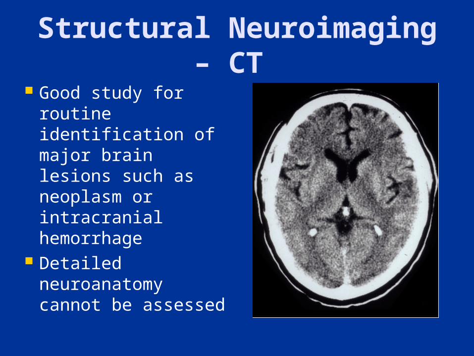
Structural Neuroimaging – CT
Good study for routine identification of major brain lesions such as neoplasm or intracranial hemorrhage
Detailed neuroanatomy cannot be assessed
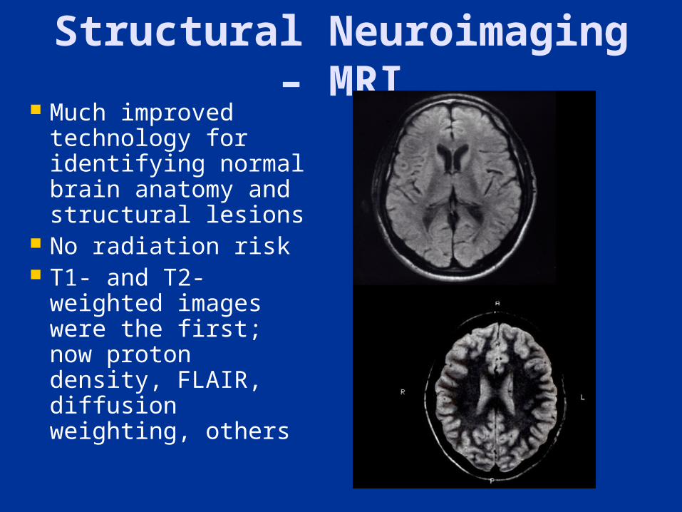
Structural Neuroimaging – MRI
Much improved technology for identifying normal brain anatomy and structural lesions
No radiation risk T1- and T2-
weighted images were the first; now proton density, FLAIR, diffusion weighting, others
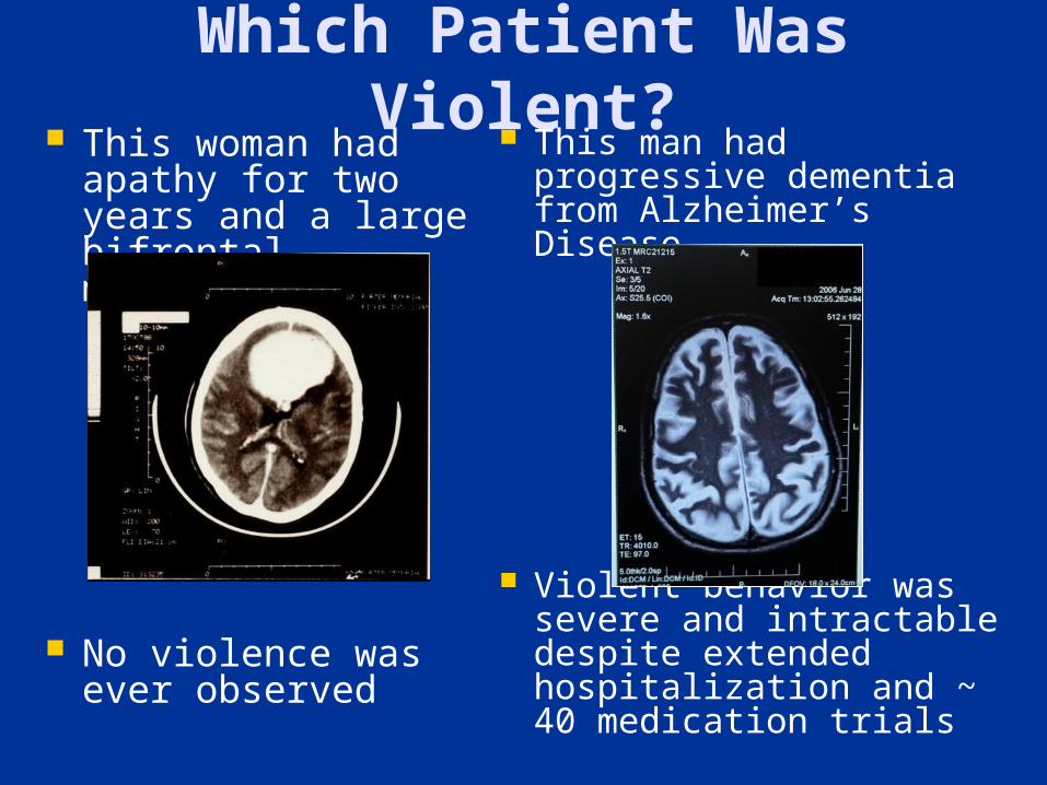
Which Patient Was Violent?
This woman had apathy for two years and a large bifrontal meningioma
No violence was ever observed
This man had progressive dementia from Alzheimer’s Disease
Violent behavior was severe and intractable despite extended hospitalization and ~ 40 medication trials
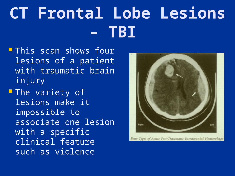
CT Frontal Lobe Lesions – TBI
This scan shows four lesions of a patient with traumatic brain injury
The variety of lesions make it impossible to associate one lesion with a specific clinical feature such as violence
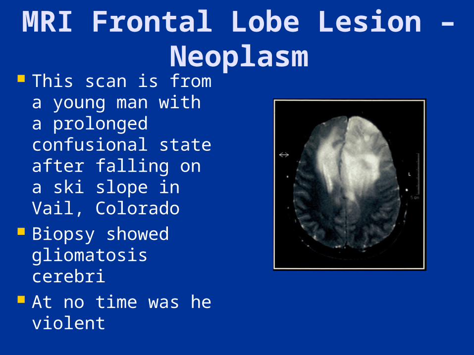
MRI Frontal Lobe Lesion – Neoplasm
This scan is from a young man with a prolonged confusional state after falling on a ski slope in Vail, Colorado
Biopsy showed gliomatosis cerebri
At no time was he violent
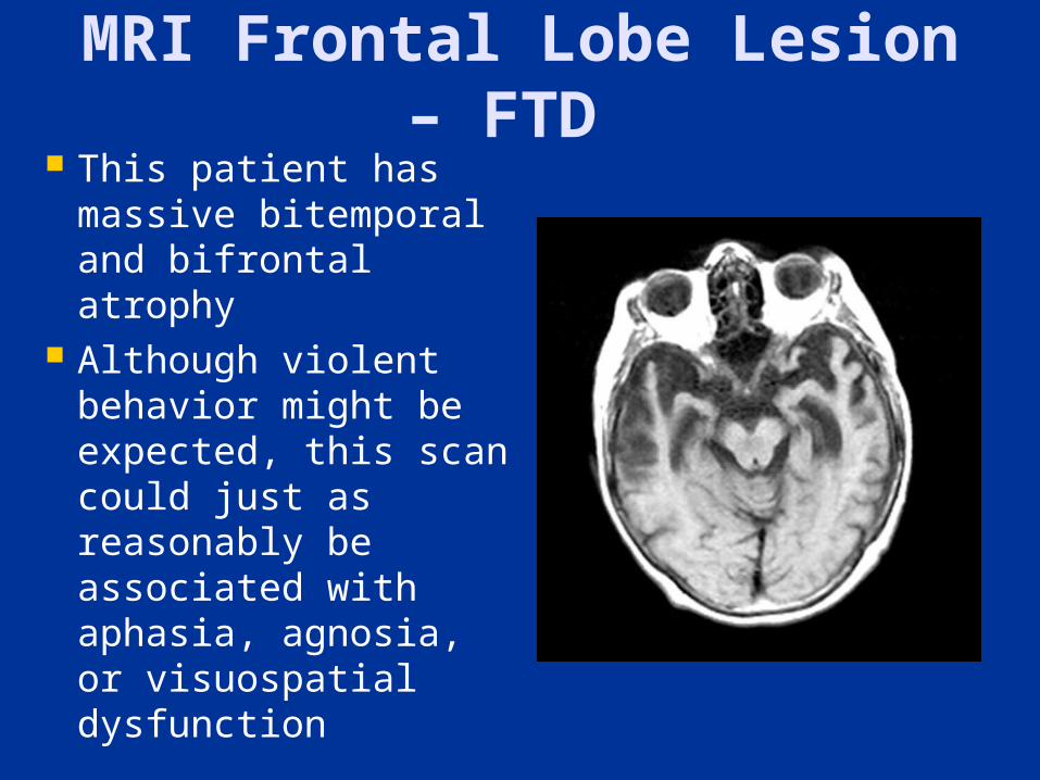
MRI Frontal Lobe Lesion – FTD
This patient has massive bitemporal and bifrontal atrophy
Although violent behavior might be expected, this scan could just as reasonably be associated with aphasia, agnosia, or visuospatial dysfunction
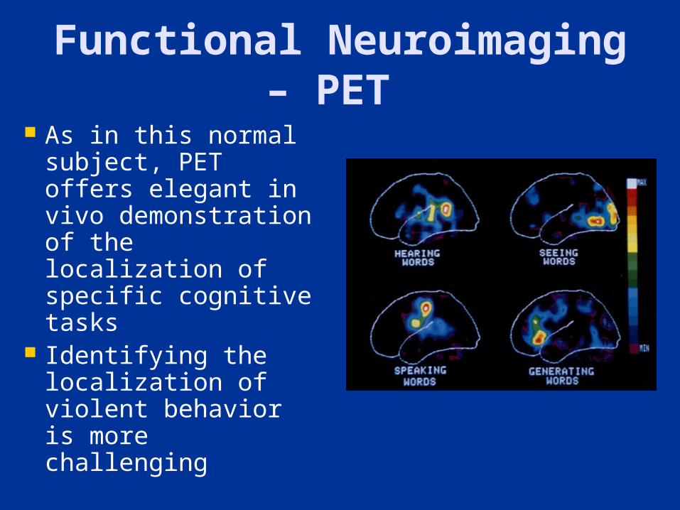
Functional Neuroimaging – PET
As in this normal subject, PET offers elegant in vivo demonstration of the localization of specific cognitive tasks
Identifying the localization of violent behavior is more challenging
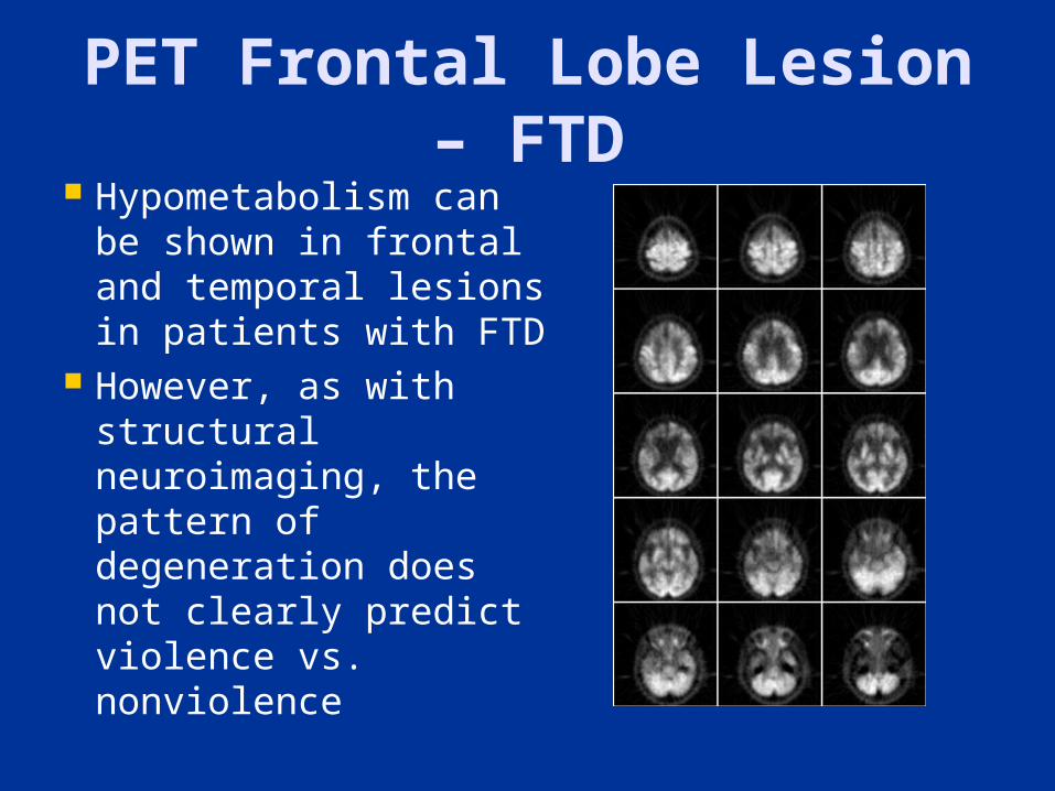
PET Frontal Lobe Lesion – FTD
Hypometabolism can be shown in frontal and temporal lesions in patients with FTD
However, as with structural neuroimaging, the pattern of degeneration does not clearly predict violence vs. nonviolence
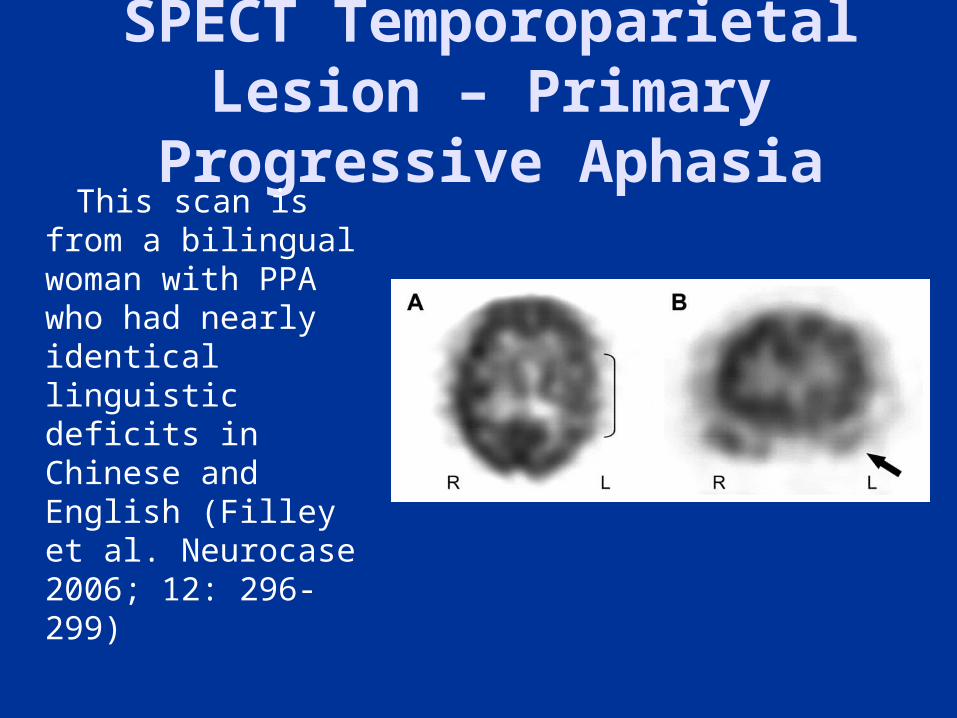
SPECT Temporoparietal Lesion – Primary
Progressive Aphasia This scan is
from a bilingual woman with PPA who had nearly identical linguistic deficits in Chinese and English (Filley et al. Neurocase 2006; 12: 296-299)
Problems with SPECT The pattern of hypometabolism is often
vague and can easily be misleading In many cases, abnormal areas are too
indistinct to be used for diagnosis, even in the case of well-known neurologic disorders
In cases of brain dysfunction without structural lesions, e.g. mild TBI, SPECT is clearly inadequate as a stand-alone diagnostic instrument in the courtroom (Wortzel et al. J Am Acad Psychiatry Law 2008; 36: 310-322)
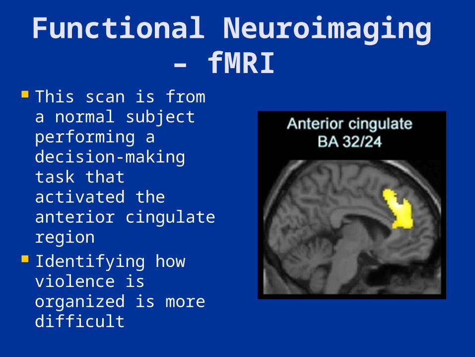
Functional Neuroimaging – fMRI
This scan is from a normal subject performing a decision-making task that activated the anterior cingulate region
Identifying how violence is organized is more difficult
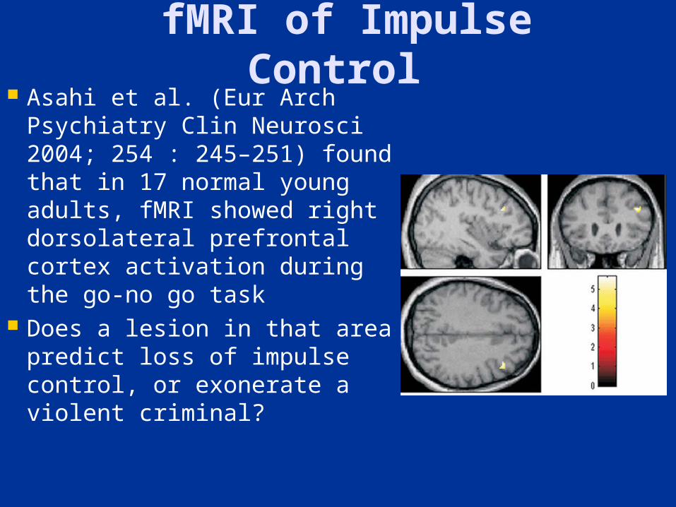
fMRI of Impulse Control
Asahi et al. (Eur Arch Psychiatry Clin Neurosci 2004; 254 : 245–251) found that in 17 normal young adults, fMRI showed right dorsolateral prefrontal cortex activation during the go-no go task
Does a lesion in that area predict loss of impulse control, or exonerate a violent criminal?
Structural Neuroimaging in Antisocial Personality
Disorder Raine et al. (Arch Gen Psychiatry 2000; 57: 119-127) used MRI to show an 11% reduction in prefrontal gray matter volume
Narayan et al. (Am J Psychiatry 2007; 164: 1418-1427) used MRI to show decreased cortical thickness in inferior mesial frontal cortices of violent antisocial personality disorder subjects
Yang et al. (Arch Gen Psychiatry 2009; 66: 986-994) used MRI to show bilateral amygdala volume reduction in psychopaths
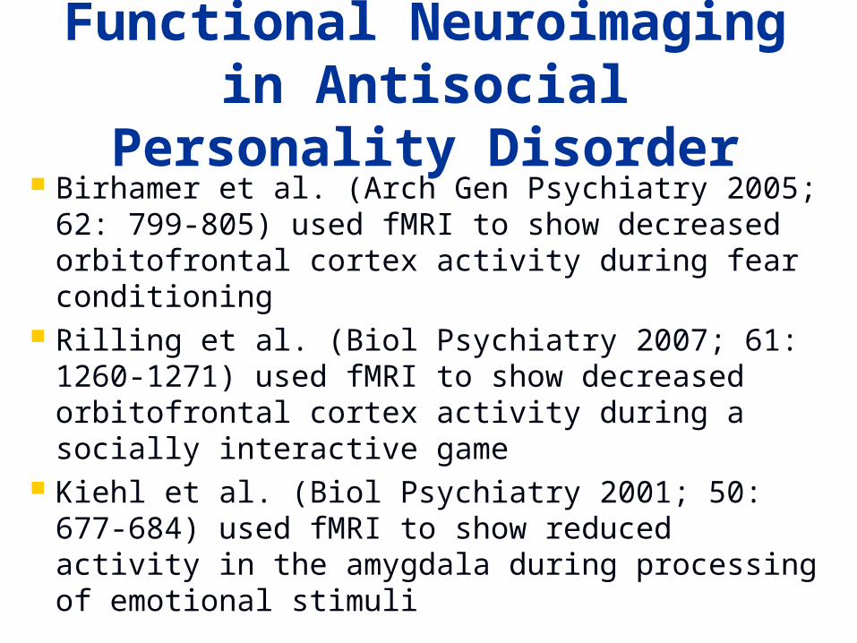
Functional Neuroimaging in Antisocial Personality
Disorder Birhamer et al. (Arch Gen Psychiatry 2005; 62:
799-805) used fMRI to show decreased orbitofrontal cortex activity during fear conditioning
Rilling et al. (Biol Psychiatry 2007; 61: 1260-1271) used fMRI to show decreased orbitofrontal cortex activity during a socially interactive game
Kiehl et al. (Biol Psychiatry 2001; 50: 677-684) used fMRI to show reduced activity in the amygdala during processing of emotional stimuli
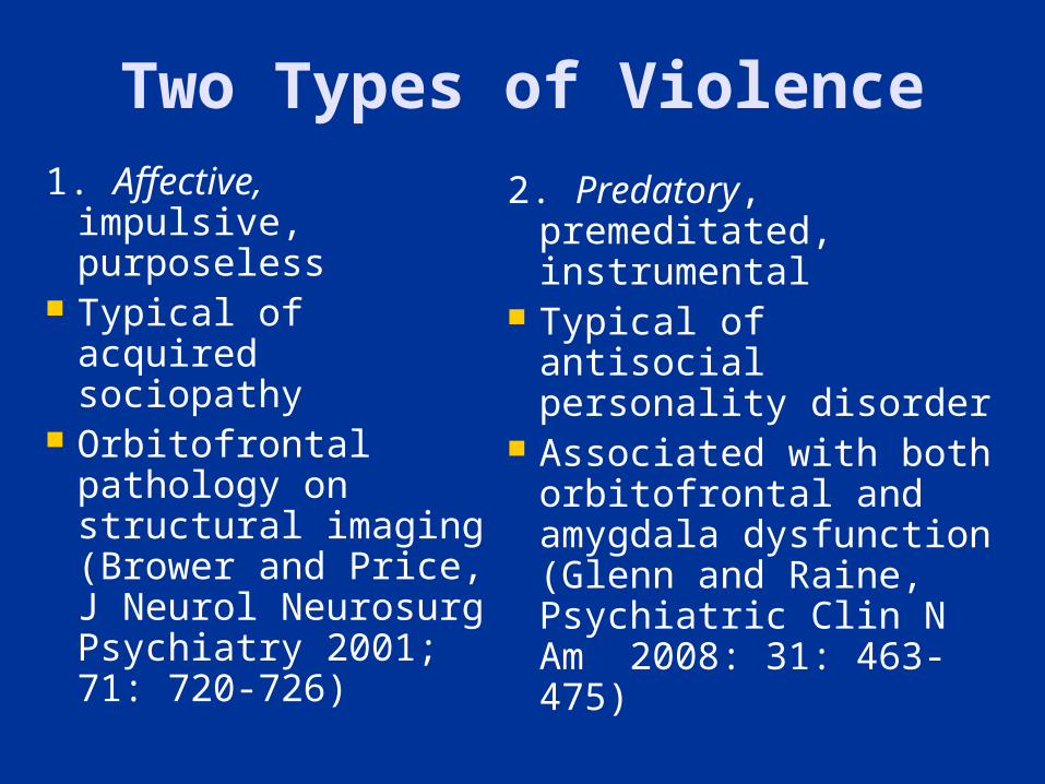
Two Types of Violence1. Affective,
impulsive, purposeless
Typical of acquired sociopathy
Orbitofrontal pathology on structural imaging (Brower and Price, J Neurol Neurosurg Psychiatry 2001; 71: 720-726)
2. Predatory, premeditated, instrumental
Typical of antisocial personality disorder
Associated with both orbitofrontal and amygdala dysfunction (Glenn and Raine, Psychiatric Clin N Am 2008: 31: 463-475)
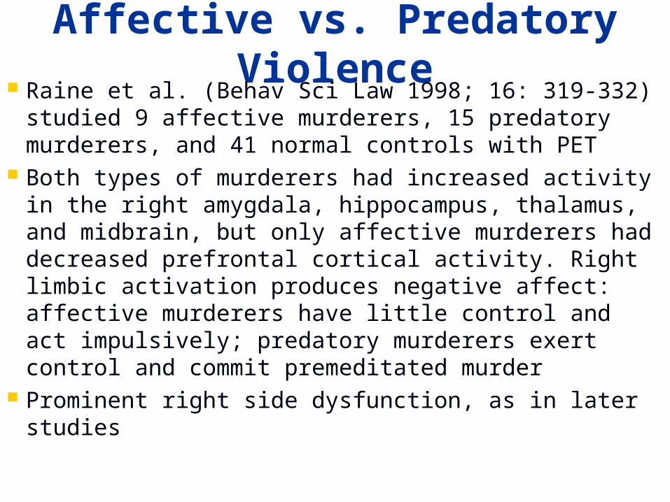
Affective vs. Predatory Violence
Raine et al. (Behav Sci Law 1998; 16: 319-332) studied 9 affective murderers, 15 predatory murderers, and 41 normal controls with PET
Both types of murderers had increased activity in the right amygdala, hippocampus, thalamus, and midbrain, but only affective murderers had decreased prefrontal cortical activity. Right limbic activation produces negative affect: affective murderers have little control and act impulsively; predatory murderers exert control and commit premeditated murder
Prominent right side dysfunction, as in later studies
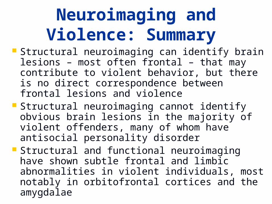
Neuroimaging and Violence: Summary
Structural neuroimaging can identify brain lesions – most often frontal – that may contribute to violent behavior, but there is no direct correspondence between frontal lesions and violence
Structural neuroimaging cannot identify obvious brain lesions in the majority of violent offenders, many of whom have antisocial personality disorder
Structural and functional neuroimaging have shown subtle frontal and limbic abnormalities in violent individuals, most notably in orbitofrontal cortices and the amygdalae
Not Guilty by Reason of Brain Damage?
Although much more study is needed of brain lesions increasing the risk of violence, people with certain disorders know the rules of conduct but fail to act by them
These people have reduced impulse control, either from frontal lobe or limbic system dysfunction
Sapolsky (Phil Trans R Soc London B 2004; 359: 1787-1796) suggests that in cases of violent crime, the insanity defense – not knowing right from wrong – should be expanded to consider the possibility of impaired volition – diminished impulse control
Outline
Overview Violence and Traumatic Brain
Injury Violence and Dementia Neuroimaging in the Study of
Violence Conclusions
Conclusions 1. The frontal lobes – mainly the orbitofrontal
cortices – normally act to regulate limbic impulses – which arise prominently from the amygdalae
2. Violence can occur as a result of dysfunction in a distributed frontolimbic network related to aggression and its regulation; the right side may be more critical
3. A distinction may exist between affective violence from orbitofrontal injury (e.g. acquired sociopathy) – vs. predatory violence from both orbitofrontal and amygdala damage (e.g. antisocial personality disorder)
Conclusions 4. Serotonin appears to inhibit violence, and
dopamine may enhance it 5. Traumatic brain injury, both penetrating
and nonpenetrating, is associated with an increased risk of violence
6. Dementia, most notably frontotemporal dementia, is associated with an increased risk of violence7. Neuroimaging has promise in the evaluation of violent behavior, but cannot accurately predict the risk of violence in an individual person
Conclusions8. Whether structural or functional,
neuroimaging is only one component of what is ideally a complete data set that permits the most reasonable assignment of factors accounting for a violent act
9. Impaired impulse control from altered brain function is relevant because some violent people are unable to act by the rules they have learned
10. Despite uncertainties introduced by neurobiology, the legal system should consider brain–behavior relationships in dealing with violent crime