Embed Size (px)
Citation preview
10/10/2019
1
The Neuro-Ophthalmology of
Multiple Sclerosis
Monterey Symposium
California Optometric Association 2019
Leonard V. Messner, OD, FAAO
Professor of Optometry
Vice President for Strategy & Institutional Advancement
Illinois College of Optometry
Disclosures
• King Devick Technologies (scientific advisory
board)
• Heidelberg Engineering (scientific advisory
board)
• Illinois Society for the Prevention of Blindness
(clinical research grants)
Key Points
• Diagnostic criteria for MS
• Pathogenesis & Classification of MS
• Neuro-ophthalmic manifestations of MS:
– Afferent system:
• Optic neuritis
– Efferent system
• Brainstem motility disorders
• Nystagmus
• Cranial neuropathies
• Visual biomarkers of disease activity
2010 Revised MacDonald Diagnostic
Criteria for MS
• Evidence of damage (clinical attacks and/or
lesions) in 2 or more separate areas of the
CNS (including the brain, spinal cord, and optic
nerves), plus evidence that the damage
happened 1 or more months apart, plus
evidence that the damage did not happen
because of another disease.
Polman C, et al. Ann Neurology 2011
2017 Changes from 2010 MacDonald
Diagnostic Criteria for MS
• CIS with CSF-specific oligoclonal bands (no
requirement for dissemination in time)
• Inclusion of cortical lesions
• Both symptomatic and asymptomatic MRI
lesions qualify for dissemination in space or
time
Push for earlier diagnosis!
Thompason AJ, et al. et al. Lancet 2017
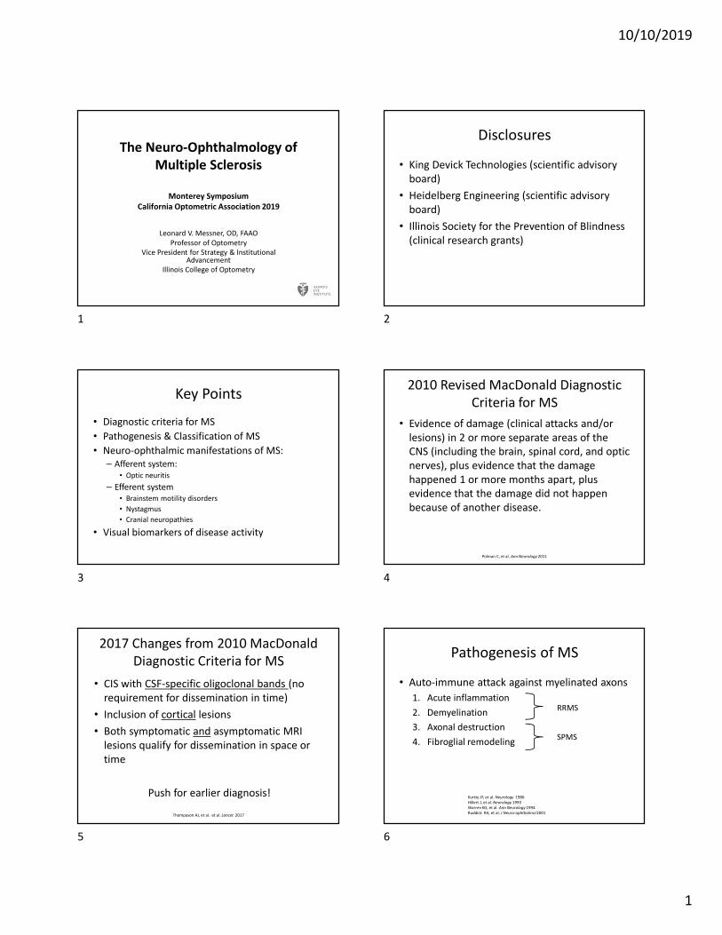
Pathogenesis of MS
• Auto-immune attack against myelinated axons
1. Acute inflammation
2. Demyelination
3. Axonal destruction
4. Fibroglial remodeling
Kurtze JF, et al. Neurology 1986
Hillert J, et al. Neurology 1993
Warren KG, et al. Ann Neurology 1994
Ruddick RA, et al. J Neuro-ophthalmol 2001
RRMS
SPMS
1 2
3 4
5 6
10/10/2019
2
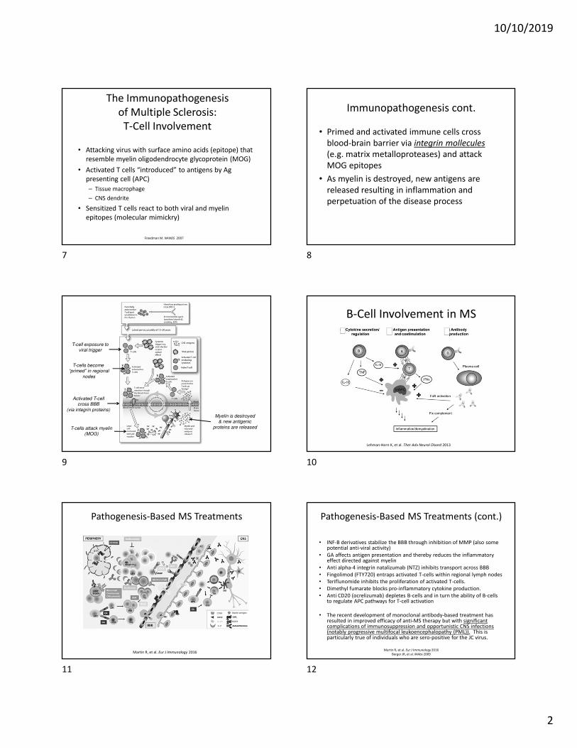
The Immunopathogenesis
of Multiple Sclerosis:
T-Cell Involvement
• Attacking virus with surface amino acids (epitope) that
resemble myelin oligodendrocyte glycoprotein (MOG)
• Activated T cells “introduced” to antigens by Ag
presenting cell (APC)
– Tissue macrophage
– CNS dendrite
• Sensitized T cells react to both viral and myelin
epitopes (molecular mimickry)
Freedman M. NANOS 2007
Immunopathogenesis cont.
• Primed and activated immune cells cross
blood-brain barrier via integrin mollecules
(e.g. matrix metalloproteases) and attack
MOG epitopes
• As myelin is destroyed, new antigens are
released resulting in inflammation and
perpetuation of the disease process
T-cell exposure toviral trigger
T-cells become “primed” in regional
nodes
Activated T-cellcross BBB
(via integrin proteins)
T-cells attack myelin(MOG)
Myelin is destroyed& new antigenic
proteins are released
B-Cell Involvement in MS
Lehman-Horn K, et al. Ther Adv Neurol Disord 2013
Pathogenesis-Based MS Treatments
Martin R, et al. Eur J Immunology 2016
Pathogenesis-Based MS Treatments (cont.)
• INF-B derivatives stabilize the BBB through inhibition of MMP (also some potential anti-viral activity)
• GA affects antigen presentation and thereby reduces the inflammatory effect directed against myelin
• Anti alpha-4 integrin natalizumab (NTZ) inhibits transport across BBB
• Fingolimod (FTY720) entraps activated T-cells within regional lymph nodes
• Teriflunomide inhibits the proliferation of activated T-cells.
• Dimethyl fumarate blocks pro-inflammatory cytokine production.
• Anti CD20 (ocrelizumab) depletes B-cells and in turn the ability of B-cells to regulate APC pathways for T-cell activation
• The recent development of monoclonal antibody-based treatment has resulted in improved efficacy of anti-MS therapy but with significant complications of immunosuppression and opportunistic CNS infections (notably progressive multifocal leukoencephalopathy (PML)). This is particularly true of individuals who are sero-positive for the JC virus.
Martin R, et al. Eur J Immunology 2016
Berger JR, et al. MAbs 2009
7 8
9 10
11 12
10/10/2019
3
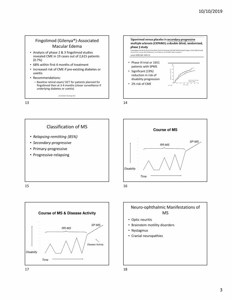
Fingolimod (Gilenya®)-Associated
Macular Edema
• Analysis of phase 2 & 3 fingolimod studies revealed CME in 19 cases out of 2,615 patients (0.7%)
• 68% within first 4 months of treatment
• Increased risk of CME if pre-existing diabetes or uveitis
• Recommendations:
– Baseline retinal exam/ OCT for patients planned for fingolimod then at 3-4 months (closer surveillance if underlying diabetes or uveitis)
Jain & Bhatti. Neurology 2012
• Phase III trial or 1651
patients with SPMS
• Significant (19%)
reduction in risk of
disability progression
• 2% risk of CME
Classification of MS
• Relapsing-remitting (85%)
• Secondary-progressive
• Primary-progressive
• Progressive-relapsing
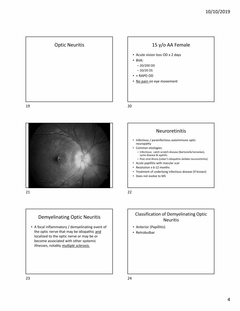
Course of MS
Time
Disability
RR-MSSP-MS
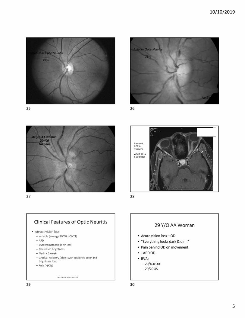
Course of MS & Disease Activity
Time
Disability
RR-MSSP-MS
Disease Activity
Neuro-ophthalmic Manifestations of
MS
• Optic neuritis
• Brainstem motility disorders
• Nystagmus
• Cranial neuropathies
13 14
15 16
17 18
10/10/2019
4
Optic Neuritis 15 y/o AA Female
• Acute vision loss OD x 2 days
• BVA:
– 20/200 OD
– 20/20 OS
• + RAPD OD
• No pain on eye movement
Neuroretinitis
• Infectious / parainfectious autoimmune optic neuropathy
• Common etiologies:
– Infectious: catch scratch disease (Bartonella henselae), Lyme disease & syphilis
– Post viral illness (Leber’s idiopathic stellate neuroretinitis)
• Acute papillitis with macular scar
• Resolution x 6-12 months
• Treatment of underlying infectious disease (if known)
• Does not evolve to MS
Demyelinating Optic Neuritis
• A focal inflammatory / demyelinating event of
the optic nerve that may be idiopathic and
localized to the optic nerve or may be or
become associated with other systemic
illnesses, notably multiple sclerosis.
Classification of Demyelinating Optic
Neuritis
• Anterior (Papillitis)
• Retrobulbar
19 20
21 22
23 24
10/10/2019
5
Retrobulbar Optic Neuritis
75%
Anterior Optic Neuritis
25%
No Blood!
34 y/o AA woman
20/400
No pain Elevated ACE &
lysozyme
+CXR (BHA & infiltrates
Clinical Features of Optic Neuritis
• Abrupt vision loss
– variable (average 20/60 x ONTT)
– APD
– Dyschromatopsia (> VA loss)
– Decreased brightness
– Nadir x 2 weeks
– Gradual recovery (albeit with sustained color and
brightness loss)
– Pain (>90%)
Beck RW, et al. N Engl J Med 1992
29 Y/O AA Woman
• Acute vision loss – OD
• “Everything looks dark & dim.”
• Pain behind OD on movement
• +APD OD
• BVA:
– 20/400 OD
– 20/20 OS
25 26
27 28
29 30
10/10/2019
6
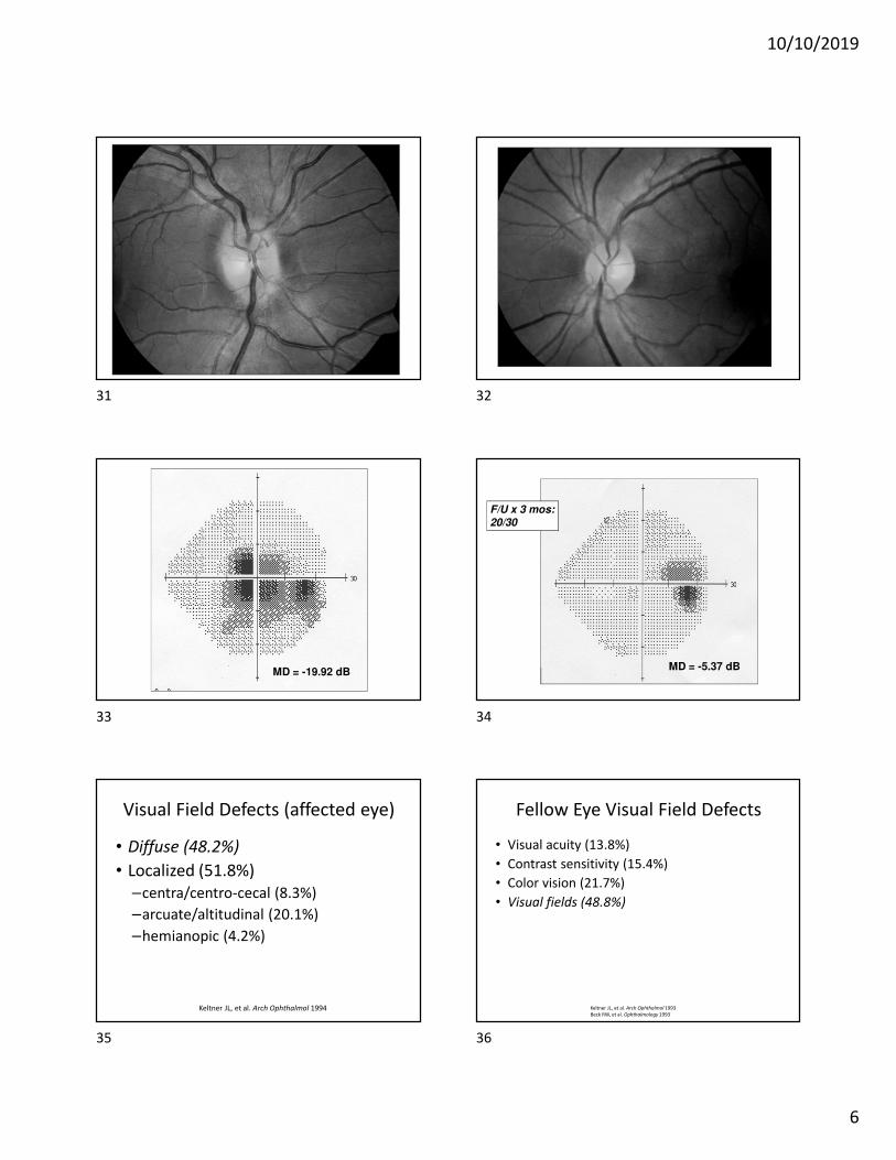
MD = -19.92 dBMD = -5.37 dB
F/U x 3 mos:
20/30
Visual Field Defects (affected eye)
• Diffuse (48.2%)
• Localized (51.8%)
–centra/centro-cecal (8.3%)
–arcuate/altitudinal (20.1%)
–hemianopic (4.2%)
Keltner JL, et al. Arch Ophthalmol 1994
Fellow Eye Visual Field Defects
• Visual acuity (13.8%)
• Contrast sensitivity (15.4%)
• Color vision (21.7%)
• Visual fields (48.8%)
Keltner JL, et al. Arch Ophthalmol 1993
Beck RW, et al. Ophthalmology 1993
31 32
33 34
35 36
10/10/2019
7
30-y/o womanc/o vision “dark & dim”
OS x 5 days (getting worse)
VA = 20/20VA = 20/80
+ APD
+Pain
25-y/o woman c/odecreased vision,
ODx one week
(getting worse)
VA = 20/40+ APD
+ Pain
VA = 20/20
37 38
39 40
41 42
10/10/2019
8
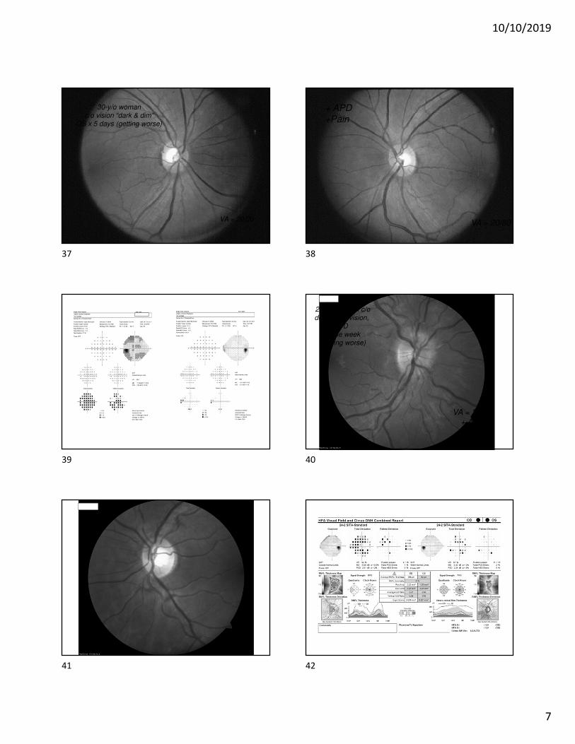
MRI Characteristics
• T1WI post Gd
– Enhancement of acute lesions
– Hypointense axonal atrophy (“black holes”)
with chronic lesions
• T2WI
• FLAIR
Hyperintense withacute & chronic lesions
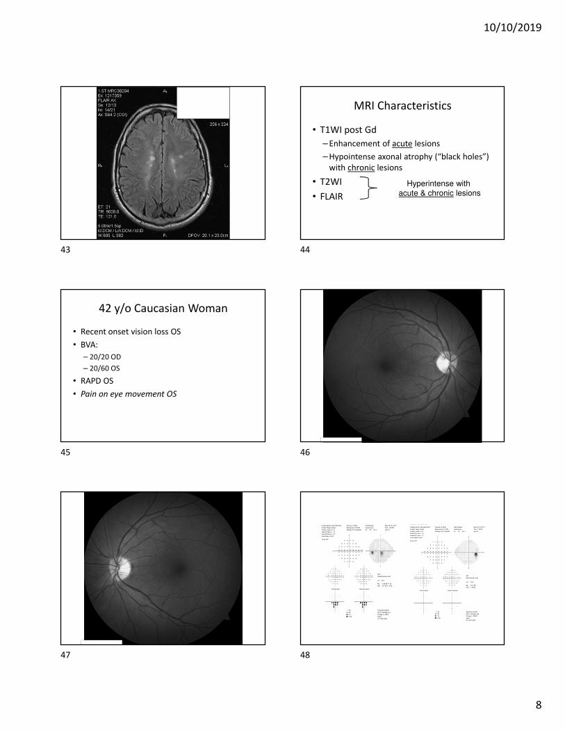
42 y/o Caucasian Woman
• Recent onset vision loss OS
• BVA:
– 20/20 OD
– 20/60 OS
• RAPD OS
• Pain on eye movement OS
43 44
45 46
47 48
10/10/2019
9
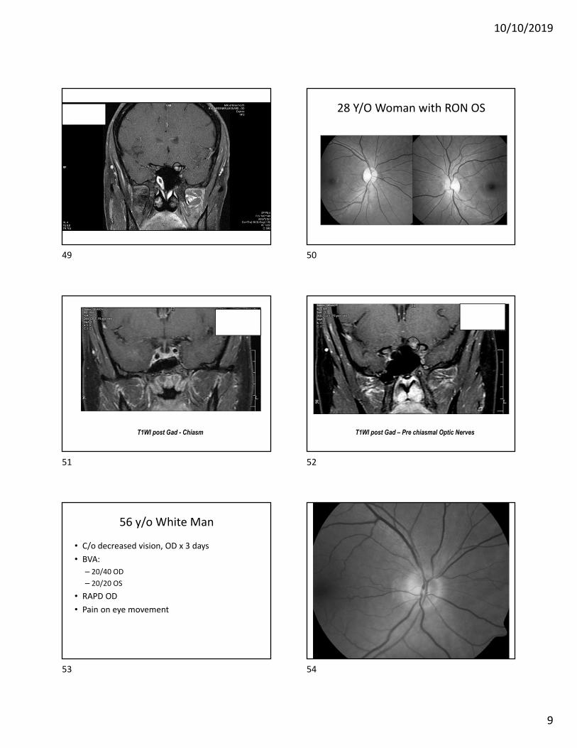
28 Y/O Woman with RON OS
T1WI post Gad - Chiasm T1WI post Gad – Pre chiasmal Optic Nerves
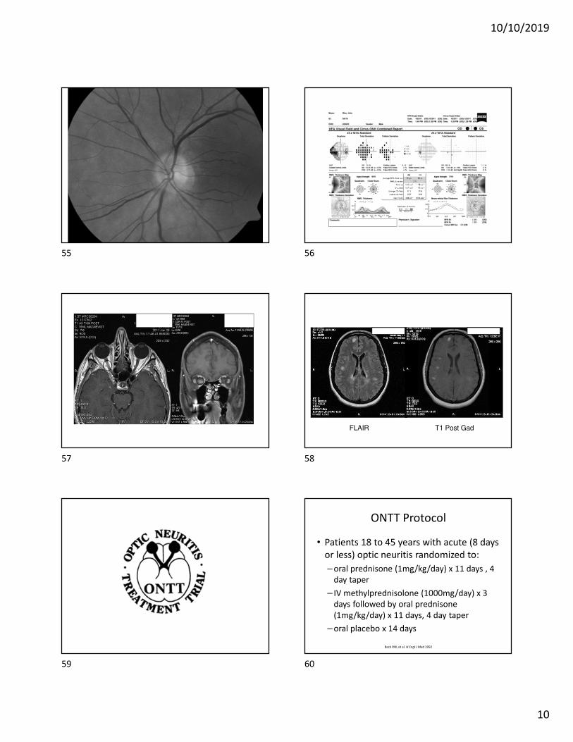
56 y/o White Man
• C/o decreased vision, OD x 3 days
• BVA:
– 20/40 OD
– 20/20 OS
• RAPD OD
• Pain on eye movement
49 50
51 52
53 54
10/10/2019
10
FLAIR T1 Post Gad
ONTT Protocol
• Patients 18 to 45 years with acute (8 days
or less) optic neuritis randomized to:
– oral prednisone (1mg/kg/day) x 11 days , 4
day taper
– IV methylprednisolone (1000mg/day) x 3
days followed by oral prednisone
(1mg/kg/day) x 11 days, 4 day taper
– oral placebo x 14 days
Beck RW, et al. N Engl J Med 1992
55 56
57 58
59 60
10/10/2019
11
ONTT Results (6 months)
• Quicker recovery with IV steroids
• Visual recovery excellent for ALL groups
• Increased # of new attacks with oral
prednisone alone
Beck RW, et al. N Engl J Med 1992
ONTT Results (2 years)
• Development of MS
– Oral prednisone (14.7%)
– Placebo (16.7%)
– IV steroids (7.5%)
Beck RW, et al. N Engl J Med 1993
ONTT Results (2 years)
• Abnormal MRI (2 or more WML’s 3mm or greater in diameter)
– Placebo (36%)
– IV steroids (16%)
Trobe JD. Arch Ophthalmol 1994
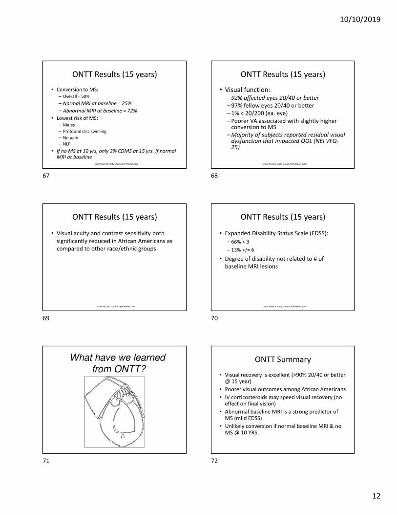
RISK OF DEFINITE MS BY TREATMENT GROUP
PlaceboPrednisoneIntravenous
.5 1 2 3 4
% w
ith
De
fin
ite
MS
Years of Follow-Up
35
30
25
20
15
10
5
0
ONTT Results (3 years)
• MRI / MS Correlation
– 3 or more plaques (43.1%)
– Normal MRI (9.3%)
• MS by TX Group
– IV steroids (24.7%)
– oral prednisone (29.8%)
– placebo (29.8)
Beck RW. Arch Ophthalmol 1995
61 62
63 64
65 66
10/10/2019
12
ONTT Results (15 years)
• Conversion to MS:
– Overall = 50%
– Normal MRI at baseline = 25%
– Abnormal MRI at baseline = 72%
• Lowest risk of MS:
– Males
– Profound disc swelling
– No pain
– NLP
• If no MS at 10 yrs, only 2% CDMS at 15 yrs. If normal MRI at baseline
Optic Neuritis Study Group Arch Neurol 2008
ONTT Results (15 years)
• Visual function:– 92% affected eyes 20/40 or better
– 97% fellow eyes 20/40 or better
– 1% < 20/200 (ea. eye)
– Poorer VA associated with slightly higher conversion to MS
– Majority of subjects reported residual visual dysfunction that impacted QOL (NEI VFQ-25)
Optic Neuritis Study Group Arch Neurol 2008
ONTT Results (15 years)
• Visual acuity and contrast sensitivity both
significantly reduced in African Americans as
compared to other race/ethnic groups
Moss HE, et al. JAMA Ophthalmol 2014
ONTT Results (15 years)
• Expanded Disability Status Scale (EDSS):
– 66% < 3
– 13% >/= 6
• Degree of disability not related to # of
baseline MRI lesions
Optic Neuritis Study Group Arch Neurol 2008
What have we learned
from ONTT?ONTT Summary
• Visual recovery is excellent (>90% 20/40 or better @ 15 year)
• Poorer visual outcomes among African Americans
• IV corticosteroids may speed visual recovery (no effect on final vision)
• Abnormal baseline MRI is a strong predictor of MS (mild EDSS)
• Unlikely conversion if normal baseline MRI & no MS @ 10 YRS.
67 68
69 70
71 72
10/10/2019
13
Can MS be prevented
in patients with
“high-risk” optic neuritis?
CHAMPS
• Controlled High-Risk Avonex®
Multiple Sclerosis Prevention Study
• Combined corticosteroids / interferon beta-1a (Avonex®)
CHAMPS - Patient Enrollment
• 50 clinical centers (US & Canada)
• 383 patients - first acute demyelinating event
– eye (optic neuritis)
– spinal cord (incomplete transverse myelitis)
– brainstem/cerebellar syndrome
• Abnormal brain MRI at baseline
CHAMPS – Treatment Protocol
• All patients given IV corticosteroids with oral taper within 14 days of symptoms (ONTT protocol)
• 50% given weekly IFN beta-1a (Avonex®) x IM injection within 27 days of symptoms
• 50% given weekly placebo injection within 27 days of symptoms
CHAMPS (3 year results)
• Development of MS significantly reduced for
Avonex® group
– 35% Avonex
– 50% placebo
• Reduction in size and number of new brain lesions
among Avonex® patients
– 53% Avonex
– 82% placebo
Jacobs LD, et al. N Engl J Med 2000
Early Treatment of MS Study
(ETOMS)
• Weekly injections of interferon beta-1a (Rebif®) vs. placebo for high-risk MS patients
• 2-year MS conversion:
– Rebif: 34%
– Placebo: 45%
• Fewer new lesions & T2 volume with Rebif ®
Comi G, et al. Lancet 2001
73 74
75 76
77 78
10/10/2019
14
Betaferon/Betaseron in Newly Emerging
Multiple Sclerosis for Initial Therapy
(BENEFIT Study)
• Evaluation of every other day injections of
interferon beta-1b for patients with first
demyelinating event & abnormal MRI
• Conversion to MS (2-year results):
– 45% with placebo
– 28% with IFN-b-1b
Kappos L, et al. Neurology 2006
PreCISe Study
• Glatiramer acetate (Copaxone®) 20 mg subQ
qd
• 2-year results:
– 45% reduction in MS
– 57% reduction in new T2 lesions
– 66% reduction if optic neuritis as first presenting
sign
Comi G, et al. Lancet 2009
Is there a penalty for delay in treatment? CHAMPS In Ongoing Neurological
Surveillance
(CHAMPIONS)• Open label extension of immediate treatment (IT)
and delayed treatment (DT) groups in CHAMPS
• 5-year results:
– 36% of IT group developed MS
– 49% of DT group developed MS
• 10-year results:
– 58% of IT group developed MS
– 69% of DT group developed MS
• 2X annualized relapse rate in DT group
CHAMPIONS Study Group Neurology 2006
Kinkel RP, et al. Arch Neurol 2012
Summary of Optic Neuritis
• Excellent visual prognosis
• Natural history suggests that CIS patients with positive MRIs will progress to CDMS
• Corticosteroids followed by disease-modifying therapy should be strongly considered for high-risk patients
• Long-term studies suggest there is a neurological penalty for not starting immunomodulatorytherapy at onset
Brainstem Motility Disorders
79 80
81 82
83 84
10/10/2019
15
The Internuclear Ophthalmoplegias
• Internuclear ophthalmoplegia (INO)
• Bilateral internuclear ophthalmoplegia (BINO)
• Wall-eyed bilateral internuclear
ophthalmoplegia (WEBINO)
• One-and-one-half syndrome
The Internuclear Ophthalmoplegias
• Internuclear ophthalmoplegia (INO)
• Bilateral internuclear ophthalmoplegia (BINO)
• Wall-eyed bilateral internuclear
ophthalmoplegia (WEBINO)
• One-and-one-half syndrome
Internuclear Ophthalmoplegia
• Lesion of MLF
• Ipsilateral adduction deficit
• Abducting nystagmus in fellow eye (+/-)
• Convergence abolished = midbrain (anterior INO)
• Convergence spared = pons (posterior INO)
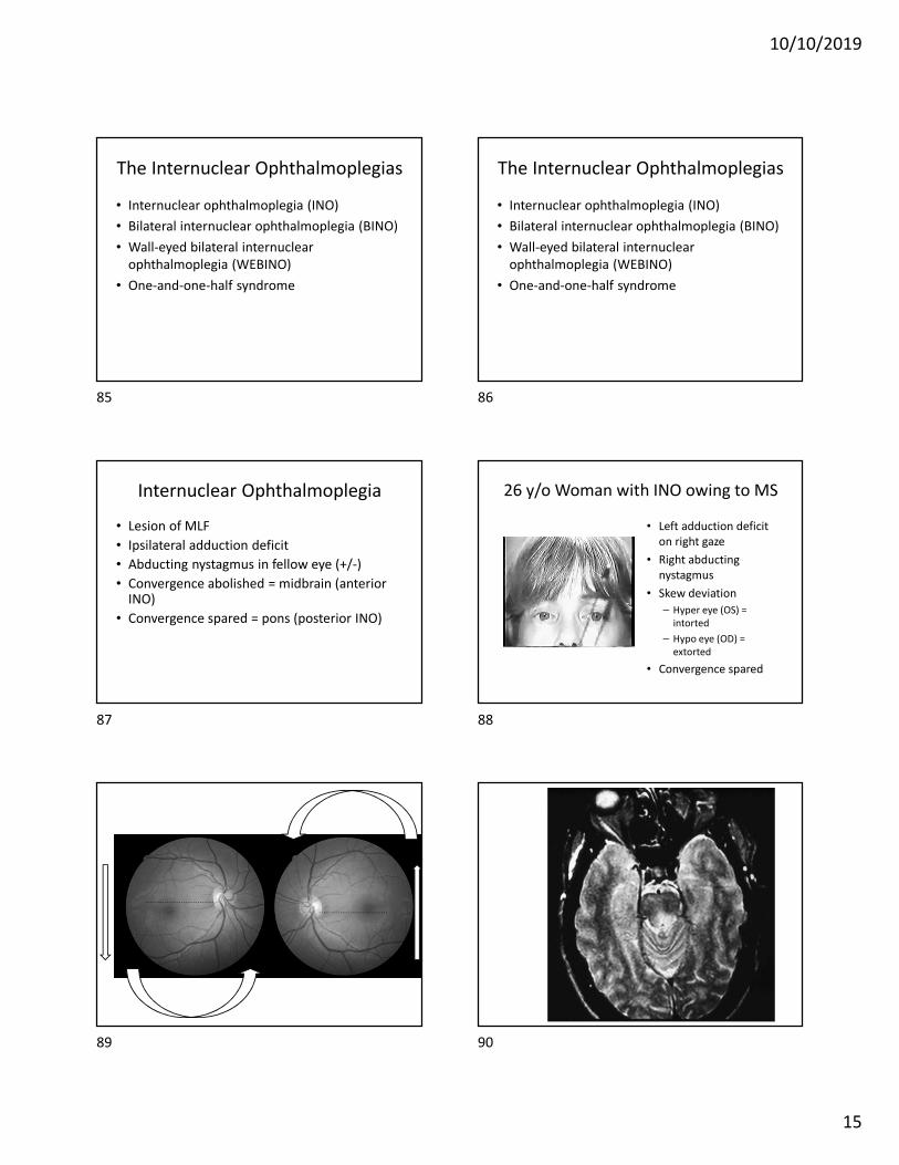
26 y/o Woman with INO owing to MS
• Left adduction deficit
on right gaze
• Right abducting
nystagmus
• Skew deviation
– Hyper eye (OS) =
intorted
– Hypo eye (OD) =
extorted
• Convergence spared
85 86
87 88
89 90
10/10/2019
16
The Internuclear Ophthalmoplegias
• Internuclear ophthalmoplegia (INO)
• Bilateral internuclear ophthalmoplegia (BINO)
• Wall-eyed bilateral internuclear
ophthalmoplegia (WEBINO)
• One-and-one-half syndrome
Bilateral Internuclear Ophthalmoplegia
• Bilateral MLF lesion
• Bilateral adduction deficits
• Contralateral abducting nystagmus (+/-)
• Convergence abolished = midbrain Convergence spared = pons
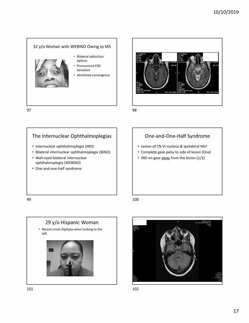
36 y/o Man with BINO Owing to MS
• Bilateral adduction
deficits (with
contralateral abducting
nystagmus)
The Internuclear Ophthalmoplegias
• Internuclear ophthalmoplegia (INO)
• Bilateral internuclear ophthalmoplegia (BINO)
• Wall-eyed bilateral internuclear
ophthalmoplegia (WEBINO)
• One-and-one-half syndrome
Wall-Eyed Bilateral Internuclear
Ophthalmoplegia
• Bilateral internuclear ophthalmoplegia with
pronounced exotropia
• Abolished convergence (midbrain)
• Involvement of convergence pathways / MR
subnuclei of CN III
91 92
93 94
95 96
10/10/2019
17
32 y/o Woman with WEBINO Owing to MS
• Bilateral adduction
deficits
• Pronounced EXO
deviation
• Abolished convergence
The Internuclear Ophthalmoplegias
• Internuclear ophthalmoplegia (INO)
• Bilateral internuclear ophthalmoplegia (BINO)
• Wall-eyed bilateral internuclear
ophthalmoplegia (WEBINO)
• One-and-one-half syndrome
One-and-One-Half Syndrome
• Lesion of CN VI nucleus & ipsilateral MLF
• Complete gaze palsy to side of lesion (One)
• INO on gaze away from the lesion (1/2)
29 y/o Hispanic Woman
• Recent onset diplopia when looking to the
left
FLAIR
97 98
99 100
101 102
10/10/2019
18
T1WI with gad FLAIR
S/P IV Methylprednisolone 28 y/o Woman with Recent-Onset Diplopia
• Prior history of left ET
(now left XT)
• Gaze palsy on right gaze
• Right INO on left gaze
T2WI
Follow-up X 3 months
• Resolution of diplopia
• Re-establishment of left
ET
103 104
105 106
107 108
10/10/2019
19
Nystagmus Common Types of Nystagmus with MS
• Found in approximately 30% of MS patients
– Acquired pendular
– Periodic alternating
– Gaze-evoked
Prasad & Galetta Neurol Clin 2010
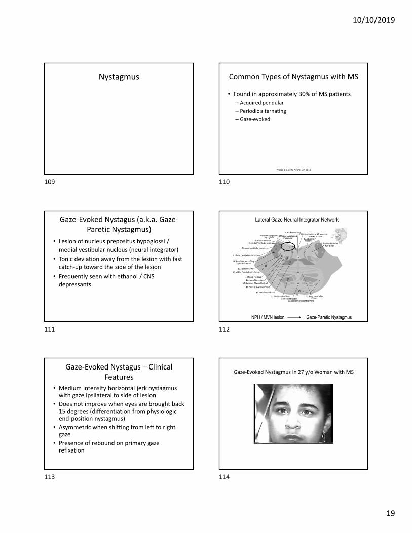
Gaze-Evoked Nystagus (a.k.a. Gaze-
Paretic Nystagmus)
• Lesion of nucleus prepositus hypoglossi /
medial vestibular nucleus (neural integrator)
• Tonic deviation away from the lesion with fast
catch-up toward the side of the lesion
• Frequently seen with ethanol / CNS
depressants
NPH / MVN lesion Gaze-Paretic Nystagmus
Lateral Gaze Neural Integrator Network
Gaze-Evoked Nystagus – Clinical
Features
• Medium intensity horizontal jerk nystagmuswith gaze ipsilateral to side of lesion
• Does not improve when eyes are brought back 15 degrees (differentiation from physiologic end-position nystagmus)
• Asymmetric when shifting from left to right gaze
• Presence of rebound on primary gaze refixation
Gaze-Evoked Nystagmus in 27 y/o Woman with MS
109 110
111 112
113 114
10/10/2019
20
Gaze-Evoked Nystagmus in 71 y/o Woman with Acute
Oscilopsia & Vestibular Dysfunction
Gaze-Evoked / Rebound Nystagmus in 34 y/o Man with MS
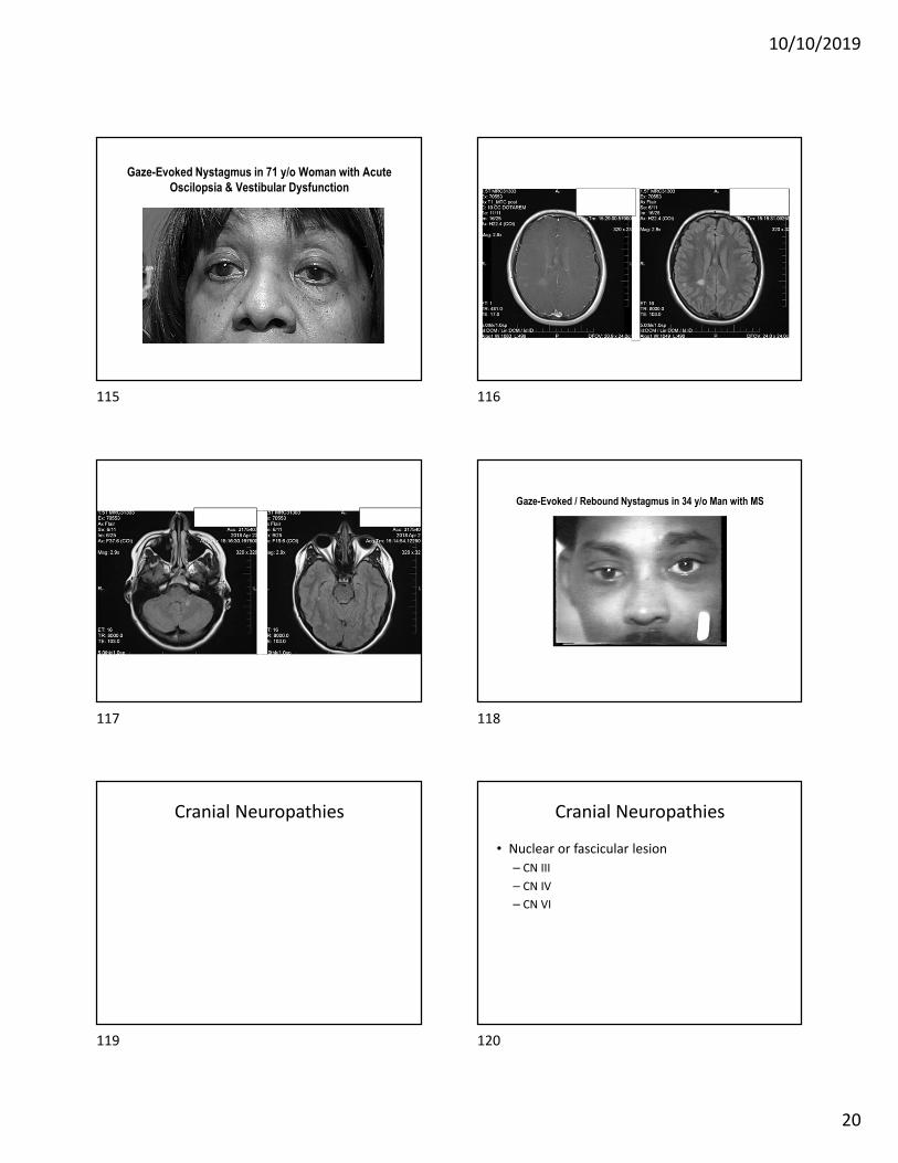
Cranial Neuropathies Cranial Neuropathies
• Nuclear or fascicular lesion
– CN III
– CN IV
– CN VI
115 116
117 118
119 120
10/10/2019
21
“The Signature” of CN VI Paresis
• Eso which increases in the action
of the paretic eye
Etiology of CN VI PalsyMayo Clinic Study of Olmstead Co. MN USA from 1978-1992
(n = 137)
• Undetermined: 26%
• Hypertension: 19%
• HTN & diabetes: 12%
• Trauma: 12%
• MS: 7%
• Neoplasm: 5% (complicated)
• Diabetes (alone): 4%
• CVA: 4%
• s/p neurosurgery: 3%
• Aneurysm: 2% (complicated)
• Other: 8%
Patel SV, et al. Ophthalmology 2004
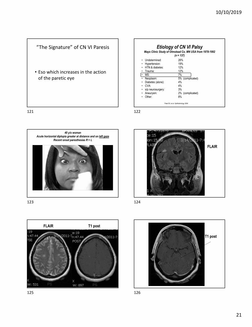
40 y/o woman
Acute horizontal diplopia greater at distance and on left gaze
Recent onset paresthesias R > L
FLAIR
FLAIR T1 post
T1 post
121 122
123 124
125 126
10/10/2019
22
Ocular Structure & Functional
Biomarkers in Multiple Sclerosis
• Optical coherence tomography (OCT)
• Low contrast acuity testing
• Visual-motor dysfunction (rapid number
naming / KD test)
OCT Findings in MS
• Acute optic neuritis associated with RNFL
thinning of 20% - 40% X 3 months
• Thinning of RNFL & GCL+IPL occurs over time
with MS in the absence of optic neuritis
(thinning of 12%)
• Incorporation of OCT, low-contrast acuity
measurement & vision-specific QOL measures
incorporated into MS clinical trials
Balcer LJ. Neuroophthalmol 2014
Sakai RE et al. J Neuroophthalmol 2011
Fisher JB, et al. Ophthalmology 2008
• Thinning of RNFL & increased VEP latencies with MS
• Normal standard assessments of vision (VA, color
vision & visual fields)
• RNFL thinning greatest temporal and inferior temp
• Thinning correlation with decreased QOL
Garcia-Martin E, et al. Ophthalmology 2017
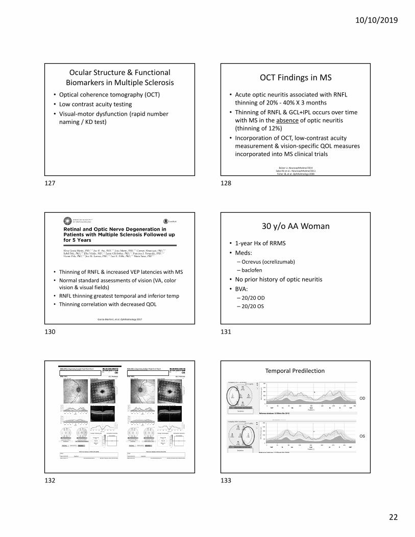
30 y/o AA Woman
• 1-year Hx of RRMS
• Meds:
– Ocrevus (ocrelizumab)
– baclofen
• No prior history of optic neuritis
• BVA:
– 20/20 OD
– 20/20 OS
Temporal Predilection
OD
OS
127 128
130 131
132 133
10/10/2019
23
31 y/o Woman
• 10-year Hx of RRMS
• Meds:
– Ocrevus (ocrelizumab)
• Prior optic neuritis OD
• BVA:
– 20/20 -1 OD
– 20/20 OS
42 y/o Woman
• 20+ year Hx of RRMS
• No prior known episodes of optic neuritis
• Meds:
– Ocrevus (ocrelizumab)
• Concomitant BINO
• BVA:
– 20/20 OD
– 20/20 OS
134 135
136 137
138 139
10/10/2019
24
RNFL N/T Comparison
OD
OS
OD
OS
RNFL Progression Analysis
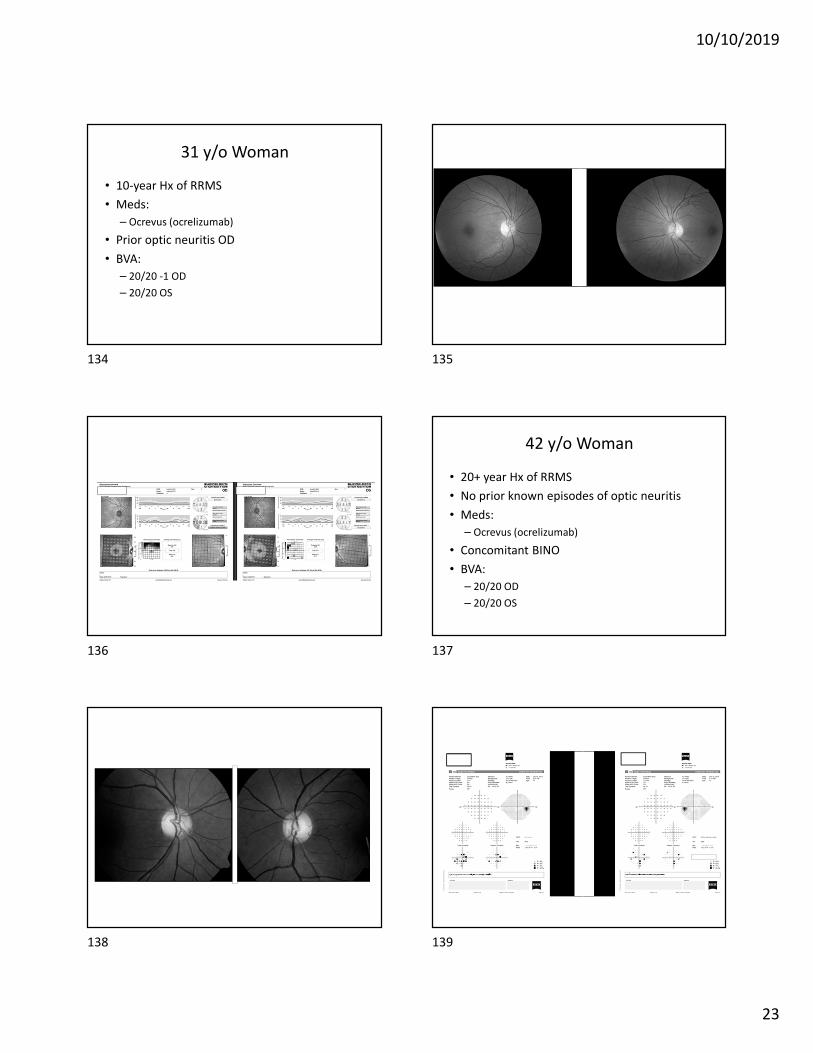
Low Contrast Acuity Testing in MS
• Low contrast charts (2.5% and 1.25%) compared to high contrast (100%) ETDRS/Snellen letters
• Comparison of LCA to OCT, QOL and standard neurology-based MS outcome measures (e.g. EDSS, MRI, cognitive performance)
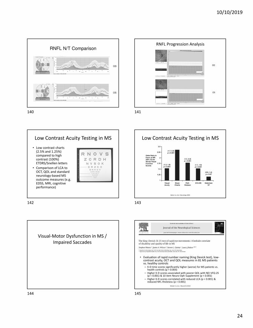
Low Contrast Acuity Testing in MS
Balcer LJ, et al. Neurology 2003
Visual-Motor Dysfunction in MS /
Impaired Saccades
• Evaluation of rapid number naming (King Devick test), low-contrast acuity, OCT and QOL measures in 81 MS patients vs. healthy controls– K-D time scores significantly higher (worse) for MS patients vs.
health controls (p = 0.003)
– Higher K-D scores associated with poorer QOL with NEI VFQ-25 (p < 0.001) & 10 item Neuro-Oph Supplement (p < 0.001)
– Higher K-D scores correlated with reduced LCA (p < 0.001) & reduced NFL thickness (p = 0.001)
Moster S, et al. J Neurol Sci 2014
140 141
142 143
144 145
10/10/2019
25
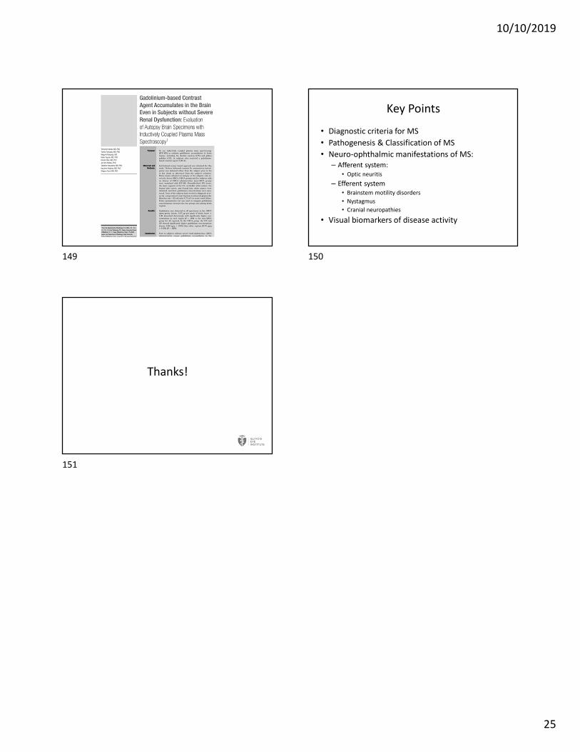
Key Points
• Diagnostic criteria for MS
• Pathogenesis & Classification of MS
• Neuro-ophthalmic manifestations of MS:
– Afferent system:
• Optic neuritis
– Efferent system
• Brainstem motility disorders
• Nystagmus
• Cranial neuropathies
• Visual biomarkers of disease activity
Thanks!
149 150
151