Embed Size (px)
Citation preview

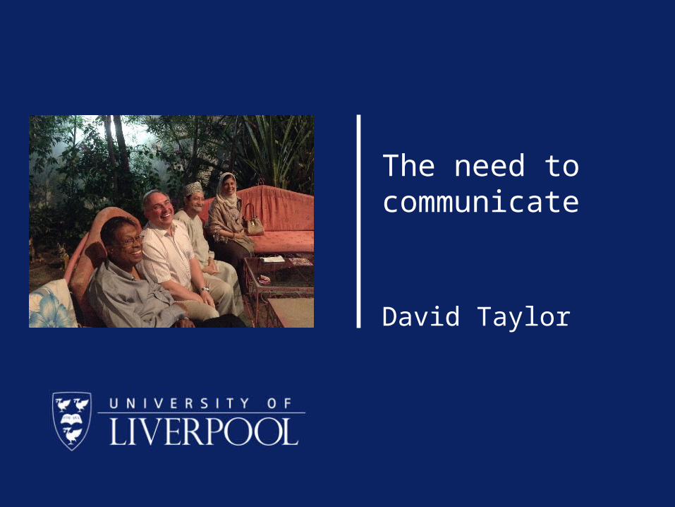
The need to communicate
David Taylor
To communicate with meThe Reverend Dr David CM Taylor
Reader in Medical Education
Cedar House 4:27
http://www.liv.ac.uk/~dcmt
To start with the obvious• We are made up of cells• But they clearly stick together • and work together• In the next couple of lectures we
will start to explore the mechanisms they use.
Cell differentiation• There are many types of cell• They all start out as stem cells• And differentiate into cells with
different and specific functions.
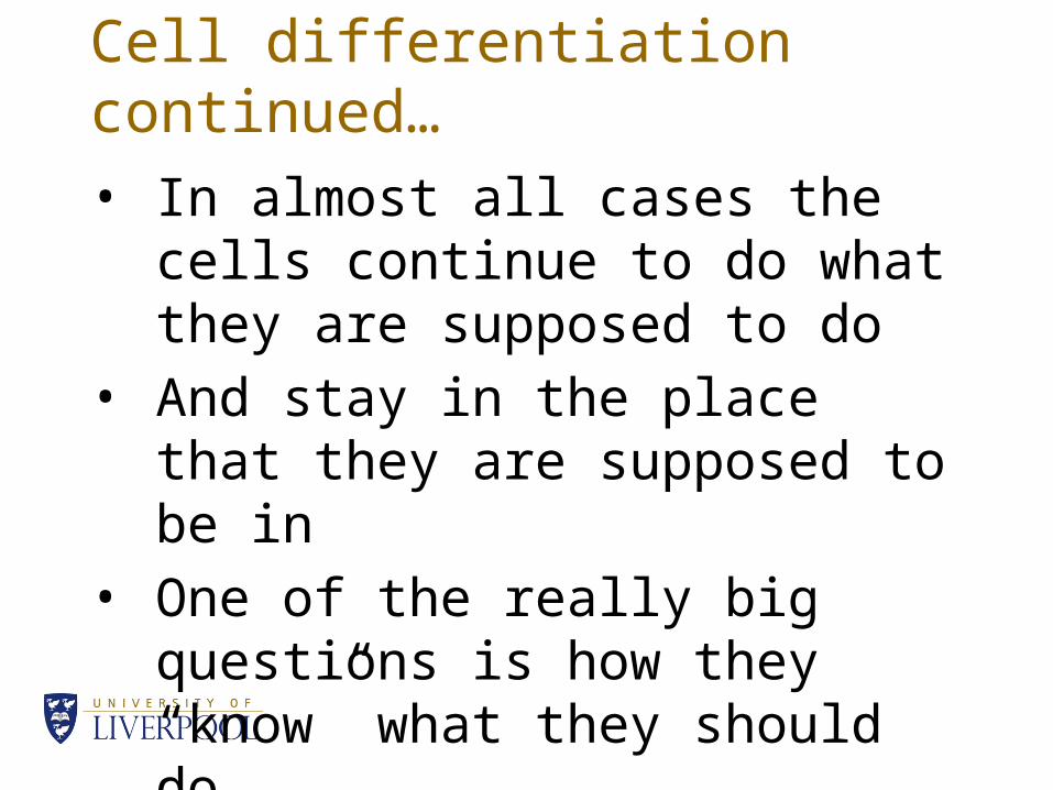
Cell differentiation continued…• In almost all cases the cells
continue to do what they are supposed to do
• And stay in the place that they are supposed to be in
• One of the really big questions is how they “know” what they should do
Short answer• The short answer is that they
communicate with each other
• But how?
I recommend Medical Sciences by Naish, Revest and Court (2009) but there is a 2014 edition published by Saunders. This lecture uses chapters 2 and 10
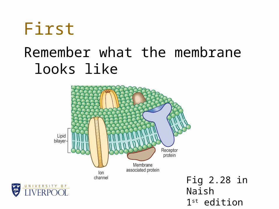
FirstRemember what the membrane
looks like
Fig 2.28 in Naish1st edition
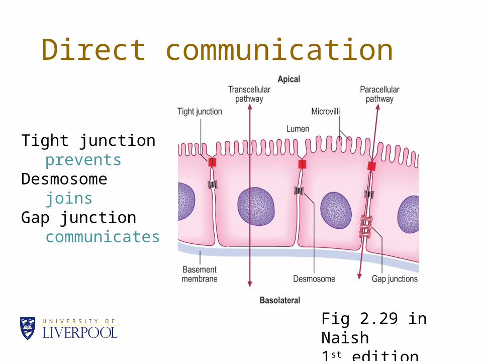
Direct communication
Tight junctionprevents
Desmosomejoins
Gap junctioncommunicates
Fig 2.29 in Naish1st edition
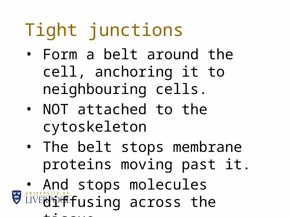
Tight junctions• Form a belt around the cell,
anchoring it to neighbouring cells.• NOT attached to the cytoskeleton• The belt stops membrane proteins
moving past it.• And stops molecules diffusing
across the tissue
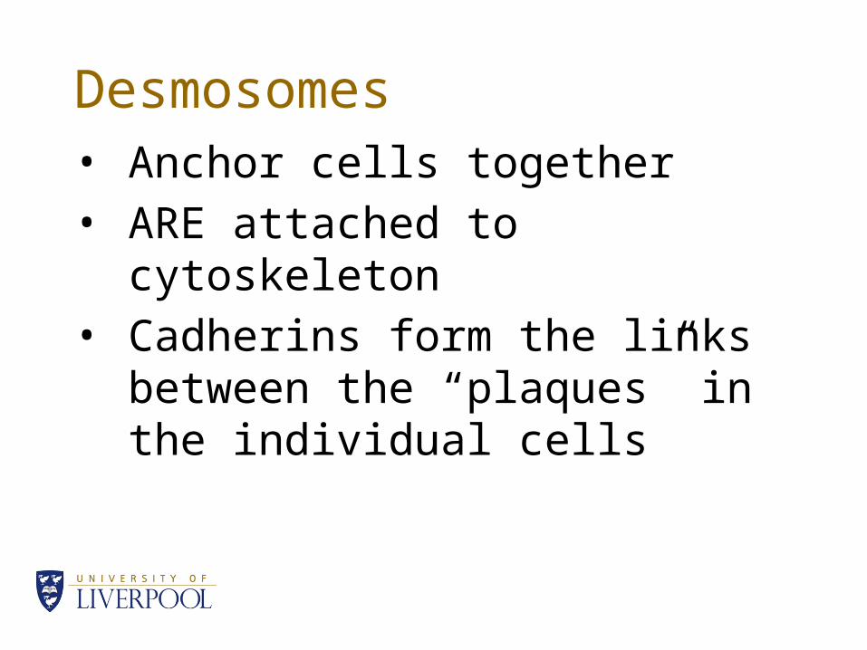
Desmosomes• Anchor cells together• ARE attached to cytoskeleton• Cadherins form the links between
the “plaques” in the individual cells
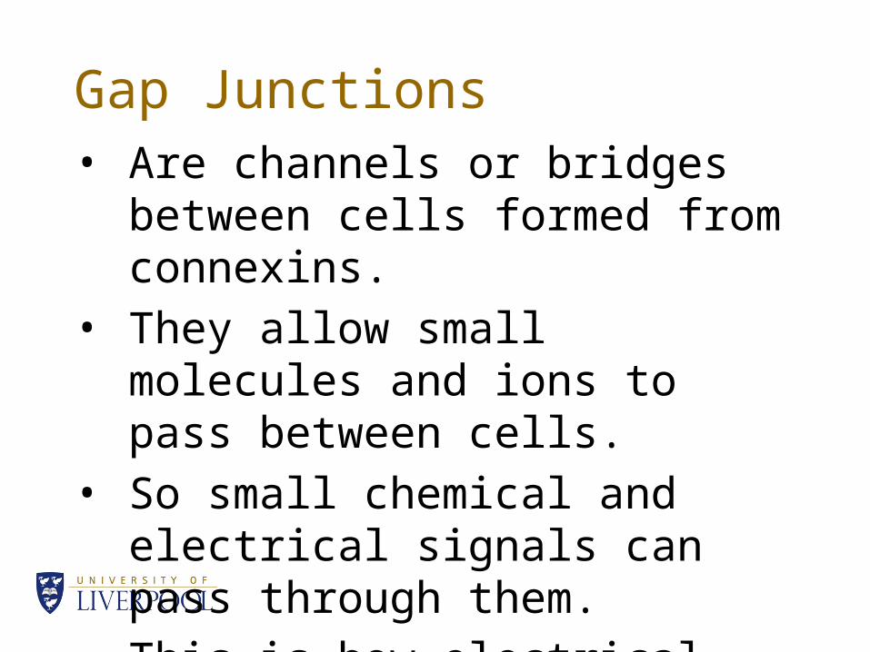
Gap Junctions• Are channels or bridges between
cells formed from connexins.• They allow small molecules and
ions to pass between cells.• So small chemical and electrical
signals can pass through them.• This is how electrical signals pass
through smooth muscle.
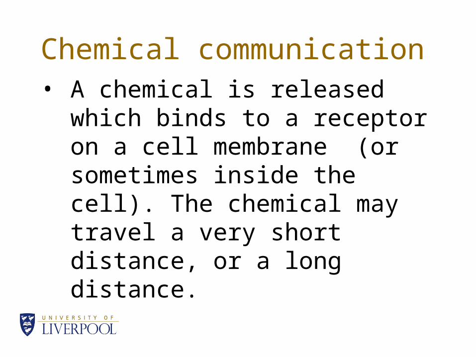
Chemical communication• A chemical is released which
binds to a receptor on a cell membrane (or sometimes inside the cell). The chemical may travel a very short distance, or a long distance.
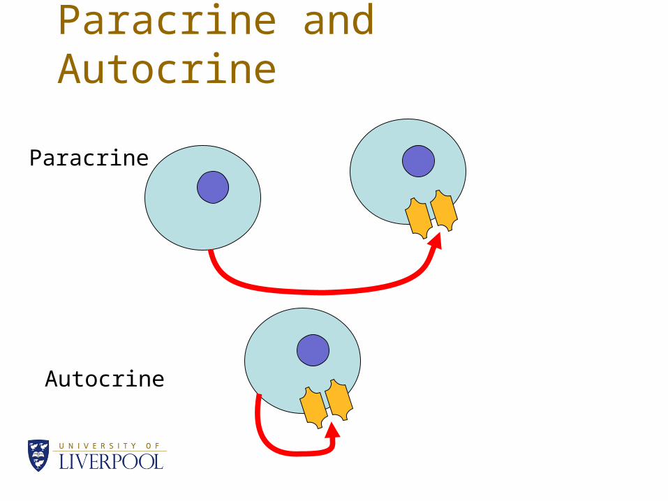
Paracrine and Autocrine
Paracrine
Autocrine
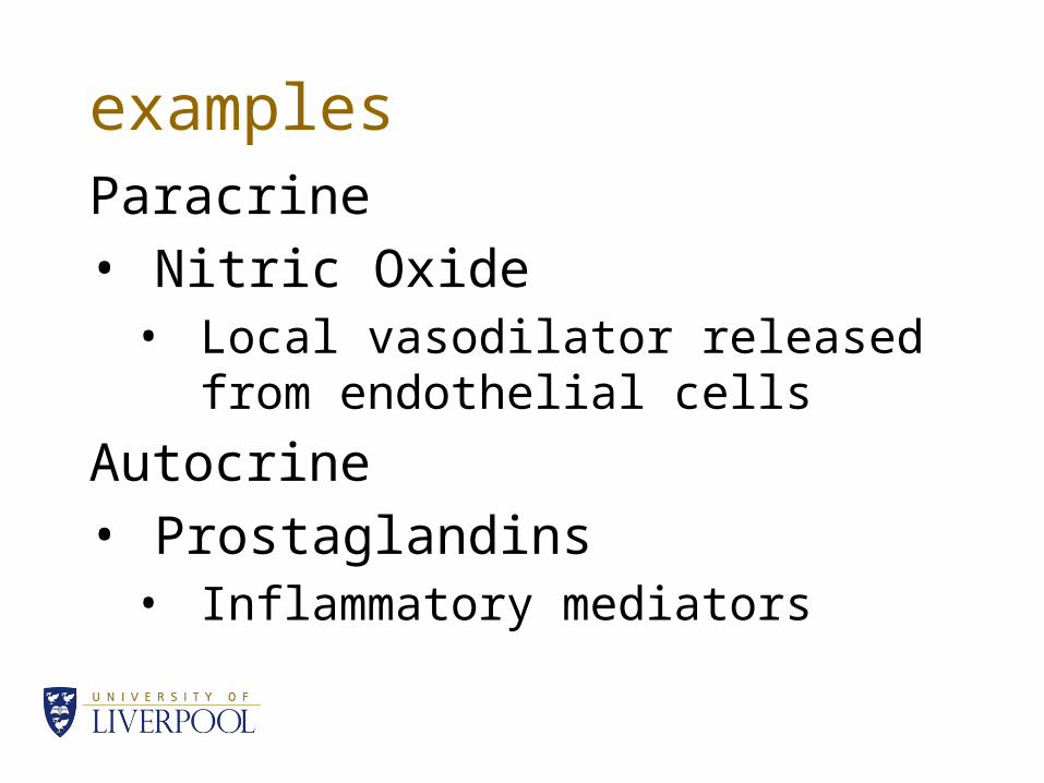
examplesParacrine• Nitric Oxide
• Local vasodilator released from endothelial cells
Autocrine• Prostaglandins
• Inflammatory mediators
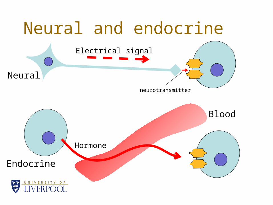
Neural and endocrine
Neural
Endocrine
Electrical signal
Hormone
neurotransmitter
Blood
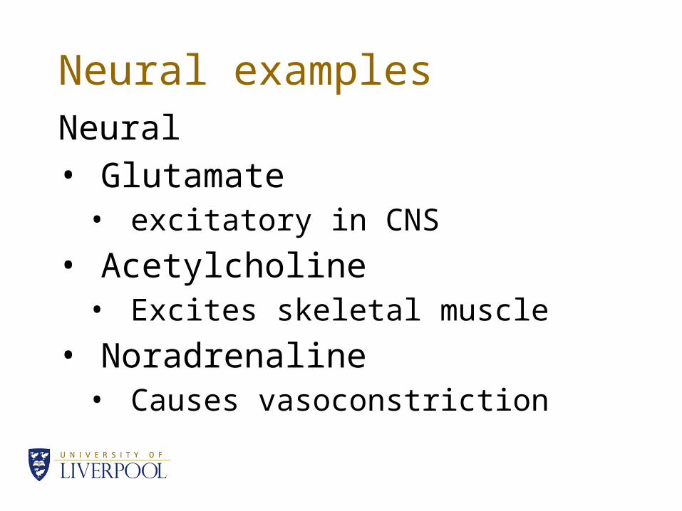
Neural examplesNeural• Glutamate
• excitatory in CNS
• Acetylcholine• Excites skeletal muscle
• Noradrenaline• Causes vasoconstriction

Hormones• The chemical type usually reflects
the way that they act on the target tissues
• Amino acid derivatives• Steroids• Peptides• Proteins• Glycoproteins
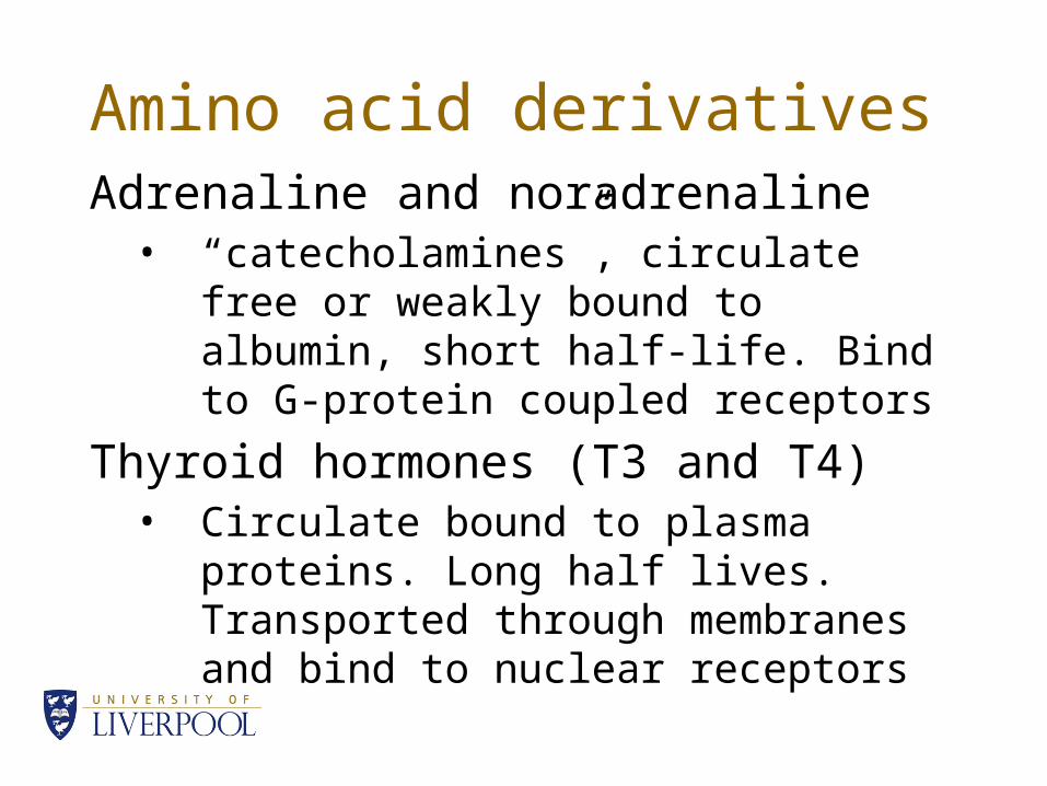
Amino acid derivativesAdrenaline and noradrenaline
• “catecholamines”, circulate free or weakly bound to albumin, short half-life. Bind to G-protein coupled receptors
Thyroid hormones (T3 and T4)• Circulate bound to plasma proteins. Long
half lives. Transported through membranes and bind to nuclear receptors
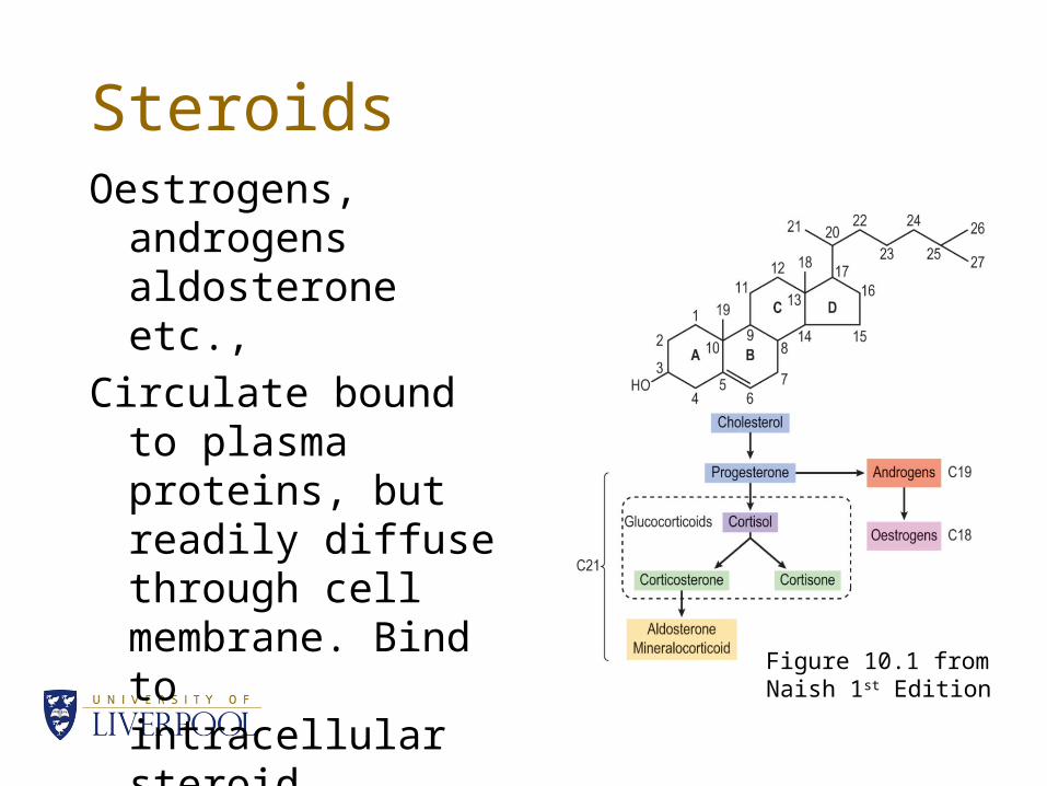
SteroidsOestrogens,
androgens aldosterone etc.,
Circulate bound to plasma proteins, but readily diffuse through cell membrane. Bind to intracellular steroid receptors
Figure 10.1 from Naish 1st Edition
Peptides etc.,Peptides, proteins and glycoproteins• Are usually carved from prohormones when
needed• Then are secreted by exocytosis• And do not usually bind to plasma proteins.• They are very different in structure so their
effects are mediated by several different mechanisms (see next lecture)
Peptides• Thyrotropin releasing factor (TRH)• Gonadotrophin releasing hormone (GnRH)• Adrenocorticotropic hormone (ACTH)• Antidiuretic hormone (ADH, Vasopressin)• Oxytocin• Glucagon• Somatostatin• Vasoactive intestinal polypeptide (VIP)
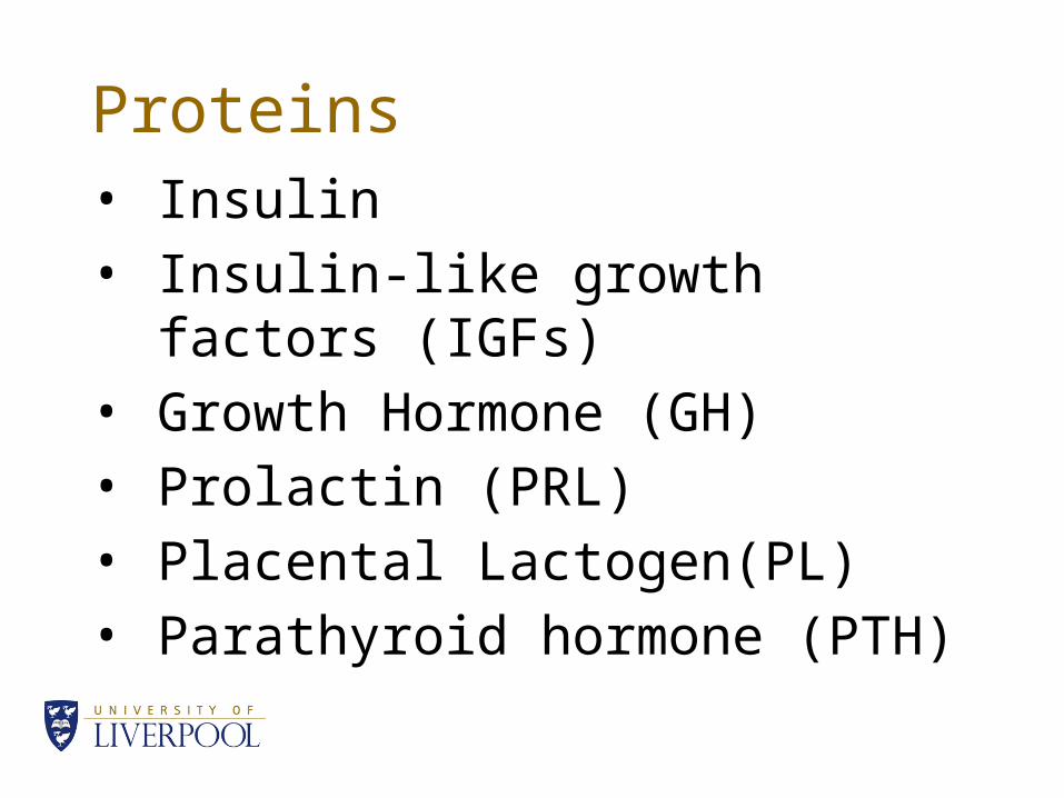
Proteins• Insulin• Insulin-like growth factors (IGFs)• Growth Hormone (GH)• Prolactin (PRL)• Placental Lactogen(PL)• Parathyroid hormone (PTH)
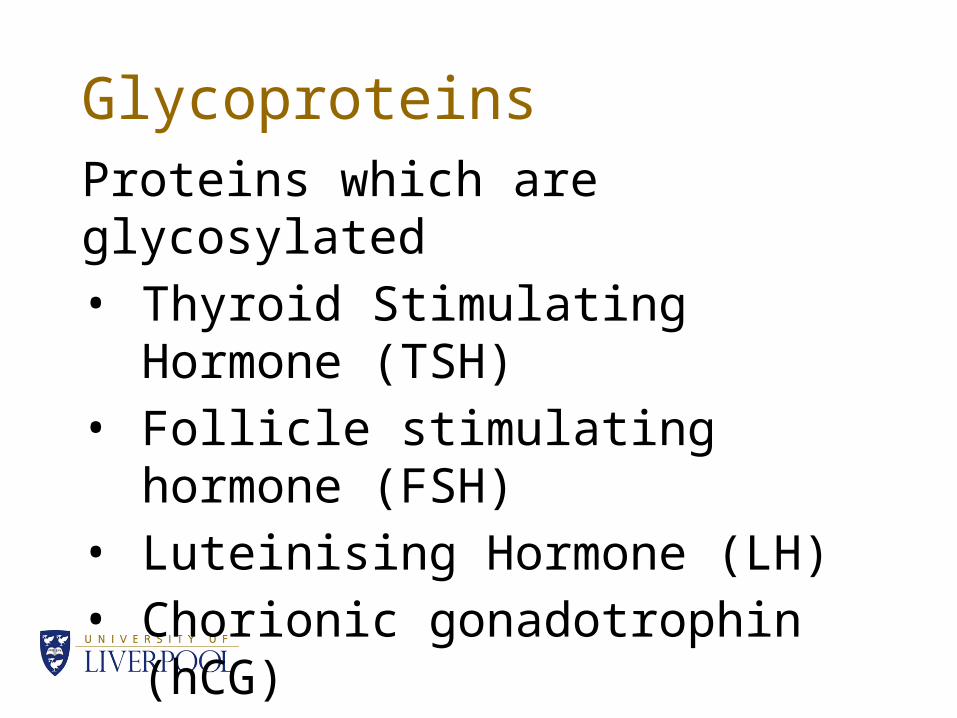
GlycoproteinsProteins which are glycosylated• Thyroid Stimulating Hormone
(TSH)• Follicle stimulating hormone (FSH)• Luteinising Hormone (LH)• Chorionic gonadotrophin (hCG)
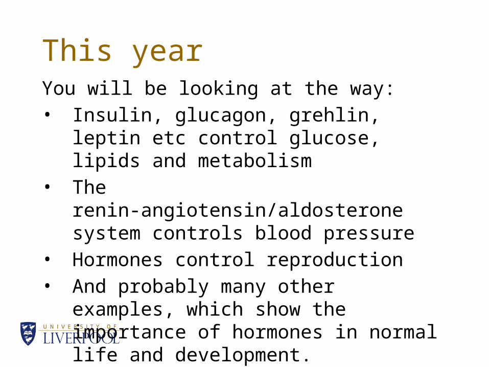
This yearYou will be looking at the way:• Insulin, glucagon, grehlin, leptin etc control
glucose, lipids and metabolism• The renin-angiotensin/aldosterone system
controls blood pressure• Hormones control reproduction• And probably many other examples, which
show the importance of hormones in normal life and development.
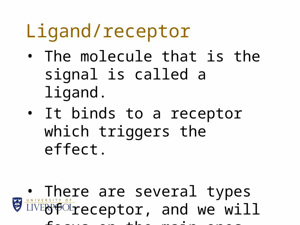
Ligand/receptor• The molecule that is the signal is
called a ligand.• It binds to a receptor which
triggers the effect.
• There are several types of receptor, and we will focus on the main ones.
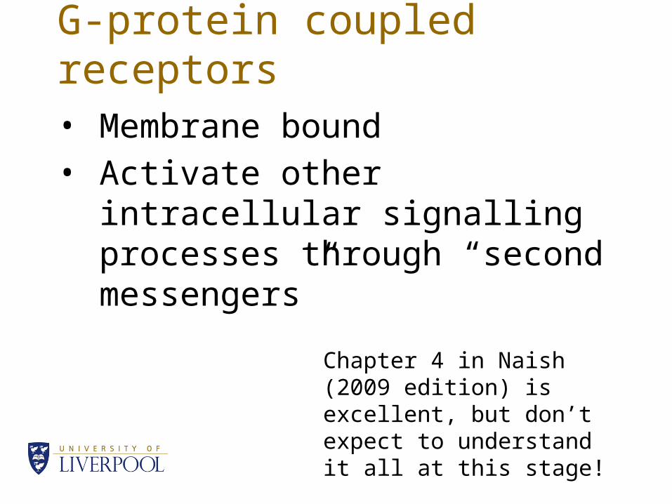
G-protein coupled receptors• Membrane bound• Activate other intracellular
signalling processes through “second messengers”
Chapter 4 in Naish (2009 edition) is excellent, but don’t expect to understand it all at this stage!
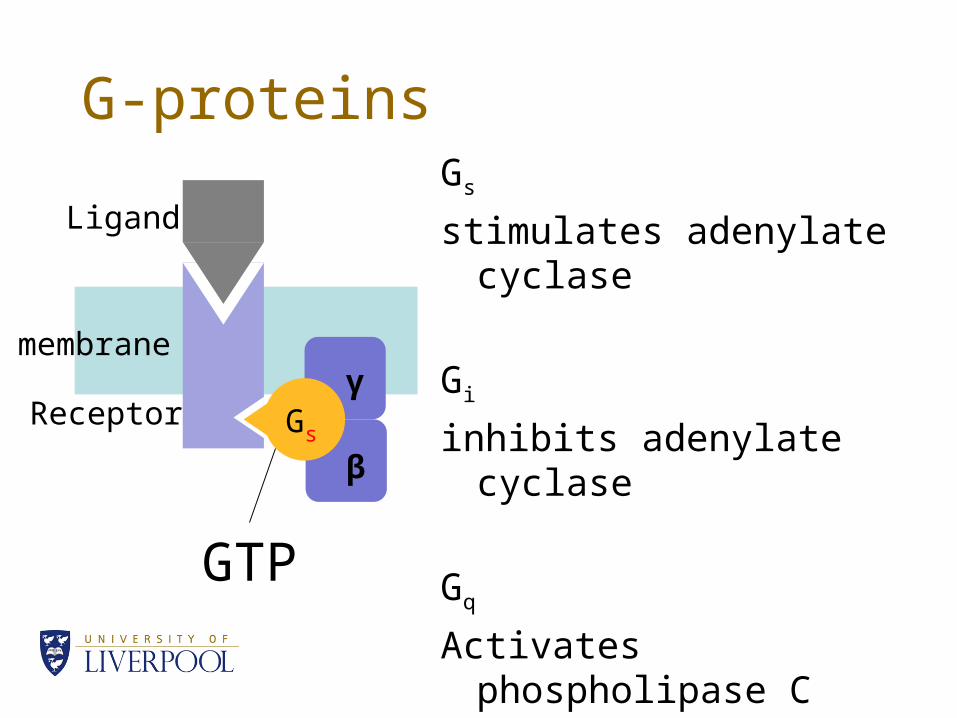
G-proteinsGs
stimulates adenylate cyclase
Gi
inhibits adenylate cyclase
Gq
Activates phospholipase C
βGs
GTP
β
γ
Ligand
Receptor
membrane
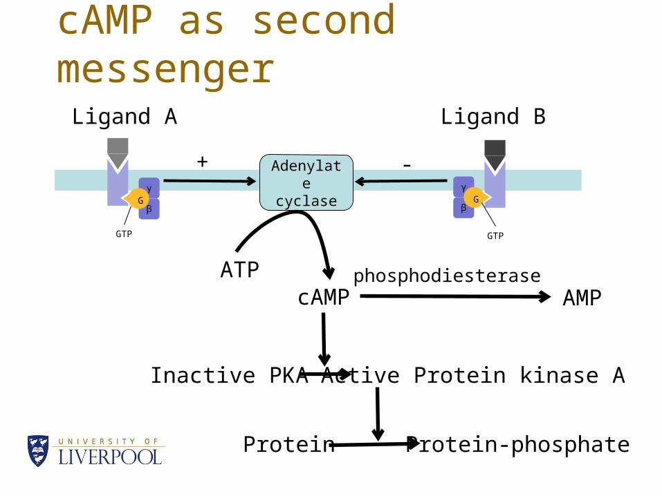
cAMP as second messenger
γ
βG
γ
βG
Adenylate cyclase
ATPcAMP AMP
Inactive PKA Active Protein kinase A
Protein Protein-phosphate
+ -
phosphodiesterase
Ligand A Ligand B
GTP GTP
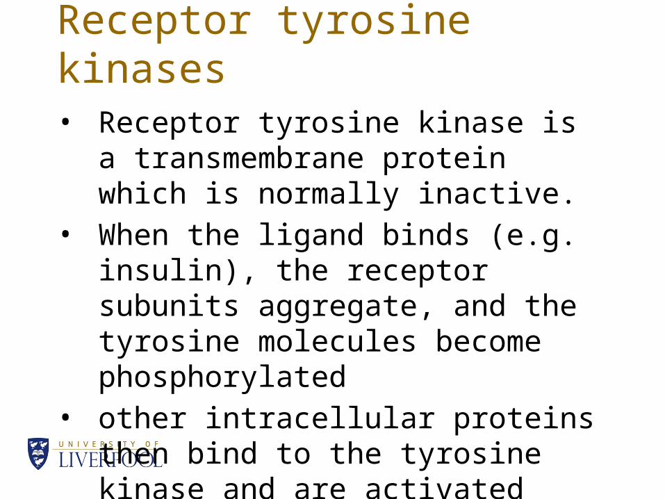
Receptor tyrosine kinases• Receptor tyrosine kinase is a
transmembrane protein which is normally inactive.
• When the ligand binds (e.g. insulin), the receptor subunits aggregate, and the tyrosine molecules become phosphorylated
• other intracellular proteins then bind to the tyrosine kinase and are activated
Nuclear receptors• Hormones like the steroid hormones are
lipid soluble and can diffuse through the plasma membrane.
• Inside the cell they bind to their receptors, causing a conformational change.
• The conformational change allows a dimer to form
• The dimer binds to recognition sites on DNA and triggers (or sometimes inhibits) transcription of specific genes
Ligand gated channels• A simple example is the acetylcholine
receptor in muscle• Acetylcholine binds to a receptor which
opens a channel to allow Na+ into the cell• The influx of Na+ depolarises the cell• The depolarisation causes the release of
intracellular Ca2+
• Which allows the actin and myosin to bind together, and contraction to occur.
Resting Membrane Potential• Cells in the body are mostly impermeable to Na+
• and mostly permeable to K+ and Cl-
• Intracellular proteins are negatively charged and can’t leave the cell.
• When the cell is “at rest” the membrane potential is a compromise between the charge carried by the diffusible ions, and the concentration gradient for each ion
• Normally this is about -90mV, or -70mV in excitable cells
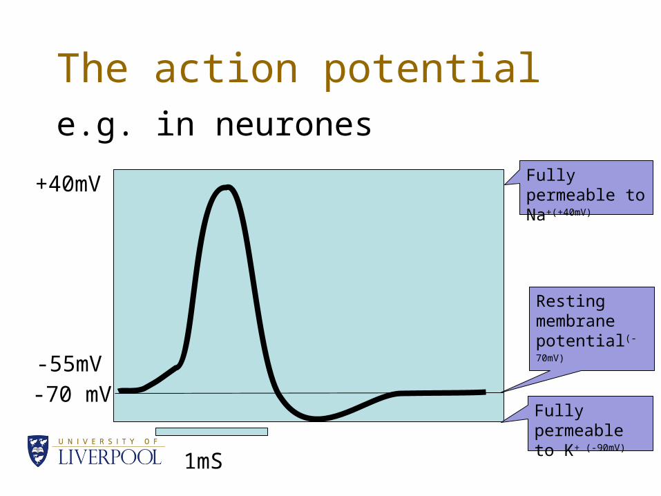
The action potentiale.g. in neurones
-70 mV-55mV
+40mV Fully permeable to Na+(+40mV)
Fully permeable to K+ (-90mV)
1mS
Resting membrane potential(-70mV)
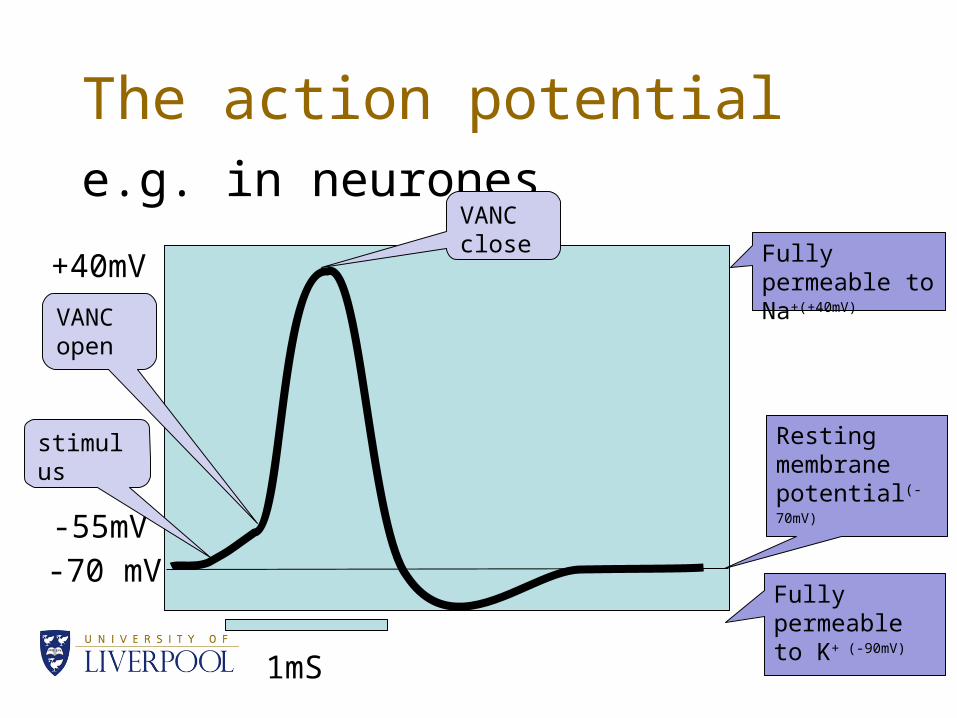
The action potentiale.g. in neurones
-70 mV-55mV
+40mV
VANC open
VANC close Fully permeable
to Na+(+40mV)
Fully permeable to K+ (-90mV)
1mS
stimulus Resting membrane potential(-70mV)
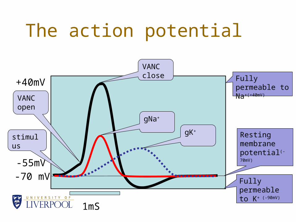
The action potential
-70 mV-55mV
+40mV
VANC open
VANC close Fully permeable
to Na+(+40mV)
Fully permeable to K+ (-90mV)
1mS
stimulus Resting membrane potential(-70mV)
gNa+
gK+
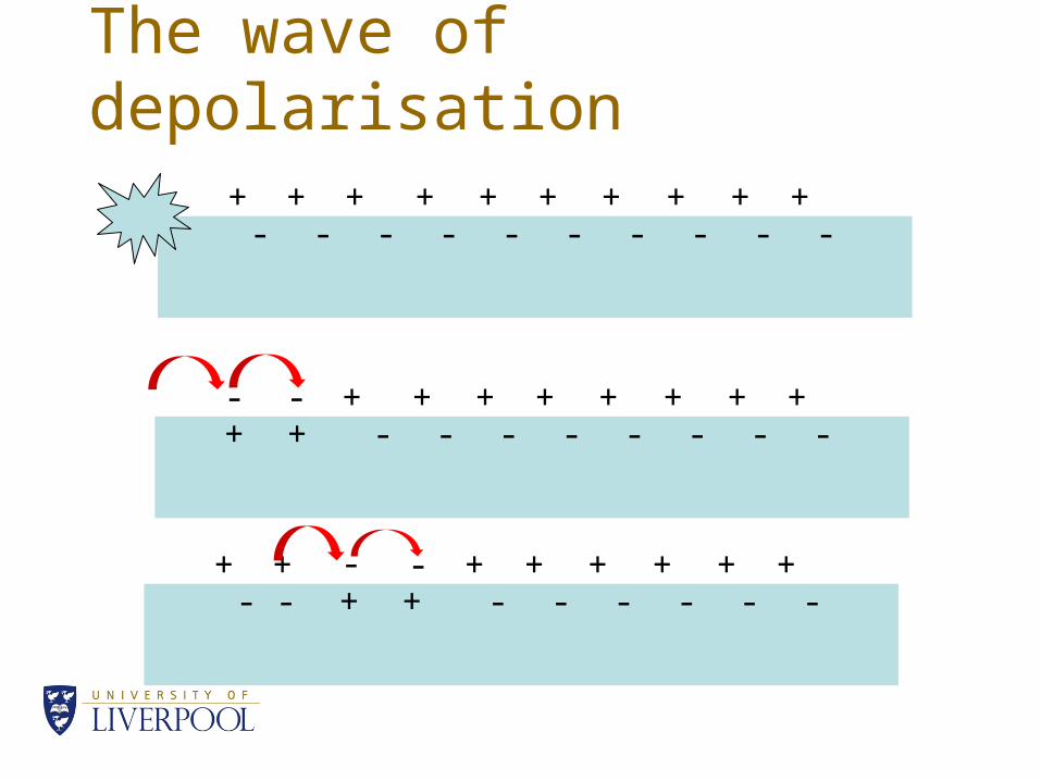
The wave of depolarisation
- -- - - - - - - -+ + + + ++ + + + +
+ -+ - - - - - - -- - + + ++ + + + +
- +- + - - - - - -+ + - - ++ + + + +
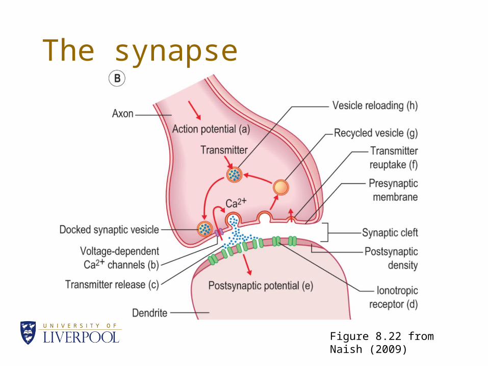
The synapse
Figure 8.22 from Naish (2009)
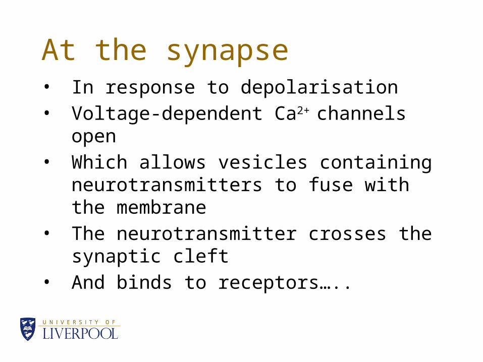
At the synapse• In response to depolarisation• Voltage-dependent Ca2+ channels open • Which allows vesicles containing
neurotransmitters to fuse with the membrane
• The neurotransmitter crosses the synaptic cleft
• And binds to receptors…..